Inhibition of MEK sensitizes human melanoma cells to endoplasmic reticulum stress-induced apoptosis
Upload
khangminh22Category
view
0download
0
BI84CH29-Prinz-Voeltz ARI 24 April 2015 11:27
Form Follows Function: TheImportance of EndoplasmicReticulum ShapeL.M. Westrate,1 J.E. Lee,1 W.A. Prinz,2,∗
and G.K. Voeltz1,∗
1Department of Molecular, Cellular, and Developmental Biology, University of Colorado,Boulder, Colorado 80303; email: [email protected] of Cell and Molecular Biology, National Institute of Diabetes and Digestiveand Kidney Diseases, National Institutes of Health, Bethesda, Maryland 20892;email: [email protected]
Annu. Rev. Biochem. 2015. 84:791–811
First published online as a Review in Advance onJanuary 12, 2015
The Annual Review of Biochemistry is online atbiochem.annualreviews.org
This article’s doi:10.1146/annurev-biochem-072711-163501
Copyright c© 2015 by Annual Reviews.All rights reserved
∗Corresponding authors
Keywords
endoplasmic reticulum, structure, dynamics, virus, neurodegeneration
Abstract
The endoplasmic reticulum (ER) has a remarkably complex structure, com-posed of a single bilayer that forms the nuclear envelope, along with a net-work of sheets and dynamic tubules. Our understanding of the biologicalsignificance of the complex architecture of the ER has improved dramati-cally in the last few years. The identification of proteins and forces requiredfor maintaining ER shape, as well as more advanced imaging techniques, hasallowed the relationship between ER shape and function to come into focus.These studies have also revealed unexpected new functions of the ER andnovel ER domains regulating alterations in ER dynamics. The importanceof ER structure has become evident as recent research has identified dis-eases linked to mutations in ER-shaping proteins. In this review, we discusswhat is known about the maintenance of ER architecture, the relationshipbetween ER structure and function, and diseases associated with defects inER structure.
791
Ann
u. R
ev. B
ioch
em. 2
015.
84:7
91-8
11. D
ownl
oade
d fr
om w
ww
.ann
ualr
evie
ws.
org
Acc
ess
prov
ided
by
Cal
ifor
nia
Inst
itute
of
Tec
hnol
ogy
on 0
3/28
/16.
For
per
sona
l use
onl
y.
BI84CH29-Prinz-Voeltz ARI 24 April 2015 11:27
Contents
INTRODUCTION . . . . . . . . . . . . . . . . . . . . . . . . . . . . . . . . . . . . . . . . . . . . . . . . . . . . . . . . . . . . . . . 792PERIPHERAL ENDOPLASMIC RETICULUM DOMAINS
AND FUNCTIONS . . . . . . . . . . . . . . . . . . . . . . . . . . . . . . . . . . . . . . . . . . . . . . . . . . . . . . . . . . . 794Cortical Endoplasmic Reticulum . . . . . . . . . . . . . . . . . . . . . . . . . . . . . . . . . . . . . . . . . . . . . . . . 795
MECHANISMS SHAPING THE ENDOPLASMIC RETICULUM . . . . . . . . . . . . . . . 795Endoplasmic Reticulum Dynamics on the Cytoskeleton . . . . . . . . . . . . . . . . . . . . . . . . . . 795Formation of Peripheral Endoplasmic Reticulum Tubules . . . . . . . . . . . . . . . . . . . . . . . . 798Formation of Peripheral Endoplasmic Reticulum Sheets . . . . . . . . . . . . . . . . . . . . . . . . . . 799Homotypic Fusion . . . . . . . . . . . . . . . . . . . . . . . . . . . . . . . . . . . . . . . . . . . . . . . . . . . . . . . . . . . . . . 800
FUNCTIONAL CONSEQUENCES OF ENDOPLASMIC RETICULUMSTRUCTURE: IMPLICATIONS FOR DISEASE PATHOLOGY . . . . . . . . . . . . . . 802Neurological Disease . . . . . . . . . . . . . . . . . . . . . . . . . . . . . . . . . . . . . . . . . . . . . . . . . . . . . . . . . . . 802Viral Infection . . . . . . . . . . . . . . . . . . . . . . . . . . . . . . . . . . . . . . . . . . . . . . . . . . . . . . . . . . . . . . . . . . 803
CONCLUSIONS AND REMAINING QUESTIONS . . . . . . . . . . . . . . . . . . . . . . . . . . . . . 804
INTRODUCTION
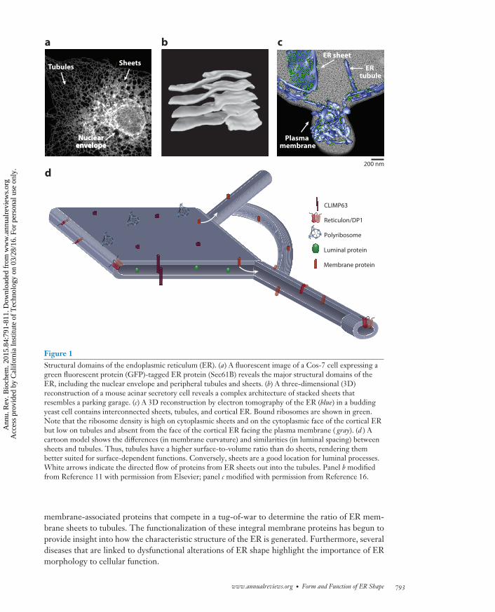
The endoplasmic reticulum (ER), despite being one of the largest organelles in the cell, was one ofthe last to be discovered. Although the ER was originally described in 1902 by Emilio Veratti (1),it took the scientific community another 50 years and the introduction of the electron microscopeto “rediscover” the ER, when George Palade (3), along with Keith Porter (4, 5), captured thestructural complexities of a fine tubular network residing within the cytoplasm (1–6). Since then,more advanced techniques, including three-dimensional electron tomography and confocal fluo-rescence microscopy, have revealed that the ER is composed of a single continuous membrane thatforms a network containing multiple domains with different structures and functions (Figure 1).
The two major domains of the ER are the nuclear envelope (NE) and the peripheral ER. Atthe center of the cell, the NE is made up of two flat ER membrane bilayers, which stack to formthe inner and outer nuclear membranes (INM and ONM). The NE shape is maintained by multipleforces, including INM proteins that bind to chromatin and lamin, linker proteins between theINM and the ONM, nuclear pores, and the cytoskeleton (7–9). Expanding from the NE, theperipheral ER branches out from the ONM into the cytosol and forms a series of cisternal sheetsand dynamic tubules (Figure 1a). This review does not discuss NE structure in detail but insteadfocuses on how the peripheral ER is shaped and how ER morphology affects function.
For many years, the relationship between the elaborate structure of the ER and the numerousfunctions of the ER was largely mysterious. Also unknown was whether defects in ER shape mightbe relevant to human disease. This situation has changed in the last few years, which have witnesseda tremendous increase in our understanding of how ER shape is determined, how morphologyis related to function, and how these processes go awry in some diseases. We are also beginningto understand how the relative ratio of the different ER structural domains in cells varies as ERarchitecture is modified to meet specific cellular demands. This understanding has been drivenprimarily by the identification of the proteins and forces that maintain ER morphology.
This review focuses on how the peripheral ER is shaped and how ER morphology affectsfunction. The formation and maintenance of peripheral ER structure are largely regulated by
792 Westrate et al.
Ann
u. R
ev. B
ioch
em. 2
015.
84:7
91-8
11. D
ownl
oade
d fr
om w
ww
.ann
ualr
evie
ws.
org
Acc
ess
prov
ided
by
Cal
ifor
nia
Inst
itute
of
Tec
hnol
ogy
on 0
3/28
/16.
For
per
sona
l use
onl
y.
BI84CH29-Prinz-Voeltz ARI 24 April 2015 11:27
Plasma membrane
ER sheet
ER tubule
NuclearenvelopeNuclear
envelopeNuclear
envelope
Tubules Sheets
Luminal protein
CLIMP63
Reticulon/DP1
Membrane protein
Polyribosome
a b c
d200 nm
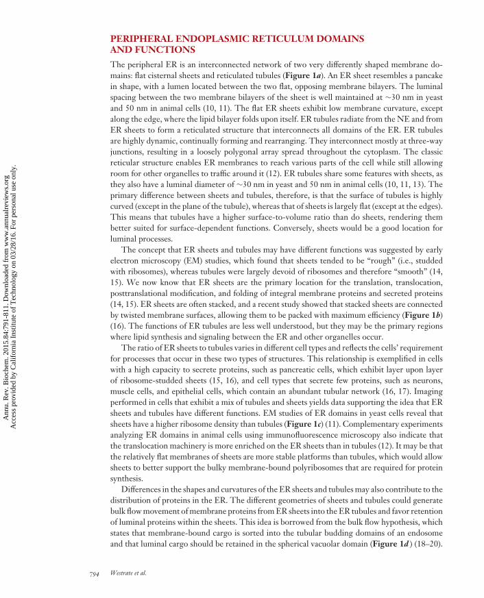
Figure 1Structural domains of the endoplasmic reticulum (ER). (a) A fluorescent image of a Cos-7 cell expressing agreen fluorescent protein (GFP)-tagged ER protein (Sec61B) reveals the major structural domains of theER, including the nuclear envelope and peripheral tubules and sheets. (b) A three-dimensional (3D)reconstruction of a mouse acinar secretory cell reveals a complex architecture of stacked sheets thatresembles a parking garage. (c) A 3D reconstruction by electron tomography of the ER (blue) in a buddingyeast cell contains interconnected sheets, tubules, and cortical ER. Bound ribosomes are shown in green.Note that the ribosome density is high on cytoplasmic sheets and on the cytoplasmic face of the cortical ERbut low on tubules and absent from the face of the cortical ER facing the plasma membrane ( gray). (d ) Acartoon model shows the differences (in membrane curvature) and similarities (in luminal spacing) betweensheets and tubules. Thus, tubules have a higher surface-to-volume ratio than do sheets, rendering thembetter suited for surface-dependent functions. Conversely, sheets are a good location for luminal processes.White arrows indicate the directed flow of proteins from ER sheets out into the tubules. Panel b modifiedfrom Reference 11 with permission from Elsevier; panel c modified with permission from Reference 16.
membrane-associated proteins that compete in a tug-of-war to determine the ratio of ER mem-brane sheets to tubules. The functionalization of these integral membrane proteins has begun toprovide insight into how the characteristic structure of the ER is generated. Furthermore, severaldiseases that are linked to dysfunctional alterations of ER shape highlight the importance of ERmorphology to cellular function.
www.annualreviews.org • Form and Function of ER Shape 793
Ann
u. R
ev. B
ioch
em. 2
015.
84:7
91-8
11. D
ownl
oade
d fr
om w
ww
.ann
ualr
evie
ws.
org
Acc
ess
prov
ided
by
Cal
ifor
nia
Inst
itute
of
Tec
hnol
ogy
on 0
3/28
/16.
For
per
sona
l use
onl
y.
BI84CH29-Prinz-Voeltz ARI 24 April 2015 11:27
PERIPHERAL ENDOPLASMIC RETICULUM DOMAINSAND FUNCTIONS
The peripheral ER is an interconnected network of two very differently shaped membrane do-mains: flat cisternal sheets and reticulated tubules (Figure 1a). An ER sheet resembles a pancakein shape, with a lumen located between the two flat, opposing membrane bilayers. The luminalspacing between the two membrane bilayers of the sheet is well maintained at ∼30 nm in yeastand 50 nm in animal cells (10, 11). The flat ER sheets exhibit low membrane curvature, exceptalong the edge, where the lipid bilayer folds upon itself. ER tubules radiate from the NE and fromER sheets to form a reticulated structure that interconnects all domains of the ER. ER tubulesare highly dynamic, continually forming and rearranging. They interconnect mostly at three-wayjunctions, resulting in a loosely polygonal array spread throughout the cytoplasm. The classicreticular structure enables ER membranes to reach various parts of the cell while still allowingroom for other organelles to traffic around it (12). ER tubules share some features with sheets, asthey also have a luminal diameter of ∼30 nm in yeast and 50 nm in animal cells (10, 11, 13). Theprimary difference between sheets and tubules, therefore, is that the surface of tubules is highlycurved (except in the plane of the tubule), whereas that of sheets is largely flat (except at the edges).This means that tubules have a higher surface-to-volume ratio than do sheets, rendering thembetter suited for surface-dependent functions. Conversely, sheets would be a good location forluminal processes.
The concept that ER sheets and tubules may have different functions was suggested by earlyelectron microscopy (EM) studies, which found that sheets tended to be “rough” (i.e., studdedwith ribosomes), whereas tubules were largely devoid of ribosomes and therefore “smooth” (14,15). We now know that ER sheets are the primary location for the translation, translocation,posttranslational modification, and folding of integral membrane proteins and secreted proteins(14, 15). ER sheets are often stacked, and a recent study showed that stacked sheets are connectedby twisted membrane surfaces, allowing them to be packed with maximum efficiency (Figure 1b)(16). The functions of ER tubules are less well understood, but they may be the primary regionswhere lipid synthesis and signaling between the ER and other organelles occur.
The ratio of ER sheets to tubules varies in different cell types and reflects the cells’ requirementfor processes that occur in these two types of structures. This relationship is exemplified in cellswith a high capacity to secrete proteins, such as pancreatic cells, which exhibit layer upon layerof ribosome-studded sheets (15, 16), and cell types that secrete few proteins, such as neurons,muscle cells, and epithelial cells, which contain an abundant tubular network (16, 17). Imagingperformed in cells that exhibit a mix of tubules and sheets yields data supporting the idea that ERsheets and tubules have different functions. EM studies of ER domains in yeast cells reveal thatsheets have a higher ribosome density than tubules (Figure 1c) (11). Complementary experimentsanalyzing ER domains in animal cells using immunofluorescence microscopy also indicate thatthe translocation machinery is more enriched on the ER sheets than in tubules (12). It may be thatthe relatively flat membranes of sheets are more stable platforms than tubules, which would allowsheets to better support the bulky membrane-bound polyribosomes that are required for proteinsynthesis.
Differences in the shapes and curvatures of the ER sheets and tubules may also contribute to thedistribution of proteins in the ER. The different geometries of sheets and tubules could generatebulk flow movement of membrane proteins from ER sheets into the ER tubules and favor retentionof luminal proteins within the sheets. This idea is borrowed from the bulk flow hypothesis, whichstates that membrane-bound cargo is sorted into the tubular budding domains of an endosomeand that luminal cargo should be retained in the spherical vacuolar domain (Figure 1d ) (18–20).
794 Westrate et al.
Ann
u. R
ev. B
ioch
em. 2
015.
84:7
91-8
11. D
ownl
oade
d fr
om w
ww
.ann
ualr
evie
ws.
org
Acc
ess
prov
ided
by
Cal
ifor
nia
Inst
itute
of
Tec
hnol
ogy
on 0
3/28
/16.
For
per
sona
l use
onl
y.
BI84CH29-Prinz-Voeltz ARI 24 April 2015 11:27
Cortical Endoplasmic Reticulum
Cortical ER refers to regions of the peripheral ER that are closely apposed and tethered to theplasma membrane (PM). Contact sites between the ER and PM allow small molecules such aslipids and signals to be exchanged. In some cell types, these contacts are quite extensive. In yeast,for example, the ER is closely opposed to ∼40% of the PM in both budding (11) and fission (11,21) yeast (Figure 1c). The shape of cortical ER is a hybrid between the properties of sheets andtubules. It forms highly fenestrated, flat ER sheets that resemble a slice of Swiss cheese. Thus, ithas regions that are flat and many regions that have high curvature. The cortical ER also has ahybrid ribosome density (Figure 1c). It has no bound ribosomes on the plane that faces the PM,probably because it is so tightly tethered there that ribosomes are excluded (11). In contrast, theother side of the cortical ER that faces the cytosol has the same high ribosome density as ER sheetsthat are not at the cortex (11).
Muscle cells are another cell type in which a significant portion of the PM has closely apposedthe ER (22, 23). Indeed, these cells have invaginations of the PM, called T-tubules, which areassociated with the ER and thus enable extensive ER–PM contact. These contacts are necessaryfor the Ca2+ signaling that occurs during muscle contraction (22, 24, 25). The ER is one ofthe major reservoirs of intracellular Ca2+ and can release or take up Ca2+ from neighboringcompartments at contact sites (23, 26, 27).
Cortical ER also exists in other cell types, but the fraction of the peripheral ER at the cortexis much smaller than in muscle cells or yeast. In these cells, Ca2+ levels in the ER lumen regulateclose contacts between the ER and the PM; when the luminal Ca2+ concentration decreases, theamount of ER contacting the PM increases. These contact sites are then the position of store-operated Ca2+ entry (SOCE) from the PM, which in turn restores Ca2+ concentration in the ERlumen (28, 29). Recent evidence indicates that cytosolic Ca2+ levels also regulate ER–PM contactsby proteins that are not part of the SOCE pathway (30, 31).
MECHANISMS SHAPING THE ENDOPLASMIC RETICULUM
The membrane of the peripheral ER network is organized into a complex shape. In addition, itis constantly rearranging its position along the cytoskeleton. Thus, generating and maintainingthe dynamic architecture of the ER require contributions from the cytoskeleton, motor proteins,proteins that mediate ER–ER fusion, and membrane-bending proteins.
Endoplasmic Reticulum Dynamics on the Cytoskeleton
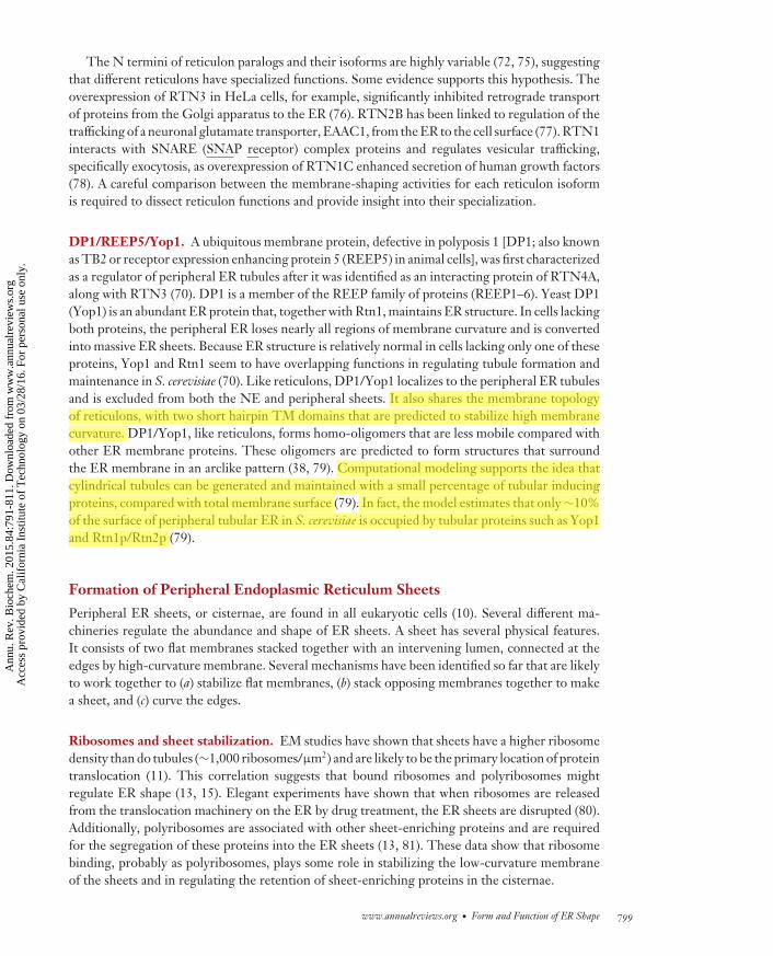
The association between ER and the cytoskeleton plays a critical role in the formation of newER tubules, ER dynamics, and the overall maintenance of the ER network. In mammalian cells,the association between the ER and microtubules (MTs) drives most of these processes (32, 33).ER tubules are extended along MTs by both retrograde and anterograde machinery (Figure 2)(34–37). When MTs are depolymerized, the dynamics of ER tubules come to a halt. Although thereticular network persists for some time without MTs in some cells, the integrity of its tubularshape is eventually lost, and ER membranes retract from the periphery into perinuclear sheets(38). Thus, interactions between the ER membrane and MTs are just as important to maintainingER shape as the membrane-shaping proteins, which are discussed below.
Research in both the plant and yeast fields has demonstrated that the ER can also be transportedthroughout the cell through interactions with the actin cytoskeleton, rather than MTs (35, 37,39, 40). Actin may play a significant role in ER structure in mammalian cells as well; several
www.annualreviews.org • Form and Function of ER Shape 795
Ann
u. R
ev. B
ioch
em. 2
015.
84:7
91-8
11. D
ownl
oade
d fr
om w
ww
.ann
ualr
evie
ws.
org
Acc
ess
prov
ided
by
Cal
ifor
nia
Inst
itute
of
Tec
hnol
ogy
on 0
3/28
/16.
For
per
sona
l use
onl
y.
BI84CH29-Prinz-Voeltz ARI 24 April 2015 11:27
EREn
doso
me
ER tubule
Organelle
cER
Endo
som
e
ER tubule
Organelle
d
ERST
IM1
b
ER tubule
Microtubule
TAC
0 s 5 s 10 s
0 s 5 s 10 s
0 s 10 s 20 s
ERM
icro
tubu
le
a
Kinesin-1/dynein
ER tubule
Microtubule
0 s 10 s 20 s
Figure 2Four types of endoplasmic reticulum (ER) dynamics. (a) ER tubules undergo motor-dependent “sliding”along the length of a microtubule. (b) During tip attachment, the ER protein stroma-interacting molecule 1(STIM1) (red ) localizes to the tip of an ER tubule ( green) and couples ER dynamics with the growth andmovement of the plus end of a microtubule. (c) ER ring rearrangements ( green) are positions where ERtubules circumscribe other organelles, here an early endosome (red ). (d ) ER tubules can also be pulledbehind organelles trafficking on the cytoskeleton. The tip of an ER tubule is linked to a trafficking endosome(red ). Abbreviation: TAC, tip attachment complex. Panel a modified with permission from Reference 46;panel c modified with permission from Reference 57; panels b and d modified from images provided by A.Rowland and P. Chitwood.
reports have demonstrated that ER sheets interact with the actin cytoskeleton (41–45). Treatmentof animal cells with the actin depolymerizer latrunculin A resulted in disrupted ER organizationcharacterized by a more reticular structure that was unevenly spread throughout the cytoplasm.Consistent with the increased reticular structure, the peripheral ER demonstrated a significantdecrease in the prevalence of ER sheets. These data suggest that actin dynamics could regulate theshape, dynamics, position, and possibly functions of ER sheets (45). Here, we focus on the role ofER association with MTs in determining ER structure and function in mammalian cells, becausethese interactions are the best understood. Live-cell imaging has revealed three main types ofmovement by ER tubules, all of which depend on MTs: (a) sliding, whereby a new tubule is pulled
796 Westrate et al.
Ann
u. R
ev. B
ioch
em. 2
015.
84:7
91-8
11. D
ownl
oade
d fr
om w
ww
.ann
ualr
evie
ws.
org
Acc
ess
prov
ided
by
Cal
ifor
nia
Inst
itute
of
Tec
hnol
ogy
on 0
3/28
/16.
For
per
sona
l use
onl
y.

BI84CH29-Prinz-Voeltz ARI 24 April 2015 11:27
out of an existing tubule along the shaft of a stable MT (Figure 2a), (b) the movement of the tipof a tubule along the cytoskeleton via a protein complex called TAC (tip attachment complex)(Figure 2b), and (c) ring rearrangements, a variation of ER sliding in which rings of ER movealong the side of MTs and occasionally close (Figure 2c).
Sliding. The most frequently observed type of ER movement is ER tubules “sliding” along MTs(34, 46, 47). This motion is driven by molecular motor proteins traveling along MTs and requireskinesin-1 or dynein, depending on whether the tubule is moving toward the plus or minus end ofan MT (Figure 2a) (34, 35, 48, 49). Inhibition of kinesin-1 function reduces ER tubule movementand extension both in vitro and in vivo (48–51). The morphological outcome of depleting kinesin-1is a reduction in tubular ER (49). Inhibition of dynein has a similar outcome: a reduction in thenumber of tubules moving toward the cell center and an increase in peripheral sheets at the expenseof tubules (49). Thus, the ratio of ER sheet to tubules is dependent on motor-driven sliding ofER tubules.
The function of ER sliding is not yet known, but it must serve an important purpose. Notonly does ER sliding occur constitutively; it also must require a great deal of energy because itdepends on molecular motors. ER sliding events track preferentially along MTs that are modifiedby acetylation (46). Acetylated MTs have unusually curved structures and prolonged stability tonocodazole treatment. Nocodazole treatment quickly (within minutes) depolymerizes the bulk ofMTs in the cytoplasm that are not modified (∼80% of the MTs), yet robust ER sliding continuesalong the nocodazole-resistant remaining acetylated MTs for up to an hour (46). It is curious thatER dynamics occur mostly on a small subset of MTs. This finding suggests that the purpose of ERsliding could be to traffic smooth ER material to a destination that is also present on modified MTs.
TAC and the role of STIM1. TAC represents another type of ER movement in which the tipof an ER tubule is linked to the plus end of an MT and the ER tubule grows and shrinks togetherwith the MT to which it is attached (Figure 2b) (34, 52). The TAC machinery includes EB1,an MT plus end–binding protein, which is linked to an integral ER membrane protein, stroma-interacting molecule 1 (STIM1) (34, 35). Because these dynamics are localized to MT plus ends,they occur in the cell periphery, just under the PM. STIM1 is an interesting protein because ithas also been shown to play a pivotal role in regulating Ca2+ homeostasis through activation ofSOCE (47, 53, 54). Depletion of ER Ca2+ stores results in a rapid relocation of STIM1 fromTACs to ER–PM contact sites, where it hetero-oligomerizes with Orai Ca2+ channels to form aCa2+ release–activated channel (CRAC), giving the ER lumen direct access to the extracellularCa2+ during SOCE (47). Surprisingly, a functional connection between TAC and SOCE has notbeen found, even though both processes require STIM1.
Ring rearrangements: the contact site connection. Ring structures of tubular ER have beenobserved to move around the cell in an MT-dependent manner at speeds consistent with molecularmotors (36, 46). These rings sometimes become smaller and smaller, leading to complete closure(33, 36, 55). Time-lapse imaging of the formation of ring structures and their closure providesinsight into ring structure organization and dynamics. Rings consist of two three-way junctions,one of which appears static or anchored with respect to the reticular ER network, whereas thesecond is dynamic with the ability to slide along the reticular ER. It is the sliding of the dynamicjunction toward the anchored junction that mediates ring closure (36, 46).
Until recently, the functions of these rings remained mysterious. Simultaneous imaging of theER and other organelles has finally revealed a clue. Numerous ER rings are positions where theER circumscribes endosomes and mitochondria (Figure 2c) (46, 57, 58). These organelles are
www.annualreviews.org • Form and Function of ER Shape 797
Ann
u. R
ev. B
ioch
em. 2
015.
84:7
91-8
11. D
ownl
oade
d fr
om w
ww
.ann
ualr
evie
ws.
org
Acc
ess
prov
ided
by
Cal
ifor
nia
Inst
itute
of
Tec
hnol
ogy
on 0
3/28
/16.
For
per
sona
l use
onl
y.

BI84CH29-Prinz-Voeltz ARI 24 April 2015 11:27
constricted where they are wrapped by ER, and subsequent studies showed that these are siteswhere mitochondrial fission and endosomal fission occur (56, 59). Thus, many ring closure eventsare the result of ER cinching at the positions where other organelles undergo fission.
The effect of interorganelle contact sites on ER shape and dynamics must be massive, giventhat, for example, a typical animal cell could have more than 100 endosomes and mitochondria andmost are attached to the tubular ER network (12, 56–59). Also, many ER sliding events may occurat positions where an ER tubule is pulled behind a trafficking organelle (Figure 2d ). Therefore, itis possible that most ER dynamics in the cell are indirectly caused by tethered organelles draggingthe ER around with them.
Formation of Peripheral Endoplasmic Reticulum Tubules
Several integral membrane proteins have been implicated in the generation and maintenance ofthe various structural domains within the ER. Unraveling the mechanisms of these proteins hasrevealed some answers to how the continuous membrane bilayer of the ER can be molded intocomplex shapes. Specifically, the significant curvature that is characteristic of peripheral ER tubulesis maintained by a series of wedge-shaped proteins that are embedded within the ER membraneand thereby maintain tubular curvature. These proteins localize exclusively to ER tubules and theedges of ER sheets, which, like tubules, are highly curved.
Reticulons. The reticulon proteins are a highly conserved and abundant family of integral mem-brane proteins that structurally shape ER tubules (60–62). In mammals, there are four reticulongenes (RTN1, RTN2, RTN3, and RTN4/Nogo), which encode several protein isoforms (RTN1A–C,RTN2A–C, RTN3A and -B, and RTN4/NogoA–C) that are generated through alternative splic-ing events or the use of different promoters (60, 63–68). Comparatively, Saccharomyces cerevisiaecontain two reticulon genes (RTN1 and RTN2), each of which encodes a single protein (Rtn1pand Rtn2p), whereas Caenorhabditis elegans has only one reticulon gene (ret-1), which encodesthree different reticulon protein isoforms (nRTN-A, nRTN-B, and nRTN-C) (69). Overexpres-sion of some reticulon isoforms drives the formation of elongated ER tubules and disruption ofperipheral ER sheets (38). In animal, yeast, and plant cells, depletion of reticulon proteins reducesER tubules and increases peripheral ER sheets (62, 70, 71). Thus, reticulons are necessary andsufficient to control ER tubule levels. Consistent with their function is the finding that reticulonproteins localize exclusively to regions of high membrane curvature, including ER tubules and theedges of ER sheets, and are excluded from the flat membrane domains of the NE and peripheralER sheets (70).
All reticulons have a core reticulon homology domain (RHD) located at their C termini.The RHD consists of two hairpin transmembrane (TM) domains separated by an interveninghydrophilic loop (72). The topology of the reticulons has been determined experimentally andreveals that the N- and C-terminal domains, along with the hydrophilic loop within the RHD, allface the cytosol (70). The hairpin TM domains within the RHD adopt an unusual topology thatsuggests how these proteins regulate membrane shape. The TM hairpins are unusually short andoccupy more space in the outer leaflet of the ER membrane than in the inner leaflet, causing themembrane bending that is necessary to generate regions of high curvature in the ER (70, 73). Thereticulon proteins also form immobile oligomers on the ER membrane (38). The structure of theseoligomers probably determines the diameter of the tubules. The RHD alone can oligomerize andpartition to ER tubules. These membrane-shaping properties are lost in a mutant in which theTM domains are lengthened to resemble the membrane-spanning α-helices found in most TMdomains (74).
798 Westrate et al.
Ann
u. R
ev. B
ioch
em. 2
015.
84:7
91-8
11. D
ownl
oade
d fr
om w
ww
.ann
ualr
evie
ws.
org
Acc
ess
prov
ided
by
Cal
ifor
nia
Inst
itute
of
Tec
hnol
ogy
on 0
3/28
/16.
For
per
sona
l use
onl
y.
BI84CH29-Prinz-Voeltz ARI 24 April 2015 11:27
The N termini of reticulon paralogs and their isoforms are highly variable (72, 75), suggestingthat different reticulons have specialized functions. Some evidence supports this hypothesis. Theoverexpression of RTN3 in HeLa cells, for example, significantly inhibited retrograde transportof proteins from the Golgi apparatus to the ER (76). RTN2B has been linked to regulation of thetrafficking of a neuronal glutamate transporter, EAAC1, from the ER to the cell surface (77). RTN1interacts with SNARE (SNAP receptor) complex proteins and regulates vesicular trafficking,specifically exocytosis, as overexpression of RTN1C enhanced secretion of human growth factors(78). A careful comparison between the membrane-shaping activities for each reticulon isoformis required to dissect reticulon functions and provide insight into their specialization.
DP1/REEP5/Yop1. A ubiquitous membrane protein, defective in polyposis 1 [DP1; also knownas TB2 or receptor expression enhancing protein 5 (REEP5) in animal cells], was first characterizedas a regulator of peripheral ER tubules after it was identified as an interacting protein of RTN4A,along with RTN3 (70). DP1 is a member of the REEP family of proteins (REEP1–6). Yeast DP1(Yop1) is an abundant ER protein that, together with Rtn1, maintains ER structure. In cells lackingboth proteins, the peripheral ER loses nearly all regions of membrane curvature and is convertedinto massive ER sheets. Because ER structure is relatively normal in cells lacking only one of theseproteins, Yop1 and Rtn1 seem to have overlapping functions in regulating tubule formation andmaintenance in S. cerevisiae (70). Like reticulons, DP1/Yop1 localizes to the peripheral ER tubulesand is excluded from both the NE and peripheral sheets. It also shares the membrane topologyof reticulons, with two short hairpin TM domains that are predicted to stabilize high membranecurvature. DP1/Yop1, like reticulons, forms homo-oligomers that are less mobile compared withother ER membrane proteins. These oligomers are predicted to form structures that surroundthe ER membrane in an arclike pattern (38, 79). Computational modeling supports the idea thatcylindrical tubules can be generated and maintained with a small percentage of tubular inducingproteins, compared with total membrane surface (79). In fact, the model estimates that only ∼10%of the surface of peripheral tubular ER in S. cerevisiae is occupied by tubular proteins such as Yop1and Rtn1p/Rtn2p (79).
Formation of Peripheral Endoplasmic Reticulum Sheets
Peripheral ER sheets, or cisternae, are found in all eukaryotic cells (10). Several different ma-chineries regulate the abundance and shape of ER sheets. A sheet has several physical features.It consists of two flat membranes stacked together with an intervening lumen, connected at theedges by high-curvature membrane. Several mechanisms have been identified so far that are likelyto work together to (a) stabilize flat membranes, (b) stack opposing membranes together to makea sheet, and (c) curve the edges.
Ribosomes and sheet stabilization. EM studies have shown that sheets have a higher ribosomedensity than do tubules (∼1,000 ribosomes/μm2) and are likely to be the primary location of proteintranslocation (11). This correlation suggests that bound ribosomes and polyribosomes mightregulate ER shape (13, 15). Elegant experiments have shown that when ribosomes are releasedfrom the translocation machinery on the ER by drug treatment, the ER sheets are disrupted (80).Additionally, polyribosomes are associated with other sheet-enriching proteins and are requiredfor the segregation of these proteins into the ER sheets (13, 81). These data show that ribosomebinding, probably as polyribosomes, plays some role in stabilizing the low-curvature membraneof the sheets and in regulating the retention of sheet-enriching proteins in the cisternae.
www.annualreviews.org • Form and Function of ER Shape 799
Ann
u. R
ev. B
ioch
em. 2
015.
84:7
91-8
11. D
ownl
oade
d fr
om w
ww
.ann
ualr
evie
ws.
org
Acc
ess
prov
ided
by
Cal
ifor
nia
Inst
itute
of
Tec
hnol
ogy
on 0
3/28
/16.
For
per
sona
l use
onl
y.
BI84CH29-Prinz-Voeltz ARI 24 April 2015 11:27
CLIMP63. EM studies have also revealed that ER sheets have a constrained luminal spacing of∼30 nm in yeast and 50 nm in mammalian cells (10, 13). This finding suggests that an intraluminaltether holds the two membranes that form an ER sheet at a constant distance. CLIMP63 (formallyp63) is a 63-kDa, nonglycosylated, type II integral membrane protein of the ER that localizes tothe ER sheets and may function to maintain the distance between the two membranes in an ERsheet (82–84). Depletion of CLIMP63 does not significantly alter the ratio of ER sheets to tubules;however, overexpression of CLIMP63 results in a dramatic increase in the number of ER sheetsand fewer tubules. Thus, CLIMP63 appears to work in opposition to the reticulons, which increasetubules and decrease sheets when overexpressed (13). Structurally, CLIMP63 is composed of anextended coiled-coil domain, a TM domain, and an N-terminal cytoplasmic segment that can bindMTs both in vivo and in vitro (82, 85). CLIMP63 has a luminal coiled-coil domain that regulatesits localization to sheets. Interactions between these charged coiled coils are predicted to driveCLIMP63 oligomerization, and these could bridge the luminal spacing of sheets (83). Indeed,CLIMP63 depletion reduces the luminal spacing from 45–50 nm to 25–30 nm (13).
Reticulons localize to the edges of ER sheets in yeast and animal cells (13). Thus, reticulonsmay also guide the curvature at the edges of sheets. Electron tomography of yeast mutants inwhich Rtn1 and Yop1 are deleted shows a massive expansion of ER sheets, accompanied by asmall change in mean luminal spacing compared with wild-type cells (11). These data suggest thatthe curvature induced by Rtn1 and Yop1 at the edges of sheets may also regulate the organizationof ER sheets.
Homotypic Fusion
A characteristic of the ER that becomes readily apparent upon live-cell imaging is that, eventhough it is very dynamic, it always remains continuous. Maintaining the continuity of the ERnetwork requires that new ER tubules undergo constitutive and efficient homotypic (ER–ER)fusion whenever they contact other ER membranes. Several recent studies have revealed that aseries of ER-localized GTPases mediate the fusion of ER tubules.
Atlastins. Atlastins are a family of conserved dynamin-related GTPases that localize to the ERand regulate ER fusion in multiple eukaryotes (86, 87). Mammals encode three atlastin paralogs(ATL1, ATL2, and ATL3), which are differentially expressed in multiple tissues. Atlastins aremost homologous to members of the dynamin/Ms/guanylate-binding protein family of GTPases(GBPs) and share the GTPase, “middle,” and other domains common to this family. Unlike thoseof many dynamins, the C termini of atlastins contain two putative TM domains, which are thoughtto form a hairpin, and an amphipathic helix–containing domain, which exposes most of the pro-tein to the cytosol (88). In this way, the structure of the atlastins resembles that of another familyof large GTPases, the mitofusins, which promote homotypic fusion of neighboring mitochon-dria through formation of hetero/homo-oligomeric complexes via heptad repeat regions in theC terminus that are similarly exposed to the cytoplasm (88, 89). However, unlike the mitofusins, at-lastins have a catalytic arginine finger (R77), which is exposed in the GDP-bound state to stimulateGTP hydrolysis upon rearrangements within the G domain (90).
In vitro experiments have demonstrated that Drosophila atlastin catalyzes fusion of syntheticliposomes (86). The mechanism of fusion has been probed by structure–function studies. Thecrystal structures of the GTPase domain revealed two dimeric confirmations corresponding toa prefusion state and a postfusion state. These structures suggest a fusion mechanism in whichthe atlastins in two apposing membranes dimerize through interactions between their GTPases,which are bound to GDP. After the GDP is exchanged for GTP, the energy of hydrolysis drives
800 Westrate et al.
Ann
u. R
ev. B
ioch
em. 2
015.
84:7
91-8
11. D
ownl
oade
d fr
om w
ww
.ann
ualr
evie
ws.
org
Acc
ess
prov
ided
by
Cal
ifor
nia
Inst
itute
of
Tec
hnol
ogy
on 0
3/28
/16.
For
per
sona
l use
onl
y.
BI84CH29-Prinz-Voeltz ARI 24 April 2015 11:27
conformational changes in the atlastins that pulls the ER membranes close enough together thatthey fuse (91, 92). Increasing the length of the linker between the N-terminal GTPase domain andthe TM domain significantly impairs ER fusion, thereby indicating the distance by which the ERmembranes are brought together upon atlastin dimerization and implicating GTP hydrolysis as acritical step for atlastin-mediated ER fusion (90, 93). Subsequent destabilization of the membraneand induced curvature by the atlastin membrane domains could be the mechanism that drivesfusion of the ER membrane (90, 94–96).
Similar to reticulons and DP1/Yop1, atlastin proteins partition into peripheral ER tubulesand are excluded from the NE and peripheral sheets. However, atlastins also have the fascinatingproperty that, when overexpressed, they accumulate at three-way junctions within the ER network(97). This localization is perfectly consistent with the observation that overexpression of a GTP-binding mutant of atlastin generates elongated ER tubules and reduces the frequency of three-way junctions (87). Although no sequence-related protein has been detected in yeast, a putativeortholog, Sey1p, was identified and found to be enriched in cortical ER and to mediate homotypicER fusion in vitro and in cells (87, 98). Yeast Sey1p localizes to three-way junctions (99) andregulates ER morphology in a fashion similar to metazoan atlastin (87, 98). Together, these datademonstrate that atlastins regulate ER shape in a GTP-dependent manner.
The highly conserved Lunapark family of proteins (Lnps) is a recent addition to what re-mains a short list of proteins that regulate peripheral ER shape. Lnp1 was identified through anS. cerevisiae screen for ER proteins that disrupt the shape of the cortical ER network. Cells that lackLnp1 contain a highly reticulated cortical ER. This phenotype is the opposite of that observed foratlastin depletion. Indeed, fluorescently labeled Lnp1p localizes to the ER, and like Sey1p/atlastin,it can also concentrate at three-way junctions (99). Mechanistically, Lnp1p works synergisticallywith reticulons and DP1/Yop1p but antagonistically to the atlastin homolog in yeast, Sey1p (99,100). These data suggest that Lunapark and atlastin proteins may play opposing roles in regulatingreticulation of the peripheral ER network. How Lunapark proteins shape ER membranes remainsan important and unanswered question.
Rab GTPases. Rab GTPases regulate membrane trafficking and fusion events at multiple distinctsteps throughout the secretory and endocytic pathways. Recently, two ER-localized Rab GTPases,Rab10 and Rab18, were found to determine ER shape by regulating ER dynamics and fusion. Anin vitro assay for ER vesicle fusion and tubule formation using membranes derived from Xenopusegg extracts was used to identify the highly conserved Rab10 as an ER-enriched GTP-bindingprotein (101). In cells, Rab10 depletion or expression of a GDP-locked Rab10 mutant (T23N)causes proliferation of peripheral ER sheets at the expense of tubules (101, 102). In these studies,fluorescently labeled Rab10 localized to the ER and, remarkably, was enriched at the leadingedge of more than 40% of ER sliding events. Moreover, ER–ER fusion usually occurred whenthe Rab10-enriched tip of an ER tubule encountered another ER membrane. In contrast, in theabsence of Rab10 or upon Rab10 T23N expression, ER dynamics and fusion were dramaticallyreduced. Thus, the defect in ER dynamics and fusion after Rab10 inactivation explains the increasein ER sheets upon loss of functional Rab10.
Interestingly, Rab10 colocalizes with at least two phospholipid biosynthetic enzymes, phos-phoinositol synthase (PIS) and choline/ethanolamine phosphotransferase (CEPT1), which alsoconcentrate at the leading edge of ER sliding events (101). Thus, Rab10-enriched domains at thetips of sliding ER tubules may promote lipid synthesis and/or delivery to various parts of the cell.Whether lipid synthesis by PIS and CEPT1 occurs at Rab10-dynamic domains, and whether PISor CEPT1 is required for regulating Rab10-dependent dynamics of the peripheral ER, remainsto be determined.
www.annualreviews.org • Form and Function of ER Shape 801
Ann
u. R
ev. B
ioch
em. 2
015.
84:7
91-8
11. D
ownl
oade
d fr
om w
ww
.ann
ualr
evie
ws.
org
Acc
ess
prov
ided
by
Cal
ifor
nia
Inst
itute
of
Tec
hnol
ogy
on 0
3/28
/16.
For
per
sona
l use
onl
y.
BI84CH29-Prinz-Voeltz ARI 24 April 2015 11:27
Rab18 is a highly conserved Rab that is targeted to the ER by a Rab3–GAP complex (102). Loss-of-function mutations in Rab18 or Rab3–GAP subunits cause a similar phenotype: an increase inperipheral ER sheets. This phenotype is reported to be even more dramatic than the phenotypeobserved upon Rab10 depletion. Interestingly, Rab10 depletion redistributes Rab18 to peripheralER sheets (102). It is not known whether these two Rabs are redundant, are synergistic, or regulatedifferent steps during ER dynamics and fusion.
It seems clear that the Rabs and the atlastins facilitate ER fusion by entirely different mech-anisms because the disruption of Rab10 or Rab18 function increases ER sheets, whereas atlastindisruption gives the opposite phenotype: an increase in unbranched tubules (87, 101, 102). Theatlastins can directly fuse ER membranes and are likely to play a role in maintaining general ERcontinuity (86), whereas the Rabs might regulate the interaction between the ER and molecularmotors during ER sliding and the rapid fusion that is occurring during these dynamics (43). In-deed, ER Rab depletion (or dominant negative expression) mimics the disruptions in ER slidingobserved in kinesin-1 and dynein disruption experiments (49, 101, 102). Thus, when Rab10 orRab18 is depleted, the inability to stabilize successful tubule fusion events ultimately deterioratesER tubule maintenance and increases the size and abundance of sheets.
FUNCTIONAL CONSEQUENCES OF ENDOPLASMIC RETICULUMSTRUCTURE: IMPLICATIONS FOR DISEASE PATHOLOGY
The organization of the ER has critical implications for maintaining not only ER function but alsooverall cellular health. Recently, ER morphology defects caused by mutations in many ER-shapingproteins, described above, have been linked to the pathology of several human diseases, includingneurological disorders and viral infections. These links show the importance of ER morphologyin ER function and organismal and cellular homeostasis.
Neurological Disease
ER morphology appears to play an important role in generating neuronal architecture (103).Neurons are highly specialized cells with an extensive peripheral ER network that packs the soma,axons, and dendrites (104–106). Functionally, axonal ER is especially interesting because it doesnot have bound ribosomes and therefore is not the site of protein translocation into the ER (106,107). Axons (and dendrites) may rely on the ER for Ca2+ stores required in pre- and postsynapticsignaling, as well as the synthesis and trafficking of lipids required to support the developmentand extension of these neuronal processes. Not surprisingly, the peripheral ER is elongated andtubular in neuronal processes. It is also very dynamic. A mechanism must exist to extend peripheralER tubules from the soma into the dendrite and axon along MTs all the way to their ends, andkeep it there. Recently, multiple links have been found between mutations in genes encoding ERstructural proteins and two common neurodegenerative diseases (103).
Alzheimer’s disease. The reticulon proteins have been linked to Alzheimer’s disease (AD). ADis one of the most common neurodegenerative diseases and affects more than 30 million peopleworldwide (108). The neuropathological features of a patient with AD include the presence ofamyloid plaques and neurofibrillary tangles within the brain, and these correlate with neuronaldegeneration and dementia (109). All four of the reticulon family members (RTN1–4) have beenimplicated in binding and modulating the activity of the type I TM aspartyl protease BACE1, whichreduces the production of amyloid-β (110). Overexpression of the reticulon proteins significantlyreduces the levels of amyloid-β. Given that BACE1 is initially expressed in the ER, interactions
802 Westrate et al.
Ann
u. R
ev. B
ioch
em. 2
015.
84:7
91-8
11. D
ownl
oade
d fr
om w
ww
.ann
ualr
evie
ws.
org
Acc
ess
prov
ided
by
Cal
ifor
nia
Inst
itute
of
Tec
hnol
ogy
on 0
3/28
/16.
For
per
sona
l use
onl
y.

BI84CH29-Prinz-Voeltz ARI 24 April 2015 11:27
with reticulons may prevent BACE1 from localizing to the Golgi network, preventing processingto the mature form and thereby inhibiting the generation of amyloid-β peptide (110).
In addition to the accumulation of amyloid plaques, other pathological features of AD includealtered phospholipid and calcium metabolism and impaired mitochondrial function (111–115).These changes might be the result of altered ER–mitochondrial contacts in the cells of AD patients(56, 111, 116), as three major functions occur at ER–mitochondrial contact sites: Ca2+ transfer,lipid biosynthesis, and mitochondrial fission. Indeed, the number of ER–mitochondrial contactsis higher in brain samples from postmortem AD patients and AD mouse models (116, 117). Thus,the increase in ER–mitochondrial contacts might cause the mitochondrial fragmentation anddysfunction and changes in metabolism that occur in AD. These links suggest how alterations inER structure affect the pathological features of AD (111, 117–120).
Hereditary spastic paraplegia. Hereditary spastic paraplegia (HSP) is a genetic neurologicaldisorder characterized by progressive lower-limb spasticity and weakness resulting from axonaldegeneration within motor neurons. HSP comprises a large family of genetic neurologic disorders(nearly 50 distinct loci) (121–123). ATL1 is the second most commonly mutated protein in HSPand represents the most common cause of early onset HSP (121, 124). Although ATL1 is ubiqui-tously expressed, it is expressed at very high levels in the brain (88, 125). Loss of ATL1 in culturedcortical neurons significantly inhibits axonal elongation, highlighting a functional requirementfor ER fusion (126). Interestingly, Rab10 depletion also attenuates axonal elongation, althoughdisruption of Rab10 has not yet been linked to a genetic neurological disorder (127).
REEP1 is another commonly mutated gene in HSP; it belongs to a family of six related proteins(REEP1–6), which were first identified through their role in trafficking olfactory receptors to thePM (121, 122, 124, 128, 129). REEP1 knockout mice demonstrate weakness and spasticity of thehind limbs, which recapitulates phenotype characteristics of HSP. Additionally, cortical motorneurons isolated from REEP1 knockout mice have a reduction in peripheral ER complexity,suggesting that REEP1 is necessary to maintain reticular ER structure in these cells. REEP5,also known as DP1, has already been implicated in regulating the formation and maintenance ofER tubules. Similar to REEP5/DP1, REEP1 localizes to the ER and contains two short hairpinTM domains, which are predicted to be capable of generating membrane curvature. Indeed,REEP1 can promote positive curvature on liposomes in vitro (130). Unlike REEP5/DP1, however,REEP1 contains an extended C-terminal domain that binds MTs through its MTBD (MT-bindingdomain) (121). Interestingly, recent evidence shows that atlastin interacts with REEP1 and maytherefore indicate that, whereas REEP5/DP1 plays a direct role in ER morphology, REEP1 mayinstead be involved in regulating the dynamics and distribution of the peripheral ER (122, 131,132). Given the close association between the ER and the MTs, REEP1’s ability to bind MTsmay therefore play a critical role in regulating ER movement and distribution within the neuron.Future studies will surely help elucidate the role REEP1 plays in HSP pathology and determinewhether the clinical presentation of the disease is due largely to alteration in ER morphology, ERdistribution, or a combination of the two.
Viral Infection
Several recent papers have shown that viruses alter ER structure to facilitate their propagation.These viruses modify ER structure to shelter the viral genome from host cell nucleases andco-opt the ER-bound ribosomes to synthesize their viral protein machineries (133–138). Forexample, infection by Japanese encephalitis virus ( JEV) causes rapid filling of the lumen with viralparticles upon cellular infection, which is accompanied by hypertrophy of the ER membranes.
www.annualreviews.org • Form and Function of ER Shape 803
Ann
u. R
ev. B
ioch
em. 2
015.
84:7
91-8
11. D
ownl
oade
d fr
om w
ww
.ann
ualr
evie
ws.
org
Acc
ess
prov
ided
by
Cal
ifor
nia
Inst
itute
of
Tec
hnol
ogy
on 0
3/28
/16.
For
per
sona
l use
onl
y.
BI84CH29-Prinz-Voeltz ARI 24 April 2015 11:27
The alteration in ER structure is also characterized by the proliferation of rough ER to supportproduction of viral proteins for replication (139). Positive-strand RNA [(+)RNA] viruses alsopromote their replication by altering ER structure. These viruses, which include the brome mosaicvirus (BMV), are responsible for several mammalian and plant diseases and utilize the ER forRNA replication through the formation of spherule vesicles on the peripheral ER (136–138,140, 141). Compared with the positive-curving properties of reticulons and DP1/Yop1, infectionby (+)RNA viruses induces ER membranes to undergo negative curvature by forming vesicularinvaginations within the outer ER membrane to form spherules, which serve as sites of RNAsynthesis (137, 142). These structures provide shelter from host cell nucleases and act as scaffoldsto concentrate replication factors and catalyze virus-dependent reactions (133–138). Interestingly,loss of reticulons and Yop1 in yeast cells significantly inhibited BMV RNA replication by morethan 80% (143). These data suggest that viruses use reticulons and DP1/Yop1 to rearrange thestructure of the tubular ER to accommodate their needs (70, 79, 143, 144).
Further evidence of the role of ER structure in promoting viral replication is that some virusesdirectly interact with proteins required for shaping the ER. For example, enterovirus 71 (EV71),a member of the Picornaviridae family of viruses, forms a mature viral polypeptide, 2C, whichbinds and interacts with RTN3 (145). Knockdown of RTN3 through targeted small interferingRNA (siRNA) significantly reduced viral replication, and expression of siRNA-resistant RTN3 wassufficient to rescue infectivity, indicating that RTN3 plays an essential role in EV71 replication andinfection (145). Although modification of ER morphology is a common theme for several virusesduring RNA synthesis, reticulons and DP1/Yop1 are not always associated with the promotion ofviral infection. Specifically, Wu et al. (146) recently provided evidence for the opposite phenotypein hepatitis C virus (HCV); RTN3 was found to bind to the HCV NS4B protein and thereby inhibitviral replication. RTN3 interacted specifically with the second amphipathic α-helical domain(AH2), thereby preventing self-oligomerization on the ER and subsequent formation of HCVparticles (146–148).
Another way that viruses reshape ER membranes is by promoting the formation of double-membrane vesicles derived from the ER. These vesicles are found in cells following infection withHCV, dengue virus (DV), and severe acute respiratory syndrome (SARS) (136, 140, 141). Three-dimensional electron tomography studies have shown that DV vesicle structures have narrownecks and pore openings similar to those of BMV-induced single-membrane spherules, whereasSARS vesicle structures are sealed off from the cytosol (136, 141). SARS virus utilizes three viralnonstructural proteins (nsps) to induce ER remodeling. SARS nsp3 and nsp4 induce membranestacking, whereas nsp6 drives membrane invagination and double-membrane vesicle formation(149). In contrast, HCV or DV can induce vesicle formation with the expression of a singleviral integral membrane protein, NS4B or NS4A, respectively (136, 141). If this is a fundamentalmechanism for double-membrane vesicle formation, then the observation that SARS infectionrequires three proteins to form double-membrane vesicles suggests that HCV NS4B and DVNS4A might employ host cell factors to stack and invaginate ER membranes. However, morestudies focused on how viral factors directly or indirectly manipulate ER structure are needed. Abetter understanding of how viruses co-opt and rearrange the ER membrane will allow drugs tobe developed that can perturb these processes.
CONCLUSIONS AND REMAINING QUESTIONS
Why the peripheral ER has such a complex and dynamic structure remained largely enigmaticfor many years. Early EM studies suggested that the various domains of the peripheral ER, forexample, the rough and smooth portions, had distinct cellular functions, but the relationship
804 Westrate et al.
Ann
u. R
ev. B
ioch
em. 2
015.
84:7
91-8
11. D
ownl
oade
d fr
om w
ww
.ann
ualr
evie
ws.
org
Acc
ess
prov
ided
by
Cal
ifor
nia
Inst
itute
of
Tec
hnol
ogy
on 0
3/28
/16.
For
per
sona
l use
onl
y.
BI84CH29-Prinz-Voeltz ARI 24 April 2015 11:27
between the structures of these domains and their functions was not well understood. Clearly, thestructure of the ER is modified to meet the particular demands of the cell, as evidenced by thediverging ER morphologies in specialized cells. In general, cells that are primed for protein orsterol secretion (such as pancreatic or adrenal cells) have evolved peripheral ER with increasedsheets, whereas cells requiring extensive Ca2+ modulation (such as muscle cells) are characterizedby peripheral ER made up of a higher percentage of ER tubules. The dynamic nature of theER also suggests that the transitions between ER sheets and tubules can occur quite rapidly inresponse to changing cellular environments.
Recent evidence suggests that much of the elaborate, dynamic structure of the ER is driven bythe apparent need for the ER to track along MTs and remain closely apposed to other organelles inthe cell. This understanding has resulted from improved multicolor live-cell imaging techniquesand from the identification of proteins and forces that maintain ER structure. These studies raiseimportant questions for the future. How are contacts between the ER and organelles generated,and what are the functions of these contacts? What cellular processes require these contacts? Whydo most cells express a variety of reticulon and reticulon-like membrane-shaping proteins? Howdo cells sense cellular stresses and modify ER size and structure in response? What role does ERmobility play in ER function? As we answer these questions, we will develop a better understandingof how the ER regulates the activities of other organelles and how defects cause various diseases.
DISCLOSURE STATEMENT
The authors are not aware of any affiliations, memberships, funding, or financial holdings thatmight be perceived as affecting the objectivity of this review.
ACKNOWLEDGMENTS
Our work is supported by a Research Scholar Grant from the American Cancer Society toG.K.V. and by grants from the National Institutes of Health to G.K.V. (GM083977) and toJ.E.L. (F32CA174158). W.A.P. is supported by the Intramural Research Program of the NationalInstitute of Diabetes and Digestive and Kidney Diseases.
LITERATURE CITED
1. Veratti E. 1961. Investigations on the fine structure of striated muscle fiber read before the Reale IstitutoLombardo, 13 March 1902. J. Biophys. Biochem. Cytol. 10(Suppl. 4):1–59
2. Schuldiner M, Schwappach B. 2013. From rags to riches—the history of the endoplasmic reticulum.Biochim. Biophys. Acta 1833:2389–91
3. Palade GE. 1955. Studies on the endoplasmic reticulum. II. Simple dispositions in cells in situ. J. Biophys.Biochem. Cytol. 1:56–7-82
4. Palade GE, Porter KR. 1954. Studies on the endoplasmic reticulum. I. Its identification in cells in situ.J. Exp. Med. 100:641–56
5. Porter KR, Palade GE. 1957. Studies on the endoplasmic reticulum. III. Its form and distribution instriated muscle cells. J. Biophys. Biochem. Cytol. 3:269–300
6. Mazzarello P, Calligaro A, Vannini V, Muscatello U. 2003. The sarcoplasmic reticulum: its discoveryand rediscovery. Nat. Rev. Mol. Cell Biol. 4:69–74
7. Hetzer MW, Walther TC, Mattaj IW. 2005. Pushing the envelope: structure, function, and dynamicsof the nuclear periphery. Annu. Rev. Cell Dev. Biol. 21:347–80
8. D’Angelo MA, Hetzer MW. 2006. The role of the nuclear envelope in cellular organization. Cell Mol.Life Sci. 63:316–32
www.annualreviews.org • Form and Function of ER Shape 805
Ann
u. R
ev. B
ioch
em. 2
015.
84:7
91-8
11. D
ownl
oade
d fr
om w
ww
.ann
ualr
evie
ws.
org
Acc
ess
prov
ided
by
Cal
ifor
nia
Inst
itute
of
Tec
hnol
ogy
on 0
3/28
/16.
For
per
sona
l use
onl
y.
BI84CH29-Prinz-Voeltz ARI 24 April 2015 11:27
9. Burke B, Ellenberg J. 2002. Remodelling the walls of the nucleus. Nat. Rev. Mol. Cell Biol. 3:487–9710. Bernales S, McDonald KL, Walter P. 2006. Autophagy counterbalances endoplasmic reticulum expan-
sion during the unfolded protein response. PLOS Biol. 4:e42311. West M, Zurek N, Hoenger A, Voeltz GK. 2011. A 3D analysis of yeast ER structure reveals how ER
domains are organized by membrane curvature. J. Cell Biol. 193:333–4612. Friedman JR, Voeltz GK. 2011. The ER in 3D: a multifunctional dynamic membrane network. Trends
Cell Biol. 21:709–1713. Shibata Y, Shemesh T, Prinz WA, Palazzo AF, Kozlov MM, Rapoport TA. 2010. Mechanisms deter-
mining the morphology of the peripheral ER. Cell 143:774–8814. Voeltz GK, Rolls MM, Rapoport TA. 2002. Structural organization of the endoplasmic reticulum. EMBO
Rep. 3:944–5015. Shibata Y, Voeltz GK, Rapoport TA. 2006. Rough sheets and smooth tubules. Cell 126:435–3916. Terasaki M, Shemesh T, Kasthuri N, Klemm RW, Schalek R, et al. 2013. Stacked endoplasmic reticulum
sheets are connected by helicoidal membrane motifs. Cell 154:285–9617. Baumann O, Walz B. 2001. Endoplasmic reticulum of animal cells and its organization into structural
and functional domains. Int. Rev. Cytol. 205:149–21418. Maxfield FR, McGraw TE. 2004. Endocytic recycling. Nat. Rev. Mol. Cell Biol. 5:121–3219. Mayor S, Presley JF, Maxfield FR. 1993. Sorting of membrane components from endosomes and sub-
sequent recycling to the cell surface occurs by a bulk flow process. J. Cell Biol. 121:1257–6920. Dunn KW, McGraw TE, Maxfield FR. 1989. Iterative fractionation of recycling receptors from lysoso-
mally destined ligands in an early sorting endosome. J. Cell Biol. 109:3303–1421. Zhang D, Vjestica A, Oliferenko S. 2012. Plasma membrane tethering of the cortical ER necessitates its
finely reticulated architecture. Curr. Biol. 22:2048–5222. Takeshima H, Komazaki S, Nishi M, Iino M, Kangawa K. 2000. Junctophilins: a novel family of junctional
membrane complex proteins. Mol. Cell 6:11–2223. Schneider MF. 1994. Control of calcium release in functioning skeletal muscle fibers. Annu. Rev. Physiol.
56:463–8424. Block BA, Imagawa T, Campbell KP, Franzini-Armstrong C. 1988. Structural evidence for direct inter-
action between the molecular components of the transverse tubule/sarcoplasmic reticulum junction inskeletal muscle. J. Cell Biol. 107:2587–600
25. Franzini-Armstrong C. 1970. Studies of the triad: I. Structure of the junction in frog twitch fibers.J. Cell Biol. 47:488–99
26. Rizzuto R, Pinton P, Carrington W, Fay FS, Fogarty KE, et al. 1998. Close contacts with the endoplasmicreticulum as determinants of mitochondrial Ca2+ responses. Science 280:1763–66
27. Csordas G, Thomas AP, Hajnoczky G. 1999. Quasi-synaptic calcium signal transmission between en-doplasmic reticulum and mitochondria. EMBO J. 18:96–108
28. Luik RM, Wang B, Prakriya M, Wu MM, Lewis RS. 2008. Oligomerization of STIM1 couples ERcalcium depletion to CRAC channel activation. Nature 454:538–42
29. Park CY, Hoover PJ, Mullins FM, Bachhawat P, Covington ED, et al. 2009. STIM1 clusters and activatesCRAC channels via direct binding of a cytosolic domain to Orai1. Cell 136:876–90
30. Giordano F, Saheki Y, Idevall-Hagren O, Colombo SF, Pirruccello M, et al. 2013. PI4,5P2-dependentand Ca2+-regulated ER–PM interactions mediated by the extended synaptotagmins. Cell 153:1494–509
31. Chang CL, Hsieh TS, Yang TT, Rothberg KG, Azizoglu DB, et al. 2013. Feedback regulation ofreceptor-induced Ca2+ signaling mediated by E-Syt1 and Nir2 at endoplasmic reticulum–plasma mem-brane junctions. Cell Rep. 5:813–25
32. Terasaki M, Song J, Wong JR, Weiss MJ, Chen LB. 1984. Localization of endoplasmic reticulum inliving and glutaraldehyde-fixed cells with fluorescent dyes. Cell 38:101–8
33. Terasaki M, Chen LB, Fujiwara K. 1986. Microtubules and the endoplasmic reticulum are highly inter-dependent structures. J. Cell Biol. 103:1557–68
34. Waterman-Storer CM, Salmon ED. 1998. Endoplasmic reticulum membrane tubules are distributed bymicrotubules in living cells using three distinct mechanisms. Curr. Biol. 8:798–806
35. Bola B, Allan V. 2009. How and why does the endoplasmic reticulum move? Biochem. Soc. Trans. 37:961–65
806 Westrate et al.
Ann
u. R
ev. B
ioch
em. 2
015.
84:7
91-8
11. D
ownl
oade
d fr
om w
ww
.ann
ualr
evie
ws.
org
Acc
ess
prov
ided
by
Cal
ifor
nia
Inst
itute
of
Tec
hnol
ogy
on 0
3/28
/16.
For
per
sona
l use
onl
y.
BI84CH29-Prinz-Voeltz ARI 24 April 2015 11:27
36. Lee C, Chen LB. 1988. Dynamic behavior of endoplasmic reticulum in living cells. Cell 54:37–4637. Fehrenbacher KL, Davis D, Wu M, Boldogh I, Pon LA. 2002. Endoplasmic reticulum dynamics, inher-
itance, and cytoskeletal interactions in budding yeast. Mol. Biol. Cell 13:854–6538. Shibata Y, Voss C, Rist JM, Hu J, Rapoport TA, et al. 2008. The reticulon and DP1/Yop1p proteins
form immobile oligomers in the tubular endoplasmic reticulum. J. Biol. Chem. 283:18892–90439. Boevink P, Oparka K, Santa Cruz S, Martin B, Betteridge A, Hawes C. 1998. Stacks on tracks: the plant
Golgi apparatus traffics on an actin/ER network. Plant J. 15:441–4740. Ueda H, Yokota E, Kutsuna N, Shimada T, Tamura K, et al. 2010. Myosin-dependent endoplasmic
reticulum motility and F-actin organization in plant cells. PNAS 107:6894–9941. Sanger JM, Dome JS, Mittal B, Somlyo AV, Sanger JW. 1989. Dynamics of the endoplasmic reticulum
in living non-muscle and muscle cells. Cell Motil. Cytoskelet. 13:301–1942. Wang Y, Mattson MP, Furukawa K. 2002. Endoplasmic reticulum calcium release is modulated by actin
polymerization. J. Neurochem. 82:945–5243. Wagner W, Brenowitz SD, Hammer JA 3rd. 2011. Myosin-Va transports the endoplasmic reticulum
into the dendritic spines of Purkinje neurons. Nat. Cell Biol. 13:40–4844. Tabb JS, Molyneaux BJ, Cohen DL, Kuznetsov SA, Langford GM. 1998. Transport of ER vesicles on
actin filaments in neurons by myosin V. J. Cell Sci. 111:3221–3445. Joensuu M, Belevich I, Ramo O, Nevzorov I, Vihinen H, et al. 2014. ER sheet persistence is coupled to
myosin 1c–regulated dynamic actin filament arrays. Mol. Biol. Cell 25:1111–2646. Friedman JR, Webster BM, Mastronarde DN, Verhey KJ, Voeltz GK. 2010. ER sliding dynamics and
ER–mitochondrial contacts occur on acetylated microtubules. J. Cell Biol. 190:363–7547. Grigoriev I, Gouveia SM, van der Vaart B, Demmers J, Smyth JT, et al. 2008. STIM1 is a MT-plus-
end-tracking protein involved in remodeling of the ER. Curr. Biol. 18:177–8248. Feiguin F, Ferreira A, Kosik KS, Caceres A. 1994. Kinesin-mediated organelle translocation revealed by
specific cellular manipulations. J. Cell Biol. 127:1021–3949. Wozniak MJ, Bola B, Brownhill K, Yang YC, Levakova V, Allan VJ. 2009. Role of kinesin-1 and cyto-
plasmic dynein in endoplasmic reticulum movement in VERO cells. J. Cell Sci. 122:1979–8950. Bannai H, Inoue T, Nakayama T, Hattori M, Mikoshiba K. 2004. Kinesin dependent, rapid, bi-
directional transport of ER sub-compartment in dendrites of hippocampal neurons. J. Cell Sci. 117:163–75
51. Wozniak MJ, Allan VJ. 2006. Cargo selection by specific kinesin light chain 1 isoforms. EMBO J.25:5457–68
52. Waterman-Storer CM, Gregory J, Parsons SF, Salmon ED. 1995. Membrane/microtubule tip attach-ment complexes (TACs) allow the assembly dynamics of plus ends to push and pull membranes intotubulovesicular networks in interphase Xenopus egg extracts. J. Cell Biol. 130:1161–69
53. Pani B, Ong HL, Liu X, Rauser K, Ambudkar IS, Singh BB. 2008. Lipid rafts determine clustering ofSTIM1 in endoplasmic reticulum–plasma membrane junctions and regulation of store-operated Ca2+
entry (SOCE). J. Biol. Chem. 283:17333–4054. Roos J, DiGregorio PJ, Yeromin AV, Ohlsen K, Lioudyno M, et al. 2005. STIM1, an essential and
conserved component of store-operated Ca2+ channel function. J. Cell Biol. 169:435–4555. Prinz WA, Grzyb L, Veenhuis M, Kahana JA, Silver PA, Rapoport TA. 2000. Mutants affecting the
structure of the cortical endoplasmic reticulum in Saccharomyces cerevisiae. J. Cell Biol. 150:461–7456. Friedman JR, Lackner LL, West M, DiBenedetto JR, Nunnari J, Voeltz GK. 2011. ER tubules mark
sites of mitochondrial division. Science 334:358–6257. Friedman JR, Dibenedetto JR, West M, Rowland AA, Voeltz GK. 2013. Endoplasmic reticulum–
endosome contact increases as endosomes traffic and mature. Mol. Biol. Cell 24:1030–4058. Zajac AL, Goldman YE, Holzbaur EL, Ostap EM. 2013. Local cytoskeletal and organelle interactions
impact molecular-motor-driven early endosomal trafficking. Curr. Biol. 23:1173–8059. Rowland AA, Chitwood PJ, Phillips MJ, Voeltz GK. 2014. ER contact sites define the position and
timing of endosome fission. Cell 159:1027–4160. Oertle T, Klinger M, Stuermer CA, Schwab ME. 2003. A reticular rhapsody: phylogenic evolution and
nomenclature of the RTN/Nogo gene family. FASEB J. 17:1238–47
www.annualreviews.org • Form and Function of ER Shape 807
Ann
u. R
ev. B
ioch
em. 2
015.
84:7
91-8
11. D
ownl
oade
d fr
om w
ww
.ann
ualr
evie
ws.
org
Acc
ess
prov
ided
by
Cal
ifor
nia
Inst
itute
of
Tec
hnol
ogy
on 0
3/28
/16.
For
per
sona
l use
onl
y.

BI84CH29-Prinz-Voeltz ARI 24 April 2015 11:27
61. Di Sano F, Bernardoni P, Piacentini M. 2012. The reticulons: guardians of the structure and functionof the endoplasmic reticulum. Exp. Cell Res. 318:1201–7
62. Tolley N, Sparkes I, Craddock CP, Eastmond PJ, Runions J, et al. 2010. Transmembrane domain lengthis responsible for the ability of a plant reticulon to shape endoplasmic reticulum tubules in vivo. Plant J.64:411–18
63. Roebroek AJ, Van de Velde HJ, Van Bokhoven A, Broers JL, Ramaekers FC, Van de Ven WJ. 1993.Cloning and expression of alternative transcripts of a novel neuroendocrine-specific gene and identifi-cation of its 135-kDa translational product. J. Biol. Chem. 268:13439–47
64. Roebroek AJ, Ayoubi TA, Van de Velde HJ, Schoenmakers EF, Pauli IG, Van de Ven WJ. 1996. Genomicorganization of the human NSP gene, prototype of a novel gene family encoding reticulons. Genomics32:191–99
65. Roebroek AJ, Contreras B, Pauli IG, Van de Ven WJ. 1998. cDNA cloning, genomic organization, andexpression of the human RTN2 gene, a member of a gene family encoding reticulons. Genomics 51:98–106
66. Moreira EF, Jaworski CJ, Rodriguez IR. 1999. Cloning of a novel member of the reticulon gene family(RTN3): gene structure and chromosomal localization to 11q13. Genomics 58:73–81
67. GrandPre T, Nakamura F, Vartanian T, Strittmatter SM. 2000. Identification of the Nogo inhibitor ofaxon regeneration as a Reticulon protein. Nature 403:439–44
68. Oertle T, Huber C, van der Putten H, Schwab ME. 2003. Genomic structure and functional character-isation of the promoters of human and mouse nogo/rtn4. J. Mol. Biol. 325:299–323
69. Iwahashi J, Kawasaki I, Kohara Y, Gengyo-Ando K, Mitani S, et al. 2002. Caenorhabditis elegans reticuloninteracts with RME-1 during embryogenesis. Biochem. Biophys. Res. Commun. 293:698–704
70. Voeltz GK, Prinz WA, Shibata Y, Rist JM, Rapoport TA. 2006. A class of membrane proteins shapingthe tubular endoplasmic reticulum. Cell 124:573–86
71. Tolley N, Sparkes IA, Hunter PR, Craddock CP, Nuttall J, et al. 2008. Overexpression of a plant reticulonremodels the lumen of the cortical endoplasmic reticulum but does not perturb protein transport. Traffic9:94–102
72. Yang YS, Strittmatter SM. 2007. The reticulons: a family of proteins with diverse functions. GenomeBiol. 8:234
73. Lin S, Sun S, Hu J. 2012. Molecular basis for sculpting the endoplasmic reticulum membrane. Int. J.Biochem. Cell Biol. 44:1436–43
74. Zurek N, Sparks L, Voeltz G. 2011. Reticulon short hairpin transmembrane domains are used to shapeER tubules. Traffic 12:28–41
75. Iwahashi J, Hamada N, Watanabe H. 2007. Two hydrophobic segments of the RTN1 family determinethe ER localization and retention. Biochem. Biophys. Res. Commun. 355:508–12
76. Wakana Y, Koyama S, Nakajima K, Hatsuzawa K, Nagahama M, et al. 2005. Reticulon 3 is involvedin membrane trafficking between the endoplasmic reticulum and Golgi. Biochem. Biophys. Res. Commun.334:1198–205
77. Liu Y, Vidensky S, Ruggiero AM, Maier S, Sitte HH, Rothstein JD. 2008. Reticulon RTN2B regulatestrafficking and function of neuronal glutamate transporter EAAC1. J. Biol. Chem. 283:6561–71
78. Steiner P, Kulangara K, Sarria JC, Glauser L, Regazzi R, Hirling H. 2004. Reticulon 1C/neuroendocrine-specific protein C interacts with SNARE proteins. J. Neurochem. 89:569–80
79. Hu J, Shibata Y, Voss C, Shemesh T, Li Z, et al. 2008. Membrane proteins of the endoplasmic reticuluminduce high-curvature tubules. Science 319:1247–50
80. Puhka M, Vihinen H, Joensuu M, Jokitalo E. 2007. Endoplasmic reticulum remains continuous andundergoes sheet-to-tubule transformation during cell division in mammalian cells. J. Cell Biol. 179:895–909
81. Rolls MM, Hall DH, Victor M, Stelzer EH, Rapoport TA. 2002. Targeting of rough endoplasmicreticulum membrane proteins and ribosomes in invertebrate neurons. Mol. Biol. Cell 13:1778–91
82. Schweizer A, Ericsson M, Bachi T, Griffiths G, Hauri HP. 1993. Characterization of a novel 63 kDamembrane protein. Implications for the organization of the ER-to-Golgi pathway. J. Cell Sci. 104:671–83
83. Klopfenstein DR, Klumperman J, Lustig A, Kammerer RA, Oorschot V, Hauri HP. 2001. Subdomain-specific localization of CLIMP-63 (p63) in the endoplasmic reticulum is mediated by its luminal α-helicalsegment. J. Cell Biol. 153:1287–300
808 Westrate et al.
Ann
u. R
ev. B
ioch
em. 2
015.
84:7
91-8
11. D
ownl
oade
d fr
om w
ww
.ann
ualr
evie
ws.
org
Acc
ess
prov
ided
by
Cal
ifor
nia
Inst
itute
of
Tec
hnol
ogy
on 0
3/28
/16.
For
per
sona
l use
onl
y.
BI84CH29-Prinz-Voeltz ARI 24 April 2015 11:27
84. Schweizer A, Rohrer J, Slot JW, Geuze HJ, Kornfeld S. 1995. Reassessment of the subcellular localizationof p63. J. Cell Sci. 108(Part 6):2477–85
85. Klopfenstein DR, Kappeler F, Hauri HP. 1998. A novel direct interaction of endoplasmic reticulumwith microtubules. EMBO J. 17:6168–77
86. Orso G, Pendin D, Liu S, Tosetto J, Moss TJ, et al. 2009. Homotypic fusion of ER membranes requiresthe dynamin-like GTPase atlastin. Nature 460:978–83
87. Hu J, Shibata Y, Zhu PP, Voss C, Rismanchi N, et al. 2009. A class of dynamin-like GTPases involvedin the generation of the tubular ER network. Cell 138:549–61
88. Zhu PP, Patterson A, Lavoie B, Stadler J, Shoeb M, et al. 2003. Cellular localization, oligomerization,and membrane association of the hereditary spastic paraplegia 3A (SPG3A) protein atlastin. J. Biol. Chem.278:49063–71
89. Chan DC. 2006. Mitochondrial fusion and fission in mammals. Annu. Rev. Cell Dev. Biol. 22:79–9990. Byrnes LJ, Singh A, Szeto K, Benvin NM, O’Donnell JP, et al. 2013. Structural basis for conformational
switching and GTP loading of the large G protein atlastin. EMBO J. 32:369–8491. Bian X, Klemm RW, Liu TY, Zhang M, Sun S, et al. 2011. Structures of the atlastin GTPase provide
insight into homotypic fusion of endoplasmic reticulum membranes. PNAS 108:3976–8192. Byrnes LJ, Sondermann H. 2011. Structural basis for the nucleotide-dependent dimerization of the large
G protein atlastin-1/SPG3A. PNAS 108:2216–2193. Pendin D, Tosetto J, Moss TJ, Andreazza C, Moro S, et al. 2011. GTP-dependent packing of a three-helix
bundle is required for atlastin-mediated fusion. PNAS 108:16283–8894. Goyal U, Blackstone C. 2013. Untangling the web: mechanisms underlying ER network formation.
Biochim. Biophys. Acta 1833:2492–9895. Moss TJ, Andreazza C, Verma A, Daga A, McNew JA. 2011. Membrane fusion by the GTPase atlastin
requires a conserved C-terminal cytoplasmic tail and dimerization through the middle domain. PNAS108:11133–38
96. Liu TY, Bian X, Sun S, Hu X, Klemm RW, et al. 2012. Lipid interaction of the C terminus and associationof the transmembrane segments facilitate atlastin-mediated homotypic endoplasmic reticulum fusion.PNAS 109:e2146–54
97. English AR, Voeltz GK. 2013. Endoplasmic reticulum structure and interconnections with other or-ganelles. Cold Spring Harb. Perspect. Biol. 5:a013227
98. Anwar K, Klemm RW, Condon A, Severin KN, Zhang M, et al. 2012. The dynamin-like GTPase Sey1pmediates homotypic ER fusion in S. cerevisiae. J. Cell Biol. 197:209–17
99. Chen S, Novick P, Ferro-Novick S. 2012. ER network formation requires a balance of the dynamin-likeGTPase Sey1p and the Lunapark family member Lnp1p. Nat. Cell Biol. 14:707–16
100. Chen S, Novick P, Ferro-Novick S. 2013. ER structure and function. Curr. Opin. Cell Biol. 25:428–33101. English AR, Voeltz GK. 2013. Rab10 GTPase regulates ER dynamics and morphology. Nat. Cell Biol.
15:169–78102. Gerondopoulos A, Bastos RN, Yoshimura S, Anderson R, Carpanini S, et al. 2014. Rab18 and a Rab18
GEF complex are required for normal ER structure. J. Cell Biol. 205:707–20103. Renvoise B, Blackstone C. 2010. Emerging themes of ER organization in the development and mainte-
nance of axons. Curr. Opin. Neurobiol. 20:531–37104. Cui-Wang T, Hanus C, Cui T, Helton T, Bourne J, et al. 2012. Local zones of endoplasmic reticulum
complexity confine cargo in neuronal dendrites. Cell 148:309–21105. Terasaki M, Slater NT, Fein A, Schmidek A, Reese TS. 1994. Continuous network of endoplasmic
reticulum in cerebellar Purkinje neurons. PNAS 91:7510–14106. Spacek J, Harris KM. 1997. Three-dimensional organization of smooth endoplasmic reticulum in hip-
pocampal CA1 dendrites and dendritic spines of the immature and mature rat. J. Neurosci. 17:190–203107. Goldberg JL. 2003. How does an axon grow? Genes Dev. 17:941–58108. Placido AI, Pereira CM, Duarte AI, Candeias E, Correia SC, et al. 2014. The role of endoplasmic
reticulum in amyloid precursor protein processing and trafficking: Implications for Alzheimer’s disease.Biochim. Biophys. Acta 1842:1444–53
109. Chiurchiu V, Maccarrone M, Orlacchio A. 2014. The role of reticulons in neurodegenerative diseases.Neuromol. Med. 16:3–15
www.annualreviews.org • Form and Function of ER Shape 809
Ann
u. R
ev. B
ioch
em. 2
015.
84:7
91-8
11. D
ownl
oade
d fr
om w
ww
.ann
ualr
evie
ws.
org
Acc
ess
prov
ided
by
Cal
ifor
nia
Inst
itute
of
Tec
hnol
ogy
on 0
3/28
/16.
For
per
sona
l use
onl
y.
BI84CH29-Prinz-Voeltz ARI 24 April 2015 11:27
110. He W, Lu Y, Qahwash I, Hu XY, Chang A, Yan R. 2004. Reticulon family members modulate BACE1activity and amyloid-β peptide generation. Nat. Med. 10:959–65
111. Schon EA, Area-Gomez E. 2013. Mitochondria-associated ER membranes in Alzheimer disease. Mol.Cell Neurosci. 55:26–36
112. Fraser T, Tayler H, Love S. 2010. Fatty acid composition of frontal, temporal and parietal neocortex inthe normal human brain and in Alzheimer’s disease. Neurochem. Res. 35:503–13
113. Pettegrew JW, Panchalingam K, Hamilton RL, McClure RJ. 2001. Brain membrane phospholipid al-terations in Alzheimer’s disease. Neurochem. Res. 26:771–82
114. Gillardon F, Rist W, Kussmaul L, Vogel J, Berg M, et al. 2007. Proteomic and functional alterations inbrain mitochondria from Tg2576 mice occur before amyloid plaque deposition. Proteomics 7:605–16
115. Wang X, Su B, Zheng L, Perry G, Smith MA, Zhu X. 2009. The role of abnormal mitochondrialdynamics in the pathogenesis of Alzheimer’s disease. J. Neurochem. 109(Suppl. 1):153–59
116. Hedskog L, Pinho CM, Filadi R, Ronnback A, Hertwig L, et al. 2013. Modulation of the endoplasmicreticulum–mitochondria interface in Alzheimer’s disease and related models. PNAS 110:7916–21
117. Area-Gomez E, del Carmen Lara Castillo M, Tambini MD, Guardia-Laguarta C, de Groof AJC, et al.2012. Upregulated function of mitochondria-associated ER membranes in Alzheimer disease. EMBO J.31:4106–23
118. Winkler E, Kamp F, Scheuring J, Ebke A, Fukumori A, Steiner H. 2012. Generation of Alzheimerdisease–associated amyloid β42/43 peptide by γ-secretase can be inhibited directly by modulation ofmembrane thickness. J. Biol. Chem. 287:21326–34
119. Schumacher MM, Choi JY, Voelker DR. 2002. Phosphatidylserine transport to the mitochondria isregulated by ubiquitination. J. Biol. Chem. 277:51033–42
120. Rusinol AE, Cui Z, Chen MH, Vance JE. 1994. A unique mitochondria-associated membrane fractionfrom rat liver has a high capacity for lipid synthesis and contains pre-Golgi secretory proteins includingnascent lipoproteins. J. Biol. Chem. 269:27494–502
121. Blackstone C. 2012. Cellular pathways of hereditary spastic paraplegia. Annu. Rev. Neurosci. 35:25–47122. Park SH, Blackstone C. 2010. Further assembly required: construction and dynamics of the endoplasmic
reticulum network. EMBO Rep. 11:515–21123. McCorquodale DS 3rd, Ozomaro U, Huang J, Montenegro G, Kushman A, et al. 2011. Mutation
screening of spastin, atlastin, and REEP1 in hereditary spastic paraplegia. Clin. Genet. 79:523–30124. Salinas S, Proukakis C, Crosby A, Warner TT. 2008. Hereditary spastic paraplegia: clinical features and
pathogenetic mechanisms. Lancet Neurol. 7:1127–38125. Rismanchi N, Soderblom C, Stadler J, Zhu PP, Blackstone C. 2008. Atlastin GTPases are required for
Golgi apparatus and ER morphogenesis. Hum. Mol. Genet. 17:1591–604126. Zhu PP, Soderblom C, Tao-Cheng JH, Stadler J, Blackstone C. 2006. SPG3A protein atlastin-1 is
enriched in growth cones and promotes axon elongation during neuronal development. Hum. Mol.Genet. 15:1343–53
127. Wang T, Liu Y, Xu XH, Deng CY, Wu KY, et al. 2011. Lgl1 activation of rab10 promotes axonalmembrane trafficking underlying neuronal polarization. Dev. Cell 21:431–44
128. Zuchner S, Wang G, Tran-Viet KN, Nance MA, Gaskell PC, et al. 2006. Mutations in the novelmitochondrial protein REEP1 cause hereditary spastic paraplegia type 31. Am. J. Hum. Genet. 79:365–69
129. Saito H, Kubota M, Roberts RW, Chi Q, Matsunami H. 2004. RTP family members induce functionalexpression of mammalian odorant receptors. Cell 119:679–91
130. Beetz C, Koch N, Khundadze M, Zimmer G, Nietzsche S, et al. 2013. A spastic paraplegia mouse modelreveals REEP1-dependent ER shaping. J. Clin. Investig. 123:4273–82
131. Evans K, Keller C, Pavur K, Glasgow K, Conn B, Lauring B. 2006. Interaction of two hereditary spasticparaplegia gene products, spastin and atlastin, suggests a common pathway for axonal maintenance.PNAS 103:10666–71
132. Sanderson CM, Connell JW, Edwards TL, Bright NA, Duley S, et al. 2006. Spastin and atlastin, twoproteins mutated in autosomal-dominant hereditary spastic paraplegia, are binding partners. Hum. Mol.Genet. 15:307–18
810 Westrate et al.
Ann
u. R
ev. B
ioch
em. 2
015.
84:7
91-8
11. D
ownl
oade
d fr
om w
ww
.ann
ualr
evie
ws.
org
Acc
ess
prov
ided
by
Cal
ifor
nia
Inst
itute
of
Tec
hnol
ogy
on 0
3/28
/16.
For
per
sona
l use
onl
y.
BI84CH29-Prinz-Voeltz ARI 24 April 2015 11:27
133. El-Hage N, Luo G. 2003. Replication of hepatitis C virus RNA occurs in a membrane-bound replicationcomplex containing nonstructural viral proteins and RNA. J. Gen. Virol. 84:2761–69
134. Gosert R, Egger D, Lohmann V, Bartenschlager R, Blum HE, et al. 2003. Identification of the hepatitisC virus RNA replication complex in Huh-7 cells harboring subgenomic replicons. J. Virol. 77:5487–92
135. Mackenzie JM, Jones MK, Young PR. 1996. Immunolocalization of the dengue virus nonstructuralglycoprotein NS1 suggests a role in viral RNA replication. Virology 220:232–40
136. Knoops K, Kikkert M, Worm SH, Zevenhoven-Dobbe JC, van der Meer Y, et al. 2008. SARS-coronavirusreplication is supported by a reticulovesicular network of modified endoplasmic reticulum. PLOS Biol.6:e226
137. Schwartz M, Chen J, Janda M, Sullivan M, den Boon J, Ahlquist P. 2002. A positive-strand RNA virusreplication complex parallels form and function of retrovirus capsids. Mol. Cell 9:505–14
138. Bamunusinghe D, Seo JK, Rao AL. 2011. Subcellular localization and rearrangement of endoplasmicreticulum by brome mosaic virus capsid protein. J. Virol. 85:2953–63
139. Su HL, Liao CL, Lin YL. 2002. Japanese encephalitis virus infection initiates endoplasmic reticulumstress and an unfolded protein response. J. Virol. 76:4162–71
140. Egger D, Wolk B, Gosert R, Bianchi L, Blum HE, et al. 2002. Expression of hepatitis C virus proteinsinduces distinct membrane alterations including a candidate viral replication complex. J. Virol. 76:5974–84
141. Welsch S, Miller S, Romero-Brey I, Merz A, Bleck CK, et al. 2009. Composition and three-dimensionalarchitecture of the dengue virus replication and assembly sites. Cell Host Microbe 5:365–75
142. den Boon JA, Ahlquist P. 2010. Organelle-like membrane compartmentalization of positive-strand RNAvirus replication factories. Annu. Rev. Microbiol. 64:241–56
143. Diaz A, Wang X, Ahlquist P. 2010. Membrane-shaping host reticulon proteins play crucial roles in viralRNA replication compartment formation and function. PNAS 107:16291–96
144. Diaz A, Ahlquist P. 2012. Role of host reticulon proteins in rearranging membranes for positive-strandRNA virus replication. Curr. Opin. Microbiol. 15:519–24
145. Tang WF, Yang SY, Wu BW, Jheng JR, Chen YL, et al. 2007. Reticulon 3 binds the 2C protein ofenterovirus 71 and is required for viral replication. J. Biol. Chem. 282:5888–98
146. Wu MJ, Ke PY, Hsu JT, Yeh CT, Horng JT. 2014. Reticulon 3 interacts with NS4B of the hepatitisC virus and negatively regulates viral replication by disrupting NS4B self-interaction. Cell Microbiol.16:1603–18
147. Hugle T, Fehrmann F, Bieck E, Kohara M, Krausslich HG, et al. 2001. The hepatitis C virus nonstruc-tural protein 4B is an integral endoplasmic reticulum membrane protein. Virology 284:70–81
148. Gretton SN, Taylor AI, McLauchlan J. 2005. Mobility of the hepatitis C virus NS4B protein on theendoplasmic reticulum membrane and membrane-associated foci. J. Gen. Virol. 86:1415–21
149. Angelini MM, Akhlaghpour M, Neuman BW, Buchmeier MJ. 2013. Severe acute respiratory syndromecoronavirus nonstructural proteins 3, 4, and 6 induce double-membrane vesicles. mBio 4:e00524-13
www.annualreviews.org • Form and Function of ER Shape 811
Ann
u. R
ev. B
ioch
em. 2
015.
84:7
91-8
11. D
ownl
oade
d fr
om w
ww
.ann
ualr
evie
ws.
org
Acc
ess
prov
ided
by
Cal
ifor
nia
Inst
itute
of
Tec
hnol
ogy
on 0
3/28
/16.
For
per
sona
l use
onl
y.
BI84-FrontMatter ARI 24 April 2015 13:54
Annual Review ofBiochemistry
Volume 84, 2015ContentsIt Seems Like Only Yesterday
Charles C. Richardson � � � � � � � � � � � � � � � � � � � � � � � � � � � � � � � � � � � � � � � � � � � � � � � � � � � � � � � � � � � � � � � � � � � � � � � � � � 1
Veritas per structuramStephen C. Harrison � � � � � � � � � � � � � � � � � � � � � � � � � � � � � � � � � � � � � � � � � � � � � � � � � � � � � � � � � � � � � � � � � � � � � � � � � � �37
Nuclear OrganizationYosef Gruenbaum � � � � � � � � � � � � � � � � � � � � � � � � � � � � � � � � � � � � � � � � � � � � � � � � � � � � � � � � � � � � � � � � � � � � � � � � � � � � � �61
The Balbiani Ring Story: Synthesis, Assembly, Processing, andTransport of Specific Messenger RNA–Protein ComplexesPetra Bjork and Lars Wieslander � � � � � � � � � � � � � � � � � � � � � � � � � � � � � � � � � � � � � � � � � � � � � � � � � � � � � � � � � � � � �65
Functions of Ribosomal Proteins in Assembly of EukaryoticRibosomes In VivoJesus de la Cruz, Katrin Karbstein, and John L. Woolford Jr. � � � � � � � � � � � � � � � � � � � � � � � � � � � � �93
Lamins: Nuclear Intermediate Filament Proteins with FundamentalFunctions in Nuclear Mechanics and Genome RegulationYosef Gruenbaum and Roland Foisner � � � � � � � � � � � � � � � � � � � � � � � � � � � � � � � � � � � � � � � � � � � � � � � � � � � � � 131
Regulation of Alternative Splicing Through Coupling withTranscription and Chromatin StructureShiran Naftelberg, Ignacio E. Schor, Gil Ast, and Alberto R. Kornblihtt � � � � � � � � � � � � � � � � 165
DNA Triplet Repeat Expansion and Mismatch RepairRavi R. Iyer, Anna Pluciennik, Marek Napierala, and Robert D. Wells � � � � � � � � � � � � � � � � 199
Nuclear ADP-Ribosylation and Its Role in Chromatin Plasticity,Cell Differentiation, and EpigeneticsMichael O. Hottiger � � � � � � � � � � � � � � � � � � � � � � � � � � � � � � � � � � � � � � � � � � � � � � � � � � � � � � � � � � � � � � � � � � � � � � � � � 227
Application of the Protein Semisynthesis Strategy to the Generationof Modified ChromatinMatthew Holt and Tom Muir � � � � � � � � � � � � � � � � � � � � � � � � � � � � � � � � � � � � � � � � � � � � � � � � � � � � � � � � � � � � � � 265
Mechanisms and Regulation of Alternative Pre-mRNA SplicingYeon Lee and Donald C. Rio � � � � � � � � � � � � � � � � � � � � � � � � � � � � � � � � � � � � � � � � � � � � � � � � � � � � � � � � � � � � � � � � 291
The Clothes Make the mRNA: Past and Present Trendsin mRNP FashionGuramrit Singh, Gabriel Pratt, Gene W. Yeo, and Melissa J. Moore � � � � � � � � � � � � � � � � � � � 325
v
Ann
u. R
ev. B
ioch
em. 2
015.
84:7
91-8
11. D
ownl
oade
d fr
om w
ww
.ann
ualr
evie
ws.
org
Acc
ess
prov
ided
by
Cal
ifor
nia
Inst
itute
of
Tec
hnol
ogy
on 0
3/28
/16.
For
per
sona
l use
onl
y.

BI84-FrontMatter ARI 24 April 2015 13:54
Biochemical Properties and Biological Functions of FET ProteinsJacob C. Schwartz, Thomas R. Cech, and Roy R. Parker � � � � � � � � � � � � � � � � � � � � � � � � � � � � � � � � � 355
Termination of Transcription of Short Noncoding RNAs by RNAPolymerase IIKaren M. Arndt and Daniel Reines � � � � � � � � � � � � � � � � � � � � � � � � � � � � � � � � � � � � � � � � � � � � � � � � � � � � � � � � 381
PIWI-Interacting RNA: Its Biogenesis and FunctionsYuka W. Iwasaki, Mikiko C. Siomi, and Haruhiko Siomi � � � � � � � � � � � � � � � � � � � � � � � � � � � � � � � 405
The Biology of Proteostasis in Aging and DiseaseJohnathan Labbadia and Richard I. Morimoto � � � � � � � � � � � � � � � � � � � � � � � � � � � � � � � � � � � � � � � � � � � � 435
Magic Angle Spinning NMR of Proteins: High-Frequency DynamicNuclear Polarization and 1H DetectionYongchao Su, Loren Andreas, and Robert G. Griffin � � � � � � � � � � � � � � � � � � � � � � � � � � � � � � � � � � � � � � 465
Cryogenic Electron Microscopy and Single-Particle AnalysisDominika Elmlund and Hans Elmlund � � � � � � � � � � � � � � � � � � � � � � � � � � � � � � � � � � � � � � � � � � � � � � � � � � � � 499
Natural Photoreceptors as a Source of Fluorescent Proteins,Biosensors, and Optogenetic ToolsDaria M. Shcherbakova, Anton A. Shemetov, Andrii A. Kaberniuk,
and Vladislav V. Verkhusha � � � � � � � � � � � � � � � � � � � � � � � � � � � � � � � � � � � � � � � � � � � � � � � � � � � � � � � � � � � � � 519
Structure, Dynamics, Assembly, and Evolution of Protein ComplexesJoseph A. Marsh and Sarah A. Teichmann � � � � � � � � � � � � � � � � � � � � � � � � � � � � � � � � � � � � � � � � � � � � � � � � 551
Mechanisms of Methicillin Resistance in Staphylococcus aureusSharon J. Peacock and Gavin K. Paterson � � � � � � � � � � � � � � � � � � � � � � � � � � � � � � � � � � � � � � � � � � � � � � � � � 577
Structural Biology of Bacterial Type IV Secretion SystemsVidya Chandran Darbari and Gabriel Waksman � � � � � � � � � � � � � � � � � � � � � � � � � � � � � � � � � � � � � � � � � 603
ATP SynthaseWolfgang Junge and Nathan Nelson � � � � � � � � � � � � � � � � � � � � � � � � � � � � � � � � � � � � � � � � � � � � � � � � � � � � � � � 631
Structure and Energy Transfer in Photosystems of OxygenicPhotosynthesisNathan Nelson and Wolfgang Junge � � � � � � � � � � � � � � � � � � � � � � � � � � � � � � � � � � � � � � � � � � � � � � � � � � � � � � � 659
Gating Mechanisms of Voltage-Gated Proton ChannelsYasushi Okamura, Yuichiro Fujiwara, and Souhei Sakata � � � � � � � � � � � � � � � � � � � � � � � � � � � � � � � 685
Mechanisms of ATM ActivationTanya T. Paull � � � � � � � � � � � � � � � � � � � � � � � � � � � � � � � � � � � � � � � � � � � � � � � � � � � � � � � � � � � � � � � � � � � � � � � � � � � � � � 711
A Structural Perspective on the Regulation of the Epidermal GrowthFactor ReceptorErika Kovacs, Julie Anne Zorn, Yongjian Huang, Tiago Barros, and John Kuriyan � � � 739
vi Contents
Ann
u. R
ev. B
ioch
em. 2
015.
84:7
91-8
11. D
ownl
oade
d fr
om w
ww
.ann
ualr
evie
ws.
org
Acc
ess
prov
ided
by
Cal
ifor
nia
Inst
itute
of
Tec
hnol
ogy
on 0
3/28
/16.
For
per
sona
l use
onl
y.
BI84-FrontMatter ARI 24 April 2015 13:54
Chemical Approaches to Discovery and Study of Sources and Targetsof Hydrogen Peroxide Redox Signaling Through NADPH OxidaseProteinsThomas F. Brewer, Francisco J. Garcia, Carl S. Onak, Kate S. Carroll,
and Christopher J. Chang � � � � � � � � � � � � � � � � � � � � � � � � � � � � � � � � � � � � � � � � � � � � � � � � � � � � � � � � � � � � � � � � 765
Form Follows Function: The Importance of EndoplasmicReticulum ShapeL.M. Westrate, J.E. Lee, W.A. Prinz, and G.K. Voeltz � � � � � � � � � � � � � � � � � � � � � � � � � � � � � � � � � 791
Protein Export into Malaria Parasite–Infected Erythrocytes:Mechanisms and Functional ConsequencesNatalie J. Spillman, Josh R. Beck, and Daniel E. Goldberg � � � � � � � � � � � � � � � � � � � � � � � � � � � � � � 813
The Twin-Arginine Protein Translocation PathwayBen C. Berks � � � � � � � � � � � � � � � � � � � � � � � � � � � � � � � � � � � � � � � � � � � � � � � � � � � � � � � � � � � � � � � � � � � � � � � � � � � � � � � � � 843
Transport of SugarsLi-Qing Chen, Lily S. Cheung, Liang Feng, Widmar Tanner,
and Wolf B. Frommer � � � � � � � � � � � � � � � � � � � � � � � � � � � � � � � � � � � � � � � � � � � � � � � � � � � � � � � � � � � � � � � � � � � � 865
A Molecular Description of Cellulose BiosynthesisJoshua T. McNamara, Jacob L.W. Morgan, and Jochen Zimmer � � � � � � � � � � � � � � � � � � � � � � � 895
Cellulose Degradation by Polysaccharide MonooxygenasesWilliam T. Beeson, Van V. Vu, Elise A. Span, Christopher M. Phillips,
and Michael A. Marletta � � � � � � � � � � � � � � � � � � � � � � � � � � � � � � � � � � � � � � � � � � � � � � � � � � � � � � � � � � � � � � � � � 923
Physiology, Biomechanics, and Biomimetics of Hagfish SlimeDouglas S. Fudge, Sarah Schorno, and Shannon Ferraro � � � � � � � � � � � � � � � � � � � � � � � � � � � � � � � � 947
Indexes
Cumulative Index of Contributing Authors, Volumes 80–84 � � � � � � � � � � � � � � � � � � � � � � � � � � � 969
Cumulative Index of Article Titles, Volumes 80–84 � � � � � � � � � � � � � � � � � � � � � � � � � � � � � � � � � � � � � 973
Errata
An online log of corrections to Annual Review of Biochemistry articles may be found athttp://www.annualreviews.org/errata/biochem
Contents vii
Ann
u. R
ev. B
ioch
em. 2
015.
84:7
91-8
11. D
ownl
oade
d fr
om w
ww
.ann
ualr
evie
ws.
org
Acc
ess
prov
ided
by
Cal
ifor
nia
Inst
itute
of
Tec
hnol
ogy
on 0
3/28
/16.
For
per
sona
l use
onl
y.
Copyright © 2022 FDOKUMEN