
Roles for Endoplasmic Reticulum-Associated Degradation and the Novel Endoplasmic Reticulum Stress...
28
ISSN: 1524-4571 Copyright © 2009 American Heart Association. All rights reserved. Print ISSN: 0009-7330. Online TX 72514 Circulation Research is published by the American Heart Association. 7272 Greenville Avenue, Dallas, DOI: 10.1161/CIRCRESAHA.109.203901 published online Nov 25, 2009; Circ. Res. Sussman and Christopher C. Glembotski Nicole Gellings Lowe, Natalie Gude, Brett Hilton, Roland Wolkowicz, Mark A. Peter J. Belmont, Wenqiong J. Chen, Matthew N. San Pedro, Donna J. Thuerauf, Endoplasmic Reticulum Stress Response Gene Derlin-3 in the Ischemic Heart Roles for Endoplasmic ReticulumAssociated Degradation and the Novel http://circres.ahajournals.org/cgi/content/full/CIRCRESAHA.109.203901/DC1 Data Supplement (unedited) at: http://circres.ahajournals.org located on the World Wide Web at: The online version of this article, along with updated information and services, is http://www.lww.com/reprints Reprints: Information about reprints can be found online at [email protected] 410-528-8550. E-mail: Fax: Kluwer Health, 351 West Camden Street, Baltimore, MD 21202-2436. Phone: 410-528-4050. Permissions: Permissions & Rights Desk, Lippincott Williams & Wilkins, a division of Wolters http://circres.ahajournals.org/subscriptions/ Subscriptions: Information about subscribing to Circulation Research is online at by on May 16, 2011 circres.ahajournals.org Downloaded from
Transcript of Roles for Endoplasmic Reticulum-Associated Degradation and the Novel Endoplasmic Reticulum Stress...

ISSN: 1524-4571 Copyright © 2009 American Heart Association. All rights reserved. Print ISSN: 0009-7330. Online
TX 72514Circulation Research is published by the American Heart Association. 7272 Greenville Avenue, Dallas,
DOI: 10.1161/CIRCRESAHA.109.203901 published online Nov 25, 2009; Circ. Res.
Sussman and Christopher C. Glembotski Nicole Gellings Lowe, Natalie Gude, Brett Hilton, Roland Wolkowicz, Mark A. Peter J. Belmont, Wenqiong J. Chen, Matthew N. San Pedro, Donna J. Thuerauf,
Endoplasmic Reticulum Stress Response Gene Derlin-3 in the Ischemic HeartRoles for Endoplasmic Reticulum�Associated Degradation and the Novel
http://circres.ahajournals.org/cgi/content/full/CIRCRESAHA.109.203901/DC1Data Supplement (unedited) at:
http://circres.ahajournals.org
located on the World Wide Web at: The online version of this article, along with updated information and services, is
http://www.lww.com/reprintsReprints: Information about reprints can be found online at
[email protected]. E-mail:
Fax:Kluwer Health, 351 West Camden Street, Baltimore, MD 21202-2436. Phone: 410-528-4050. Permissions: Permissions & Rights Desk, Lippincott Williams & Wilkins, a division of Wolters
http://circres.ahajournals.org/subscriptions/Subscriptions: Information about subscribing to Circulation Research is online at
by on May 16, 2011 circres.ahajournals.orgDownloaded from

Roles for Endoplasmic Reticulum–Associated Degradationand the Novel Endoplasmic Reticulum Stress Response Gene
Derlin-3 in the Ischemic HeartPeter J. Belmont, Wenqiong J. Chen, Matthew N. San Pedro, Donna J. Thuerauf,
Nicole Gellings Lowe, Natalie Gude, Brett Hilton, Roland Wolkowicz,Mark A. Sussman, Christopher C. Glembotski
Rationale: Stresses, such as ischemia, impair folding of nascent proteins in the rough endoplasmic reticulum (ER),activating the unfolded protein response, which restores efficient ER protein folding, thus leading to protectionfrom stress. In part, the unfolded protein response alleviates ER stress and cell death by increasing thedegradation of terminally misfolded ER proteins via ER-associated degradation (ERAD). ERAD is increased bythe ER stress modulator, activating transcription factor (ATF)6, which can induce genes that encode componentsof the ERAD machinery.
Objective: Recently, it was shown that the mouse heart is protected from ischemic damage by ATF6; however,ERAD has not been studied in the cardiac context. A recent microarray study showed that the Derlin-3 (Derl3)gene, which encodes an important component of the ERAD machinery, is robustly induced by ATF6 in the mouseheart.
Methods and Results: In the present study, activated ATF6 induced Derl3 in cultured cardiomyocytes, and in theheart, in vivo. Simulated ischemia (sI), which activates ER stress, induced Derl3 in cultured myocytes, and in anin vivo mouse model of myocardial infarction, Derl3 was also induced. Derl3 overexpression enhanced ERAD andprotected cardiomyocytes from simulated ischemia–induced cell death, whereas dominant-negative Derl3decreased ERAD and increased simulated ischemia–induced cardiomyocyte death.
Conclusions: This study describes a potentially protective role for Derl3 in the heart, and is the first to investigatethe functional consequences of enhancing ERAD in the cardiac context. (Circ Res. 2010;106:00-00.)
Key Words: endoplasmic reticulum stress � unfolded protein response � ischemia� endoplasmic reticulum-associated degradation
Synthesis and folding of secreted, membrane-bound, andorganelle-targeted proteins takes place in the endoplas-
mic reticulum (ER).1,2 The ER environment is sensitive tostresses that impair folding of ER proteins.2 The aggregationof misfolded proteins can be toxic and has been implicatedin nearly sixty diseases,3 including desmin-related cardio-myopathy.4 In addition, a recent study has shown thattransgenic overexpression of preamyloid oligomers is toxic tocardiomyocytes.5
An accumulation of misfolded ER proteins triggers the ERstress response (ERSR), also known as the unfolded proteinresponse (UPR),6 in many different cell and tissue types.7–9
Initial ERSR signaling is oriented toward resolving the stressand reducing the protein folding load on the ER, leading tosurvival. Such prosurvival responses include ER-associatedprotein degradation (ERAD), through which terminally mis-folded proteins that accumulate in the ER are degraded.
Terminally misfolded ER proteins are translocated out of theER lumen to be degraded by ERAD.10 Although the impor-tance of ERAD in the heart has been discussed,11,12 fewstudies have analyzed the functionality of ERAD in themyocardium.
Although many details of ERAD remain to be elucidated,it is believed that in part, the activating transcription factor(ATF)6 branch of the ERSR induces some of the ERADgenes. ATF6 is an ER-transmembrane protein that is cleavedon ER stress; the N-terminal fragment of ATF6 translocatesto the nucleus and acts as a transcription factor to inducenumerous protective ERSR genes. ATF6 can bind to cisregulatory elements in ERSR genes, including the ER stressresponse element (ERSE), ERSE-II, and, to a lesser extent,the unfolded protein response element (UPRE).13
To investigate the effects of ATF6 and ERAD in the heartwe generated a line of transgenic (TG) mice featuring
Original received June 29, 2009; revision received November 12, 2009; accepted November 16, 2009.From the San Diego State University Heart Institute and the Department of Biology, San Diego State University, Calif.Correspondence to Christopher C. Glembotski, The SDSU Heart Institute and the Department of Biology, San Diego State University, San Diego, CA
92182. E-mail [email protected]© 2009 American Heart Association, Inc.
Circulation Research is available at http://circres.ahajournals.org DOI: 10.1161/CIRCRESAHA.109.203901
1 by on May 16, 2011 circres.ahajournals.orgDownloaded from

cardiac-restricted expression of a novel protein in which themutant mouse estrogen receptor (MER) was fused to the Cterminus of the active fragment of ATF6. This fusion protein,ATF6-MER, is constitutively expressed in ATF6-MER TGmouse hearts, but is active only on tamoxifen administra-tion.14 Activation of ATF6 in TG mouse hearts enhancesfunction and reduces necrosis and apoptosis during ex vivoI/R,14 suggesting that ATF6 contributes to reducing damageand enhancing recovery from I/R. It is possible that some ofthese effects might be exerted through ATF6-mediated en-hancement of ERAD.
Whole-genome microarray analyses of ATF6-MER TGmouse hearts showed that 381 transcripts exhibited ATF6-dependent increases.15 One of the most induced genes wasDerlin-3 (Derl3), a member of a family comprised of Derl1,-2, and -3. The Derlins were named “der” for “degradation inthe ER.”16 Derl1 facilitates the retro-translocation of mis-folded proteins from the ER lumen to the cytosolic face of theER.17 Thus, it is possible that the other Derl family members,including Derl3, exert similar functions.
Although studies of Derl1, -2, and -3 have been done withnoncardiac cell types,18 the mechanism of Derl3 induction inany cell type is unknown, and the Derlin family has not beenstudied in the heart. Accordingly, we examined the mecha-nism of induction and function of Derl3 in the heart.
MethodsAnimalsThe transgenic mice used in this study have been described previ-ously.14 Approximately 100 neonatal rats and 24 adult male mice
were used in this study. All procedures involving animals were inaccordance with the San Diego State University Institutional AnimalCare and Use Committee.
Cultured Cardiac MyocytesPrimary neonatal rat ventricular myocytes (NRVMCs) were preparedand maintained in culture, as previously described.19
An expanded Methods section is available in the Online DataSupplement at http://circres.ahajournals.org.
ResultsPromoter Sequence Analysis of ATF6-RegulatedGenes in the Heart Identifies Derl3We previously identified ATF6-regulated genes in ATF6-MER TG mouse hearts.15 To determine which of the genesfrom the microarray analyses of ATF6-MER TG mousehearts might be direct targets of ATF6, the regulatory regionsof each were analyzed for the ATF6 binding sites, ERSE,ERSE-II and UPRE.20–22 ERSE, ERSE-II and UPRE se-quences were found 9.6-, 8.4- and 2.1-fold more frequently,respectively, than in the whole mouse genome (Figure 1).Among the 607 ATF6-regulated genes in the heart, 16 havecanonical ERSEs, ERSE-IIs, and/or UPREs (Online Tables Ithrough III), whereas 211 have elements, with 1 mismatch(Online Tables IV through VI); mismatches of 1 bp have beenshown in other studies to be potential ATF6-binding ele-ments.13,20 Thus, 227 genes are likely to be direct targets ofATF6.
One of only 2 ATF6-regulated genes with 2 canonicalERSEs is Derlin-3 (see Online Table I; Derl3, NM_024440).Because it may play a role in ERAD, we investigated themechanism of induction and the function of Derl3 in the heartand cultured cardiac myocytes. To examine transcriptionalregulation cultured cardiac myocytes were transfected withseveral luciferase reporter constructs (Figure 2A) with orwithout subsequent infection with either a control (AdVCon)or an adenovirus that encodes activated ATF6 (AdVATF6).Luciferase activation in cultures infected with AdVATF6 andtransfected with reporter construct 1 was 200-fold of control(Figure 2B, bar 4). ATF6-mediated luciferase induction wasreduced by 75% to 80% in constructs 2 and 3, which eachcontain 1 mutated ERSE (Figure 2B, bars 5 and 6). TheERSR activator tunicamycin (TM) conferred robust inductionof construct 1 (Figure 2B, bar 7); however, constructs 2 and3 exhibited decreased induction (Figure 2B, bars 8 and 9).Thus, maximal ATF6- or TM-mediated induction of theDerl3 promoter was dependent on both ERSE1 and ERSE2.
ATF6 Induces Derl3 in Mouse HeartsThe ability of ATF6 to induce the other Derlin familymembers was also examined. Whereas neither Derl1 norDerl2 was induced by tamoxifen in the ATF6 TG mousehearts (Figure 3A, bars 1 through 8), Derl3 was induced by400-fold by tamoxifen, but only in the TG mouse hearts(Figure 3A, bars 9 through 12); thus, only Derl3 wasATF6-inducible in the heart, consistent with the lack ofERSEs in the Derl1 and Derl2 genes (Online Table I).
Derlin levels were relatively low in sections from vehicle-treated ATF6-MER TG hearts (Figure 3B, Derl3) andtamoxifen-treated nontransgenic (NTG mouse hearts (not
Non-standard Abbreviations and Acronyms
A1ATmut �-1 antitrypsin mutant
AdV adenoviral
ATF activating transcription factor
CHOP C/EBP homologous protein
Derl Derlin
DN dominant negative
ER endoplasmic reticulum
ERAD endoplasmic reticulum–associated degradation
ERSE endoplasmic reticulum stress–response element
ERSR endoplasmic reticulum stress response
GRP glucose-regulated protein
MER mutant mouse estrogen receptor
miDerl3 microRNA directed to Derl3
miRNA microRNA
NRVMC neonatal rat ventricular myocyte
NTG nontransgenic
PI propidium iodide
RT-qPCR quantitative RT-PCR
sI simulated ischemia
sI/R simulated ischemia/reperfusion
TG transgenic
TM tunicamycin
UPR unfolded protein response
UPRE unfolded protein response element
2 Circulation Research February 5, 2010
by on May 16, 2011 circres.ahajournals.orgDownloaded from

AveGenome
A ERSE
C ERSEII
E UPRE
0 1 2 3 4 5 6 7 8 9 10 11 12 13
0
100
200
300
400
500
# of ERSEs found
Sear
ches
0 1 2 3 4 5 6 7 8 9 10 11 12 13 14 15 16 17 18
GenomeATF6-Regulated
# of ERSEIIs found
# of UPREs found
0 1 2 3 4 5 6 7 8 90
100
200
300
400
500
600
10 131211
Sear
che s
GenomeATF6-Regulated
GenomeATF6-Regulated
0
100
200
300
Sear
ches
B ERSE Totals
D ERSEII Totals
F UPRE Totals
# of ERSEs Found
Genome Searches
ATF6-RegulatedSearches
0 417 01 336 02 174 03 50 44 16 155 4 396 3 737 0 1398 0 1549 0 18210 0 14311 0 10812 0 7213 0 3814 0 2115 0 616 0 317 0 218 0 1
Ave # of ERSEs found:
0.94 9.01
Fold Enrichment: 9.62
# of ERSEIIs Found
Genome Searches
ATF6-RegulatedSearches
0 500 01 336 32 127 163 29 594 4 1225 4 1966 0 2337 0 1698 0 1189 0 5410 0 2311 0 612 0 113 0 0
Ave # of ERSEIIs found:
0.71 6.00
Fold Enrichment: 8.41
# of UPREs Found
Genome Searches
ATF6-RegulatedSearches
0 35 01 110 12 199 33 235 74 173 465 137 1336 64 2097 23 2108 14 2039 8 11510 1 4711 1 2312 0 313 0 0
Ave # of UPREs found: 3.34 7.02
Fold Enrichment: 2.10
# of
#
of
# of
Ave ATF6
AveGenome Ave ATF6
AveGenome Ave ATF6
fold enriched 9.62
fold enriched 8.41
fold enriched 2.10
Figure 1. ATF6-MER TG mouse microarray is enriched in genes with ERSEs, ERSE-IIs, and/or UPREs. To determine the enrichment ofthe numbers of ERSEs in the ATF6-regulated gene list, a bootstrapping analysis was performed, as described in Methods. The numberof times these elements were found in each search was plotted as a function of the number of searches, resulting in a representationof the frequency with which each was found in the whole genome (A, C, and E, black bars) and in the ATF6-regulated gene list (A, C,and E, white bars). The data used to generate each bar graph are shown in the tables in B, D, and F. For example, 417 of 1000searches of the whole genome resulted in the identification of zero ERSEs (row 1, B). The average number of times each element wasfound per search (ie, average frequency) is shown as AveGenome and AveATF6. The average frequency with which each element wasfound in the ATF6-regulated gene list was divided by the average in whole genome, and defined as the fold enrichment of the array.
Belmont et al Derl3 in the Heart 3
by on May 16, 2011 circres.ahajournals.orgDownloaded from

shown). In contrast, tamoxifen-treated ATF6-MER TGmouse hearts exhibited robust Derl3 expression that colocal-ized primarily with actin-positive cardiomyocytes (Figure 3C,overlay). Thus, ATF6 induces Derl3 expression in cardiacmyocytes, in vivo.
Derl3 Is an ATF6-Inducible ER StressResponse GeneThe effects of ATF6 or ER stress on Derlin mRNA incultured cardiac myocytes were examined. Derl3 was theonly family member that exhibited ATF6-inducibility, incultured cardiac myocytes (Figure 4A, bars 1 through 6).When cells were treated with TM, Derl1 and Derl2 mRNAlevels were 3- and 8-fold of control, respectively (Figure 4B,bars 2 and 6); however, neither was affected by dominant-negative ATF6 (Figure 4B, bars 4 and 8). In contrast, on TMtreatment, Derl3 mRNA was 200-fold of control (Figure 4B,bar 10), and this induction was attenuated by more than halfby dominant-negative ATF6 (Figure 4B, bar 12).
Simulating ischemia (sI) activates ER stress in cardiacmyocytes19; accordingly, the effect of sI on Derlin expressionwas examined. Whereas Derl1 and Derl2 have been shown tobe inducible by TM in XBP1 knockout MEFs,18 in the present
study in NRVMCs, sI had no effect on Derl1, and Derl2 wasonly slightly increased, but this increase did not reachsignificance (Figure 4C, bars 2 and 6). In contrast, sIsignificantly increased Derl3 to �2.5-fold of control (Figure4C, bar 10); moreover, sI-mediated Derl3 induction wascompletely blocked by dominant-negative ATF6 (Figure 4C,bar 12), which was different than the partial blockage ofDerl3 induction following TM treatment. This could beattributable to the higher dynamic range of induction experi-enced with TM or attributable to other potential mediators ofDerl3 induction during TM but not sI. Thus, Derl1 and Derl2were induced by the prototypical ER stressor, TM, indepen-dently of ATF6. Moreover, only Derl3 was induced by thephysiological ER stressor, sI, in an ATF6-dependent manner.
ATF6 Is Activated and Derl3 Is Induced in an InVivo Model of Myocardial InfarctionWe previously showed that ER stress is activated in themouse heart by myocardial infarction (MI).19 During ERstress, full-length, 90-kDa ATF6 is converted to 50-kDaATF6, which is an active transcription factor.6,23 Accord-ingly, the effects of ischemia on ATF6 and Derl3 on MI wereexamined in vivo. An MI time course indicated that the
A Diagram of Derl3-Luc Reporter Construct Mutations
Luciferase-1359 +26
ERSE2 ERSE1{-285 -267 {-183 -165
Derl3 5'-flanking region
1
3
2 Luciferase-1359
ERSE2 ERSE1
{-285 -267 {-183 -165
Luciferase-1359 +26
ERSE2 ERSE1
{-285 -267 {-183 -165
M1
M2
+26
B ATF6 or TM
1 2 3 4 5 6 7 8 9
AdVCon AdVATF6 AdVCon+TM
0
50
1 00
1 50
2 00
250
Rel
ativ
e Lu
cife
ras e
(fold
of A
dvC
on C
onst
ruct
1) *
+
Constructs1
2
3
Figure 2. Effect of ATF6 overexpression andtunicamycin on Derl3 promoter activity in cul-tured cardiac myocytes. A, The mouse Derl3promoter (�1359 to �26, construct 1) and ver-sions harboring mutations in putative ERSEelements located at either �183 to �165 (con-struct 2) or �285 to �267 (construct 3) werecloned into a luciferase expression construct.B, NRVMCs were transfected with luciferaseconstructs 1, 2, or 3 and CMV-�-gal and theninfected with AdV-Con or AdV-ATF6, as previ-ously described.19 Twenty-four hours afterinfection, cultures were treated with or withoutTM; 16 hours later, cultures were extracted andluciferase and �-galactosidase reporter enzymeactivities were determined, as previouslydescribed.19 Shown are the mean relative lucif-erase activities (luciferase/�-galactosidase),expressed as the fold of construct 1 treatedwith AdVCon (bar 1)�SE for each treatment(n�3 cultures per treatment, sum of 3 separateexperiments). *P�0.05, �P�0.05 from all othervalues by ANOVA.
4 Circulation Research February 5, 2010
by on May 16, 2011 circres.ahajournals.orgDownloaded from

50-kDa form of ATF6 increased significantly after 16 hour ofMI, remained elevated after 1 day and 3 days and decreasedat 4 days of MI, consistent with activation of ATF6 (Figure5A and 5B).24 The 50-kDa band migrated slightly further thana 3xFlag-tagged form of cleaved, N-terminal ATF6, consis-tent with its identity as cleaved ATF6 (Online Figure I, A).
The mRNA levels of all Derl family members increased in4 days MI mouse hearts, compared to sham, with Derl1 andDerl3 reaching significance and Derl3 exhibiting the mostrobust upregulation of �6-fold (Figure 5C, bars 2, 4, and 6).An MI time course showed that although Derl3 mRNAshowed a trend of being increased after 1 day MI, it wassignificantly increased after 4 days and 7 days of MI (OnlineFigure I, B; Online Data Supplement). The increase in p50ATF6 by 16 hours of MI and continued elevation after 1 and3 days of MI were consistent with the possibility that ATF6could induce Derl3 mRNA as early as 1 day after MI.However, because the elevation of p50 ATF6 after 1 and 3
days of MI, and the elevated Derl3 mRNA after 1 day of MIdid not reach statistical significance, it is formally possiblethat ATF6 activation/inactivation may precede Derl3 induc-tion, suggesting that ATF6 may induce Derl3 expressionindirectly. For example, ATF6 is known to induce the ERstress response transcription factor, XBP1, which could
B Confocal ICF: TG VehDerl3 actin
Overlay
Derl3 actinC Confocal ICF: TG TX
Overlay
A Mouse Heart mRNA
Derl1 Derl2 Derl3
0.0
2.5
5.0
300
400
500 **
1 2 3 4 5 6 7 8 9 10 11 12
NTG Vehicle
NTG Tamoxifen
TG Vehicle
TG Tamoxifen
50 µM 50 µM
Targ
et g
ene/
GA
PD
H m
RN
A(fo
ld o
ver N
TG v
ehic
le)
Figure 3. Effect of activated ATF6 on Derlin family memberinduction in ATF6-MER TG mouse hearts. A, NTG and ATF6-MER TG mice were treated with or without vehicle or tamoxifen,and RNA was extracted from hearts, as previously described.14
RNA samples were subjected to quantitative RT-PCR (RT-qPCR) to examine the levels of mRNAs for Derl1, Derl2, Derl3,and GAPDH. Shown are the mean values of each target gene/GAPDH mRNA, expressed as the fold of NTG vehicle�SE foreach target gene (n�3 mice per treatment). TG indicates ATF6-MER transgenic; Veh, vehicle; Tx, tamoxifen. **P�0.01 from allother values by ANOVA. B and C, TG mice were treated with orwithout vehicle (B) or tamoxifen (C), and hearts were sectionedfor immunofluorescence confocal microscopy. Sections werecostained with Derl3 and actin, as described in Methods.
Derl1 Derl2 Derl30.0
2.5
5.0100
200
300
**
0
5
10
1550
100
150
200
A ATF6
1 2 3 4 5 6
B Tunicamycin
1 2 3 4 5 6 7 8 9 10 11 12
Simulated Ischemia (sI)
** **
****
**
++
AdV-Con
AdV-ATF6
Derl1 Derl2 Derl3
Con
Con + TM
DN-ATF6DN-ATF6 + TM
0
1
2
3
1 2 3 4 5 6 7 8 9 10 11 12
*
Derl3
Con
Con + sI
DN-ATF6DN-ATF6 + sI
Derl1 Derl2
Targ
et g
ene/
GA
PD
H m
RN
A (fo
ld o
ver A
dVC
on)
Targ
et g
ene/
GA
PD
H m
RN
A (fo
ld o
ver C
on)
Targ
et g
ene/
GA
PD
H m
RN
A (fo
ld o
ver C
on)
C
Figure 4. Derl3 mRNA is induced by ATF6 and TM in culturedcardiac myocytes. A, NRVMCs were infected with either AdV-Con or AdV-ATF6 (n�3 cultures per treatment). Forty-eighthours after infection, cultures were extracted and the RNA wassubjected to RT-qPCR to examine the levels of mRNA for thetarget genes described in Figure 3. Shown are the means�SEfor each target gene (n�3 cultures per treatment). *P�0.05 dif-ferent from all other values by ANOVA. B and C, NRVMCs wereinfected with either AdV-Con or AdV-DNATF6 (n�3 cultures pertreatment). Twenty-four hours after infection, cultures weretreated with TM for 16 hours (10 mg/mL) (B) or sI for 20 hours(C). Cells were extracted and subjected to RT-qPCR to examinethe levels of mRNA for the target genes described in A. Shownare the means�SE for each target gene (n�3 cultures pertreatment).
Belmont et al Derl3 in the Heart 5
by on May 16, 2011 circres.ahajournals.orgDownloaded from

induce Derl3. These limitations leave open the question ofwhether ATF6 directly induces Derl3 mRNA in the heart,which we intend to investigate in future studies; nonetheless,ATF6 is a potent inducer of Derl3 gene expression in themyocardium.
Derl3 was low in sham mouse hearts (Figure 5D, image 1)but elevated in surviving myocytes in the infarct zone (Figure5E, images 1 and 3, arrow 1), as well as other tropomyosin-negative cells, which were most likely nonmyocytes (Figure5E, images 1 and 3, arrow 2). Derl3 staining was perinuclearin the myocytes, consistent with expression in the ER.
Derl3 Protects Cardiac Myocytes From Cell DeathERAD reduces ER stress by degrading misfolded ER pro-teins; accordingly, the effect of Derl3 on ER stress wasexamined. Following sI or simulated ischemia/reperfusion
(sI/R), control cells exhibited a significant increase in theprototypical ER stress response proteins glucose-regulatedprotein (GRP)94 and GRP78 (Figure 6A, lanes 1 through 3versus 7 through 9 and 11 through 13; Figure 6B and 6C, bars1, 3, and 5). In contrast, cells infected with AdV-Derl3exhibited reduced GRP94 and GRP78 (Figure 6A, lanes 4through 6, 10 through 12 and 14 through 16; Figure 6B and6C, bars 2, 4, and 6). GRP94 and GRP78 mRNA levels werealso assessed following sI and sI/R (Online Figure II, A andB). Because neither GRP94 nor GRP78 mRNA levels wereelevated following 20 hours of sI (Online Figure II, A and B,bars 3 through 4), we hypothesized that these mRNAs wereincreased at shorter times of sI. As indicated in Online FigureII (C and D), GRP94 and GRP78 mRNAs increased at shortertimes of sI, reaching a maximum at 12 hours, and AdV-Derl3reduced the induction seen at these sI time points, consistent
G Cell Death sI/RF Caspase Activi ty sI/R
1 2 3 4 5 6
Con sI sIR
A Immunoblot: ER Stress Markers
B GRP94: Quant C . GRP78: Quant
Con Der3Con sI sIR
1 2 3 4 5 6Con sI sIR
0
5
10
15
20AdVCon
AdVDerl3
* *
+
0
5
10
15
20
25
30
35AdVCon
AdVDerl3
1 2 3 4 5 6
*
*
+
!
Con sI sIR
- 94 kDa- 78 kDa- 37 kDa
Con Der3 Con Der3Treatment:
AdV:
KDEL IB:
GAPDH IB:
Con Derl3
CHOP IB:
GAPDH IB:
Con Derl3 Con Derl3Con sI sIR
0
5
10
15
20
25
30
35*
+
AdVCon
AdVDerl3
Con sI sIR1 2 3 4 5 6
D CHOP Immunoblot E CHOP: Quant
1 2 3 4 5 60.0
0.5
1.0
1.5
2.0
2.5AdVCon
AdVDer3
Con sI sIR
0
1
2
3
4
5AdVCon
AdVDerl3*
*
*
1 2 3 4 5 6 7 8 9 10 11 12 11 12 13 14 15 16
1 2 3 4 5 6 7 8 9 10 11 12
Cas
pase
Act
ivity
(fo
ld o
f AdV
Con
Con
)
Cel
l Dea
th
(fold
of A
dVC
on C
on)
CH
OP
/GA
PD
H
(fold
of A
dVC
on C
on)
Rel
blo
t int
ensi
ty
(fold
of A
dVC
on C
on)
Rel
blo
t int
ensi
ty
(fold
of A
dVC
on C
on)
Figure 6. Derl3 overexpressionattenuates ER stress activa-tion, caspase activity, and celldeath following sI and/or sI/R.A through C, NRVMCs wereinfected with or without AdV-Con or AdV-Derl3, and 24hours later, cultures weretreated with or without sI orsI/R. Cultures were thenextracted, and cell lysateswere analyzed for the levels ofthe prototypical ERSR proteinsGRP78 and GRP94 using ananti-KDEL antibody (A), as pre-viously described. B and C,Relative blot intensities of eachprotein/GAPDH, expressed asthe fold of control (bar 1)�SEfor each treatment (n�3 cul-tures per treatment). D and E,NRVMCs were treated as in A,and cultures were extractedand analyzed for the levels ofCHOP using an anti-CHOPantibody in D and quantified inE. F, NRVMCs were treated asin A. Cultures were thenassayed for caspase-3 activa-tion, as described in Methods.G, NRVMCs were treated as inA. Cultures were then stainedwith Hoechst and propidiumiodide (PI), and images of 5randomly chosen fields perculture were viewed at �10magnification on a fluorescentmicroscope. The numbers ofHoechst-positive (total) cellsand PI-positive (dead) cellswere then quantified using NIHImageJ software (n�3 culturesper treatment, sum of 3 sepa-rate experiments). Shown isthe relative amount of celldeath, expressed as fold ofAdVCon Con�SE for eachtreatment. *P�0.05, �P�0.05,!P�0.05 different from all othervalues by ANOVA.
6 Circulation Research February 5, 2010
by on May 16, 2011 circres.ahajournals.orgDownloaded from

with the reduced levels of these proteins following sI seen inFigure 6A through 6C. In addition, sI/R significantly in-creased GRP78 and GRP94 mRNA in AdVCon infected cellsby �15 and 17 fold, respectively (Online Figure II, A and B,bar 5). AdV-Derl3–infected cells exhibited a significantreduction in sI/R-mediated GRP78 and GRP94 mRNA (On-line Figure II, A and B, bar 5 versus 6). Together, theseresults suggest that overexpression of Derl3 attenuated sI-and sI/R-activated ER stress.
Prolonged ER stress activates apoptosis6,23,25,26; accord-ingly, we assessed whether Derl3 overexpression decreasedthe ER stress-inducible, proapoptotic protein, C/EBP homol-ogous protein (CHOP) and caspase-3 activation. AdV-Concells exhibited �25- and 5-fold increase in CHOP followingsI and sI/R, respectively (Figure 6D, lanes 1 and 2 versus 5and 6 and 9 and 10; Figure 6E, bars 1, 3, and 5). In contrast,AdV-Derl3 cells exhibited decreased CHOP induction fol-lowing sI and sI/R (Figure 6D, lanes 3 and 4, 7 and 8, and 11and 12; Figure 6E, bars 2, 4, and 6). sI did not activatecaspase-3 in AdV-Con– or AdV-Derl3–infected cells, whichis consistent with the lack of ATP required to activate caspaseduring sI (Figure 6F, bars 1 and 2 versus 3 and 4). However,sI/R resulted in a �2-fold increase in caspase-3 in AdV-Con–infected cells, which was attenuated in AdV-Derl3–infectedcells (Figure 6F, bars 5 and 6). In addition, AdV-Con–
infected cells exhibited a �2-fold and 3.5-fold increases incell death following sI and sI/R, respectively (Figure 6G, bars1 versus 3 and 5). In contrast, AdV-Derl3–infected cellsexhibited significantly less cell death in response to sI/R(Figure 6G, bar 6). Thus, overexpression of Derl3 attenuatedER stress and apoptosis in cardiac myocytes subjected tothese stressors.
Derl3 Enhances ERAD and Reduces ER StressThe effect of Derl3 on ERAD was examined using a mutantform of the �-1 anti-trypsin protein (A1ATmut), which isconstitutively misfolded in the ER.27 HeLa cells were cotrans-fected with plasmids expressing A1ATmut and Derl3-encoding plasmid. Derl3 overexpression decreased A1ATmut(Figure 7A and 7B), indicating that Derl3 augmented clear-ance of misfolded proteins in the ER during ERAD.
The effect of A1ATmut overexpression on ER stress wasdetermined by measuring GRP78 promoter activation. Cellswere transfected with plasmids expressing a GRP78-luciferase promoter and A1ATwt, which folds properly orA1ATmut. Whereas A1ATwt had no effect, promoter activitywas �2-fold of control in cells transfected with A1ATmut(Figure 7C, bars 2 and 3). Moreover, cotransfecting Derl3decreased A1ATmut-mediated GRP78 promoter activation(Figure 7C, bars 3 versus 5). Thus, Derl3 enhanced theremoval of A1ATmut and, in so doing, decreased ER stress.
Figure 5. Derl3 is upregulated by myocardial infarction inmouse hearts. A and B, NTG mice were subjected tosham infarct surgery or to permanent occlusion myocar-dial infarction for the indicated times. Animals wereeuthanized, and hearts were used to prepare tissueextracts for Western blot analysis, as previouslydescribed.29 *P�0.05 different from sham (determined by2-way t test; n�3 animals per time point). C, NTG micewere subjected to sham infarct surgery or to permanentocclusion myocardial infarction for 4 days. Animals werethen euthanized and hearts were used to prepare tissueextracts for RT-qPCR, as previously described.29
*P�0.05 different from all other values determined byANOVA (sham, n�4; MI, n�7 to 9). D and E, Mice weresubjected to surgeries as in C and then euthanized, andhearts were sectioned for confocal immunocytofluores-cence as previously described29; n�3 mice per treat-ment, 1 heart/treatment shown in this figure. Heart sec-tions were stained for Derl3 (green) (1) or tropomyosin(red) (2), and an overlay is shown (3). Samples wereviewed by laser scanning confocal immunofluorescentmicroscopy as previously described.19 Arrow labeled asno. 1 point to Derl3-positive cardiac myocytes in theinfarct border zone, and arrows labeled as no. 2 point toDerl3-positive noncardiac myocytes in the infarct zone.
Belmont et al Derl3 in the Heart 7
by on May 16, 2011 circres.ahajournals.orgDownloaded from

Dominant Negative Derl3 and microRNA Directedto Derl3 Enhance sI/R-Mediated CardiacMyocyte DeathAn expression construct encoding an inactive, dominant-negative (DN) form of Derl3 was designed based on previousstudies using Derl1 and 2 DN constructs.17 HeLa cells weretransfected with A1ATmut, control, Derl3, or Derl3-DNplasmids. Derl3 conferred an approximate 70% reduction inthe level of A1ATmut, whereas Derl3-DN did not signifi-
cantly change the levels of A1ATmut (Figure 8A and 8B).Thus, unlike wild-type Derl3, Derl3-DN does not increase theclearance of A1ATmut.
Cultured cardiac myocytes were transfected with Derl3-DN, subjected to sI or sI/R, and then analyzed for cell deathby flow cytometry. Compared to control, cells transfectedwith Derl3-DN exhibited slight increases in cell death undercontrol conditions, although this did not reach significance(Figure 8C, bars 1 versus 2). Cells transfected with Derl3-DNexhibited significant increases in cell death on sI and sI/R(Figure 8C, bars 3 versus 4 and 5 versus 6).
To examine roles for Derl3 on cell survival, recombinantadenovirus encoding Derl3-targeted microRNA (miRNA)(AdVmiDerl3) was generated. Compared to control, Ad-VmiDerl3 decreased basal Derl3 mRNA levels by �70%(Figure 8D). To determine whether sI increased Derl3 pro-tein, and whether AdVmiDerl3 could attenuate this increase,NRVMCs were treated with AdVmiDerl3, subjected to sI,and lysates were examined for Derl3 protein. Derl3 wasincreased with sI (Figure 8E, bars 1 through 3 versus 4through 6; Figure 8F, bars 1 versus 2). This increase wassignificantly attenuated by AdVmiDerl3 (Figure 8E, lanes 4through 6 versus 10 through 12; Figure 8F, bars 2 versus 4).AdVmiDerl3 slightly increased cell death under basal condi-tions, although this increase did not reach significance (Fig-ure 8G, bars 1 versus 2); however, compared to control,AdVmiDerl3 significantly increased cell death during sI(Figure 8G, bars 3 versus 4) and sI/R (Figure 8G, bars 5versus 6).
Thus, Derl3-DN or Derl3 knockdown attenuated clearanceof misfolded ER proteins, and augmented sI and sI/R-mediated cell death, suggesting that the ability to clearmisfolded proteins from the ER is especially critical duringischemic stress.
DiscussionER stress is activated in the heart during ischemia and thatATF6 protects the heart from ischemic damage, in vivo.ATF6 induces numerous known and novel ER stress responsegenes, however, only recently have potential ATF6 genetargets been identified in the heart15; however, it is unclearwhich genes contribute to myocardial protection. In thepresent study, a detailed promoter analysis identified 16ATF6-regulated genes which contained canonical ATF6-binding elements (Online Tables I through III). Only 2 ofthese genes contain 2 canonical ER stress response elements:GRP94, the well-known ER stress responsive chaperone; andDerl3, which encodes a protein likely to be involved inERAD. Neither Derl3 nor the functional consequences ofERAD have been studied in the cardiac context; therefore, thepresent study focused on this gene and roles for ERAD incultured cells and in the mouse heart, in vivo.
The Derlins are all ER transmembrane proteins that asso-ciate with other proteins that participate in the degradation ofmisfolded proteins in the ER. Derl1 and Derl2 associate withthe p97 AAA ATPase and VIMP,17 Derl2, and Derl3 associ-ate with proteins known to be involved in ERAD, EDEM, andp97,18 and the Derl3 yeast homolog Der3p/Hrd1p interactswith Hrd3p and the retro-translocon pore complex protein
A A1ATmut clearance
A1ATmut IB:
Con Der3 40µg
Der3 20µg
Der3 10µg
GAPDH IB:
B Quantification
A1A
TT re
mai
ning
(% o
f con
)
0
20
40
60
80
100
Con Der3 40µg
Der3 20µg
Der3 10µg
C GRP78 luciferase
Rel
ativ
e Lu
cife
rase
(fol
d of
con
)
0
1
2
3
Con A1ATmutA1AT A1ATmutDer3
A1ATDer3
**
**
++++
1 2 3 4 5 6 7 8
1 2 3 4 5
1 2 3 4
mut
Figure 7. Derl3 overexpression enhances misfolded proteinclearance and attenuates ER stress activation. A and B, HeLacells were cotransfected with plasmids encoding A1ATmut,along with either an empty vector or increasing concentrationsof a plasmid expressing Derl3. Forty-eight hours later, cultureswere extracted, and lysates were analyzed for A1ATmut by im-munoblot. Shown are the relative blot intensities of A1ATmut/GAPDH, expressed as the fold of control (bar 1)�SE for eachtreatment (n�3 cultures per treatment). **P�0.01, ��P�0.01different from all other values by ANOVA. C, NRVMCs weretransfected with a GRP78 promoter/luciferase construct andCMV-�-gal as described previously,19 along with plasmidsencoding A1ATwt or A1ATmut and Derl3. Forty-eight hourslater, cells were extracted and luciferase and �-galactosidasereporter enzyme activities were determined, as previouslydescribed.19 Shown are the mean relative luciferase (luciferase/�-galactosidase), expressed as the fold-of-control (bar 1)�SEfor each treatment (n�3 cultures per treatment, sum of 3 sepa-rate experiments). **P�0.01 different from all other values byANOVA.
8 Circulation Research February 5, 2010
by on May 16, 2011 circres.ahajournals.orgDownloaded from
Sec61p.28 Overexpression of Derl2 and Derl3 acceleratesdegradation of known misfolded substrates in HEK293 cells,whereas knockdown blocks degradation.18 In addition, theIRE1/XBP1 pathway was implicated to play a role in induc-tion of Derl1 and Derl2; however, the specific mechanism ofDerl3 induction was not determined.18
In the present study, it was shown that Derl3 was stronglyinduced by ATF6 in vivo, a finding that was replicated incultured cardiac myocytes. Derl3 was also induced in the infarctborder zone in an in vivo mouse model of myocardial infarctionand by simulated ischemia in cultured cardiac myocytes.
Because Derl3 enhances ER-associated degradation of mis-folded proteins in other cell types, we investigated the possibilitythat Derl3 may serve a potentially beneficial role during physi-ologically relevant stresses in cardiac myocytes, such as ische-
mia and/or reperfusion. We found that overexpressing Derl3attenuated long-term ER stress response signaling and cell deathin response to sI and sI/R, suggesting that enhancing elements ofthe ERAD machinery in the heart can protect from ischemicinjury. Although the present study indicates that Derl3 is inducedin the heart, in vivo, by physiological stress, such as myocardialinfarction, additional studies must be carried out using overex-pression or knockdown of Derl3 or other ERAD components, invivo, to determine whether enhancing or inhibiting ERAD in theheart can result in attenuation or exacerbation of ischemicdamage in cardiac tissue.
Sources of FundingThis work was supported by NIH grants HL-075573 and HL-085577. P.J.B. and N.G. are fellows of the Rees-Stealy Research
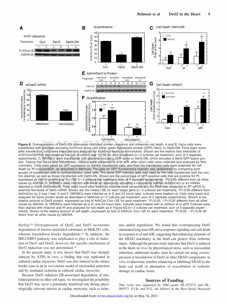
Figure 8. Overexpression of Derl3-DN attenuates misfolded protein clearance and enhances cell death. A and B, HeLa cells weretransfected with plasmids encoding A1ATmut along with either green fluorescent protein (GFP), Derl3, or Derl3-DN. Forty-eight hoursafter transfection, cells were extracted and analyzed for A1ATmut levels by immunoblot. Shown are the relative blot intensities ofA1ATmut/GAPDH, expressed as the fold of control (bar 1)�SE for each treatment (n�3 cultures per treatment, sum of 3 separateexperiments). C, NRVMCs were transfected with plasmids encoding GFP alone or Derl3-DN, which encodes a Derl3-GFP fusion pro-tein. Twenty-four hours after transfection, cultures were subjected to sI or sI/R, after which cells were collected and analyzed by flowcytometry. Cells were gated for GFP expression to identify transfected cells, and then the transfected cells were assessed for celldeath by PI incorporation, as described in Methods. The gate for GFP fluorescence intensity was established by comparing bothgroups of transfected cells to nontransfected, naïve cells. The same GFP intensity gate was used for the cells transfected with the con-trol plasmid, as well as those transfected with Derl3-DN. Shown are the percentage of GFP-positive cells that are positive for PI,expressed as fold of control (bar 1) (�SE, n�3 cultures per treatment, sum of 3 separate experiments). *P�0.05 different from all othervalues by ANOVA. D, NRVMCs were infected with either an adenovirus encoding a nonspecific miRNA (AdVmiCon) or an miRNAdirected to Derl3 (AdVmiDerl3). Forty-eight hours after infection, cultures were extracted and the RNA was subjected to RT-qPCR toexamine the levels of Derl3 mRNA. Shown are the means�SE for each target gene (n�3 cultures per treatment). *P�0.05 different fromAdVmiCon by 2-way t test. E and F, NRVMCs were infected as in D and 24 hours later, cultures were treated�sI. Cells were lysed andanalyzed for Derl3 protein levels as described in Methods (n�3 cultures per treatment, sum of 2 separate experiments). Shown is therelative amount of Derl3 protein, expressed as fold of AdVCon Con�SE for each treatment. *P�0.05, �P�0.05 different from all othervalues by ANOVA. G, NRVMCs were infected as in D, and 24 hours later, cultures were treated with or without sI or sI/R. Cultures werethen stained with Hoechst and PI and analyzed for cell death as in Figure 6G (n�3 cultures per treatment, sum of 3 separate experi-ments). Shown is the relative amount of cell death, expressed as fold of AdVCon Con�SE for each treatment. *P�0.05, �P�0.05 dif-ferent from all other values by ANOVA.
Belmont et al Derl3 in the Heart 9
by on May 16, 2011 circres.ahajournals.orgDownloaded from

Foundation and the San Diego State University Heart Institute. P.J.B.is a scholar of the San Diego Chapter of the Achievement Rewardsfor College Scientists (ARCS) Foundation and a recipient of anAmerican Heart Association Western States Affiliate PredoctoralFellowship (Award 0815210F). M.N.S.P. is a Minority Access toResearch Careers (MARC) scholar and a recipient of a MARCfellowship (NIH/NIGMS SDSU MARC 5T34 GM08303).
DisclosuresNone.
References1. Szegezdi E, Duffy A, O’Mahoney ME, Logue SE, Mylotte LA, O’Brien
T, Samali A. ER stress contributes to ischemia-induced cardiomyocyteapoptosis. Biochem Biophys Res Commun. 2006;349:1406–1411.
2. Kaufman RJ. Orchestrating the unfolded protein response in health anddisease. J Clin Invest. 2002;110:1389–1398.
3. Aridor M. Visiting the ER: the endoplasmic reticulum as a target fortherapeutics in traffic related diseases. Adv Drug Deliv Rev. 2007;59:759–781.
4. Wang X, Osinska H, Klevitsky R, Gerdes AM, Nieman M, Lorenz J,Hewett T, Robbins J. Expression of R120G-alphaB-crystallin causesaberrant desmin and alphaB-crystallin aggregation and cardiomyopathyin mice. Circ Res. 2001;89:84–91.
5. Pattison JS, Sanbe A, Maloyan A, Osinska H, Klevitsky R, RobbinsJ. Cardiomyocyte expression of a polyglutamine preamyloid oligomercauses heart failure. Circulation. 2008;117:2743–2751.
6. Glembotski CC. The role of the unfolded protein response in the heart. JMol Cell Cardiol. 2008;44:453–459.
7. Lee AS. Mammalian stress response: induction of the glucose-regulatedprotein family. Curr Opin Cell Biol. 1992;4:267–273.
8. Shamu CE, Cox JS, Walter P. The unfolded-protein-response pathway inyeast. Trends Cell Biol. 1994;4:56–60.
9. McMillan DR, Gething MJ, Sambrook J. The cellular response tounfolded proteins: intercompartmental signaling. Curr Opin Biotechnol.1994;5:540–545.
10. Nakatsukasa K, Huyer G, Michaelis S, Brodsky JL. Dissecting theER-associated degradation of a misfolded polytopic membrane protein.Cell. 2008;132:101–112.
11. Razeghi P, Taegtmeyer H. Cardiac remodeling: UPS lost in transit. CircRes. 2005;97:964–966.
12. Wang X, Robbins J. Heart failure and protein quality control. Circ Res.2006;99:1315–1328.
13. Yamamoto K, Yoshida H, Kokame K, Kaufman RJ, Mori K. Differentialcontributions of ATF6 and XBP1 to the activation of endoplasmicreticulum stress-responsive cis-acting elements ERSE, UPRE andERSE-II. J Biochem. 2004;136:343–350.
14. Martindale JJ, Fernandez R, Thuerauf D, Whittaker R, Gude N, SussmanMA, Glembotski CC. Endoplasmic reticulum stress gene induction andprotection from ischemia/reperfusion injury in the hearts of transgenicmice with a tamoxifen-regulated form of ATF6. Circ Res. 2006;98:1186–1193.
15. Belmont PJ, Tadimalla A, Chen WJ, Martindale JJ, Thuerauf DJ,Marcinko M, Gude N, Sussman MA, Glembotski CC. Coordination ofgrowth and endoplasmic reticulum stress signaling by regulator of cal-cineurin 1 (RCAN1), a novel ATF6-inducible gene. J Biol Chem. 2008;283:14012–14021.
16. Hiller MM, Finger A, Schweiger M, Wolf DH. ER degradation of amisfolded luminal protein by the cytosolic ubiquitin-proteasomepathway. Science. 1996;273:1725–1728.
17. Lilley BN, Ploegh HL. Multiprotein complexes that link dislocation,ubiquitination, and extraction of misfolded proteins from the endoplasmicreticulum membrane. Proc Natl Acad Sci U S A. 2005;102:14296–14301.
18. Oda Y, Okada T, Yoshida H, Kaufman RJ, Nagata K, Mori K. Derlin-2and Derlin-3 are regulated by the mammalian unfolded protein responseand are required for ER-associated degradation. J Cell Biol. 2006;172:383–393.
19. Thuerauf DJ, Marcinko M, Gude N, Rubio M, Sussman MA, GlembotskiCC. Activation of the unfolded protein response in infarcted mouse heartand hypoxic cultured cardiac myocytes. Circ Res. 2006;99:275–282.
20. Yoshida H, Haze K, Yanagi H, Yura T, Mori K. Identification of thecis-acting endoplasmic reticulum stress response element responsible fortranscriptional induction of mammalian glucose-regulated proteins.Involvement of basic leucine zipper transcription factors. J Biol Chem.1998;273:33741–33749.
21. Kokame K, Kato H, Miyata T. Identification of ERSE-II, a new cis-actingelement responsible for the ATF6-dependent mammalian unfoldedprotein response. J Biol Chem. 2001;276:9199–9205.
22. DenBoer LM, Hardy-Smith PW, Hogan MR, Cockram GP, Audas TE, LuR. Luman is capable of binding and activating transcription from theunfolded protein response element. Biochem Biophys Res Commun. 2005;331:113–119.
23. Glembotski CC. Endoplasmic reticulum stress in the heart. Circ Res.2007;101:975–984.
24. Thuerauf DJ, Morrison L, Glembotski CC. Opposing roles for ATF6alphaand ATF6beta in endoplasmic reticulum stress response gene induction.J Biol Chem. 2004;279:21078–21084.
25. Oyadomari S, Koizumi A, Takeda K, Gotoh T, Akira S, Araki E, Mori M.Targeted disruption of the Chop gene delays endoplasmic reticulumstress-mediated diabetes. J Clin Invest. 2002;109:525–532.
26. Austin RC. The unfolded protein response in health and disease. AntioxidRedox Signal. In press.
27. Sifers RN, Brashears-Macatee S, Kidd VJ, Muensch H, Woo SL. Aframeshift mutation results in a truncated alpha 1-antitrypsin that isretained within the rough endoplasmic reticulum. J Biol Chem. 1988;263:7330–7335.
28. Plemper RK, Egner R, Kuchler K, Wolf DH. Endoplasmic reticulumdegradation of a mutated ATP-binding cassette transporter Pdr5 proceedsin a concerted action of Sec61 and the proteasome. J Biol Chem. 1998;273:32848–32856.
29. Tadimalla A, Belmont PJ, Thuerauf DJ, Glassy MS, Martindale JJ, GudeN, Sussman MA, Glembotski CC. Mesencephalic astrocyte-derived neu-rotrophic factor is an ischemia-inducible secreted endoplasmic reticulumstress response protein in the heart. Circ Res. 2008;103:1249–1258.
10 Circulation Research February 5, 2010
by on May 16, 2011 circres.ahajournals.orgDownloaded from

SUPPLEMENT MATERIAL
Expanded Materials and Methods:
Animals-
The transgenic mice used in this study have been described previously.1 Approximately 100 neonatal
rats and 24 adult male mice were used in this study. All procedures involving animals were in accordance
with the San Diego State University Institutional Animal Care and Use Committee.
Promoter Searches- The 2kb region lying 5’ of the start sites for each of the ATF6-regulated transcripts identified in our
previous array study2 were retrieved using Ensembl Biomart (http://www.ensembl.org/biomart/martview).
The same regions of each of the roughly 40,000 transcripts on the Affymetrix mouse 430 2.0 whole
genome array chips (Affymetrix, Inc., part #900496) used in this previous study were also acquired.
These sequences were then searched for ERSEs, ERSE-IIs and UPREs using a custom Perl script and the
following sequences:
ERSE- CCAAT-N9-CCACG3
ERSE 1bp mismatch- Allows for 1bp mismatch in any nucleotide in either of the 5bp flanking regions of
the consensus ERSE
ERSE-II- ATTGG-N-CCACG4
ERSE-II 1bp mismatch- Allows for 1bp mismatch in any nucleotide in either of the 5bp flanking regions
of the consensus ERSEII
UPRE: TGACGTGGA5
UPRE 1bp mismatch: Allows for 1bp mismatch in any nucleotide of the consensus UPRE
Enrichment of ATF6 Array for Putative ER Stress Response Genes-
To determine whether the genes previously identified in the mouse heart as ATF6-regulated2 were
enriched for ERSEs, ERSE-IIs and UPREs, a bootstrapping analysis was performed, essentially as
previously described.6,7
Briefly, 1,000 promoter sets, each of which contains 607 promoters, were
generated from genes either from the mouse whole-genome Affymetrix GeneChip array, or from the
ATF6-regulated gene cluster, using Ensembl Biomart (http://www.ensembl.org/biomart/martview).
Bootstrapped promoter sets were generated using a custom Perl script. The number of times these
elements were found in each bootstrapped promoter set, i.e. the frequency of appearance in the whole
genome and in the ATF6-regulated genes, is shown in Figure 1.
Cultured Cardiac Myocytes-
Primary neonatal rat ventricular myocyte cultures (NRVMC) were prepared and maintained in
culture, as previously described.8
Derl3-luciferase and Derl3-mut-luciferase Constructs- The mouse Derl3 gene from nt -1359 to +26 was amplified by PCR and cloned into a pGL2 luciferase
reporter vector (Clontech, Inc.) and designated Construct 1. Two consensus ERSE sequences5,
CCAAT(N)9CCACG, located between nt -183 to -165 and nt -285 to -267 were designated ERSE1 and
ERSE2, respectively. Using PCR-based mutagenesis, ERSE1 of Derl3-luciferase Construct 1 was
mutated to AACCG(N)9AACAT, creating Construct 2. ERSE2 of Derl3-luciferase Construct 1 was also
mutated to AACCG(N)9AACAT, creating Construct 3.
by on May 16, 2011 circres.ahajournals.orgDownloaded from

Reporter Enzyme Assays- Reporter enzyme assays for luciferase and β-galactosidase were carried out in NRVMC extracts as
previously described.8
Simulated Ischemia/Simulated Reoxygenation- NRVMCs were subjected to conditions that simulate ischemia (sI), or simulate ischemia followed by
reoxygenation (sI/R), essentially as previously described.8 Briefly, for sI, the medium was replaced with
glucose-free DMEM/F12 containing 2% dialyzed fetal bovine serum and 3 mM 2-deoxyglucose (Sigma,
catalog #D6134), and cultures were placed in a chamber (PROOX model 110, Sensor Part #E702,
BioSpherix, Ltd Redfield, NY), filled with N2/CO2 (95% N2/ 5% CO2) for 20h. For sI/R, following sI, the
medium was replaced with glucose-containing DMEM/F-12 supplemented with 2% bovine serum
albumin, and cultures were incubated in O2/CO2 (18% O2) for 24h. For immunoblot analysis, cells were
lysed as previously described.9 GRP78 and GRP94 protein levels were assessed using an anti-KDEL
antibody (Stressgen, catalog # SPA-827); Derl3 protein levels were assessed using an anti-Derl3 antibody
(LifeSpan Biosciences, catalog #LS-C80661).
Tamoxifen Treatment- Non-transgenic (NTG) and transgenic (TG) mice were treated with vehicle or tamoxifen, n=3
mice per treatment group, and RNA was extracted from mouse heart ventricles, as described previously.1
Myocardial Infarction- NTG mice were subjected to in vivo permanent myocardial infarction for 6h, 16h, 1d, 3d, 4d, 7d, or
14d. After each time point, mice were sacrificed and sections of the hearts were prepared for
immunocytofluoescence confocal microscopy. Tissue extracts were also prepared for qRT-PCR or
western blot analysis. All of these procedures have been previously described.8,10
An antibody which
recognizes cleaved, 50KD ATF6 was used (ATF6 H-280, Santa Cruz, catalog #22799).
Immunocytofluorescence- Immunofluorescent confocal microscopy was carried out as previously
described.
10 Sections were
stained using an anti-Derl3 antibody (Sigma, catalog #D2194) at a dilution of 1:40.
Real Time Quantitative-PCR- Real time quantitative-PCR (RT-qPCR) was performed, as previously described.
1 The following rat
primer pairs were used:
Derl1: 5’-CTTAATGGCCGAGCTCTTGC; 3’-CATTCAGCGTGGGTCAGGT
Derl2: 5’-CTTTCTGCCCTGGGTGCTC; 3’-AATCGAGTTCCCCAGCAACA
Derl3: 5’-CCAGCAACACCATGCACTTC; 3’-TCACTGTGGTTGAGCGGAGA
GAPDH: 5;- CCTGGCCAAGGTCATCCAT; 3’- GTCATGAGCCCTTCCACGAT
The following mouse primer pairs were used:
Derl1: 5’-CTAATGTCCTGGTACCCGGC; 3’-CCCTTCGAAAAGCCAGTCCT
Derl2: 5’-ACCTTGCTTGTCTCGTTCAGC; 3’-AGACGTGATTGCAGACTTCGG
Derl3: 5’-GAGCAGGAAGCCCACTCTGA; 3’-GAGCTGAGGTGGAGGGAAGG
GAPDH: 5;- CCTGGCCAAGGTCATCCAT; 3’- GTCATGAGCCCTTCCACGAT
by on May 16, 2011 circres.ahajournals.orgDownloaded from

Plasmid Constructs-
3xFlag-ATF6- A plasmid encoding a 3xFlag-tagged version of the N-terminal portion of ATF6
(amino acids 1-373) has been constructed as previously described.11
α-1Antitrypsin and Mutant α-1Antitrypsin- A construct encoding human α-1 antitrypsin (A1AT,
NCBI RefSeq NM_000295) was generated by PCR using the appropriate primers to create an amplicon
with Xho1 on the 5’ end of the start site, and a termination codon and EcoRI on the 3’ end. This PCR
product was cloned into the pCDNA3.1 vector. Mutant α-1 antitrypsin (A1ATmut) was generated by
making a two nucleotide deletion at aa318 of A1AT, leading to a frameshift mutation, as previously
described,12
using PCR-based mutagenesis. Clones were generated
using QuikChange from Stratagene
and the appropriate primers.
Derl3-GFP Fusion Protein- A construct
encoding GFP fused to the C-terminus of Derl3 (Derl3-DN) was created by PCR, using
mouse Derl3 (NCBI RefSeq NM_024440) as the template, and the appropriate primers, to create an
amplicon with an Xho1 site on the 5'-end, and a termination codon and
EcoRI site on the 3'-end. This PCR
product was then cloned into the pEGFP-N1 vector (Clontech, catalog #6085-1, GenBank
TM accession
number U55762). Clones were generated
using QuikChange from Stratagene and the appropriate primers.
Adenovirus- Recombinant adenovirus (AdV) encoding only GFP (AdVCon), GFP and constitutively active
ATF6 (AdVATF6), or GFP and dominant-negative ATF6 (AdVDN-ATF6), which encode proteins of the
expected physical characteristics, were prepared as previously described 8. A recombinant AdV encoding
GFP and Derl3 (AdVDerl3) was generated by cloning a construct encoding mus Derl3 (NCBI RefSeq
NM_024440) into the adenovirus shuttle vector, pAdTrack-CMV, and was used to create a new AdV
strain, as previously described.13
Recombinant AdV encoding GFP and A1AT (AdVA1AT) and GFP and
mutA1AT (AdVmutA1AT) were generated by cloning constructs encoding human A1AT (NCBI RefSeq
NM_000295) or mutA1AT, a mutated version of human A1AT, described above, into the adenovirus
shuttle vector, pAdTrack-CMV, and used to
create new AdV strains, as previously described.13
Recombinant adenovirus encoding either miRNA targeted to rat Derl3 (AdVmiDerl3) or a negative
control (AdVmiCon) were created using the Gateway System from Invitrogen, as previously described.9
Caspase-3 Activity Assay-
NRVMCs were infected with AdVCon vs AdVDerl3, or AdVmiCon vs AdVmiDerl3, in 2% FCS-
containing medium for 6 hours, after which, cells were washed and fed with the same medium but
without the AdV. After 24h, cells were treated +/- sI or sI/R, as described above. Cultures were extracted
in caspase assay buffer containing 50 mM Hepes, pH 7.4, 0.1% CHAPS, 0.1 mM EDTA. A total of 50 μL
of the lysate and 10 μL of the assay buffer were then combined with 45 μL of reaction buffer (40 μL
caspase assay buffer, 1 mM DTT, 40 μM DEVD-AFC in DMSO (Sigma, catalog no. A0466). After 1
hour at 37°C, fluorescence was measured at an excitation wavelength of 400 nm and an emission
wavelength of 505 nm. Caspase activity was defined as fluorescence/protein.
Live/Dead Assay-
Assessment of cell death in NRVMCs was performed using Hoescht (catalog no. H21486; Invitrogen)
and propidium iodide (PI, Invitrogen, catalog no. P1304MP), as previously described.
10
by on May 16, 2011 circres.ahajournals.orgDownloaded from

Protein Clearance Assays-
HeLa cells were co-transfected with A1ATmut and varying concentrations of Derl3, or co-transfected
with A1ATmut and GFP or Derl3-DN. After 24h, cultures were scraped in protein lysis buffer, and
immunoblotted with anti-A1AT antibody (Dako, Denmark A/S, Catalog #A 0012).
Flow Cytometry-
NRVMCs were electroporated with plasmids encoding GFP alone or Derl3-DN, which encodes a
Derl3-GFP fusion protein, and plated onto 6-well dishes. Cultures were subjected to sI or sI/R, as
described above. Cells were then collected using TripLE (Gibco, catalog #12605), washed with PBS,
resuspended in PBS, and analyzed by flow cytometry on a BD FACSAriaTM
cell sorter. Approximately
150,000 events were recorded for each condition. Cells were first gated to eliminate debris and
aggregates. Transfected cells were identified by gating for GFP expression, and the percentage of these
transfected cells that were also PI-positive was determined. This percentage was normalized to the
percent of overall PI-positive cells for each condition. Data is presented as fold of control. Shown is the
average of three independent experiments.
Statistical Analyses-
Data are reported as mean ± SEM and analyzed via 1-way ANOVA with Newman–Keuls post-hoc
analysis using SPSS version 11.0. Unless otherwise stated in the figure legends, *, +, #, or ! = p < 0.05
different from all other values, and **, ++ = p < 0.01 different from all other values.
Supplemental Table Legends
Online Table I. Genes containing consensus ERSE elements within 2KB promoter regions.
Each unique gene containing a consensus ERSE (CCAAT-N9-CCACG) is numbered once. All ATF6-
regulated genes are sorted by the fold change from our previous array study.2 All genes from the full
genome are sorted alphabetically. The MGI symbol, alias or common name, and NCBI Reference
Sequence ID, or accession number, is shown for each gene. More information about each gene can be
obtained by entering the MGI symbol into the Mouse Genome Informatics (MGI 3.54) web site. Also
shown is the Start and End location for each ERSE identified, as determined by retrieving the 2000bp 5'
flanking promoter sequence using Ensembl Biomart (http://www.ensembl.org/biomart/martview) and
searching for each element with our custom Perl script.
Online Table II. Genes containing consensus ERSEII elements within 2KB promoter regions.
Each unique gene containing a consensus ERSEII (ATTGG-N-CCACG) is numbered once. All genes are
sorted as in Online Table I, and all information is presented as in Online Table I.
Online Table III. Genes containing consensus UPRE elements within 2KB promoter regions. Each
unique gene containing a consensus UPRE (TGACGTGGA) is numbered once. All genes are sorted as in
Online Table I, and all information is presented as in Online Table I.
Online Table IV. Genes containing 1bp-mismatched ERSE elements within 2KB promoter regions.
Each unique gene containing an ERSE (CCAAT-N9-CCACG) with 1bp-mismatched anywhere on either
flanking region, is numbered once. All ATF6-regulated genes are sorted by the fold change from our
by on May 16, 2011 circres.ahajournals.orgDownloaded from

previous array study.2 The MGI symbol, alias or common name, and NCBI Reference Sequence ID, or
accession number, is shown for each gene. More information about each gene can be obtained by entering
the MGI symbol into the Mouse Genome Informatics (MGI 3.54) web site. Also shown is the Start and
End location for each 1bp-mismatched ERSE identified, as determined by retrieving the 2000bp 5'
flanking promoter sequence using Ensembl Biomart (http://www.ensembl.org/biomart/martview) and
searching for each element with our custom Perl script.
Online Table V. Genes containing 1bp-mismatched ERSEII elements within 2KB promoter regions.
Each unique gene containing an ERSEII (ATTGG-N-CCACG) with 1bp-mismatched anywhere on either
flanking region, is numbered once. All genes are sorted as in Online Table IV, and all information is
presented as in Online Table IV.
Online Table VI. Genes containing 1bp-mismatched UPRE elements within 2KB promoter regions.
Each unique gene containing an UPRE (TGACGTGGA) with 1bp-mismatched anywhere on the element,
is numbered once. All genes are sorted as in Online Table IV, and all information is presented as in
Online Table IV.
Online Figure I. MI Increases 50KD ATF6 and Derl3 mRNA
Panels A-B: NTG mice were subjected to sham infarct surgery or to permanent occlusion myocardial
infarction for the indicated times. Animals were then sacrificed and hearts were used to prepare tissue
extracts for western blot analysis, as previously described (lanes 1-9).10
To confirm the identity of the
50KD band, cell lysates from HeLa cells transfected with a 3xFlag-tagged ATF6 plasmid and subjected to
the protease inhibitor ALLN (3mM) in order to preserve the labile, N-terminal form of ATF6, were
loaded (lanes 10-11).
Panel C: NTG mice were subjected to sham infarct surgery or to permanent occlusion myocardial
infarction for 1d, 4d, or 7d. Animals were then sacrificed and hearts were used to prepare tissue extracts
for RT-qPCR, as previously described.10
The numbers of animals used for each trial were as follows:
sham n = 4; MI n= 7 to 8. * = p<0.05 different from all other values determined by ANOVA.
Online Figure II. Derl3 Overexpression Attenuates ER Stress mRNA Activation Following sI and
sI/R.
NRVMCs were infected + AdVCon or AdV-Derl3 and 24h later, cultures were treated + sI for 20h
followed by sI/R for 24h (Panels A and B), or + sI for the indicated times (Panels C and D). Cultures
were then extracted and cell lysates were analyzed for the levels of the prototypical ERSR mRNAs,
GRP78 and GRP94 (n = 3 cultures per treatment, sum of 2 separate experiments). Shown is the relative
amount of each target mRNA/GAPDH, expressed as fold of AdVCon Con, + S.E for each treatment.
Panels A-B: *,+ = p<0.05 different from all other values by ANOVA, Panels C-D: * = p<0.05 from
AdVCon Con by ANOVA.
The following primers were used:
GRP78: 5’-CACGTCCAACCCGGAGAA; 3’ –ATTCCAAGTGCGTCCGATG
GRP94: 5’ – TTGTGGATTCCGATGATCTCC; 3’ – CAGAGTTTTGCGGACAAGCTT
GAPDH: 5’ –CCTGGCCAAGGTCATCCAT; 3’ – GTCATGAGCCCTTCCACGAT
by on May 16, 2011 circres.ahajournals.orgDownloaded from

References
1. Martindale JJ, Fernandez R, Thuerauf D, Whittaker R, Gude N, Sussman MA,
Glembotski CC. Endoplasmic reticulum stress gene induction and protection from
ischemia/reperfusion injury in the hearts of transgenic mice with a tamoxifen-regulated
form of ATF6. Circ Res. 2006;98:1186-93.
2. Belmont PJ, Tadimalla A, Chen WJ, Martindale JJ, Thuerauf DJ, Marcinko M, Gude N,
Sussman MA, Glembotski CC. Coordination of growth and endoplasmic reticulum stress
signaling by regulator of calcineurin 1 (RCAN1), a novel ATF6-inducible gene. J Biol
Chem. 2008;283:14012-21.
3. Aridor M. Visiting the ER: the endoplasmic reticulum as a target for therapeutics in
traffic related diseases. Adv Drug Deliv Rev. 2007;59:759-81.
4. Kokame K, Kato H, Miyata T. Identification of ERSE-II, a new cis-acting element
responsible for the ATF6-dependent mammalian unfolded protein response. J Biol Chem.
2001;276:9199-205.
5. Yoshida H, Haze K, Yanagi H, Yura T, Mori K. Identification of the cis-acting
endoplasmic reticulum stress response element responsible for transcriptional induction
of mammalian glucose-regulated proteins. Involvement of basic leucine zipper
transcription factors. J Biol Chem. 1998;273:33741-9.
6. Efron B, Tibshirani R. Statistical Data Analysis in the Computer Age. Science.
1991;253:390-395.
7. Chen WH, Sun LT, Tsai CL, Song YL, Chang CF. Cold-stress induced the modulation of
catecholamines, cortisol, immunoglobulin M, and leukocyte phagocytosis in tilapia. Gen
Comp Endocrinol. 2002;126:90-100.
8. Thuerauf DJ, Marcinko M, Gude N, Rubio M, Sussman MA, Glembotski CC. Activation
of the unfolded protein response in infarcted mouse heart and hypoxic cultured cardiac
myocytes. Circ Res. 2006;99:275-82.
9. Doroudgar S, Thuerauf DJ, Marcinko MC, Belmont PJ, Glembotski CC. Ischemia
activates the ATF6 branch of the endoplasmic reticulum (ER) stress response. J Biol
Chem. 2009.
10. Tadimalla A, Belmont PJ, Thuerauf DJ, Glassy MS, Martindale JJ, Gude N, Sussman
MA, Glembotski CC. Mesencephalic astrocyte-derived neurotrophic factor is an
ischemia-inducible secreted endoplasmic reticulum stress response protein in the heart.
Circ Res. 2008;103:1249-58.
11. Thuerauf DJ, Morrison L, Glembotski CC. Opposing roles for ATF6alpha and ATF6beta
in endoplasmic reticulum stress response gene induction. J Biol Chem. 2004;279:21078-
84.
12. Sifers RN, Brashears-Macatee S, Kidd VJ, Muensch H, Woo SL. A frameshift mutation
results in a truncated alpha 1-antitrypsin that is retained within the rough endoplasmic
reticulum. J Biol Chem. 1988;263:7330-5.
13. Hoover HE, Thuerauf DJ, Martindale JJ, Glembotski CC. alpha B-crystallin gene
induction and phosphorylation by MKK6-activated p38. A potential role for alpha B-
crystallin as a target of the p38 branch of the cardiac stress response. J Biol Chem.
2000;275:23825-33.
by on May 16, 2011 circres.ahajournals.orgDownloaded from

Number MGI symbol Alias or Protein Name NCBI RefSeq Start End Strand Fold Change1 Derlin-3 Degraded in ER protein 3 NM_024440 257 239 - 18.51 Derlin-3 Degraded in ER protein 3 NM_024440 155 137 - 18.52 DNAJC3 P58; P58IPK NM_008929 146 128 + 12.753 Hsp90b1 Tra1; GRP94 NM_011631 41 23 - 7.073 Hsp90b1 Tra1; GRP94 NM_011631 64 46 + 7.074 Calr Calreticulin NM_007591 161 143 - 5.295 Hspa5 GRP78 NM_022310 148 130 + 4.716 Syvn1 Synoviolin 1 NM_028769 129 111 + 4.67 XBP1 X-box binding protein 1 NM_013842 60 42 + 2.95
Number MGI symbol Alias or Protein Name NCBI RefSeq Start End Strand1 Adamtsl5 ADAMTS-like 5 NM_001024139 591 573 -2 Adcy3 adenylate cyclase 3 NM_027857 216 198 +3 Alg12 asparagine-linked glycosylation 12 homolog NM_145477 219 201 +4 Calr Calreticulin NM_007591 161 143 -5 Cd209d CD209d antigen NM_130904 666 648 +6 Clec12a C-type lectin domain family 12, member a NM_177686 1919 1901 +7 CNX calnexin NM_001110499 161 143 +8 Creld2 cysteine-rich with EGF-like domains 2 NM_029720 125 107 -
9 Ddefl1development and differentiation enhancing factor-like
1 NM_001008232 1504 1486 +
10 Derlin-3 Degraded in ER protein 3 NM_024440 257 239 -10 Derlin-3 Degraded in ER protein 3 NM_024440 155 137 -
11 Dnajb14 DnaJ (Hsp40) homolog, subfamily B, member 14 XM_001473173 1784 1766 -
12 DNAJC3 P58; P58IPK NM_008929 146 128 +13 Ero1lb ERO1-like beta NM_026184 200 182 -14 Foxk1 forkhead box K1 NM_010812 1227 1209 -15 Grp45 G protein-coupled receptor 44 NM_009962 173 155 -16 Hnrpa3 heterogeneous nuclear ribonucleoprotein A3 NM_053263 243 225 -17 Hsp90b1 Tra1; GRP94 NM_011631 41 23 -17 Hsp90b1 Tra1; GRP94 NM_011631 64 46 +18 Hspa5 GRP78 NM_022310 64 46 +
19 Kbtbd8 kelch repeat and BTB (POZ) domain containing 8 NM_001008785 1165 1147 +
20 Kcna6potassium voltage-gated channel, shaker-related,
subfamily, member 6NM_013568 1668 1650 +
21 Kcnmb2potassium large conductance calcium-activated
channel, subfamily M, beta member 2NM_028231 1920 1902 -
22 Klk1b8 kallikrein 1-related peptidase b8 NM_008457 321 303 +23 Klk1b22 kallikrein 1-related peptidase b22 NM_010114 318 300 +
24 Mfsd11 major facilitator superfamily domain containing 11 NM_178620 579 561 +
25 Nox3 NADPH oxidase 3 NM_198958 1045 1027 -26 Nt5dc3 5'-nucleotidase domain containing 3 NM_175331 338 320 +26 Nt5dc3 5'-nucleotidase domain containing 3 NM_175331 233 215 -27 Pdia6 protein disulfide isomerase associated 6 NM_027959 103 85 +28 Setbp1 SET binding protein 1 NM_053099 322 304 -29 Sfrs2 splicing factor, arginine/serine-rich 2 (SC-35) NM_011358 358 340 -30 Syvn1 Synoviolin 1 NM_028769 129 111 +31 Tmprss11e transmembrane protease, serine 11e NM_172880 1554 1536 +32 Tmprss12 transmembrane protease, serine 12 NM_183109 1003 985 +33 Usp52 ubiquitin specific peptidase 52 NM_133992 47 29 -34 XBP1 X-box binding protein 1 NM_013842 60 42 +35 Xlr3a X-linked lymphocyte-regulated 3A NM_001110784 171 153 +36 Xlr3b X-linked lymphocyte-regulated 3B NM_001081643 148 130 +37 Xlr3c X-linked lymphocyte-regulated 3C NM_011727 168 150 +38 Zfp57 zinc finger protein 57 NM_001013745 49 31 -39 Zfp777 zinc finger protein 777 NM_001081382 1948 1930 -
Online Table I.Genes containing consensus ERSE elements within 2KB promoter regions
Full Genome
ATF6-Regulated Genes
by on May 16, 2011 circres.ahajournals.orgDownloaded from

Number MGI symbol Alias or Protein Name NCBI RefSeq Start End Strand Fold Change1 Dnajb11 DnaJ (Hsp40) homolog, subfamily B, member 11 NM_026400 11 1 + 12.392 Armet arginine-rich, mutated in early stage tumors NM_029103 110 100 - 8.213 Hyou1 hypoxia up-regulated 1 NM_021395 178 168 - 3.8643 Hyou1 hypoxia up-regulated 1 NM_021395 77 67 - 3.864
4 Herpud1homocysteine-inducible, endoplasmic reticulum stress-
inducible, ubiquitin-like domain member 1 NM_022331 117 107 + 2.43
Number MGI symbol Alias or Protein Name NCBI RefSeq Start End Strand1 1110067D22Rik Grpa NM_173752 80 70 -2 4933421E11Rik Rif1 NM_001039478 67 57 +3 1500005K14Rik Fam101b XM_893392 1067 1057 +4 Apon apolipoprotein N NM_133996 1464 1454 -5 Armet arginine-rich, mutated in early stage tumors NM_029103 110 100 -6 Cxcl12 chemokine (C-X-C motif) ligand 12 NM_001012477 479 469 +7 D430018E03Rik RIKEN cDNA D430018E03 gene NM_001002769 1449 1439 -8 Dnajb11 DnaJ (Hsp40) homolog, subfamily B, member 11 NM_026400 11 1 +9 Efhc1 EF-hand domain-containing protein 1 XM_129694 349 339 -10 EG665305 LOC665305 XM_975981 123 113 -11 Gal3st4 galactose-3-O-sulfotransferase 4 NM_001033416 865 855 +
12 Herpud1homocysteine-inducible, endoplasmic reticulum stress-
inducible, ubiquitin-like domain member 1 NM_022331 117 107 +
13 Hyou1 hypoxia up-regulated 1 NM_021395 178 168 -13 Hyou1 hypoxia up-regulated 1 NM_021395 77 67 -14 Iqcf5 IQ motif containing F5 XM_356185 740 730 -
15 Mcm9 minichromosome maintenance complex component 9 NM_027830 867 857 -
16 Ninj1 Ninjurin-1 NM_013610 781 771 +17 Nrxn2 Neurexin II XM_978630 100 90 -18 Nucb1 nucleobindin 1 NM_008749 40 30 -19 Rasd1 Dexras1 NM_009026 768 75820 Rbm39 RNA binding motif protein 39 NM_133242 80 70 +21 Rcl1 RNA terminal phosphate cyclase-like 1 NM_021525 81 71 -22 Rnf151 ring finger protein 151 NM_026205 33 23 +23 Tbccd1 TBCC domain containing 1 NM_001081368 323 313 -24 Tbx2 T-box 2 NM_009324 1913 1903 -25 Tmed2 transmembrane emp24 domain trafficking protein 2 NM_019770 202 192 +26 Tmem119 transmembrane protein 119 NM_146162 380 370 +27 Trim46 tripartite motif-containing 46 NM_183037 1300 1290 -28 Ufd1 ubiquitin fusion degradation 1 like NM_011672 657 647 +29 Ung UDG NM_001040691 1152 1142 -
ATF6-Regulated Genes
Full Genome
Genes containing consensus ERSEII elements within 2KB promoter regionsOnline Table II.
by on May 16, 2011 circres.ahajournals.orgDownloaded from

Number MGI symbol Alias or Protein Name NCBI RefSeq Start End Strand Fold Change1 Timp1 tissue inhibitor of metalloproteinase 1 NM_001044384 83 75 + 7.782 Stat3 signal transducer and activator of transcription 3 NM_011486 563 555 - 2.7773 Fra-2 fos-like antigen 2 NM_008037 1118 1110 - 2.4774 Nmor2 NAD(P)H dehydrogenase, quinone 2 NM_020282 539 531 + 0.34135 Kcnv2 potassium channel, subfamily V, member 2 NM_183179 124 116 - 0.179
Number MGI symbol Alias or Protein Name NCBI RefSeq Start End Strand1 1110049F12Rik RIKEN cDNA 1110049F12 gene NM_025411 62 54 -2 1810007P19Rik RIKEN cDNA 1810007P19 gene NM_172701 827 819 +3 2010203O07Rik DnaJ (Hsp40) homolog, subfamily C , member 25 NM_001033165 48 40 +4 4921507L20Rik RIKEN cDNA 4921507L20 gene AK014837 1959 1951 +5 4921530L21Rik RIKEN cDNA 4921530L21 gene NM_025733 1067 1059 +6 4930579K19Rik RIKEN cDNA 4930579K19 gene NM_175227 138 130 +7 9230112E08Rik RIKEN cDNA 9230112E08 gene NM_177264 197 189 +8 9930038B18Rik RIKEN cDNA 9930038B18 gene NM_176929 1502 1494 -9 Adam32 a disintegrin and metallopeptidase domain 32 NM_153397 1850 1842 +
10 Adamts17a disintegrin-like and metallopeptidase (reprolysin type) with
thrombospondin type 1 motif, 17 NM_001033877 660 652 -
11 Adrp adipose differentiation related protein NM_007408 1062 1054 +12 Aff4 AF4/FMR2 family, member 4 NM_033565 470 462 -13 Afg3l1 AFG3(ATPase family gene 3)-like 1 (yeast) NM_054070 466 458 +14 Alx3 aristaless-like homeobox 3 XM_973424 37 29 +15 Aox3 aldehyde oxidase 3 NM_023617 228 220 -16 Apbb1 amyloid beta (A4) precursor protein-binding, family B, member 1 NM_009685 778 770 -17 Apc2 adenomatosis polyposis coli 2 NM_011789 1732 1724 -18 Apobec3 apolipoprotein B editing complex 3 NM_030255 372 364 +18 Apobec3 apolipoprotein B editing complex 3 NM_030255 1851 1843 +19 App amyloid beta (A4) precursor protein NM_007471 638 630 -20 Azi1 5-azacytidine induced gene 1 NM_001109658 698 690 -21 B530045E10Rik RIKEN cDNA B530045E10 gene NM_177302 881 873 +22 BC022687 cDNA sequence BC022687 NM_145450 1466 1458 +23 BC031781 cDNA sequence BC031781 NM_145943 1503 1495 +24 Brd9 bromodomain containing 9 NM_001024508 1813 1805 -25 C130021I20Rik Riken cDNA C130021I20 gene NM_177842 110 102 -26 C230094A16Rik RIKEN cDNA C230094A16 gene NM_146016 332 324 -27 C330006K01Rik RIKEN cDNA C330006K01 gene NM_172725 186 178 +28 Camta2 calmodulin binding transcription activator 2 NM_178116 1116 1108 +29 Ccdc126 coiled-coil domain containing 126 NM_175098 1514 1506 +30 CCS copper chaperone for superoxide dismutase NM_016892 1998 1990 +31 Cd101 immunoglobulin superfamily, member 3 NM_207205 507 499 +32 Cd3e CD3 antigen, epsilon polypeptide NM_007648 73 65 +33 Cdk2ap1 CDK2 (cyclin-dependent kinase 2)-associated protein 1 NM_013812 1981 1973 +34 Cenpp centromere protein P NM_025495 1690 1682 -35 Chchd4 coiled-coil-helix-coiled-coil-helix domain containing 4 NM_133928 888 880 -36 CHLFH1a CKLF-like MARVEL transmembrane domain containing 1 NM_181990 599 591 -37 Chrnb2 cholinergic receptor, nicotinic, beta polypeptide 2 (neuronal) NM_009602 198 190 -38 Clcn6 chloride channel 6 NM_011929 1740 1732 +39 Clstn3 calsyntenin 3 NM_153508 367 359 +40 Cmkbr6 chemokine (C-C motif) receptor 6 NM_009835 634 626 -41 Col20a1 collagen, type XX, alpha 1 XM_001473208 664 656 -42 Crtap cartilage associated protein NM_019922 1145 1137 +43 Ctbs chitobiase, di-N-acetyl- NM_028836 1853 1845 +44 Cul4a cullin 4A NM_146207 703 695 +45 Cyb561 cytochrome b-561 NM_007805 236 228 +46 Dchs1 dachsous 1 XM_993405 660 652 +47 Dcun1d1 DCN1, defective in cullin neddylation 1, domain containing 1 NM_033623 320 312 +48 Defb38 defensin beta 38 NM_183036 1399 1391 +49 Dkk3 dickkopf homolog 3 NM_015814 1414 1406 -50 Dscaml1 Down syndrome cell adhesion molecule-like 1 NM_001081270 787 779 -51 Ebf4 early B-cell factor 4 NM_001110513 951 943 +52 EG245174 predicted gene, EG245174 XM_001480860 403 395 -53 Eif2a eukaryotic translation initiation factor 2a NM_001005509 823 815 -54 Fank1 fibronectin type 3 and ankyrin repeat domains 1 NM_025850 71 63 -55 Fbxo9 f-box protein 9 NM_001081490 188 180 +56 Fer1l4 fer-1-like 4 XM_001481335 417 409 +57 Fgfbp1 fibroblast growth factor binding protein 1 NM_008009 1931 1923 -58 Fpgt fucose-1-phosphate guanylyltransferase NM_029330 69 61 +59 Fra-2 fos-like antigen 2 NM_008037 1118 1110 -
60 Fscn3fascin homolog 3, actin-bundling protein, testicular
(Strongylocentrotus purpuratus) NM_019569 1230 1222 +
61 Gabarap gamma-aminobutyric acid receptor associated protein NM_019749 464 456 +62 Gabpb1 GA repeat binding protein, beta 1 NM_207669 568 560 -63 Gcg glucagon NM_008100 1175 1167 +64 Gfra1 glial cell line derived neurotrophic factor family receptor alpha 1 NM_010279 696 688 +65 Glut4 solute carrier family 2 (facilitated glucose transporter), member 4 NM_009204 1890 1882 -66 Gnai3 guanine nucleotide binding protein (G protein), alpha inhibiting 3 NM_010306 1028 1020 +
ATF6-Regulated Genes
Full Genome
Genes containing consensus UPRE elements within 2KB promoter regionsOnline Table III.
by on May 16, 2011 circres.ahajournals.orgDownloaded from
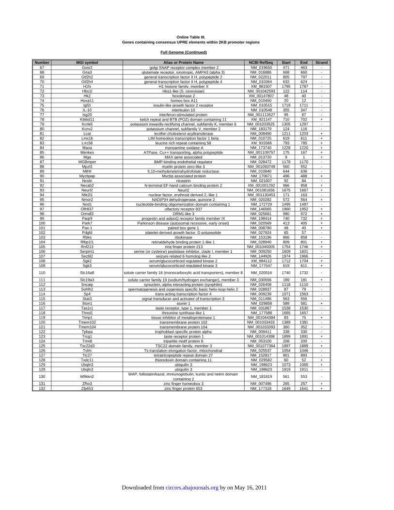
Full Genome (Continued)
Number MGI symbol Alias or Protein Name NCBI RefSeq Start End Strand67 Gosr2 golgi SNAP receptor complex member 2 NM_019650 471 463 -68 Gria3 glutamate receptor, ionotropic, AMPA3 (alpha 3) NM_016886 668 660 -69 Gtf2h2 general transcription factor II H, polypeptide 2 NM_022011 805 797 -70 Gtf2h4 general transcription factor II H, polypeptide 4 NM_010364 632 624 -71 H1fx H1 histone family, member X XM_981507 1795 1787 -72 Hbs1l Hbs1-like (S. cerevisiae) NM_001042593 122 114 -73 Hk2 hexokinase 2 XM_00147807 48 40 -74 Hoxa11 homeo box A11 NM_010450 20 12 -75 Igf2r insulin-like growth factor 2 receptor NM_010515 1719 1711 -76 IL-10 interleukin 10 NM_010548 355 347 -77 Isg20 interferon-stimulated protein NM_001113527 95 87 -78 Kbtbd11 kelch repeat and BTB (POZ) domain containing 11 XM_921147 710 702 +79 Kcnk6 potassium inwardly-rectifying channel, subfamily K, member 6 NM_001033525 1305 1297 -80 Kcnv2 potassium channel, subfamily V, member 2 NM_183179 124 116 -81 Lcat lecithin cholesterol acyltransferase NM_008490 1211 1203 +82 Lmx1b LIM homeobox transcription factor 1 beta NM_010725 619 611 +83 Lrrc58 leucine rich repeat containing 58 XM_915566 793 785 +84 Maoa monoamine oxidase A NM_173740 1228 1220 +85 Menkes ATPase, Cu++ transporting, alpha polypeptide NM_001109757 175 167 +86 Mga MAX gene associated NM_013720 9 1 +87 MGBmper BMP-binding endothelial regulator NM_028472 1178 1170 -88 Mpzl3 myelin protein zero-like 3 NM_001093749 560 552 -89 Mthfr 5,10-methylenetetrahydrofolate reductase NM_010840 644 636 -90 Mycbpap Mycbp associated protein NM_170671 496 488 +91 Ncstn nicastrin NM_021607 92 84 -92 Necab2 N-terminal EF-hand calcium binding protein 2 XM_001001292 966 958 +93 Neurl2 Neurl2 NM_001081656 1675 1667 +94 Nfe2l1 nuclear factor, erythroid derived 2,-like 1 NM_001130453 171 163 -95 Nmor2 NAD(P)H dehydrogenase, quinone 2 NM_020282 572 564 +96 Nod1 nucleotide-binding oligomerization domain containing 1 NM_172729 1495 1487 -97 Olfr837 olfactory receptor 837 NM_146565 1960 1952 +98 Ormdl3 ORM1-like 3 NM_025661 980 972 +99 Paqr9 progestin and adipoQ receptor family member IX NM_198414 740 732 +
100 Park7 Parkinson disease (autosomal recessive, early onset) NM_020569 413 405 +101 Pax-1 paired box gene 1 NM_008780 48 40 +102 Pdgfd platelet-derived growth factor, D polypeptide NM_027924 65 57 -103 Rbks ribokinase NM_153196 866 858 -104 Rlbp1l1 retinaldehyde binding protein 1-like 1 NM_028940 809 801 +105 Rnf213 ring finger protein 213 NM_001040005 1754 1746 +106 Serpini1 serine (or cysteine) peptidase inhibitor, clade I, member 1 NM_009250 1609 1601 -107 Sez6l2 seizure related 6 homolog like 2 NM_144926 1974 1966 -108 Sgk2 serum/glucocorticoid regulated kinase 2 XM_984112 1712 1704 +109 Sgk3 serum/glucocorticoid regulated kinase 3 NM_177547 619 611 +
110 Slc16a8 solute carrier family 16 (monocarboxylic acid transporters), member 8 NM_020516 1740 1732 -
111 Slc19a3 solute carrier family 19 (sodium/hydrogen exchanger), member 3 NM_030556 189 181 +112 Sncaip synuclein, alpha interacting protein (synphilin) NM_026408 1118 1110 -113 Sohlh2 spermatogenesis and oogenesis specific basic helix-loop-helix 2 NM_028937 87 79 -114 Sp4 trans-acting transcription factor 4 NM_009239 1571 1563 -115 Stat3 signal transducer and activator of transcription 3 NM_011486 563 555 -116 Ston1 stonin 1 NM_029858 589 581 +117 Tas1r1 taste receptor, type 1, member 1 NM_031867 1538 1530 -118 Thnsl1 threonine synthase-like 1 NM_177588 1665 1657 -119 Timp1 tissue inhibitor of metalloproteinase 1 NM_001044384 83 75 +120 Tmem102 transmembrane protein 102 NM_001033433 1389 1381 -121 Tmem104 transmembrane protein 104 NM_001033393 360 352 -122 Tpbpa trophoblast specific protein alpha NM_009411 338 330 -123 Trcg1 taste receptor protein 1 NM_001014398 1899 1891 +124 Trim8 tripartite motif protein 8 NM_053100 208 200 -125 Tsc22d3 TSC22 domain family, member 3 NM_001077364 1897 1889 +126 Tsfm Ts translation elongation factor, mitochondrial NM_025537 1054 1046 -127 Ttc27 tetratricopeptide repeat domain 27 NM_152817 901 893 -128 Txdc11 thioredoxin domain containing 11 NM_029582 60 52 +129 Ubqln3 ubiquilin 3 NM_198623 1073 1065 +129 Ubqln3 ubiquilin 3 NM_198623 1919 1911 -
130 Wfikkn2WAP, follistatin/kazal, immunoglobulin, kunitz and netrin domain
containing 2 NM_181819 561 553 -
131 Zfhx3 zinc finger homeobox 3 NM_007496 265 257 +132 Zfp653 zinc finger protein 653 NM_177318 1649 1641 +
Online Table III.Genes containing consensus UPRE elements within 2KB promoter regions
by on May 16, 2011 circres.ahajournals.orgDownloaded from

Number MGI symbol Alias or Protein Name NCBI RefSeq Start End Strand Fold Change1 Alkbh2 alkB, alkylation repair homolog 2 NM_175016 534 516 + 47.452 Il-6 interleukin 6 NM_031168 134 116 + 38.453 Pdia4 protein disulfide isomerase associated 4 NM_009787 107 89 - 16.374 Ears2 glutamyl-tRNA synthetase 2 (mitochondrial)(putative) NM_026140 91 73 - 7.8415 Eda2r ectodysplasin A2 isoform receptor NM_175540 1590 1572 + 7.4646 Hsp90b1 heat shock protein 90, beta (Grp94), member 1 NM_011631 1287 1269 - 7.0716 Hsp90b1 heat shock protein 90, beta (Grp94), member 1 NM_011631 185 167 - 7.0717 Kpnb1 karyopherin (importin) beta 1 NM_008379 1328 1310 + 6.428 Sel1h sel-1 suppressor of lin-12-like NM_001039089 190 172 + 6.2979 Tubb3 tubulin, beta 3 NM_023279 777 759 + 5.79510 Edem1 ER degradation enhancer, mannosidase alpha-like 1 NM_138677 299 281 - 5.3611 G530011O06Rik RIKEN cDNA G530011O06 gene NM_001039559 595 577 + 5.21612 Trim37 tripartite motif-containing 37 NM_197987 239 221 - 5.20812 Trim37 tripartite motif-containing 37 NM_197987 228 210 + 5.208
13 Kcnq1 potassium voltage-gated channel, subfamily Q, member 1 NM_008434 1891 1873 + 5.136
14 Ubfd1 ubiquitin family domain containing 1 NM_138589 79 61 + 5.04815 Pdia3 protein disulfide isomerase associated 3 NM_007952 28 10 + 4.71816 Arntl aryl hydrocarbon receptor nuclear translocator-like NM_007489 291 273 + 4.26717 Tbcb tubulin folding cofactor B NM_025548 35 17 - 4.188
18 Pin1protein (peptidyl-prolyl cis/trans isomerase) NIMA-
interacting 1NM_023371 100 82 - 4.119
19 2610036L11Rik RIKEN cDNA 2610036L11 gene NM_001109747 47 29 + 4.10620 Hyou1 hypoxia up-regulated 1 NM_021395 513 495 + 3.86421 p97/VCP valosin containing protein NM_009503 263 245 + 3.28822 Rabepk Rab9 effector protein with kelch motifs NM_145522 168 150 - 3.06623 Ptpn14 protein tyrosine phosphatase, non-receptor type 14 NM_001033287 1377 1359 + 3.04524 Gsk3b glycogen synthase kinase 3 beta NM_019827 217 199 + 2.99824 Gsk3b glycogen synthase kinase 3 beta NM_019827 1441 1423 - 2.998
25 Sfpqsplicing factor proline/glutamine rich (polypyrimidine tract
binding protein associated) XM_994784 1725 1707 - 2.906
26 Nmt1 N-myristoyltransferase 1 NM_008707 29 11 + 2.83227 Fkbp11 FK506 binding protein 11 NM_024169 1089 1071 + 2.81428 Ormdl2 ORM1-like 2 NM_024180 1673 1655 - 2.78529 Tmem45a transmembrane protein 45a NM_019631 961 943 + 2.75630 Rasl11b RAS-like, family 11, member B NM_026878 760 742 - 2.5430 Rasl11b RAS-like, family 11, member B NM_026878 87 69 - 2.5431 Nip7 nuclear import 7 homolog NM_025391 32 14 - 2.513
32 Herpud1homocysteine-inducible, endoplasmic reticulum stress-
inducible, ubiquitin-like domain member 1NM_022331 83 65 + 2.4315
33 Ngdn neuroguidin, EIF4E binding protein NM_026890 1844 1826 + 2.37934 Mrps18b mitochondrial ribosomal protein S18B NM_025878 499 481 - 2.22835 Lip1 lysosomal acid lipase A NM_001111100 91 73 - 2.2136 Ddx54 DEAD (Asp‐Glu‐Ala‐Asp) box polypeptide 54 NM_028041 1142 1124 + 2.18936 Ddx54 DEAD (Asp-Glu-Ala-Asp) box polypeptide 54 NM_028041 1204 1186 + 2.18937 Stmn1 stathmin 1 NM_019641 1034 1016 - 2.14338 Dnaja4 DnaJ (Hsp40) homolog, subfamily A, member 4 NM_021422 1834 1816 + 2.10738 Dnaja4 DnaJ (Hsp40) homolog, subfamily A, member 4 NM_021422 350 332 + 2.10739 Tubb5 tubulin, beta 5 NM_011655 158 140 + 2.08440 Popdc3 popeye domain containing 3 NM_024286 1592 1574 + 2.04341 Crls1 cardiolipin synthase 1 NM_025646 662 644 - 2.03742 Enpp5 ectonucleotide pyrophosphatase/phosphodiesterase 5 NM_032003 741 723 - 0.49543 Jam2 junction adhesion molecule 2 NM_023844 470 452 + 0.48744 Auh AU RNA binding protein/enoyl‐coenzyme A hydratase NM_016709 1410 1392 + 0.47545 Ppil1 Cypl1 NM_025646 913 895 - 0.47
46 B3gnt6UDP-GlcNAc:betaGal beta-1,3-N-
acetylglucosaminyltransferase 6 (core 3 synthase)NM_001081167 1138 1120 + 0.46
47 2410012H22Rik zinc finger, SWIM-type containing 7 XM_001473892 575 557 + 0.4548 Egflam EGF-like, fibronectin type III and laminin G domains NM_178748 189 171 + 0.43549 Hrasls HRAS-like suppressor NM_013751 141 123 - 0.42850 Myh6 myosin, heavy polypeptide 6, cardiac muscle, alpha NM_010856 773 755 - 0.39851 Cog8 component of oligomeric golgi complex 8 NM_139229 188 170 + 0.38452 Siae sialic acid acetylesterase NM_011734 1525 1507 + 0.36553 Nit2 nitrilase family, member 2 NM_023175 1041 1023 + 0.3654 Asb14 ankyrin repeat and SOCS box-containing 14 NM_080856 1045 1027 + 0.18755 Fbp2 fructose bisphosphatase 2 NM_007994 1068 1050 + 0.15956 Acsm5 acyl-CoA synthetase medium-chain family member 5 NM_178758 287 269 - 0.096157 Ogdh oxoglutarate dehydrogenase (lipoamide) NM_010956 133 115 + 0.0828
58 Apba3amyloid beta (A4) precursor protein binding, family A,
member 1 NM_177034 600 582 - 0.0753
ATF6-Regulated Genes
Genes containing 1bp-mismatched ERSE elements within 2KB promoter regionsOnline Table IV.
by on May 16, 2011 circres.ahajournals.orgDownloaded from

Number MGI symbol Alias or Protein Name NCBI RefSeq Start End Strand Fold Change1 Sdf2l1 stromal cell-derived factor 2-like 1 NM_022324 166 156 - 17.412 Tspyl2 TSPY-like 2 NM_029836 1326 1316 + 9.2943 Ears2 glutamyl-tRNA synthetase 2 (mitochondrial)(putative) NM_026140 351 341 + 7.8414 Sel1h sel-1 suppressor of lin-12-like NM_001039089 161 151 - 6.2975 D16Ertd472e DNA segment, Chr 16, ERATO Doi 472, expressed NM_025967 315 305 - 6.2556 Edem1 ER degradation enhancer, mannosidase alpha-like 1 NM_138677 201 191 - 5.367 Ebp phenylalkylamine Ca2+ antagonist (emopamil) binding protein NM_007898 460 450 + 5.2878 Atf4 activating transcription factor 4 NM_009716 81 71 - 4.9289 Hspa5 heat shock protein 5, GRP78 NM_022310 103 93 - 4.706
10 Syvn1 synovial apoptosis inhibitor 1, synoviolin NM_028769 154 144 + 4.599511 Arntl aryl hydrocarbon receptor nuclear translocator-like NM_007489 288 278 + 4.26712 Egr-1 early growth response 1 NM_007913 342 332 - 3.66713 Junb Jun-B oncogene NM_008416 360 350 - 2.63914 Fra-2 fos-like antigen 2 NM_008037 1051 1041 - 2.47715 3110050N22Rik RIKEN cDNA 3110050N22 gene NM_173181 23 13 - 2.39616 Stt3b STT3, subunit of the oligosaccharyltransferase complex, homolog B NM_024222 191 181 + 2.22817 Rpl14 ribosomal protein L14 NM_025974 80 70 - 2.08718 Pank1 pantothenate kinase 1 NM_001114339 81 71 - 0.40119 Slc22a5 solute carrier family 22 (organic cation transporter), member 5 NM_011396 246 236 - 0.389520 Acot11 acyl-CoA thioesterase 11 NM_025590 160 150 + 0.3421 Tfdp2 transcription factor Dp 2 XM_001481271 110 100 - 0.33522 Tfdp2 transcription factor Dp 2 XM_001481271 557 547 + 0.33523 Asb10 ankyrin repeat and SOCS box-containing 10 NM_080444 516 506 - 0.23124 ACH2 acyl-CoA thioesterase 1 NM_012006 1238 1228 - 0.22525 Acot1 acyl-CoA thioesterase 1 NM_012006 686 676 - 0.225
ATF6-Regulated Genes
Genes containing 1bp-mismatched ERSEII elements within 2KB promoter regionsOnline Table V.
by on May 16, 2011 circres.ahajournals.orgDownloaded from

Number MGI symbol Alias or Protein Name NCBI RefSeq Start End Strand Fold Change1 Far1 fatty acyl CoA reductase 1 NM_027379 131 123 + 120.92 Spp1 secreted phosphoprotein 1 NM_009263 1645 1637 + 91.473 Il-6 interleukin 6 NM_031168 886 878 + 38.454 Tk-1 thymidine kinase 1 NM_009387 745 737 + 20.575 Sdf2l1 stromal cell-derived factor 2-like 1 NM_022324 1539 1531 - 17.415 Sdf2l1 stromal cell-derived factor 2-like 1 NM_022324 1456 1448 - 17.416 Pdia4 protein disulfide isomerase associated 4 NM_009787 838 830 - 16.377 Dnajc3 DnaJ (Hsp40) homolog, subfamily C, member 3 NM_008929 194 186 - 12.758 Pgp phosphoglycolate phosphatase NM_025954 1099 1091 - 10.098 Pgp phosphoglycolate phosphatase NM_025954 386 378 + 10.099 eIF-1A eukaryotic translation initiation factor 1A NM_010120 81 73 + 9.65710 P4hb prolyl 4-hydroxylase, beta polypeptide NM_011032 924 916 - 9.17311 Chac1 ChaC, cation transport regulator-like 1 NM_026929 1603 1595 - 8.06511 Chac1 ChaC, cation transport regulator-like 1 NM_026929 979 971 - 8.06512 Hsp90b1 heat shock protein 90, beta (Grp94), member 1 NM_011631 823 815 + 7.07112 Hsp90b1 heat shock protein 90, beta (Grp94), member 1 NM_011631 665 657 + 7.07113 Itgam integrin alpha M NM_001082960 590 582 + 6.87814 Hn1l hematological and neurological expressed 1-like NM_198937 1061 1053 + 6.77215 Dbf4 DBF4 homolog NM_013726 1383 1375 - 6.44216 Sel1h sel-1 suppressor of lin-12-like NM_001039089 1652 1644 - 6.297
17 D16Ertd472e DNA segment, Chr 16, ERATO Doi 472, expressed NM_025967 1986 1978 + 6.255
18 2810474O19Rik RIKEN cDNA 2810474O19 gene NM_026054 1509 1501 + 6.048519 M(beta)2 tubulin, beta 2a NM_009450 1604 1596 + 5.57919 M(beta)2 tubulin, beta 2a NM_009450 1222 1214 - 5.57920 Tubb2b tubulin, beta 2b NM_023716 1957 1949 - 5.57921 Fem1b feminization 1 homolog b NM_010193 881 873 + 5.32522 Morf4l2 mortality factor 4 like 2 NM_019768 902 894 - 5.3222 Morf4l2 mortality factor 4 like 2 NM_019768 1105 1097 + 5.3223 G530011O06Rik RIKEN cDNA G530011O06 gene NM_001039559 1815 1807 - 5.216
24 Kcna9potassium voltage-gated channel, subfamily Q,
member 1 NM_008434 1928 1920 - 5.136
25 TSA903 uridine-cytidine kinase 2 NM_030724 423 415 + 5.027326 Rbm15b RNA binding motif protein 15B NM_175402 1302 1294 - 4.98927 Rras2 related RAS viral (r-ras) oncogene homolog 2 NM_025846 1075 1067 - 4.94727 Rras2 related RAS viral (r-ras) oncogene homolog 2 NM_025846 224 216 - 4.94728 Aldh18a1 aldehyde dehydrogenase 18 family, member A1 NM_019698 1768 1760 + 4.804529 Grp58 protein disulfide isomerase associated 3 NM_007952 484 476 + 4.718
30 Ms4a4cmembrane-spanning 4-domains, subfamily A,
member 4CNM_029499 694 686 + 4.565
31 Plac8 placenta-specific 8 NM_139198 721 713 - 4.55931 Plac8 placenta-specific 8 NM_139198 1433 1425 + 4.55932 Kif5b kinesin family member 5B NM_008448 119 111 + 4.55
33 NansN-acetylneuraminic acid synthase (sialic acid
synthase)NM_053179 42 34 + 4.5345
34 Nola2 nucleolar protein family A, member 2 NM_026631 1215 1207 - 4.44135 Map3k3 mitogen-activated protein kinase kinase kinase 3 NM_011947 13 5 + 4.287
36 Bmal1 aryl hydrocarbon receptor nuclear translocator-like NM_007489 1388 1380 - 4.267
37 Tbcb tubulin folding cofactor B NM_025548 1014 1006 + 4.18838 2610036L11Rik RIKEN cDNA 2610036L11 gene NM_001109747 1175 1167 + 4.10639 Hmgb1 high mobility group box 1 NM_010439 962 970 + 4.08340 Hagh hydroxyacyl glutathione hydrolase NM_024284 1761 1753 + 3.141340 Hagh hydroxyacyl glutathione hydrolase NM_024284 1504 1496 + 3.141340 Hagh hydroxyacyl glutathione hydrolase NM_024284 794 786 - 3.141341 Mesdc2 mesoderm development candidate 2 NM_023403 306 298 + 3.139
42 Serpinh1serine (or cysteine) peptidase inhibitor, clade H,
member 1 NM_001111044 315 307 - 3.097
43 Gsk3b glycogen synthase kinase 3 beta NM_019827 1288 1280 - 2.99844 2700049P18Rik RIKEN cDNA 2700049P18 gene NM_175382 570 562 + 2.98445 Flnc filamin C, gamma (actin binding protein 280) NM_001081185 1973 1965 - 2.910546 Dlgap4 discs, large homolog-associated protein 4 NM_001042488 407 399 + 2.90746 Dlgap4 discs, large homolog-associated protein 4 NM_001042488 1591 1583 - 2.90747 Nol12 nucleolar protein 12 NM_133800 1268 1260 + 2.88848 Mettl1 methyltransferase-like 1 NM_010792 1029 1021 - 2.87349 Srm spermidine synthase NM_009272 905 897 - 2.81250 H2-Ke6 H2-K region expressed gene 6 NM_013543 934 926 + 2.71951 Os9 amplified in osteosarcoma NM_177614 1935 1927 - 2.672
52 Med19mediator of RNA polymerase II transcription, subunit
19 homologNM_025885 989 981 - 2.66
53 Apex1 apurinic/apyrimidinic endonuclease 1 NM_009687 569 561 - 2.632
Genes containing 1bp-mismatched UPRE elements within 2KB promoter regionsOnline Table VI.
ATF6-Regulated Genes
by on May 16, 2011 circres.ahajournals.orgDownloaded from

Number MGI symbol Alias or Protein Name NCBI RefSeq Start End Strand Fold Change54 Cyb5r1 cytochrome b5 reductase 1 NM_028057 220 212 - 2.59755 Pja2 praja 2, RING-H2 motif containing NM_001025309 968 960 + 2.56855 Pja2 praja 2, RING-H2 motif containing NM_001025309 706 698 + 2.56855 Pja2 praja 2, RING-H2 motif containing NM_001025309 676 668 + 2.56855 Pja2 praja 2, RING-H2 motif containing NM_001025309 646 638 + 2.56855 Pja2 praja 2, RING-H2 motif containing NM_001025309 616 608 + 2.56855 Pja2 praja 2, RING-H2 motif containing NM_001025309 586 578 + 2.56855 Pja2 praja 2, RING-H2 motif containing NM_001025309 556 548 + 2.56855 Pja2 praja 2, RING-H2 motif containing NM_001025309 526 518 + 2.56855 Pja2 praja 2, RING-H2 motif containing NM_001025309 496 488 + 2.56855 Pja2 praja 2, RING-H2 motif containing NM_001025309 466 458 + 2.56855 Pja2 praja 2, RING-H2 motif containing NM_001025309 436 428 + 2.56855 Pja2 praja 2, RING-H2 motif containing NM_001025309 939 931 + 2.56855 Pja2 praja 2, RING-H2 motif containing NM_001025309 406 398 + 2.56855 Pja2 praja 2, RING-H2 motif containing NM_001025309 376 368 + 2.56855 Pja2 praja 2, RING-H2 motif containing NM_001025309 346 338 + 2.56855 Pja2 praja 2, RING-H2 motif containing NM_001025309 316 308 + 2.56855 Pja2 praja 2, RING-H2 motif containing NM_001025309 286 278 + 2.56855 Pja2 praja 2, RING-H2 motif containing NM_001025309 256 248 + 2.56855 Pja2 praja 2, RING-H2 motif containing NM_001025309 226 218 + 2.56855 Pja2 praja 2, RING-H2 motif containing NM_001025309 196 188 + 2.56855 Pja2 praja 2, RING-H2 motif containing NM_001025309 910 902 + 2.56855 Pja2 praja 2, RING-H2 motif containing NM_001025309 881 873 + 2.56855 Pja2 praja 2, RING-H2 motif containing NM_001025309 852 844 + 2.56855 Pja2 praja 2, RING-H2 motif containing NM_001025309 823 815 + 2.56855 Pja2 praja 2, RING-H2 motif containing NM_001025309 794 786 + 2.56855 Pja2 praja 2, RING-H2 motif containing NM_001025309 765 757 + 2.56855 Pja2 praja 2, RING-H2 motif containing NM_001025309 736 728 + 2.56856 Sec11a SEC11 homolog A NM_019951 1759 1751 - 2.52857 Trove2 TROVE domain family, member 2 NM_013835 236 228 - 2.514558 Nip7 nuclear import 7 homolog NM_025391 561 553 - 2.51359 Heatr5a HEAT repeat containing 5A NM_177171 1031 1023 + 2.51260 Rap2b RAP2B, member of RAS oncogene family NM_028712 1437 1429 + 2.454
61 Ywhaztyrosine 3-monooxygenase/tryptophan 5-
monooxygenase activation protein, zeta polypeptide NM_011740 92 84 - 2.446
62 Gmppb GDP-mannose pyrophosphorylase B NM_177910 1739 1731 - 2.43763 Ehd4 EH-domain containing 4 NM_133838 353 345 - 2.40364 3110050N22Rik RIKEN cDNA 3110050N22 gene NM_173181 191 183 + 2.39665 Ngd neuroguidin, EIF4E binding protein NM_026890 1576 1568 - 2.37965 Ngd neuroguidin, EIF4E binding protein NM_026890 743 735 + 2.37966 LOC217431 nucleolar protein 10 NM_001008421 282 274 + 2.37367 Ing1 inhibitor of growth family, member 1 XM_976943 518 510 + 2.36967 Ing1 inhibitor of growth family, member 1 XM_976943 1333 1325 + 2.36968 Cldnd1 claudin domain containing 1 NM_171826 809 801 - 2.33869 Pdap1 PDGFA associated protein 1 NM_001033313 931 923 + 2.336
70 Nucks1nuclear casein kinase and cyclin-dependent kinase
substrate 1NM_175294 1921 1913 + 2.326
70 Nucks1nuclear casein kinase and cyclin-dependent kinase
substrate 1NM_175294 274 266 + 2.326
71 Gcs1 glucosidase 1 NM_020619 33 25 + 2.32372 1700025G04Rik RIKEN cDNA 1700025G04 gene NM_197990 1978 1970 - 2.262573 Pde4b phosphodiesterase 4B, cAMP specific NM_019840 253 245 - 2.21673 Pde4b phosphodiesterase 4B, cAMP specific NM_019840 1062 1054 - 2.21674 Fads3 fatty acid desaturase 3 NM_021890 1516 1508 - 2.21
75 Slc6a6solute carrier family 6 (neurotransmitter transporter,
taurine), member 6 NM_009320 1768 1760 + 2.207
75 Slc6a6solute carrier family 6 (neurotransmitter transporter,
taurine), member 6 NM_009320 480 472 - 2.207
76 Adamts15a disintegrin-like and metallopeptidase (reprolysin
type) with thrombospondin type 1 motif, 15 NM_001024139 52 44 - 2.182
77 Tln1 talin 1 NM_011602 1444 1436 + 2.13777 Tln1 talin 1 NM_011602 1060 1052 + 2.137
Online Table VI.Genes containing 1bp-mismatched UPRE elements within 2KB promoter regions
Full Genome (Continued)
by on May 16, 2011 circres.ahajournals.orgDownloaded from

Number MGI symbol Alias or Protein Name NCBI RefSeq Start End Strand Fold Change78 Tmem57 transmembrane protein 57 NM_025382 735 727 + 2.13679 Rrbp1 ribosome binding protein 1 NM_024281 626 618 - 2.11480 Rrbp1 ribosome binding protein 1 NM_024281 311 303 + 2.114
81 Ralbv-ral simian leukemia viral oncogene homolog B (ras
related)NM_022327 1719 1711 + 2.109
82 tTGas transglutaminase 2, C polypeptide NM_009373 1498 1490 - 2.10683 Srxn1 sulfiredoxin 1 homolog NM_029688 750 742 + 2.08484 Stip1 stress-induced phosphoprotein 1 NM_016737 1476 1468 + 2.05185 Rps27l ribosomal protein S27-like NM_026467 1848 1840 - 2.0586 Crls1 cardiolipin synthase 1 NM_025646 1192 1184 + 2.03787 B3galtl beta 1,3-galactosyltransferase-like NM_194318 586 578 - 0.49488 Rhobtb1 Rho-related BTB domain containing 1 NM_001081347 624 616 - 0.47889 Lmod2 leiomodin 2 (cardiac) NM_053098 615 607 + 0.473
90 Clip4CAP-GLY domain containing linker protein family,
member 4NM_175378 1672 1664 - 0.472
91 3110002H16Rik RIKEN cDNA 3110002H16 gene NM_029623 1093 1085 + 0.46191 3110002H16Rik RIKEN cDNA 3110002H16 gene NM_029623 23 15 + 0.46192 Lpl lipoprotein lipase NM_008509 1987 1979 + 0.45993 Cbr1 carbonyl reductase 1 NM_007620 1061 1053 + 0.45194 Ccni cyclin I NM_017367 519 511 - 0.44595 Grb14 growth factor receptor bound protein 14 NM_016719 1187 1179 + 0.4496 Neo1 neogenin NM_001042752 1193 1185 - 0.427
97 Atp6v0e2 ATPase, H+ transporting, lysosomal V0 subunit E2 NM_133764 1191 1183 + 0.42
97 Atp6v0e2 ATPase, H+ transporting, lysosomal V0 subunit E2 NM_133764 542 534 + 0.42
98 Sirt5sirtuin 5 (silent mating type information regulation 2
homolog) 5NM_178848 20 12 + 0.403
98 Sirt5sirtuin 5 (silent mating type information regulation 2
homolog)NM_178848 1344 1336 - 0.403
99 Gramd1b GRAM domain containing 1B NM_172768 1577 1569 + 0.396100 Mrpl49 mitochondrial ribosomal protein L49 NM_026246 1216 1208 - 0.395100 Mrpl49 mitochondrial ribosomal protein L49 NM_026246 1230 1222 - 0.395101 Asb11 ankyrin repeat and SOCS box-containing 11 NM_026853 90 82 - 0.383102 Mfsd4 major facilitator superfamily domain containing 4 NM_001114662 973 965 - 0.361102 Mfsd4 major facilitator superfamily domain containing 4 NM_001114662 235 227 + 0.361103 D930001I22Rik RIKEN cDNA D930001I22 gene NM_173397 427 419 - 0.357104 Ak3 adenylate kinase 3 NM_021299 454 446 - 0.3505105 Gdc1 glycerol-3-phosphate dehydrogenase 1 (soluble) NM_010271 1384 1376 + 0.342106 Retsat retinol saturase (all trans retinol 13,14 reductase) NM_026159 1863 1855 - 0.34106 Ccdc69 coiled-coil domain containing 69 NM_177471 82 74 + 0.331
107 gamma-SGsarcoglycan, gamma (dystrophin-associated
glycoprotein) NM_011892 1227 1219 + 0.317
108 Gpcr10 endothelin receptor type A NM_010332 1954 1946 - 0.3167109 Cutc cutC copper transporter homolog NM_001113562 128 120 - 0.307110 ORF28 DnaJ (Hsp40) homolog, subfamily C, member 28 NM_001099738 1820 1812 + 0.302111 Sord sorbitol dehydrogenase NM_146126 218 210 - 0.295112 Ucp3 uncoupling protein 3 (mitochondrial, proton carrier) NM_009464 1723 1715 - 0.27112 Ucp3 uncoupling protein 3 (mitochondrial, proton carrier) NM_009464 925 917 - 0.27113 Asb2 ankyrin repeat and SOCS box-containing 2 XM_977692 51 43 - 0.249114 Fndc5 fibronectin type III domain containing 5 NM_027402 166 158 + 0.243115 Acy3 aspartoacylase (aminoacylase) 3 NM_027857 306 298 - 0.239116 Asb-10 ankyrin repeat and SOCS box-containing 10 NM_080444 1338 1330 - 0.231117 Wdr21 WD repeat domain 21 NM_030246 412 404 + 0.19118 Pdlim4 PDZ and LIM domain 4 NM_019417 1371 1363 - 0.102
119 Acsm5 acyl-CoA synthetase medium-chain family member 5 NM_178758 1356 1348 + 0.0961
120 Apba3amyloid beta (A4) precursor protein-binding, family A,
member 3NM_018758 844 836 + 0.0753
Online Table VI.Genes containing 1bp-mismatched UPRE elements within 2KB promoter regions
Full Genome (Continued)
by on May 16, 2011 circres.ahajournals.orgDownloaded from
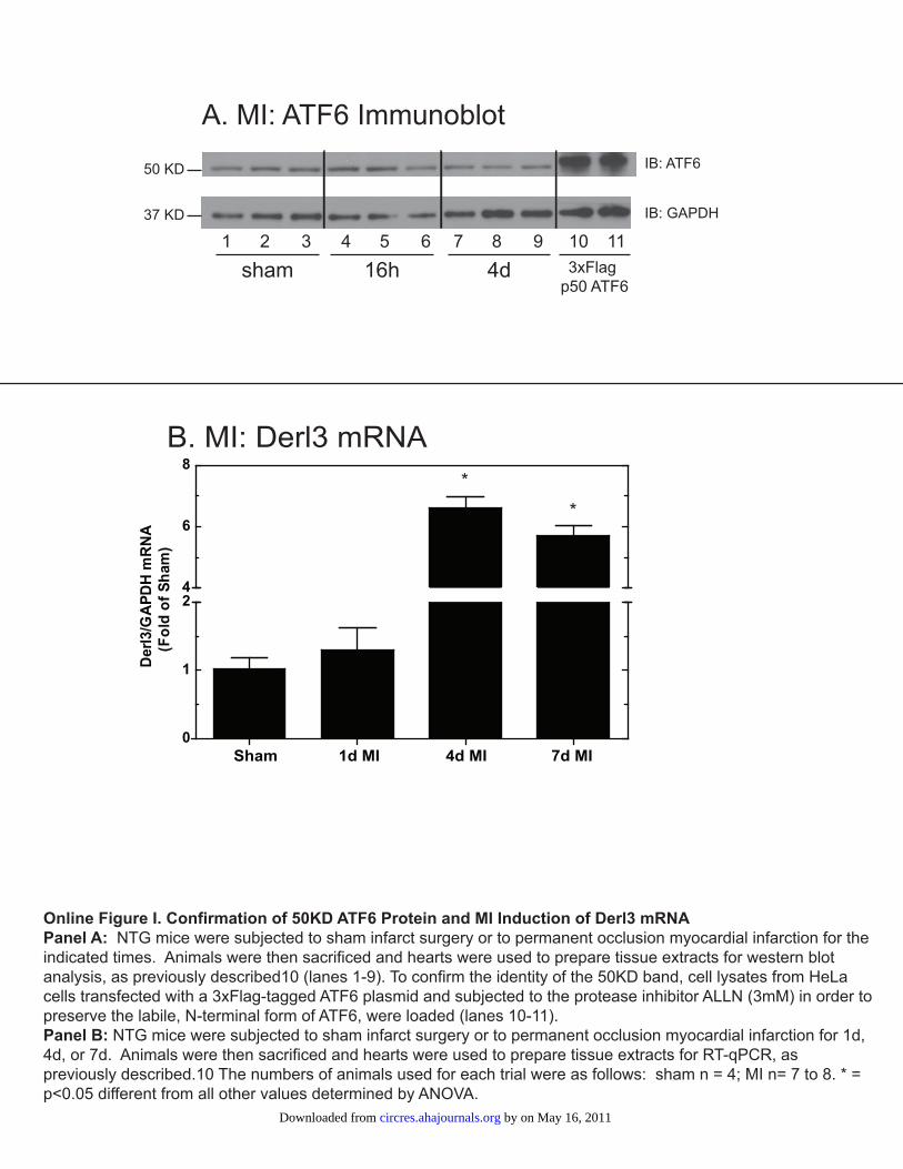
B. MI: Derl3 mRNA
Sham 1d MI 4d MI 7d MI
Der
l3/G
APD
H m
RN
A(F
old
of S
ham
)
Online Figure I. Confirmation of 50KD ATF6 Protein and MI Induction of Derl3 mRNAPanel A: NTG mice were subjected to sham infarct surgery or to permanent occlusion myocardial infarction for the indicated times. Animals were then sacrificed and hearts were used to prepare tissue extracts for western blot analysis, as previously described10 (lanes 1-9). To confirm the identity of the 50KD band, cell lysates from HeLa cells transfected with a 3xFlag-tagged ATF6 plasmid and subjected to the protease inhibitor ALLN (3mM) in order to preserve the labile, N-terminal form of ATF6, were loaded (lanes 10-11). Panel B: NTG mice were subjected to sham infarct surgery or to permanent occlusion myocardial infarction for 1d, 4d, or 7d. Animals were then sacrificed and hearts were used to prepare tissue extracts for RT-qPCR, as previously described.10 The numbers of animals used for each trial were as follows: sham n = 4; MI n= 7 to 8. * = p<0.05 different from all other values determined by ANOVA.
IB: ATF6
IB: GAPDH
sham 3xFlag p50 ATF6
4d16h
A. MI: ATF6 Immunoblot
50 KD
37 KD
1 32 7 984 65 10 11
0
1
24
6
8*
*
by on May 16, 2011 circres.ahajournals.orgDownloaded from
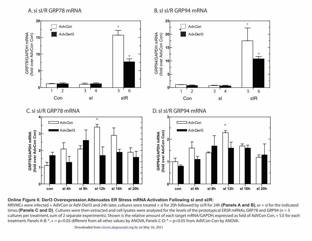
Con sI sIR
0
5
10
15
20AdvCon
AdvDerl3
GR
P78/
GA
PDH
mR
NA
(fol
d ov
er A
dvC
on C
on)
Con sI sIR
0
5
10
15
20
25AdvCon
AdvDerl3
GR
P94/
GA
PDH
mR
NA
(fol
d ov
er A
dvC
on C
on)
1 2 3 4 5 6 1 2 3 4 5 6
*
+
*
+
con sI 4h sI 8h sI 12h sI 16h sI 20h0
1
2
3
4AdvCon
AdvDerl3
GR
P78/
GA
PDH
mR
NA
(fol
d ov
er A
dvC
on C
on)
con sI 4h sI 8h sI 12h sI 16h sI 20h0
1
2
3AdvCon
AdvDerl3
GR
P94/
GA
PDH
mR
NA
(fol
d ov
er A
dvC
on C
on)
A. si sI/R GRP78 mRNA B. sI sI/R GRP94 mRNA
C. si sI/R GRP78 mRNA D. sI sI/R GRP94 mRNA
Online Figure II. Derl3 Overexpression Attenuates ER Stress mRNA Activation Following sI and sI/R:NRVMCs were infected + AdVCon or AdV-Derl3 and 24h later, cultures were treated + sI for 20h followed by sI/R for 24h (Panels A and B), or + sI for the indicated times (Panels C and D). Cultures were then extracted and cell lysates were analyzed for the levels of the prototypical ERSR mRNAs, GRP78 and GRP94 (n = 3 cultures per treatment, sum of 2 separate experiments). Shown is the relative amount of each target mRNA/GAPDH, expressed as fold of AdVCon Con, + S.E for each treatment. Panels A-B: *, + = p<0.05 different from all other values by ANOVA, Panels C-D: * = p<0.05 from AdVCon Con by ANOVA.
**
by on May 16, 2011 circres.ahajournals.orgDownloaded from