Gene Delivery of Sarcoplasmic Reticulum Calcium ATPase Inhibits Ventricular Remodeling in Ischemic...
18
Gene Delivery of Sarcoplasmic Reticulum Calcium ATPase Inhibits Ventricular Remodeling in Ischemic Mitral Regurgitation Ronen Beeri, MD *,# , Miguel Chaput, MD * , J. Luis Guerrero, BS * , Yoshiaki Kawase, MD * , Chaim Yosefy, MD * , Suzan Abedat, MSc # , Ioannis Karakikes, PhD $ , Charlotte Morel, BS $ , Ashley Tisosky, BS $ , Suzanne Sullivan, BS * , Mark Handschumacher, BS * , Dan Gilon, MD # , Gus J. Vlahakes, MD * , Roger J. Hajjar, MD, PhD $ , and Robert A. Levine, MD * * Cardiac Ultrasound Lab and Cardiac Surgery Department, The Heart Center, Massachusetts General Hospital, Boston MA # Cardiovascular Research Center, Heart Institute, Hadassah-Hebrew University Medical Center, Jerusalem, Israel $ Cardiovascular Research Center, Cardiovascular Institute, Mt Sinai School of Medicine, New-York, NY Abstract Background—Mitral regurgitation (MR) doubles mortality following myocardial infarction (MI). We have demonstrated that MR worsens remodeling after MI, and that early correction reverses remodeling. SERCA2a is downregulated in this process. We hypothesized that upregulating SERCA2a may inhibit remodeling in a surgical model of apical MI (no intrinsic MR) with independent MR-type flow. Methods and Results—In 12 sheep, percutaneous gene delivery was performed using a validated protocol to perfuse both LAD and circumflex coronary arteries with occlusion of venous drainage. We administered adeno-associated virus 6 (AAV6) carrying SERCA2a under CMV promoter control in 6 sheep, and a reporter gene in 6 controls. After 2 weeks, standardized apical MI was created, and a shunt implanted between the LV and LA, producing regurgitant fractions of ~30%. Animals were compared at baseline, 1 and 3 months using 3D echo, Millar hemodynamics and biopsies. The SERCA2a group had well-maintained preload-recruitable stroke work at 3 months (decrease by 8 ±10% vs. 42±12% with reporter gene controls (p<0.001)). LV dP/dt followed the same pattern (no change vs. 55% decrease, p<0.001). LVESV was lower with SERCA2a (82.6±9 6 vs 99.4±9.7 ml, p=0.03); LVEDV, reflecting volume overload, was not significantly different (127.8±6.2 vs 134.3 ±9.4 ml). SERCA2a sheep showed 15% rise in anti-apoptotic pAkt vs. 30% reduction with reporter gene (P<0.001). Pro-hypertrophic activated STAT3 was also 41% higher with SERCA2a than in controls (p<0.001). Pro-apoptotic activated caspase-3 rose over 5-fold over 1 month in both SERCA2a and controls (p=NS), and decreased by 19% at 3 months, remaining elevated in both groups. Conclusions—In this controlled model, upregulating SERCA2a induces better function and lesser remodeling, with improved contractility, smaller volume and activation of pro-hypertrophic/anti- apoptotic pathways. Although caspase-3 remains activated in both arms, SERCA2a sheep had increased molecular anti-remodeling “tone”. We therefore conclude that upregulating SERCA2a Correspondence to: Robert A. Levine, MD, Cardiac Ultrasound Laboratory, YAW5, Massachusetts General Hospital, 55 Fruit Street, Boston, MA 02114, [email protected], Telephone: (617) 724-1995, Fax: (617) 643-1616. DISCLOSURES None. NIH Public Access Author Manuscript Circ Heart Fail. Author manuscript; available in PMC 2011 September 1. Published in final edited form as: Circ Heart Fail. 2010 September 1; 3(5): 627–634. doi:10.1161/CIRCHEARTFAILURE.109.891184. NIH-PA Author Manuscript NIH-PA Author Manuscript NIH-PA Author Manuscript
Transcript of Gene Delivery of Sarcoplasmic Reticulum Calcium ATPase Inhibits Ventricular Remodeling in Ischemic...

Gene Delivery of Sarcoplasmic Reticulum Calcium ATPaseInhibits Ventricular Remodeling in Ischemic Mitral Regurgitation
Ronen Beeri, MD*,#, Miguel Chaput, MD*, J. Luis Guerrero, BS*, Yoshiaki Kawase, MD*,Chaim Yosefy, MD*, Suzan Abedat, MSc#, Ioannis Karakikes, PhD$, Charlotte Morel, BS$,Ashley Tisosky, BS$, Suzanne Sullivan, BS*, Mark Handschumacher, BS*, Dan Gilon, MD#,Gus J. Vlahakes, MD*, Roger J. Hajjar, MD, PhD$, and Robert A. Levine, MD** Cardiac Ultrasound Lab and Cardiac Surgery Department, The Heart Center, MassachusettsGeneral Hospital, Boston MA# Cardiovascular Research Center, Heart Institute, Hadassah-Hebrew University Medical Center,Jerusalem, Israel$ Cardiovascular Research Center, Cardiovascular Institute, Mt Sinai School of Medicine, New-York,NY
AbstractBackground—Mitral regurgitation (MR) doubles mortality following myocardial infarction (MI).We have demonstrated that MR worsens remodeling after MI, and that early correction reversesremodeling. SERCA2a is downregulated in this process. We hypothesized that upregulatingSERCA2a may inhibit remodeling in a surgical model of apical MI (no intrinsic MR) withindependent MR-type flow.
Methods and Results—In 12 sheep, percutaneous gene delivery was performed using a validatedprotocol to perfuse both LAD and circumflex coronary arteries with occlusion of venous drainage.We administered adeno-associated virus 6 (AAV6) carrying SERCA2a under CMV promoter controlin 6 sheep, and a reporter gene in 6 controls. After 2 weeks, standardized apical MI was created, anda shunt implanted between the LV and LA, producing regurgitant fractions of ~30%. Animals werecompared at baseline, 1 and 3 months using 3D echo, Millar hemodynamics and biopsies. TheSERCA2a group had well-maintained preload-recruitable stroke work at 3 months (decrease by 8±10% vs. 42±12% with reporter gene controls (p<0.001)). LV dP/dt followed the same pattern (nochange vs. 55% decrease, p<0.001). LVESV was lower with SERCA2a (82.6±9 6 vs 99.4±9.7 ml,p=0.03); LVEDV, reflecting volume overload, was not significantly different (127.8±6.2 vs 134.3±9.4 ml). SERCA2a sheep showed 15% rise in anti-apoptotic pAkt vs. 30% reduction with reportergene (P<0.001). Pro-hypertrophic activated STAT3 was also 41% higher with SERCA2a than incontrols (p<0.001). Pro-apoptotic activated caspase-3 rose over 5-fold over 1 month in bothSERCA2a and controls (p=NS), and decreased by 19% at 3 months, remaining elevated in bothgroups.
Conclusions—In this controlled model, upregulating SERCA2a induces better function and lesserremodeling, with improved contractility, smaller volume and activation of pro-hypertrophic/anti-apoptotic pathways. Although caspase-3 remains activated in both arms, SERCA2a sheep hadincreased molecular anti-remodeling “tone”. We therefore conclude that upregulating SERCA2a
Correspondence to: Robert A. Levine, MD, Cardiac Ultrasound Laboratory, YAW5, Massachusetts General Hospital, 55 Fruit Street,Boston, MA 02114, [email protected], Telephone: (617) 724-1995, Fax: (617) 643-1616.DISCLOSURESNone.
NIH Public AccessAuthor ManuscriptCirc Heart Fail. Author manuscript; available in PMC 2011 September 1.
Published in final edited form as:Circ Heart Fail. 2010 September 1; 3(5): 627–634. doi:10.1161/CIRCHEARTFAILURE.109.891184.
NIH
-PA Author Manuscript
NIH
-PA Author Manuscript
NIH
-PA Author Manuscript
inhibits MR-induced post-MI remodeling in this model, and thus may constitute a useful approachto reduce the vicious cycle of remodeling in ischemic MR.
Keywordsmitral regurgitation; valvular heart disease; echocardiography; remodeling
Expansion of infarcted tissue begins acutely after myocardial infarction (MI), but a moregradual remodeling process also involves noninfarcted areas;1 initially compensatory, thisprocess becomes maladaptive, as the ventricle enlarges and contracts poorly2 with reducedsurvival.3
MI also causes ischemic mitral regurgitation (MR) by altering ventricular geometry andfunction,4, 5 doubling the risk of death. Severe non-ischemic MR has been shown to promoteLV remodeling and reduce survival.6–8 We have previously demonstrated 9 that moderate MR,simulated by an LV-to-LA shunt, added to a small antero-apical MI (causing no intrinsic MR)causes greater ventricular remodeling than a comparable infarction alone, with an earliertransition to a failure phenotype. We have also shown that repairing the regurgitant-type flowat an early stage after the MI reverses the remodeling-related processes.10 Whole heart changesparallel cellular and molecular abnormalities in the non-infarcted myocardium that reflect thecomplex remodeling process. These molecular events also progress differently with MR thanwith comparable infarction alone, with an initial rise in pro-hypertrophic and anti-apoptoticsignals followed by their exhaustion.
Most experimental models of post-MI remodeling use infero-posterior MIs,11, 12 but thisnecessarily links the MI-induced remodeling to the development of MR. The shunt modelallows MR to be varied independently in the presence of MI and without interventions such asinfarct patching13 that might themselves influence remodeling.
Upregulating genes encoding for proteins of interest has been demonstrated to be an effectiveapproach to modulate and treat heart failure. One candidate for such gene therapy is thesarcoplasmic (SR) Ca+2-ATPase (SERCA2a), which is down-regulated in that model, andplays a pivotal role in the regulation of intracellular Ca2+ in cardiomyocytes14. Calcium entryinto the cytosol during systole induces Ca2+ release from the SR through the ryanodynereceptor, coupling excitation and contraction. During relaxation, Ca2+ is returned to the SR bythe SR Ca2+-ATPase (SERCA2a). Some is also extruded by the sarcolemmal Na+/Ca2+
exchanger (NCX), which is upregulated in cardiac hypertrophy and failure.15, 16 BecauseSERCA2a is the major determinant of the amount of Ca2+ available to be released during theupcoming systole, changes in SERCA function significantly affect cardiac excitation-contraction coupling. SERCA2a activity also has a major influence on myocardial relaxation,17, 18 Ca2+ extrusion via SERCA2a being more efficient energetically than the alternative NCXpathway.19 SERCA2a mRNA levels are reduced in failing hearts.20 Using a gene therapyapproach, up-regulating SERCA2a levels in different models of heart failure resulted inimprovement in systolic21–23 and diastolic function,17 as well as improving metabolism, 24,25 potentially reducing arrhythmias26, 27 and improving survival.25 We have demonstrated thatSERCA2A is down-regulated in the remote zones of post-MI remodeling ventricles-significantly more so when MR was also present, accompanied reduction in contractility of thewhole ventricle and of isolated cells, and reduction in single cell calcium transients.9 Pathwaysinvolved in the compensatory hypertrophic response in were initially up-regulated, only to fallbelow baseline at 3 months, when severe dilatation and failure were present. As repairing MRin the early phase, before these processes have been activated, induces reversal of remodeling,10 SERCA2a may have a unique role in determining this reversibility. This is emphasized bythe recently reported effects of SERCA2a up-regulation in an MR-only pig model of heart
Beeri et al. Page 2
Circ Heart Fail. Author manuscript; available in PMC 2011 September 1.
NIH
-PA Author Manuscript
NIH
-PA Author Manuscript
NIH
-PA Author Manuscript
failure,28 where it induced inhibition of ventricular enlargement and myocardial dysfunctionapparent in the control animals.
This study aims to apply the gene therapy approach in a clinically relevant large-animal modelof actively evolving remodeling induced by the combination of ischemic and valvular lesionsin which a biphasic pattern of compensatory and decompensatory changes has beendemonstrated. An intriguing question to address in this model is whether the potentiallybeneficial effects of SERCA2a gene therapy are accompanied by molecular changes typicalof compensated hypertrophy9 as seen early in the course of MR-augmented remodeling, oronly by measurable reductions in ventricular volumes and improvements in contractiledysfunction without molecular changes in other aspects of the remodeling processes. Geneticmodification of such a key pathway can thereby help dissect its contribution to the entire diseaseprocess,
Thus, we hypothesized that up-regulating SERCA2a levels by gene delivery using a viral vectormay reverse the remodeling process in our model of “ischemic-type” MR. that is, MRassociated with myocardial infarction.9 We also hypothesized that this reversal will be manifestboth in ventricular volumes and function, and in persistent activation of pro-hypertrophic andanti-apoptotic pathways.
In this context, prolonged and sustained expression of the transgene is critical, as is the lackof host immune response to the vector. Adeno-associated vector has been demonstrated toconfer prolonged and sustained expression of myocardial transgenes, while lackingimmunogenic and cardiotoxic effects,29 and was therefore used in this study.
METHODSAnimal studies
A total of 12 male Dorsett hybrid sheep (20–30 kg) were included. Our established model ofindependent MI and MR-type flow9, 10 was implemented using an 8-cm long, 8-mm diameterreinforced Teflon (PTFE) graft (Edwards, cross-sectional area 0.50 cm2) implanted understerile conditions into the mid-lateral LV and LA appendage with intramuscular portionsstiffened with epoxy resin (Figure 1). The regurgitant flow was confirmed during eachthoracotomy using a Transonic flow probe and color Doppler. The standardized shunt diameterand length consistently produced moderate MR (regurgitant fractions of ~30%30). Animalswere treated with heparin (3 days) and then oral aspirin.
Vector designVector production, harvest, purification, and testing were done as previously described.31 TherAAV6.SERCA2a vector used in this study contains an AAV serotype 6 viral capsid and asingle-stranded ~4.5 kb DNA containing the human SERCA2a cDNA driven by a CMVimmediate-early promoter/enhancer, a hybrid intron, and a bovine growth hormone poly-adenylation signal, all flanked by 145 nt AAV2 inverted terminal repeat sequences necessaryfor replication and packaging of the vector DNA in the capsid. The vector was manufacturedusing standard calcium phosphate transfection methods in adherent 293 cells. Three plasmidswere used, 1 containing helper functions from adenovirus, 1 containing the AAV rep2 and cap1genes, and the third containing the vector genome. Final vector preparations were more than95% pure as judged by SDS-PAGE (Invitrogen, Carlsbad, California).
Gene deliveryTwo weeks prior to the first thoracotomy (in order to obtain significant gene expression atmodel creation), antegrade coronary arterial injection with concomitant great cardiac vein
Beeri et al. Page 3
Circ Heart Fail. Author manuscript; available in PMC 2011 September 1.
NIH
-PA Author Manuscript
NIH
-PA Author Manuscript
NIH
-PA Author Manuscript
blockade was performed with AAV6 as a vehicle for the reporter gene β-galactosidase (β-gal-control) and SERCA2a, each at a titer of 5×10¹⁴ genomes/ml. The great cardiac vein wascannulated via internal jugular access and occluded with a standard balloon-tipped catheter.The left anterior descending coronary artery (LAD) was cannulated via the femoral artery andoccluded with a standard angioplasty balloon before the first diagonal branch. With both arterialand venous balloons transiently inflated for 2 minutes, intracoronary adenosine wasadministered to increase permeability and prolong dwell time32, followed by 5×10¹² genomesof either AAV6.βgal or AAV6.SERCA2a (six sheep each). This sequence was repeated for theleft circumflex (LCX) artery. Previous work has shown increased tissue expression in the wholeadult heart using this delivery method33.
Model creationSheep were loaded for 3 days with amiodarone (200 mg PO BID), anesthetized withthiopentothal (0.5 ml/kg), intubated and ventilated at 15 ml/kg with 2% isoflurane-oxygen,receiving glycopyrrolate (0.4 mg IV) and prophylactic vancomycin (0.5 g IV) and amiodarone(150 mg IV drip). Surface ECG was monitored and a sterile left thoracotomy performed withpericardial cradle creation. A high-fidelity micromanometer-tipped catheter (Millar, Houston,TX) was placed into the LV. After baseline 2D and 3D echo imaging, a septal MI was producedby ligating the mid- to distal left anterior descending coronary artery, known to producesubstantial MIs without MR.34 2D echo confirmed that wall motion abnormality involvedapproximately one-third of the anteroseptum from apex to base for standardization. In additionto analgesia, propranolol, 1 mg IV in two doses, was given for evident stress and tachycardia(>150) upon extubation. Antibiotics (Cephapirin, 0.5 gm IV) and analgesics (Buprenorphine,0.3 mg BID) were administered for 5 days, and oral amiodarone (200 mg BID) for three.
During repeat sterile thoracotomy at day 30, 3D echo evaluated LV remodeling and function,with directed TruCut needle biopsies of the noninfarcted myocardium near and remote fromthe border zone. At day 90, 3D echo and blood sampling were repeated at thoracotomy,followed by euthanasia. Animal studies conformed to NIH guidelines (National ResearchCouncil, Washington, DC, 1996) and were IRB-approved.
3D echo and LV function.35
Rotated apical images were obtained at 10-degree intervals with an epicardial 5MHz TEE probe(Sonos 7500, Philips, Andover, MA), rotated by software and gated to ECG and respiration.Digital images were analyzed on a workstation with custom programs, by an operator blindedto treatment assignment. Endocardial surfaces were traced to calculate LV volumes validatedagainst a 36-crystal sonomicrometer array. Remodeling was quantified in terms of increasingLV volumes. Regurgitant fraction was calculated as MR-equivalent flow by Transonic flow-meter, divided by LV ejection volume, with verification of MR flow by pulsed Doppler time-velocity integral of shunt flow multiplied by shunt cross-sectional area. LV pressure-volumeloops at the initial and final thoracotomies were obtained using Millar catheters andsubendocardial crystals (Sonometrics, London, ON, Canada), with IVC occlusion to obtainpressure-volume curves and derive preload-recruitable stroke work.36 Crystals were not placedat day 30 to maximize sterility and survival. Maximal systolic dP/dt was obtained by the high-fidelity Millar catheter.
Molecular assaysWe measured levels of several molecular species associated with remodeling that modulatecell hypertrophy and death and are responsible for extracellular matrix turnover.9, 10 All proteinassays were performed for each treatment group and stage on each individual sheep separately,and the results were averaged.
Beeri et al. Page 4
Circ Heart Fail. Author manuscript; available in PMC 2011 September 1.
NIH
-PA Author Manuscript
NIH
-PA Author Manuscript
NIH
-PA Author Manuscript
Calcium cycleSarcoplasmic reticulum (SR) membrane was obtained using sucrose gradient centrifugation.37 Proteins were separated and an immunoblot using monoclonal anti-SERCA2 and anti-phospholamban (Santa Cruz Biotechnology, Santa Cruz, CA) was performed, normalized tototal protein. Na+/Ca2+ exchanger (NCX) levels were measured by Western blot, usingmonoclonal anti-NCX antibodies (Santa Cruz Biotechnology, Santa Cruz, CA).
Pro-hypertrophic and pro-apoptotic cascadesWe measured levels of Akt (protein kinase B) and gp130, which are both at their respectivelevels (cytosol and membrane) important crossroads in pro-hypertrophic signaling;phosphorylated (activated) STAT3, an important downstream effector of gp130; and activatedcaspase-3, the final common pathway for intra-cellular apoptosis signaling. Western blotanalysis was performed on cell lysates from biopsies at baseline and days 30 and 90. Anti-gp130, anti-phosphoAkt, anti-phospho STAT3 and anti-activated caspase-338 (Santa CruzBiotech-nology, Santa Cruz, CA) were detected with peroxidase-conjugated anti-mouse IgGand chemiluminescence, with α-actin as housekeeping control. Integrated blot pixel densitywas assessed using standard software (ImageJ, NIH) by an operator blinded to treatmentassignments.
StatisticsAll values are reported as mean±SD. Statistical analysis used 2-tailed Student’s t-test forcontinuous variables compared at specific time-points; the Bonferroni correction was appliedwhen appropriate. Repeated measures over time were analyzed with repeated-measuresANOVA (JMP 8, SAS Institute). P<0.05 was considered significant. Inter- and intra-observervariability for 3D echo-measured LV volumes in our lab were 3.5% as previously reported.9
RESULTSInfarct size, traced and integrated by 3D echo, was 12–22% of the endocardial surface area,with a mean of 17±3% (n=12).
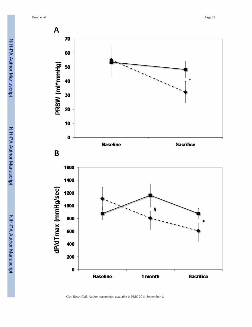
Function and volumesThe SERCA2a group had well-maintained preload-recruitable stroke work at 3 monthssacrifice (decrease by 8±10%) vs. a 42±12% decrease with reporter gene controls (p<0.001,Fig. 2). Peak systolic LV dP/dt followed the same pattern (no change vs. 55% decrease,p<0.001, Fig. 2). Although 3D echo-derived LVEF was decreased in both groups beginningwith the post-MI baseline, it was better maintained at sacrifice with SERCA2a (35.2±4.0% vs.26.1±3.5%, p=0.01, Fig. 2). This was accompanied by a lower LVESV with SERCA2a (82.6±9.6 ml vs 99.4±9.7 ml, p=0 03, Fig. 3); LVEDV, reflecting the volume overload, was notsignificantly different at sacrifice (127.8±6.2 ml vs 134.3±9.4 ml, p=NS, Fig. 3).
Although no quantitative assessment of animal well-being could be performed, the animals inthe control group were less active and seemed more short of breath.
Molecular pathways of remodelingAs expected, in the SERCA group there was a very significant increase in SERCA2a proteinlevels in both remote and border zones at sacrifice, which was already apparent at 1 month,follow up, while control sheep demonstrated a sharp reduction in SERCA2a levels at 1 monthfollow-up (average integrated density 85.6±15.2 vs 54.2±10.8 p<0.001, Fig. 4) and even moreso at sacrifice (average integrated density 93.6±20.1 vs 34.2±6.3, p<0.001, Fig. 4). Of note, nosignificant change was noted in regulatory phospholamban levels (Fig. 4). NCX levels were
Beeri et al. Page 5
Circ Heart Fail. Author manuscript; available in PMC 2011 September 1.
NIH
-PA Author Manuscript
NIH
-PA Author Manuscript
NIH
-PA Author Manuscript
significantly more elevated in the control sheep as compared with the SERCA sheep atsacrifice, consistent with a more active remodeling process39 (P=0.025, Fig. 5).
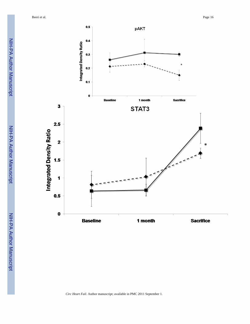
SERCA2a sheep showed at 3 months’ sacrifice a 15% rise in anti-apoptotic phospho-Akt vs.30% reduction with reporter gene (P<0.001, Fig. 5). The sample mean of STAT3 was also 41%higher at sacrifice with SERCA2a than reporter gene (p<0.001, Fig. 5). In contrast, gp130 fellby 25%–26% in both groups (p=NS by repeated-measures ANOVA), raising the possibilitythat improved contractility blunted the stimulus for pro-hypertrophic compensation, oralternatively, that SERCA2a over-expression compensates for but does not entirely eliminatethe remodeling drive. Pro-apoptotic activated caspase-3 rose over 5-fold and to a comparableextent over 1 month in both SERCA2a and reporter gene animals (p=NS, Fig. 5), and decreasedby only 19% from 1 to 3 months, remaining elevated in both groups at sacrifice.
DISCUSSIONA large number of patients with MI develop MR and progress to congestive heart failure. Ourprevious results9 have shown that, for a comparable infarct size, the presence of MR-typevolume over-load leads to greater LV dilatation and dysfunction and to more severe changesat a cellular and molecular level. Molecular changes are biphasic, with initial upregulation andsubsequent exhaustion of pro-hypertrophic and anti-apoptotic pathways that otherwise remainelevated when MI is not accompanied by MR. Maintained elevation of caspase-3 andextracellular matrix turnover lead to a failure phenotype with abnormal cellular morphology,decreased calcium cycling, and reduced sarcoplasmic reticulum Ca+2-ATPase (SERCA2a).9This reduction was more pronounced in the border zones of the infarction, reflecting a probablelarger element of cell loss through ischemic damages, but was also significant in the remotezones, possibly reflecting stretch-induced activation of the fetal program in these myocytes,resulting in diminution of SERCA2a levels. We have also demonstrated the corollary that earlyrepair of such moderate MR-type volume overload reverses these progressive remodelingprocesses, and activates intracellular signals promoting hypertrophy, opposing apoptosis, andinhibiting matrix proteolysis.10 Manipulating the expression of key proteins and the activityof specific down-stream signaling pathways involved in cardiac hypertrophy and failure willallow us to understand their contribution to the disease process.
AAV (adeno-associated virus) is a gene therapy vector that provides gene expression lastingmore than a year in muscle and brain with little or no immune reaction.40–42 SERCA2a waschosen as transgene because its expression is reduced in our MI+MR model9, and itsoverexpression improves contractility21, 23, 43 and might also decrease apoptosis by reducingintracellular diastolic Ca2+ concentrations.44, 45 In fact, a phase 1 clinical trial usingAAV1.SERCA2a has been completed in patients with severe heart failure showing safety andpositive biological effects (albeit in an open label trial) 46.
In our model, using a percutaneous delivery system for AAV6 encoding SERCA2a, wemanaged to secure robust transduction, as manifested by sustained elevation of SERCA2alevels as compared with a significant reduction in controls. We did not detect a compensatoryincrease in inhibitory pospholamban expression. This up-regulation translated into preservedLV contractility as measured by preload-recruitable stroke work, a relatively load-independentmeasure of LV function; LV dP/dt was also preserved, while these measurements wheresignificantly depressed in the control animals. Morphologically, there was less evidence ofremodeling in the SERCA animals, manifested as relatively preserved LV end-systolicvolumes throughout the experiment. On the other hand, we did not detect a significant changein activated-caspase3 levels - suggesting that while the net tone in the cell is shifted to anti-apoptosis, as demonstrated by Akt and STAT3 activation, upregulating SERCA might notablate all aspects of the intracellular remodeling cascade. One interesting aspect of the
Beeri et al. Page 6
Circ Heart Fail. Author manuscript; available in PMC 2011 September 1.
NIH
-PA Author Manuscript
NIH
-PA Author Manuscript
NIH
-PA Author Manuscript
molecular changes was the effect on NCX expression in our model. An increase in NCXexpression has been observed in a number of models of heart failure and has been associatedwith an increased risk of ventricular arrhythmias.47 In our model, NCX was increased in thecontrol group but was remained at baseline levels with overexpression of SERCA2a. Likewise,we did not detect increased levels of gp130, as we have previously seen with MR repair;10
however, activated STAT3, downstream from gp130, was significantly increased, suggestingthe possibility of greater activation of gp130-containing cytokine receptors, STAT3 activationby an alternative pathway, or decreased feedback inhibition. The results suggest that improvingcontractility and relaxation is insufficient to reverse the remodeling process completely at amolecular level. Nonetheless, the improvement in contraction, volumes and intracellular pro-hypertrophic pathways suggests that SERCA2a upregulation does at least strongly inhibit theremodeling process. As SERCA2a upregulation has been demonstrated, in different models,to improve function and retard progression to heart failure,17, 23, 26, 33, 43, 48, 49 our resultsare consistent with previously reported data.
This study has several limitations: Ischemic MR affecting a native valve often progressivelyincreases,11, 50–52 but is inherently linked to the underlying MI and not standardized. Basedon the study motivation, it was critical to separate the two processes of infarction andregurgitation to determine the incremental role of MR and to do so with a standardized orifice,which provided stable regurgitant fractions of ~30% throughout the study. In the clinicalsituation of the tethered mitral valve, SERCA2a may have an even more pronounced effect byreducing the severity of this dynamic MR: increased LV contractility and decreased LVvolumes will increase the closing forces and decrease the tethering forces on the mitral valve,thereby improving coaptation and reducing MR.49 This will be examined in a separate study.We performed gene delivery 2 weeks before induction of MR+MI. This was done in order tohave an established up-regulation of SERCA2a coincident with the initiation of the remodelingprocess, which starts immediately after infarction, to provide a proof of concept about the roleof SERCA2a in this situation. Variations in timing of SERCA2a therapy relative to MR repairwill also be assessed, for example, in the fully dilated remodeling state, as there may be a “pointof no return” beyond which these interventions may be ineffective.53
In conclusion, we have demonstrated that up-regulating SERCA2a in a model of MR+MI mayinhibit the remodeling process, as manifested by ventricular function, volumes, and intra-cellular pathways of hypertrophy. This may constitute a potentially useful approach to reducethe vicious cycle of remodeling in ischemic MR.
AcknowledgmentsSOURCES OF FUNDING
Supported in part by grants R01 HL72265, R01 HL 078731, R01 HL080498, R01 HL083156 and K24 HL67434 fromNIH/NHLBI, and grant 2005250 from the USA/Israel Binational Science Foundation (BSF). Dr Levine was alsosupported by grant 07CVD04 for the Transatlantic MITRAL Network, Leducq Foundation, Paris, France.
References1. Pfeffer MA, Braunwald E. Ventricular remodeling after myocardial infarction: experimental
observations and clinical implications. Circulation 1990;81:1161–1172. [PubMed: 2138525]2. Picard MH, Wilkins GT, Ray PA, Weyman AE. Progressive changes in ventricular structure and
function during the year after acute myocardial infarction. Am Heart J 1992;124:24–31. [PubMed:1535474]
3. Cohn JN, Ferrari R, Sharpe N. Cardiac remodeling- concepts and clinical implications: a consensuspaper from an international forum on cardiac remodeling. J Am Coll Cardiol 2000;35:569–582.[PubMed: 10716457]
Beeri et al. Page 7
Circ Heart Fail. Author manuscript; available in PMC 2011 September 1.
NIH
-PA Author Manuscript
NIH
-PA Author Manuscript
NIH
-PA Author Manuscript

4. Otsuji Y, Handshumacher MD, Schwammethal E, Jiang L, Song J-K, Guerrero JL, Vlahakes GJ, LevineRA. Insights from three-dimensional echocardiography into the mechanisms of functional mitralregurgitation: direct in vivo demonstration of altered leaflet geometry. Circulation 1997;96:1999–2008. [PubMed: 9323092]
5. Komeda M, Glasson JR, Bolger AF, Daughters GT 2nd, Ingels NB Jr, Miller DC. Papillary muscle-left ventricular “complex”. J Thorac Cardiovasc Surg 1997;113:292–300. [PubMed: 9040623]
6. Carabello BA, Nakano K, Corin W, Biederman R, Spann JF Jr. Left ventricular function in experimentalvolume overload hypertrophy. Am J Physiol 1989;256:H974–981. [PubMed: 2523200]
7. Spinale FG, Ishihra K, Zile MR, DeFryte G, Crawford FA, Carabello BA. Structural basis for changesin left ventricular function and geometry because of chronic mitral regurgitation and after correctionof volume overload. J Thorac Cardiovasc Surg 1993;106:1147–1157. [PubMed: 8246553]
8. Ling LH, Enriquez-Sarano M, Seward JB, Tajik AJ, Schaff HV, Bailey KR, Frye RL. Clinical outcomeof mitral regurgitation due to flail leaflet. N Engl J Med 1996;335:1417–1423. [PubMed: 8875918]
9. Beeri R, Yosefy C, Guerrero JL, Nesta F, Abeidat S, Chaput M, del Monte F, Handschumacher MD,Stroud R, Sullivan S, Pugatsch T, Gilon D, Vlahakes GJ, Spinale FG, Hajjar RJ, Levine RA. MitralRegurgitation Augments Post-Myocardial Infarction Remodeling: Failure of HypertrophicCompensation. J Am Coll Cardiol 2008;51:476–486. [PubMed: 18222360]
10. Beeri R, Yosefy C, Guerrero JL, Abedat S, Handschumacher MD, Stroud RE, Sullivan S, Chaput M,Gilon D, Vlahakes GJ, Spinale FG, Hajjar RJ, Levine RA. Early repair of moderate ischemic mitralregurgitation reverses left ventricular remodeling: a functional and molecular study. Circulation2007;116:I288–293. [PubMed: 17846319]
11. Llaneras MR, Nance ML, Streicher JT, Lima JA, Savino JS, Bogen DK, Deac RF, Ratcliffe MB,Edmunds LH. Large animal model of ischemic mitral regurgitation. Ann Thorac Surg 1994;57:432–439. [PubMed: 8311608]
12. Moainie SL, Guy TS, Gorman JH, Plappert T, Jackson BM, St John-Sutton MG, Edmunds LH,Gorman RC. Infarct restraint attenuates remodeling and reduces chronic ischemic mitral regurgitationafter postero-lateral infarction. Ann Thorac Surg 2002;74:444–449. [PubMed: 12173827]
13. Hung J, Guerrero JL, Handschumacher MD, Supple G, Sullivan S, Levine RA. Reverse ventricularremodeling reduces ischemic mitral regurgitation: echo-guided device application in the beatingheart. Circulation 2002;106:2594–2600. [PubMed: 12427657]
14. Gwathmey JK, Slawsky MT, Hajjar RJ, Briggs GM, Morgan JP. Role of intracellular calcium handlingin force-interval relationships of human ventricular myocardium. J Clin Invest 1990;85:1599–1613.[PubMed: 2332508]
15. Sipido KR, Volders PGA, Vos MA, Verdonck F. Altered Na/Ca exchange activity in cardiachypertrophy and heart failure: a new target for therapy? Cardiovasc Res 2002;53:782–805. [PubMed:11922890]
16. Hasenfuss G, Schillinger W, Lehnart SE, Preuss M, Pieske B, Maier LS, Prestle J, Minami K, JustH. Relationship between Na+-Ca2+-exchanger protein levels and diastolic function of failing humanmyocardium. Circulation 1999;99:641–648. [PubMed: 9950661]
17. Schmidt U, del Monte F, Miyamoto MI, Matsui T, Gwathmey JK, Rosenzweig A, Hajjar RJ.Restoration of diastolic function in senescent rat hearts through adenoviral gene transfer ofsarcoplasmic reticulum Ca(2+)-ATPase. Circulation 2000;101:790–796. [PubMed: 10683354]
18. Schmidt U, Hajjar RJ, Helm PA, Kim CS, Doye AA, Gwathmey JK. Contribution of abnormalsarcoplasmic reticulum ATPase activity to systolic and diastolic dysfunction in human heart failure.J Mol Cell Cardiol 1998;30:1929–1937. [PubMed: 9799647]
19. Sakata S, Lebeche D, Sakata N, Sakata Y, Chemaly ER, Liang LF, Takewa Y, Jeong D, Park WJ,Kawase Y, Hajjar RJ. Targeted gene transfer increases contractility and decreases oxygen cost ofcontractility in normal rat hearts. Am J Physiol Heart Circ Physiol 2007;292:H2356–2363. [PubMed:17220178]
20. Gianni D, Chan J, Gwathmey JK, del Monte F, Hajjar RJ. SERCA2a in heart failure: role andtherapeutic prospects. J Bioenerg Biomembr 2005;37:375–380. [PubMed: 16691468]
21. del Monte F, Harding SE, Schmidt U, Matsui T, Kang ZB, Dec GW, Gwathmey JK, Rosenzweig A,Hajjar RJ. Restoration of contractile function in isolated cardiomyocytes from failing human heartsby gene transfer of SERCA2a. Circulation 1999;100:2308–2311. [PubMed: 10587333]
Beeri et al. Page 8
Circ Heart Fail. Author manuscript; available in PMC 2011 September 1.
NIH
-PA Author Manuscript
NIH
-PA Author Manuscript
NIH
-PA Author Manuscript

22. Hajjar RJ, Schmidt U, Matsui T, Guerrero JL, Lee KH, Gwathmey JK, Dec GW, Semigran MJ,Rosenzweig A. Modulation of ventricular function through gene transfer in vivo. Proc Natl Acad SciU S A 1998;95:5251–5256. [PubMed: 9560262]
23. Miyamoto MI, del Monte F, Schmidt U, DiSalvo TS, Kang ZB, Matsui T, Guerrero JL, GwathmeyJK, Rosenzweig A, Hajjar RJ. Adenoviral gene transfer of SERCA2a improves left-ventricularfunction in aortic-banded rats in transition to heart failure. Proc Natl Acad Sci U S A 2000;97:793–798. [PubMed: 10639159]
24. Sakata S, Lebeche D, Sakata N, Sakata Y, Chemaly ER, Liang LF, Tsuji T, Takewa Y, del Monte F,Peluso R, Zsebo K, Jeong D, Park WJ, Kawase Y, Hajjar RJ. Restoration of mechanical and energeticfunction in failing aortic-banded rat hearts by gene transfer of calcium cycling proteins. J Mol CellCardiol 2007;42:852–861. [PubMed: 17300800]
25. del Monte F, Williams E, Lebeche D, Schmidt U, Rosenzweig A, Gwathmey JK, Lewandowski ED,Hajjar RJ. Improvement in Survival and Cardiac Metabolism After Gene Transfer of SarcoplasmicReticulum Ca2+-ATPase in a Rat Model of Heart Failure. Circulation 2001;104:1424–1429.[PubMed: 11560860]
26. Davia K, Bernobich E, Ranu HK, del Monte F, Terracciano CM, MacLeod KT, Adamson DL,Chaudhri B, Hajjar RJ, Harding SE. SERCA2A overexpression decreases the incidence ofaftercontractions in adult rabbit ventricular myocytes. J Mol Cell Cardiol 2001;33:1005–1015.[PubMed: 11343422]
27. del Monte F, Lebeche D, Guerrero JL, Tsuji T, Doye AA, Gwathmey JK, Hajjar RJ. Abrogation ofventricular arrhythmias in a model of ischemia and reperfusion by targetin myocardial calciumcycling. Proc Natl Acad Sci U S A 2004;101:5622–5627. [PubMed: 15044708]
28. Kawase Y, Ly HQ, Prunier F, Lebeche D, Shi Y, Jin H, Hadri L, Yoneyama R, Hoshino K, TakewaY, Sakata S, Peluso R, Zsebo K, Gwathmey JK, Tardif J-C, Tanguay J-F, Hajjar RJ. Reversal ofCardiac Dysfunction After Long-Term Expression of SERCA2a by Gene Transfer in a Pre-ClinicalModel of Heart Failure. J Am Coll Cardiol 2008;51:1112–1119. [PubMed: 18342232]
29. Palomeque J, Chemaly ER, Colosi P, Wellman JA, Zhou S, Del Monte F, Hajjar RJ. Efficiency ofeight different AAV serotypes in transducing rat myocardium in vivo. Gene Ther 2007;14:989–997.[PubMed: 17251988]
30. Zoghbi WA, Enriquez-Sarano M, Foster E, Grayburn PA, Kraft CD, Levine RA, NihoyannopoulosP, Otto CM, Quinones MA, Rakowski H, Stewart WJ, Waggoner A, Weissman NJ.Recommendations for evaluation of the severity of native valvular regurgitation with two-dimensional and Doppler echocardiography. J Am Soc Echocardiogr 2003;16:777–7802. [PubMed:12835667]
31. Sandalon Z, Bruckheimer EM, Lustig KH, Rogers LC, Peluso RW, Burstein H. Secretion of aTNFR:Fc Fusion Protein following Pulmonary Administration of Pseudotyped Adeno-AssociatedVirus Vectors. J Virol 2004;78:12355–12365. [PubMed: 15507622]
32. Beeri R, Guerrero JL, Supple G, Sullivan S, Levine RA, Hajjar RJ. New efficient catheter-basedsystem for myocardial gene delivery. Circulation 2002;106:1756–1759. [PubMed: 12356625]
33. Kawase Y, Ly HQ, Prunier F, Lebeche D, Shi Y, Jin H, Hadri L, Yoneyama R, Hoshino K, TakewaY, Sakata S, Peluso R, Zsebo K, Gwathmey JK, Tardif JC, Tanguay JF, Hajjar RJ. Reversal of cardiacdysfunction after long-term expression of SERCA2a by gene transfer in a pre-clinical model of heartfailure. J Am Coll Cardiol 2008;51:1112–1119. [PubMed: 18342232]
34. Gorman JH, Gorman RC, Plappert T, Jackson BM, Hiramatsu Y, St John-Sutton MG, Edmunds LH.Infarct size and location determine development of mitral regurgitation in the sheep model. J ThoracCardiovasc Surg 1998;115:615–622. [PubMed: 9535449]
35. Handschumacher MD, Lethor J-P, Siu SC, Mele D, Rivera M, Picard MH, Weyman AE, Levine RA.A new integrated system for three-dimensional echocardiographic reconstruction: development andvalidation for ventricular volume with application in human subjects. J Am Coll Cardiol1993;21:743–753. [PubMed: 8436757]
36. Glower DD, Spratt JA, Snow ND, Kabas JS, Davis JW, Olsen CO, Tyson GS, Sabiston DC, RankinJS. Linearity of the Frank-Starling relationship in the heart: the concept of preload recruitable strokework. Circulation 1985;71:994–1009. [PubMed: 3986986]
Beeri et al. Page 9
Circ Heart Fail. Author manuscript; available in PMC 2011 September 1.
NIH
-PA Author Manuscript
NIH
-PA Author Manuscript
NIH
-PA Author Manuscript
37. Chu A, Dixon MC, Saito A, Seiler S, Fleischer S. Isolation of sarcoplasmic reticulum fractionsreferable to longitudinal tubules and junctional terminal cisternae from rabbit skeletal muscle.Methods Enzymol 1988;157:36–46. [PubMed: 2976466]
38. Molkentin JD, Dorn GW II. Cytoplasmic signaling pathways that regulate cardiac hypertrophy. AnnuRev Physiol 2001;63:391–426. [PubMed: 11181961]
39. Ito K, Yan X, Tajima M, Su Z, Barry WH, Lorell BH. Contractile Reserve and Intracellular CalciumRegulation in Mouse Myocytes From Normal and Hypertrophied Failing Hearts. Circ Res2000;87:588–595. [PubMed: 11009564]
40. Malik AK, Monahan PE, Samulski RJ, Kurachi K. Kinetics of recombinant adeno-associated virus-mediated gene transfer. J Virol 2000;74:3555–3565. [PubMed: 10729130]
41. Samulski, RJ. AAV vectors, the future workhorse of human gene therapy. Ernst Schering ResearchFoundation Workshop; 2003. p. 25-40.
42. Young SM Jr, McCarty DM, Degtyareva N, Samulski RJ. Roles of adeno-associated virus Rep proteinand human chromosome 19 in site-specific recombination. J Virol 2000;74:3953–3966. [PubMed:10756007]
43. del Monte F, Williams E, Lebeche D, Schmidt U, Rosenzweig A, Gwathmey JK, Lewandowski ED,Hajjar RJ. Improvement in survival and cardiac metabolism after gene transfer of sarcoplasmicreticulum Ca(2+)-ATPase in a rat model of heart failure. Circulation 2001;104:1424–1429. [PubMed:11560860]
44. Faulk EA, McCully JD, Tsukube T, Hadlow NC, Krukenkamp IB, Levitsky S. Myocardialmitochondrial calcium accumulation modulates nuclear calcium accumulation and DNAfragmentation. Ann Thorac Surg 1995;60:338–344. [PubMed: 7544101]
45. Sakata Y, Masuyama T, Yamamoto K, Nishikawa N, Yamamoto H, Kondo H, Ono K, Otsu K, KuzuyaT, Miwa T, Takeda H, Miyamoto E, Hori M. Calcineurin inhibitor attenuates left ventricularhypertrophy, leading to prevention of heart failure in hypertensive rats. Circulation 2000;102:2269–2275. [PubMed: 11056104]
46. Jaski BE, Jessup ML, Mancini DM, Cappola TP, Pauly DF, Greenberg B, Borow K, Dittrich H, ZseboKM, Hajjar RJ. Calcium upregulation by percutaneous administration of gene therapy in cardiacdisease (CUPID Trial), a first-in-human phase 1/2 clinical trial. J Card Fail 2009;15:171–181.[PubMed: 19327618]
47. Pogwizd SM, Bers DM. Na/Ca exchange in heart failure: contractile dysfunction andarrhythmogenesis. Annals of the New York Academy of Sciences 2002;976:454–465. [PubMed:12502595]
48. Sakata S, Lebeche D, Sakata N, Sakata Y, Chemaly ER, Liang LF, Takewa Y, Jeong D, Park WJ,Kawase Y, Hajjar RJ. Targeted gene transfer increases contractility and decreases oxygen cost ofcontractility in normal rat hearts. Am J Physiol Heart Circ Physiol 2007;292:H2356–2363. [PubMed:17220178]
49. Sakata S, Lebeche D, Sakata Y, Sakata N, Chemaly ER, Liang L, Nakajima-Takenaka C, Tsuji T,Konishi N, del Monte F, Hajjar RJ, Takaki M. Transcoronary gene transfer of SERCA2a increasescoronary blood flow and decreases cardiomyocyte size in a type 2 diabetic rat model. Am J PhysiolHeart Circ Physiol 2007;292:H1204–1207. [PubMed: 17012346]
50. Liel-Cohen N, Guerrero JL, Otsuji Y, Handshumacher MD, Rudski LG, Hunziker PR, Tanabe H,Scherrer-Crosbie M, Sullivan S, Levine RA. Design of a new surgical approach for ventricularremodeling to relieve ischemic mitral regurgitation. Circulation 2000;101:2756–2763. [PubMed:10851215]
51. Hung J, Papakostas L, Tahta SA, Hardy BG, Bollen BA, Duran CM, Levine RA. Mechanism ofRecurrent Ischemic Mitral Regurgitation Post-Annuloplasty: Continued LV Remodeling as a MovingTarget. Circulation 2004;110:85–90.
52. Otsuji Y, Handschumacher MD, Liel-Cohen N, Tanabe H, Jiang L, Schwammethal E, Guerrero JL,Nicholls LA, Vlahakes GJ, Levine RA. Mechanism of ischemic mitral regurgitation with segmentalleft ventricular dysfunction: Three-dimensional echocardiographic studies in models of acute andchronic progressive regurgitation. J Am Coll Cardiol 2001;37:641–648. [PubMed: 11216991]
53. Enriquez-Sarano M, Loulmet DF, Burkhoff D. The conundrum of functional mitral regurgitation inchronic heart failure. J Am Coll Cardiol 2008;51:487–489. [PubMed: 18222361]
Beeri et al. Page 10
Circ Heart Fail. Author manuscript; available in PMC 2011 September 1.
NIH
-PA Author Manuscript
NIH
-PA Author Manuscript
NIH
-PA Author Manuscript
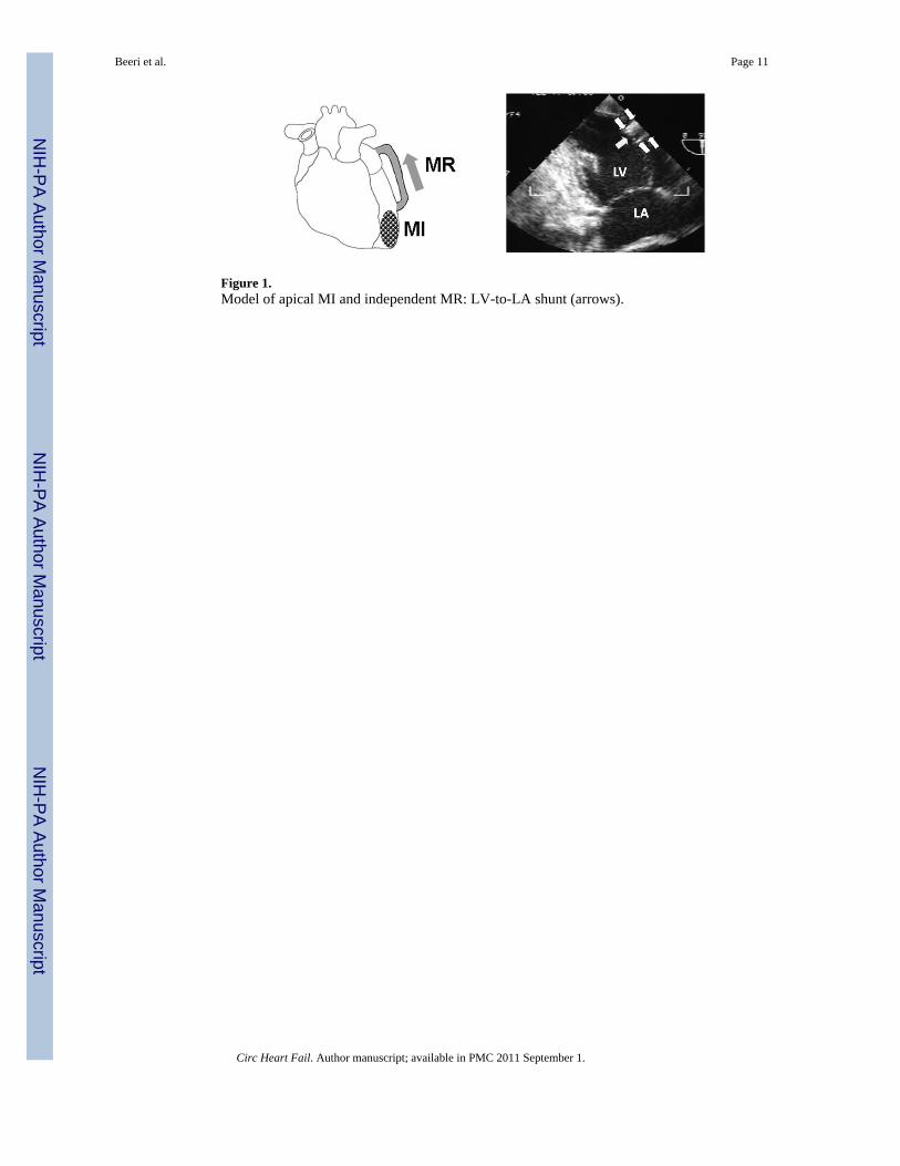
Figure 1.Model of apical MI and independent MR: LV-to-LA shunt (arrows).
Beeri et al. Page 11
Circ Heart Fail. Author manuscript; available in PMC 2011 September 1.
NIH
-PA Author Manuscript
NIH
-PA Author Manuscript
NIH
-PA Author Manuscript
Beeri et al. Page 12
Circ Heart Fail. Author manuscript; available in PMC 2011 September 1.
NIH
-PA Author Manuscript
NIH
-PA Author Manuscript
NIH
-PA Author Manuscript
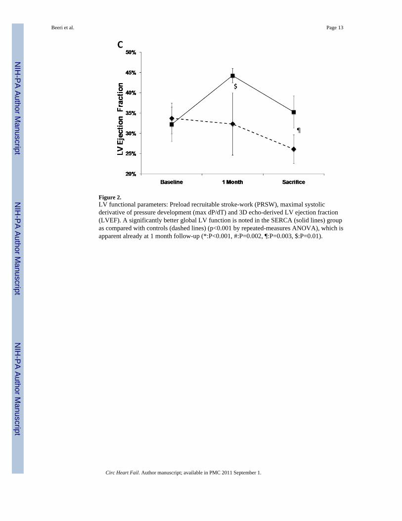
Figure 2.LV functional parameters: Preload recruitable stroke-work (PRSW), maximal systolicderivative of pressure development (max dP/dT) and 3D echo-derived LV ejection fraction(LVEF). A significantly better global LV function is noted in the SERCA (solid lines) groupas compared with controls (dashed lines) (p<0.001 by repeated-measures ANOVA), which isapparent already at 1 month follow-up (*:P<0.001, #:P=0.002, ¶:P=0.003, $:P=0.01).
Beeri et al. Page 13
Circ Heart Fail. Author manuscript; available in PMC 2011 September 1.
NIH
-PA Author Manuscript
NIH
-PA Author Manuscript
NIH
-PA Author Manuscript
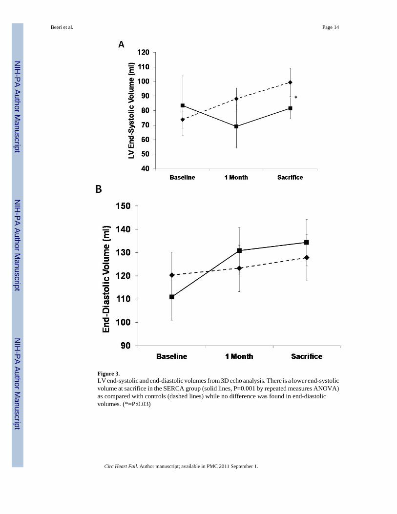
Figure 3.LV end-systolic and end-diastolic volumes from 3D echo analysis. There is a lower end-systolicvolume at sacrifice in the SERCA group (solid lines, P=0.001 by repeated measures ANOVA)as compared with controls (dashed lines) while no difference was found in end-diastolicvolumes. (*=P:0.03)
Beeri et al. Page 14
Circ Heart Fail. Author manuscript; available in PMC 2011 September 1.
NIH
-PA Author Manuscript
NIH
-PA Author Manuscript
NIH
-PA Author Manuscript
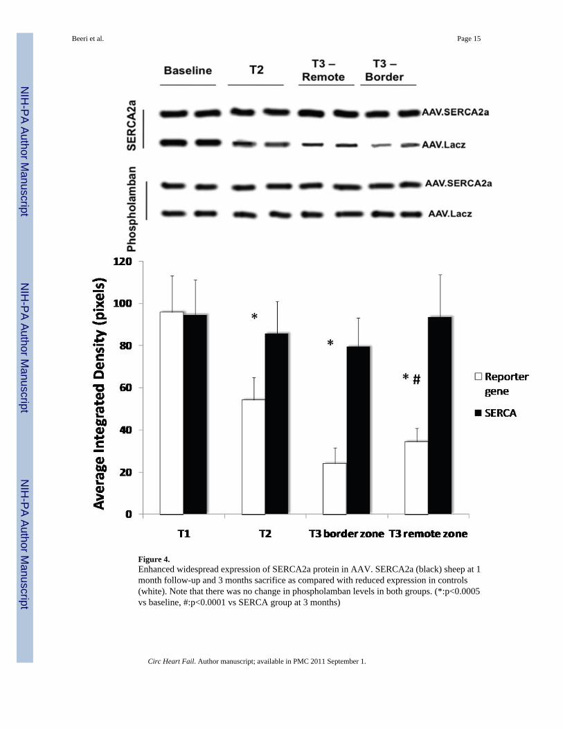
Figure 4.Enhanced widespread expression of SERCA2a protein in AAV. SERCA2a (black) sheep at 1month follow-up and 3 months sacrifice as compared with reduced expression in controls(white). Note that there was no change in phospholamban levels in both groups. (*:p<0.0005vs baseline, #:p<0.0001 vs SERCA group at 3 months)
Beeri et al. Page 15
Circ Heart Fail. Author manuscript; available in PMC 2011 September 1.
NIH
-PA Author Manuscript
NIH
-PA Author Manuscript
NIH
-PA Author Manuscript
Beeri et al. Page 16
Circ Heart Fail. Author manuscript; available in PMC 2011 September 1.
NIH
-PA Author Manuscript
NIH
-PA Author Manuscript
NIH
-PA Author Manuscript

Beeri et al. Page 17
Circ Heart Fail. Author manuscript; available in PMC 2011 September 1.
NIH
-PA Author Manuscript
NIH
-PA Author Manuscript
NIH
-PA Author Manuscript
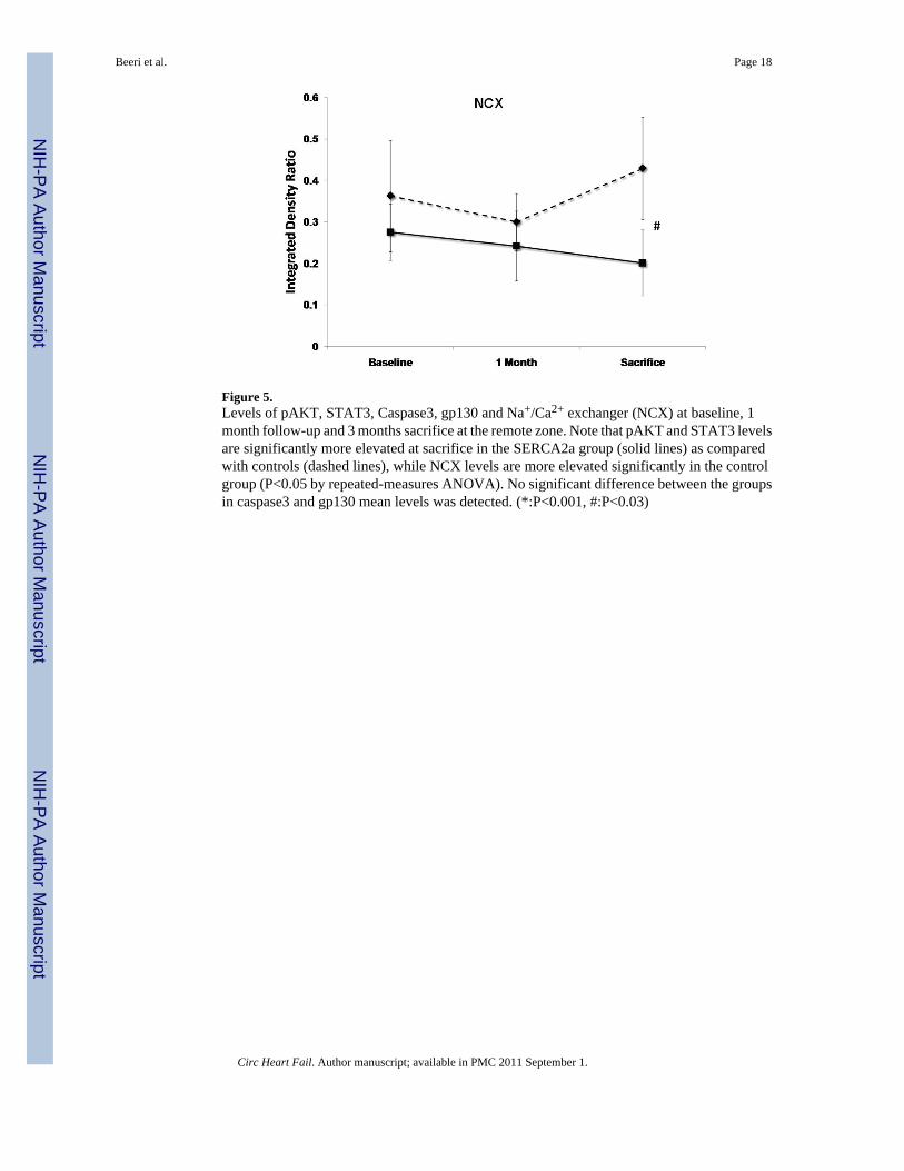
Figure 5.Levels of pAKT, STAT3, Caspase3, gp130 and Na+/Ca2+ exchanger (NCX) at baseline, 1month follow-up and 3 months sacrifice at the remote zone. Note that pAKT and STAT3 levelsare significantly more elevated at sacrifice in the SERCA2a group (solid lines) as comparedwith controls (dashed lines), while NCX levels are more elevated significantly in the controlgroup (P<0.05 by repeated-measures ANOVA). No significant difference between the groupsin caspase3 and gp130 mean levels was detected. (*:P<0.001, #:P<0.03)
Beeri et al. Page 18
Circ Heart Fail. Author manuscript; available in PMC 2011 September 1.
NIH
-PA Author Manuscript
NIH
-PA Author Manuscript
NIH
-PA Author Manuscript