Mitral Regurgitation
30
From: Contemporary Cardiology: Essential Echocardiography: A Practical Handbook With DVD Edited by: S. D. Solomon © Humana Press, Totowa, NJ 255 Mitral Regurgitation Jacqueline Suk Danik, MD, MPH and Bernard E. Bulwer, MD, MSC CONTENTS INTRODUCTION ASSESSMENT OF MR ETIOLOGY AND MECHANISMS/CASE PRESENTATIONS CASE PRESENTATIONS ASSESSMENT OF MR SEVERITY TWO-DIMENSIONAL ECHOCARDIOGRAPHIC P ARAMETERS FOR GRADING MR SEVERITY DOPPLER METHODS FOR GRADING MR SEVERITY COLOR FLOW DOPPLER P ARAMETERS OTHER DOPPLER METHODS INTEGRATING INDICES OF SEVERITY SURGICAL CONSIDERATIONS IN MR/CASE PRESENTATIONS CASE PRESENTATIONS TEE AND V ALVE ANALYSIS SUGGESTED READING 14 INTRODUCTION Mitral regurgitation (MR) is one of the most com- mon acquired valvular heart diseases seen in the adult echocardiography practice. Its onset may be acute or chronic, and the etiology can stem from any process that disturbs the architecture of the mitral valve appara- tus (Table 1; Figs. 1 and 2). Echocardiography has several important roles in MR, including: 1. Evaluation of the etiology of MR. 2. Grading the severity of MR. 3. Assessment of its impact on overall cardiac function, especially left ventricular (LV) function. 4. Guidance for further management, including timing of surgical intervention. Transthoracic echocardiography (TTE) is the modal- ity of choice for evaluation and follow-up of MR and global cardiac function, but transesophageal echocardio- graphy (TEE) plays an important role when differences exist between clinical assessment and TTE findings, or when mitral valve surgery is needed (Table 2). ASSESSMENT OF MR ETIOLOGY AND MECHANISMS The structure of the entire mitral valve apparatus which include the mitral leaflets, chordal attachments, annulus, papillary muscles, and the supporting ventric- ular walls affect overall function. Defining the etiology of MR is important as it can influence management and prognosis of such patients. Diseases that are primarily structural in origin can be managed differently from individuals with functional MR. The cases that follow were chosen to highlight sev- eral common etiologies of MR. The most common causes of MR in North America include degenerative disease, ischemia, and functional MR. CASE PRESENTATION 1: TRACE MITRAL REGURGITATION A 26-yr-old woman was referred for TTE follow- ing clinical detection of a faint systolic murmur. She was completely asymptomatic and had no significant medical history (Fig. 2A).
-
Upload
khangminh22 -
Category
Documents
-
view
8 -
download
0
Transcript of Mitral Regurgitation

From: Contemporary Cardiology: Essential Echocardiography: A Practical Handbook With DVDEdited by: S. D. Solomon © Humana Press, Totowa, NJ
255
Mitral Regurgitation
Jacqueline Suk Danik, MD, MPH and Bernard E. Bulwer, MD, MSC
CONTENTS
INTRODUCTION
ASSESSMENT OF MR ETIOLOGY AND MECHANISMS/CASE PRESENTATIONS
CASE PRESENTATIONS
ASSESSMENT OF MR SEVERITY
TWO-DIMENSIONAL ECHOCARDIOGRAPHIC PARAMETERS FOR GRADING MR SEVERITY
DOPPLER METHODS FOR GRADING MR SEVERITY
COLOR FLOW DOPPLER PARAMETERS
OTHER DOPPLER METHODS
INTEGRATING INDICES OF SEVERITY
SURGICAL CONSIDERATIONS IN MR/CASE PRESENTATIONS
CASE PRESENTATIONS
TEE AND VALVE ANALYSIS
SUGGESTED READING
14
INTRODUCTION
Mitral regurgitation (MR) is one of the most com-mon acquired valvular heart diseases seen in the adultechocardiography practice. Its onset may be acute orchronic, and the etiology can stem from any processthat disturbs the architecture of the mitral valve appara-tus (Table 1; Figs. 1 and 2).
Echocardiography has several important roles in MR,including:
1. Evaluation of the etiology of MR.2. Grading the severity of MR.3. Assessment of its impact on overall cardiac function,
especially left ventricular (LV) function.4. Guidance for further management, including timing
of surgical intervention.
Transthoracic echocardiography (TTE) is the modal-ity of choice for evaluation and follow-up of MR andglobal cardiac function, but transesophageal echocardio-graphy (TEE) plays an important role when differencesexist between clinical assessment and TTE findings, orwhen mitral valve surgery is needed (Table 2).
ASSESSMENT OF MR ETIOLOGY AND MECHANISMS
The structure of the entire mitral valve apparatuswhich include the mitral leaflets, chordal attachments,annulus, papillary muscles, and the supporting ventric-ular walls affect overall function. Defining the etiologyof MR is important as it can influence management andprognosis of such patients. Diseases that are primarilystructural in origin can be managed differently fromindividuals with functional MR.
The cases that follow were chosen to highlight sev-eral common etiologies of MR. The most commoncauses of MR in North America include degenerativedisease, ischemia, and functional MR.
CASE PRESENTATION 1:TRACE MITRAL REGURGITATION
A 26-yr-old woman was referred for TTE follow-ing clinical detection of a faint systolic murmur. Shewas completely asymptomatic and had no significantmedical history (Fig. 2A).

256 Suk Danik and Bulwer
The images show trace (trivial) MR. There is acentrally directed jet confined to an area just distalto the mitral leaflet closure line, seen in multiplewindows. It is of short duration and low velocityand may simply represent volume displacementwhen the mitral leaflets close. This can be seen inup to 80% of normal adults increases in prevalencewith age and has little pathological significance.
CASE PRESENTATION 2:MITRAL VALVE PROLAPSE
The second case is a 33-yr-old woman with amidsystolic click and an end-systolic murmur
(Figs. 3–7; please see companion DVD for corre-sponding video for Figs. 3–6).
These images show classic mitral valve prolapse(MVP), one of the most common cardiac valvularabnormalities, which has been reported in 1–3% ofNorth American populations. This particular exampledemonstrates prolapse of both anterior and posteriorleaflets past the mitral annular plane. The leaflets arethickened (>5 mm) and there is concomitant severeregurgitation.
Table 1Causes of MR
Mechanism Diagnosis
Acute MRStructural Ruptured chordae tendinae
Ischemic papillary muscle dysfunction and/or rupture
Infectious Bacterial endocarditis (both normal and prosthetic heart valves)Chronic MR
Degenerative/structural Myxomatous degeneration of leaflets (mitral valve prolapse)Chordal rupture with flail leafletPapillary muscle dysfunction and/or rupture with flail leafletParavalvular leak following prosthetic valve replacementMitral annular calcificationCollagen disorders, for example, Marfan syndrome and Ehlers Danlos syndrome
Ischemic and functional Dilated cardiomyopathy with mitral annular dilatation and apical tetheringInfectious Bacterial endocarditis (of both normal and prosthetic heart valves)Inflammatory Rheumatic heart disease
Connective tissue disorders, for example, systemic lupus erythematosus, scleroderma
Table 2Major Indications for Echocardiography in MR
• Diagnosis and evaluation of the etiology/mechanism of MR
• Asessment of hemodynamic severity, including impact on ventricular size, function, and hemodynamics
• Initial assessment and re-evaluation of asymptomatic andsymptomatic patients with MR
• Assessment of effects of medical and surgical therapies inMR, including mitral valve repair or replacement
• TEE for pre-operative, intra-operative, and post-operativeevaluation of patients with MR
Classic MVP exists when there is exaggerated (>2 mm)superior displacement (“buckling” or “hammocking”) ofthickened mitral leaflets (>5 mm thick in diastole)beyond the plane of the mitral annulus during late sys-tole. One of the most common reasons for MVP isfibromyxomatous degeneration of the mitral valve,which can lead to leaflet prolapse, chordal rupture, orpartial flail of a segment of one or both leaflets. Strictechocardiographic features of MVP are shown in(Table 3). The parasternal long-axis and the apicallong-axis views are the best views for the optimalassessment of MVP, and Figs. 4 and 5 (please see com-panion DVD for corresponding video) are classicexamples of MVP. Of note, the normal saddle-shapedprofile of the mitral valve leafleats may be exaggeratedwhen viewed from the apical four-chamber window,resulting in overdiagnosis of MVP or “echocardio-graphic heart disease.”

Chapter 14 / Mitral Regurgitation 257
Fig. 1. (A) Mitral valve topography: base of heart view. (B) Anterior and posterior mitral valve leaflets and scallops.
Fig. 2. Illustrations showing mitral valve leaflets, scallops (Carpentier nomenclature) and supporting structures. The mitral valvecomplex includes the mitral annulus, valve leaflets, chordae tendinae (primary, secondary, and tertiary), and their papillary muscleorigins, as well as the supporting left ventricle, left atrium, and the aortic root.

258 Suk Danik and Bulwer
Caveats about detection of mitral valve leaflets includemistaking myxomatous or floppy valves for vegetationsor masses (see Chapter 19, Fig. 11). MR may be absent atrest, but can be precipitated by exercise in some patientswith MVP, so exercise stress echocardiography may beuseful for diagnosis.
Although echocardiography is useful in definingMVP, the MVP syndrome includes associated clinicalfeatures and auscultatory findings. Furthermore, thepathology underlying the primary MVP may alsoaffect other intracardiac valves, and assessment forconcomitant prolapse of the tricuspid, pulmonary,
Fig. 3. Parasternal long-axis (PLAX) view showing trace mitral regurgitation (arrow). It is also termed “mitral regurgitation closingvolume”—indicating mere volume displacement during leaflet closure. (Please see companion DVD for corresponding video.)
Fig. 4. Systolic frame of the parasternal long-axis (PLAX) shows significant prolapse (>2 mm) of both mitral valve leaflets above themitral annular plane. (Please see companion DVD for corresponding video.)

and aortic valves should be performed during theechocardiographic examination.
Important potential sequelae of MVP include pro-gressive MR, potentially from progressive leaflet mal-coaptation, disintegration of chordal integrity overtime, and infective endocarditis.
CASE PRESENTATION 3:POST-MI MITRAL REGURGITATION
This 66-yr-old male with myocardial infarction(MI) 1 yr previously presented with congestive cardiac failure.
Chapter 14 / Mitral Regurgitation 259
Fig. 5. During diastole, the myxomatous leaflets can be measured. Thickened leaflets more than 5 mm support the diagnosis ofclassic MVP. (Please see companion DVD for corresponding video.)
Fig. 6. Bileaflet prolapse is easily visualized in the apical four-chamber (A4C) view. However, the mitral valve profile is normallyexaggerated in this view—and may lead to overdiagnosis of mitral valve prolapse (left panel). Severe mitral regurgitation was pres-ent (right panel). (Please see companion DVD for corresponding video.)

260 Suk Danik and Bulwer
Fig. 7. M-mode echocardiogram of patient (Figs. 4–6) with classic mitral valve prolapse. (A) Both thickened valve leaflets show pos-terior displacement of the C, D segment during mid- to late systole. (B) Only the posterior leaflet is affected.
Fig. 8. Mitral regurgitation in dilated ischemic cardiomyopathy. This results in apical tethering (“tenting”) of mitral valve leaflets dur-ing ventricular systole (A–C), impaired leaflet coaptation, and mitral regurgitation (B). “Tenting” is best seen in C and D (arrows). Thisterm describes closure of the mitral valve well within the left ventricular cavity rather than at the level of the mitral annular plane (C).Valve leaflet morphology is normal, but left ventricular dilation (D) leads to lateral displacement of papillary muscles (see Fig. 5). Thiscauses leaflet malcoaptation that is typically symmetric (C), and the jet of ischemic mitral regurgitation is centrally directed (A,B).

Chapter 14 / Mitral Regurgitation 261
Fig. 9. The mechanism of mitral regurgitation in dilated ischemic cardiomyopathy. The three major contributors as shown preventnormal leaflet coaptation and functional mitral regurgitation. The mitral leaflets are morphologically normal.
Table 3Echocardiographic Features of MVP
• Superior displacement of botha mitral valve leaflets (≥ 2 mma above mitral annulus) with coaptation point at or above the mitral annular plane.
• Diffuse leaflet thickening (>5 mma) in diastole (“myxomatous” appearance)
• Mitral annular dilatation
• Redundant mitral valve leaflets
• Chordal rupture with leaflet flail (partial)
• Mitral regurgitation
• Characteristic M-mode findings (Fig. 4A)aStrict criteria for classical MVP or “floppy mitral valve.”
The images (Figs. 8 and 9) show MR that resultfrom left ventricular remodeling and dilatation.Such changes to LV geometry significantly changethe spatial relationships between mitral leaflets,papillary muscles, and chordae tendinae. In thisexample, lateral displacement of the papillary mus-cles and chordae lead to poor leaflet coaptation andMR from LA dilatation. The resulting MR is cen-trally directed.
CASE PRESENTATION 4:FLAIL MITRAL LEAFLET
A 44-yr-old man complained of increasingshortness of breath and was found to have a mur-mur consistent with MR (Fig. 10; please see com-panion DVD for corresponding video).
These echocardiographic images illustrate howcomplete or partial flail of one or both mitral leafletscan lead to moderate to severe MR. Partial leafletflail most commonly involves the posteriorleaflet and is usually secondary to chordal rupture.The resultant MR may be severe, and the jet is typi-cally directed anteriorly. Conversely, an anteriorleaflet flail usually results in a posteriorly directedjet of regurgitation (see Chapter 7, Fig. 7).
Papillary muscle rupture in the setting of acute MI canpresent with acute severe regurgitation within the first 5 days post-MI. It more commonly involves dehiscenceof the tip or a head of the muscle, and less commonly,complete transection (Fig. 11; please see companionDVD for corresponding video). In particular, the postero-medial papillary muscle is more susceptible to injury dur-ing infarction as its blood supply is derived almost solelyfrom the posterior descending branch of the right coro-nary artery. The antero-lateral papillary muscle and thoseof the right ventricle have a dual blood supplies.
Different degrees of MR may result from ischemicdysfunction of the papillary muscles and supportingventricular wall. This is seen in approx 30% of patientswith coronary artery disease being considered for car-diac surgery.
Another example of disruption of mitral valvearchitecture is seen with degenerative mitral valvedisease. For example, with mitral annular calcifica-tion, which can be seen with older patients, the excur-sion of the mitral leaflets can be limited, affectingleaflet coaptation. This also results in MR.

262 Suk Danik and Bulwer
Fig. 10. (A) Parasternal long-axis view (PLAX) and apical four-chamber view (A4C) showing a partial flail of posterior mitral valveleaflet. Note that tip of leaflet points toward the left ventricle indicating that flail is incomplete (top panels). Marked turbulence withthis valvulopathy are seen on color flow Doppler examination. Note that the mitral regurgitant jet is anteriorly directed. M-modeexamination shows that flail affected the posterior leaflet only. (Please see companion DVD for corresponding video.)

CASE PRESENTATION 5:RHEUMATIC MITRAL REGURGITATION
A 39-yr-old female with known history of rheu-matic heart disease and worsening shortness of breathwas referred for an echocardiogram prior to consider-ation for percutaneous mitral balloon valvuloplasty.
Rheumatic heart disease is the major underlyingcause of mitral stenosis. The postinflammatory
Chapter 14 / Mitral Regurgitation 263
Fig. 11. Ruptured papillary muscle. (A) Transthoracic images a patient 5 d post-MI showed a mobile linear echodensity that ping-ponged between left atrium and left ventricle with each cardiac cycle (left panel). Attached at its end was the avulsed tip (head) ofthe posterior-medial papillary muscle. Right panel showed large postero-laterally directed MR jet as seen in from the apical four-chamber view. (B) Mid-esophageal views from patient showed the mobile tip of the dehisced papillary muscle (arrow in both panels)within the left atrium. Note the partial flail of the anterior mitral valve leaflet (left panel) with the resultant postero-laterally directedMR jet. (Please see companion DVD for corresponding video.)
Table 4Indicators for Poor Prognosis in MR
1. Symptoms of heart failure
2. Left ventricular ejection fraction (LVEF) < 50% withsymptoms
3. Acute-onset MR
4. Acute flail of mitral valve leaflets
5. Significant MR accompanying acute myocardial infarction
264 Suk Danik and Bulwer
Fig. 12. (A) Mitral regurgitation in the setting of rheumatic heart disease is the result of structural distortion, thickening, and restrictedleaflet motion. Mitral stenosis (MS) and aortic regurgitation (AR) were also present in this patient. (B) This color M-mode throughthe left atrium and ventricle in the same patient shows mixed mitral stenosis-mitral regurgitation jets.

Chapter 14 / Mitral Regurgitation 265
ASSESSMENT OF MR SEVERITY
A comprehensive assessment of a patient with MRshould pay special attention to ventricular performance,with emphasis on the parameters of LA systolic func-tion (see Chapter 5).
In addition to defining etiological and anatomicmechanisms of MR, assessment of MR severity and its
Table 5Grading MR Severity: Recommended Parameters
Mild Moderate Severe
Structural parameters
Left atrial size N N or Dilated Usually D
Left ventricular size N N or Dilated Usually D
Mitral leaflets and N or Abnormal N or abnormal Abnormal/Flail supporting complex leaflet/Ruptured
papillary muscle
Doppler parameters
Color jet area Small, central jet Variable Large central jet (usually(Niquist limit 50–60 cm/s) (usually <4 cm2 Variable >10 cm2 or >40% left atrium
or <20% of left atrium area) area) or variable sizewall-impinging jetswirling in left atrium
Mitral E- point velocity – PW A-wave dominant Variable E-wave dominant (E usually >1.2 m/)
Jet density – CW Incomplete or faint Dense Dense
Jet contour – CW parabolic Usually parabolic Early peaking-triangular
Pulmonary vein flow – PW Systolic dominance Systolic blunting Systolic flowreversal
Quantitative parameters
Vena contracta width (cm) <0.3 cm 0.3–0.69 cm ≥0.7 cm
Regurgitant volume (mL/beat) <30 30–59 ≥60 mL/beat
Regurgitant fraction (%) <30% 30–49% ≥50%
EROA (effective regurgitant <0.2 cm2 0.2–0.29 0.3–0.39 cm2 ≥0.4 cm2
area orifice)
Modified from ASE, American Society of Echocardiography Report (2003)—ASE/ACC/AHA/ESC Guidelines. See ref. 3.
hemodynamic impact on risk stratification and timingof intervention (Table 4).
The American Society of Echocardiography guide-lines for the classification of grades of MR severityare shown in Table 5. Although the recommendationto grade MR severity is mild, moderate and severe,distinguishing mild from moderate and moderate fromsevere can be challenging. Thus, several qualititativeand quantitive measures have been proposed, althoughlimitations of each should be noted during their use;the most popular methods in use will be further delin-eated in this chapter. For example, most of the Doppler-based parameters apply only to isolated MR, and not tomixed valvular lesions, or MR secondary to LV dys-function. Quantitative assessment of MR using thesemethods may involve relatively complex calculations,
fibrosis and thickening of leaflets and chordae thatlead to mitral stenosis also prevent normal mitralleaflet coaptation during systole. Combined mitralstenosis-MR is the result. (Fig. 12; see Chapter 13,Table 3).

266 Suk Danik and Bulwer
and small errors in measurement easily lead to largeerrors in calculation. Therefore, they should beapplied in the context of the patient, without relianceon a single measure.
TWO-DIMENSIONALECHOCARDIOGRAPHIC PARAMETERS
FOR GRADING MR SEVERITY
LV PerformanceIndices of LV systolic function—left ventricular
end-systolic and end-diastolic dimensions, ejectionfraction, wall thickness, and fractional shortening—are
the most important echocardiographic barometers ofthe hemodynamic effects of MR on global cardiacfunction (see Chapter 5 for LV quantification).
Three major phases of MR on and their impact onLV performance are summarized in Fig. 13.
1. Acute MR phase.2. Chronic compensated MR.3. Chronic decompensated MR.
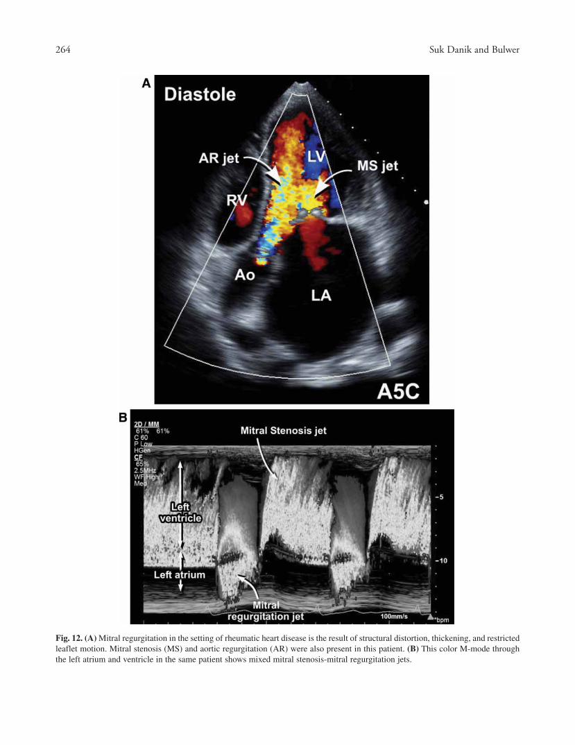
LV end-systolic dimensions (Fig. 14) and LV ejectionfraction (LVEF) reflect the heart’s ability to adapt, andtherefore influences the frequency of clinical follow-up,timing of surgical intervention, and outcomes following
Fig. 13. Mitral regurgitation and the left ventricle: three phases of dysfunction. (Modified from Carabello BA. Progress in mitral andaortic regurgitation. Curr Probl Cardiol 2003;28:553.)
Table 6Recommended Echocardiography Follow-up in Chronic Asymptomatic MR
MR severity LV size LVEF Frequency of follow-up
Mild MR ESD normal Normal EF 5 yr
Moderate MR ESD normal Normal EF 1–2 yr
Moderate MR ESD > 40 mm <65% 1 yr
Severe MR ESD normal Normal EF 1 yr
Severe MR ESD > 40 mm <65% 6 mo
Adapted from: Otto CM. Evaluation and management of chronic asymptomatic mitral regurgitation. N Engl.J Med 2001;345:740–746.
mitral valve surgery (Tables 6 and 7). In chronic compen-sated MR, cardiac output is maintained via an increase inthe LVEF, and such patients typically have LVEFs greaterthan 65%. LV hypertrophy is not a feature of isolatedMR as the regurgitant chamber—the left atrium (LA)—usually adapts and dilates to accommodate increases inpreload (end-diastolic volume [EDV]; Fig. 13C). Evenin the acute setting, LV contractility and LVEF increasein response to an increase in preload (Fig. 13B).However, LV contractility can decrease silently and irre-versibly in chronic MR. For this reason, increasingly ear-lier surgical interventions for less severe degrees of MRare being recommended (Fig. 13D).
LA SizeThe LA will dilate in response to chronic volume
and pressure overload. Its dimensions may help toassess MR severity and chronicity (Figs. 15 and 16).Acute-onset severe MR, as occurs with papillary mus-cle rupture, does not cause LA dilatation. The excess
regurgitant blood entering the small noncompliantatrium causes acute increase in LA pressures and canprecipitate acute pulmonary hypertension and rightheart pressure overload. Increased LA pressures andsystolic flow into reversal pulmonary veins may bethe only echocardiographic findings that documentthe severity of acute-onset MR. LA size may predictthe onset of atrial fibrillation, but is otherwise of littleprognostic value in MR itself, in the absence of heartfailure. Measurements should be adjusted for age and body surface area.
DOPPLER METHODS FOR GRADING MR SEVERITY
Color Flow Doppler ParametersColor flow Doppler imaging is perhaps the most intu-
itive of all measures, is useful for detecting the jet origin,direction, and spatial relationships and has excellentsensitivity and specificity. Each of the three components
Chapter 14 / Mitral Regurgitation 267
Table 7Echocardiographic Signs and Surgical Considerations in MR
Variables Measures Comment
Clinical symptoms Symptomatic vs Asymptomatic Presence of symptoms favors earlier intervention
LVEF <50 mm Sign of decompensation
ESD (LVIDs) <50–55 mm Sign of decompensation
EDD (LVIDd) >70 mm Sign of decompensation
Surgical considerations Repair vs Replacement Earlier interventions with valve-sparing surgery favored
Fig. 14. Left ventricle dimensions in mitral regurgitation. Values from cardiac ultrasound laboratory, Massachusetts General Hospital.

268 Suk Danik and Bulwer
of the MR jet—flow convergence zone, vena contracta,and jet profile—can provide a semiquantitative or quanti-tative measure of MR severity (Fig. 17).
MR jets are best assessed using multiple windows toobtain a three-dimensional (3D) perspective. Qualitativeestimates of MR jets are categorized on a scale of 0–4:grade 0 = none or trace MR, grade 1 = mild MRthrough to grade 4 = severe MR (Fig. 18). The notation
1+, 2+, and so on, to denote increasing grades of MRseverity is also popular (Table 8).
COLOR JET AREA
The same color jet profiles can be measured withinthe LA. Color jet areas are influenced by jet velocity,momentum, and direction. Mild MR jets cover less than20% of total LA area (or a maximal jet area < 4.0 cm2),
Fig. 15. (A) Measuring left atrial size. (B) Left atrial measurements. Values from cardiac ultrasound laboratory, MassachusettsGeneral Hospital.

Chapter 14 / Mitral Regurgitation 269
Fig. 16. Left atrial volume. (Reproduced with permission from Roberto M. Lang et al. Recommendations for chamber quantification.A report from the American Society of Echocardiography’s Nomenclature and Standards Committee and the Task Force on ChamberQuantification American Society of Echocardiography, 2005.)
Fig. 17. Mitral regurgitation: color jet profile.
Table 8Eyeball Estimation of Color Jet Area in MR
Advantages Disadvantages
1. Ease of use 1. Can be unreliable for estimation of regurgitation severity
2. Good screening test for mild versus severe regurgitation 2. Not good at distinguishing moderate MR from mild or severe MR
3. Evaluate in at least 2 views (PLAX, A4C, A3, or A2C) 3. Influenced by transducer frequency and other instrumentsettings, especially pulse repetition frequency and color gain
4. Best “estimate” using scale of 1+ (mild) to 4+ (severe) 4. Size of color jet may be misleading, especially witheccentric jets

270 Suk Danik and Bulwer
with severe MR jets more than 40% of total LA area (ora maximal jet area > 10 cm2). At least two orthogonalviews should be used with the Nyquist limit set at50–60 cm/s (Fig. 19; please see companion DVD forcorresponding video).
Larger color jet areas indicate more severe MR whenthe jet is centrally directed, but can be misleading witheccentrically directed jets. Hugging or entrainment(Coanda effect) of the eccentric jet to the LA wallresults in smaller jet areas even when MR is severe. Athorough evaluation of eccentrically directed jets shouldinclude evaluation for etiologies, such as a flail leaflet,prolapse or perforation. Severe MR with eccentricallydirected jets sometimes exhibit a “wrap around” effectin the LA (Fig. 20).
In acute MR, even centrally directed jets may bemisleadingly small. A small nondilated atrium in thesetting of acute regurgitation constrains the regurgitantjet momentum and hence the visible color jet area.
VENA CONTRACTA WIDTH
The vena contracta is the narrow neck of the MR jet asit traverses the regurgitant orifice (Fig. 21). The vena con-tracta measured from the parasternal long-axis view is bestoptimized by using a narrow sector scan, optimal colorgain, and Nyquist limit between 40–70 cm/s. The venacontracta appears as the well-defined light blue or light
yellow high-velocity core on the red-blue color Dopplerscale. This portion of the regurgitant jet, unlike the flowconvergence zone and the distal turbulent jet profile, mostclosely mirrors that of the actual regurgitant orifice. It is,therefore, a more reliable marker of MR severity with sig-nificant advantages over other methods provided that therecommended technique is used (Table 9).
A single vena contracta width (VCW) measurement,however, is a 2D snapshot across an elongate regurgi-tant orifice area that extends to a variable degree alongthe crescenticleaflet coaptation line (Fig. 1B). A closerapproximation of the actual regurgitant orifice area isbest obtained by scanning through multiple planes andselecting the greatest VCW. Such considerations arebetter appreciated and measured on 3D echocardiogra-phy. Averaging VCW measurements over at least threebeats and using two orthogonal planes is recom-mended. A VCW less than 0.3 cm indicates mild MR;a VCW more than 0.7 cm indicates severe regurgitation(Fig. 21; Table 5).
The effective regurgitant orifice area (EROA)—amarker of MR severity that is less affected by loadingconditions—can be calculated from the VCW using theformula:
EROA = π(VCW/2)2
Good agreement exists between this EROA formulaand other validated measures of MR severity. VCWmeasurements are not valid for assessing MR severitywith multiple MR jets (Fig. 22).
PROXIMAL FLOW CONVERGENCE
AND PROXIMAL ISOVELOCITY SURFACE AREA
According to fluid dynamics, fluids within an enclo-sure stream symmetrically toward a narrowed outlet ororifice in near concentric isovelocity zones. This is akinto water being drained from a kitchen sink, or when waterfrom a lake flows into a narrowed estuary (Fig. 23). Thesame principle applies when blood in the LV stream con-verges toward a narrowed (stenosed or regurgitant) ori-fice. This method can be used for estimating the area ofthe regurgitant orifice—which is hard to measure directlybecause actual regurgitant orifice is dynamic, functional,and 3D. As regurgitant blood converges toward the regur-gitant orifice at the proximal convergence zone, the sizeand velocity of the innermost shell or hemisphere can bemeasured (Fig. 24).
Furthermore, according to the continuity principle(see Chapter 11, Fig. 11), the amount of fluid thatpasses through the regurgitant orifice is the same
Fig. 18. Schema illustration of jet profile of mitral regurgitationas viewed from the apical four-chamber window. Color Dopplermapping allows visualization of the spatial distribution of bloodflow within the heart by displaying the blood flow velocities interms of ranges of color. Visual estimation of the jet profile is asimple method of estimating mitral regurgitation severity. ColorDoppler mapping, however, is very sensitive to instrument set-tings, hemodynamic status, regurgitant orifice geometry (affectsjet eccentricity), and atrial dynamics—e.g., wall constraint, wallimpingement, and the presence of secondary flow.

Chapter 14 / Mitral Regurgitation 271
amount that flows in the regurgitant jet (the law of con-servation of mass). Therefore, total flow at the proximalisovelocity surface area (PISA) will equal total flow inthe distal MR jet.
The apical four-chamber view is recommended foroptimal visualization of the MR jet PISA measurement.
The area of interest is optimized by lowering imagingdepth and lowering the Nyquist limit (on the colorDoppler scale) to approx 40 cm/s. The velocity atwhich the blue-red color shift occurs identifies thePISA shell. The PISA radius (r) is then measured andmultiplied by the PISA velocity, i.e., the aliasing velocity
Fig. 19. Color flow jet areas. (Please see companion DVD for corresponding video.)
Fig. 20. Severe mitral regurgitation showing left atrial “wrap-around.”

272 Suk Danik and Bulwer
Table 9Vena Contracta Width Measurements in MR
Advantages Disadvantages
1. Relatively quick and easy to assesses using standard 1. Not good at distinguishing mild from moderate MR windows or moderate from severe MR
2. Good for extremes of MR (mild and severe MR) 2. Small values; small measurement errors are multiplied
3. Assesses basic size of defect 3. True cross-sectional area may be difficult to obtain—usetwo apical diameteres
4. Relatively independent of flow rate, driving pressure, 4. VCW measurement—a single temporal measurementor entrainment (Coanda effect)
5. Not influenced by the presence of another regurgitant 5. Overestimates true regurgitant orifice area—a problem ofleak, for example, aortic regurgitation resolution
6. No need for correction for convergence angle as with 6. Not valid for multiple MR jetsproximal isovelocity suface area measurement
MR, mitral regurgitation.
Fig. 21. Vena contracta width measurement.
(Nyquist limit)—VALIAS —to give the regurgitant flowrate (Fig. 24). If the base of the PISA hemisphere is nothorizontal, it should be corrected to 180°.
Reguritant flow rate = 2πr2 × VALIAS (mL/s)
From this, the EROA can be quantified using thecontinuity principle equation (Figs. 25 and 26; seeChapter 11, Fig. 11) for flow rate: Area1 × Velocity1 =Area2 × Velocity2; and VMAX is the peak velocity of theMR jet on CW Doppler:
EROA = 2πr2 × VALIAS/VMAX
Regurgitant volume and the regurgitant fraction canthen be calculated (Fig. 27).
The PISA method can also be assessed by trans-esophageal echocardiography when indicated (Fig. 28).
The PISA method makes several assumptions(Table 10)—many of which are violated in the clinicalsetting. 3D echocardiography may ultimately assist inovercoming some of these limitations.
Other Doppler MethodsSYSTOLIC FLOW REVERSAL IN THE PULMONARY VEINS
The presence and the degree of reversal of bloodflow from the LA into the pulmonary veins can indicatethe hemodynamic impact of the MR jet. Visualizationof flow reversal into one or more pulmonary veins on

Chapter 14 / Mitral Regurgitation 273
color flow Doppler, or more reliably—pulsed Dopplerevidence of flow reversal into the pulmonary veins aremeasures of MR severity (Fig. 29). In normal indivi-duals, a positive systolic (S) wave followed by a smallerpositive diastolic (D) wave is seen, but with moderateor severe MR, blunting or reversal of this pattern maybe seen (see Chapter 6). Systolic flow reversal may beindicative of severe MR even if the color jet area sug-gests milder disease.
Atrial fibrillation and elevated LA pressures fromany cause can blunt forward systolic pulmonary veinflow. Blunting of pulmonary forward flow may lackspecificity, but is nonetheless a useful parameter thatprovides information independent from color Dopplermethods used to assess MR severity.
MITRAL E-POINT VELOCITY
The progressive increase in trans-mitral flow thatoccurs with increasing MR severity can be detected ashigher flow velocities during early diastolic filling. Adominant E-wave more than 1.2 m/s may indicate moresevere MR, providing there is no concomitant mitralstenosis (Fig. 30).
CONTINUOUS-WAVE JET INTENSITY
AND MORPHOLOGY
Peak MR jet velocities by continuous-wave (CW)Doppler typically range between 4 and 6 m/s—areflection of the systolic pressure gradient between LVand LA. If the blood pressure at the time of the study islow, the peak velocities and gradients will also be low.Peak MR jet velocities alone, therefore, are not reliablemeasures of MR severity.
The signal intensity (jet density) of the CW envelopeof the MR jet can be a guide to MR severity, but thisshould be assessed relative to the density of antegradeflow signal (mitral inflow). A dense mitral regurgitantsignal with a full envelope of equal in intensity to theantegrade flow signal indicates more severe regurgita-tion than a faint signal (Fig. 31).
The CW Doppler envelope may show blunting ornotching of the CW envelope. This results from therapid surge in LA pressures in severe MR—the atrialV-wave (Fig. 32). This may reflect an LA that has notyet dilated and show discordance with color Dopplerseverity.
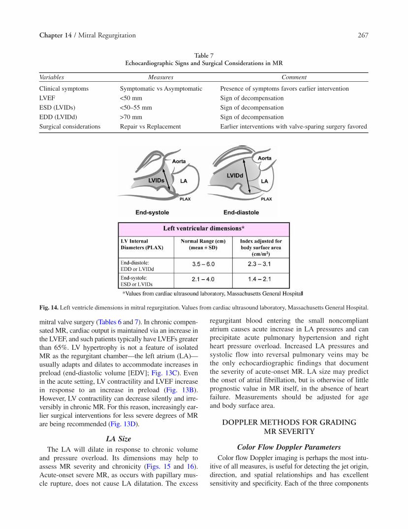
LV SYSTOLIC PERFORMANCE
The rate of rise of LV systolic pressure over time(dP/dT) may be a useful index of the LV systolic func-tion in MR (Fig. 33). In patients with preserved systolicfunction, the MR jet velocity shows a rapidly early insystole. A lower dP/dT can unmask patients withdeclining systolic function, and therefore serve as aguide to more aggressive intervention, especially whensupported by other indicators of severity.
Integrating Indices of SeverityIntegrated scores have been devised to improve the
diagnostic validity of parameters of MR severity. TheMR index is a composite score comprising six echocar-diographic parameters—jet length, PISA, CW jet den-sity, pulmonary artery systolic pressure, pulsed waveDoppler, pulmonary vein flow pattern, and LA size.Such scores provide a better overall assessment of MR
Fig. 22. Narrow sector scan (color flow Doppler) showing twoseparate mitral regurgitant (MR) jets viewed from the apicallong axis view. This was confirmed when viewed from multi-ple windows. When assessing multiple MR jets, each jetshould be analyzed and reported separately. Vena contractwidths (VCW) measurements are invalid when multiple MRjets are present.

274 Suk Danik and Bulwer
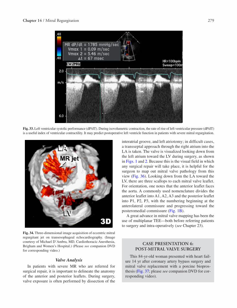
severity itself, but should be further integrated withglobal cardiac function and the patient’s clinical status.Newer methods of assessing MR severity show promise,e.g., 3D echocardiography and power Doppler imaging,but are not yet routinely used in clinical practice (Fig. 34; please see companion DVD for correspondingvideo).
SURGICAL CONSIDERATIONS IN MR
Anatomical Mechanisms of MRPrecise determination of the mechanism of MR
results is necessary for successful reconstructive sur-gery of the mitral valve. Thus, the Carpentier’s func-tional classification is often used (Fig. 35).
Fig. 23. The proximal isovelocity surface area method. Fluids within a confined space accelerate toward an outlet or orifice in con-centric isovelocities. A familiar example of this occurs when water is released from a kitchen sink.
Fig. 24. Proximal isovelocity surface area (PISA).

Chapter 14 / Mitral Regurgitation 275
Fig. 25. Mitral regurgitation severity: proximal isovelocity surface area (PISA) method. Step 1: select the best apical four-chamber view(A4C) to indentify the region of interest—the regurgitant mitral valve (panel 1). Step 2: lower imaging depth, then zoom (panel 2).Step 3: apply color Doppler, then narrow color sector scan (panel 3). Step 4: shift color flow baseline on color Doppler scale down-ward (to lower Nyquist limit) to approx 40 cm/s. This optimizes the aliasing velocities enables the PISA hemispheric zone bigger andmore measurable (panel 4). Step 5: record the Nyquist limit on the color Doppler Scale. This is the aliasing velocity of the PISA. Scrollthrough cine loop and optimize largest PISA (mid-systole) and measure radius (panel 5). Step 6: obtain the maximum velocity acrossthe mitral regurgitation jet using continuous-wave Doppler (panel 6). (Please see companion DVD for corresponding video.)
Fig. 26. Proximal isovelocity surface area (PISA) method: calculation of effective regurgitant orifice area (EROA) by the continuityequation. The PISA method is used to calculate the EROA according to the principle of conservation of mass. The continuity equa-tion is used to calculate the EROA from the variables measured in Fig. 25.
Carpentier’s Functional Approachto Mitral Regurgitation
The Carpentier functional classification of MR isbased on the opening and closing motions of bothleaflets. Normal mitral leaflet coaptation requires coor-dination of many structures including a normal-sized LV
and a chordal apparatus that is not calcified and is welltethered to the mitral leaflets that coapt well (Fig. 35).
Functional Classification: Type IMR that occurs despite normal leaflet motion is termed
type I MR. This can be owing to annular dilatation seen

276 Suk Danik and Bulwer
Fig. 27. Regurgitant volume and regurgitant fraction.
Fig. 28. (A) Transesophageal echocardiography (TEE) midesophageal view at omniplane 0° showing partial flail of posterior mitralvalve leaflet. (B) Color Doppler imaging shows severe mitral regurgitation with proximal convergence zone with an anteriorlydirected jet. Color Doppler scale set at 59 cm/s. (C) Color Doppler scale is then shifted upward (on TEE) to 43 cm/s showing clearlydefined proximal isovelocity surface area (PISA) measuring 0.966 cm, consistent with severe mitral regurgitation.
Table 10PISA Assumptions and Pitfalls
• Accurate measurements (subject to error, errors are squared, interobserver variability)
• Unconstrained flow (constrainment exists)
• Circular point-like orifice (regurgitant orifice irregular, often “smiley” shaped)
• Flat regurgitant orifice (actual orifice not flat)
• PISA is a hemisphere (PISA is more a hemi-ellipse)
• Constant orifice (PISA calculation is an instantaneous measurement, but PISA—like the cardiac cycle—is dynamic)
• PISA method not suitable for eccentric jets, or concomitant mitral stenosis

in cardiomyopathy. With cardiomyopathy, the annulusbecomes dilated and the exoskeleton of the LV becomesstretched. Despite preserved leaflet motion, the leafletsare unable to coapt because of the incompetent architec-ture of the LV and annulus. This often results in a centraljet of MR (Figs. 8 and 9). Another etiology of MRdespite preserved leaflet motion is leaflet perforation, asa sequelae of endocarditis.
Functional Classification: Type IIType II regurgitation refers to MR that occurs
because of leaflet prolapse. This may be owing to simple
elongation of the leaflets, with prolapse into the leftatrium. This may be owing to chordal rupture or papil-lary muscle rupture. An example of chordal rupture isshown in Fig. 35.
Functional Classification: Type IIIa RestrictedLeaflet Motion
Valvular and subvalvular thickening can restrictmitral leaflet motion. Mitral annular calcification andthickening of the subvalvular apparatus are seen withincreasing age, or as the sequelae of rheumatic heartdisease. The resulting poor coaptation results in MR.
Chapter 14 / Mitral Regurgitation 277
Fig. 29. Pulmonary vein systolic flow reversal. Pulsed Doppler examination right upper pulmonary vein flow in severe mitral regur-gitation shows reversal of pulmonary vein systolic flow, as depicted by the negative S-wave.
Fig. 30. Mitral E-point velocity. (A) Pulsed Doppler of normal mitral inflow characteristically shows an early diastolic E-wave fol-lowed by the atrial A wave. (B) In severe mitral regurgitation (MR), marked E-wave dominance is seen (>1.2 m/s), reflecting amarked increased in early diastolic flow—typical of severe MR.

278 Suk Danik and Bulwer
Fig. 35 shows an example of such thickening of thevalvular and subvalvular apparatus.
Functional Classification: Type IIIb RestrictedLeaflet Motion
When leaflet motion is restricted, with displace-ment of the papillary muscles, this is referred to astype IIIb MR. The most common culprit is a dilated
cardiomyopathy, where enlargement of LV chambersare accompanied by elongation of the papillary mus-cles and displacement of such muscles to the apex,relative to the mitral valve leaflets. Figure 35 showssuch a schema and still frame of an enlarged LV anddownwardly displaced papillary muscles. The result-ing mitral valve incompetence often results in a cen-trally directed jet of MR.
Fig. 31. Continuous-wave (CW) Doppler intensity. CW Doppler envelopes showing a higher intensity signal in moderate-to-severeregurgitation (A) compared to mild mitral regurgitation (B). Compare these densities to the antegrade (mitral inflow) signals abovethe baseline.
Fig. 32. Continuous-wave (CW) Jet with V-wave. Notching of the CW envelope can occur in severe mitral regurgitation, a reflectionof the large atrial V-wave (insert). Left ventricular systolic performance (dP/dT).
interatrial groove, and left atriotomy; in difficult cases,a transseptal approach through the right atrium into theLA is taken. The valve is visualized looking down fromthe left atrium toward the LV during surgery, as shownin Figs. 1 and 2. Because this is the visual field in whichany surgical repair will take place, it is helpful for thesurgeon to map out mitral valve pathology from thisview (Fig. 36). Looking down from the LA toward theLV, there are three scallops to each mitral valve leaflet.For orientation, one notes that the anterior leaflet facesthe aorta. A commonly used nomenclature divides theanterior leaflet into A1, A2, A3 and the posterior leafletinto P1, P2, P3, with the numbering beginning at theanterolateral commissure and progressing toward theposteromedial commissure (Fig. 1B).
A great advance in mitral valve mapping has been theuse of multiplanar TEE—both before referring patientsto surgery and intra-operatively (see Chapter 23).
Chapter 14 / Mitral Regurgitation 279
Fig. 33. Left ventricular systolic performance (dP/dT). During isovolumetric contraction, the rate of rise of left ventricular pressure (dP/dT)is a useful index of ventricular contractility. It may predict postoperative left ventricle function in patients with severe mitral regurgitation.
Fig. 34. Three-dimensional image acquisition of eccentric mitralregurgitant jet on transesophageal echocardiography. (Imagecourtesy of Michael D’Ambra, MD, Cardiothoracic Anesthesia,Brigham and Women’s Hospital.) (Please see companion DVDfor corresponding video.)
Valve AnalysisIn patients with severe MR who are referred for
surgical repair, it is important to delineate the anatomyof the anterior and posterior leaflets. During surgery,valve exposure is often performed by dissection of the
CASE PRESENTATION 6:POST-MITRAL VALVE SURGERY
This 84-yr-old woman presented with heart fail-ure 14 yr after coronary artery bypass surgery andmitral valve replacement with a porcine biopros-thesis (Fig. 37; please see companion DVD for cor-responding video).

280 Suk Danik and Bulwer
Fig. 35. Functional classification of mitral regurgitation (by Carpentier). (Figure courtesy of Dr. David Adams, Mount Sinai MedicalCenter, New York, NY.)
Fig. 36. Orientation of mitral valve leaflets and scallops (Carpentier’s nomenclature) during intra-operative (TEE) echocardio-graphy. The aorta is assigned the 12 o’clock position, the commissures—at the 3 o’clock and 9 o’clock positions as shown. Theleft atrial appendage (LAA) at the 9 o’clock/antero-lateral commissure position. The TEE operator’s view is the mirror-imageof the surgeon’s view.

Chapter 14 / Mitral Regurgitation 281
Fig. 37. A 84-yr-old woman after coronary artery bypass grafting and 14 yr status after porcine mitral valve replacement with con-gestive heart failure. (Please see companion DVD for corresponding video.)
Fig. 38. A 72-yr-old man 2 wk after coronary artery bypass graft mitral valve repair, left ventricle aneurysm resection.

282 Suk Danik and Bulwer
These images illustrate a common complicationafter mitral valve surgery. Transthoracic imagesand subsequent transesophageal images confirmmoderate to severe MR because of a paravalvularleak and dehiscence of a bioprothetic valve. Color
flow doppler now illustrate that some of the regur-gitation occurs outside of the struts of the porcinemitral valve. Superimposed on a central jet of MRis the regurgitant jet resulting from a paravalvularleak with dehiscence.
Fig. 39. Prosthetic mitral valve: paravalvular leak. Prosthetic mitral valve dysfunction leading to prosthetic mitral regurgitation canoccur secondary to thrombus formation or pannus ingrowth that can interfere with normal valve closure. This is most often seen withmechanical valves compared to bioprosthetic valves. The most common type of regurgitation following mitral valve replacement isowing to a paravalvular leak which occurs outside the sewing ring. The nature and severity of the paravalular regurgitation can bedetected on transthoracic echocardiography, but transesophageal echocardiography is necessary to map the extent of the dehiscence,which may be crescent-shaped and guide surgical repair. (Please see companion DVD for corresponding video.)
Fig. 40. Prosthetic mitral valve: normal signature regurgitation. Midesophageal two-chamber view showing St. Jude bileaflet prosthesis dur-ing systole at omniplane 80°. Normal leaflet appearance in closed position (arrows, A) with normal acoustic shadow pattern and reverber-tion seen within left ventricular cavity. St. Jude valve with leaflets in open position is show in insert A. The two tiny jets of mitralregurgitation (arrows, B) seen at in this midesophageal two-chamber view are “signature MR” of normal functioning St. Jude mitral valveprosthesis. Other prosthetic valves have their own normal signature mitral regurgitations. They do not indicate prosthetic valve dysfunction.

Chapter 14 / Mitral Regurgitation 283
CASE PRESENTATION 7: MITRALREGURGITATION ENDOCARDITIS
This 72-yr-old man had coronary artery bypasssurgery, mitral valve repair and LV aneurysm resec-tion. Fourteen days after surgery, he was still in theintensive care unit with signs of heart failure andlow-grade temperatures, despite no evidence of bac-teremia (Fig. 38).
Transthoracic images demonstrate a mitralregurgitant jet that appears to originate from out-side the ring annulus. Transesophageal imagesreveal a thick mass on the anterior mitral leaflet.When this patient was brought back to the oper-ating room, the sutures were noted to be incom-petent. No vegetations were seen. The ring,coiled and loosened from its original points ofattachment, resulted in a mass-like appearanceon the anterior mitral valve leaflet, and MR.
CASE PRESENTATION 8: STATUS-POSTMITRAL VALVE REPLACEMENT
This 56-yr-old man had mitral valve replace-ment 1 yr earlier, but was discovered to have anew MR murmur (Fig. 39; please see companionDVD for corresponding video).
Mitral valve mapping has an important appli-cation in the evaluation of prosthetic valves.Mapping of the regurgitant jet on TEE revealed acrescent-shaped dehiscence. Prosthetic valvesnormally demonstrate a small jet(s) that are char-acteristic of each model (Fig. 40). Such “signa-ture” regurgitations are clinically insignificant.
Cape EG, Yoganathan AP, Levine RA. Increased heart rate cancause underestimation of regurgitant jet size by Dopplercolor flow mapping. J Am Coll Cardiol 1993;21:1029–1037.
Cape EG, Yoganathan AP, Weyman AE, Levine RA. Adjacent solidboundaries alter the size of regurgitant jets on Doppler colorflow maps. J Am Coll Cardiol 1991;17:1094–1102.
Carabello BA, Crawford FA Jr. Valvular heart disease. N Engl JMed 1997;337:32–41.
Carabello BA. Progress in mitral and aortic regurgitation. CurrProbl Cardiol 2003;28:553.
Chen C, Rodriguez L, Lethor JP, et al. Continuous wave Dopplerechocardiography for noninvasive assessment of left ventric-ular dP/dt and relaxation time constant from mitral regurgi-tant spectra in patients. J Am Coll Cardiol 1994;23:970– 976.
Chen C, Thomas JD, Anconina J, et al. Impact of impinging wall jeton color Doppler quantification of mitral regurgitation.Circulation 1991;84:712–720.
Dujardin KS, Enriquez-Sarano M, Bailey KR, Nishimura RA,Seward JB, Tajik AJ. Grading of mitral regurgitation by quanti-tative Doppler echocardiography: calibration by left ventricularangiography in routine clinical practice. Circulation 1997;96:3409–3415.
Enriquez-Sarano M, Avierinos J-F, Messika-Zeitoun D, et al.Quantitative determinants of the outcome of asymptomaticmitral regurgitation. N Engl J Med 2005;352:875–883.
Enriquez-Sarano M, Bailey KR, Seward JB, Tajik AJ, Krohn MJ,Mays JM. Quantitative Doppler assessment of valvular regur-gitation. Circulation 1993;87:841–848.
Enriquez-Sarano M, Seward JB, Bailey KR, Tajik AJ. Effectiveregurgitant orifice area: a noninvasive Doppler developmentof an old hemodynamic concept. J Am Coll Cardiol1994;23:443–451.
Enriquez-Sarano M, Tajik AJ, Bailey KR, Seward JB. Color flowimaging compared with quantitative Doppler assessment ofseverity of mitral regurgitation: influence of eccentricity ofjet and mechanism of regurgitation. J Am Coll Cardiol 1993;21:1211–1229.
Foster GP, Isselbacher EM, Rose GA, Torchiana DF, Akins CW,Picard MH. Accurate localization of mitral regurgitantdefects using multiplane transesophageal echocardiography.Ann Thorac Surg 1998;65:1025–1031.
Freed LA, Levy D, Levine RA, et al. Prevalence and clinical out-come of mitral-valve prolapse.N Engl J Med 1999;341:1–7.
Freeman WK, Schaff HV, Khandheria BK, et al. Intraoperativeevaluation of mitral valve regurgitation and repair bytransesophageal echocardiography: incidence and signifi-cance of systolic anterior motion. J Am Coll Cardiol 1992;20:599.
Haffajee CI. Chronic mitral regurgitation. In: Dalen JE, Alpert JS,eds. Valvular Heart Disease. 2nd ed. Boston: Little, Brown,and Co, 1987:112.
Hall SA, Brickner ME, Willett DL, Irani WN, Afridi I, GrayburnPA. Assessment of mitral regurgitation severity by Dopplercolor flow mapping of the vena contracta. Circulation1997;95:636–642.
Heinle SK, Hall SA, Brickner ME, Willett DL, Grayburn PA. Comparison of vena contracta width by multiplanetransesophageal echocardiography with quantitative Dopplerassessment of mitral regurgitation. Am J Cardiol 1998;81:175–179.
Helmcke F, Nanda NC, Hsiung MC, et al. Color Doppler assess-ment of mitral regurgitation with orthogonal planes.Circulation 1987;75:175–183.
SUGGESTED READINGAklog L, Filsoufi F, Flores KQ, et al. Does coronary artery bypass
grafting alone correct moderate ischemic mitral regurgita-tion? Circulation 2001;104:I68–I75.
Ballester M, Jajoo J, Rees S, Rickards A, McDonald L. The mecha-nism of mitral regurgitation in dilated left ventricle. ClinCardiol 1983;6:333–338.
Bonow RO, Carabello B, de LA Jr, et al. Guidelines for the man-agement of patients with valvular heart disease: executivesummary; areport of the American College of Cardiology/American Heart Association Task Force on PracticeGuidelines (Committee on Management of Patients withValvular Heart Disease). Circulation 1998;98:1949–1984.
Cape EG, Skoufis EG, Weyman AE, Yoganathan AP, Levine RA.A new method for noninvasive quantification of valvularregurgitation based on conservation of momentum: in vitrovalidation. Circulation 1989;79:1343–1353.

284 Suk Danik and Bulwer
Jutzy KR, Al-Zaibag M. Acute mitral and aortic valve regurgitation.In: Al-Zaibag M, Duran CMG, eds. Valvular Heart Disease.New York: Marcel Dekker, 1994:342–382.
Lamas GA, Mitchell GF, Flaker GC, et al. Clinical significance ofmitral regurgitation after acute myocardial infarction.Survival and Ventricular Enlargement Investigators.Circulation 1997;96:827–833.
Lambert AS, Miller JP, Merrick SH, et al. Improved evaluation ofthe location and mechanism of mitral valve regurgitationwith a systematic transesophageal echocardiography exami-nation. Anesth Analg 1999;88:1205–1212.
Lester SJ, Ryan EW, Schiller NB, Foster E. Best method in clinicalpractice and in research studies to determine left atrial size.Am J Cardiol 1999;84:829–832.
McQuillan BM, Weyman AE. Severe mitral regurgitation secondaryto partial papillary muscle rupture following myocardialinfarction. Rev Cardiovasc Med 2000;1:57–60.
Mele D, Schwammenthal E, Torp H, et al. A semiautomated objec-tive technique for applying the proximal isovelocity surfacearea method to quantitate mitral regurgitation: clinical stud-ies with the digital flow map. Am Heart J 2001;141:653–660.
Mele D, Vandervoort P, Palacios I, et al. Proximal jet size byDoppler color flow mapping predicts severity of mitral regur-gitation: clinical studies. Circulation 1995;91:746–754.
Otto CM. Mitral Regurgitation. In: Otto CM, ed. ValvularHeartDdisease, 2nd ed. Philadelphia: Saunders, 2004:336–367.
Playford D, Weyman AE. Mitral valve prolapse: time for a freshlook. Rev Cardiovasc Med 2001;2:73–81.
Rokey R, Sterling LL, Zoghbi WA, et al. Determination of regurgi-tant fraction in isolated mitral or aortic regurgitation bypulsed Doppler two-dimensional echocardiography. J AmColl Cardiol 1986;7:1273–1278.
Rossi A, Cicoira M, Golia G, Anselmi M, Zardini P. Mitral regurgi-tation and left ventricular diastolic dysfunction similarlyaffect mitral and pulmonary vein flow Doppler parameters:the advantage of end-diastolic markers. J Am SocEchocardiogr 2001;14:562–568.
Sahn DJ. Instrumentation and physical factors related to visualiza-tion of stenotic and regurgitant jets by Doppler color flowmapping. J Am Coll Cardiol 1988;12:1354–1365.
Saiki Y, Kasegawa H, Kawase M, et al. Intraoperative TEE duringmitral valve repair: does it predict early and late postoperativemitral valve dysfunction? Ann Thorac Surg 1998;66:1277.
Schiller NB, Shah PM, Crawford M, et al. Recommendations forquantitation of the left ventricle by two-dimensional echocar-diography: American Society of Echocardiography Committee
on Standards, Subcommittee on Quantitation of Two-Dimensional Echocardiograms. J Am Soc Echocardiogr1989;2:358–367.
Schiller NB, Foster E, Redberg RF. Transesophageal echocardiogra-phy in the evaluation of mitral regurgitation. The twenty-foursigns of severe mitral regurgitation. Cardiol Clin 1993;11:399.
Schwammenthal E, Chen C, Benning F, Block M, Breithardt G,Levine RA. Dynamics of mitral regurgitant flow and orificearea: physiological application of the proximal flow conver-gence method; clinical data and experimental testing.Circulation 1994;90:307–322.
Schwammenthal E, Chen C, Giesler M, et al. New method foraccurate calculation of regurgitant flow rate based on analy-sis of Doppler color flow maps of the proximal flow field:validation in a canine model of mitral regurgitation withinitial application in patients. J Am Coll Cardiol 1996;27:161–172.
Stewart WJ, Currie PJ, Salcedo EE, et al. Intraoperative Dopplercolor flow mapping for decision-making in valve repair formitral regurgitation: technique and results in 100 patients.Circulation 1990;81:556–566.
Stewart WJ, Salcedo EE, Cosgrove DM. The value of echocardiogra-phy in mitral valve repair. Cleve Clin J Med 1991;58:177–183.
Thomas JD, Liu CM, Flachskampf FA, O’Shea JP, Davidoff R, WeymanAE. Quantification of jet flow by momentum analysis: an in vitrocolor Doppler flow study. Circulation 1990;81:247–259.
Thomas L, Foster E, Hoffman JI, Schiller NB. The MitralRegurgitation Index: an echocardiographic guide to severity.J Am Coll Cardiol 1999;33:2016–2022.
Thomas L, Foster E, Schiller NB. Peak mitral inflow velocity pre-dicts mitral regurgitation severity. J Am Coll Cardiol1998;31:174–179.
Tribouilloy C, Shen WF, Quere JP, et al. Assessment of severity ofmitral regurgitation by measuring regurgitant jet width at itsorigin with transesophageal Doppler color flow imaging.Circulation 1992;85:1248–1253.
Wang Y, Gutman JM, Heilbron D, Wahr D, Schiller NB. Atrial vol-ume in a normal adult population by two-dimensionalechocardiography. Chest 1984;86:595–601.
Yoshida K, Yoshikawa J, Shakudo M, et al. Color Doppler evalua-tion of valvular regurgitation in normal subjects. Circulation1988;78:840–847.
Zoghbi WA, Enriquez-Sarano M, Foster E, et al. American Societyof Echocardiography. Recommendations for evaluation of theseverity of native valvular regurgitation with two-dimensionaland Doppler echocardiography. J Am Soc Echocardiogr 2003;16:777–802.