Increased Oxidative Stress and Cardiomyocyte Myofibrillar Degeneration in Patients With Chronic...
16
Increased Oxidative Stress and Cardiomyocyte Myofibrillar Degeneration in Patients with Chronic Isolated Mitral Regurgitation and Ejection Fraction > 60% Mustafa I Ahmed, MD * , James D Gladden, BS * , Silvio H Litovsky, MD * , Steven G Lloyd, MD PhD * , Himanshu Gupta, MD * , Seidu Inusah, MS * , Thomas Denney Jr, PhD ‡ , Pamela Powell, BS * , David C McGiffin, MD * , and Louis J Dell’Italia, MD *,† * Center for Heart Failure Research, Departments of Medicine, Cardiovascular Surgery, Pathology, and Biostatistics, University of Alabama at Birmingham (UAB) † Birmingham Veteran Affairs Medical Center, Alabama ‡ Auburn University Samuel Ginn College of Engineering, Alabama Abstract Objectives—We assessed myocardial damage in patients with chronic isolated mitral regurgitation (MR) and LV ejection fraction (EF) > 60%. Background—MR patients typically have decreased LVEF after mitral valve (MV) repair despite normal pre-operative LVEF. Methods—27 patients with isolated MR had LV biopsies taken at time of MV repair. Magnetic resonance imaging with tissue tagging was performed in 40 normal subjects and in MR patients pre- and 6 months post-MV repair. Results—LVEF (66 ± 1 to 54 ± 2% p<0.0001) and LV end-diastolic volume index (108 ± 5 to 78 ± 5 ml/m 2 p<0.0001) decreased, while LV end-systolic volume index (ESVI) was 60% above normal pre- and post-MV repair (p<0.05). LV circumferential and longitudinal strain rates decreased below normal post-MV repair (6.38 ± 0.30 vs. 5.11 ± 0.25 p=0.0009, and 7.51 ± 0.50 vs. 5.31 ± 0.30, %RR, p<0.0001), as LVES stress (σ)/LVESVI ratio was depressed at baseline and post-MV repair vs. normals (0.25 ± 0.02 and 0.28 ± 0.01 vs. 0.33 ± 0.02, p < 0.01). LV biopsies demonstrated cardiomyocyte myofibrillar degeneration vs. normals (p=0.0016). Immunostaining and immunoblotting demonstrated increased xanthine oxidase (XO) in MR vs. normals (p<0.05). Lipofuscin deposition was increased in cardiomyocytes of MR vs. normals (0.62 ± 0.04 vs. 0.33 ± 0.04, % area, p <0.01). Conclusions—Decreased LV strain rates and LVES σ/ESVI post-MV repair indicate contractile dysfunction, despite pre-surgical LVEF > 60%. Increased oxidative stress could cause myofibrillar degeneration and lipofuscin accumulation resulting in LV contractile dysfunction in MR. Keywords Mitral regurgitation; oxidative stress; magnetic resonance imaging; tissue tagging Address review correspondence to: Louis J Dell’Italia, MD, UAB Center for Heart Failure Research, Division of Cardiology, 434 BMR2, 901 19 th Street South, Birmingham, Alabama 35294-2180, Telephone: (205) 934-3969 Fax: (205) 996-2586, [email protected]. Disclosures None NIH Public Access Author Manuscript J Am Coll Cardiol. Author manuscript; available in PMC 2011 May 11. Published in final edited form as: J Am Coll Cardiol. 2010 February 16; 55(7): 671–679. doi:10.1016/j.jacc.2009.08.074. NIH-PA Author Manuscript NIH-PA Author Manuscript NIH-PA Author Manuscript
-
Upload
independent -
Category
Documents
-
view
4 -
download
0
Transcript of Increased Oxidative Stress and Cardiomyocyte Myofibrillar Degeneration in Patients With Chronic...

Increased Oxidative Stress and Cardiomyocyte MyofibrillarDegeneration in Patients with Chronic Isolated MitralRegurgitation and Ejection Fraction > 60%
Mustafa I Ahmed, MD*, James D Gladden, BS*, Silvio H Litovsky, MD*, Steven G Lloyd, MDPhD*, Himanshu Gupta, MD*, Seidu Inusah, MS*, Thomas Denney Jr, PhD‡, Pamela Powell,BS*, David C McGiffin, MD*, and Louis J Dell’Italia, MD*,†
* Center for Heart Failure Research, Departments of Medicine, Cardiovascular Surgery,Pathology, and Biostatistics, University of Alabama at Birmingham (UAB)† Birmingham Veteran Affairs Medical Center, Alabama‡ Auburn University Samuel Ginn College of Engineering, Alabama
AbstractObjectives—We assessed myocardial damage in patients with chronic isolated mitralregurgitation (MR) and LV ejection fraction (EF) > 60%.
Background—MR patients typically have decreased LVEF after mitral valve (MV) repairdespite normal pre-operative LVEF.
Methods—27 patients with isolated MR had LV biopsies taken at time of MV repair. Magneticresonance imaging with tissue tagging was performed in 40 normal subjects and in MR patientspre- and 6 months post-MV repair.
Results—LVEF (66 ± 1 to 54 ± 2% p<0.0001) and LV end-diastolic volume index (108 ± 5 to78 ± 5 ml/m2 p<0.0001) decreased, while LV end-systolic volume index (ESVI) was 60% abovenormal pre- and post-MV repair (p<0.05). LV circumferential and longitudinal strain ratesdecreased below normal post-MV repair (6.38 ± 0.30 vs. 5.11 ± 0.25 p=0.0009, and 7.51 ± 0.50vs. 5.31 ± 0.30, %RR, p<0.0001), as LVES stress (σ)/LVESVI ratio was depressed at baseline andpost-MV repair vs. normals (0.25 ± 0.02 and 0.28 ± 0.01 vs. 0.33 ± 0.02, p < 0.01). LV biopsiesdemonstrated cardiomyocyte myofibrillar degeneration vs. normals (p=0.0016). Immunostainingand immunoblotting demonstrated increased xanthine oxidase (XO) in MR vs. normals (p<0.05).Lipofuscin deposition was increased in cardiomyocytes of MR vs. normals (0.62 ± 0.04 vs. 0.33 ±0.04, % area, p <0.01).
Conclusions—Decreased LV strain rates and LVES σ/ESVI post-MV repair indicate contractiledysfunction, despite pre-surgical LVEF > 60%. Increased oxidative stress could cause myofibrillardegeneration and lipofuscin accumulation resulting in LV contractile dysfunction in MR.
KeywordsMitral regurgitation; oxidative stress; magnetic resonance imaging; tissue tagging
Address review correspondence to: Louis J Dell’Italia, MD, UAB Center for Heart Failure Research, Division of Cardiology, 434BMR2, 901 19th Street South, Birmingham, Alabama 35294-2180, Telephone: (205) 934-3969 Fax: (205) 996-2586,[email protected]
NIH Public AccessAuthor ManuscriptJ Am Coll Cardiol. Author manuscript; available in PMC 2011 May 11.
Published in final edited form as:J Am Coll Cardiol. 2010 February 16; 55(7): 671–679. doi:10.1016/j.jacc.2009.08.074.
NIH
-PA Author Manuscript
NIH
-PA Author Manuscript
NIH
-PA Author Manuscript

BackgroundChronic left ventricular (LV) volume overload from isolated mitral regurgitation (MR)results in increased LV diastolic wall stress and eccentric hypertrophy, fostering an initialadaptive LV chamber enlargement (1). A combination of increased preload and ejection intothe low pressure left atrium fosters favorable loading conditions which falsely elevate LVejection fraction (EF) despite underlying cardiomyocyte contractile impairment (2).Therefore, in order to preserve LV systolic function and improve survival, corrective mitralvalve (MV) surgery is recommended if LVEF falls below 60% (3).
There is currently no effective medical therapy to attenuate the progressive LV remodelingresulting from the chronic volume overload stress in isolated MR, and mechanisms of LVmyocardial remodeling are poorly understood (3). It is of interest that echocardiographicstudies of patients with isolated MR have reported that LV end-diastolic (ED) dimensiondecreases as LV end-systolic (ES) dimension remains unchanged resulting in a decrease inLVEF in patients with isolated MR after mitral valve repair, even when LVEF is > 55% (4–7). Thus, it is well accepted that LVEF may belie the degree of dysfunction in the setting ofMR. Indeed, some studies have reported myocardial dysfunction from LV muscle strips,derangement of calcium handling proteins, and increased cytokines in patients with isolatedMR despite LVEF > 55% (8–10)—all of which can be associated with and/or attributed toincreased oxidative stress. Xanthine oxidase (XO) is well characterized as a majorcontributor to free radical generation and oxidative stress in the cardiovascular system (11).Furthermore increased XO has been implicated in cardiovascular disease states includingheart failure (12). Recent studies have demonstrated that excessive mechanical stretch in thelung increases XO activity which plays a prominent role in acute lung injury (13). WhetherXO plays a role in adverse LV remodeling and functional impairment resulting frommyocardial stretch in isolated MR has not previously been investigated. Accordingly, weobtained LV biopsies at the time of surgery to evaluate the level of myocardial oxidativestress and cardiomyocyte damage in patients who were within conventional guidelines forvalve repair (LVEF>60%). In addition, we utilized magnetic resonance imaging (MRI) withtissue tagging and 3-dimensional analysis to define LV function at the myocardial level pre-and post-MV repair.
MethodsStudy Subjects
The study protocol was approved by the University of Alabama at Birmingham InstitutionalReview Board and informed consent was obtained from all participants. The study groupconsisted of 27 patients (age mean 53 ± 3, median 54, range 34–71) with severe isolated MRsecondary to degenerative mitral valve disease who were referred for corrective MV surgeryand all had estimated LVEF > 60%. Severe MR was documented on echocardiogram/Doppler studies and cine-MRI in all cases. All patients had cardiac catheterization prior tosurgery and patients with obstructive coronary artery disease (> 50% stenosis), aortic valvedisease, or concomitant mitral stenosis were excluded from the study. Patients underwentMRI with tissue tagging prior to surgery and six months after MV repair. At the time ofsurgery, LV myocardial tissue was taken from the lateral endocardial wall of the LV at thelevel of the tips of the papillary muscles in all patients. MRI with tissue tagging was alsoperformed in control volunteers (age 40 ± 3, median 38, range 21–62 years) who had noprior history of cardiovascular disease and were not taking any cardiovascular medications.It is important to note that six control subjects were between the ages of 45 and 50 and ninewere over 50 years of age.
Ahmed et al. Page 2
J Am Coll Cardiol. Author manuscript; available in PMC 2011 May 11.
NIH
-PA Author Manuscript
NIH
-PA Author Manuscript
NIH
-PA Author Manuscript
Magnetic resonance ImagingMagnetic resonance imaging was performed on a 1.5-T MRI scanner (Signa GE,Milwaukee, Wisconsin) optimized for cardiac application. Electrocardiographically gatedbreath-hold steady-state free precision technique was used to obtain standard (2-, 3-, and 4-chamber short-axis) views using the following parameters: slice thickness of the imagingplanes 8 mm, field of view 44 × 44, scan matrix 256 × 128, flip angle 45°, repetition/echotimes 3.8/1.6 ms).
Three-dimensional (3D) LV geometric parameters were measured from endocardial andepicardial contours manually traced on cine-MR images acquired near end-diastole and end-systole. The contours were traced to exclude the papillary muscles. Cubic B-spline surfaceswere fit to the endocardial and epicardial contours for each time frame. Three-dimensionalwall thickness was computed by measuring the distance from a point on the endocardialsurface to the closest point on the epicardial surface along a line perpendicular to theepicardial surface.
Tagged magnetic resonance images were acquired on the same scanner with repetition/echotimes 8/44 ms, and tag spacing 7mm. Three-dimensional LV strain was measured fromtagged images at end-systole, which was defined by visual inspection of the image data asthe time frame with maximum contraction. Strain computations were conducted using an in-house software package (14,15). Two-dimensional strain rates were measured usingharmonic phase (HARP) analysis (16–18). HARP analysis measures the local, two-dimensional strain of the myocardium based on the local spatial frequency of the tag lines.During myocardial contraction, the tag lines become closer to each other and the tagfrequency increases in proportion to that contraction. Strain rates were computed at midwallsegments as defined by Cerqueira et al(19).
CalculationsThree-dimensional wall thickness was computed at the same segments by measuring thedistance from a point on the epicardial surface to the closest point on the epicardial surfacealong a line perpendicular to the epicardial surface. The radius of curvature to wall thicknessratio (R/T) was computed as the reciprocal of the product of the endocardial circumferentialcurvature (κ) and wall thickness (T). End-systolic wall stress was computed according to the
formula: where P is mean arterial LV blood pressuremeasured by a cuff measurement at the time of the MR scan. Mean arterial pressure (MAP)was calculated as MAP = (systolic blood pressure + 2[diastolic pressure])/3. (20)
Surgical MethodsAll patients underwent mitral valve repair. The operation was performed through a mediansternotomy and employed standard hypothermic cardiopulmonary bypass and cold bloodcardioplegia. A variety of methods were used to repair the mitral valve including leafletresection, chordal replacement or a combination of each and all patients had implantation ofa flexible annuloplasty ring. The adequacy of repair was assessed by intraoperativetransesophageal echocardiography.
Histopathological AnalysisControl LV myocardial specimens for immunohistochemistry of XO and lipofuscin wereobtained at time of autopsy in patients of comparable age with no evidence of cardiacdisease on autopsy (ages 24, 28, 31, 34, 45, 49, 49 years). Biopsies from MR patients wereimmersion-fixed in 10% neutral-buffered formalin and embedded in paraffin. 5μm sections
Ahmed et al. Page 3
J Am Coll Cardiol. Author manuscript; available in PMC 2011 May 11.
NIH
-PA Author Manuscript
NIH
-PA Author Manuscript
NIH
-PA Author Manuscript

were stained with Hematoxylin and Eosin for examination by light microscopy, using highpower (40 x objectives, 1500x total magnification). A semi-quantitative method, with agrading scale of 0 to 4, was used to evaluate ten randomly selected fields for presence ofmyofibrillar degeneration. A score of 0 represented no degeneration; 1. 1-<25%degeneration; 2. 25-<50% degeneration; 3. 50-<75% degeneration; 4 – 75–100%degeneration. A mean grade for each biopsy was used to express myofibrillar degenerationobserved in the LV tissue with all measurements performed in a blinded manner.
ImmunohistochemistryXanthine Oxidase—5μm sections were mounted on slides, deparaffinized in xylene andrehydrated in graded solutions of ethanol. After blocking with 5% normal serum, sectionswere incubated with XO antibody (NeoMarkers, Fremont, CA; 1:50) for 1 hour at roomtemperature, followed with Alexa Fluor 488-conjugated secondary antibody incubation(Molecular Probes, Eugene, OR; 1:150) for 1 hour at room temperature. Slides weremounted with Vectashield Mounting Medium with DAPI for nuclear staining (VectorLaboratories, Burlingame, CA). Image acquisition and intensity measurements wereperformed on a Leica DM6000 epifluorescence microscope with SimplePCI software(Compix, Inc., Cranberry Township, PA). Primary antibody absorbed with purified enzymeserved as a negative control for each biopsy section to measure background fluorescence.
Nitrotyrosine—Slides were prepared and mounted as for XO (see above). After blockingwith 5% normal serum, sections were incubated with nitrotyrosine antibody (Upstate, LakePlacid, NY; 1:100) overnight at 4°C, followed with Alexa Fluor 594-conjugated secondaryantibody incubation (Molecular Probes, Eugene, OR; 1:200) for 1 hour RT. Primaryantibody absorbed with 10mM nitrotyrosine served as a negative control for each biopsysection to measure background fluorescence.
Lipofuscin—5μm sections were stained with lipofuscin stain (AFIP method, LaboratoryMethods in Histotechnology, Armed Forces Institute of Pathology, 1994) for imaging.Quantitative analysis was accomplished by light microscopy at medium power (20xobjective, 700x total magnification), using a 540-nm (green) filter to provide grayscalecontrast for lipofuscin granules. Using images collected by the digital camera, wedetermined the percent lipofuscin of 30 to 40 randomly selected fields in each section, andthe mean value was calculated. All measurements were performed in a blinded manner.Results are presented as the mean standard error with values computed from the average ofindividual measurements obtained from each biopsy.
Western BlotsLV biopsies were obtained from 5 patients undergoing valve repair (4 men, 1 woman, meanage 52 ± 7 years). Normal LV tissue (4 men, 1 woman, mean age 46 ± 4) was obtained fromImgenex Corporation (San Diego, CA). Samples were homogenized in RIPA buffercontaining protease inhibitors. Cell debris and fragments were then removed bycentrifugation and the Bio-Rad Bradford assay was performed to determine proteinconcentration. Fifteen μg total protein was separated on a Tris-Hcl gel and then transferredto a nitrocellulose membrane. Membranes were then probed with a goat polyclonal antibodydirected against human xanthine oxidoreductase (Santa Cruz Biotechnology, Santa Cruz,CA). Bound primary antibodies were detected with horseradish peroxidase conjugatedsecondary antibodies rabbit-anti goat IgG, followed by a chemiluminescent system.Densitometry analysis was conducted using ImageJ software. Membranes were stripped andreprobed with a rabbit polyclonal antibody directed towards calsequestrin (Abcam,Cambridge, MA). All reported densitometry values are normalized to the loading controlcalsequestrin.
Ahmed et al. Page 4
J Am Coll Cardiol. Author manuscript; available in PMC 2011 May 11.
NIH
-PA Author Manuscript
NIH
-PA Author Manuscript
NIH
-PA Author Manuscript

Statistical AnalysisValues are presented as mean ± standard deviation. Fisher’s exact test was used to comparethe gender while a repeated measures logistic regression was used to compare the otherbinary characteristics. Continuous patient characteristics and MRI functional data wereanalyzed with repeated measures ANOVA models. Using the MIXED procedure in SAS,these models allowed for comparison of the three groups (Normals, Baseline MR, and sixmonth post-MV repair) while adjusting for the within patient correlation. To avoid inflatingthe probability of a Type I error for the MRI variables, the Bonferroni-Holm step down testprocedure was utilized to adjust the significance level accordingly. This method is used toadjust the level of significance 0.05, by the number of tests. When comparing biopsymyofibrillar degeneration grades between MR patients and controls, Wilcoxon Rank Sumtest, a nonparametric analog of the t test, was used to compare the differences in the meanranks of the measurements between the MR and control groups. Simple linear regressionwas used to test associations between biopsy findings (i.e. XO, lipofuscin) and patientcharacteristics including age and MRI parameters. P values less than 0.05 were consideredsignificant. All statistical analysis was performed using SAS version 9.1.3.
ResultsPatient Characteristics (Table 1)
There were no significant differences in body surface areas, heart rates, or blood pressuresamong control and pre- and post-MV repair groups. Mean age of control subjects was 40 ± 2and MR patients was 53 ± 2. All MR patients were New York Heart Association class I(45%), or had only very mild symptoms (Class II, 55%) prior to surgery. No MR patientshad atrial fibrillation. All patients had pre-operative LVEF > 60%. Nine patients (33%) hadpre-surgical LV end systolic dimension (LVESD) ≥ 40mm at time of surgery. Sevenpatients (26%) had LVESD < 40mm and were asymptomatic at time of surgery.
LV Geometry and Function by MRI with Tissue Tagging (Table 2)LVED volume index was increased by > 60% in pre-MV repair patients vs. controls (108 ±5 vs. 66 ± 2 ml/m2 p<0.05), decreased post-MV repair (78 ± 5 ml/m2 p<0.0001) and did notdiffer from controls. Pre-MV repair LVES volume index was increased above controls (37 ±3 vs. 23 ± 1 ml/m2, p=0.0018) and did not change six months post-MV repair. LVED andLVES dimensions changed in similar directions as LV volumes.
LVEF decreased from 66 ± 1 to 54 ± 2% (p <0.001) post-MV repair. LV systoliccircumferential and longitudinal strain rates did not differ in control vs. MR patients (Figure1A). LV strain rates not only decreased six months post-MV repair but were also reducedbelow controls (6.38 ± 0.30 vs. 5.11 ± 0.25 p=0.0009, and 7.51 ± 0.50 vs. 5.31 ± 0.30 %/%RR, p<0.0001 respectively, Figure 1A). LVES radius/wall thickness (R/T) ratios (Figure1B) did not differ significantly between groups (p=0.192). In contrast, LV mass/volumeratio was decreased (0.76 ± 0.03 vs. 0.67 ± 0.03, p<0.01) and LV 3D radius/wall thicknesswas increased (3.72 ± 0.15 vs. 4.32 ± 0.21, p<0.01) pre-MV repair and returned to normalpost-MV repair, consistent with a reversal of eccentric LV remodeling. Furthermore, whencompared to normal subjects, the LV contracted to a higher ESV at a relatively smallincrease in mean arterial pressure that did not achieve statistical significance (91 ± 2 vs. 97 ±2 mmHg, p = 0.15) but did increase post-MV repair (100 ± 2 mmHg, p < 0.01, Figure 2).LVES wall stress was increased pre- (p = 0.053) but did increase post-MV repair (p < 0.01)vs. normals. However, LVES stress/ESVI ratio was decreased pre- and post-MV repair vs.normals (0.25 ± 0.02 and 0.28 ± 0.01 vs. 0.33 ± 0.02, p < 0.01).
Ahmed et al. Page 5
J Am Coll Cardiol. Author manuscript; available in PMC 2011 May 11.
NIH
-PA Author Manuscript
NIH
-PA Author Manuscript
NIH
-PA Author Manuscript

Myocardial Biopsy Histology FindingsMR hearts demonstrated marked myofibrillar degeneration (2.32 ± 0.23 vs. 1.25 ± 0.13,p=0.0016 [mean degeneration grade 1–4]), as demonstrated in Figure 3a–c. Immunostainingof biopsies for XO/XDH (Figure 3d–f) demonstrated mean intensity increase in MR heartsvs. controls (88 ± 7 vs. 33 ± 4%, p <0.01). XO was diffusely distributed and clearlyassociated with myofibrillar Z bands. In MR patients however, XO showed a punctate,perinuclear pattern (arrows), with distribution in close proximity to areas of myofibrillardegeneration. Immunostaining for lipofuscin (Figure 3g–i) demonstrated marked depositionin MR hearts compared to controls (0.59 ± 0.04 vs. 0.33 ± 0.05 volume%, p <0.005). Ofnote, no significant association was noted between age and lipofuscin deposition in MRpatients. Nitrotyrosine staining was prominent in areas of lipofuscin accumulation andmyofibrillar degeneration (Figure 3). Scanning electron microscopy in a representativepatient with MR demonstrated the electron dense bodies that are consistent with lipofuscinlocated in a perinuclear distribution (Figure 4). There were no significant associations notedon linear regression analyses between biopsy measures of oxidative stress (XO, lipofuscin)and various MRI measures of LV function in MR patients.
Western Blot Analysis for Xanthine OxidaseXO is produced by either a transient or permanent modification of xanthine dehydrogenase(XDH). On western blot analysis (Figure 5), XDH and transiently modified XO (145kDband) were increased >5-fold in density in MR patients vs. controls (p=0.0125).Permanently modified XO is represented by a 125kD and an 85kD band. The 125kD banddemonstrated a >5 fold increase in MR vs. normal controls (p=0.00098) while the 85kDband did not differ significantly between groups (p=0.128). Overall, total XDH/XOexpression (145kD + 125kD +85kD) was increased 2.6 fold (p=0.032) in MR patients vs.control subjects.
DiscussionIn this study we demonstrate LV systolic dysfunction six-month post-MV repair in patientswith isolated degenerative MR despite pre-surgical LVEF > 60%. Decreased LV strain ratesin the absence of increases in indices of LVES wall stress, along with a decreased LV end-systolic stress/end-systolic volume ratio, strongly suggests underlying myocyte dysfunctionpost-MV repair. This contention is further supported by the presence of marked myocytemyofibrillar degeneration and lipofuscin accumulation in hearts of isolated MR patientsprior to surgery.
The decrease in LVEF post-MV repair has been attributed to a decrease in preload and arelative increase in afterload due to the correction of regurgitation into the low pressure leftatrium (2,21). However, other studies report a stable LVEF with chordal preservation (22).In the current study, in which all patients had MV repair, MRI-derived LVEDVI and 3Dradius/wall thickness ratio decreased as LV mass/volume ratio returned to normal—allindicative of a reversal of eccentric cardiac hypertrophy. However, the LVES stress/ESVIratio, which has been shown to predict adverse outcomes in patients with isolated MR (23),was decreased both pre- and post-MV repair suggesting a decrease LV contractility. LVESwall stress was increased at baseline and at 6 months post repair. Further, when compared tonormals, the LV contracted to a higher ESVI in the presence of a relatively small increase inmean arterial pressure, which also suggests decreased LV contractility at baseline and at 6months post MV repair.
Previous studies in patients with isolated MR and LVEF > 55% have demonstrateddecreases in the force-frequency effect in isolated LV muscle strips (9) and in LV calcium
Ahmed et al. Page 6
J Am Coll Cardiol. Author manuscript; available in PMC 2011 May 11.
NIH
-PA Author Manuscript
NIH
-PA Author Manuscript
NIH
-PA Author Manuscript

handling proteins (8), indicative of mechanical and biochemical markers of heart failure. Inanother study, LV TNF-α was increased in patients with isolated MR and well preservedLVEF (13). In our patients, there was marked deposition of lipofuscin, a non degradablematerial primarily composed of oxidatively modified protein and lipid degradation residues(24). Lipofuscin accumulation is usually seen in the senile heart and is considered to be anirreversible end product of excessive oxidative stress that overwhelms protectivemechanisms (25). Lipofuscin accumulation has been shown to have deleterious effects oncellular function including triggering of mitochondrial pro-apoptotic pathways incardiomyocytes and fibroblasts (26,27) and its accumulation in the heart is irreversible (24).Thus, it is tempting to speculate that the cumulative effects of prolonged oxidative stress andlipofuscin accumulation in the volume overloaded heart could account for LV contractiledysfunction at baseline and at 6 months post MV repair. It must be noted however that LVfunction in patients with aortic regurgitation and aortic stenosis may continue to improve foryears following surgery, thus the definitive impact of our pre-op findings may be unknownat this time (28,29).
Another marker of heart failure was cardiomyocyte vacuolization with extensivemyofibrillar degeneration. This has also been reported in the clinically relevant model ofMR in the dog (30). Myofibrillar degeneration can occur as a result of increased oxidativestress and indeed degraded myofibrils may also constitute a part of lipofuscin. We alsofound increased levels of xanthine oxidoreductase (XOR), which when transformed from itsparent enzyme XDH into its oxidase form XO, generates superoxide and hydrogen peroxideupon conversion of xanthine to hypoxanthine and hypoxanthine to uric acid, respectively(11). XO activity was upregulated in the LV of humans with dilated cardiomyopathy andintracoronary infusion of allopurinol improved LV contractile performance withoutincreasing myocardial oxygen consumption(12). In vitro studies demonstrated that XOdepresses myofilament sensitivity to calcium and that it colocalized with nitric oxidesynthase-1 in the sarcoplasmic reticulum in the mouse cardiomyoctye, which could regulateexcitation-contraction coupling as well as myofilament oxidative damage (31,32).Interestingly, staining of nitrotyrosine—a marker of oxidative and nitrosative damage—wasincreased in areas of myofibrillar degeneration and lipofuscin accumulation in the MRhearts. XO is a major source of superoxide and its combination with NO producesperoxynitrite. Thus, the proximity of XO to these markers of excessive nitration in themyocyte suggested a pathophysiological role for XO in the cardiomyocyte myofibrillardegeneration.
The current study cannot directly determine cause and effect of XO-mediated oxidativedamage and the decrease in LV function post MV-repair. Nevertheless, the markedlipofuscin accumulation and myofibrillar degeneration in LV endomyocardial biopsies areindeed markers of heart failure that were present in patients with otherwise normal LV strainrates and LVEF prior to surgery. This underscores the unreliability of ejection phase indicesin reflecting underlying LV myocardial damage on ours and other studies (8–10). Thesefindings may be of use in defining the optimal timing of mitral valve surgery. Future studiesare required to determine whether XO and persistent oxidative stress are causative inmaladaptive LV remodeling and offer potential therapeutic targets in ameliorating LVdamage in patients with isolated MR.
AcknowledgmentsFunding Sources
This study was supported by: SCCOR in Cardiac Dysfunction P50HL077100.
Ahmed et al. Page 7
J Am Coll Cardiol. Author manuscript; available in PMC 2011 May 11.
NIH
-PA Author Manuscript
NIH
-PA Author Manuscript
NIH
-PA Author Manuscript

Abbreviations
ED End diastolic
EF Ejection fraction
ES End systolic
LV Left ventricular
MRI Magnetic resonance imaging
MV Mitral valve
XO Xanthine oxidase
XDH Xanthine dehydrogenase
References1. Grossman W, Jones D, McLaurin LP. Wall stress and patterns of hypertrophy in the human left
ventricle. J Clin Invest. 1975; 56:56–64. [PubMed: 124746]2. Borer JS, Bonow RO. Contemporary approach to aortic and mitral regurgitation. Circulation. 2003;
108:2432–8. [PubMed: 14623790]3. Bonow RO, Carabello BA, Chatterjee K, et al. ACC/AHA 2006 guidelines for the management of
patients with valvular heart disease: a report of the American College of Cardiology/AmericanHeart Association Task Force on Practice Guidelines (writing Committee to Revise the 1998guidelines for the management of patients with valvular heart disease) developed in collaborationwith the Society of Cardiovascular Anesthesiologists endorsed by the Society for CardiovascularAngiography and Interventions and the Society of Thoracic Surgeons. J Am Coll Cardiol. 2006;48:e1–148. [PubMed: 16875962]
4. Le Tourneau T, de Groote P, Millaire A, et al. Effect of mitral valve surgery on exercise capacity,ventricular ejection fraction and neurohormonal activation in patients with severe mitralregurgitation. J Am Coll Cardiol. 2000; 36:2263–9. [PubMed: 11127471]
5. Madaric J, Watripont P, Bartunek J, et al. Effect of mitral valve repair on exercise tolerance inasymptomatic patients with organic mitral regurgitation. Am Heart J. 2007; 154:180–5. [PubMed:17584574]
6. Matsumura T, Ohtaki E, Tanaka K, et al. Echocardiographic prediction of left ventriculardysfunction after mitral valve repair for mitral regurgitation as an indicator to decide the optimaltiming of repair. J Am Coll Cardiol. 2003; 42:458–63. [PubMed: 12906972]
7. Shyu KG, Chen JJ, Lin FY, et al. Regression of left ventricular mass after mitral valve repair of puremitral regurgitation. Ann Thorac Surg. 1994; 58:1670–3. [PubMed: 7979733]
8. Leszek P, Korewicki J, Klisiewicz A, et al. Reduced myocardial expression of calcium handlingprotein in patients with severe chronic mitral regurgitation. Eur J Cardiothorac Surg. 2006; 30:737–43. [PubMed: 16996747]
9. Mulieri LA, Leavitt BJ, Martin BJ, Haeberle JR, Alpert NR. Myocardial force-frequency defect inmitral regurgitation heart failure is reversed by forskolin. Circulation. 1993; 88:2700–4. [PubMed:8252681]
10. Oral H, Sivasubramanian N, Dyke DB, et al. Myocardial proinflammatory cytokine expression andleft ventricular remodeling in patients with chronic mitral regurgitation. Circulation. 2003;107:831–7. [PubMed: 12591752]
11. Berry CE, Hare JM. Xanthine oxidoreductase and cardiovascular disease: molecular mechanismsand pathophysiological implications. J Physiol. 2004; 555:589–606. [PubMed: 14694147]
12. Cappola TP, Kass DA, Nelson GS, et al. Allopurinol improves myocardial efficiency in patientswith idiopathic dilated cardiomyopathy. Circulation. 2001; 104:2407–11. [PubMed: 11705816]
13. Abdulnour RE, Peng X, Finigan JH, et al. Mechanical stress activates xanthine oxidoreductasethrough MAP kinase-dependent pathways. Am J Physiol Lung Cell Mol Physiol. 2006; 291:L345–53. [PubMed: 16632522]
Ahmed et al. Page 8
J Am Coll Cardiol. Author manuscript; available in PMC 2011 May 11.
NIH
-PA Author Manuscript
NIH
-PA Author Manuscript
NIH
-PA Author Manuscript

14. Denney TS Jr, Gerber BL, Yan L. Unsupervised reconstruction of a three-dimensional leftventricular strain from parallel tagged cardiac images. Magn Reson Med. 2003; 49:743–54.[PubMed: 12652546]
15. Li J, Denney TS Jr. Left ventricular motion reconstruction with a prolate spheroidal B-splinemodel. Phys Med Biol. 2006; 51:517–37. [PubMed: 16424579]
16. Liu W, Chen J, Ji S, et al. Harmonic phase MR tagging for direct quantification of Lagrangianstrain in rat hearts after myocardial infarction. Magn Reson Med. 2004; 52:1282–90. [PubMed:15562486]
17. Osman NF, Kerwin WS, McVeigh ER, Prince JL. Cardiac motion tracking using CINE harmonicphase (HARP) magnetic resonance imaging. Magn Reson Med. 1999; 42:1048–60. [PubMed:10571926]
18. Osman NF, Prince JL. Visualizing myocardial function using HARP MRI. Phys Med Biol. 2000;45:1665–82. [PubMed: 10870717]
19. Cerqueira MD, Weissman NJ, Dilsizian V, et al. Standardized myocardial segmentation andnomenclature for tomographic imaging of the heart: a statement for healthcare professionals fromthe Cardiac Imaging Committee of the Council on Clinical Cardiology of the American HeartAssociation. Circulation. 2002; 105:539–42. [PubMed: 11815441]
20. Corin WJ, Sutsch G, Murakami T, Krogmann ON, Turina M, Hess OM. Left ventricular functionin chronic mitral regurgitation: preoperative and postoperative comparison. J Am Coll Cardiol.1995; 25:113–21. [PubMed: 7798487]
21. Goldfine H, Aurigemma GP, Zile MR, Gaasch WH. Left ventricular length-force-shorteningrelations before and after surgical correction of chronic mitral regurgitation. J Am Coll Cardiol.1998; 31:180–5. [PubMed: 9426038]
22. Solomon NA, Pranav SK, Naik D, Sukumaran S. Importance of preservation of chordal apparatusin mitral valve replacement. Expert Rev Cardiovasc Ther. 2006; 4:253–61. [PubMed: 16509820]
23. Carabello BA, Nolan SP, McGuire LB. Assessment of preoperative left ventricular function inpatients with mitral regurgitation: value of the end-systolic wall stress-end-systolic volume ratio.Circulation. 1981; 64:1212–7. [PubMed: 7296794]
24. Brunk UT, Terman A. Lipofuscin: mechanisms of age-related accumulation and influence on cellfunction. Free Radic Biol Med. 2002; 33:611–9. [PubMed: 12208347]
25. Terman A, Brunk UT. Ceroid/lipofuscin formation in cultured human fibroblasts: the role ofoxidative stress and lysosomal proteolysis. Mech Ageing Dev. 1998; 104:277–91. [PubMed:9818731]
26. Powell SR, Wang P, Divald A, et al. Aggregates of oxidized proteins (lipofuscin) induce apoptosisthrough proteasome inhibition and dysregulation of proapoptotic proteins. Free Radic Biol Med.2005; 38:1093–101. [PubMed: 15780767]
27. Terman A, Dalen H, Brunk UT. Ceroid/lipofuscin-loaded human fibroblasts show decreasedsurvival time and diminished autophagocytosis during amino acid starvation. Exp Gerontol. 1999;34:943–57. [PubMed: 10673148]
28. Borer JS, Herrold EM, Hochreiter C, et al. Natural history of left ventricular performance at restand during exercise after aortic valve replacement for aortic regurgitation. Circulation. 1991;84:III133–9. [PubMed: 1934401]
29. Villari B, Vassalli G, Monrad ES, Chiariello M, Turina M, Hess OM. Normalization of diastolicdysfunction in aortic stenosis late after valve replacement. Circulation. 1995; 91:2353–8.[PubMed: 7729021]
30. Spinale FG, Ishihra K, Zile M, DeFryte G, Crawford FA, Carabello BA. Structural basis forchanges in left ventricular function and geometry because of chronic mitral regurgitation and aftercorrection of volume overload. J Thorac Cardiovasc Surg. 1993; 106:1147–57. [PubMed:8246553]
31. Khan SA, Lee K, Minhas KM, et al. Neuronal nitric oxide synthase negatively regulates xanthineoxidoreductase inhibition of cardiac excitation-contraction coupling. Proc Natl Acad Sci U S A.2004; 101:15944–8. [PubMed: 15486091]
32. Perez NG, Gao WD, Marban E. Novel myofilament Ca2+-sensitizing property of xanthine oxidaseinhibitors. Circ Res. 1998; 83:423–30. [PubMed: 9721699]
Ahmed et al. Page 9
J Am Coll Cardiol. Author manuscript; available in PMC 2011 May 11.
NIH
-PA Author Manuscript
NIH
-PA Author Manuscript
NIH
-PA Author Manuscript

Figure 1. LV systolic strain rates and remodeling pre- and six months post-MV repairLV circumferential and longitudinal systolic strain rates (A) are significantly decreased post-MV repair vs. pre-surgery and vs. normal controls. 3-D LV end-systolic (B) radius/wallthickness ratios did not differ between normal and post-surgery groups. RR denotes R to Rinterval. Graphs displaying LV mass/volume (C) and LV end-diastolic radius/wall thicknessdemonstrate reversal of eccentric remodeling following MV repair. * = p <0.05 vs. controlgroup. † = p <0.05 vs. pre-MV repair.
Ahmed et al. Page 10
J Am Coll Cardiol. Author manuscript; available in PMC 2011 May 11.
NIH
-PA Author Manuscript
NIH
-PA Author Manuscript
NIH
-PA Author Manuscript
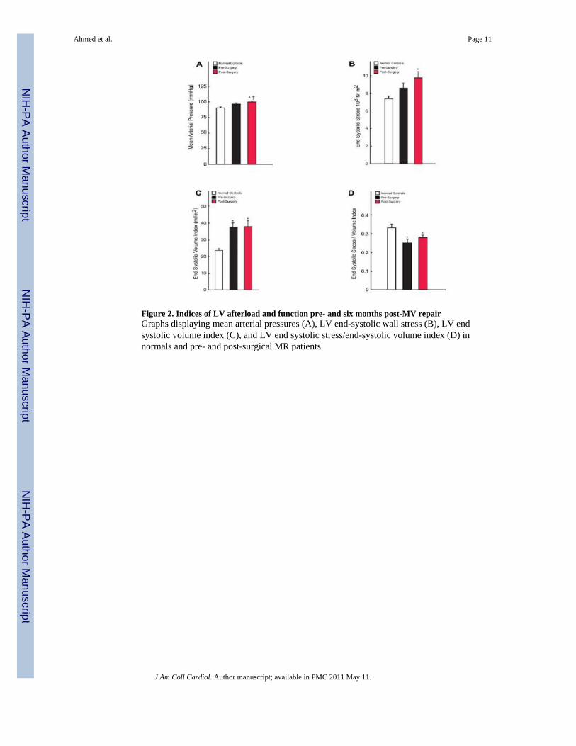
Figure 2. Indices of LV afterload and function pre- and six months post-MV repairGraphs displaying mean arterial pressures (A), LV end-systolic wall stress (B), LV endsystolic volume index (C), and LV end systolic stress/end-systolic volume index (D) innormals and pre- and post-surgical MR patients.
Ahmed et al. Page 11
J Am Coll Cardiol. Author manuscript; available in PMC 2011 May 11.
NIH
-PA Author Manuscript
NIH
-PA Author Manuscript
NIH
-PA Author Manuscript

Figure 3. Myofibrillar loss and oxidative stress in patients with isolated MRMyocardial biopsy findings in controls (n=10) (a,d,g) and MR patients (n=27)demonstrating myofibrillar degeneration (b,c), increased xanthine oxidase (e,f), andincreased lipofuscin (h,i). Nitrotyrosine staining in the LV of an MR patient demonstratingincreased staining in areas of lipofuscin accumulation (j) and a corresponding image (k) withimmunoabsorbed antibody with no uptake of antibody and only autofluorescence oflipofuscin. Scale bar = 20 μm. * = p <0.05 vs. normal control group. Scale bar = 20 μm.
Ahmed et al. Page 12
J Am Coll Cardiol. Author manuscript; available in PMC 2011 May 11.
NIH
-PA Author Manuscript
NIH
-PA Author Manuscript
NIH
-PA Author Manuscript

Figure 4. Lipofuscin in electron micrographs of patients with isolated MRTransmission electron microscopy of endomyocardial biopsy samples demonstrating markedlipofuscin deposition (arrows) in the hearts of MR patients.
Ahmed et al. Page 13
J Am Coll Cardiol. Author manuscript; available in PMC 2011 May 11.
NIH
-PA Author Manuscript
NIH
-PA Author Manuscript
NIH
-PA Author Manuscript

Figure 5. Protein quantification of xanthine oxidase in LV of patients with isolated MRWestern blot analysis for xanthine oxidoreductase (A) depicts a band at 145kD whichrepresents both XDH and transiently modified XO and bands at 125kD and 85kD whichrepresent permanently modified XO. Densitometry depicting average band intensitynormalized to respective calsequestrin loading control (B). *=p<0.05 vs. normal controlgroup.
Ahmed et al. Page 14
J Am Coll Cardiol. Author manuscript; available in PMC 2011 May 11.
NIH
-PA Author Manuscript
NIH
-PA Author Manuscript
NIH
-PA Author Manuscript

NIH
-PA Author Manuscript
NIH
-PA Author Manuscript
NIH
-PA Author Manuscript
Ahmed et al. Page 15
Table 1
Patient Characteristics
Characteristics Normal Pre-Surgery Post-Surgery
Sample Size 39 27 27
Age, years 40 ± 11 53 ± 12* -
Male, % 48 85 85*
BSA, m2 1.92 ± 0.23 2.02 ± 0.21 1.99 ± 0.22
Heart Rate, beats/min 76 ± 11 71 ± 11 80 ± 16
Mean Systolic BP, mmHg 116 ± 12 123 ± 15 120 ± 17
Mean Diastolic BP, mmHg 74 ± 11 74 ± 10 77 ± 9
ACE-Inhibitor/AR Blocker (%) 0 22 17
Beta-blocker (%) 0 26 35
Calcium Channel blocker (%) 0 7 0
BSA, body surface area; BP, blood pressure; ACE, Angiotensin converting enzyme; AR, Angiotensin receptor; Values are ± standard error of themean
*p <0.05 vs. normal control group.
J Am Coll Cardiol. Author manuscript; available in PMC 2011 May 11.

NIH
-PA Author Manuscript
NIH
-PA Author Manuscript
NIH
-PA Author Manuscript
Ahmed et al. Page 16
Table 2
Magnetic Resonance Imaging Variables
Variables Normal Pre-Surgery Post-Surgery
Sample size 39 27 27
LV EF, % 65 ± 6 66 ± 7 54 ± 9 *†
LV ESD, mm 35 ± 4 42 ± 6 * 43 ± 8
LVEDD, mm 51 ± 4 62 ± 5* 54 ± 7†
LV ESV index, ml/m2 23 ± 6 37 ± 14 * 37 ± 18 *
LV EDV index, ml/m2 66 ± 11 108 ± 28 * 78 ± 24 †
LV SV index, ml/m2 43 ± 7 71 ± 18 * 41 ± 9 †
LVEF, LV ejection fraction; ESD, end-systolic dimension; ESV, end-systolic volume;
EDV, end-diastolic volume, SV, stroke volume. Values are ± standard error of the mean.
*p <0.05 vs. normal control group.
†p <0.05 vs. pre-MV repair.
J Am Coll Cardiol. Author manuscript; available in PMC 2011 May 11.