Edwards v. Mortgage Electronic Registration Respondent's ...
Upload
independentCategory
view
0download
0
A prospective ‘‘oversizing’’ strategy of the Edwards SAPIENbioprosthesis: Results and impact on aortic regurgitation
Mariam Samim, BSc,a Pieter R. Stella, MD, PhD,a Pierfrancesco Agostoni, MD, PhD,a
Jolanda Kluin, MD, PhD,b Faiz Ramjankhan, MD,b Gertjan Sieswerda, MD, PhD,a
Ricardo Budde, MD, PhD,a Marijke van der Linden, RN,a Francis Juthier, MD,c Carlo Banfi, MD,c
Christopher Hurt, MD,c Morsal Samim, BSc,a Marieke Hillaert, MD,a Lex van Herwerden, MD, PhD,b
Michel E. Bertrand, MD,c Pieter A. M. Doevendans, MD, PhD,a and Eric Van Belle, MD, PhDc
Objective:Moderate to severe aortic regurgitation is occurring in 20% to 30% of cases after transcatheter aorticvalve implantation.
Methods: The purpose of the study was to investigate the impact of a prospective policy of ‘‘oversizing’’ theEdwards SAPIEN bioprosthesis (Edwards Lifesciences LLC, Irvine, Calif) relative to the diameter of the aorticannulus on the rate and severity of aortic regurgitation in 28 consecutive patients initially considered eligible fortranscatheter aortic valve implantation on the basis of angiography, multislice computed tomography, and trans-thoracic echocardiography. This policy included the systematic use of transesophageal echocardiography to ex-clude borderline patients and the modification of the procedure to use the larger device possible. The results werestudied on an individual patient basis.
Results: Because 6 of 28 patients (21%) had an annulus diameter greater than 24 mm by transesophageal echo-cardiography, 22 patients underwent implantation of the Edwards SAPIEN prosthesis. In 6 of 22 patients, theprocedure was adapted to follow our ‘‘oversizing’’ policy. As a result, the ‘‘prosthesis/annulus cover index’’was 12.4% ! 4.3%. The procedure was successful in 21 of 22 patients (95%), and 18 patients were availablefor echocardiography at 1 month. Although a moderate to severe aortic regurgitation was observed pretreatmentin 4 of 18 patients (22%), it was no longer the case at 1 month (0/18, 0%; P" .03). The improvement was sec-ondary to a disappearance of the aortic regurgitation in all 7 patients with a significant aortic regurgitation atpretreatment, whereas the new aortic regurgitations appearing in 5 of the 11 patients with no aortic regurgitationat pretreatment were only mild aortic regurgitations.
Conclusions: In patients with a successful implantation of an Edwards SAPIEN valve, a simple ‘‘oversizing’’policy based on a systematic use of transesophageal echocardiography and modification of the procedure mayprevent the occurrence ofmoderate and severe aortic regurgitations. (J ThoracCardiovasc Surg 2013;145:398-405)
Transcatheter aortic valve implantation (TAVI) is a develop-ing technique to treat patients with aortic stenosis.1,2
Because questions remain concerning safety anddurability, its use currently is limited to patientscontraindicated or at very high risk for conventional aorticvalve replacement.
After TAVI, a moderate to severe paravalvular aortic re-gurgitation (AR) can occur in approximately 20% to 30%of cases after implantation.3,4 Although this is usually
considered as acceptable in the elderly population, it isa limitation to a broader use of this technique.Implantation of an undersized prosthesis has beenproposed as a major cause of this issue.3,5#7 Although it isusually recommended to ‘‘oversize’’ the implantation ofthe Edwards SAPIEN bioprosthesis (Edwards LifesciencesLLC, Irvine, Calif) relative to the annulus diameter asmeasured by transesophageal echocardiography (TEE) by2 to 4 mm, this rule is not strictly applied, particularly inpatients with a large annulus or borderline vascularaccesses.3 To date, the benefit of a strict ‘‘oversizing’’ policyon the severity of AR has never been investigated.
When starting our TAVI program with the Edwards SA-PIEN bioprosthesis, we decided to implement a prospectivepolicy of systematic ‘‘oversizing’’ the bioprosthesis relativeto the diameter of the aortic annulus and to test its impact onthe rate and severity of AR. This issue was studied bymeansof serial 2-dimensional transthoracic echocardiography(TTE) Doppler cardiography performed before the proce-dure, before discharge, and at 1 month.
From the Departments of Cardiologya and Thorax Surgery,b University Medical Cen-ter, Utrecht, The Netherlands; and Department of Cardiology,c University Hospital,Lille, France.
Disclosures: Dr Sieswerda reports receiving lecture fees from Actelion. The other au-thors have nothing to disclose with regard to commercial support.
Received for publication Aug 14, 2011; revisions received Nov 20, 2011; accepted forpublication Dec 14, 2011; available ahead of print Feb 29, 2012.
Address for reprints: Eric Van Belle, MD, PhD, EA 2693, University Lille Nord deFrance and Department of Cardiology, CHRU de Lille, 59037 Lille Cedex, France(E-mail: [email protected]).
0022-5223/$36.00Copyright ! 2013 by The American Association for Thoracic Surgerydoi:10.1016/j.jtcvs.2011.12.067
398 The Journal of Thoracic and Cardiovascular Surgery c February 2013
Acquired Cardiovascular Disease Samim et al
ACD
MATERIALS AND METHODSPatient Population and Selection
Patients with severe aortic stenosis considered to be at high or prohibi-tive surgical risk were considered to undergo TAVI with the Edwards SA-PIEN valve at University Medical Center, Utrecht, The Netherlands.Consensus was achieved between cardiologists and cardiac surgeons re-garding the surgical risk, and all patients provided informed writtenconsent.
To determine the initial eligibility for TAVI and to choose between thetransfemoral (TF) aortic valve implantation (AVI) or the transapical (TA)-AVI approach, all patients underwent coronary angiography, aortobife-moral angiography, multislice computed tomography, and TTE. Potentialcandidates for TAVI demonstrated symptomatic degenerative aortic steno-sis with at least New York Heart Association class 2 symptoms and an aor-tic valve area of 1.0 cm2 or mean gradient greater than 40 mm Hg or peakgradient greater than 80 mm Hg. All candidates were contraindicated forconventional surgery or considered a high surgical risk with an operativemortality risk of greater than 20% as assessed by at least 2 cardiovascularsurgeons and 2 cardiologists.8
Patients were excluded from TAVI if they had at least 1 of the following:(1) aortic annulus diameter less than 16 mm or greater than 24 mm by TTE;(2) congenital unicuspid or bicuspid valve; (3) noncalcified valve; (4) un-treated clinically significant proximal coronary artery disease; (5) severeleft ventricular (LV) dysfunction less than 20%; or (6) hemodynamic insta-bility requiring inotropic support.
All patients were considered as potential candidates for TF-AVI unlessthey had at least 1 of the following: (1) mobile aortic arch atheroma greaterthan 5mm or (2) iliofemoral dimensions, morphology, or calcifications thatwould preclude insertion of a 22F sheath or 24F introducer sheath based ona detailed assessment of aortobifemoral angiography and multislice com-puted tomography, in which case they were considered for TA-AVI.
On the basis of this initial selection process, 28 patients were consideredpotential candidates. All patients underwent TEE before the final planningof the procedure. As part of our ‘‘oversizing’’ policy (see below), thosewithan annulus diameter greater than 24 mm by TEE were further excluded anddid not undergo the procedure.
Choice of Bioprosthesis Size and ‘‘Oversizing’’ PolicyThe Edwards SAPIEN transcatheter heart valve is a catheter-delivered
heart valve that is composed of a stainless-steel balloon-expandable stent,with an integrated trileaflet tissue valve and a polyethylene terephthalatefabric cuff. The valve tissue is made from 3 equal sections of bovine peri-cardium treated with a proprietary tissue treatment. During the time of thestudy, the bioprosthesis was available in 2 sizes (23 and 26 mm) and couldbe delivered through the TF or TA approach.
Our ‘‘oversizing’’ policy for the implantation of the Edwards SAPIENbioprosthesis was defined as follows: (1) All patients with an aortic annu-lus greater than 24 mm by TEE during the final step of the screening were
strictly excluded (see above); (2) a 26-mm prosthesis was implanted in allpatients with an annulus greater than 21 mm, and the TA approach wasused every time it was needed; and (3) in patients with an annulus 21mm or less, a 26-mm prosthesis was also implanted every time the balloonpredilatation with a 23-mm balloon associated with contrast angiographyat maximum balloon inflation demonstrated a significant aorticregurgitation.
To appreciate the degree of ‘‘oversizing,’’ the ‘‘prosthesis/annulus coverindex’’ expressed as 1003 ([prosthesis diameter – TTE annulus diameter]/prosthesis diameter) was used, as suggested by Detaint and colleagues.3
Transcatheter Aortic Valve ImplantationThe prosthetic stented valve was mechanically crimped on a balloon
catheter immediately before implantation. The Retroflex delivery system(Edwards Lifesciences) was used for device implantation.6,9,10
TF and TA-AVI were performed under general anesthesia in the cathe-terization laboratory as previously described.6,9,10 TEE was used for allprocedures. Patients were premedicated with aspirin and antibiotics.Heparin was used to maintain an activated clotting time greater than 300seconds. The activated clotting time was reversed with protamine at theend of the procedure.
For TF-AVI, our technique was similar to that in previously describedreports.6,9 A surgical cut-down of the femoral arty was performed, anda 14F sheath was initially placed. A temporary transvenous electrodewas introduced into the right ventricle. Balloon aortic valvuloplasty witha 20- or 23-mm balloon was performed. The balloon-mounted valve waspositioned using angiographic techniques and echocardiographic guid-ance, and subsequently deployed under rapid pacing (180–220 beats/min). Exit peripheral angiography was performed to ensure no extravasa-tion of contrast before removal of femoral sheath. The sheath was removedand closed surgically in the operating room by a surgeon immediatelypostprocedure.
The TA-AVI procedure was performed as previously described.10 Forthe purpose of rapid ventricular pacing, 2 unipolar epicardial pacer wireswere secured and tested with a high-output epicardial pacing system to en-sure ventricular capture at rates of 180 to 220 beats/min.
Procedural success was defined as the implantation of a functional pros-thetic valvewithin the aortic annulus at the end of the procedurewithout in-laboratory mortality. Patients received aspirin (81 mg/d) and clopidogrel(75 mg/d) indefinitely. Warfarin was substituted for clopidogrel in patientswith atrial fibrillation.
Echocardiography Follow-upTEE was the final step of the screening process and was performed dur-
ing the procedure in all patients. TTE was performed during the screeningprocess, pretreatment (last echocardiography study before TAVI), post-treatment (at discharge), and postdischarge (first outpatient clinic visit) us-ing a Philips 5500 or ie33 system (Philips, Eindhoven, The Netherlands).Complete echocardiographic studies were performed for each patient ina standard fashion. Standard parameters were recorded.11 By using the par-asternal long-axis view and M-mode or 2-dimensional echocardiography,LV diameters, function, and outflow track diameter were obtained.
To assess the severity of aortic stenosis peak aortic velocity, peak instan-taneous gradient, mean transaortic gradient, and velocity-time integralwere measured. Aortic valve area was estimated using the continuity equa-tion approach.12
The color-flow Doppler signal was used to assess aortic regurgitation.According to the European Society of Cardiology guidelines and AmericanSociety of Echocardiography recommendations, valvular insufficiency wasgraded in 5 groups as none ("0), trivial ("1), mild ("2), moderate ("3), orsevere ("4).13,14 A valvular insufficiency of 2 or greater was consideredsignificant. More specifically, regurgitation was assessed by visualinspection and by using color-flow mapping of the regurgitation jet as de-scribed by Zoghbi and colleagues14 and Helmcke and colleagues.15 A
Abbreviations and AcronymsAR " aortic regurgitationAVI " aortic valve implantationLV " left ventricularTA " transapicalTAVI " transcatheter aortic valve implantationTEE " transesophageal echocardiographyTF " transfemoralTTE " transthoracic echocardiography
Samim et al Acquired Cardiovascular Disease
The Journal of Thoracic and Cardiovascular Surgery c Volume 145, Number 2 399
ACD
single experienced and independent cardiologist, blinded to the patientclinical status, graded AR.
Statistical AnalysisContinuous variables were presented as mean ! standard deviation.
Discrete variables were presented as absolute numbers and percentages.For comparison between categoric variables, a chi-square test was used.A 1-way analysis of variance test was used for comparison between contin-uous variables. A 1-way repeated-measures Friedman test with post hocanalysis was used to evaluate changes over time. Analyses were performedusing SPSS 18.0 (SPSS Inc, Chicago, Ill).
RESULTSCharacteristics of the Study Population andProcedure
The baseline clinical and echocardiography characteris-tics of the study population are shown in Table 1. On the ba-sis of the measurement of the annulus diameter by TTE(<24 mm), 28 patients were initially considered eligiblefor implantation. On the basis of the subsequent
measurement of the annulus diameter by TEE (>24 mm)during the final step of the screening process, 6 of 28 pa-tients (21%) were excluded. By definition and comparisonwith the remaining 22 patients, the annulus diameter ofthese 6 patients as measured by TEE was larger (P " .01,Table 1). This difference was not detectable by TTE(P " .10, Table 1). The 6 excluded patients were male(100%) and had a higher body mass index (P " .04) thanthe remaining 22 patients (Table 1).
The medical decision in the 6 patients in whom the TAVIprocedure was not performed was as follows: One patientunderwent conventional aortic replacement and was aliveat 12 months follow-up; 1 patient was eligible for the im-plantation of a 29-mmCoreValve (Medtronic Inc, Minneap-olis, Minn) (the procedure was successful, and the patientdied at 7 months follow-up); the last 4 patients underwentconservative medical management (2 patients died within12 months and 2 patients remained alive at 1 year underconservative treatment; 1 of the last 2 patients recently
TABLE 1. Clinical and echocardiography characteristics of the 28 patients
Total
Excluded
N " 6
Included
N " 22 P Transfemoral Transapical P
Age (y) 80 ! 8 80 ! 9 79 ! 7 .71 78 ! 7 80 ! 8 .61
Male, n (%) 17 (61) 6 (100) 11 (50) .02 4 (40) 7 (58) .66
BMI (kg/m2) 27 ! 6 28 ! 4 26 ! 5 .04 26 ! 7 26 ! 4 .85
Hypertension, n (%) 21 (75) 5 (83) 16 (73) .58 7 (70) 9 (75) .80
Atrial fibrillation, n (%) 12 (43) 2 (33) 10 (45) .59 5 (50) 5 (42) .70
Diabetes, n (%) 5 (18) 1 (17) 4 (18) .93 2 (20) 2 (17) .84
Coronary artery disease, n (%) 16 (57) 3 (50) 13 (59) .69 3 (30) 10 (83) .02
Prior myocardial infarction, n (%) 8 (29) 1 (17) 7 (32) .45 2 (20) 5 (42) .38
Percutaneous coronary intervention, n (%) 11 (39) 2 (33) 9 (41) .73 2 (20) 7 (58) .06
Coronary artery bypass, n (%) 7 (25) 1 (17) 6 (27) .58 2 (20) 4 (33) .64
Cerebrovascular events, n (%) 8 (29) 1 (17) 7 (32) .45 3 (30) 4 (33) .87
Peripheral vascular disease, n (%) 3 (11) 1 (17) 2 (9) .61 0 (0) 2 (17) .48
Renal disease, n (%) 6 (21) 1 (17) 5 (23) .74 2 (20) 3 (25) .78
COPD, n (%) 8 (29) 2 (33) 6 (27) .77 3 (30) 3 (25) .79
NYHA class, n (%)
I 0 (0) 0 (0) 0 (0) 0 (0) 0 (0)
II 3 (11) 1 (17) 2 (9) .74 1 (10) 1 (8) .40
III 22 (78) 4 (66) 18 (82) 9 (90) 9 (75)
IV 3 (11) 1 (17) 2 (9) 0 (0) 2 (17)
Logistic euroSCORE 21.8 ! 13.3 23.2 ! 16.3 21.3 ! 14.1 .81 18.1 ! 15.5 24.1 ! 12.8 .33
LVEF<35%, n (%) 6 (21) 1 (17) 5 (23) .74 2 (20) 3 (25) .78
LV end-diastolic diameter (mm) 46.2 ! 4.5 47.5 ! 5.3 45.7 ! 5.3 .43 45.3 ! 6.1 46.2 ! 4.8 .71
Mean aortic gradient (mm Hg) 42 ! 15 44 ! 15 41 ! 16 .55 40 ! 16 41 ! 17 .88
Peak aortic gradient (mm Hg) 71 ! 24 73 ! 21 70 ! 26 .65 70 ! 27 71 ! 26 .95
Aortic valve area (cm2) 0.72 ! 0.19 0.69 ! 0.14 0.73 ! 0.19 .25 0.73 ! 0.17 0.73 ! 0.21 .96
Annulus diameter by TTE (mm) 21.8 ! 1.2 22.5 ! 1.1 21.6 ! 1.3 .10 21.5 ! 1.4 21.6 ! 1.5 .91
Annulus diameter by TEE (mm) 22.6 ! 1.4 24.9 ! 0.4 22.0 ! 1.3 .01 22.1 ! 1.3 22.0 ! 1.3 .86
No significant AR, n (%) 16 (57) 3 (50) 13 (59) .69 5 (50) 8 (67) .72
Significant AR, n (%) 12 (43) 3 (50) 9 (41) 5 (50) 4 (33)
Aortic regurgitation grade (0–4) 1.55 ! 0.95 1.33 ! 0.82 1.55 ! 1.01 .35 1.80 ! 1.40 1.33 ! 0.49 .29
BMI, Body mass index; COPD, chronic obstructive pulmonary disorder; NYHA, New York Heart Association; euroSCORE, European System for Cardiac Operative Risk Eval-uation; LVEF, left ventricular ejection fraction; LV, left ventricular; TTE, transthoracic echocardiography; AR, aortic regurgitation. Data are mean! standard deviation otherwisestated.
Acquired Cardiovascular Disease Samim et al
400 The Journal of Thoracic and Cardiovascular Surgery c February 2013
ACD
underwent the implantation of a 29-mm Edwards SAPIENbioprosthesis).
Among the 22 remaining patients, 12 underwent TA-AVIand 10 underwent TF-AVI. Patients who underwent TA-AVI rather than TF-AVI were more likely to have a historyof coronary artery disease (P " .02). The other characteris-tics were not different between the groups.
The relatively low mean annulus diameter (22.0 ! 1.3mm, Tables 1 and 2) of the population in whom theprocedure was performed reflects the exclusion of allpatients with an annulus greater than 24 mm by TEE aspart of our ‘‘oversizing’’ policy. Furthermore, in 6 of 22patients the procedure was modified: In 3 patients with anannulus greater than 21 mm and suitable for a 22F but notfor a 24F iliofemoral sheath, a TA approach was used. In3 other patients with an annulus 21 or less by TTE, a 26-mm rather than a 23-mm prosthesis was implanted on thebasis of the results of balloon predilatation associatedwith angiography. As a result, the ‘‘prosthesis/annuluscover index’’ was 12.4%! 4.3%. Using a 23-mm prosthe-sis in the 6 cases mentioned above would have led to a sig-nificantly lower ‘‘cover index’’ (9.9% ! 3.6%, P " .02).
The procedure was successful in 21 of 22 patients (95%,Table 2). The patient with unsuccessful valve implant wasan 85-year-old man with a history of myocardial infarction,bypass surgery, and peripheral vascular disease. The tech-nical failure was related to the inability to perform appro-priate hemostasis at the level of the purse at the apex of the
left ventricle related to pericardial adherences. The proce-dure was aborted and converted to open surgery for con-ventional valve replacement. Although the surgicalintervention was successful, the patient died of multiorganfailure on day 11. Another patient died of right ventricularfailure secondary to an inferior myocardial infarction inhospital on day 3 from. These 2 in-hospital deaths were ob-served among the 12 patients who underwent a TA-AVIprocedure. No procedural failure and no hospital deathwere observed in the 10 patients who underwent a TF-AVIprocedure. One patient who underwent TA-AVI requireda permanent pacemaker implantation before discharge.No rupture of the aortic annulus was observed. No caseof severe AR was observed immediately after balloonvalvuloplasty by TEE. After implantation of the biopros-thesis, fluoroscopy did not show any distortion of theedges of the stent supporting the valve. Likewise, TEE per-formed in short axis showed that the circularity of the stentwas preserved. Overall, 20 patients (91%) underwent a suc-cessful procedure and were discharged alive from thehospital.Within the first 30 days, 2 additional patients died of pul-
monary infection in the TA-AVI group (1) and sudden death(1) in the TF-AVI group. No stroke was observed. Eighteen
TABLE 2. Procedural characteristics and 30-day outcome
Total
N " 22
Transfemoral
N " 10
Transapical
N " 12 P
Annulus diameter
(mm), mean ! SD
22.0 ! 1.3 22.1 ! 1.3 22.0 ! 1.3 .86
Aortic valve area (cm2),
mean ! SD
0.73 ! 0.19 0.73 ! 0.17 0.73 ! 0.21 .95
Prosthesis diameter,
n (%)
23 mm 6 (27) 3 (30) 3 (25) .79
26 mm 16 (73) 7 (70) 9 (75)
Cover index, %,
mean ! SD
12.4 ! 4.3 11.8 ! 5.5 12.8 ! 3.2 .58
Successful valvuloplasty,
n (%)
22 (100) 10 (100) 12 (100) 1
Successful valve
deployment, n (%)
21 (95) 10 (100) 11 (92) .35
Intraprocedural death,
n (%)
0 (0) 0 (0) 0 (0) 1
30-d outcome
Myocardial infarction 1 (4) 0 (0) 1 (8) .35
Stroke, n (%) 0 (0) 0 (0) 0 (0) 1
Permanent pacemaker 1 (4) 0 (0) 1 (8) .35
Mortality, n (%) 4 (18) 1 (10) 3 (25) .35
SD, Standard deviation. Cover index as 100 3 (prosthesis diameter – annulus diam-eter by TEE/prosthesis diameter).
FIGURE 1. Patient flow chart. TEE, Transesophageal echocardiography;
TTE, transthoracic echocardiography.
Samim et al Acquired Cardiovascular Disease
The Journal of Thoracic and Cardiovascular Surgery c Volume 145, Number 2 401
ACD
patients were available for echocardiography after dis-charge (Figure 1).
Echocardiography AnalysisThe echocardiography findings of the 18 patients with an
echocardiography at 1 month are summarized in Table 3.There was a statistically significant decrease in the peak(pretreatment 73 ! 26 mm Hg vs post-treatment 17 ! 6mm Hg vs 1 month 15 ! 6 mm Hg, P<.0001) and meangradient (pretreatment 42 ! 16 mm Hg vs post-treatment9 ! 4 mm Hg vs 1 month 8 ! 3 mm Hg, P<.0001) afterEdwards implantation, which was associated with an in-crease in the aortic valve area (pretreatment 0.72 ! 0.21cm2 vs post-treatment 1.87 ! 0.45 cm2 vs 1 month 1.81! .32 cm2, P<.0001). Ventricular function did not changesignificantly during follow-up. No difference was observedbetween patients treated through the TF or TA approach.
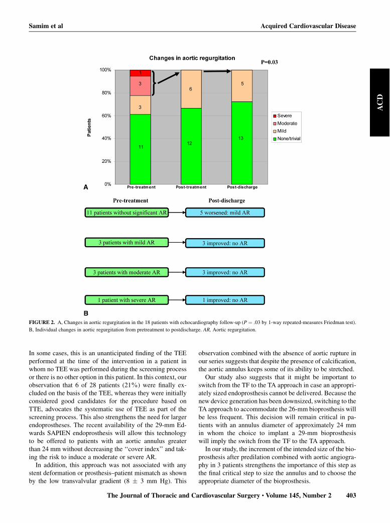
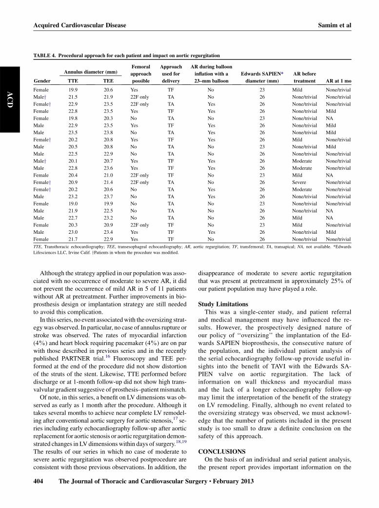
Before treatment, a significant AR was observed in 9 of22 patients (41%, Table 1). AR was mild, moderate, and se-vere in 5 patients (23%), 3 patients (13%), and 1 patient(5%), respectively. The degree of AR before and 1 monthafter Edwards SAPIEN valve implantation in the 18 patientswith echocardiography follow-up is shown in Table 3 andFigure 2,A. Although amoderate to severeARwas observedin 4 of 18 patients (22%) before treatment, it was no longer
the case at 1 month (P" .03, Figure 2, A). The improvementwas secondary to a disappearance of the AR in all 7 patientswith a significant AR before treatment, whereas the newARs appearing in 5 of the 11 patients with no AR at pretreat-ment were only mild (Table 4; Figure 2, B).
DISCUSSIONThe present study is the first to show that in patients re-
ferred for TAVI for aortic stenosis, a simple prospectivestrategy of ‘‘oversizing’’ the implantation of the EdwardsSAPIEN bioprosthesis may prevent the occurrence of mod-erate and severe ARs.
Impact of ‘‘Oversizing’’ the Edwards SAPIENBioprosthesis on Aortic Regurgitation
The key finding of this study is that the use of a simple‘‘oversizing’’ policy was associated with no occurrence ofmoderate or severe ARs at 1 month with no case of ruptureof the aortic annulus and no prosthesis–patient mismatch.This benefit was the result of 3 simple measures, including(1) exclusion of all patients with an aortic annulus greaterthan 24 mm by TEE during the screening process, (2) useof a 26-mm prosthesis in all patients with aortic annulusgreater than 21, and (3) use of a 26-mm prosthesis in pa-tients with an aortic annulus 21 or less whenever sizingbased on balloon valvuloplasty with a 23-mm balloon andcontrast angiography suggested that it was feasible.
Our results are consistent with a recent retrospectiveanalysis of determinants of AR after Edwards SAPIEN bio-prosthesis implantation by Detaint and colleagues.3 Theyobserved that oversizing the prosthesis, as estimated bythe ‘‘prosthesis/annulus cover index,’’ was the major factorassociated with the absence of severe AR after implanta-tion. Of note, in our population the cover index was twiceas high as in the population studied by Detaint and col-leagues (12.4 ! 4.3 vs 6.7 ! 4). Detaint and colleaguesalso found that a moderate to severe AR was never observedin patients with a ‘‘cover index’’ greater than 8 or an aorticannulus less than 22 mm. Of note, this combination waspresent in 20 of 22 patients (91%) in our population butin only half of the population studied by Detaint andcolleagues.3
Analysis of the respective role of the criteria used in theselection process demonstrates that the exclusion of patientswith an annulus greater than 24 mm would have already re-sulted in a high cover index of 9.9%! 3.6%. The use of the2 additional criteria further increased the cover index of ourpopulation to 12.4% ! 4.3%.
The exclusion of patients with an aortic annulus greaterthan 24 mm is already recommended as a common practice.However, this rule is not strictly applied, and in a recentlypublished series, Detaint and colleagues3 reported that anEdwards SAPIEN was implanted in patients with an annu-lus greater than 24 mm in more than one third of the cases.
TABLE 3. Echocardiography findings during follow-up (N " 18)
Pretreatment
Post-
treatment Postdischarge P
LVEF<35%, n (%) 3 (23) 2 (15) 3 (17) .57
TF 1 (11) 1 (11) 1 (11)
TA 2 (22) 1 (11) 2 (25)
LV end-diastolic
diameter (mm)
45.7 ! 5.9 45.2 ! 5.5 44.7 ! 5.3 .03
TF 45.0 ! 6.3 44.7 ! 5.5 44.3 ! 4.9
TA 46.4 ! 5.5 45.8 ! 5.6 45.1 ! 5.7
Mean aortic gradient
(mm Hg)
42 ! 17 9 ! 4 8 ! 3 .0001
TF 41 ! 17 12 ! 4 9 ! 3
TA 44 ! 18 7 ! 2 7 ! 2
Peak aortic gradient
(mm Hg)
73 ! 26 17 ! 6 15 ! 6 .0001
TF 73 ! 28 21 ! 6 17 ! 8
TA 75 ! 27 13 ! 4 13 ! 4
Aortic valve area
(cm2)
0.72 ! 0.21 1.87 ! 0.45 1.81 ! 0.31 .0001
TF 0.73 ! 0.18 1.87 ! 0.50 1.86 ! 0.34
TA 0.71 ! 0.24 1.88 ! 0.43 1.75 ! 0.30
Aortic regurgitation
grade (0–4)
1.56 ! 1.10 1.17 ! 0.71 0.89 ! 0.83* .03
TF 1.78 ! 1.48 1.22 ! 0.67 1.11 ! 0.78
TA 1.33 ! 0.50 1.11 ! 0.78 0.67 ! 0.87
All parameters are mean ! standard deviation, otherwise stated. P reflects the differ-ence among the 3 time points by 1-way repeated measures Friedman test. LVEF, Leftventricular ejection fraction; TF, transfemoral; TA, transapical; LV, left ventricular.*P<.05 compared with ‘‘pretreatment’’ by post hoc analysis of the Friedman test.
Acquired Cardiovascular Disease Samim et al
402 The Journal of Thoracic and Cardiovascular Surgery c February 2013
ACD
In some cases, this is an unanticipated finding of the TEEperformed at the time of the intervention in a patient inwhom no TEE was performed during the screening processor there is no other option in this patient. In this context, ourobservation that 6 of 28 patients (21%) were finally ex-cluded on the basis of the TEE, whereas they were initiallyconsidered good candidates for the procedure based onTTE, advocates the systematic use of TEE as part of thescreening process. This also strengthens the need for largerendoprostheses. The recent availability of the 29-mm Ed-wards SAPIEN endoprosthesis will allow this technologyto be offered to patients with an aortic annulus greaterthan 24 mm without decreasing the ‘‘cover index’’ and tak-ing the risk to induce a moderate or severe AR.
In addition, this approach was not associated with anystent deformation or prosthesis–patient mismatch as shownby the low transvalvular gradient (8 ! 3 mm Hg). This
observation combined with the absence of aortic rupture inour series suggests that despite the presence of calcification,the aortic annulus keeps some of its ability to be stretched.Our study also suggests that it might be important to
switch from the TF to the TA approach in case an appropri-ately sized endoprosthesis cannot be delivered. Because thenew device generation has been downsized, switching to theTA approach to accommodate the 26-mm bioprosthesis willbe less frequent. This decision will remain critical in pa-tients with an annulus diameter of approximately 24 mmin whom the choice to implant a 29-mm bioprosthesiswill imply the switch from the TF to the TA approach.In our study, the increment of the intended size of the bio-
prosthesis after predilation combined with aortic angiogra-phy in 3 patients strengthens the importance of this step asthe final critical step to size the annulus and to choose theappropriate diameter of the bioprosthesis.
FIGURE 2. A, Changes in aortic regurgitation in the 18 patients with echocardiography follow-up (P " .03 by 1-way repeated-measures Friedman test).
B, Individual changes in aortic regurgitation from pretreatment to postdischarge. AR, Aortic regurgitation.
Samim et al Acquired Cardiovascular Disease
The Journal of Thoracic and Cardiovascular Surgery c Volume 145, Number 2 403
ACD
Although the strategy applied in our population was asso-ciated with no occurrence of moderate to severe AR, it didnot prevent the occurrence of mild AR in 5 of 11 patientswithout AR at pretreatment. Further improvements in bio-prosthesis design or implantation strategy are still neededto avoid this complication.
In this series, no event associatedwith the oversizing strat-egywas observed. In particular, no case of annulus rupture orstroke was observed. The rates of myocardial infarction(4%) and heart block requiring pacemaker (4%) are on parwith those described in previous series and in the recentlypublished PARTNER trial.16 Fluoroscopy and TEE per-formed at the end of the procedure did not show distortionof the struts of the stent. Likewise, TTE performed beforedischarge or at 1-month follow-up did not show high trans-valvular gradient suggestive of prosthesis–patientmismatch.
Of note, in this series, a benefit on LV dimensions was ob-served as early as 1 month after the procedure. Although ittakes several months to achieve near complete LV remodel-ing after conventional aortic surgery for aortic stenosis,17 se-ries including early echocardiography follow-up after aorticreplacement for aortic stenosis or aortic regurgitation demon-strated changes in LV dimensionswithin days of surgery.18,19
The results of our series in which no case of moderate tosevere aortic regurgitation was observed postprocedure areconsistent with those previous observations. In addition, the
disappearance of moderate to severe aortic regurgitationthat was present at pretreatment in approximately 25% ofour patient population may have played a role.
Study LimitationsThis was a single-center study, and patient referral
and medical management may have influenced the re-sults. However, the prospectively designed nature ofour policy of ‘‘oversizing’’ the implantation of the Ed-wards SAPIEN bioprosthesis, the consecutive nature ofthe population, and the individual patient analysis ofthe serial echocardiography follow-up provide useful in-sights into the benefit of TAVI with the Edwards SA-PIEN valve on aortic regurgitation. The lack ofinformation on wall thickness and myocardial massand the lack of a longer echocardiography follow-upmay limit the interpretation of the benefit of the strategyon LV remodeling. Finally, although no event related tothe oversizing strategy was observed, we must acknowl-edge that the number of patients included in the presentstudy is too small to draw a definite conclusion on thesafety of this approach.
CONCLUSIONSOn the basis of an individual and serial patient analysis,
the present report provides important information on the
TABLE 4. Procedural approach for each patient and impact on aortic regurgitation
Gender
Annulus diameter (mm)Femoral
approach
possible
Approach
used for
delivery
AR during balloon
inflation with a
23–mm balloon
Edwards SAPIEN*
diameter (mm)
AR before
treatment AR at 1 moTTE TEE
Female 19.9 20.6 Yes TF No 23 Mild None/trivial
Maley 21.5 21.9 22F only TA No 26 None/trivial None/trivial
Femaley 22.9 23.5 22F only TA Yes 26 None/trivial None/trivial
Female 22.8 23.5 Yes TF Yes 26 None/trivial Mild
Female 19.8 20.3 No TA No 23 None/trivial NA
Male 22.9 23.5 Yes TF Yes 26 None/trivial Mild
Male 23.5 23.8 No TA Yes 26 None/trivial Mild
Femaley 20.2 20.8 Yes TF Yes 26 Mild None/trivial
Male 20.5 20.8 No TA No 23 None/trivial Mild
Male 22.5 22.9 No TA No 26 None/trivial None/trivial
Maley 20.1 20.7 Yes TF Yes 26 Moderate None/trivial
Male 22.8 23.6 Yes TF Yes 26 Moderate None/trivial
Female 20.4 21.0 22F only TF No 23 Mild NA
Femaley 20.9 21.4 22F only TA No 26 Severe None/trivial
Femaley 20.2 20.6 No TA Yes 26 Moderate None/trivial
Male 23.2 23.7 No TA Yes 26 None/trivial None/trivial
Female 19.0 19.9 No TA No 23 None/trivial None/trivial
Male 21.9 22.5 No TA No 26 None/trivial NA
Male 22.7 23.2 No TA No 26 Mild NA
Female 20.3 20.9 22F only TF No 23 Mild None/trivial
Male 23.0 23.4 Yes TF Yes 26 None/trivial Mild
Female 21.7 22.9 Yes TF No 26 None/trivial None/trivial
TTE, Transthoracic echocardiography; TEE, transesophageal echocardiography; AR, aortic regurgitation; TF, transfemoral; TA, transapical; NA, not available. *EdwardsLifesciences LLC, Irvine Calif. yPatients in whom the procedure was modified.
Acquired Cardiovascular Disease Samim et al
404 The Journal of Thoracic and Cardiovascular Surgery c February 2013
ACD
outcome of AR after implantation of an Edwards SAPIENvalve in the aortic position. In particular, the results suggestthat a simple ‘‘oversizing’’ policy may prevent the occur-rence of moderate and severe ARs. Although these findingsneed confirmation in studies including more patients andwith a longer follow-up, they support the use of larger endo-prostheses (29 mm) to adequately treat patients with an an-nulus greater than 24 mm.
References1. Sun JC, Ghanta RK, Davidson MJ. Highlights from the transcatheter cardiovas-
cular therapeutics conference 2010: Washington, DC, September 21-25, 2010. JThorac Cardiovasc Surg. 2011;142:468-71.
2. Higgins J, Ye J, Humphries KH, Cheung A,Wood DA,Webb JG, et al. Early clin-ical outcomes after transapical aortic valve implantation: a propensity-matchedcomparison with conventional aortic valve replacement. J Thorac CardiovascSurg. 2011;142:e47-52.
3. Detaint D, Lepage L, Himbert D, Brochet E, Messika-Zeitoun D, Iung B, et al.Determinants of significant paravalvular regurgitation after transcatheter aorticvalve: Implantation impact of device and annulus discongruence. JACC Cardio-vasc Interv. 2009;2:821-7.
4. Moss RR, Ivens E, Pasupati S, Humphries K, Thompson CR, Munt B, et al. Roleof echocardiography in percutaneous aortic valve implantation. JACC Cardio-vasc Imaging. 2008;1:15-24.
5. Takagi K, Latib A, Al-Lamee R, Mussardo M, Montorfano M, Maisano F, et al.Predictors of moderate-to-severe paravalvular aortic regurgitation immediatelyafter CoreValve implantation and the impact of postdilatation. Catheter Cardio-vasc Interv. 2011;78:432-43.
6. Webb JG, Pasupati S, Humphries K, Thompson C, Altwegg L, Moss R, et al. Per-cutaneous transarterial aortic valve replacement in selected high-risk patientswith aortic stenosis. Circulation. 2007;116:755-63.
7. Descoutures F, Himbert D, Lepage L, Iung B, Detaint D, Tchetche D, et al. Con-temporary surgical or percutaneous management of severe aortic stenosis in theelderly. Eur Heart J. 2008;29:1410-7.
8. Vahanian A, Alfieri O, Al-Attar N, Antunes M, Bax J, Cormier B, et al. Trans-catheter valve implantation for patients with aortic stenosis: a position statementfrom the European Association of Cardio-Thoracic Surgery (EACTS) and theEuropean Society of Cardiology (ESC), in collaboration with the European As-
sociation of Percutaneous Cardiovascular Interventions (EAPCI). Eur Heart J.2008;29:1463-70.
9. Cribier A, Eltchaninoff H, Tron C, Bauer F, Agatiello C, Nercolini D, et al. Treat-ment of calcific aortic stenosis with the percutaneous heart valve: mid-term fol-low-up from the initial feasibility studies: the French experience. J Am CollCardiol. 2006;47:1214-23.
10. Walther T, Simon P, Dewey T, Wimmer-Greinecker G, Falk V, Kasimir MT, et al.Transapical minimally invasive aortic valve implantation: multicenter experi-ence. Circulation. 2007;116:I240-5.
11. Tzikas A, Piazza N, van Dalen BM, Schultz C, GeleijnseML, vanGeuns RJ, et al.Changes in mitral regurgitation after transcatheter aortic valve implantation.Catheter Cardiovasc Interv. 2009;75:43-9.
12. Oh JK, Taliercio CP, Holmes DR Jr, Reeder GS, Bailey KR, Seward JB, et al. Pre-diction of the severity of aortic stenosis by Doppler aortic valve area determina-tion: prospective Doppler-catheterization correlation in 100 patients. J Am CollCardiol. 1988;11:1227-34.
13. Vahanian A, Baumgartner H, Bax J, Butchart E, Dion R, Filippatos G, et al.Guidelines on the management of valvular heart disease: the task force on themanagement of valvular heart disease of the European Society of Cardiology.Eur Heart J. 2007;28:230-68.
14. Zoghbi WA, Enriquez-Sarano M, Foster E, Grayburn PA, Kraft CD, Levine RA,et al. Recommendations for evaluation of the severity of native valvular regurgi-tation with two-dimensional and Doppler echocardiography. J Am Soc Echocar-diogr. 2003;16:777-802.
15. Helmcke F, Nanda NC, Hsiung MC, Soto B, Adey CK, Goyal RG, et al. ColorDoppler assessment of mitral regurgitation with orthogonal planes. Circulation.1987;75:175-83.
16. Leon MB, Smith CR, Mack M, Miller DC, Moses JW, Svensson LG, et al. Trans-catheter aortic-valve implantation for aortic stenosis in patients who cannot un-dergo surgery. N Engl J Med. 2010;363:1597-607.
17. Yarbrough WM, Mukherjee R, Ikonomidis JS, Zile MR, Spinale FG. Myocardialremodeling with aortic stenosis and after aortic valve replacement: mechanismsand future prognostic implications. J Thorac Cardiovasc Surg. 2011 Jul 13 [Epubahead of print].
18. Petrov G, Regitz-Zagrosek V, Lehmkuhl E, Krabatsch T, Dunkel A, Dandel M,et al. Regression of myocardial hypertrophy after aortic valve replacement: fasterin women? Circulation. 2010;122:S23-8.
19. Carroll JD, Gaasch WH, Zile MR, Levine HJ. Serial changes in left ventricularfunction after correction of chronic aortic regurgitation. Dependence on earlychanges in preload and subsequent regression of hypertrophy. Am J Cardiol.1983;51:476-82.
Samim et al Acquired Cardiovascular Disease
The Journal of Thoracic and Cardiovascular Surgery c Volume 145, Number 2 405
ACD
Copyright © 2022 FDOKUMEN