Insight, symptomatic dimensions, and cognition in patients with acute-phase psychosis
Upload
independentCategory
view
2download
0
Kligfield and O. Wayne IsomAschermann, Detlef Wencker, Richard B. Devereux, Mary J. Roman, Massimiliano Szulc, Paul
Jeffrey S. Borer, Clare Hochreiter, Edmond McM. Herrold, Phyllis Supino, MichaelPerformance
Symptomatic Patients With Chronic Aortic Regurgitation and Normal Left Ventricular Prediction of Indications for Valve Replacement Among Asymptomatic or Minimally
Print ISSN: 0009-7322. Online ISSN: 1524-4539 Copyright © 1998 American Heart Association, Inc. All rights reserved.
is published by the American Heart Association, 7272 Greenville Avenue, Dallas, TX 75231Circulation doi: 10.1161/01.CIR.97.6.525
1998;97:525-534Circulation.
http://circ.ahajournals.org/content/97/6/525World Wide Web at:
The online version of this article, along with updated information and services, is located on the
http://circ.ahajournals.org//subscriptions/
is online at: Circulation Information about subscribing to Subscriptions:
http://www.lww.com/reprints Information about reprints can be found online at: Reprints:
document. Permissions and Rights Question and Answer this process is available in the
click Request Permissions in the middle column of the Web page under Services. Further information aboutOffice. Once the online version of the published article for which permission is being requested is located,
can be obtained via RightsLink, a service of the Copyright Clearance Center, not the EditorialCirculationin Requests for permissions to reproduce figures, tables, or portions of articles originally publishedPermissions:
by guest on October 20, 2014http://circ.ahajournals.org/Downloaded from by guest on October 20, 2014http://circ.ahajournals.org/Downloaded from

Prediction of Indications for Valve Replacement AmongAsymptomatic or Minimally Symptomatic Patients With
Chronic Aortic Regurgitation and Normal LeftVentricular Performance
Jeffrey S. Borer, MD; Clare Hochreiter, MD; Edmond McM. Herrold, MD, PhD; Phyllis Supino, EdD;Michael Aschermann, MD; Detlef Wencker, MD; Richard B. Devereux, MD; Mary J. Roman, MD;
Massimiliano Szulc, PhD; Paul Kligfield, MD; O. Wayne Isom, MD
Background—Optimal criteria for valve replacement are unclear in asymptomatic/minimally symptomatic patients with aorticregurgitation (AR) and normal left ventricular (LV) performance at rest. Moreover, previous studies have not assessed theprognostic capacity of load-adjusted LV performance (“contractility”) variables, which may be fundamentally related toclinical state. Therefore, 18 years ago, we set out to test prospectively the hypothesis that objective noninvasive measuresof LV size and performance and, specifically, of load-adjusted variables, assessed at rest and during exercise (ex), couldpredict the development of currently accepted indications for operation for AR.
Methods and Results—Clinical variables and measures of LV size, performance, and end-systolic wall stress (ESS) were assessedannually in 104 patients by radionuclide cineangiography at rest and maximal ex and by echocardiography at rest; ESS wasderived during ex. During an average 7.3-year follow-up among patients who had not been operated on, 39 of 104 patientseither died suddenly (n54) or developed operable symptoms only (n522) or subnormal LV performance with or withoutsymptoms (n513) (progression rate56.2%/y). By multivariate Cox model analysis, change (D) in LV ejection fraction (EF)from rest to ex, normalized for DESS from rest to ex (DLVEF-DESS index), was the strongest predictor of progression toany end point or to sudden cardiac death alone. Unadjusted DLVEF was almost as efficient. Symptom status modifiedprediction on the basis of the DLVEF-DESS index. The population tercile at highest risk by DLVEF-DESS progressed toend points at a rate of 13.3%/y, and the lowest-risk tercile progressed at 1.8%/y.
Conclusions—Currently accepted symptom and LV performance indications for valve replacement, as well as sudden cardiacdeath, can be predicted in asymptomatic/minimally symptomatic patients with AR by load-adjusted DLVEF-DESS index,which includes data obtained during exercise. (Circulation. 1998;97:525-534.)
Key Words: valves n heart failure n regurgitation n ventricles
Among patients with AR, appropriate criteria for valvereplacement in the asymptomatic or minimally symptom-
atic patient are controversial despite publication of severalprospective prognostic studies during the past 15 years.1–16
Bases of the lack of consensus include the relatively small sizeof well-studied populations, the relative paucity of end pointsachieved by these populations, application of predictors ofpostoperative results (measured immediately before operationin patients sent to valve replacement for other reasons) topatients who may be relatively early in the natural course oftheir disease, and possible alteration in the natural history of thedisease itself associated with use of prophylactic drug thera-py.17–19 In addition, most studies have based prediction onobjective descriptors of LV size and performance, which reflectthe impact of extrinsic and varying loading factors as well asintrinsic myocardial characteristics.20–24 Indeed, currently ac-
cepted criteria for operation include both subnormal LVperformance at rest and symptoms of early pulmonary vascularcongestion,16 which often occurs with normal resting LVsystolic performance.
See p 518
In AR, the stress of exercise can unmask subnormal perfor-mance not apparent at rest.2,6,10,11,13,14,25 This finding has prog-nostic importance,2,6,10,11,13,14 although its optimal application inclinical prognostication remains unclear. Moreover, few datahave defined the predictive value of load-adjusted performancedescriptors, more reflective of intrinsic contractility than per-formance descriptors alone21–24; intrinsic myocardial impair-ment is presumed to be a fundamental basis of clinicaldebility.9,22–24 Eighteen years ago, we began a prospective studyof clinical and noninvasive prognostication in regurgitant
Received June 19, 1997; revision received November 25, 1997; accepted December 2, 1997.From The New York Hospital–Cornell Medical Center, New York, NY. Dr Aschermann is currently at the Department of Medicine, Charles University,
Prague, The Czech Republic.Reprint requests to Jeffrey S. Borer, MD, The New York Hospital–Cornell Medical Center, 525 E 68th St, New York, NY 10021.© 1998 American Heart Association, Inc.
525
C l i n i c a l I n v e s t i g a t i o n a n d R e p o r t s
by guest on October 20, 2014http://circ.ahajournals.org/Downloaded from

valvular diseases that included measurement of loads andperformance, at rest and with exercise, allowing definition ofload-adjusted variables. We have now determined the absoluteand relative strengths of association between these descriptorsand clinical outcome in 104 initially asymptomatic or mini-mally symptomatic patients who, at entry, had normal LVEF atrest and thus were not considered candidates for valve replace-ment. In addition, we tested the hypothesis that objectiveassessment could identify patients who, although currently notcandidates for valve replacement, would develop characteristicsknown to confer relatively high postoperative risk by the timethey satisfied conventional criteria for operation.
MethodsStudy PopulationIn 1979, entry began into our ongoing prospective study of prognos-tication among patients with regurgitant valvular diseases. Studydetails, including entry criteria for patients with AR, have beendescribed previously.26–29 The present analysis involves the cohort ofpatients who, at study entry, manifested hemodynamically severe,isolated, pure AR; were asymptomatic or minimally symptomatic; hadnormal LVEF at rest; and had at least 1 year of objective follow-upafter study entry. In all patients, AR was confirmed as hemodynam-ically severe either at cardiac catheterization (n55) or by physical andechocardiographic evidence of severe AR, including supranormal LVdiastolic dimension and/or severe Doppler echocardiographic AR.30,31
Patients were excluded if at entry they had evidence of previousmyocardial infarction, a history of typical angina pectoris (unlesscoronary arteriography revealed normal coronary arteries), more thanminimal mitral regurgitation or mild aortic stenosis, or any mitralstenosis.
Our study plan requires clinical evaluation supplemented at studyentry by several objective tests, including radionuclide cineangiogra-phy at rest and during exercise and echocardiography at rest. Decisionsregarding medications and surgery are not influenced by studyprotocol. Repetitions of entry evaluations are planned annually, butsome annual evaluations were not performed in the reported patientsbecause of logistic difficulties or patient inconvenience. This fact didnot affect the primary analysis, because the development of symptom-based criteria for operation was obtained by history in all patients andobjective evaluations of LV function were available within 1 year ofthe last follow-up or of surgery in all but 3 patients. In addition,catheterization data were available to supplement previous noninva-sive determinations in all but 1 patient who underwent surgery.
As of July 1994, when data entry for this analysis was closed, 165patients with severe, isolated AR had entered our study and had atleast 1 year of potential follow-up. Of these, 23 had reachedsymptom-based criteria for valve surgery at study entry, despite
normal LV performance at rest; 29 had subnormal LV performance atrest at study entry despite absence of symptoms and consequently werereferred for operation; and 5 others had both operable symptoms andsubnormal LV performance at rest at study entry. Four additionalpatients were lost to follow-up shortly after study entry; for thesepatients, no follow-up was available, and they were not included inthe analyses presented here. The remaining 104 patients had severeAR with normal LV performance at rest and were asymptomatic (83patients) or minimally symptomatic (at most, early NYHA FC II, 21patients) at study entry. Each had normal LVEF ($45% by radionu-clide cineangiography29). These 104 patients compose the cohort forthe present analysis. (Three of these patients subsequently underwentoperation without symptoms or subnormal LV performance on thebasis of their personal physicians’ directives; their follow-up wascensored at operation, and they were not considered to have reachedend points.)
Demographic characteristics at baseline are presented in Table 1.For patients who had not yet died or reached an end point, the timefrom study entry to the last evaluable follow-up averaged 7.463.7years (1 to 12.9 years). No patients entered the study with knownischemic heart disease. However, by the time of operation severalyears later, 9 patients had clinically unsuspected coronary luminaldiameter narrowing of $50% at coronary angiography, affecting 1artery in 5 patients, 2 arteries in 3 patients, and 3 arteries in 1 patient.In 7 of these 9 patients, coronary artery bypass graft surgery wasperformed at the time of valve replacement. None evidenced func-tional aortic stenosis by clinical and echocardiographic evaluation atentry, but 3 had developed aortic stenosis by the time of operation (2among the 9 with coronary disease); 2 additional patients haddeveloped mild to moderate mitral regurgitation. Among the 104-patient cohort, 35 had severe AR confirmed at cardiac catheterization.All others had physical and study-mandated entry echocardiographicfindings of severe AR. Definable cause of disease was predominantlynon-Marfan’s idiopathic aortic root dilatation (26 patients), althoughin 36 patients, no cause could be defined (Table 1).
At entry, long-term therapy included digoxin (20 patients), diuret-ics (13 patients), b-blockers (6 patients), antiarrhythmics (5 patients),calcium channel blockers (1 patient), and vasodilators (13 patients;ACE inhibitor in 7); 65 were taking no medications, 9 were receivingcombination therapy, and in 1 patient medication information was notavailable. During follow-up, 3 of the 38 patients receiving medicationat entry stopped their drugs; 26 patients who, at entry, were notreceiving medications began such therapy; and 25 who had beentaking single agents at entry received combination therapy at sometime subsequently. According to the study plan, medications werestopped before yearly evaluations, although at the primary physician’sdirection, drugs could be continued during testing.
To define normal radionuclide cineangiographic variables, studiesidentical with those performed in patients also were performed in 26normal subjects, 23 to 61 years old (average, 37 years), who showed noclinical and rest and stress ECG abnormalities; most of these have beenreported previously.29,32,33
End Points and PredictorsClinical and objective data were analyzed to define rate of progressionfrom study entry to the development of one or more of the followingend points: (1) “operable symptoms,” defined as well-establishedNYHA FC II or worse dyspnea, angina, or fatigue; (2) “operable LVperformance descriptors,” defined as subnormal LVEF at rest (,45%)determined by radionuclide cineangiography34 or, among patientswho became operably symptomatic, subnormal LVEF at preoperativecontrast angiography at catheterization5; and (3) cardiac death in theabsence of operable symptoms or subnormal LV performance. De-scriptors assessed for predictive capacity included those previouslyreported to be prognostically valid in populations who either had orhad not undergone surgery (LVEF [rest or exercise2,5,10,11,14], change inEF from rest to exercise,14 fractional shortening, LVIDs andLVIDd,2,12,14 LVEF-ESS indices, effective prognostically in our pre-liminary studies,13,35 and clinical symptoms36,37).
Selected Abbreviations and Acronyms
AR 5 aortic regurgitationD 5 change
EF 5 ejection fractionESS 5 end-systolic stressFC 5 functional classIVS 5 interventricular septal thicknessLV 5 left ventricular
LVEDV 5 end-diastolic volumeLVEF-ESS index 5 ESS-adjusted LV performance variables
LVEFex 5 unadjusted LVEF during exerciseDLVEF-DESS index 5 DLVEF normalized for DESS
LVIDd 5 LV internal dimension in diastoleLVIDs 5 LV internal dimension in systolePWT 5 posterior wall thickness
526 Prognostication in Chronic Aortic Regurgitation
by guest on October 20, 2014http://circ.ahajournals.org/Downloaded from
Procedures
EchocardiographyStandard M-mode and two-dimensional echocardiograms were per-formed as previously described.27–29 M-mode measurements of end-diastolic and end-systolic LV wall thicknesses and internal dimensions(LVIDd and LVIDs) and of left atrial dimensions were obtainedaccording to American Society of Echocardiography recommenda-tions.38 LV mass was calculated by an anatomically validated formula.39
ESS at rest was calculated according to the method of Reichek et al40:ESS5(0.3343SBP3LVIDs)/(PWTs [11PWTs/LVIDs]), where SBPis systolic blood pressure and PWTs is posterior wall thickness insystole. LV end-systolic volume and LVEDV were calculated fromLVIDs and LVIDd according to the angiographically validated for-mula of Teichholz et al.41 LVIDs, LVIDd, ESV, LV mass, and left atrialdimension were indexed for body surface area; in alternative analyses,the linear dimensions of LV and left atrial diameters also were indexedby body height. Doppler echocardiography, from the time thisprocedure became available in our patients, was evaluated by standardmethods we have previously identified for this study.27
Radionuclide CineangiographyRadionuclide cineangiography was performed with the patient in thesupine position at rest and during maximal, symptom-limited bicycleexercise by an ECG-gated equilibrium method analogous to one wehave previously described,25,42 after in vivo labeling of red blood cellswith intravenous injection of stannous pyrophosphate and 15 to 25mCi of 99mTc. In our laboratory, exercise is performed beginning at aload of 25 W; load generally is increased by 25 W at 2-minute intervalsuntil limited by fatigue, dyspnea, or hemodynamically importantarrhythmias. Exercise EF is determined at peak exercise, generallyinvolving at least the last 90 seconds of data collection. LVEF wasdetermined by a method analogous to our previously reportedcount-based procedure,25,42–44 validated by comparison with contrastangiography at rest and exercise.43,44
ESS During ExerciseESS during exercise was determined by combining exercise systolicblood pressure with derived exercise echocardiographic wall thick-nesses and chamber dimensions calculated from these measurementsmade at rest by taking into account the changes in LV chambervolumes from rest to exercise determined by radionuclide cineangiog-
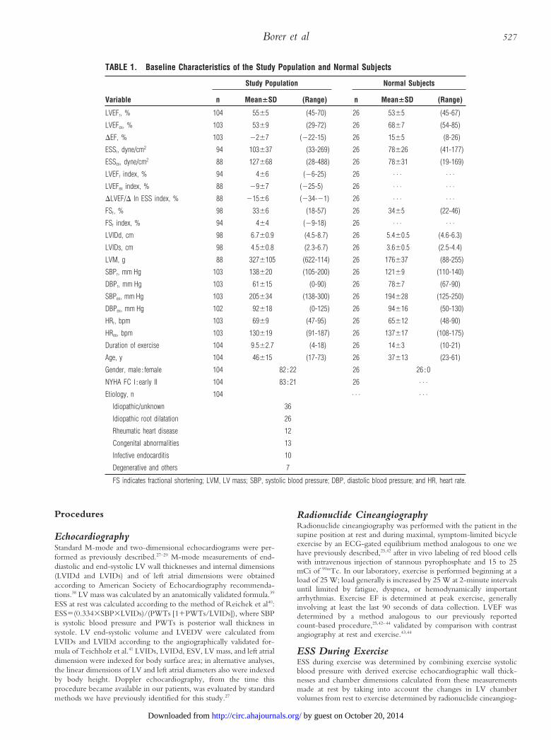
TABLE 1. Baseline Characteristics of the Study Population and Normal Subjects
Variable
Study Population Normal Subjects
n Mean6SD (Range) n Mean6SD (Range)
LVEFr, % 104 5565 (45-70) 26 5365 (45-67)
LVEFex, % 103 5369 (29-72) 26 6867 (54-85)
DEF, % 103 2267 (222-15) 26 1565 (8-26)
ESSr, dyne/cm2 94 103637 (33-269) 26 78626 (41-177)
ESSex, dyne/cm2 88 127668 (28-488) 26 78631 (19-169)
LVEFr index, % 94 466 (26-25) 26 . . . . . .
LVEFex index, % 88 2967 (225-5) 26 . . . . . .
DLVEF/D ln ESS index, % 88 21566 (234-21) 26 . . . . . .
FSr, % 98 3366 (18-57) 26 3465 (22-46)
FSr index, % 94 464 (29-18) 26 . . . . . .
LVIDd, cm 98 6.760.9 (4.5-8.7) 26 5.460.5 (4.6-6.3)
LVIDs, cm 98 4.560.8 (2.3-6.7) 26 3.660.5 (2.5-4.4)
LVM, g 88 3276105 (622-114) 26 176637 (88-255)
SBPr, mm Hg 103 138620 (105-200) 26 12169 (110-140)
DBPr, mm Hg 103 61615 (0-90) 26 7867 (67-90)
SBPex, mm Hg 103 205634 (138-300) 26 194628 (125-250)
DBPex, mm Hg 102 92618 (0-125) 26 94616 (50-130)
HRr, bpm 103 6969 (47-95) 26 65612 (48-90)
HRex, bpm 103 130619 (91-187) 26 137617 (108-175)
Duration of exercise 104 9.562.7 (4-18) 26 1463 (10-21)
Age, y 104 46615 (17-73) 26 37613 (23-61)
Gender, male;female 104 82;22 26 26;0
NYHA FC I;early II 104 83;21 26 . . .
Etiology, n 104 . . . . . .
Idiopathic/unknown 36
Idiopathic root dilatation 26
Rheumatic heart disease 12
Congenital abnormalities 13
Infective endocarditis 10
Degenerative and others 7
FS indicates fractional shortening; LVM, LV mass; SBP, systolic blood pressure; DBP, diastolic blood pressure; and HR, heart rate.
Borer et al 527
by guest on October 20, 2014http://circ.ahajournals.org/Downloaded from

raphy. Logistic difficulties preclude concurrent performance of echo-cardiography and radionuclide cineangiography; therefore, data forthe calculation were obtained from studies performed sequentiallyduring the same visit. ESS at peak exercise was estimated from theformulas below on the basis of the following assumptions: (1) Theratio of rest and peak exercise LVEDV determined by radionuclidecineangiography is the same as this ratio determined from echocar-diographic measurements of LVEDV; (2) LV mass and density remainconstant from rest to exercise; (3) the ratio of PWT to IVS is identicalat rest and during exercise. The standard echocardiographic formulasfor LV mass15 and for LV chamber volume were used to develop theformula for exercise ESS.
(1a) LVM (g)50.83{1.043[(LVID1IVS1PWT)32LVID3]}10.6
(1b) Mean wall thickness (MWT)5(IVS1PWT)/2
(2) LV volume (ESV or EDV)5[7/(2.41LVID)]3LVID3
(3) ESS5[(0.3343SBP3LVIDs)/PWTs]3[11(PWTs/LVIDs)]
By calculating the ratio of the rest (r) and exercise (ex) LVEDV fromradionuclide cineangiography, echocardiographic (echo) dimensionsat peak exercise were estimated on the basis of assumption 1 and therelationship between the measured resting echo variables and restingecho LVEDV. Thus,
(4) LVEDVecho,ex/LVEDVecho,r5[LVEDV(RNCAex/LVEDV(RNCAr]
or
LVEDVecho,ex5LVEDVecho,r3[LVEDV(RNCAex/LVEDV(RNCAr]
LVIDex was determined from LVEDVecho,ex from Equation 2 solved byiterative estimation of the LVID that produced the LVEDVecho,ex
within 0.001 mL of the LVEDV from Equation 4. With LVIDex
known, MWTex was calculated from LVIDecho, ex from Equations 1aand 1b and assumption 2 (ie, LV myocardial volume is identical at restand exercise). Given assumption 3, the two unknowns, PWT and IVS,were determined from Equations 1a and 1b for the sum and the ratioof PWT and IVS. Exercise ESS was calculated from Equation 3 byentering the SBP (measured by cuff sphygmomanometry) and calcu-lated values of PWTs and LVIDs at peak exercise.
Calculation of LVEF-ESS IndicesFor each of the three indices (LVEFrest normalized for ESSrest, LVEFex
normalized for ESSex, and DLVEF from rest to exercise normalized forchange in ESS from rest to exercise [DESS]), the normal relationbetween the LVEF variable and the natural log of the ESS variable wasdefined by least-squares linear regression analysis. Logarithmic trans-formation of ESS previously has been found to improve prediction ofLV chamber function.45 The relations between rest, exercise, andDLVEF and the corresponding ESS variable in the 26 normal subjectswere used to develop regression equations to predict the average valueof each performance variable expected in normal subjects for a givenvalue of ESS, as previously reported for functional stratification byechocardiography46,47 alone and combined with radionuclide cinean-giography.35,48 The LVEF variable recorded for each patient then wassubtracted from the normally expected LVEF variable. The difference,the LVEF-ESS index variable, was used in statistical analyses, as notedbelow (Fig 1).
Statistical AnalysisBaseline variables screened for prognostic significance are denoted inTable 1. The Kaplan-Meier product limit estimate method49 was usedto determine rate of progression from study entry to the initial primaryend points. These end points included (1) operable symptoms alone,(2) operable symptoms or LV dysfunction (determined by radionu-clide cineangiography or cardiac catheterization), (3) operable symp-toms or cardiac death, and (4) all cardiac events (symptoms, LVdysfunction, death). When an end point was reached, or whenoperation was performed for reasons other than development ofsymptoms or LV dysfunction (3 patients), or when documentednoncardiac death occurred (3 patients), the patient was censored from
further analysis. Kaplan-Meier product limit estimate curves werecompared by the log-rank test to provide univariate assessment of therelation of selected clinical risk factors, hemodynamic variables, andfunctional and other descriptors measured during index radionuclidestudy or echocardiography to each of the primary end points both forthe patient population of 104 and among the 88 patients for whomechocardiographic data were sufficiently complete to support ESS-based analysis. Because of the limited number of end points, analysesof the relation of prognostic indices to two additional end points, (5)cardiac death and (6) LV dysfunction alone, were undertaken on aunivariate, exploratory basis only. For all but the exploratory analyses,continuous variables were partitioned according to statistical terciles(ie, the group was divided into thirds according to the distribution ofthe variable of interest, with no a priori assumptions or biases relativeto cut points). Variables that were significantly (P,.05) related to endpoints were entered into a series of multivariate Cox proportionalhazards models50 to confirm their independent predictive value foreach end point. Separate Cox models were constructed for baselineclinical descriptors and for LV function or size predictors. Variablesfound to be independently predictive in these separate models thenwere entered into a final, summative multivariate Cox model.
ResultsObjective measures of LV size and performance at index studyare presented in Table 1. During follow-up (average, 7.4 yearsfor patients remaining alive and not operated on), 39 of 104patients reached a “surgical” end point or died suddenlywithout indications for operation. Of these, 22 developed asymptomatic indication for surgery (dyspnea FC II, III, IV[n59, 3, 6], angina FC $II [n53], fatigue FC $II [n51])without LV dysfunction; 7 developed LV dysfunction withoutsymptoms; 6 developed symptoms (dyspnea FC II, III, IV[n51, 2, 2], fatigue FC II [n51]) and evidenced concomitantLV dysfunction for the first time when studied immediatelybefore operation; and 4 died suddenly without LV dysfunctionor operable symptoms at their last evaluation 6 to 10 monthsbefore death. Three additional patients died of documentednoncardiac cause (infection in 1, cancer in 2) and werecensored from analysis at the time of this event. The annual risk
Figure 1. A, Relation of a performance descriptor, LVEF, to aloading descriptor, ESS, after log transformation (ln) of ESS, asdefined from 26 normal subjects. To define LVEF-ESS index fora patient, patient’s ln ESS is determined and, from normalregression line, expected LVEF is defined. Patient’s measuredLVEF (EF observed) is then subtracted from expected value. Dif-ference is LVEF-ESS index. B, Relation of DLVEF to D ln ESS,analogous to relation depicted in A, for 88 patients with severeaortic regurgitation. Solid regression line defines this relation in26 normal subjects; dashed lines represent 1 and 2 SD belowthis mean normal relation. Clinical outcome (end points) inpatients is defined by symbols, as noted.
528 Prognostication in Chronic Aortic Regurgitation
by guest on October 20, 2014http://circ.ahajournals.org/Downloaded from

of reaching a surgical end point or cardiac death was 6.2% (Fig2), and for a surgical end point alone, 6.0%. At 5 years, 75% ofpatients had not yet died of cardiac cause or developedindications for valve replacement; the 5-year risk of cardiacdeath alone was 2.4%. Half the patients were projected to reachcardiac end points by '10 years after study entry (Fig 2).
Univariate Predictors of Single and CombinedEnd Points
Clinical VariablesWhen clinical variables were considered by univariate analysisfor the 88 patients with complete data, NYHA FC and agewere significantly associated with development of operablesymptoms alone and of symptoms and/or subnormal LVperformance, although not with subnormal LV performancealone. FC, but not age, was associated with cardiac death(Table 2). FC and age similarly were significantly related tomost end points among the entire 104-patient cohort. Con-versely, assessment for the impact of AR cause on outcomerevealed no association between cause and cardiac end points.
LV Size, Performance, or FunctionUnivariate analysis of LV function or size descriptors revealedsignificant associations between several variables and $1 of the6 single or combined morbidity and mortality end points(Table 2). The only variables that significantly predicted all endpoints were DLVEF alone (P5.0006) and DLVEF-DESS index(P5.0002). Significant univariate associations also existed be-tween at least 1 but not all end points and LVEF at restnormalized for ESS at rest (LVEFrest-ESSrest index), exerciseLVEF normalized for exercise ESS (LVEFex-ESSex index),unadjusted LVEFex, and fractional shortening, LVIDd, andLVIDs at rest (Table 2).
Multivariate Analysis for IndependentContribution to End Point PredictionAlthough several variables provided significant prediction byunivariate analysis, when a multivariate Cox proportionalhazards model was constructed, the only independent objec-tive predictor of risk of end points was the DLVEF-DESSindex (Table 2). When this complex variable was not consid-
TABLE 2. Clinical and Objective Univariate* andMultivariate† Predictors of Cardiac End Points
Independent VariableUnivariate
PMultivariate
P
Death, operable symptoms or LV dysfunction
DLVEF-DESS index .0002 ,.001‡
DLVEF .0006 NS
LVEFex .0044 NS
LVIDs .0113 NS
FSr .0227 NS
LVEFr-ESSr index .0486 NS
NYHA FC (I vs early II) ,.0001 ,.001
Operable symptoms or LV dysfunction
DLVEF-DESS index .0015 ,.001‡
DLVEF .0099 NS
FSr .0238 NS
LVIDs .0259 NS
LVEFex .0260 NS
NYHA FC (I vs early II) .0001 .001
Operable symptoms or cardiac death
DLVEF-DESS index .0006 ,.001‡
DLVEF .0036 . . .
LVEFr-ESSr index .0413 . . .
LVEFex .0481 . . .
NYHA FC (I vs early II) ,.0001 ,.001
Age .0171 . . .
Operable symptoms
DLVEF-DESS index .0041 ,.001‡
DLVEF .0395 . . .
NYHA FC (I vs early II) ,.0001 ,.001
Age .0198 . . .
LV dysfunction
LVEFex .0004 . . .
DLVEF-DESS index .0090 . . .
LVIDd .0305 . . .
LVIDs .0423 . . .
LVEFex-ESSex index .0494 . . .
DLVEF .0636 . . .
NYHA FC (I vs early II) .0881 . . .
Cardiac death
DLVEF-DESS index .0212 . . .
DLVEF .0345 . . .
NYHA FC (I vs early II) .0274 . . .
D indicates change from rest to exercise; FS, fractional shortening.*Independent variables analyzed by comparison (log-rank test) of Kaplan-Meier
product limit estimate curves incorporating all data available in 104 patients; Pvalues are unadjusted.
†Independent variables analyzed by forward stepwise Cox regression analysisamong 88 patients with complete data: final model constructed with variablessignificantly predictive in initial, separate noninvasive and clinical multivariatemodels; P values are adjusted, computed with all variables considered together inthe final model.
‡Enters first in final multivariate Cox models conducted among 88 study patientsand among the study patients without coronary artery disease, aortic stenosis, ormitral regurgitation.
Figure 2. Natural history of progression to end points in severechronic aortic regurgitation (see text).
Borer et al 529
by guest on October 20, 2014http://circ.ahajournals.org/Downloaded from

ered, all independent prognostic information for operablesymptoms and for symptoms and/or subnormal LV perfor-mance was carried by DLVEF alone; the latter variable also wasthe only predictor of cardiac death by univariate analysis otherthan DLVEF-DESS index.
When FC was included in the multivariate model, thisclinical descriptor provided independent information thatmodified prediction on the basis of the prognostically strongerDLVEF-DESS index. When multivariate analysis was per-formed only on the asymptomatic (NYHA FC I) patients,DLVEF-DESS index continued to be the primary provider ofindependent significant prognostic information for all cardiacevents (P,.001), with LVIDs providing lesser but significantmodification of the model. When patients who subsequentlywere found to have developed coronary artery disease and/oraortic stenosis or mitral regurgitation were excluded fromanalysis, again DLVEF-DESS index was statistically significantand was the predominant independent predictor of operablesymptoms alone, operable symptoms or death, operable symp-toms or LV dysfunction, and total cardiac events (P,.001, allend points, Table 2). Although not as powerful in prediction asDLVEF-DESS, FC also was significantly predictive and wassecond to DLVEF-DESS for all outcomes in this group; finally,after FC, LVIDs was predictive of all events and of operablesymptoms or LV dysfunction, whereas use of antiarrhythmicdrugs was predictive of operable symptoms alone and ofsymptoms or sudden death. No other variables were predictivein multivariate analysis among patients with AR but withoutconfounding conditions.
A DLVEF-DESS index of 217% was the upper boundary ofthe tercile comprising the patients at highest risk (Fig 3).Patients with DLVEF-DESS indices lower than this valuemanifested a 13.3%/y annual rate of progression to any cardiacend point. When unadjusted DLVEF was considered, theupper bound of the high-risk tercile was DLVEF of 25%;patients with DLVEF less than this value manifested a 12.5%/yannual rate of progression to any cardiac end point. Con-versely, the relatively lowest-risk tercile was bounded by a
lower DLVEF-DESS index of 211%. Patients with higherindices evidenced a 1.8%/y annual rate of progression to anycardiac end point. The corresponding “low-risk” unadjustedDLVEF lower bound was 13%; higher values predicted a1.9%/y annual rate of progression to any cardiac end point.
Predictors of Subnormal LV Performance andCardiac DeathSubnormal LV performance at rest and cardiac death occurredtoo infrequently to permit stable multivariate analyses and wereevaluated only for univariate association with predictors. Forsubnormal LV performance, the strongest univariate associa-tion was with absolute LVEFex, followed closely by DLVEF-DESS index (Fig 4); other, less prominent but significantassociations also were apparent (Table 2). The highest-risktercile, bounded by LVEFex #49%, evidenced an 8.8%/ylikelihood of developing subnormal LV performance at rest,whereas the low-risk tercile (LVEFex $57%) had a 0%/ylikelihood of developing this end point during our follow-up.For the previously defined high- and low-risk DLVEF-DESSterciles, the rates of progression to subnormal LV performancewere 6.7%/y and 0%, respectively. For cardiac death, thestrongest association was with DLVEF-DESS index; the 5-yearsudden death risk was 3.3% in the high-risk tercile (Fig 5). Theonly other significant objective prognosticator was DLVEFalone, which predicted this outcome almost as well. Althoughless strongly associated with death than DLVEF-DESS index,early FC II symptoms also bore a significant relation tosubsequent sudden death.
Effect of Cardioactive DrugsBy univariate analysis, adverse outcomes were associated withuse of any drug at entry (all cardiac end points, symptomsalone, and symptoms and/or subnormal LV performance);antiarrhythmic drugs or digoxin alone at entry predicteddevelopment of cardiac end points. However, in multivariateCox models, neither use of specific agents nor of any cardio-active drug added independently to prediction by DLVEF-DESS index.
Figure 3. Relation of DLVEF-DESS index at study entry tooccurrence of any cardiac end point (cardiac death, operablesymptoms, and/or subnormal LV performance at rest) duringfollow-up. Population of 88 patients with evaluable data for thisanalysis has been divided statistically into terciles. DLVEF-DESSindex boundaries for each tercile are boxed.
Figure 4. Relation of DLVEF-DESS (stress) index at study entryto occurrence of subnormal LV performance at rest during sub-sequent follow-up. The 88 patients evaluable for this purposehave been divided into statistical terciles, as in Fig 3.
530 Prognostication in Chronic Aortic Regurgitation
by guest on October 20, 2014http://circ.ahajournals.org/Downloaded from

DiscussionOur data support our hypothesis that the development ofcurrently accepted indications for aortic valve replacement,including symptoms or subnormal LV performance, can bepredicted among patients with AR on the basis of objectivedescriptors of LV size, performance, and function. This findingis consistent with the results of previous studies of patients notoperated on,10–12,14,51 which also concluded that assessment ofLV functional and size characteristics could predict develop-ment of indications for operation. Our findings also areconsistent with the several series in which preoperative char-acteristics have significantly predicted late postoperative out-come,3–5,9,13,15,34 thus indicating the prognostic utility of objec-tive LV size and performance assessment in the setting of AR.
Our data add to those of previous investigators of load-adjusted performance indices in patients with chronic AR,9,52,53
first, by demonstrating the relative prognostic power of ESS-normalized LVEF (and specifically, of the DLVEF-DESSindex) in comparison with more conventional descriptors andsecond, by demonstrating that subnormal LV performance andcardiac death alone can be predicted effectively. Thus, wefound that DLVEF-DESS index was the best single predictor ofall cardiac end points, of symptoms alone, of symptoms and/orsubnormal LV performance, and of sudden cardiac death and,together with absolute LVEFexercise, was among the two stron-gest predictors of subnormal LV performance. The relativeutility of an LVEF-ESS index is consistent with our previouspreliminary report13,35 among patients studied immediatelybefore valve replacement, which indicated that exercise LVEFnormalized to resting ESS was the best and only independentpredictor of asymptomatic survival during prolonged fol-low-up after operation, whereas ESS-normalized resting LVEFwas the best and only independent predictor of survival alone.However, for that study, we could not yet assess ESS duringexercise and could not perform all analyses reported here. Thevalue of the LVEF-ESS indices is not surprising, because theyare more closely related to myocardial contractility,20–23 anintrinsic property of the heart muscle, than is LVEF (orfractional shortening or LVIDs), which is directly and impor-
tantly affected by loading conditions as well as by intrinsiccontractility. Therefore, it is intuitively reasonable that theload-adjusted performance variable should be more closelyrelated to clinical outcome than the performance variable.Consistent with this premise, Carabello et al9 demonstratedthat another contractility descriptor, ESS/ESV index, whenmeasured immediately before operation, significantly predictsoutcome after valve replacement in patients with any valvulardisease, although the inclusion of only 9 patients with ARprecluded conclusions specifically about prognostication in thisdisease alone. Similarly, Gaasch et al53 found that a combina-tion of preoperative end-systolic dimension and an index ofsystolic wall stress was the most useful predictor of poorpostoperative outcome among 32 patients with aortic AR.
Our results are consistent with our experimental studies,which indicate the presence of profound abnormalities inmyocardial contractile protein metabolism in a model ofchronic moderate to severe AR, with markedly subnormalmyocardial protein degradation rate associated with heartfailure and LV dysfunction.54–56 These studies have suggestedthe possible relation between disordered contractile proteinmetabolism, subnormal LV contractility, and clinical outcome,a relation mirrored in our clinical findings.
Our data demonstrate that subnormal LV performance andcardiac death can be predicted objectively. Previous seriesincluded relatively few patients with subnormal LV perfor-mance at the time of operation10,11 or did not specifically assesspredictors of this outcome.12,14 For example, Bonow et al14
identified 15 patients who developed LV dysfunction, but theyevaluated only their 4 patients who developed LV dysfunctionwithout symptoms and found no clear predictor. However,identification of patients imminently at risk of subnormal LVperformance, with or without symptoms, is potentially impor-tant: outcome is worse after operation among those withsubnormal LV performance at rest than those who undergooperation with symptoms and normal LV performance.3,5,34
Our 13 subnormal LVEF end points do not constitute a largeseries and were not sufficiently numerous to support multivar-iate analyses. Nonetheless, our analysis indicates the significantcapacity of LVEFex and of DLVEF-DESS index to predictdevelopment of an important risk factor and represents the firstsuch report of which we are aware.
Previous studies have reported few cardiac deaths withoutoperation among asymptomatic patients: Bonow et al14 ob-served 2 among 104 patients during an 8-year follow-up, andHenry et al,3 Lindsay et al,11 and Siemienczuk et al10 saw noneamong a total of 118 patients followed for shorter periods. Forthis reason, sudden death generally has not been considered animportant risk for patients with AR who are asymptomatic andhave normal LV performance at rest. Moreover, Bonow et alwere unable to identify potential predictors of the suddendeaths they observed. In the present series, the occurrence ofsudden death clearly is related to LV dysfunction duringexercise at study entry. Although the absolute frequency of thisevent in an AR population cannot be defined rigorously fromour relatively small series, the prominence of this end point inour group suggests that potential sudden death may requirespecific consideration when management decisions are madefor patients with AR. Subnormal LV performance at rest and
Figure 5. Relation of DLVEF-DESS index at study entry to car-diac death during follow-up. Because of small number of endpoints, group is divided on basis of a binary split for this figure.For risk values associated with tercile boundaries noted in Figs3 and 4, see text.
Borer et al 531
by guest on October 20, 2014http://circ.ahajournals.org/Downloaded from
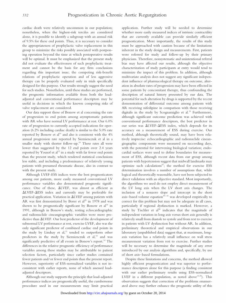
cardiac death were relatively uncommon in our population;nonetheless, when the highest-risk terciles are consideredalone, it is possible to identify a subgroup with an annual riskof 9.5% for these end points. Thus, it is necessary to considerthe appropriateness of prophylactic valve replacement in thisgroup to minimize the risks possibly associated with postpon-ing operation beyond the time at which postoperative resultswill be optimal. It must be emphasized that the present studydid not evaluate the effectiveness of such prophylactic treat-ment and cannot be the basis for any firm conclusionsregarding this important issue; the competing risk-benefitrelations of prophylactic operation and of less aggressivetherapy can be properly evaluated only in trials specificallydesigned for this purpose. Our results strongly suggest the needfor such studies. Nonetheless, until these studies are performed,the prognostic information available from both the load-adjusted and conventional performance descriptors may beuseful in decisions in which the known competing risks ofvalve replacement are considered.
Our data support those of Bonow et al concerning the rateof progression to end points among asymptomatic patientswith AR who have normal LV performance at rest. Our 6.0%rate of progression to currently accepted indications for oper-ation (6.2% including cardiac death) is similar to the 5.0% ratereported by Bonow et al14 and also is consistent with the 4%annual progression rate reported by Siemienczuk et al in asmaller study with shorter follow-up.10 These rates all werelower than suggested by the 12 end points over 2.4 yearsreported by Yousof et al12 in a study with far fewer end pointsthan the present study, which rendered statistical conclusionsless stable, and including a predominance of relatively youngpatients with presumed rheumatic disease, also in contrast towith the present study.
Although LVEF-ESS indices were the best prognosticatorsamong our patients, more easily measured conventional LVperformance variables also demonstrated prognostic signifi-cance. One of these, DLVEF, was almost as efficient asDLVEF-DESS index and currently may be preferable forpractical application. Variation in DLVEF among patients withAR was first demonstrated by Borer et al25 in 1978 and wasshown to be prognostically significant by Bonow et al14 in1991, although in Bonow’s study, resting echocardiographicand radionuclide cineangiographic variables were more pre-dictive than DLVEF. Our best predictor of the development ofsubnormal LV performance at rest, exercise LVEF, also was theonly significant predictor of combined cardiac end points inthe study by Lindsay et al,11 tended to outperform otherpredictors in the study by Siemienczuk et al,10 and wassignificantly predictive of all events in Bonow’s report.14 Thedifferences in the relative prognostic efficiency of performancevariables among these studies may be attributable to patientselection factors, particularly since earlier studies containedfewer patients and/or fewer end points than the present report.However, superiority of ESS-normalized variables is not in-consistent with earlier reports, none of which assessed load-adjusted descriptors.
Although our study supports the principle that load-adjustedperformance indices are prognostically useful, the cumbersomeprocedure used in our measurements may limit practical
application. Further study will be needed to determinewhether more easily measured indices of intrinsic contractilitythat are currently available can provide similarly efficientprognostication. More importantly, the results of this studymust be approached with caution because of the limitationsinherent in the study design and measurements. First, patientswere referred for study and follow-up by their primaryphysicians. Therefore, nonsystematic and unintentional referralbias may have affected our results, although the objectivecharacterization of study participants at entry would tend tominimize the impact of this problem. In addition, althoughmultivariate analysis does not suggest any significant indepen-dent influence of pharmacological therapy on outcome, alter-ation in absolute rates of progression may have been effected insome patients by concomitant therapy, thus confounding thedescription of natural history presented by our data. Thepotential for such alteration by drugs is supported by the recentdemonstration of differential outcome among patients withAR receiving nifedipine in comparison with those receivingdigitalis in the study by Scognamiglio et al.19 Furthermore,although significant outcome prediction was achieved withconventional performance descriptors, the best predictor inour series was DLVEF-DESS index, which depends for itsaccuracy on a measurement of ESS during exercise. Ourmethod, although theoretically sound, may have been rela-tively imprecise: echocardiographic and radionuclide cinean-giographic components were measured on succeeding days,with the potential for intervening biological variation; endo-cardial surfaces were used to define boundaries for measure-ment of ESS, although recent data from our group amongpatients with hypertension suggest that midwall landmarks mayoptimize such calculations57; the method for exercise ESSdetermination involves a number of assumptions that, whilelogical and theoretically reasonable, have not been subjected todirect validation with an objective standard. Most importantly,the algorithms we used do not employ direct measurement ofthe LV long axis when the LV short axis changes. Theinclusion of a nonzero slope and intercept in the shortaxis–based volume equation39–41 was developed empirically tocorrect for this problem but may not be adequate in all cases,particularly if regional dysfunction is marked. However, astudy by Tischler et al58 indicates that the magnitude ofindependent variation in long axis versus short axis generally isrelatively small from diastole to systole and from rest to exercisein patients with LV dysfunction but without AR. In addition,preliminary theoretical and empirical observations in ourlaboratory (unpublished data) suggest that, at maximum, long-axis variation has a relatively small influence on wall stressmeasurement variation from rest to exercise. Further studieswill be necessary to determine the magnitude of any errorintroduced by our analysis algorithms and, specifically, by useof short axis–based formulations.
Despite these limitations and concerns, the method allowedhighly efficient prognostication and was superior to perfor-mance descriptors alone for this purpose (a finding consistentwith our earlier preliminary results using ESS-normalizedLVEF in a different population, as noted above13,35). Thisobservation suggests that correction of the problems enumer-ated above may further enhance the prognostic utility of this
532 Prognostication in Chronic Aortic Regurgitation
by guest on October 20, 2014http://circ.ahajournals.org/Downloaded from

descriptor, a hypothesis that requires empirical testing. Finally,we used each patient’s initial study to predict events that oftenoccurred many years later. Because one goal of evaluation is toidentify imminent development of “high-risk” descriptors, it ispossible that serial determination of the most predictive vari-ables would enhance the precision and practical utility ofprognostication, as suggested by the study by Bonow et al.14
Further study will be needed to evaluate this possibility.
AcknowledgmentsThis work was supported in part by National Heart, Lung, and BloodInstitute grant RO1-HL-26504 (Dr Borer, principal investigator) andby grants from The Howard Gilman Foundation, the Lasdon Foun-dation, the American Cardiovascular Research Foundation, theDaniel and Elaine Sargent Charitable Trust, the Charles and JeanBrunie Foundation, and the David Margolis Foundation (all NewYork, NY), and by much appreciated gifts from Ronald and JeanSchiavone, William and Donna Aquavella, William and MaryjaneVoute, and Milton and Rita Weinick. The authors wish to acknowl-edge the invaluable assistance of Gerald Casadei, BA, DeborahLauterstein, MA, Dawn Fishman, BA, and John Teevan III, BA, fortheir indefatigable assistance in arranging patient follow-up, compilingand archiving data, and aiding in analytical procedures; of Ben Deyi,MS, for his invaluable assistance in statistical analysis; and of JohnClement, MA, for his critical help in the preparation ofthis manuscript.
References1. Gaasch WH, Andrias CW, Levine HJ. Chronic aortic regurgitation: the
effect of aortic valve replacement on left ventricular volume, mass andfunction. Circulation. 1978;58:825–836.
2. Borer JS, Rosing DR, Kent KM, Bacharach SL, Green MV, Johnston GS,Morrow AG, Epstein SE. Left ventricular function at rest and duringexercise after aortic valve replacement in patients with aortic regurgitation.Am J Cardiol. 1979;44:1297–1305.
3. Henry WL, Bonow RO, Borer JS, Ware JH, Kent KM, Redwood DR,McIntosh CL, Morrow AG, Epstein SE. Observations on the optimumtime for operative intervention for aortic regurgitation, I: evaluation of theresults of aortic valve replacement in symptomatic patients. Circulation.1980;61:471–483.
4. Bonow RO, Borer JS, Rosing DR, Henry WL, Pearlman AS, McIntoshCL, Morrow AG, Epstein SE. Preoperative exercise capacity in symptom-atic patients with aortic regurgitation as a predictor of postoperative leftventricular function and long-term prognosis. Circulation. 1980;62:1280–1290.
5. Greves J, Rahimtoola SH, McAnulty JH, DeMots H, Clark DG,Greenberg B, Starr A. Preoperative criteria predictive of late survivalfollowing valve replacement for severe aortic regurgitation. Am Heart J.1981;101:300–308.
6. Bonow RO, Rosing DR, McIntosh CL, Jones M, Maron BJ, Lan KKG,Lakatos E, Bacharach SL, Green MV, Epstein SE. The natural history ofasymptomatic patients with aortic regurgitation and normal left ventricularfunction. Circulation. 1983;68:509–517.
7. Bonow RO, Rosing DR, Maron BJ, McIntosh CL, Jones M, BacharachSL, Green MV, Clark RE, Epstein SE. Reversal of left ventricular dys-function after aortic valve replacement for chronic aortic regurgitation:influence of duration of preoperative left ventricular dysfunction. Circu-lation. 1984;70:570–579.
8. Daniel WG, Hood WP, Siart A, Hausmann D, Nellessen U, Oelert H,Lichtlen PR. Chronic aortic regurgitation: reassessment of the prognosticvalue of left ventricular end-systolic dimension and fractional shortening.Circulation. 1985;71:669–680.
9. Carabello BA, Williams H, Gash AK, Kent R, Belber D, Maurer A, SiegelJ, Blasius K, Spann JF. Hemodynamic predictors of outcome in patientsundergoing valve replacement. Circulation. 1986;74:1309–1316.
10. Siemienczuk D, Greenberg B, Morris C, Massie B, Wilson RA, Topic N,Bristow JD, Cheitlin M. Chronic aortic insufficiency: factors associatedwith progression to aortic valve replacement. Ann Intern Med. 1989;110:587–592.
11. Lindsay J, Silverman A, Van Voorhees LB, Nolan NG. Prognostic impli-cations of left ventricular function during exercise in asymptomatic patientswith aortic regurgitation. Angiology. 1987;38:386–392.
12. Yousof AM, Mohammed MMJ, Khan N, Shuhaiber H, Cherian G.Chronic severe aortic regurgitation: a prospective follow-up of 60 asymp-tomatic patients. Am Heart J. 1988;116:1262–1267.
13. Borer, JS. Prognostication strategies in heart failure and valvular heartdiseases: current concepts and their support. In: Yacoub MH, ed. 1989Annual of Cardiac Surgery. London, UK: Current Science, Ltd, 115–124.
14. Bonow RO, Lakatos E, Maron BJ, Epstein SE. Serial long-term assessmentof the natural history of asymptomatic patients with chronic aortic regur-gitation and normal left ventricular systolic function. Circulation. 1991;84:1625–1635.
15. Recke SH, Marienhagen J, Feistel H, Platsch G, Bock E, von der Emde J.Electrocardiographic characteristics indicating a risk of irreversiblyimpaired myocardial function in chronic aortic regurgitation. Int J Cardiol.1993;42:129–138.
16. Bonow RO. Asymptomatic aortic regurgitation: indications for operation.J Cardiac Surg. 1994;9(suppl):170–173.
17. Borer JS, Bonow RO, Bacharach SL, Green MV. The effects of nitro-glycerin in patients with valvular heart disease: hemodynamic and radio-nuclide cineangiographic studies. In: Lichtlen PR, Engel H-J, Schrey A,Swann HJC, eds. Proceedings of the Third International Symposium on Nitrates,Monte Carlo, 1980. Berlin, Germany: Springer-Verlag; 1981: Nitrates3:546–551.
18. Greenberg BH, Massie B, Bristow JD, Cheitlin M, Siemienczuk D, TopicN, Wilson RA, Szlachcic J, Thomas D. Long-term vasodilator therapy ofchronic aortic insufficiency: a randomized double-blinded, placebo-controlled clinical trial. Circulation. 1988;78:92–103.
19. Scognamiglio R, Rahimtoola SH, Fasoli G, Nistri S, Dalla Volta S. Ni-fedipine in asymptomatic patients with severe aortic regurgitation andnormal left ventricular function. N Engl J Med. 1994;331:689–694.
20. Suga H, Sugawa K. Instantaneous pressure-volume relationships and theirratio in the excised, supported canine left ventricle. Circ Res. 1974;35:117–127.
21. Mirsky I, Henschke C, Hess OM, Krayenbuehl HP. Prediction of post-operative performance in aortic valve disease. Am J Cardiol. 1981;48:295–303.
22. Shen FW, Roubin GS, Choong CY-P, Hutton BF, Harris PJ, Fletcher PJ,Kelly DT. Evaluation of the relationship between myocardial contractilestate and left ventricular function in patients with aortic regurgitation.Circulation. 1985;71:1–38.
23. Starling MR, Kirsh MM, Montgomery DG, Gross MD. Mechanisms forleft ventricular systolic dysfunction in aortic regurgitation: importance forpredicting the functional response to aortic valve replacement. J Am CollCardiol. 1991;17:887–897.
24. Booth D, DeMaria A, Nissen S, Waters J. Relationship of contractile stateto ejection performance in patients with chronic aortic valve disease.Circulation. 1986;73:47–53.
25. Borer JS, Bacharach SL, Green MV, Kent KM, Henry WL, Rosing DR,Seides SF, Johnston GS, Epstein SE. Exercise-induced left ventriculardysfunction in symptomatic and asymptomatic patients with aortic regur-gitation: assessment by radionuclide cineangiography. Am J Cardiol. 1978;42:351–357.
26. Borer JS, Herrold EM, Hochreiter C, Roman MJ, Supino P, DevereuxRB, Kligfield P, Nawaz H. Natural history of left ventricular performanceat rest and during exercise after aortic valve replacement for aortic regur-gitation. Circulation. 1991;84(suppl III):III-133-III-139.
27. Roman MJ, Klein L, Devereux RB, Kligfield P, Niles NW, Hochreiter C,Isom OW, Borer JS. Reversal of left ventricular dilatation, hypertrophy,and dysfunction by valve replacement in aortic regurgitation. Am Heart J.1989;118:553–563.
28. Roman MJ, Devereux RB, Niles NW, Hochreiter C, Kligfield P, Sato N,Spitzer M, Borer JS. Aortic root dilatation as a cause of isolated, severeaortic regurgitation. Ann Intern Med. 1987;106:800–807.
29. Niles NW, Borer JS, Kamen M, Hochreiter C, Devereux RB, Kligfield P.Preoperative left and right ventricular performance in combined aortic andmitral regurgitation and comparison with isolated aortic or mitral regurgi-tation. Am J Cardiol. 1990;65:1372–1378.
30. Wallerson DC, Dubin J, Devereux RB. Assessment of cardiac hemody-namics and valvular function by Doppler echocardiography. Bull N Y AcadMed. 1987;63:762–796.
31. Perry GJ, Helmcke F, Nanda NC, Byard C, Soto B. Evaluation of aorticinsufficiency by Doppler color flow mapping. J Am Coll Cardiol. 1987;9:952–959.
Borer et al 533
by guest on October 20, 2014http://circ.ahajournals.org/Downloaded from
32. Herrold EM, Carter JN, Borer JS. Volume overload related shape changelimits mass increase with wall thickening but only minimally reduces wallstress. Computers in Cardiology 1992. IEEE Computer Society publication0276–6547/92:287–290.
33. Hochreiter C, Niles N, Devereux RB, Kligfield P, Borer JS. Mitralregurgitation: relationship of non-invasive descriptors of right and leftventricular performance to clinical and hemodynamic findings and toprognosis in medically and surgically treated patients. Circulation. 1986;73:900–912.
34. Bonow RO, Picone AL, McIntosh CL, Jones M, Rosing DR, Maron BJ,Lakatos E, Clark RE, Epstein SE. Survival and functional results after valvereplacement for aortic regurgitation from 1976 to 1983: impact of preop-erative left ventricular function. Circulation. 1985;72:1244–1256.
35. Borer JS, Herrold EM, Hochreiter C, Niles N, Devereux R, Kligfield P,Roman M, Chlouverakis G. Aortic regurgitation: stress-normalized ven-tricular performance predicts post-operative prognosis. Circulation. 1991;84(suppl II):II-639. Abstract.
36. Goldschlager N, Pfeifer J, Cohn K, Popper R, Selzer A. The natural historyof aortic regurgitation: a clinical and hemodynamic study. Am J Med.1973;54:577–588.
37. Smith HJ, Neutze JM, Roche AHG, Agnew TM, Barratt-Boyes BG. Thenatural history of rheumatic aortic regurgitation and the indications forsurgery. Br Heart J. 1976;38:147–154.
38. Sahn DS, DeMaria A, Kisslo J, Weyman A, for the Committee on M ModeStandardization of the American Society of Echocardiography. Recom-mendations regarding quantitation in M-mode echocardiography: results ofa survey of echocardiographic measurements. Circulation. 1978;58:1072–1083.
39. Devereux RB, Alonso DR, Lutas EM, Gottlieb GJ, Camp E, Sachs I,Reichek N. Echocardiographic assessment of left ventricular hypertrophyin comparison to necropsy findings. Am J Cardiol. 1986;57:450–458.
40. Reichek N, Wilson J, St John Sutton M, Plappert TA, Goldberg S,Hirshfeld JW. Non-invasive determination of end systolic stress: validationof the method and initial application. Circulation. 1982;65:99–108.
41. Teichholz LE, Kreulen T, Herman MV, Gorlin R. Problems in echo-cardiographic volume determinations: echocardiographic-angiographiccorrelation in the presence and absence of asynergy. Am J Cardiol.1976;37:7–11.
42. Borer JS, Bacharach SL, Green MV, Kent KM, Epstein SE, Johnston GS.Real-time radionuclide cineangiography in the non-invasive evaluation ofglobal and regional left ventricular function at rest and during exercise inpatients with coronary artery disease. N Engl J Med. 1977;297:839–844.
43. Goldberg HL, Herrold EM, Hochreiter C, Moses JW, Fisher J, Tamari I,Borer JS. Videodensitometric determination of right ventricular and leftventricular ejection fraction. Am J Noninvasive Cardiol. 1987;1:18–23.
44. Goldberg HL, Moses JW, Borer JS, Fisher J, Tamari I, Skelly NT, CohenB. Exercise left ventriculography utilizing intravenous digital angiography.J Am Coll Cardiol. 1983;2:1092–1098.
45. Lutas EM, Devereux RB, Reis G, Alderman MH, Pickering TG, Borer JS,Laragh JH. Increased cardiac performance in mild essential hypertension:left ventricular mechanics. Hypertension. 1985;7:979–988.
46. Hammond IW, Devereux RB, Alderman MH, Laragh JH. Relation ofblood pressure and body build to left ventricular mass in normotensive andhypertensive employed adults. J Am Coll Cardiol. 1988;12:996–1004.
47. Ganau A, Devereux RB, Atlas SA, Pecker M, Roman MJ, Vargia P, CodyR, Laragh JH. Plasma atrial natriuretic factor in essential hypertension:relation to cardiac size, function and systemic hemodynamics. J Am CollCardiol. 1989;14:715–724.
48. Borer JS, Jason M, Devereux RB, Pickering T, Erle S, Laragh JH. Leftventricular performance in the hypertensive patient: exercise-mediatedseparation of loading influences from intrinsic muscle dysfunction. Chest.1983;83(suppl):S314–S316.
49. Kaplan EL, Meier P. Non-parametric estimation from incomplete obser-vations. J Am Stat Assoc. 1958;53:457–481.
50. Cox DR. Regression models and life-tables. J R Stat Soc. 1972;34(seriesB):187–220.
51. Henry WL, Bonow RO, Rosing DR, Epstein SE. Observations on theoptimum time for operative intervention for aortic regurgitation, II: serialechocardiographic evaluation of asymptomatic patients. Circulation. 1980;61:484–492.
52. Wisenbaugh T, Booth D, DeMaria A, Nissen S, Waters J. Relationship ofcontractile state to ejection performance in patients with chronic aorticvalve disease. Circulation. 1986;73:47–53.
53. Gaasch WH, Carroll JD, Levine HJ, Criscitiello MG. Chronic aorticregurgitation: prognostic value of left ventricular end-diastolic radius/thickness ratio. J Am Coll Cardiol. 1983;1:775–782.
54. Magid NM, Borer JS, Young MS, Wallerson DC, DeMonteiro C. Sup-pression of protein degradation in the progressive cardiac hypertrophy ofchronic aortic regurgitation. Circulation. 1993;87:1249–1257.
55. Magid NM, Wallerson DC, Borer JS. Myofibrillar protein turnover incardiac hypertrophy due to aortic regurgitation. Cardiology. 1993;82:20–29.
56. Magid NM, Opio G, Wallerson DC, Borer JS. Suppressed proteinturnover in congestive heart failure due to severe chronic aortic regurgi-tation. Circulation. 1992;86(suppl I):I-540. Abstract.
57. DeSimone G, Devereux RB, Camango MC, Wallerson DC, Laragh JH.Left ventricular mid-wall mechanics in Goldblatt hypertension: effect of saltintake. Am J Hypertens. 1993;6:34A. Abstract.
58. Tischler MD, Niggel J, Borowski DT, LeWinter MM. Relation betweenleft ventricular shape and exercise capacity in patients with left ventriculardysfunction. J Am Coll Cardiol. 1993;22:751–757.
534 Prognostication in Chronic Aortic Regurgitation
by guest on October 20, 2014http://circ.ahajournals.org/Downloaded from
Copyright © 2022 FDOKUMEN