Myofibrillar protein oxidation affects filament charges ... - CORE
26
1 Myofibrillar protein oxidation affects filament charges, aggregation and water- holding Yulong Bao a , Sjef Boeren b , Per Ertbjerg a * a Department of Food and Environmental Sciences, University of Helsinki, 00014, Helsinki, Finland b Laboratory of Biochemistry, Wageningen University, 6708WE, Wageningen, The Netherlands *Corresponding author: Tel.: +358 503183909; E-mail address: [email protected] Abstract: Hypochlorous acid (HClO) is a strong oxidant that is able to mediate protein oxidation. In order to study the effect of oxidation on charges, aggregation and water-holding of myofibrillar proteins, extracted myofibrils were oxidized by incubation with different concentrations of HClO (0, 1, 5, and 10 mM). Loss of free thiols, loss of histidine and formation of carbonyls were greater with increasing oxidation level and the particle size increased. Water-holding in the 5 and 10 mM HClO groups were greater than in the non-oxidized control. Isoelectric focusing (IEF) showed that the isoelectric point (pI) of oxidized proteins were lower compared to non-oxidized ones. The lower pI values of oxidized proteins suggests that oxidation increased the overall net negative charge of myofibrillar proteins solubilized for IEF. Here we propose a hypothesis that oxidation-induced increase in net negative charges is the driving force for improved water-holding in myofibrils, whereas protein cross-linking and aggregation have an opposing effect by decreasing the water-holding. Keywords: carbonyls, free thiols, isoelectric point, particle size, histidine brought to you by CORE View metadata, citation and similar papers at core.ac.uk provided by Helsingin yliopiston digitaalinen arkisto
-
Upload
khangminh22 -
Category
Documents
-
view
3 -
download
0
Transcript of Myofibrillar protein oxidation affects filament charges ... - CORE

1
Myofibrillar protein oxidation affects filament charges, aggregation and water-
holding
Yulong Baoa, Sjef Boerenb, Per Ertbjerga*
a Department of Food and Environmental Sciences, University of Helsinki, 00014,
Helsinki, Finland
b Laboratory of Biochemistry, Wageningen University, 6708WE, Wageningen, The Netherlands
*Corresponding author: Tel.: +358 503183909; E-mail address: [email protected]
Abstract:
Hypochlorous acid (HClO) is a strong oxidant that is able to mediate protein oxidation. In order to study
the effect of oxidation on charges, aggregation and water-holding of myofibrillar proteins, extracted
myofibrils were oxidized by incubation with different concentrations of HClO (0, 1, 5, and 10 mM). Loss
of free thiols, loss of histidine and formation of carbonyls were greater with increasing oxidation level
and the particle size increased. Water-holding in the 5 and 10 mM HClO groups were greater than in the
non-oxidized control. Isoelectric focusing (IEF) showed that the isoelectric point (pI) of oxidized proteins
were lower compared to non-oxidized ones. The lower pI values of oxidized proteins suggests that
oxidation increased the overall net negative charge of myofibrillar proteins solubilized for IEF. Here we
propose a hypothesis that oxidation-induced increase in net negative charges is the driving force for
improved water-holding in myofibrils, whereas protein cross-linking and aggregation have an opposing
effect by decreasing the water-holding.
Keywords: carbonyls, free thiols, isoelectric point, particle size, histidine
brought to you by COREView metadata, citation and similar papers at core.ac.uk
provided by Helsingin yliopiston digitaalinen arkisto

2
1. Introduction
Protein oxidation, which can readily occur in post-mortem muscle or during meat processing, involves
modifications of some amino acid side chains, such as formation of cross-links and protein carbonylation
(Estévez, 2011; Lund, Heinonen, Baron, & Estevez, 2011). These modifications of proteins are able to
affect meat quality and nutritional and health aspects (Estévez & Luna, 2017). For example, it has been
reported that protein oxidation leads to increased meat toughness (Bao & Ertbjerg, 2015; Lund, Lametsch,
Hviid, Jensen, & Skibsted, 2007). However, the effect of protein oxidation on water-holding of meat,
especially fresh meat, is unclear. Decker, Xiong, Calvert, Crum, & Blanchard (1993) found decreased
water-holding of gels made from oxidized turkey myofibrils. In support of their view, some authors
(Estévez, 2011; Lund et al., 2011) have argued that protein oxidation leads to decreased water-holding.
Recently, decreased water-holding of myofibril gels prepared from beef packaged with oxygen (20 – 80%)
has been reported (Wang, Luo, & Ertbjerg, 2017).
In fresh raw meat oxidation-induced reactions are very complex. Lund et al. (2007) observed higher drip
loss in high oxygen packaged pork as compared to vacuum packaged samples. In agreement, Delles &
Xiong (2014) found higher centrifugation loss in high oxygen packaged pork loins. However, other studies
did not find a clear effect of high oxygen packaging on water-holding when compared to vacuum packaged
beef (Clausen, Jakobsen, Ertbjerg, & Madsen, 2009; Lindahl, Lagerstedt, Ertbjerg, Sampels, & Lundström,
2010; Łopacka, Półtorak, & Wierzbicka, 2017). In contrast, some studies even reported higher drip loss or
purge loss in vacuum packaged meat compared to high oxygen packaged meat (Chen, Zhou, & Zhang,
2015; Sekar, Dushyanthan, Radhakrishnan, & Babu, 2006; Yang et al., 2016). Thus there is currently no
general agreement about the effect of protein oxidation on water-holding of fresh meat.
To understand the effect of protein oxidation on water-holding of meat, it is important to know how protein
oxidation affects physicochemical factors that contribute to water-holding. As reviewed by Puolanne &
3
Halonen (2010), various forces including electrostatic forces and osmotic forces are hypothesized to
contribute to water-holding in meat. Both electrostatic forces and osmotic forces can affect swelling of
myofibrils, and the swelling of myofibrils generally depends on two aspects: the net charges on
myofilaments and structural constraints (e.g., Z-disks, actomyosin cross-bridges and intermediate
filaments). Increased net charges are believed to induce swelling of myofibrils and lead to improved water-
holding, while structural constraints prohibit unlimited swelling (Hamm, 1986; Offer & Trinick, 1983).
In meat, oxidation is known to induce protein cross-links (Decker et al., 1993; Lund et al., 2007) and
oxidation-induced disulfide cross-links between myosin molecules was suggested as a restricting factor
that inhibit swelling of oxidized myofibrils during brine irrigation (Liu, Xiong, & Chen, 2009). However,
there is a lack of information about how oxidation can affect the net charges on myofilaments. Many of
the amino acid sidechains can pick up or lose protons whereby the charges on the protein molecules are
altered. The electrostatic properties of myofibrils are affected by charged amino acid side chains of proteins
and peptides as those constituting the myosin and actin filaments. It has been suggested that oxidative
modification of the side chains lead to modified electronic arrangement on myofilaments (Estévez,
Ventanas, Heinonen, & Puolanne, 2011) and the modification of filament charges was speculated to affect
water-holding.
This paper aims to examine the effect of oxidation on the net charges of myofibrillar proteins by isoelectric
focusing gel analysis, and to provide insight into the mechanism of oxidation-induced changes in water-
holding and aggregation.
2. Materials and methods
2.1. Extraction of myofibrils
4
The pigs were slaughtered at a commercial slaughterhouse in Finland and muscles were excised at 24
hours postmortem, vacuum packaged and transported refrigerated to the lab. Each muscle was trimmed
of visible connective tissue and external fat. Then the muscle was cut into small pieces (about 1 cm
thick) and frozen stored at -20 ºC. Meat pieces were thawed before extraction of myofibrils. One part of
meat samples was homogenized in four parts of cold MES buffer (100 mM KCl, 50 mM MES (2-(N-
Morpholino) ethanesulfonic acid hydrate), 2 mM MgCl2, 2 mM EGTA (ethylene glycol tetraacetic
acid), pH 5.5) for 40 s (two cycles of 20 s each) with a speed of 20,500 rpm using an IKA Ultra Turrax
T25 homogenizer (Labortechnik, Staufen, Germany). The homogenate was passed through a metal
mesh with an average pore size of 1 mm to remove any unbroken meat pieces and connective tissue.
The filtrate was centrifuged at 1500 g for 15 min at 4 ºC. The pellet was washed twice with 20 mL of
MES buffer followed by centrifugation at 1500 g for 15 min at 4 ºC.
2.2. Oxidation of myofibrils
Hypochlorous acid (HClO) is a strong oxidant that is able to mediate protein oxidation (Pattison, Hawkins,
and Davies, 2007). Although no obvious practical application exists, HClO has been used to induce protein
oxidation in meat as an alternative to the Fenton reaction (Soglia, Petracci & Ertbjerg, 2016). The myofibril
pellet was suspended in four volumes of MES buffer. Aliquots of 0.5 mL of the myofibril suspension were
added with different amounts of MES buffer and NaClO (100 mM, pH 8.0, freshly made) to give a final
concentration of 0, 1, 5, and 10 mM HClO in 1 mL reaction mixture. A household NaClO solution was
used and the concentration of NaClO was determined and adjusted to 100 mM by measurement of the
absorbance at 292 nm using an extinction coefficient of 350 M-1 cm-1 (Morris, 1966) and the pH of the 100
mM NaClO solution was adjusted to pH 8.0 with acetic acid. The pH values of the reaction mixtures
(myofibril suspension added with oxidant) were between 5.4 and 5.5. The oxidant exist mainly in the form

5
of hypochlorous acid in the incubations as the pH is well below its pKa value of 7.5. The reaction mixtures
were kept at cold room (5 ºC). After overnight incubation, the reaction mixtures were centrifuged at 2400
g for 10 min to obtain myofibril pellets. The pellets were subsequently washed with MES buffer and
centrifuged at 2400 g for 10 min at room temperature, and the washing was repeated once. The resultant
myofibril pellet was used for determination of protein oxidation, particle size distribution, isoelectric
focusing (IEF) gel analysis, amino acid analysis, and water-holding.
2.3. Determination of protein oxidation
Protein oxidation was measured as loss of free thiols and formation of carbonyl groups. For the free
thiol content measurement, the pellet was suspended in 1 mL solution (pH 8.0) containing 5% SDS and
0.1 M Tris-HCl. The suspensions were heated at 80 ºC for 30 min in a dry bath, and then cooled to
room temperature followed by centrifugation at 10,000 g for 5 min. The free thiol content in the
supernatant was determined using a DTNB-based (DTNB, 5,5’-Dithiobis(2-nitrobenzoic acid) ) method
(Ellman, 1959) with modifications described by Bao, Puolanne, & Ertbjerg (2016). The carbonyl
content was determined according to Soglia et al. (2016), exept that the myofibril pellet was suspended
in 1 mL MES buffer and the centrifugation speed was 10,000 g.
2.4. Particle size
The particle size distribution of the pellet suspended in MES buffer was determined by a Mastersizer 3000
(Malvern Instruments Ltd., Malvern, UK). Every suspension was analyzed five times using tap water as
dispersant. The refractive index was set to 1.46 and the absorption coefficient to 0.01, and the particles

6
were considered as non-spherical. Volume weighted distribution and the following statistical parameters
were reported:
D(v,0.1) – the size of the particle for which 10% of the sample is below this size;
D(v,0.5) – the size of the particle for which 50% of the sample is below this size;
D(v,0.9) – the size of the particle for which 90% of the sample is below this size;
D(3,2) – the surface area moment mean diameter, D(3,2) = ∑ nidi3 / ∑ nidi
2, where ni is the number of
particles with diameter di and was calculated from the size distribution;
D(4,3) – the volume moment mean diameter, D(4,3) = ∑ nidi4 / ∑ nidi
3, where ni is the number of particles
with diameter di and was calculated from the size distribution.
2.5. IEF gels and western blot analysis
The myofibril pellet was added with 1 mL of extraction solution (8 M urea, 2 M thiourea, 1% CHAPS)
and kept at room temperature. After overnight extraction, the mixture was centrifuged at 10,000 g for 5
min. One part of the supernatant was mixed with one part of 2x sample buffer (8 M urea, 2 M thiourea,
1% CHAPS, 80 mM lysine, 15% glycerol) and centrifuged at 10,000 g for 5 min. The resultant
supernatant was loaded (10 µL on each well) onto Novex pH 3 – 7 Isoelectric Focusing gels (Thermo
Fisher Scientific). After isoelectric focusing separation, the gel was washed for 5 min in deionized
water and then fixed in 12% trichloroacetic acid (TCA) for 30 min. After fixation, the gel was washed
3 x 10 min with deionized water to remove TCA and subsequently stained in Coomassie Brilliant Blue
R250 solution.

7
For western blot analysis, the gel was treated according to Anderson & Peck (2014) before blotting to
PVDF membrane. Briefly, the gel was first washed for 5 min in deionized water and placed in 12%
TCA at 5 ºC overnight. Then the gel was washed 3 x 10 min followed by incubation in resolubilization
buffer (7 M urea, 2 M thiourea, 5 mM dithiothreitol) for 10 min and deionized water for 5 min. Finally,
the gel was incubated in SDS equilibration buffer (0.37 M Tris-HCl pH 8.8, 0.1% SDS) for 3 x 5 min.
The blotting and detection was carried out as described by Bao & Ertbjerg (2015) except that the
primary antibody chain (mouse monoclonal anti-myosin (Skeletal, Fast) antibody clone MY-32
(Sigma-Aldrich, Saint Louis)) was diluted 1:5000.
2.6. Protein identification by nano-LC-MS/MS
Selected protein bands in IEF gel were cut out of the gel and prepared for in-gel digestion. Disulfide
bridges in proteins were reduced with 10 mM dithiothreitol, pH 7.6, in 50 mM NH4HCO3 at 60 °C for 1
h. Alkylation was performed for 30 minutes in 10 mM iodoacetamide in 100 mM Tris-HCl, pH 8.0.
Gel slices were thoroughly rinsed with 50 mM NH4HCO3. Enzymatic digestion was done by adding 50
μl of trypsin solution (5 ng/μl trypsin in 50 mM NH4HCO3) and by incubating at room temperature
overnight with gentle shaking. The supernatant was transferred to a clean protein LoBind tube.
Trifluoroacetic acid (10%) was added to the peptide supernatant to reach a pH between 2 and 4. Peptide
samples were measured by nLC–MS/MS with a Proxeon EASY nLC and a LTQ-Orbitrap XL mass
spectrometer as previously described (Lu et al., 2011).
MaxQuant 1.5.2.8 was used for protein identification and quantitation of the LC–MS data (Cox &
Mann, 2008). The “Specific Trypsin/P” Digestion mode was used with maximally 2 missed cleavages
and further default settings for the Andromeda search engine (Cox et al., 2011). The Maxquant

8
proteinGroups result file was filtered to accept only proteins identified with at least two peptides of
which at least one should be unique and at least one should be unmodified.
2.7. Amino acid analysis
Myofibrillar proteins were digested in HCl and the amino acids were derivatized with Waters (Milford,
MA, USA) AccQ•Tag Ultra reagent and analyzed by ultra high performance liquid chromatography
(UHPLC). Myofibrillar pellet (section 2.2; 14 mg protein) was digested with 5 mL of 6 M HCl
containing 2 mM D-Norvaline (internal standard) and 0.1% phenol at 110 ºC for 24 h. After digestion,
1 mL of sample was centrifuged at 15,000 g for 10 min. An aliquot (200 μL) of the supernatant was
neutralized with 535 μL 2 M NaOH followed by 4 times dilution with AccQ•Tag Ultra Borate buffer.
Then 10 μL of the neutralized and diluted sample was taken into a UHPLC vial and added 70 μL
Borate buffer and 20 μL AccQ•Tag reagent. The reaction mixture was kept at room temperature for 1
min, heated at 55 ºC for 10 min and 1 μL was injected after cooling.
UHPLC was performed on Acquity system (Waters) equipped with a Waters BEH C18 column (100
mm * 2.1 mm, particle size 1.7 μm). The flow rate was 0.7 mL / min and column temperature at 55 ºC.
The solvent system consisted of two eluents from Waters: (A) AccQ•Tag ultra eluent A, diluted 20
times (B) AccQ•Tag ultra eluent B. The following gradient was used: 0–0.54 min, 99.9% A–0.1% B;
5.74 min, 90.9% A–9.1% B; 7.74 min, 78.8% A–21.2% B; 8.04 min, 40.4% A–59.6% B; 8.05 min,
10% A–90% B; 8.64 min, 10% A–90% B; 8.73 min, 99.9% A–0.1% B; 9.50 min, 99.9% A–0.1% B.
The peaks were identified by the retention time of individual amino acid. The quantity was calculated
based on peak areas of samples and known amount of standard amino acid mixtures. The amount of
each amino acid was converted to mg per gram protein.

9
2.8. Determination of water-holding
Eppendorf tubes of known weight (W0) were used for the oxidation of myofibrils. After oxidation,
washing and centrifugation (section 2.2), the weight of tubes (containing wet myofibril pellet) was
recorded (W1). Then the tubes were dried in an oven at 100 ºC for 6 h, and the weight of tubes
(containing dry myofibril protein pellet) was recorded (W2). The water holding was defined using the
following equation:
Water holding (g H2O / g protein) = (W1 - W2) / (W2 – W0)
2.9. Data analysis
Two independent repetitions of the experiment were performed involving isolation and oxidation of
myofibrils. For each repetition, one porcine longissimus thoracis et lumborum (LTL) muscle was used.
Independent isolations of myofibrils were done for the various measurements followed by triplicate
determinations of free thiol groups, particle size and amino acid analysis within each repetition. Six
replicates was done for carbonyls measurement and eight replicates was done for water-holding
determination. Data were analyzed by the IBM SPSS Statistics 24 software using one way ANOVA with
concentration of HClO as fixed factor. Tukey HSD test was used to find significant differences at a level
of P < 0.05.
3. Results
3.1. Protein oxidation

10
Protein oxidation has previously been measured as a decline in free thiols and an increase in protein
carbonyls. Incubation with hypochlorous acid greatly (P < 0.001) reduced the free thiols (Fig. 1) of
myofibrillar proteins and increased the content of carbonyls (Fig. 2). Around 40% of the free thiols
were lost after incubation with 10 mM HClO compared to the control without oxidant, declining from
98 nmol / mg protein to 55 nmol / mg protein (Fig. 1). In parallel, the carbonyls increased from 2.0
nmol / mg protein in the control to 11 nmol / mg protein at 10 mM HClO (Fig. 2).
Fig. 1. Free thiol groups of myofibrillar proteins after oxidation with different concentrations of
HClO (0, 1, 5, and 10 mM). Means with standard deviations (n = 6) are shown. a-cMeans with
different letters differ (P < 0.05).
cc
b
a
0
20
40
60
80
100
120
0 1 5 10
Fre
e th
iols
(nm
ol
/ m
g p
rote
in)
HClO (mM)

11
Fig. 2. Carbonyl content of myofibrillar proteins after oxidation with different concentrations of
HClO (0, 1, 5, and 10 mM). Means with standard deviations (n = 12) are shown. a-dMeans with
different letters differ (P < 0.05).
3.2. Particle size
Particle size measurement is increasingly used as a basic measurement in meat science and is often a
critical parameter. It has been shown to be very useful in estimating myofibril fragmentation due to its
convenience (Lametsch, Knudsen, Ertbjerg, Oksbjerg, & Therkildsen, 2007). Here we used the particle
size distribution of oxidized myofibrils to evaluate the structural changes due to oxidation. With
increasing concentration of hypochlorous acid, the distribution curve of particle size shifted as the
fraction of small particles decreased along with an increase in the faction of large particles (Fig. 3). All
a
b
c
d
0
2
4
6
8
10
12
14
16
0 1 5 10
Car
bonyls
(nm
ol
/ m
g p
rote
in)
HClO (mM)

12
the reported statistical parameters (D(v,0.1), D(v,0.5), D(v,0.9), D(3,2), and D(4,3)) increased (P <
0.001) with increasing concentration of HClO (Table 1). The presented data clearly indicates that the
myofibrils form larger particles upon oxidation, and thus illustrates pronounced aggregation of
myofibrils.
Fig. 3. Particle size distribution of myofibrils (n = 6) after oxidation with different concentrations
of HClO (0, 1, 5, and 10 mM).
0
1
2
3
4
5
6
7
1 10 100 1000 10000
Volu
me
Inte
nsi
ty (
%)
Particle Size (µm)
0 mM HClO
1 mM HClO
5 mM HClO
10 mM HClO

13
Table 1. Particle size (µm) of myofibrils after oxidation with different concentrations of HClO (0, 1, 5, and
10 mM).
µm HClO (mM) SEM
0 1 5 10
D(v,0.1) 8a 15b 57c 69d 4
D(v,0.5) 56a 83b 226c 269d 13
D(v,0.9) 292a 251a 562ab 659b 41
D(3,2) 22a 34a 110b 134c 7
D(4,3) 80a 133a 273b 322b 15
a-dMeans (n = 6) in the same row with different superscripts differ, P < 0.05.
SEM: standard error of the mean.
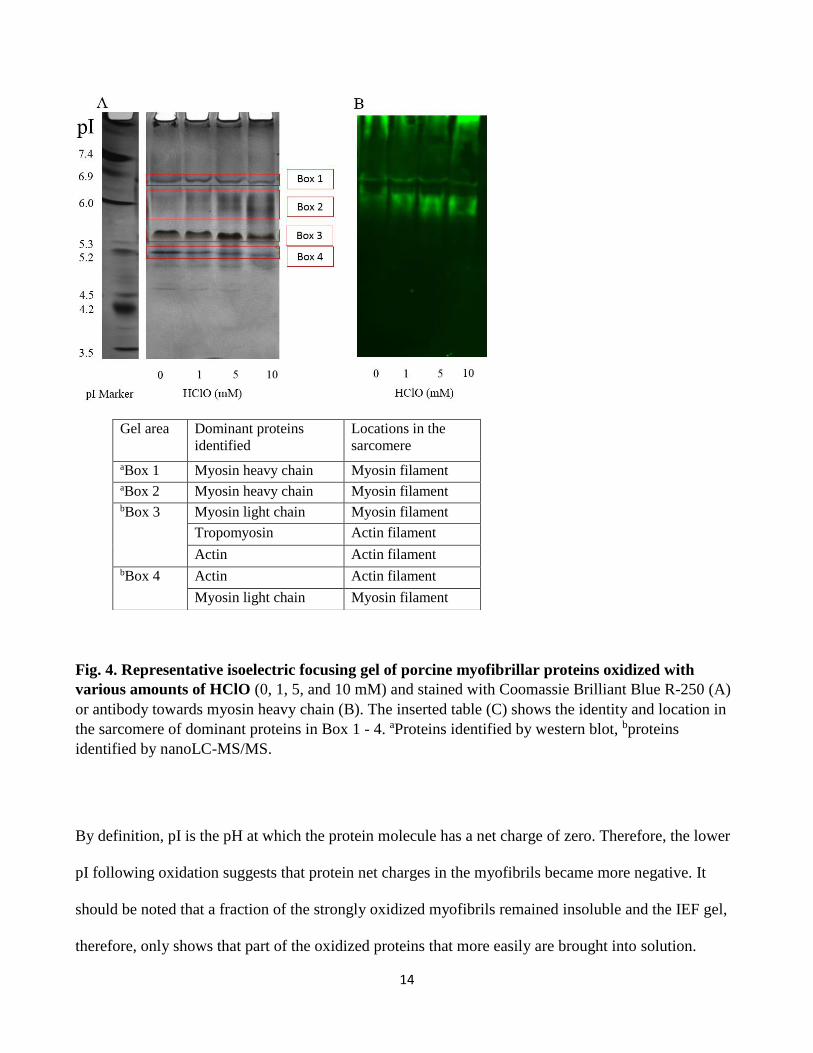
3.3. Isoelectric focusing and western blot
Myofibrillar proteins were extracted following oxidation and separated according to their isoelectric
point (pI) (Fig. 4). The isoelectric focusing gel showed that the pI of the proteins generally shifted to a
slightly more acidic area (Box 1 – 4, Fig. 4A). The proteins in Box 1 and 2 were recognized by the
myosin heavy chain antibody (Fig. 4B) and thus contained myosin heavy chain, and the dominating
protein bands migrated to a slightly lower position in the 10 mM HClO group compared to the non-
oxidized control, indicating that the pI value of the myosin heavy chain had shifted towards the acidic
area. Box 3 (pI around 5.4) mainly contained tropomyosin, actin and myosin light chain and Box 4 (pI
around 5.2) mainly contained actin and myosin light chain as identified by mass spectroscopy (Fig.
4C).
14
Fig. 4. Representative isoelectric focusing gel of porcine myofibrillar proteins oxidized with
various amounts of HClO (0, 1, 5, and 10 mM) and stained with Coomassie Brilliant Blue R-250 (A)
or antibody towards myosin heavy chain (B). The inserted table (C) shows the identity and location in
the sarcomere of dominant proteins in Box 1 - 4. aProteins identified by western blot, bproteins
identified by nanoLC-MS/MS.
By definition, pI is the pH at which the protein molecule has a net charge of zero. Therefore, the lower
pI following oxidation suggests that protein net charges in the myofibrils became more negative. It
should be noted that a fraction of the strongly oxidized myofibrils remained insoluble and the IEF gel,
therefore, only shows that part of the oxidized proteins that more easily are brought into solution.
Gel area Dominant proteins
identified
Locations in the
sarcomere
aBox 1 Myosin heavy chain Myosin filament aBox 2 Myosin heavy chain Myosin filament bBox 3 Myosin light chain Myosin filament
Tropomyosin Actin filament
Actin Actin filament bBox 4 Actin Actin filament
Myosin light chain Myosin filament
15
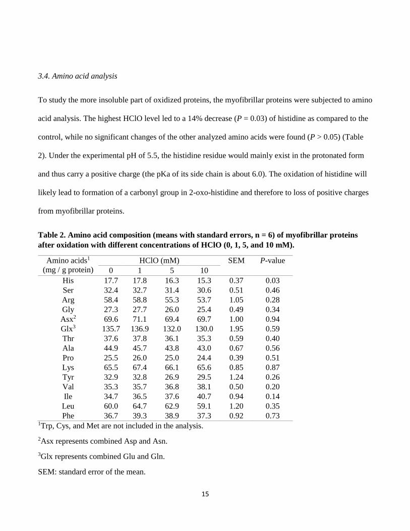
3.4. Amino acid analysis
To study the more insoluble part of oxidized proteins, the myofibrillar proteins were subjected to amino
acid analysis. The highest HClO level led to a 14% decrease (P = 0.03) of histidine as compared to the
control, while no significant changes of the other analyzed amino acids were found (P > 0.05) (Table
2). Under the experimental pH of 5.5, the histidine residue would mainly exist in the protonated form
and thus carry a positive charge (the pKa of its side chain is about 6.0). The oxidation of histidine will
likely lead to formation of a carbonyl group in 2-oxo-histidine and therefore to loss of positive charges
from myofibrillar proteins.
Table 2. Amino acid composition (means with standard errors, n = 6) of myofibrillar proteins
after oxidation with different concentrations of HClO (0, 1, 5, and 10 mM).
Amino acids1
(mg / g protein)
HClO (mM) SEM P-value
0 1 5 10
His 17.7 17.8 16.3 15.3 0.37 0.03
Ser 32.4 32.7 31.4 30.6 0.51 0.46
Arg 58.4 58.8 55.3 53.7 1.05 0.28
Gly 27.3 27.7 26.0 25.4 0.49 0.34
Asx2 69.6 71.1 69.4 69.7 1.00 0.94
Glx3 135.7 136.9 132.0 130.0 1.95 0.59
Thr 37.6 37.8 36.1 35.3 0.59 0.40
Ala 44.9 45.7 43.8 43.0 0.67 0.56
Pro 25.5 26.0 25.0 24.4 0.39 0.51
Lys 65.5 67.4 66.1 65.6 0.85 0.87
Tyr 32.9 32.8 26.9 29.5 1.24 0.26
Val 35.3 35.7 36.8 38.1 0.50 0.20
Ile 34.7 36.5 37.6 40.7 0.94 0.14
Leu 60.0 64.7 62.9 59.1 1.20 0.35
Phe 36.7 39.3 38.9 37.3 0.92 0.73 1Trp, Cys, and Met are not included in the analysis.
2Asx represents combined Asp and Asn.
3Glx represents combined Glu and Gln.
SEM: standard error of the mean.
16
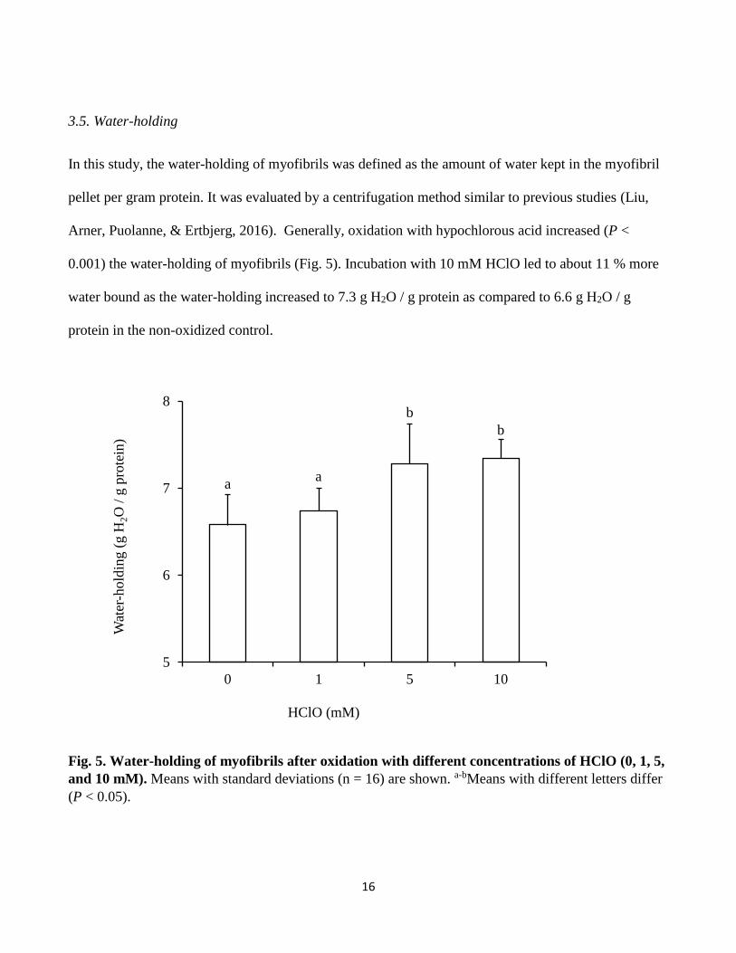
3.5. Water-holding
In this study, the water-holding of myofibrils was defined as the amount of water kept in the myofibril
pellet per gram protein. It was evaluated by a centrifugation method similar to previous studies (Liu,
Arner, Puolanne, & Ertbjerg, 2016). Generally, oxidation with hypochlorous acid increased (P <
0.001) the water-holding of myofibrils (Fig. 5). Incubation with 10 mM HClO led to about 11 % more
water bound as the water-holding increased to 7.3 g H2O / g protein as compared to 6.6 g H2O / g
protein in the non-oxidized control.
Fig. 5. Water-holding of myofibrils after oxidation with different concentrations of HClO (0, 1, 5,
and 10 mM). Means with standard deviations (n = 16) are shown. a-bMeans with different letters differ
(P < 0.05).
aa
b
b
5
6
7
8
0 1 5 10
Wat
er-h
old
ing (
g H
2O
/ g
pro
tein
)
HClO (mM)
17
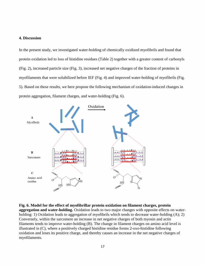
4. Discussion
In the present study, we investigated water-holding of chemically oxidized myofibrils and found that
protein oxidation led to loss of histidine residues (Table 2) together with a greater content of carbonyls
(Fig. 2), increased particle size (Fig. 3), increased net negative charges of the fraction of proteins in
myofilaments that were solubilized before IEF (Fig. 4) and improved water-holding of myofibrils (Fig.
5). Based on these results, we here propose the following mechanism of oxidation-induced changes in
protein aggregation, filament charges, and water-holding (Fig. 6).
Fig. 6. Model for the effect of myofibrillar protein oxidation on filament charges, protein
aggregation and water-holding. Oxidation leads to two major changes with opposite effects on water-
holding: 1) Oxidation leads to aggregation of myofibrils which tends to decrease water-holding (A); 2)
Conversely, within the sarcomere an increase in net negative charges of both myosin and actin
filaments tends to improve water-holding (B). The change in filament charges on amino acid level is
illustrated in (C), where a positively charged histidine residue forms 2-oxo-histidine following
oxidation and loses its positive charge, and thereby causes an increase in the net negative charges of
myofilaments.

18
Upon oxidation, the loss of free thiol groups and increase in particle size indicate formation of protein
cross-links and aggregates which act to inhibit swelling of myofibrils. An opposite effect from
electrostatic charges is simultaneously promoting swelling. Histidine side chains are carbonylated to
form 2-oxohistidine whereby positive charges are reduced. As a consequence, the overall net negative
charges in myofibrillar proteins including myosin, actin and tropomyosin increase with oxidation
which leads to increased net negative charges on both myosin and actin filaments. More net negative
charges on the myofilaments cause greater swelling pressure, which is linked to better water-holding
(Offer & Trinick, 1983). The overall result of protein oxidation on water-holding is therefore a balance
between promoting factors (such as increased net charges) and inhibiting factors (such as protein cross-
linking and aggregation).
It has been suggested that protein oxidation in meat lead to an alteration in electronic arrangement, and
thereby affects the chemical interaction between myofibrillar proteins and water molecules (Estévez et
al., 2011). Also Baraibar et al. (2011) reported that some proteins involved in the oxidative stress
response in human myoblasts were shifted to a more acidic pI upon oxidative stress. In agreement, the
present study found that the isoelectric point of oxidized proteins that are solubilized were shifted to a
more acidic area in the IEF gel (Fig. 4). At pH around 5.0, both myosin and actin filaments have a low
or zero net charge (Offer and Knight, 1988). In the present study the pH was around 5.5, and hence
both filaments were net negatively charged. After oxidation, the pI of solubilized myofibrillar proteins
generally shifted to a more acidic area and the affected proteins were mainly identified as myosin
heavy chain, myosin light chain, actin and tropomyosin, all of which are located in either myosin- or
actin filaments. Myosin, actin and tropomyosin together account for about 73% of the total
myofibrillar protein and the observed changes (Fig. 4) is therefore likely able to lower the pI of
myofilaments. Consequently, the difference between the isoelectric point of myofilaments and their

19
environmental pH of 5.5 would be larger following oxidation, indicating an overall increase in net
negative charges of myofilaments.
One mechanism that oxidation may affect protein charges is through protein carbonylation which
involves the loss of positively charged amino acids such as lysine, arginine and histidine (Stadtman,
1993; Utrera & Estévez, 2012). The loss of a positively charged amino acid will increase the overall net
negative charge. Histidine and lysine are major targets for oxidative modification and the histidine
derived carbonyl, 2-oxo-histidine, has been detected in several proteins subjected to in vitro oxidation
(Uchida, 2003). Park & Xiong (2007) found that oxidative modifications on amino acids depend on the
oxidizing system. Specific protein carbonyls, such as lysine-derived α-aminoadipic semialdehydes
(AAS) and arginine or proline derived γ-glutamic semialdehydes (GGS) have been suggested to be
major products of protein oxidation in meat (Estévez, 2011). The present study suggests that also
histidine is a major target of oxidative reactions in postmortem muscle. Another mechanism whereby
oxidation can affect protein charges may be protein denaturation and unfolding. According to Sun,
Zhou, Sun, & Zhao (2013), protein oxidation may expose buried hydrophobic groups and induce an
increase in the number of ionized groups, causing an increase in net negative charges.
As the charges of a protein is critical to its biochemical properties, oxidation-induced changes in net
negative charges may be one of the key factors to understand the functionality of oxidized proteins. Protein
net charges have been linked to protein functionality in meat such as the emulsifying properties of
myofibrillar proteins (Sun et al., 2013). Utrera & Estévez (2012) found that protein carbonylation correlates
to many functionality indicators (solubility, water-holding, foaming, etc. ) and they suggested that loss of
protonable amino acids would lead to an alteration of the overall charge arrangement of myofibrillar
proteins, and therefore affect funtional properties of proteins. Liu et al. (2016) studied effect of
sarcoplasmic protein denaturation on water-holding of myofibrils and hypothesized that denatured

20
sarcoplasmic proteins may precipitate onto the surface of myofilaments. The denatured sarcoplasmic
proteins was suggested to add positive charges to the overall negatively charged filaments and thus
decrease the net charge and subsequently the electrical repulsion – an effect which would be able to explain
an observed decrease in the distance between actin and myosin filaments due to sarcoplasmic protein
denaturation..
In our study, myofibrils were used to investigate the effect of oxidation on water-holding. Previous
studies mainly focused on oxidation-induced changes in myofibrillar structures. Huff-Lonergan &
Lonergan (2005) suggested that oxidation may reduce the proteolytic degradation of myofibrillar
proteins, including proteins in costameric linkages. Kristensen & Purslow (2001) suggested that
proteolysis of costamers plays an important role in water-holding of meat. The costameres can act as a
structural constraint and limit the swelling of myofibers. Recently, Zeng, Li, & Ertbjerg (2017) showed
that proteolytic degradation of proteins in and around the Z-disk is also a determining factor for
increased swelling of myofibrils, and thereby affect water-holding of myofibrils. Protein oxidation has
also been suggested to inhibit swelling of myofibrils due to structural constraint of oxidation-induced
disulfide cross-links (Liu et al., 2009).
If structural constraint is the only factor affecting water-holding of oxidized meat, we would expect
decreased water-holding following oxidation. However, we observed increased water-holding with
oxidation (Fig. 5), suggesting that other factors exerted stronger effects on water-holding. Oxidation led
to a shift of pI of both myosin and actin towards the more acidic area, suggesting an increase in net
negative charges due to oxidative modification of histidine. This would lead to increased net negative
charges on myosin and actin filaments. In addition, fragmentation of peptides upon oxidation may
produce additional charges to myofibrillar proteins. An increase in filament net charges is generally
believed to improve water-holding of meat. Therefore, in this study, the oxidation-induced increase in

21
water-holding was likely driven by increased net charges on myofilaments whereas opposing effects of
protein cross-linking and aggregation did not balance out or overrule the effects of increased net
negative charges. This balance may, however, differ depending on a variety of factors which can
explain why there is no general agreement on the effect of protein oxidation on water-holding.
Protein oxidation can eventually lead to intra- and inter-molecular cross-linking and protein
aggregation (Estévez, 2011; Lund et al., 2011). Therefore, it was expected that the particle size of
oxidized myofibrils became larger (Fig. 3, Table 1). In agreement, we previously reported (Bao et al.,
2016) that when minced beef was packaged under high oxygen atmosphere, protein oxidation was
induced and resulted an increased particle size of both raw meat and cooked meat. Protein cross-links
or aggregates altered the myofibrillar structure, which may limit the swelling of myofibrils and
therefore have a negative effect on water-holding.
In addition to filament net charges and protein cross-linking, chaotropic (structure-breaking) and
kosmotropic (structure-making) effects may play a role on oxidation-induced changes in water-holding.
The positively charged histidine residues in myofibrillar proteins are strong chaotropes which make
water molecules less structured and more mobile (Collins, 1997) and chaotropic effects are
hypothesized to be linked to reduced water-holding (Puolanne & Halonen, 2010). Formation of
carbonyls from histidine will likely reduce the chaotropic effect and therefore increase water-holding.
In the present study, myofibrils were used as a model to investigate the effect of protein oxidation on
water-holding. However, additional factors such as the presence of sarcoplasmic proteins, sarcolemma
and connective tissue may be of importance to understand the role of protein oxidation on water-
holding in fresh meat.
22
5. Conclusion
Oxidation of myofibrils resulted in greater loss of free thiol groups, loss of histidine residues and formation
of carbonyls in myofibrillar proteins and generally induced larger particle size. The myofibrillar proteins
myosin, actin and tropomyosin were identified in protein bands that shifted to a more acidic isoelectric
point upon oxidation, suggesting an increase in net negative charges of myofilaments. The water-holding
of myofibrils were generally improved following oxidation. The overall net negative charges of
myofilaments provide a novel perspective to understand oxidation-induced meat quality changes related to
water-holding. We here propose a hypothesis that water-holding of oxidized meat is a balance between
promoting factors such as oxidation-induced net negative charges and opposing factors such as cross-
linking and aggregation due to oxidation. However, the oxidation system was based on HClO alone, and it
would be of interest to extend the study to other model systems and meat products.
Acknowledgments
The authors would like to thank the China Scholarship Council for financial support; Simon Allegaert
and Cristina Ababei for analytical help; Minnamari Edelmann and Miikka Olin for help in
determination of amino acids and professor emeritus Eero Puolanne for his valuable comments on the
manuscript.
References
Anderson, J. C., & Peck, S. C. (2014). Detection of protein phosphorylation and charge isoforms using
vertical one-dimensional isoelectric focusing gels. Plant MAP Kinases: Methods and Protocols,
39-46.

23
Bao, Y., & Ertbjerg, P. (2015). Relationship between oxygen concentration, shear force and protein
oxidation in modified atmosphere packaged pork. Meat Science, 110, 174-179.
Bao, Y., Puolanne, E., & Ertbjerg, P. (2016). Effect of oxygen concentration in modified atmosphere
packaging on color and texture of beef patties cooked to different temperatures. Meat Science,
121, 189-195.
Baraibar, M. A., Hyzewicz, J., Rogowska-Wrzesinska, A., Ladouce, R., Roepstorff, P., Mouly, V., &
Friguet, B. (2011). Oxidative stress-induced proteome alterations target different cellular
pathways in human myoblasts. Free Radical Biology and Medicine, 51, 1522-1532.
Chen, L., Zhou, G., & Zhang, W. (2015). Effects of high oxygen packaging on tenderness and water
holding capacity of pork through protein oxidation. Food and Bioprocess Technology, 8, 2287-
2297.
Clausen, I., Jakobsen, M., Ertbjerg, P., & Madsen, N. T. (2009). Modified atmosphere packaging
affects lipid oxidation, myofibrillar fragmentation index and eating quality of beef. Packaging
Technology and Science, 22, 85-96.
Collins, K. D. (1997). Charge density-dependent strength of hydration and biological structure.
Biophysical Journal, 72, 65-76.
Cox, J., & Mann, M. (2008). MaxQuant enables high peptide identification rates, individualized ppb-
range mass accuracies and proteome-wide protein quantification. Nature Biotechnology, 26,
1367-1372.
Cox, J., Neuhauser, N., Michalski, A., Scheltema, R. A., Olsen, J. V., & Mann, M. (2011). Andromeda:
a peptide search engine integrated into the MaxQuant environment. Journal of Proteome
Research, 10, 1794-1805.

24
Decker, E. A., Xiong, Y. L., Calvert, J. T., Crum, A. D., & Blanchard, S. P. (1993). Chemical, physical,
and functional properties of oxidized turkey white muscle myofibrillar proteins. Journal of
Agricultural and Food Chemistry, 41, 186-189.
Delles, R. M., & Xiong, Y. L. (2014). The effect of protein oxidation on hydration and water-binding
in pork packaged in an oxygen-enriched atmosphere. Meat Science, 97, 181-188.
Ellman, G. L. (1959). Tissue sulfhydryl groups. Archives of biochemistry and biophysics, 82, 70-77.
Estévez, M. (2011). Protein carbonyls in meat systems: A review. Meat Science, 89, 259-279.
Estévez, M., & Luna, C. (2017). Dietary protein oxidation: A silent threat to human health? Critical
Reviews in Food Science and Nutrition, 57, 3781-3793.
Estévez, M., Ventanas, S., Heinonen, M., & Puolanne, E. (2011). Protein carbonylation and water-
holding capacity of pork subjected to frozen storage: Effect of muscle type, premincing, and
packaging. Journal of Agricultural and Food Chemistry, 59, 5435-5443.
Hamm, R. (1986). Functional properties of the myofibrillar system and their measurements. In Bechtel,
P. J. (Eds). Muscle as food. Food Science and Technology (pp. 135-191). London: Academic
Press Inc.
Huff-Lonergan, E., & Lonergan, S. M. (2005). Mechanisms of water-holding capacity of meat: The
role of postmortem biochemical and structural changes. Meat Science, 71, 194-204.
Kristensen, L., & Purslow, P. P. (2001). The effect of ageing on the water-holding capacity of pork:
Role of cytoskeletal proteins. Meat Science, 58, 17-23.
Lametsch, R., Knudsen, J. C., Ertbjerg, P., Oksbjerg, N., & Therkildsen, M. (2007). Novel method for
determination of myofibril fragmentation post-mortem. Meat Science, 75, 719-724.
Lindahl, G., Lagerstedt, Å., Ertbjerg, P., Sampels, S., & Lundström, K. (2010). Ageing of large cuts of
beef loin in vacuum or high oxygen modified atmosphere – Effect on shear force, calpain
activity, desmin degradation and protein oxidation. Meat Science, 85, 160-166.
25
Liu, J., Arner, A., Puolanne, E., & Ertbjerg, P. (2016). On the water-holding of myofibrils: Effect of
sarcoplasmic protein denaturation. Meat Science, 119, 32-40.
Liu, Z., Xiong, Y. L., & Chen, J. (2009). Identification of restricting factors that inhibit swelling of
oxidized myofibrils during brine irrigation. Journal of Agricultural and Food Chemistry, 57,
10999-11007.
Łopacka, J., Półtorak, A., & Wierzbicka, A. (2017). Effect of reduction of oxygen concentration in
modified atmosphere packaging on bovine M. longissimus lumborum and M. gluteus medius
quality traits. Meat Science, 124, 1-8.
Lu, J., Boeren, S., de Vries, S. C., van Valenberg, H. J. F., Vervoort, J., & Hettinga, K. (2011). Filter-
aided sample preparation with dimethyl labeling to identify and quantify milk fat globule
membrane proteins. Journal of Proteomics, 75, 34-43.
Lund, M. N., Heinonen, M., Baron, C. P., & Estevez, M. (2011). Protein oxidation in muscle foods: A
review. Molecular Nutrition & Food Research, 55, 83-95.
Lund, M. N., Lametsch, R., Hviid, M. S., Jensen, O. N., & Skibsted, L. H. (2007). High-oxygen
packaging atmosphere influences protein oxidation and tenderness of porcine longissimus dorsi
during chill storage. Meat Science, 77, 295-303.
Morris, J. C. (1966). The acid ionization constant of HOCl from 5 to 35°. The Journal of Physical
Chemistry, 70, 3798-3805.
Offer, G., & Knight, P. (1988). Structural basis of water-holding in meat. Part 1: General principles and
water uptake in meat processing. In R. Lawrie (Ed.), Developments in Meat Science, Vol 4 (pp.
63-171). London: Elsevier Applied Science.
Offer, G., & Trinick, J. (1983). On the mechanism of water holding in meat: The swelling and
shrinking of myofibrils. Meat Science, 8, 245-281.

26
Park, D., & Xiong, Y. L. (2007). Oxidative modification of amino acids in porcine myofibrillar protein
isolates exposed to three oxidizing systems. Food Chemistry, 103, 607-616.
Puolanne, E., & Halonen, M. (2010). Theoretical aspects of water-holding in meat. Meat Science, 86,
151-165.
Sekar, A., Dushyanthan, K., Radhakrishnan, K., & Babu, R. N. (2006). Effect of modified atmosphere
packaging on structural and physical changes in buffalo meat. Meat Science, 72, 211-215.
Soglia, F., Petracci, M., & Ertbjerg, P. (2016). Novel DNPH-based method for determination of protein
carbonylation in muscle and meat. Food Chemistry, 197, 670-675.
Stadtman, E. R. (1993). Oxidation of free amino acids and amino acid residues in proteins by radiolysis
and by metal-catalyzed reactions. Annual Review of Biochemistry, 62, 797-821.
Sun, W., Zhou, F., Sun, D.-W., & Zhao, M. (2013). Effect of oxidation on the emulsifying properties of
myofibrillar proteins. Food and Bioprocess Technology, 6, 1703-1712.
Uchida, K. (2003). Histidine and lysine as targets of oxidative modification. Amino Acids, 25, 249-257.
Utrera, M., & Estevez, M. (2012). Oxidation of myofibrillar proteins and impaired functionality:
underlying mechanisms of the carbonylation pathway. Journal of Agricultural and Food
Chemistry, 60, 8002-8011.
Wang, H., Luo, Y., & Ertbjerg, P. (2017). Myofibrillar protein gel properties are influenced by oxygen
concentration in modified atmosphere packaged minced beef. Food Chemistry, 230, 475-481.
Yang, X., Zhang, Y., Zhu, L., Han, M., Gao, S., & Luo, X. (2016). Effect of packaging atmospheres on
storage quality characteristics of heavily marbled beef longissimus steaks. Meat Science, 117,
50-56.
Zeng, Z., Li, C., & Ertbjerg, P. (2017). Relationship between proteolysis and water-holding of
myofibrils. Meat Science, 131, 48-55.