Isolated left ventricular noncompaction in association with rheumatic mitral stenosis
127
Indian Heart Journal (ISSN: ʹʹ͵ͽ-ͼͷͶ) January–February 2012 Volume 64 Issue 01 Honorary Editor K. Sarat Chandra, Hyderabad Executive Editors G. Ramesh, Hyderabad Deepak Kumar Saha, Hyderabad Associate Editor Rakesh Gupta, New Delhi Editorial Board Niteen Deshpande, Nagpur J.C. Mohan, New Delhi C. Narsimhan, Hyderabad Jacob Jose, Vellore Nakul Sinha, Lucknow V. Dayasagar Rao, Hyderabad Sandeep Mishra, New Delhi K.K. Talwar, New Delhi Consulting Editors Upendra Kaul, New Delhi P. Krishnam Raju, Hyderabad A.K. Banerjee, Kolkata P.K. Deb, Kolkata Santanu Guha, Kolkata Ravi Bathina, Hyderabad S.C. Manchanda, New Delhi K.C. Goswami, New Delhi Section Editors Imaging Johann Christopher, Hyderabad Gurpreet Gulati, New Delhi Interventional Cardiology R.K. Jain, Hyderabad Harinder K. Bali, Chandigarh Ajit Mullasari, Chennai Balram Bhargava, New Delhi Paediatric Cardiology Anita Saxena, New Delhi K. Nageshwar Rao, Hyderabad R. Prasada Reddy, Hyderabad Rohit Kumar Manoj, Chandigarh Preventive Cardiology B. Ramesh Babu, Hyderabad Dorairaj Prabhakaran, Delhi Electro Physiology B. Hygriv Rao, Hyderabad Echo K.K. Kapoor, New Delhi Sameer Srivastava, New Delhi K. Raghu, Hyderabad A.V. Anjaneyulu, Hyderabad Pulmonary Hypertension B.K.S. Sastry, Hyderabad Hypertrophic Cardiomyopathy and Heart Failure Ajay Bahl, Chandigarh Sandeep Seth, New Delhi Nuclear Cardiology K. Kumaresan, Hyderabad G.N. Mahapatra, Mumbai Peripheral Vascular Pinjala Ramakrishna, Hyderabad N.N. Khanna, New Delhi Cardiac Surgery Vijay Dixit, Hyderabad O.P. Yadav, New Delhi A. Dharma Rakshak, Hyderabad R.V. Kumar, Hyderabad R.C. Mishra, Hyderabad S. Lokeshwar Rao, Hyderabad International Editorial Board Enas Enas, USA Gautam Reddy, USA Y.C. Chandra Shekar, USA P.S. Reddy, USA Sumeet Chugh, USA Samuel Ashirvadam, USA Bharat K. Kantharia, USA Nalini Rajamannan, USA James Min, USA 001-IHJ-EB.indd i 1/25/2012 5:14:13 PM
-
Upload
independent -
Category
Documents
-
view
1 -
download
0
Transcript of Isolated left ventricular noncompaction in association with rheumatic mitral stenosis

Indian Heart Journal(ISSN: - )
January–February 2012 Volume 64 Issue 01
Honorary Editor
K. Sarat Chandra, Hyderabad
Executive Editors
G. Ramesh, HyderabadDeepak Kumar Saha, Hyderabad
Associate Editor
Rakesh Gupta, New Delhi
Editorial Board
Niteen Deshpande, NagpurJ.C. Mohan, New DelhiC. Narsimhan, HyderabadJacob Jose, Vellore
Nakul Sinha, LucknowV. Dayasagar Rao, HyderabadSandeep Mishra, New DelhiK.K. Talwar, New Delhi
Consulting Editors
Upendra Kaul, New DelhiP. Krishnam Raju, HyderabadA.K. Banerjee, KolkataP.K. Deb, Kolkata
Santanu Guha, KolkataRavi Bathina, HyderabadS.C. Manchanda, New DelhiK.C. Goswami, New Delhi
Section Editors
ImagingJohann Christopher, HyderabadGurpreet Gulati, New Delhi
Interventional CardiologyR.K. Jain, HyderabadHarinder K. Bali, ChandigarhAjit Mullasari, ChennaiBalram Bhargava, New Delhi
Paediatric CardiologyAnita Saxena, New DelhiK. Nageshwar Rao, HyderabadR. Prasada Reddy, HyderabadRohit Kumar Manoj, Chandigarh
Preventive CardiologyB. Ramesh Babu, HyderabadDorairaj Prabhakaran, Delhi
Electro PhysiologyB. Hygriv Rao, Hyderabad
EchoK.K. Kapoor, New DelhiSameer Srivastava, New DelhiK. Raghu, HyderabadA.V. Anjaneyulu, Hyderabad
Pulmonary HypertensionB.K.S. Sastry, Hyderabad
Hypertrophic Cardiomyopathy and Heart FailureAjay Bahl, ChandigarhSandeep Seth, New Delhi
Nuclear CardiologyK. Kumaresan, HyderabadG.N. Mahapatra, Mumbai
Peripheral VascularPinjala Ramakrishna, HyderabadN.N. Khanna, New Delhi
Cardiac SurgeryVijay Dixit, HyderabadO.P. Yadav, New DelhiA. Dharma Rakshak, HyderabadR.V. Kumar, HyderabadR.C. Mishra, HyderabadS. Lokeshwar Rao, Hyderabad
International Editorial Board
Enas Enas, USAGautam Reddy, USAY.C. Chandra Shekar, USAP.S. Reddy, USASumeet Chugh, USA
Samuel Ashirvadam, USABharat K. Kantharia, USANalini Rajamannan, USAJames Min, USA
001-IHJ-EB.indd i 1/25/2012 5:14:13 PM

Advisory Board
J.S.N. Murthy, ChennaiKhurshid Iqbal, SrinagarP.K. Goel, LucknowV. Rajasekhar, TirupathiH.N. Mishra, CuttackR.K. Ghokroo, AjmerS.K. Kaushik, UdaipurM. Khalilullah, New DelhiK.K. Sethi, New DelhiI. Satya Murthy, ChennaiShanmuga Sundaram, ChennaiR. Subramaniam, ChennaiR. Yadav, New DelhiAjay K. Sinha, PatnaR. Arora, New DelhiSameer Dani, AhmedabadSunil Modi, New DelhiUmesh Chandra, HyderabadVivek Gupta, New DelhiPradyut Ghosh, New DelhiB. Soma Raju, HyderabadShiva Kumar, HyderabadP.C. Rath, HyderabadShailendar Singh, HyderabadV. Suryaprakasa Rao, HyderabadS.K. Malani, LucknowVijay Trehan, New DelhiArupdas Biswas, KolkataRajeev Gupta, JaipurA.K. Pancholia, IndoreV.H. Bang, MumbaiP.P. Mohanan, TrissurMantosh Panja, KolkataR.K. Saran, LucknowK. Venugopal, CalicutSoumitra Ray, KolkataM.K. Das, KolkataAnil Barani, IndoreT.S. Kler, New Delhi
Vidyut Jain, IndoreUlhas Pandurangi, ChennaiC.N. Manjunath, BengaluruT.R. Raghu, BengaluruRanga Nayak, BengaluruS.S. Ramesh, BengaluruRajnish Kapoor, New DelhiAtul Mathur, New DelhiV. Mohan, ChennaiV. Amuthan, MaduraiRajeev Bajaj, New DelhiRajeev Agarwal, New DelhiPraveen Jain, JhansiA. Srinivas Kumar, HyderabadBalbir Singh, New DelhiChenniappan, ChennaiSatyanarayana Routary, CuttackKrishna Kumar, KochiS. Radha Krishnan, New DelhiVikas Kohli, New DelhiS.S. Kothari, New DelhiS.K. Chugh, New DelhiSoumitra Kumar, KolkataS. Harikrishnan, ThiruvananthapuramSunita Maheshwari, BengaluruPraveen Chandra, New DelhiSanjay Mehrotra, BengaluruBharat Dalvi, MumbaiAditya Kapoor, LucknowHarshavardhan, New DelhiKeyur Parikh, AhmedabadR.R. Kasliwal, New DelhiJamshed Dalal, MumbaiI.B. Vijaya Lakshmi, BengaluruGeorge Joseph, VelloreG. Karthikeyan, New DelhiS. Ramakrishnan, New DelhiH.M. Mardikar, NagpurV.K. Bahl, New Delhi
Srinivas Reddy, ChandigarhTiny Nair, ThiruvananthapuramM.N. Krishnan, CalicutSandeep Seth, New DelhiA.B. Mehta, MumbaiD.B. Pahalajani, MumbaiBrian Pinto, MumbaiUday Khanolkar, GoaVitul K. Gupta, BhatindaMan Mohan Singh, PatialaG.S. Wander, ChandigarhAdarsh Kumar, AmritsarJag Mohan Verma, ChandigarhYash Pal Sharma, ChandigarhV.S. Narain, LucknowPraful Kerkar, MumbaiRajeev Lochan, DubaiA.N. Patnaik, HyderabadO. Sai Satish, HyderabadMohan Nair, New DelhiRavi Kishore, BengaluruAmit Vohra, MumbaiK. Sharada, HyderabadAnil Saxena, New DelhiKartikeya Bhargava, New DelhiShomu Bohora, BarodaVanita Arora, New DelhiRajneesh Sardana, New DelhiR. Juneja, New DelhiRabin Chakraborty, KolkataK.K. Haridas, KochiC.G. Bahuleyan, ThiruvananthapuramN.V. Rayudu, HyderabadP.V.R. Sarma, GunturAshok Seth, New DelhiNagaraj Desai, BengaluruH.S. Rissam, New DelhiJ.P. Sawhney, New Delhi
Indian Heart Journal(ISSN: - )
January–February 2012 Volume 64 Issue 01
Copyright © 2012, Cardiological Society of India. All rights reserved.
No part of this publication may be reproduced or transmitted in any form or by any means, electronic or mechanical, without written permission from the Editor-in-Chief.
Publishing services by Reed Elsevier India Pvt. Ltd.
Published at EIH Limited-Unit Printing Press, IMT Manesar, Gurgaon.
001-IHJ-EB.indd ii 1/25/2012 5:14:13 PM

President – CSIAshok Seth (New Delhi)
[email protected] 098100 25814
President Elect & Chairman Scientific Committee – CSIP.K. Deb (Kolkata)
[email protected] 098310 38261
Immediate Past President – CSISamuel K. Mathew (Chennai)
[email protected] 098410 71118
General Secretary – CSISantanu Guha (Kolkata)
[email protected] 098310 16367
Arup Dasbiswas (Kolkata)[email protected] 098300 22609
Vice President – CSIH.K. Chopra (New Delhi)
[email protected] 098110 90204Harshwardhan Mohan Mardikar (Nagpur)
[email protected] 098230 82609
Treasurer – CSIMrinal Kanti Das (Kolkata)
[email protected] 098300 34263
Editor – Indian Heart JournalK. Sarat Chandra (Hyderabad)
[email protected] 098480 12212
Associate Editor – Indian Heart JournalRakesh Gupta (New Delhi)
[email protected]/[email protected] 098110 13246
EC Member – CSI
K.C. Goswami (New Delhi)[email protected] 098113 78890
Nakul Sinha (Lucknow)[email protected] 093359 27360
Biswakes Majumdar (Kolkata)[email protected] 098300 60809
P.P. Mohanan (Thrissur)[email protected] 098460 76006
Ajay Kumar Sinha (Patna)[email protected] 098350 37785
A.K. Pancholia (Indore)[email protected] 098270 27920
Sundeep Mishra (New Delhi)[email protected] 098714 21390
S.B. Gupta (Mumbai)[email protected] 098213 64565
Praveen Jain (Jhansi)[email protected]/[email protected] 094150 30615
Soumitra Kumar (Kolkata)[email protected]/[email protected] 098310 32519
P.B. Jayagopal (Palakkad)[email protected] 098470 23777
P. Rajendra Kumar (Secunderabad)[email protected] 098480 29443
Satyanarayan Routray (Cuttack)[email protected] 094372 25072
Shuvanan Ray (Kolkata)[email protected] 098300 24266
Gurpreet Singh Wander (Ludhiana)[email protected] 098155 45316
R. Subramanian (Chennai)[email protected] 094441 04759
Vijay H. Bang (Mumbai)[email protected] 098200 30363
Joint Secretary – CSISaumitra Ray (Kolkata)
[email protected] 098300 22317
Assistant Secretary – CSIKajal Ganguly (Kolkata)
[email protected] 098311 18148
Co-opted Member – (From Past President of CSI)S.K. Parashar (Noida)
[email protected] 098101 46231
S.S. Chatterjee (Kolkata)[email protected] 098302 66075
Co-opted Member (From Armed Forces) – CSIS.K. Malani (Kolkata)
[email protected] 098360 01818
Executive Committee
Cardiological Society of Indiawww.csi.org.in
002-IHJ-Exec commitee.indd i 1/25/2012 12:43:46 PM

Indian Heart Journal(ISSN: - )
January–February 2012 Volume 64 Issue 01
Table of Contents
Editorial 1
Original articles
Correlation between peripheral arterial disease and coronary artery disease using ankle brachial index—a study in Indian population 2Sharmistha Sarangi, Banumathy Srikant, Dayasagar V. Rao, Laxmikant Joshi, G. Usha
Outcomes of in-hospital, out of intensive care and operation theatre cardiac arrests in a tertiary referral hospital 7Murali Chakravarthy, Sona Mitra, Latha Nonis
Once weekly azithromycin in secondary prevention of rheumatic fever 12Rakesh Gopal, S. Harikrishnan, S. Sivasankaran, V.K. Ajithkumar, T. Titus, J.M. Tharakan
Discrepancy between myocardial perfusion and fatty acid metabolism following acute myocardial infarction for evaluating the dysfunctional viable myocardium 16Shankar K. Biswas, Masayoshi Sarai, Hiroshi Toyama, Hitoshi Hishida, Yukio Ozaki
64 slice computed tomography—a novel diagnostic method for evaluation of patients after coronary artery bypass grafts 23S. Balashankar Gomathi, P. Nandhini, R. Ravikumar, S. Mullasari Ajit
Role of lifestyle variables on the lipid profile of selected South Indian subjects 28Uma Chitra, N. Krishna Reddy, N. Balakrishna
Association between adipocytokines and insulin resistance in Indian hypertensive patients 35Sujata R. Mahadik
Vertical P-wave axis: the electrocardiographic synonym for pulmonary emphysema and its severity 40Lovely Chhabra, Pooja Sareen, Daniel Perli, Indu Srinivasan, David H. Spodick
Correlation of myocardial perfusion SPECT with invasive and computed tomography coronary angiogram 43S. Shelley, M. Indirani, I. Sathyamurthy, K. Subramanian, N. Priti, K. Harshad, D. Padma
International patients with congenital heart disease: what brings them to India? 50Sunita Maheshwari, B.A. Animasahun, O.F. Njokanma
Review articles
Platelet adenosine diphosphate receptor antagonists: ticlopidine to ticagrelor—a long continuing journey 54Upendra Kaul, Aijaz H. Mansoor
Pulmonary hypertension—“state of the art” management in 2012 60Anita Saxena
Case reports
Thrombolytic therapy in prosthetic valve thrombosis during early pregnancy 74B.C. Srinivas, Nagaraja Moorthy, Arora Kuldeep, Harsha Jeevan, Danalakshmi Chandrasekaran, C.N. Manjunath
Submitral aneurysm of the left ventricle 77D.K. Baruah, P.V. Naresh Kumar, G.S.P. Reddy, V. Ramesh Babu
Left main coronary artery bifurcation angioplasty and stenting after aortic valve replacement: a case report 80Sanjeeb Roy, Ajeet Bana, Rajeev Gupta, Rakesh Chittora, Sameer Sharma, Navneet Mehta
A case of arrhythmogenic right ventricular cardiomyopathy—Naxos disease 84R.R. Saravanan, V. Amuthan, R.A. Janarthanan, S. Balasubramanian, S. Naina Mohamed
003-IHJ-TOC.indd i 1/25/2012 5:13:52 PM

Percutaneous balloon pericardiotomy for the treatment of recurrent malignant pericardial effusion 88Aniket Puri, Nitin Agarwal, S.K. Dwivedi, V.S. Narain
Isolated left ventricular non-compaction in association with ventricular tachycardia 90Rajesh Vijayvergiya, Mukesh Yadav, Anand Subramaniyan
Parachute tricuspid valve in an asymptomatic adult 93Jagdish C. Mohan, Chandra Shekhar, Vipul Mohan, Bimalpreet Kaur, Shivesh Kumar Singh
Tetralogy of Fallot with Holt-Oram syndrome 95Vikas Kumar, Vikas Agrawal, Dharmendra Jain, Om Shankar
Magnetic resonance imaging findings in apical ballooning syndrome or takotsubo cardiomyopathy 99Jambhekar Kedar, Tarun Pandey, Chhavi Kaushik, Sanjaya Viswamitra, Behzad Molavi
Recurrent and rapidly occurring pericardial tamponade in Erdheim Chester disease 103Oommen K. George, M.S.K. Subhendu
Systemic lupus erythematosus presenting as cardiac tamponade—a case report 106Mohan Ashok Kumar, I. Sathyamurthy, K. Jayanthi, Ramakrishnan, Ramasubramanian
Journal review
Long term outcome of patients with isolated thin discrete subaortic stenosis treated by balloon dilation: a 25 year study 108
A comparison of Blalock-Taussig shunts with and without closure of the ductus arteriosus in neonates with pulmonary atresia 108
Efficacy and safety of Bosentan for pulmonary arterial hypertension in adults with congenital heart disease 109
Pulse Oximetry screening for congenital heart defects in newborn infants (PulseOx): a test accuracy study 110Saurabh Kumar Gupta, Anita Saxena
Rivaroxaban versus warfarin in nonvalvular atrial fibrillation 110
Apixaban versus warfarin in patients with atrial fibrillation 111Niteen Deshpande
Dronedarone in high-risk permanent atrial fibrillation 112
Prevention of ventricular fibrillation episodes in Brugada syndrome by catheter ablation over the anterior right ventricular outflow 112tract epicardium
Endocardial radiofrequency ablation for hypertrophic obstructive cardiomyopathy acute results and 6 months’ follow-up in 19 patients 112
Use of secondary prevention drugs for cardiovascular disease in the community in high-income, middle-income, and low-income 113countries (the PURE Study): a prospective epidemiological survey. Hygriv Rao
Four year follow up of SYNTAX trial; optimal revascularization strategy in patients with three vessel disease and/or left main disease 114Dheeaj Gandotra, Upendra Kaul
RadIal Vs FemorAL (RIVAL) trial for coronary angiography and intervention in patients with acute coronary syndromes 114Abid Hussain, Upendra Kaul
Rivaroxaban in patients with a recent acute coronary syndrome 115
Effect of two intensive statin regimens on progression of coronary disease 116Deepak Kumar Saha, K. Sarat Chandra
Cardiac magnetic resonance imaging pericardial late gadolinium enhancement and elevated inflammatory markers can predict the 117reversibility of constrictive pericarditis after antiinflammatory medical therapy: a pilot studyJohann Christopher
Arrhythmia graphics
What is your diagnosis? 118Shomu Bohora
Instructions to authors 120
Images in cardiology 27Book review 87Obituary 98
003-IHJ-TOC.indd ii 1/25/2012 5:13:52 PM

Indian Heart Journal 6401 (2012) 1
Contents lists available at SciVerse ScienceDirect
Indian Heart Journal
ISSN: 0019-4832 Copyright © 2012. Cardiological Society of India. All rights reserved.doi: 10.1016/S0019-4832(12)60001-7
Editorial
Dear Colleagues,
It may sound like a ‘cliche’, yet I would say that it is a great honour and privilege to be elected as the Editor of Indian Heart Journal (IHJ). It has been my long time desire, as it is a unique position with a lot of responsibility and is time con-suming, but it is well worth the efforts. I promise that I will under-take this job with utmost sincerity
and professionalism as I did the earlier jobs given to me by cardiological society of India.
A journal is the mirror of any scientific society and as such all of us as members of the society have a responsibility to-wards the journal to ensure that it is moulded and sculpted into great shape. If we want the journal to do well and take its appropriate place among the internationally reputed jour-nals of cardiology it needs everyone’s co-operation. There is no reason why any work done in India should be accepted for publication if it is not of the highest standard. We need to spend quality time in planning our trials. Naturally those stud-ies which are planned in collaboration with IHJ would get a priority in publication.
We also solicit contribution of articles from beyond our borders and it will be our privilege to have international au-thors. The reviewers need to be prompt and positive in their approach towards their job.
The field of cardiology has seen tremendous advances in the last decade, so also the field of medical publication. In this background we have gone for a tie-up with Elsevier, a well-known house in medical publishing. The submission of the articles will be completely paperless and will be done through Elsevier Editorial System (EES) which is an exclusive online submission website. The instructions to the authors are available in this issue and online and I request all the potential authors to register themselves with EES.
The regular website of IHJ is a separate website and would have archives of the journal for at least 10 years. Eventually we intend to have archives of the journal right from 1949 onwards.
Look forward to a wonderful 2012.
Yours Dr. K. Sarat Chandra
Honorary EditorIndian Heart Journal
Dr. K. Sarat Chandra
01-IHJ-Editorial-Sarat.indd 1 1/25/2012 7:03:42 PM
Contents lists available at SciVerse ScienceDirect
Indian Heart Journal
ISSN: 0019-4832 Copyright © 2012. Cardiological Society of India. All rights reserved.doi: 10.1016/S0019-4832(12)60002-9
Indian Heart Journal 6401 (2012) 2–6
* Corresponding a uthor. E-mail address: [email protected]
Original article
Correlation between peripheral arterial disease and coronary artery disease using ankle brachial index—a study in Indian populationSharmistha Sarangi1, Banumathy Srikant2*, Dayasagar V. Rao4, Laxmikant Joshi5, G. Usha3
, Registrar, Consultant, Professor and Head, Department of General Medicine, Professor and Head, Department of Cardiology, Durgabai Deshmukh Hospital and Research Centre, Hyderabad.
A B S T R A C T
Objective: To study the prevalence of peripheral arterial disease (PAD) of the lower limbs in a high-risk population and its correlation with coronary artery disease (CAD), using the ankle brachial index (ABI).Methods: The present study was conducted in randomly selected indoor patients >45 years of age with one or more risk factors for PAD admitted in the cardiology and medicine wards in a tertiary care institute.Results: Based on ABI <0.9, PAD was diagnosed in 32 of the 182 (18%) patients. Coronary artery disease was present in 15 cases of PAD which was statistically significant.Conclusion: There is a definite and strong correlation between PAD and CAD. Correct diagnosis and supervision of patients with PAD is important for preventing the local progression of the disease and effective secondary prevention of future coronary and cerebrovascular events.
Copyright © 2012, Cardiological Society of India. All rights reserved.
K E Y W O R D S
Ankle brachial indexCoronary artery diseasePeripheral arterial disease
Introduction
Peripheral arterial disease (PAD) is the occlusive disease of arteries distal to the aortic bifurcation.1 The prevalence of PAD in the lower limbs in a general population >55 years of age is between 10% and 25% and it increases with age.2 Majority of affected population have asymptomatic disease. Peripheral arterial disease, whether symptomatic or asymp-tomatic, is a risk factor for non-fatal and fatal coronary disease and cerebrovascular events.3 Patients with PAD alone have the same relative risk of death from cardiovascular cause as those with coronary or cerebrovascular disease.4 Risk of death in patients of PAD within 10 years is 4 times more than those without the disease.5 Several studies have shown that the ankle brachial index (ABI), an index for occlusive vascular disease, is now considered an independent predictor of coro-nary and cerebrovascular morbidity and mortality.6 Our study in an Indian population was carried out to correlate
and substantiate the relation of PAD with coronary artery disease (CAD) using the ABI.
Methods
The present study was conducted in randomly selected in-patients admitted in the cardiology and medicine wards in a tertiary care institute between October 2004 and March 2005. The following inclusion criteria were followed:1. Above 45 years of age.2. History of one or more conventional risk factors of PAD
like diabetes mellitus (DM), smoking, hypertension or dy-slipidaemia and/or were on treatment for the same.
3. Angiographic confirmation of CAD in addition to clinical history and electrocardiogram (ECG) abnormalities in sus-pected cases.A detailed clinical history of the patients was taken fol-
lowed by a detailed clinical examination, which was recorded in a proforma sheet. The diagnosis of intermittent claudica-tion (IC) was made based on the response to the questions in the proforma sheet.
01-IHJ-OA-Sharmistha.indd 2 1/25/2012 9:15:17 PM

S. Sarangi et al. / Indian Heart Journal 6401 (2012) 2–6 3
The measurement of ABI was done with the help of VP-1000 from Colins, Japan which works on the oscillometric principle. Blood pressure cuffs are tied to all four limbs and systolic pressure of all the limbs is measured at the same time and the ABI is calculated for each side.
Results
The present study consisted of a study population of 182 pa-tients. Based on ABI <0.9, PAD was diagnosed in 32 patients and 150 patients had ABI >0.9 on both sides and hence were considered normal. The occurrence of PAD in the study popu-lation was 18% (Figure 1 and Table 1).
Of the study population 143 (78%) were males and 39 (22%) were females. Among males, 22 (15.38%) were detected as PAD-positive cases. Among females 10 (25.64%) out of the 39 cases had PAD (Table 2).
Nine (4.8%) patients among the 182 described symptoms of IC; of the 9, 7 patients had PAD. The overall occurrence of IC in the study population was 3.8%. Among the 32 cases who
had PAD, 7 had IC. Twenty-five (13%) patients had PAD with-out symptoms of IC.
The minimum recorded ABI value on the right-side was 0.3 and the maximum was 1.29. The median value was 1.06. The minimum value on the left-side was 0.37 and the maxi-mum was 1.39 with a median of 1.06. Of the ABI recordings 13.19% were abnormal on the right-side and 9.34% were ab-normal on the left-side (Tables 3 and 4).
The presence of PAD in diabetics was significantly higher as shown in Table 5 (P = 0.021; statistically significant).
As shown in Table 6, 14 (35.9%) out of the 39 smokers had PAD whereas 25 (16%) out of the 150 non-smokers had PAD. This proved the predominance of PAD amongst smokers. Amongst the PAD-positive patients 44% were smokers, i.e. 14 of the 32 patients (P = 0.001; statistically significant).
Of the 182 patients in the study population, 102 had hy-pertension and 19 patients (18.63%) amongst them had PAD (Table 7). Occurr ence in hypertensives and non-hyperten-sives was similar (P = 0.676; statistically insignificant).
Table 2The age wise occurrence of peripheral arterial disease.
Age (yr) Normal PAD (%) Total
45–54 70 8 (10.2) 7855–64 37 9 (18.7) 46>65 43 15 (24.8) 58Total 150 32 (18) 182
PAD: peripheral arterial disease.
Table 1The number of normal and peripheral arterial disease patients in the study population.
Diagnosis Total (%)
Normal 150 (82)Peripheral arterial disease 32 (18)Total 182 (100)
Table 3The variation of peripheral pulsations in the study population.
Peripheral pulse Normal PAD Total
Abnormal 13 9 22Normal 137 23 160Total 150 32 182
PAD: peripheral arterial disease.
Table 5The relation of peripheral arterial disease to diabetic status.
Diabetic status Normal PAD Total
Non-diabetic 37 (22)* 2 (0)* 39 (22)*Diabetic 113 (54)* 30 (24)* 143 (78)*Total 150 (76)* 32 (24)* 182 (100)*
*Figures in brackets indicate patients >55 years of age. PAD: peripheral arterial disease.
82%
18%
Normal
PAD
Figure 1 The pie graft distribution of normal and peripheral arterial disease patients in the study population. PAD: peripheral arterial disease.
Table 4The distribution of ankle brachial index values in both lower limbs and their relation to severity of peripheral arterial disease.
Severity (ABI value) Left Right Total
Mild (0.89–0.7) 12 19 31Moderate (0.69–0.4) 5 4 9Severe (<0.4) 1 2 3Normal (>0.9) 164 157 321Total 182 182 364
ABI: ankle brachial index, PAD: peripheral arterial disease.
Table 6Relation of smoking to peripheral arterial disease.
Smoking status Normal PAD Total
Non-smokers 125 18 143Smokers 25 14 39Total 150 32 182
PAD: peripheral arterial disease.
01-IHJ-OA-Sharmistha.indd 3 1/25/2012 9:15:18 PM

4 S. Sarangi et al. / Indian Heart Journal 6401 (2012) 2–6
As shown in Table 8, 45 patients (24.73%) in the study popu-lation had CAD. The occurrence of CAD in PAD-positive cases was 46.88% while in PAD-negative cases it was 20% (P = 0.001; statistically significant).
Discussion
Peripheral arterial disease is the occlusive disease of arteries distal to the aortic bifurcation.1 The term, however, is widely used to refer to chronic arterial disease of the legs of athero-sclerotic origin. Athero sclerosis is by far the most common cause (>90%) of arterial problems in the legs.7 The pathology was designated as arteriosclerosis obliterans by the World Health Organization (WHO) study group.8 The ratio between systolic arterial pressure at the ankle and brachial artery, i.e. the ABI was established as a valid index to identify pa-tients with asymptomatic PAD.1 Some of the landmark stud-ies like Edinburgh Artery Study (1992), Framingham Study (1970–1996), The San Diego Study (1992), and The Rotterdam Study (1998) using the ABI have shown that the prevalence of asymptomatic PAD is much higher than the symptomatic disease.1 The measurement of ABI is the single most useful diagnostic tool in the evaluation of PAD.6
The San Diego Study found a high-risk of cardiovascular mortality among subjects with an abnormal ABI (<0.8).6 Criqui et al. using multivariate analysis in a population inves-tigated for carotid stenosis, ECG anomalies, and presence of PAD, diagnosed on the basis of the ABI, found that after 8 years of follow-up, ABI <0.9 was associated with total mortal-ity 2.4 times higher than normal and double the risk of cardio-vascular mortality.9 The Cardiovascular Health Study and the Edinburgh Artery Study using multivariate analysis on pro-spective observations of a large series (5888 and 1592 sub-jects, respectively) with an adequate follow-up (6 and 5 years, respectively), showed that the risk of total and cardio-vascular mortality was higher in patients with ABI <0.9, with a relative risk estimate between 1.5 and 1.8.10,11 The risk of death and non-fatal vascular events was higher in patients who had a low ABI together with risk factors such as diabetes or high blood cholesterol. In Italian ADEP (Associazione
Diaspora e Pace) Study, a low ABI was one of the predictors of vascular events—fatal or non-fatal in a population with IC.4
While the strength of the ABI as a negative prognostic in-dicator seems clear, it also appears that subclinical abnor-malities in the index imply a prognosis as negative as in symptomatic patients.6 In the Cardiovascular Health Study, subclinical vascular abnormalities detected instrumentally and with the ABI, involved a greater risk of developing the disease than in patients with no subclinical disorder.10
In this study, done on a defined population comprising in-patients >45 years of age with one or more conventional risk factors for PAD using the ABI as the diagnostic parameter, 18% of the subjects had PAD. The PARTNERS program which stud-ied the population aged between 50 years and 69 years with diabetes or smoking and age >70 years found a prevalence of 29%.12 The Rotterdam study with a study population <55 years of age had a prevalence of 19%.13 The Edinburgh Artery Study studied the age stratified sample between 55 years and 74 years and found a prevalence of 9%.11 However, the Swiss Atherothrombosis Survey carried out on a population >55 years of age with stroke, TIA, CAD or two or more risk factors found a prevalence of only 6.4%.14 All the compared studies used ABI as the diagnostic parameter.
This study population when analysed age wise, the preva-lence in age group of 45–54 years was only 10.2%. It increased to 18.7% in 55–64 years age group and was 24.8% in the age group >65 years. Peripheral arterial disease occurrence in-creased with age. Most studies have shown a linear relation between age and PAD. The Rotterdam Study showed a preva-lence of 7.6% in age group of 55–59 years, which increased to 59.6% in age >85 years.13 Newman et al. have shown a preva-lence of 26% in a population aged ≥ 60 years.10 This is compa-rable to the present study which has a prevalence of PAD of 24% of the patients in the study population >55 years of age.
In this study population, the male subjects comprised 78% and female subjects comprised 22%. The occurrence of PAD among males was 15.38% and among females was 25.64%. The impact of sex on PAD, however, did not reach statistical sig-nificance in this study. Most of the studies have shown a sim-ilar incidence of PAD with men to be slightly more than women. Schroll and Munk have shown an incidence of 16% in men and 13% in women.1 The Cardiovascular Health Study has shown a prevalence of 14% for men and 11% for women.10 Vogt et al. have shown the gap in prevalence narrows after 70 years of age.15 However, Meijer et al. in the Rotterdam Study found a higher prevalence rate among women being at 20.5% and for men being at 16.9%.13
An important aspect of this study was assessing the occur-rence of symptomatic and asymptomatic PAD based on the presence of IC. In this study, 4.8% of subjects described symp-toms of IC. Of these 1% did not have PAD. The overall occur-rence of IC in the study population was 3.8%. This is similar to most of the other studies. The Edinburgh Artery Study shows the prevalence of IC at 4.5%.11 Reunanen et al. had shown the prevalence of IC at 2%.16 Schroll and Munk have shown a prevalence of 3.5% for IC.1 Though the prevalence of PAD in this study was 18%, the prevalence of symptomatic PAD is only 3.8%. The occurrence of IC has risen steeply with age.
Table 7The relation of hypertension to peripheral arterial disease.
Hypertension status Normal PAD Total
Negative 67 13 80Positive 83 19 102Total 150 32 182
PAD: peripheral arterial disease.
Table 8Relation of coronary artery disease to peripheral arterial disease.
CAD status Normal PAD Total
Negative 120 17 137Positive 30 15 45Total 150 32 182
CAD: coronary artery disease; PAD: peripheral arterial disease.
01-IHJ-OA-Sharmistha.indd 4 1/25/2012 9:15:18 PM

S. Sarangi et al. / Indian Heart Journal 6401 (2012) 2–6 5
Subjects <55 years of age had an occurrence of 1.2%, between 55 years and 64 years of age the occurrence was 6.5%, while in those subjects >65 years of age an occurrence of 9.6% was observed. This again corroborates the fact that progression of PAD occurs with increasing age. Ouriel et al. has reported that the incidence of symptomatic PAD increases with age from about 0.3% per year for men aged 40–55 years to about 1% per year for men aged over 75 years.17 Reunanen et al. have shown a prevalence of IC of 2% in a population aged <60 years.16 Newman et al. have shown that the prevalence of IC in population >60 years is 6.4% which is comparable to the fig-ures discussed in this study.10
Another interesting fact observed was the presence of clau-dication seen more among subjects who also had associated CAD. Reunanen et al. had also made a similar observation in their study.16 In this study, among the patients diagnosed to having PAD, 21% were symptomatic. Hence, screening with ABI detected 79% asymptomatic PAD subjects. Stoffers et al. have shown that among those diagnosed with PAD, only 22% had symptoms.18 Meijer et al. have shown the prevalence of symptoms in 15% of PAD-positive cases.13
Taking into consideration the whole study population, 25 subjects (13%) out of 182 had asymptomatic PAD. Stoffers et al. have reported a prevalence of 6.9% of asymptomatic PAD.18 In the Edinburgh Artery Study, 8% had major asymptomatic PAD.11 The occurrence of both asymptomatic disease (13%) and diseased subjects being without symptoms (79%) sug-gests that screening the population at risk by a simple test like ABI measurement should be done in regular clinical practice. In the present study 28% of patients with PAD had an abnormal peripheral pulse examination. The remaining 72% had normal peripheral pulse. The PARTNERS program has highlighted that as many as 50% of cases may have a nor-mal peripheral pulse.12
The spread of ABI showed a median value of 1.06 on each side. The maximum ABI value was 1.39. As none of the values were >1.5, which is indicative of non-compressible calcific arteries, no second method of evaluation was required in this study. Disease occurrence on the right-side was 13.19% and on the left-side was 9.34%. The Edinburgh Artery Study has also shown unilateral predisposition to disease, but it was to the left-side as opposed to the right-side in our study.11
Based on ABI, the study population was divided into mild, moderate, and severe disease and 72% of subjects were re-ported to have had mild disease (ABI 0.7–0.89). Doobay and Anand have shown that a low ABI between 0.8 and 0.9 has a high specificity of 92% to predict CAD and 87% for cardiovas-cular mortality.19 Lee et al. have shown that ABI <0.9 can in-dependently predict fatal myocardial infarction in addition to the conventional risk factors.20 Majority of the study popula-tion have a low ABI but asymptomatic PAD. However, they are at a high-risk for coronary and cardiovascular events and hence should be the target for preventive measures.
This study showed that 20.98% of patients with DM had PAD and the P value for DM as a risk factor was statistically significant. A cross-sectional study by Adler et al. found a prevalence of 23.5% PAD among type 2 DM patients.21 In the study by Beckman et al., 50% of patients with DM were found
to have PAD.22 In our study, the occurrence of PAD in diabet-ics >55 years of age went up to 24%. However, regardless of high prevalence and complication that can result from PAD, it is still not a common practice to routinely screen for the dis-ease in diabetics.
In our study, 21% were smokers and all of them were males. Occurrence of PAD among smokers was around 36% which was significantly higher than among non-smokers (20%). Forty-four percent of the PAD-positive cases were smokers. Smoking as a risk factor had a statistically significant P value (0.001). Studies like Framingham Study, Cardiovascular Health Study, and Edinburgh Artery Study showed that amongst smokers PAD was 2–5 times higher.10,11,23 Willingdael et al. have shown that PAD is 2.5 times more in smokers.24 In our study, PAD was 2 times more in smokers than in non-smokers with a significant P value.
The present study had 55% of subjects as hypertensives; 18.63% had PAD whereas a similar proportion of 16% among non-hypertensives had PAD. The P value was not statistically significant (P value 0.676). According to the Framingham Heart Study, hypertension doubles the risk of PAD.23 However, Reunanen et al. showed that hypertension was not significantly related to PAD.16 In our study occurrence of PAD in both groups was similar.
The present study population of 182 patients had 45 pa-tients (24.73%) who had CAD. However, the occurrence of CAD among patients who had PAD was 2 times more than those without PAD. Among PAD-positive cases, CAD was present in 46.88%. Only 20% of PAD-negative cases had CAD. A strong correlation was found to occur between PAD and CAD (P = 0.001; statistically significant). The PARTNERS pro-gram showed that 16% of patients had PAD and CAD, 13% had only PAD, and 24% had only CAD.12 In our study 10% had PAD and CAD, 12% had only PAD, and 16% had only CAD.
Another interesting observation in our study was that only 6 patients were subjected to ABI measurement previously comprising only about 3% of the study population. Among the 6 patients, 2 of them had symptomatic disease and 2 of them had associated CAD. Hence, it was observed that though ABI is a relatively simple test to conduct, it is still used very sparingly in clinical practice.
Conclusion
There is a definite and strong correlation between PAD and CAD. In view of the increasingly aging population and associ-ated increase in atherosclerotic vascular disease, confronta-tion with patients of PAD will increase, which however, continues to be under diagnosed and under treated. The awareness and implementation of ABI in general clinical practice is poor. A simple, inexpensive test like ABI can im-prove the diagnosis of PAD in clinical practice and thus help in preventing CAD and consequent death by a range of medi-cal therapies. Correct diagnosis and supervision of patients with these disorders is important for the prevention of local progression of the disease and effective secondary preven-tion of any future coronary and cerebrovascular events.
01-IHJ-OA-Sharmistha.indd 5 1/25/2012 9:15:18 PM

6 S. Sarangi et al. / Indian Heart Journal 6401 (2012) 2–6
References
1. Lanzer P. Peripheral Vascular Disease—The Textbook of Peripheral Vascular Medicine (ed.). Eric J Topol 388–96.
2. Cimminiello C. Peripheral arterial disease – epidemiology and pathophysiology. Thromb Res 2002;106:295–301.
3. Norman PE, Eikelboom JW, Hankey GJ. Peripheral arterial disease – prognostic significance and prevention of atherothrom-botic complications. MJA 2004;181:150–4.
4. Brevetti G, Oliva G, Silvestro A, Francesco S, Chiariello M. Prevalence, risk factors and cardiovascular comorbidity of symp-tomatic peripheral arterial disease in Italy. Atherosclerosis 2004;175:131–8.
5. Hertzer NR. The natural history of peripheral vascular disease – implications for its management. Circulation 1991;83(Suppl 1):9–12.
6. Leng GC, Fowkes FGR, Lee AJ. Use of the ankle brachial pressure index to predict cardiovascular events and death – a Cohort study. BMJ 1996;313:1440–4.
7. Halperin JL. Evaluation of patients with peripheral vascular dis-ease. Thromb Res 2002;106:303–11.
8. World Health Organization Study Group. Classification of athero-sclerotic lesions – report of a study group. WHO Tech Rep Ser 1958;143:1–20.
9. Criqui MH, Fronek A, Klauber MR, Barrett CE, Gabriel S. The sen-sitivity, specificity and predictive value of traditional clinical evaluation of peripheral arterial disease – results from non inva-sive testing in a definite population. Circulation 1985;71:516–22.
10. Newman AB, Siscovick BS, Manolio TA. Ankle arm index as a marker of atherosclerosis – the Cardiovascular Health Study. Circulation 1993;88:837–45.
11. Fowkes FG, Housley E, Cawood EH, Macintyre CC, Ruckby CV, Prescott RJ. Edinburgh Artery Study – prevalence of asympto-matic and symptomatic peripheral arterial disease in the general population. Int J Epidemiol 1991;20:384–92.
12. Hirsch AT, Criqui MH, Jacobson TD, et al. Peripheral arterial disease – detection, awareness and treatment in primary care. JAMA 2001;286:1317–24.
13. Meijer WT, Hoes AW, Dominique R, Bots ML, Hofman A, Grobbee DE. Peripheral arterial disease in the elderly – the Rotterdam Study. Arterioscler Thromb Vasc Biol 1998;18:185–92.
14. Tomson J, Lip GH. Peripheral arterial disease – high risk but neglected disease population. BMC Cardiovasc Disord 2005;5:15–8.
15. Vogt MT, Wolfson SK, Kuller LH. Lower extremity arterial disease and the aging process – review. J Clin Epidemiol 1992;45:529–42.
16. Reunanen A, Takkunen H, Aromaa A. Prevalence of intermittent claudication and its effect on mortality. Acta Med Scand 1982;211:249–56.
17. Ouriel K. Detection of peripheral arterial disease in primary care. JAMA 2001;286:1380–1.
18. Stoffers HE, Rinkens PE, Kester AD, Kaiser V, Knottnerus JA. The prevalence of asymptomatic and unrecognized peripheral arte-rial occlusive disease. Int J Epidemiol 1996;25:282–90.
19. Doobay AV, Anand SS. Sensitivity and specificity of the ankle bra-chial index to predict future cardiovascular outcomes. Arterioscler Thromb Vasc Biol 2005;25:1463–65.
20. Lee AJ, Price JF, Russell MJ, Smith FB, Wijk MW, Fowkes F. Improved prediction of fatal myocardial infarction using the ankle brachial index in addition to conventional risk factors – The Edinburgh Artery Study. Circulation 2004;110:3075–80.
21. Adler AL, Stevens RJ, Neil A, Stratton IM, Boulton AJ, Holman RR. UKPDS 59 – hyperglycemia and other potentially modifiable risk factors for peripheral vascular disease in type 2 diabetes. Diabetes Care 2002;25:894–9.
22. Beckman JA, Creager MA, Libby P. Diabetes in atherosclerosis – epidemiology, pathophysiology and management. JAMA 2002;287:2570–81.
23. McKee PA, Castelli WP, McNamara PM, Kannel WB. The natural history of congestive heart failure – the Framingham Study. N Engl J Med 1971;285:1441–6.
24. Willingdael EM, Teijink JA, Bartelink ML, et al. Influence of smok-ing on incidence and prevalence of peripheral arterial disease. J Vasc Surg 2004;40:1158–65.
01-IHJ-OA-Sharmistha.indd 6 1/25/2012 9:15:18 PM

Contents lists available at SciVerse ScienceDirect
Indian Heart Journal
ISSN: 0019-4832 Copyright © 2012. Cardiological Society of India. All rights reserved.doi: 10.1016/S0019-4832(12)60003-0
Indian Heart Journal 6401 (2012) 7–11
* Corresponding a uthor. E-mail address: [email protected]
Original article
Outcomes of in-hospital, out of intensive care and operation theatre cardiac arrests in a tertiary referral hospitalMurali Chakravarthy1*, Sona Mitra2, Latha Nonis3
Chief Consultant, Research Fellow, Department of Anaesthesia, Critical Care, Pain Relief, Senior Executive, Department of Nursing, Fortis Hospitals, Bengaluru – , India.
A B S T R A C T
Objective: Cardiac arrest in the hospital wards may not receive as much attention as it does in the operation theatre and intensive care unit (ICU). The experience and the qualifications of person-nel in the ward may not be comparable to those in the other vital areas of the hospital. The out-come of cardiac arrest from the ward areas is a reasonable surrogate of training of the ward nurses and technicians in cardiopulmonary resuscitation. We conducted an audit to assess the issues surrounding the resuscitation of cardiac arrest in areas other than operation theatre and ICU in a tertiary referral hospital.Aims of the audit: To assess the outcomes of cardiac arrest in a tertiary referral hospital. Areas such as wards, dialysis room and emergency room were considered for the audit.Methods: This is a retrospective observational audit of the case records of all the adult patients who were resuscitated from ‘code blue’. Data for 2 years from 2007 was analysed by a research fellow unconnected with the resuscitations.Results: Twenty-two thousand three hundred and forty-four patients were admitted as in-patients to the hospital during the 2 years, starting May 2007 through May 2009. One hundred code blue calls were received during this time. Twenty-two of the total calls received were false. Among the 78 confirmed cardiac arrests 69 occurred in the wards, 2 in emergency room, 1 in cardiac cathe-terisation laboratory and 3 in dialysis room. Twenty-eight patients were declared dead after un-successful cardiopulmonary resuscitation. Among the 50 who were resuscitated with a return of spontaneous rhythm 26 died. Twenty-four patients were discharged (survival rate of 30%). The survival decreased significantly as the age progressed beyond 60. The resuscitation rates were better in day shifts in contrast to the night. Higher survival was noted in patients who received resuscitation in less than a minute.Conclusion: A overall survival to discharge rate of 30% was noted in this audit. Higher survival rates might be attributable to high rate and degree of training at the time of their employment, which was repeated at yearly interval.
Copyright © 2012, Cardiological Society of India. All rights reserved.
K E Y W O R D S
Cardiac arrestCardiopulmonary resuscitationCode blueIntensive care unitReturn of spontaneous circulation
Introduction
The basic techniques of cardiopulmonary resuscitation (CPR) have been established since 1960 when Kouwenhoven, Jude and Knickerbocker described closed chest massage.1 There have been a number of studies after that, which have re-ported outcome and the predictors affecting outcome.2–5 Cardiopulmonary resuscitation is a frequently performed
medical intervention in healthcare facilities. Successful car-diopulmonary resuscitation after in-hospital cardiac arrest depends on basic and advanced life support systems, the ability to immediately defibrillate the arrested heart, and the quality of the CPR intervention (Beuret et al.; Jorgenson 1997). But studies show a wide range of ‘survival on discharge’ (3–27%), which could be due to differences in the settings in which the CPR is performed and differences in inclusion/ex-clusion criteria.5–8 To overcome this problem, the in-hospital Utstein style data collection (Utstein template) recommen-dation were published in 1997 and revised in 2004.5,6 These
02-IHJ-OA-Murali.indd 7 1/25/2012 9:14:07 PM

8 M. Chakravarthy et al. / Indian Heart Journal 6401 (2012) 7–11
recommendations defined a set of data elements that are es-sential for documenting in-hospital cardiac arrests and sug-gested guidelines for reviewing, reporting, and conducting research on this topic.7 In the present audit, we sought to de-termine how well CPR is utilised at our institution by estimat-ing the incidence and the outcome from in hospital cardiac arrest and also predict the factors affecting the outcome. Key factors include presenting rhythm, time to definite therapy, episode being witnessed, provision of basic life support, time from collapse to defibrillation, age, gender, location of arrest, and associated risk factors. The purpose of this audit is to identify and facilitate necessary improvements both in the prevention of in-hospital cardiac arrest, and in the organisa-tion, delivery, and outcomes from pre-arrest, resuscitation, and post resuscitation care in the hospital and providing feed-back to relevant staff at all levels across the hos pital. Twenty-two thousand three hundred and forty-four patients were admitted to the hospital during the period of May 2007–2009. Seventy-eight cardiac arrests were encountered during this period of time. Twenty-four patients were discharged home.
Methods
This study is a retrospective observational audit conducted on the patient data obtained in our patients who received CPR during the 2 years period from March 2007 to April 2009. We received Institu tional Review Board approval to perform a retrospective medical records review on adult pa-tients who received CPR at our hospital. Such of the cardiac arrests resuscitated in the operation theatres and intensive care units (ICUs) were excluded from the audit. Ours is a multi-specialty hospital conducting cardiac, neurosurgical, or-thopaedic and minimal access general surgery. The hospital was commissioned in July 2006. The hospital has wards dis-tributed on the 6th and 7th floors of the hospital building. A standardised protocol termed ‘code 555’ is in place at the hos-pital for treating individuals with cardiorespiratory collapse. A phone call to 555 from any of the house phones by any of the staff (nurses, doctors or technicians) would automatically dial the emergency room staff, who by pressing buttons 1–4 would dial automatically dial the ‘code phone’ manned by a registrar from medical ICU, coronary care unit, anaesthesiol-ogy and nursing supervisor who are all located within the hos-pital. This phone call also informs the phone holder from where the code has been raised. The receiver will immedi-ately rush to the location from where the code has been raised and attend to the victim of cardio respiratory arrest. At the time of appointment, the employees of the hospital (nurses, technicians, lift operators among others), resident doctors, and registrars were trained to provide basic life sup-port. A review and recertification of their basic life support tal-ent was made at 1 year interval. After the victim had been attended to, depending on the decision of the leader of the resuscitation team, the patient would be transferred to medi-cal ICU for further care. After the patient transfer, the nursing supervisor, who is also a member of the code team, would fill up the code report form. This form mentions details of the
patient, details of concomitant diseases, location of cardiac arrest, time taken to start resuscitation, the initial rhythm, drugs administered, whether the code was successful or not, therapeutic manoeuvres employed among others.
Formation of emergency response (code blue) team (ERT): The hospital standard operating protocol (SOP) guided the for-mation and standardisation of the function of ERT. An ERT was formed and all its members were advanced cardiac life support providers. The ERT consisted of anaesthesiologist, intensivists, intensive care nurse, and ward nurse. The nurse assigned to the team was responsible for knowing the location of the crash cart. A separate SOP guided the maintenance of crash carts. The crash cart would not be opened unless there was a cardi-opulmonary arrest. The ward nurse would check the equip-ment on every day. Utmost care would be given to the check of defibrillators. Presence of a well charged battery, cleanliness of paddles, constant supply of electrocardiogram cable, jelly, and recording paper are assured every day. Defibrillator check is performed during every shift and the result of the test is pasted in the log book. It was made certain that other resusci-tative equipments such as functioning laryngoscope, availa-bility of endotracheal tubes and other support equipments were available. Emergency drugs commonly used during re-suscitation were kept handy in the crash cart. The contents were checked only if the crash cart seal was found broken.
The recommendations of the 2005 American Heart Association (AHA) cardiopulmonary resuscitation (CPR) guidelines were used by all the resuscitators.9
A research fellow unconnected with the study collected the data. The details about the patients, event characteristics, and outcome were noted as per the guidelines of in-hospital Utstein style using a standard questionnaire. The research as-sistant was a medical graduate and was trained by the princi-pal investigator in data retrieval from medical records.
The following data were obtained from all patient records—patient demographics, diagnosis, cardiac arrest char-acteristics (which included time of the day, physical location at the time of the arrest, whether arrest was witnessed or monitored, whether it was respiratory or cardiac arrest), days in the hospital prior to arrest and survival on discharge. The study population consisted of adults who underwent CPR be-tween March 2007 to April 2009 at a location other than ICU and operation theatre. Our definition of cardiac arrest was the same as described in the Utstein style template; the ces-sation of cardiac mechanical activity confirmed by the ab-sence of detectable pulse, unresponsiveness and/or apnoea (agonal respirations).6,7 In instances where the patient suf-fered multiple cardiac arrests, only the initial in-hospital ar-rest was recorded during the period of hospitalisation. This was done to avoid falsely elevated rate of successful CPR. Patients who had ‘do not resuscitate’ or ‘do not intubate or ventilate’ were not included in this audit.
Results
Twenty-two thousand three hundred and forty-four patients were admitted as in-patients to the hospital during the
02-IHJ-OA-Murali.indd 8 1/25/2012 9:14:08 PM

M. Chakravarthy et al. / Indian Heart Journal 6401 (2012) 7–11 9
bicarbonate, 17 of them died. Figure 1 shows the survival rate based on age. Table 4 shows the survival decreased signifi-cantly as the age progressed beyond 60. There was one survi-vor among the 14 who were above the age of 80 years. Table 5 shows the survival rate and associated co-morbidities. The best survival of 64% was among those who had no or 1 co-morbidity. The survival decreased to 9.6% if the patients had 2 co-morbidities. There were no survivors among those who had <2 co-morbidities. Survival at various locations of car-diac arrest can be seen in Table 6. The survival in cardiac catheterisation laboratory was 60%, 30% in the ward, and none in the emergency room and dialysis room. Twenty-five
Table 1Patient characteristics.
Hypertension 23Ischaemic heart disease 21Heart disease 33Diabetes mellitus 23Hyperlipidaemia 39Chronic renal failure 13Malignancy 6Chronic obstructive pulmonary disease 11Neurological dysfunction 11Sepsis 6Head injury 2Electrolyte abnormality 2Post coronary artery bypass surgery status 14Post valve surgery 3Post coronary angioplasty 6Others 11
Table 3Manoeuvres during resuscitation.
Intubation 73Atropine 43Adrenaline 47Sodium bicarbonate 19Direct current shock 16
Table 4Variables affecting survival.
n Survivors
Age (yr) <60 19 8 >60 42 15 >80 14 1
Male 47 15
Female 31 9
50
45
40
35
30
25
20
15
10
5
0
<60 >8060–80Age
Male Female
n
Survivors
Figure 1 Survival rate based on age.
Table 5Survival rate and number of co-morbidities/patients.
No. of co-morbidities/patients n Survived % survival
Up to 1 25 16 642 50 8 9.6>2 3 0 0
2 years, starting May 2007 through May 2009. During this period, there were 100 cardiac arrest calls received by the ERT during the study period. About 1 per week was the aver-age ‘code blue’ call received by the ERT. Twenty-two of the total calls received were false; they were: gasping in 13, gen-eralised tonic clonic seizures in 2 and chest pain in 7 patients. Among the 78 confirmed cardiac arrests, 47 were observed in males and 31 in females. Sixty-nine of these cardiac arrests occurred in the wards, 2 in emergency room, 1 in cardiac catheterisation laboratory and 3 in dialysis room. Twenty-eight patients (35%) did not have return of spontaneous cir-culation (ROSC) and were declared dead subsequently. Returns of spontaneous circulation occurred in 50 patients (65%) and were admitted to the ICU. Twenty-six of these 50 died subsequently, 24 patients were discharged (survival rate of 30%). Thirty-two males and 22 females died. Table 1 shows the pre-existing and existing characteristics in those patients. Table 2 shows the initial cardiac rhythm that was noted by the ERT. A majority of them were in pulseless electrical activ-ity (39%), the next common rhythm was asystole (37%). Table 3 shows the manoeuvres conducted at the time of resuscitation. Nearly every patient (93%) received endotracheal intubation. Those not requiring intubation survived to discharge. Nineteen patients received intravenous injection of sodium
Table 2The initial cardiac rhythm.
Pulseless electrical activity 31Asystole 29Ventricular fibrillation/tachycardia 18Bradycardia 2
Table 6Survival rate and location of cardiac arrest.
Location n Survivors % survival
Wards 69 23 30Cardiac catheterisation laboratory 5 3 60Emergency room 1 0 0Dialysis room 3 0 0
02-IHJ-OA-Murali.indd 9 1/25/2012 9:14:08 PM

10 M. Chakravarthy et al. / Indian Heart Journal 6401 (2012) 7–11
patients had cardiac arrest in the day shift and 12 of them survived (48% survival), while 53 during evening or night shift; 11 (20%) survived. Twenty-seven patients received re-suscitation within 1 minute, 12 survived (44%), but 51 pa-tients received resuscitation after 1 minute. The survival in that group was 12 (23%).
Discussion
We describe our first 2 years data of out of intensive care cardiac arrests encountered in 22,344 in-patients. The co-morbidities in the subjects studied are described in Table 1. The ERT managed the ‘code blue’ services 24 hours daily and all 7 days of the week. Twenty-two percent of the calls at-tended by ERT after ‘code blue’ were not cardiac arrests. The incidence of false cardiac arrest of 22% appears to be in syn-chrony with the other reports. In a report on false cardiac arrest calls, Kenward et al.10 found 150 incidents of false car-diac arrests among 512 calls (29%). The authors point out that the incidence of this depends on the training and knowledge level of the individual raising the call. They also relevantly point out that the survival among the patients for whom false cardiac arrest alarm is lower than the general hospital population, but higher than those who suffer cardiac arrest. This data may not reflect the total number of cardiac arrests in the hospital, since the ICUs usually manage such events on their own, without calling for the ERTs. The male:female distribution in our audit was 47:31. But Ahmed et al. reported a ratio of 18:24; this observation suggests ab-sence of any particular gender preference when cardiac arrest occurs.11
Return of spontaneous circulation occurred in 65% of the patients who suffered cardiac arrest in our audit. The rate of ROSC appears to vary from one institution to other. Peters et al. have reported an incidence of 76% in their series.12 The rate of ROSC is higher in patients who are monitored, well oxygenated and haemodynamically not unstable. The inci-dence of survival to discharge was 30% in this audit. This ap-pears better than the rate (21%) reported by Cohn et al.13 They also identified a few predictors of survival after cardiac ar-rest. ‘Predictors of successful resuscitation included a pri-mary cardiac admission diagnosis, monitoring at the time of the arrest, a longer duration of resuscitation and the absence of the need for endotracheal intubation. Patients with ven-tricular tachycardia/fibrillation were more likely to survive to hospital discharge than those with asystolic or pulseless electrical activity (45 vs 12 vs 20%, P = 0.01). The sole inde-pendent predictor of survival to hospital discharge was the absence of the need for endotracheal intubation (odds ratio 0.14, 95% confidence interval 0.02–0.88, P < 0.01).’13 The observation in the present audit appears to differ from that of Cohn et al. Despite finding >70% of the patients in either asystole or pulseless activity, the survival to discharge was reasonable. The discharge ratio in this study is 26%. The data on survival rate to hospital discharge for patients with in-hospital cardiac arrest in the United States of America has been documented by the National Cardiopulmonary Resuscitation
Registry as 17%.14 The better incidence of discharge in our study may be due to the fact that it was a prerequisite for all the doctors and nurses in providing basic life support prior to their appointment. Nearly every patient received endotra-cheal intubation. All those who did not require endotracheal intubation survived. It has been observed by others that pa-tients not requiring pre-hospital endotracheal intubation survive in comparison to those who require it.15 The number of patients not requiring endotracheal intubation is relatively small in our audit.
Nineteen patients received sodium bicarbonate injection and 17 of them died. This observation has been made earlier by other authors. Use of sodium bicarbonate during resusci-tation of cardiac arrest is controversial.16 We currently how-ever do not use sodium bicarbonate injection or infusion as earlier.
The survival was inversely related to the age. Best survival to discharge was observed in patient <60 years of age; the worst outcome was observed in those over the age of 80 years. Eight patients survived in the group of 11 patients under the age of 60 years (73%) in comparison to 1 in the group of 14 among patients aged over 80 years (7%). Similar observation can be found in the literature. Age, used as a continuous vari-able, was strongly related to survival (odds ratio = 0.984; P < 0.0001) in a study by Parish et al.17,18 Presence of co-morbidities prevented successful outcome after CPR. A sur-vival rate of 64% has been shown in out audit in patients with no co-morbidities. It significantly decreased to 9.6% in pa-tients with >2 co-morbidities. Association of multiple co-morbidities in the pre-arrest stage has been shown to adversely affect the outcome.19 The survival to discharge var-ied with the location of the cardiac arrest.
Among the four locations studied—wards, cardiac cathe-terisation laboratory, emergency room, and dialysis room, the outcome was best in the cardiac catheterisation labora-tory followed by wards. The outcome was worst in the emer-gency room and dialysis room. It may not be possible to arrive at any significant inference from the data about loca-tion in our audit. The majority of our cardiac arrests occurred at the wards. However, it has been documented that survival after cardiac arrest in the dialysis room is significantly lower in comparison to the other areas of the hospital; this finding does not come as a surprise considering the illness of the pa-tients nursed there.20 The data in this audit confirmed the better outcome during day shifts in comparison to those in night shifts, weekends and holidays.21
Weakness of the audit
The drawbacks of retrospective studies apply to our audit. The data collected in this audit belonged to elective surgical patients and not to patients suffering from trauma and/or sepsis. The data may therefore not be a true reflection of the prevailing situation in the society. Therapeutic hypothermia which is now recommended strongly in patients having ROSC was not applied to our patients for logistic reasons. Therefore the outcome may be distorted.
02-IHJ-OA-Murali.indd 10 1/25/2012 9:14:08 PM

M. Chakravarthy et al. / Indian Heart Journal 6401 (2012) 7–11 11
Conclusion
A reasonable rate of successful discharge may be achieved with appropriate training of the ERT. Despite the best efforts to resuscitate, favourable outcome may not be possi-ble in elderly patients with multiple co-morbidities who suf-fer cardiac arrest.
References
1. Kouwenhoven WB, Jude JR, Knickerbocker GG. Closed-chest car-diac massage. JAMA 1960;173:1064–7.
2. Hershey CO, Fisher L. Why outcome of cardiopulmonary resusci-tation in general wards is poor. Lancet 1982;1:31–4.
3. Peatfield RC, Taylor D, Sillet RW, McNicol MW. Survival after car-diac arrest in Hospital. Lancet 1977;1:1223–5.
4. Bedell SE, Delebanco TL, Cook EF, Epstein FH. Survival after cardi-opulmonary resuscitation in the hospital. NEJM 1983;309:569–76.
5. Khan NU, Razzak JA, Ahmed H, et al. Cardiopumonary resuscita-tion: outcome and its predictors among hospitalized adult pa-tients in Pakistan. Int J Emerg Med 2008;1:27–34.
6. Cummins RO, Chamberlain D, Hazinski MF, et al. Recommended guidelines for reviewing, reporting, and conducting research on in-hospital resuscitation: the in-hospital ‘Utstein Style’. Circulation 1997;95:2213–39.
7. Jacobs I, Nadkarni V, Bahr J, et al. Cardiac arrest and cardiopul-monary resuscitation outcome reports: update and simplification of the Utstein templates for resuscitation registries. Resuscitation 2004;63:233–49.
8. Idris AH, Berg RA, Bierens J, et al. American Heart Association. Recommended guidelines for uniform reporting of data from drowning: the “Utstein style”. Circulation 2003;108:2565–74.
9. Hazinski MF, Nadkarni VM, Hickey RW, O’Connor R, Becker LB, Zarisky A. Major changes in the 2005 AHA Guidelines for CPR and ECC reaching the tipping point for change. Circulation 2005;112(24 Suppl):IV206–11.
10. Kenward G, Robinson A, Bradburn S, Steeds R. False cardiac arrests: the right time to turn away? Postgrad Med J 2007;83:344–7.
11. Ahmed A, Ali M, Khan EA, Khan MU. An audit of perioperative car-diac arrests in a Southeast Asian university teaching hospital over 15 years. Anaesth Intensive Care 2008;36:710–6.
12. Peters R, Boyde M. Improving survival after in-hospital cardiac arrest: the Australian experience. Am J Crit Care 2007;16:240–6.
13. Cohn AC, Wilson WM, Yan B, et al. Analysis of clinical outcomes following in-hospital adult cardiac arrest. Intern Med J 2004;34:398–402.
14. Peberdy MA, Kaye W, Ornato JP, Larkin GL, Nadkarni V, Mancini ME. Cardio pulmonary resuscitation of adults in the hospital: a re-port of 14,720 cardiac arrests from the National Registry of Cardiopulmonary Resuscitation. Resuscitation 2003;58:297–308.
15. Studnek JR, Thestrup L, Vandeventer S, et al. The association be-tween prehospital endotracheal intubation attempts and sur-vival to hospital discharge among out-of-hospital cardiac arrest patients. Acad Emerg Med 2010;17:918–25.
16. Geraci MJ, Klipa D, Heckman MG, Persoff J. Prevalence of sodium bicarbonate-induced alkalemia in cardiopulmonary arrest pa-tients. Ann Pharmacother 2009;43:1245–50.
17. Snyder JE, Loschner AL, Kepley HO. The effect of patient age on perceived resuscitation outcomes by practitioners. N C Med J 2010;71:199–205.
18. Parish DC, Dane FC, Montgomery M, Wynn LJ, Durham MD. Resuscitation in the hospital: differential relationships between age and survival across rhythms. Crit Care Med 1999;27:2137–41.
19. Sandroni C, Nolan J, Cavallaro F, Antonelli M. In-hospital cardiac arrest: incidence, prognosis and possible measures to improve survival. Intensive Care Med 2007;33:237–4.
20. Lafrance JP, Nolin L, Senécal L, Leblanc M. Predictors and out-come of cardiopulmonary resuscitation (CPR) calls in a large haemodialysis unit over a seven-year period. Nephrol Dial Transplant 2006;21:1006–12.
21. Horimoto Y, Yoshizawa M, Okazaki A, Hasumi K. Five years expe-rience of cardio pulmonary resuscitation in a children’s hospital. Resuscitation 1985;13:47–55.
02-IHJ-OA-Murali.indd 11 1/25/2012 9:14:09 PM

ISSN: 0019-4832 Copyright © 2012. Cardiological Society of India. All rights reserved.doi: 10.1016/S0019-4832(12)60004-2
Contents lists available at SciVerse ScienceDirect
Indian Heart Journal
Indian Heart Journal 6401 (2012) 12–15
* Corresponding a uthor. E-mail address: [email protected], [email protected]
Original article
Once weekly azithromycin in secondary prevention of rheumatic feverRakesh Gopal1, S. Harikrishnan2*, S. Sivasankaran3, V.K. Ajithkumar3, T. Titus3, J.M. Tharakan3
Consultant, Additional Professor, Professor, Department of Cardiology, Sree Chitra Tirunal Institute for Medical Sciences and Technology, Thiruvananthapuram, Kerala.
A B S T R A C T
Rheumatic fever and rheumatic heart disease (RHD) are still important problems in developing countries. Secondary prophylaxis which is the most cost-effective method in preventing recur-rences of rheumatic fever is fraught with problems of drug compliance. The utility of 500 mg once weekly azithromycin (AZT), an orally effective long-acting antibiotic was evaluated against oral penicillin (phenoxy methyl penicillin 250 mg twice daily) in this study. Forty-eight consecutive patients (44% males, mean age 29.4 years) with established RHD were randomised into two groups—26 patients received AZT and 22 received oral penicillin. Patients were evaluated at ran-domisation, at 1 month, 3 months, and 6 months, clinically, serologically and by throat swab culture. End points were absence of streptococcal colonisation, infection or fever at the end of 6 months. During the study, 4 patients (15.4%) in the AZT group developed sore throat and fever, had positive throat culture and positive serology indicating streptococcal infection. None satisfied the criteria for rheumatic fever reactivation. None in the oral penicillin group developed streptococcal infection. In conclusion, weekly 500 mg of AZT is not effective in the prevention of streptococcal throat infec-tion compared to oral penicillin therapy in adult patients with established RHD.
Copyright © 2012, Cardiological Society of India. All rights reserved.
K E Y W O R D S
AzithromycinPenicillinProphylaxisRecurrenceRheumatic feverStreptococcus
Introduction
Rheumatic fever and rheumatic heart disease (RHD) are still important problems in developing countries like India.1–4 Recurrent subclinical or manifest streptococcal infection and rheumatic carditis will lead to the development or progres-sion of rheumatic valvular lesions.3
Secondary prophylaxis is the most cost-effective method in preventing recurrences of rheumatic fever.5–7 Of the avail-able options, injectable benzathine penicillin is better than oral penicillin or sulfadiazine.8 The main problem with the different regimens of secondary prophylaxis is compliance.9,10 So, we are on the look-out for safer alternatives with improved patient compliance.
Azithromycin (AZT) is an orally effective antibiotic and there are reports highlighting its utility in the prevention of streptococcal infection.11–13 It has a long half-life and hence can be given once a week. The effectiveness of once weekly oral AZT in preventing group A beta haemolytic streptococcal
throat colonisation, infection, and acute rheumatic fever was evaluated against oral penicillin in this study.
Methods
Consecutive patients attending the RHD clinic of SCTIMST, who were initiated on oral rheumatic prophylaxis for the first time, and willing to be followed up as per protocol, not allergic to pencillin and AZT were randomised to receive either weekly 500 mg AZT orally or phenoxy methyl penicillin 250 mg twice daily were included in this open label study. Patients who were changed over from injectable benzathine penicillin to oral penicillin for many reasons (e.g. non-availability) were also included. All patients gave a formal informed consent. The study was approved by the departmental ethics committee.
The following definitions were made.1. Streptococcal colonisation: those with positive throat cul-
ture alone.2. Streptococcal throat infection: those associated with posi-
tive throat swab culture and two-fold rise in anti-strep-tolysin-O (ASO) titre.
03-IHJ-OA-Rakesh.indd 12 1/25/2012 9:13:30 PM

R. Gopal et al. / Indian Heart Journal 6401 (2012) 12–15 13
3. Rheumatic fever: diagnosis based on modified Jones criteria (World Health Organization [WHO] 2003 modification).4
4. Cure of group A beta haemolytic streptococcus (GABHS) infection was defined as negative throat culture at the end of 10 days of antibiotic treatment. Further evaluation for rheumatic fever recurrence was done at 3 weeks.Every attempt was made to prevent rheumatic reactiva-
tion following a throat infection during the study period. All patients were instructed to report immediately if they devel-oped sore throat for evaluation and ‘sledgehammer treatment’ as per WHO recommendation4 was initiated at the earliest, to eradicate the nidus of infection.
It was planned to cross over the groups if recurrence of throat infection occurred. A third recurrence was taken as an indication to change over to benzathine penicillin. Patients were evaluated at randomisation, at 1 month, 3 months and 6 months, clinically and by ASO and throat swab culture. End points were absence of streptococcal colonisation, infection or fever at the end of 6 months.
Laboratory studies
Lab personnel were blinded with regard to the treatment arms. Throat culture, antibiotic sensitivity and serology were done by standard methods. Throat swab was obtained and immediate plating was done in blood agar. Gram-stain was done after 48 hours of culture and sub-culture was done whenever necessary. Anti-streptolysin-O titre was estimated using latex agglutination in serial dilutions.
Results
There were 48 patients in the study who were randomised into two groups—26 patients receiving AZT and 22 receiving oral penicillin. Twenty-one patients (44%) were males and the mean age was 29 years, and the median age was 30 years for the whole group. Nineteen patients (%) were from poor socio-economic class. Base line characters were comparable in both groups (Table 1).
Twenty-five patients (42%) gave a prior history of rheu-matic fever (Table 2). The median age of first attack of rheu-matic fever obtained from history was 11.5 years. All patients who had rheumatic fever reported antecedent sore throat at the time of their first ever attack. Mitral valve disease was the most common RHD of which mitral stenosis was the pre-dominant lesion.
Most patients were in New York Heart Association (NYHA) functional class II symptom status (56.3%). Rest of the pa-tients were in class I, 94% of the patients were in normal sinus rhythm, while the rest had atrial fibrillation.
One patient among the 48 had an episode of rheumatic fever 2 months prior to the enrolment, for which he received treatment with aspirin for 6 weeks. None of the other patients had recent history of rheumatic fever. None of the patients at entry to the study had isolation of GABHS from throat culture or history of rheumatic fever.
Table 1Baseline characteristics.
Azithromycin* Penicillin*
n 26 22Mean age (yr) 29.2 30Sex (male) 11 10Low socio-economic class (%) 54.6 45.4History of rheumatic fever (%) 54 50RHDMS 13 12MR 4 5AR 4 3Sinus rhythm 24 21Symptom class NYHA class I (%) 45 55NYHA class II (%) 51 43
*P = not significant. AR: aortic regurgitation, MR: mitral regurgitation, MS: mitral stenosis, NYHA: New York Heart Association, RHD: rheumatic heart disease.
Table 3Features of patient who had sore throat while on azithromycin prophylaxis.
Age Sex SES Valve lesions Clinical features Time of recurrence (mo)
24 M L Mild MR Sore throat, cervical 3 adenopathy31 M L Post BMV Sore throat 342 F H Mild MR Sore throat, cervical 1 adenopathy37 F H Mild MS, MR Sore throat 2
BMV: balloon mitral valvotomy, MR: mitral regurgitation, MS: mitral ste-nosis, SES: socio-economic status.
Table 2Data on first attack of rheumatic fever (n = 25).
Mean age (yr) 11.8Fever (%) 50Sore throat (%) 50Arthritis (%) 41Chorea Nil
Median duration since the last episode of rheumatic fever in the study population was 10 years. Two patients in the AZT group and 3 patients in the penicillin group gave history of throat pain lasting 3–4 days within the last 1 year prior to entry into the study. One patient had received antibiotics from the local doctor. None of the remaining patients had consulted a doctor for the sore throat.
During the study, 4 patients (15.4%) in the AZT group devel-oped sore throat and fever. Cervical lymphadenopathy was seen in 2 of them. All 4 patients who had throat infection had positive throat culture for group A streptococcal (GAS) and el-evated ASO indicating GAS infection of throat. None satisfied the criteria for rheumatic fever reactivation.
The clinical details of patients who suffered of GABHS in-fection while on prophylaxis are outlined in Table 3. All pa-tients who had sore throat reported within 3 days of onset of
03-IHJ-OA-Rakesh.indd 13 1/25/2012 9:13:30 PM

14 R. Gopal et al. / Indian Heart Journal 6401 (2012) 12–15
symptoms since they were instructed to do so. ‘Sledgehammer’ therapy was initiated as per the WHO recommendation.4 On follow-up for 4 weeks, no evidence of rheumatic reactiva-tion was confirmed in any of them. Acute phase reactants (C-reactive protein) erythrocyte sedimentation rate (ESR), and PR interval in electrocardiogram remained normal.
As per the protocol, these patients were put on oral peni-cillin prophylaxis. No further recurrence of infection occurred in any of the patients. Three patients (11.5%) in the AZT group complained of symptoms of gastric irritation, but they could tolerate the drug, so the treatment was continued. None of the patients in the penicillin group reported of any gastroin-testinal problem.
The mean follow-up period was 12.2 ± 2.3 months. Patients who had failure of AZT therapy was initiated on oral penicillin prophy laxis and after a mean follow-up of 7.2 months none of the 4 patients had any recurrence of sore throat or rheu-matic fever.
The status of valvular lesions and cardiac function remained the same throughout the study period in all patients. None required hospitalisation for any purpose.
Since the AZT group had significant failure, all patients were started on rheumatic prophylaxis with oral penicillin at the end of the study, except for 1 patient who was allergic to penicillin was started on erythromycin 250 mg twice daily.
Cost-effectiveness—Treatment cost of weekly oral 500 mg AZT and twice daily 250 mg oral penicillin is the same.
Discussion
Rheumatic fever and its sequelae, RHD is still an important public health problem in developing countries.1–4 The compli-ance to different prophylactic regimens is relatively poor.9,10
Azithromycin, with a long half-life, which can be adminis-tered once weekly was thought to improve the compliance.14 So we decided to study the effectiveness of AZT in the sec-ondary prophylaxis of rheumatic fever.
Females predominated (58%) in our study population in contrast to the male predominance in a usual cohort of RHD patients.14 This was because there is a referral bias for pa-tients with mitral valve disease who were referred to our hospital for percutaneous and surgical interventions.
We could enrol only patients with established RHD, since ours is a tertiary care centre. No patient at entry into the study had isolation of GABHS in throat culture or had fea-tures of acute rheumatic fever.
Median age of the study population was 30 years. This is because of the referral bias of our centre, which primarily caters to those patients requiring valvular interventions. We included older patients who changed over from benzathine penicillin to oral penicillin in the study population.
Past history of rheumatic fever was present in 50% of our patients. This is in concordance with the studies reporting prevalence of this history in patients with established RHD.15,16 Incidence of arthritis in our population was 41%, though in the literature it is 75%. It is reported that arthralgia predominates in the Indian population rather than arthritis.17
None of the patients in the penicillin group had treatment failure, i.e. either GAS throat infection or colonisation. But the reported streptococcal throat infection rate in patients under ‘good’ oral penicillin prophylaxis is 7.3–16.2 per 100 patient years.18,19
A significant number of patients (15.4%) in AZT group in our study had GABHS throat infection as evidenced by clini-cal pharyngitis, positive throat culture, and elevated ASO titre. However, none had recurrence of rheumatic fever as per the modified Jones criteria. After curative treatment, when the treatment was changed over to penicillin, no recurrence was noted.
There are no data in the literature on the use of AZT in the secondary prophylaxis of rheumatic fever. But there are reports of the successful use of once weekly AZT in prevent-ing colonisation and recurrences of streptococcal throat infections.11,12
Gray et al.12 reported superiority of weekly oral AZT in the prevention of upper respiratory infection over penicillin when used as prophylaxis in 1016 US marine trainees at high-risk of respiratory disease. Azithromycin group reported less side-effects, respiratory symptoms and serological evidence for sterptococcal, mycoplasmal, and chamydial infections.
However, there is a report by Ghirga20 on the occurrence of rheumatic fever after a successful treatment of GAS throat infection by AZT.
Our study showed a recurrence of infection as high as 15.4%. This is definitely high for this small cohort of patients. It is possible that these patients with established RHD consti-tute a high-risk group.
Why AZT failed to prevent GAS infection in 15.4% of pa-tients is not very clear. One possibility is that drug dosage was too widely spaced. Though AZT has a long half-life, drug concentration might not have been adequate in this high-risk population at the end of the dosage interval.
Treatment with a 3-day, once daily 10 mg/kg AZT for GABHS pharyngitis is associated with similar high levels of clinical efficacy, but lower levels of bacteriologic eradication, than with 10-day 100,000 IU/kg/day penicillin V.21
Casey et al.22 in a meta-analysis has reported that in chil-dren, AZT administered at 60 mg/kg per course was superior to the 10-day course of penicillin, with treatment failure oc-curring 5 times more often in patients receiving penicillin. Azithromycin administered at 30 mg/kg per course was infe-rior to the 10-day courses of penicillin, with bacterial failure occurring 3 times more frequently in patients receiving AZT. Three-day AZT regimens were inferior to 5-day regimens. So, AZT treatment may be required in higher doses and for a more prolonged duration to be effective in preventing recur-rences of GABHS throat infection. Azithromycin treatment was cost-effective in the regimen which we used in this study. If we increase the dosage or the frequency, it may not be cost-effective.
Other possibilities of failure of AZT might include poor patient compliance, failure of the drug to reach adequate concentration in the mucosa, microbial tolerance to AZT, re-current exposure of patients to virulent strains of GAS, sup-pression of natural immunity and disturbance of normal
03-IHJ-OA-Rakesh.indd 14 1/25/2012 9:13:30 PM

R. Gopal et al. / Indian Heart Journal 6401 (2012) 12–15 15
flora of throat. Azithromycin inhibits growth of alpha strep-tococci that are normal defenders of pharyngeal mucosa against pathogens at lower MIC.23
Intracellular accumulation of macrolides have been shown in leucocytes but not in epithelial cells, which are probably the principal cells targeted by GABHS. In leucocytes AZT ac-cumulates predominantly in lysosomes, whereas intracellular GABHS is found in phagosomes and cytosol.24
Recently single 2.0-g dose of AZT microspheres has become available and found to be as effective and well tolerated as a 7-day course of extended-release clarithromycin in the treat-ment of adults with mild-to-moderate community acquired pneumonia.25 A further advantage of single-dose therapy is the potential for use as directly-observed therapy, which may be useful in prophylaxis of rheumatic fever.26
In conclusion, weekly 500 mg of AZT is not effective in pre-vention of streptococcal throat infection compared to oral penicillin therapy in adult patients with established RHD.
It is worthwhile evaluating newer long-acting prepara-tions of AZT as the compliance rate of the available regimens are very poor.
Limitations
1. Age of the study population, well above the usual age of rheumatic fever, 5–15 years.
2. Small number of patients.3. All patients were having established RHD.4. Microbiological studies to assess the rheumatogenicity of
streptococcal strains were not undertaken.
References
1. Grover A, Vijayvergiya R, Thingam ST. Burden of rheumatic and congenital heart disease in India: lowest estimate based on the 2001 census. Indian Heart J 2002;54:104–7.
2. Mishra TK, Routray SN, Behera M, Pattniak UK, Satpathy C. Has the prevalence of rheumatic fever/rheumatic heart disease really changed? A hospital-based study. Indian Heart J 2003;55:152–7.
3. Padmavathy S. Rheumatic fever and rheumatic heart disease in India at the turn of the century. Indian Heart J 2001;53:35–7.
4. Rheumatic fever and rheumatic heart disease. World Health Organ Tech Rep Ser 2004;923:1–122.
5. Chandrasekhar Y. Secondary prevention of rheumatic fever—the-ory, practice and analysis of available studies. In: Rheumatic Fever Narula J, Virmani R, Reddy KS, Tandon R, eds. Washington: American Registry of Pathology 1999:399–442.
6. Carapetis JR, McDonald M, Wilson NJ. Acute rheumatic fever—Authors’ reply. Lancet 2005;366:1355–6.
7. Michaud C, Rammohan R, Narula J. Cost-effectiveness analysis of intervention strategies for reduction of the burden of rheumatic heart disease. In: Rheumatic Fever Narula J, Virmani R, Reddy KS, Tandon R, eds. Washington: American Registry of Pathology 1999:485–97.
8. Wood HF, Feinstein AR, Taranta A, Epstein JA, Simpson R. Rheumatic fever in children and adolescents: a long-term epide-miological study of subsequent prophy laxis, streptococcal infec-tions, and clinical sequelae, III—comparative effectiveness of three
prophylaxis regimens in preventing streptococcal infections and rheumatic recurrences. Ann Intern Med 1964;60:31–46.
9. Ravisha MS, Tullu MS, Kamat JR. Rheumatic fever and rheumatic heart disease: clinical profile of 550 cases in India. Arch Med Res 2003;34:382–7.
10. Stewart T, McDonald R, Currie B. Acute rheumatic fever: adher-ence to secondary prophylaxis and follow up of Indigenous pa-tients in the Katherine region of the Northern Territory. Aust J Rural Health 2007;15:234–40.
11. Gray GC, McPhate DC, Leinonen M, et al. Weekly oral azithromy-cin as prophylaxis for agents causing acute respiratory disease. Clin Infect Dis 1998;26:103–10.
12. Gray GC, Witucki PJ, Gould MT, et al. Randomized, placebo-controlled clinical trial of oral azithromycin prophylaxis against respiratory infections in a high-risk, young adult population. Clin Infect Dis 2001;33:983–9.
13. Putnam SD, Gray GC, Biedenbach DJ, Jones RN. Pharyngeal colonization prevalence rates for Streptococcus pyogenes and Streptococcus pneumoniae in a respiratory chemoprophylaxis intervention study using azithromycin. Clin Microbiol Infect 2000;6:2–8.
14. Powers JL. Properties of azithromycin that enhance the potential for compliance in children with upper respiratory tract infections. Pediatr Infect Dis J 1996;15:S30–7.
15. Anonymous. United Kingdom and United States joint report: the natural history of rheumatic fever and rheumatic heart disease—ten-year report of a cooperative clinical trial of ACTH, cortisone, and aspirin. Circulation 1965;32:457–76.
16. Selzer A, Cohn KE. Natural History of mitral stenosis: a review. Circulation 1972;45:878–90.
17. Saxena A. Diagnosis of rheumatic fever: current status of jones criteria and role of echocardiography. Indian J Pediatr 2000;67:283–6.
18. Feinstein AR, Wood HF, Epstein JA, Taranta A, Simpson R, Tursky E. A controlled study of three methods of prophylaxis against streptococcal infection in a population of rheumatic children. II. Results of the first three years of the study, including methods for evaluating the maintenance of oral prophylaxis. N Engl J Med 1959;260:697–702.
19. Feinstein AR, Spagnuolo M, Wood HF, Taranta A, Tursky E, Kleinberg E. Rheumatic fever in children and adolescents a long term epidemiologic study of subsequent prophylaxis, strepto-coccal infection and clinical sequalae. Vi. Clinical Features of streptococcal infection and rheumatic recurrences. Ann Intern Med 1964;60:68–86.
20. Ghirga G. Inefficacy of a course of Azithromycin in preventing acute rheumatic fever after group A streptococcal infection (Scarlet fever) in an 8 year old child. Letters. J Pediatr 1999;134:123–4.
21. Schaad UB, Kellerhals P, Altwegg M. Azithromycin versus penicil-lin V for treatment of acute group A streptococcal pharyngitis. Swiss Pharyngitis Study Group. Pediatr Infect Dis J 2002;21:304–8.
22. Casey JR, Pichichero ME. Higher dosages of azithromycin are more effective in treatment of group A streptococcal tonsillopharyngitis. Clin Infect Dis 2005;40:1748–55.
23. Podbielski A, Kreikemeyer B. Persistence of group A streptococci in eukaryotic cells—a safe place? Lancet 2001;358:3–4.
24. Harold C. Neu Clinical microbiology of Azithromycin. Am J Med 1991;91:S12–8.
25. Drehobl MA, De Salvo MC, Lewis DE, Breen JD. Single-dose azi-thromycin microspheres vs clarithromycin extended release for the treatment of mild-to-moderate community-acquired pneu-monia in adults. Chest 2005;128:2230–7.
26. Blasi F, Aliberti S, Tarsia P. Clinical applications of azithromycin microspheres in respiratory tract infections. Int J Nanomedicine 2007;2:551–9.
03-IHJ-OA-Rakesh.indd 15 1/25/2012 9:13:30 PM

Contents lists available at SciVerse ScienceDirect
Indian Heart Journal
ISSN: 0019-4832 Copyright © 2012. Cardiological Society of India. All rights reserved.doi: 10.1016/S0019-4832(12)60005-4
Indian Heart Journal 6401 (2012) 16–22
* Corresponding a uthor. E-mail address: [email protected], [email protected]
Original article
Discrepancy between myocardial perfusion and fatty acid metabolism following acute myocardial infarction for evaluating the dysfunctional viable myocardiumShankar K. Biswas1*, Masayoshi Sarai2, Hiroshi Toyama3, Hitoshi Hishida4, Yukio Ozaki5
Research Fellow, Assistant Professor, Professor, Professor and Head, Department of Cardiology, Associate Professor, Nuclear Medicine Division, Department of Radiology, Fujita Health University Hospital - , Dengakugakubo, Kutsukake, Toyoake, Aichi- - , Japan.
A B S T R A C T
Objective: Following acute myocardial infarction (AMI) the area of myocardial perfusion and metab-olism mismatch is designated as dysfunctional viable myocardium. 123I-beta-methyl iodophenyl pentadecanoic acid (BMIPP) is clinically very useful for evaluating myocardial fatty acid metabo-lism, and 99mTc-Tetrofosmin (TF) is a widely used tracer for myocardial perfusion. This study was designed to evaluate the degree of discrepancy between BMIPP and TF at the subacute state of AMI.Methods: Fifty-two patients (aged 59 ± 10 years; mean 46 years) with AMI were enrolled, and all of them underwent percutaneous coronary intervention (PCI). Patients were classified according to ST-T change and PCI timing. 123I-beta-methyl iodophenyl pentadecanoic acid and TF cardiac scintigraphy were performed on 7 ± 3.5 days of admission using a dual headed gamma camera. Perfusion and fatty acid metabolism defect were scored on a 17 segments model.Results: The mean BMIPP defect score on early and delayed images were 16.67 ± 10.19 and 16.25 ± 10.40, respectively. The mean TF defect score was 10 ± 7.69. Defect score of BMIPP was signifi-cantly higher than that of the TF (P < 0.0001; 95% CI 4.32–7.02), and there was a strong correlation between perfusion and metabolism defect score (r = 0.89, P < 0.00001). Forty-seven (90%) patients showed mismatched defect (BMIPP > TF), and 5 (10%) patients showed matched defect (BMIPP = TF). Mismatched defect score (MMDS) was significantly higher in patients with ST-segment elevation myocardial infarction (STEMI) than that of non-ST-segment elevation myocardial infarction (NSTEMI) (P < 0.041; 95% CI 0.11–5.19).Conclusion: At the subacute state of AMI, most of the patients showed perfusion-metabolism mis-match, which represents the dysfunctional viable myocardium, and patients with STEMI showed higher mismatch.
Copyright © 2012, Cardiological Society of India. All rights reserved.
K E Y W O R D S
Acute myocardial infarctionFatty acid metabolismMyocardial perfusionPerfusion-metabolism mismatchSingle photon emission computed
tomography
Introduction
During the acute phase of myocardial infarction, the majority of myocyte loss in the infarct zone occurs via coagulation necro-sis and proceeds to inflammation, phagocytosis of necrotic myocytes, and repair eventuating in scar formation.1 As the extent of myocardial infarction is a major determinant of patient’s care and rehabilitation with acute myocardial infarc-tion (AMI) it is very important to differentiate dysfunctional viable segments ‘stunned myocardium’ from the infarcted
segments in the area of abnormal left ventricular function early after AMI.2,3
Among several iodinated fatty acid tracers studied for SPECT, 123I-15-(p-iodophenyl)-3-(R, S)-methylpentadecanoic acid (123I-BMIPP) has been the most commonly used tracer in evaluating ischaemic heart disease, especially in Europe and Japan.4 Gated myocardial perfusion imaging with tech-netium labelled tetrofosmin (99mTc-TF) permits the assess-ment and quantification of various parameters of global and regional left ventricular (LV) function.5 A combined study of metabolic tracer with perfusion tracer is useful for assessing salvaged myocardium after revascularisation in patients with AMI.3,6–8
04-IHJ-OA-Shankar.indd 16 1/25/2012 9:12:13 PM

S.K. Biswas et al. / Indian Heart Journal 6401 (2012) 16–22 17
Previous studies remarked that following AMI, and after coronary reperfusion therapy, there is decreased 123I-BMIPP uptake compared to 201Tl uptake, this discordant BMIPP up-take may relate to stunned myocardium.6,9 Franken et al. also reported that mismatching of BMIPP and sestamibi uptake is predictive of long-term functional recovery following AMI.10 In contrast, segments with matched defects contain only scar tissue and absence of functional improvement, and it is noteworthy that combined BMIPP and sestamibi scintigraphy offers increased accuracy compared to dobutamine echocar-diography. Thus, scoring of discordant BMIPP uptake may play apivotal role in estimating salvaged myocardium early after coronary reperfusion therapy.
The purpose of this study was: (1) to evaluate the degree of impaired myocardial perfusion and fatty acid metabolism at the subacute state of AMI; (2) to assess the discrepancy be-tween myocardial perfusion tracer (TF) and fatty acid meta-bolic tracer (BMIPP) in a view to assess the dysfunctional viable myocardium even after successful percutaneous coro-nary intervention (PCI).
Materials and methods
Patient population
This prospective and cross-sectional study was carried out in 52 consecutive patients (mean age 59 ± 10 years; mean 46 years) with first attack of AMI who got admission in this hos-pital between April 2007 and October 2008. The diagnosis of AMI was made on the basis of ≥ 2 of the following criteria: (1) typical chest pain lasting for >30 minutes; (2) typical ST-T change in at least two contiguous electrocardiographic (ECG) leads; and (3) raised cardiac biomarkers (troponin I and creatine kinase). Patients with old myocardial infraction, car-diogenic shock, severe congestive heart failure, conduction abnormalities, and valvular heart disease were excluded from this study.
Patient classification
According to the timing of PCI patients were classified as: (i) emergency PCI group, those who underwent PCI within 6 hours of attack and (ii) delayed PCI group, those who un-derwent PCI >6 hours of attack. Again on the basis of ST-T change they were divided into 2 groups: (i) patients with chest pain, ST-segment elevation on ECG, and elevation of troponin level were considered as ST-segment elevation myo-cardial infarction (STEMI); (ii) patients with ischaemic discom-fort without ST-segment elevation on ECG but elevation of troponin level were considered as non-ST-segment elevation myocardial infarction (NSTEMI).
Study protocolBoth STEMI and NSTEMI patients were managed according to American College of Cardiology (ACC)/American Heart Association (AHA) guidelines.11,12 All patients underwent PCI.
123I-BMIPP and 99mTc-TF cardiac scan were performed on 7 ± 3.5 days of admission. Cardiac biomarkers were checked serially and the peak level was recorded. Written and in-formed consent were taken from all patients and the study protocol was approved by the human research ethics com-mittee of our institution.
Estimation of cardiac biomarkersVenous blood was collected immediately after the patient arrived in the emergency department for the analysis of cardiac biomarkers. Cardiac troponin I (TP-I) was measured using chemiluminescent enzyme immuno assay (CLEIA), and creatine kinase-myocardial band (CK-MB) by enzyme im-mune assay (EIA). The normal level of TP-I of our institute is <0.06 ng/mL and for CK-MB it is 0.6–3.5 ng/mL. The levels also checked serially while the patients were in the coronary care unit, and the peak concentration was recorded accordingly.
Coronary angiography and reperfusionSelective coronary cine angiography was performed to ascer-tain the infarct-related coronary artery soon after admission. Patients with STEMI and NSTEMI with high-risk12 patients arriving 6 hours after attack underwent emergency PCI. Experi-enced interventional cardiologists reviewed the angiograms without having any prior information about the study design. The anterior and septal segments were considered to be in the left anterior descending coronary artery distribution (LAD), the lateral segments in the left circumflex coronary artery distribution (LCx) and the inferior segments in the right coro-nary artery distribution (RCA). Coronary flow in the infarct-related artery before and after percutaneous intervention was graded according to the classification of the thromboly-sis in myocardial infarction (TIMI) trial.13 Percutaneous coro-nary intervention was considered successful in the presence of TIMI-grade 3 coronary flow in the treated vessel, with a residual stenosis <20%.14
Radiopharmaceuticals123I-BMIPP (Cardiodine; 111 MBq/0.03-0.01 mg) was purchased from Nihon Medi-Physics Co. Ltd. Japan. 99mTc-TF was pre-pared using a kit vial of tetrofosmin (Myoview; Nihon Medi-Physics Co. Ltd., Japan) and freshly eluted 99mTc-pertechnetate from 99mTc generator system.
Imaging protocolBMIPP and TF cardiac scintigraphy was performed at the same day using a dual headed SPECT gamma camera (ADAC, VERTEX-plus EPIC, USA) well-equipped with a low-energy gen-eral purpose (LEGP) collimator. After an overnight fast early imaging of BMIPP began 20 minutes after intravenous injec-tion of 111 MBq of 123I-BMIPP. Before SPECT data acquisition, an anterior chest planar image was acquired for 360 seconds in a 128 × 128 matrix. Immediately after planar imaging, 64 projection images were obtained in a 64 × 64 matrix for 55 seconds each, with 180° rotation and an energy window of 10% centred at 160 keV. Delayed planar imaging and SPECT imaging were performed 4 hours after the injection following the same acquisition mode.
04-IHJ-OA-Shankar.indd 17 1/25/2012 9:12:14 PM

18 S.K. Biswas et al. / Indian Heart Journal 6401 (2012) 16–22
After finishing BMIPP scan, standard electrocardiographi-cally quantitative gated SPECT (QGS) of TF was performed on an RR interval divided into 16 frames per cardiac cycle. Images were taken 20 minutes after intravenous administration of 740 MBq of 99mTc-TF radiotracer. Planar images were taken for 120 seconds in a 128 × 128 matrix. Immediately after pla-nar imaging, for SPECT images 64 projection images were ob-tained in a 64 × 64 matrix for 30 seconds each with an energy window of 10% centred at 140 keV.
A series of contiguous transaxial images of 5.12 mm thick-ness were reconstructed without attenuation correction by a filtered back-projection algorithm for BMIPP and TF studies. After preprocessing with a Butterworth filter (order, 5; cut-off frequency, 0.44 cycle/pixel), transaxial images were recon-structed with a ramp filter. Long and short axial slices were then produced by axial reorientation (SPECT imaging protocol, Figure 1).
Perfusion and metabolism defect scoreTwo experienced nuclear cardiologists independently inter-preted the result unaware of the patient clinical information, and disagreements were resolved by consensus. Perfusion and metabolism defects in this study were graded in a 17 segments
model according to the quality assurance (QA) committee of the American Society of Nuclear Cardiology for semiquanti-tative visual analysis15 (Figures 2A and B). For interpretation of radiotracer defect in the LV anterior, inferior, septal, and lateral wall short axis slices were considered; for apical re-gion vertical long axis slices were considered. 123IBMIPP and 99mTc-TF uptake was scored in each segment using a visual five-grade scale: 0, normal; 1, mildly decreased uptake; 2, moderately decreased uptake; 3, severely decreased uptake; and 4, complete defect. When BMIPP defect score was higher than that of the TF defect score, it was considered as mis-matched defect, and when BMIPP defect score was similar to the TF defect score it was considered as matched defect.
Statistical analysisStatistical analysis was performed using the statistical package for social science (SPSS) (Version 12 for windows; Chicago, USA). Continuous variables were expressed as the mean ± SD. To find out significant intergroup differences Student’s un-paired t test was used and paired t test was applied for de-tecting significant differences of variables in a certain group. Correlations between continuous variables were assessed using linear regression analysis. A P < 0.05 was considered statistically significant.
Results
The clinical information of the patients with AMI have been summarised in the Table 1. Out of 52 patients 46 (88%) were males, and the mean ± SD age of the patients was 59 ± 10 years. Twenty-five patients (48%) had inferior, 14 (35%) had anterior, and rest 6 (17%) had lateral myocardial infraction. Among the patients, 27 (52%) had STEMI, and rest 25 (48%) NSTEMI. Out of 27 STEMI patients 23 (85%) and out of 25 NSTEMI patients 10 (40%) underwent emergency PCI. Sub-sequently, 33 (64%) patients underwent emergency PCI and the rest 19 underwent delayed PCI. Forty (77%) patients re-ceived stent during PCI, rest of the patients received periop-erative balloon angioplasty (POBA) or combination of both.
Left ventricular segmentation
1
7
13
15
10
4
1714
82
93
16
115
126
ALAD RCA LCx
B
Coronary artery territories
MidBasal
1
2 6
5
4
3
Vertical long axis
Short axis
Mid
7
8 12
119
10
13
14 16
15
Apical 17
Figure 2 Left ventricular segmentation. (A) Diagrammatic representation of a circumferential polar plot of 17 segmental model of the LV for perfusion and metabolism defect scoring (modified from American Society of Nuclear Cardiology (ASNC) recommendations15): 1, basal ante-rior; 2, basal anteroseptal; 3, basal inferoseptal; 4, basal inferior; 5, basal inferolateral; 6, basal anterolateral; 7, mid anterior; 8, mid anteroseptal; 9, mid inferoseptal; 10, mid inferior; 11, mid inferolateral; 12, mid anterolateral; 13, apical anterior; 14, apical septal; 15, apical inferior; 16, apical lateral; 17, apex. (B) Myocardial slices according to coronary artery territories (modified from ASNC recommendations15): assignment of the 17 myocardial segments to the territories of the left anterior descending (LAD), right coronary artery (RCA), and the left circumflex coronary artery (LCx).
123I-BMIP inj. (111 MBq)(Cardiodine)
99mTc-TF inj. (740 MBq)(Myoview)
Initial image Delayed image Signal image at rest
20 min
TF
20 min
4 hr
BMIPP BMIPP
Imageprocessing
Figure 1 SPECT imaging protocol. About 20 minutes and 4 hour after intravenous (i.v.) administration of BMIPP, initial and delayed images were taken, respectively. Quantitative gated SPECT was performed 20 minutes after i.v. administration of TF. BMIPP: 123I-beta-methyl iodophenyl pentadecanoic acid, TF: 99mTc-Tetrofosmin.
04-IHJ-OA-Shankar.indd 18 1/25/2012 9:12:15 PM

S.K. Biswas et al. / Indian Heart Journal 6401 (2012) 16–22 19
The PCI was successful (TIMI-grade 3) in 50 (96%) patients, and no patients had major cardiac events or fatal outcome during the procedure and first 1 week in the hospital.
Cardiac scintigraphy findings
The mean BMIPP defect score on early and delayed images were 15.67 ± 10.19 and 16.25 ± 10.40, respectively, but the dif-ference was not significant (P = 0.20; 95% CI −1.47 to 0.32). The mean TF defect score was 10 ± 7.69. Defect score of BMIPP (early) was significantly higher (16 ± 10) than that of the TF defect score (10 ± 8) (P < 0.0001; 95% CI 4.32–7.02), and there was a significant positive correlation between the BMIPP de-fect score and TF defect score (r = 0.89, P < 0.00001) (Figure 3). Heart to mediastinum ratio (H/M) of BMIPP on early and de-layed planar images were 2.30 ± 0.25 and 2.03 ± 0.22, respec-tively, and the overall washout rate retrieved from the polar map of BMIPP images was 21 ± 7.34.
Out of the 52 patients, 47 (90%) had mismatched defect, and only 5 (10%) patients had matched defect. The mismatch defect score (MMDS) was 5.78 ± 4.7 considering BMIPP early images. Considering the subgroups: MMDS was significantly higher in patients with STEMI than that in patients with NSTEMI (7.77 ± 4.49 vs 4.81 ± 4.86; P < 0.053; 95% CI −0.04 to 6.32); MMDS was higher in emergency PCI group than that of delayed PCI but did not show significant difference (7.77 ± 5.26 vs 3.83 ± 2.51; P = 0.07; 95% CI −0.28 to 6.35). Additional information we got from the gated SPECT: ejection fraction (EF%), end diastolic volume (EDV), and end systolic volume (ESV) were 60 ± 12%, 94 ± 23 mL, and 39 ± 17 mL, respectively.
The SPECT images of one representative case of our study is presented in Figure 4.
Discussion
We noticed some important findings in this study that was carried out at the subacute state (day 7 ± 3 days) of myocar-dial infarction. The present study demonstrated that most of the patients (90%) revealed discrepancy between myocardial perfusion and fatty acid metabolism which is designated as dysfunctional viable myocardium “stunning” following acute myocardial infarction. The rest 10% patients showed perfusion-metabolism matched defect which denoted the non-viable area. The area of perfusion-metabolism mismatch was sig-nificantly higher in patients with STEMI than that of NSTEMI. As the stunned myocardium is important indeed for future cardiac events, for better risk stratification this combined cardiac scintigraphy findings would be very informative for watchful waiting for a cardiologist.
Kinetics of I-beta-methyl iodophenyl pentadecanoic acid
The precise uptake mechanism of BMIPP is still unclear. It is distributed in the myocardium according to regional blood flow and subsequently incorporated into the endogenous lipid pool in the myocardium.16,17 Free fatty acid (FFA) is a major myocardial energy source involving β-oxidation after the tri-carboxylic acid cycle in the basal oxygen state. The BMIPP is a methyl-branched fatty acid analogue-designed to resist β-oxidation.18 Most injected BMIPP is transported into myo-cytes, followed by adenosine triphosphate (ATP) dependent activation to coenzyme A; subsequently, substantial quanti-ties of BMIPP are esterified and retained in the triglyceride pool.19,20 Therefore, the myocardial BMIPP image is related
Table 1Baseline patients’ characteristics (n = 52).
Age (yr ± SD) 59 ± 10
Gender (M:F) 46:6
BMI (kg/m2) 24.26 ± 3.5
Risk factors Smoking 31 (60%) Diabetes 15 (29%) Hypertension 25 (48%) Hyperlipidaemia 13 (25%) ST elevation AMI 27 (52%)
Biomarkers Peak TP-I (ng/mL) 76 ± 94 Peak CK-MB (ng/mL) 210 ± 219
Myocardial infarct-related coronary artery RCA 25 (48%) LAD 14 (35%) LCx 6 (17%)
Time to cardiac scintigraphy (d) 7 ± 3.5
Emergency PCI (≤6 hr of AMI) 33 (64%)
TIMI flow grade 3 after PCI 50 (96%)
AMI: acute myocardial infarction, BMI: body mass index, CK-MB: creatine kinase-myocardial band, LAD: left anterior descending, LCx: left circum-flex, PCI: percutaneous coronary intervention, RCA: right coronary artery, SD: standard deviation, TIMI: thrombolysis in myocardial infarction, TP-I: troponin-I.
50 n = 52r = 0.89r2 = 0.79P < 0.00001
40
30
20
10
0
0 5 10 15 20 25 30
BM
IPP
-def
ect s
core
TF-defect score
Figure 3 Linear regression analysis of BMIPP and TF defect score. Plots of linear regression analysis between the BMIPP defect score and TF defect score, showing a strong positive correlation between them. BMIPP: 123I-beta-methyl iodophenyl pentadecanoic acid, TF: 99mTc-Tetrofosmin.
04-IHJ-OA-Shankar.indd 19 1/25/2012 9:12:15 PM

20 S.K. Biswas et al. / Indian Heart Journal 6401 (2012) 16–22
predominantly to the triglyceride pool. Ischaemic insult is known to increase the size of the triglyceride pool, and ele-vated BMIPP uptake observed just after AMI should be a reflection of increased capacity of the triglyceride pool.21,22
Under aerobic condition 60–90% of myocardial energy me-tabolism is provided by the fatty acids which require a large amount of oxygen. Conversely under ischaemic state, fatty acid metabolism is replaced by glucose metabolism with less oxygen consumption.23 123I-labelled 15-(p-iodophenyl)-3-R, S-methyl pentadecanoic acid (123I-BMIPP) can demonstrate myocardial fatty acid metabolism and clinically very useful to evaluate myocardial infarction, angina pectoris and their prognosis.23,24 In ischaemic state blood catecholamine con-centration increases and the degradation of fat tissues are accelerated throughout the body. Excess FFA produced in this way may exert adverse effects on cardiac muscle cells, in-cluding the induction of fatal arrhythmias, decreased cardiac contractility, and membranous dysfunction.25,26 To prevent these events, the lipid pool in cardiac muscle cells expands, and incorporates excess free fatty acids.27,28
Among various clinical, angiographic, and radionuclide indices, discrepant BMIPP uptake was the best predictor of future cardiac events.29 It has been shown that the additional information provided by BMIPP substantially increases the accuracy of perfusion tracer uptake alone or dobutamine echocardiography alone to predict functional outcome early after AMI.30 Recently, Higuchi et al.31 in an animal experiment noticed that BMIPP uptake was higher than 201Tl uptake in the acute phase (20 minutes and 1 day), lower than 201Tl during the subacute phase (7 day), and BMIPP uptake was similar to 201Tl uptake in the chronic phase (30 day) of acute ischaemia followed by reperfusion. The present study also demonstrated the perfusion and metabolism mismatch (BMIPP uptake lower
than TF) on cardiac scintigraphy at 7 ± 3.5 days following acute MI even after establishment of reperfusion. There was a strong correlation between the BMIPP and TF defect score, and BMIPP defect score was characteristically higher than that of the TF defect score (Figure 4).
One important thing is that we were intended to know the perfusion-metabolism mismatch at the subacute state (7 ± 3.5 days) of AMI. Because Tanaka et al.32 suggested that a 7 day interval should be placed between reperfusion therapy and 99mTc-TF SPECT to evaluate a salvage effect (i.e. SPECT im-aging immediately after reperfusion therapy underestimates the salvaged area) and that TF delayed images and BMIPP im-ages were both useful in estimating the risk area. Moreover, we classified the patients according to ST-T change on ECG, and according to PCI timing. We noticed that MMDS was sig-nificantly higher in patients with STEMI than that of NSEMI, and MMDS was higher in patients underwent emergency PCI than that of delayed PCI though the difference not signifi-cant. This is for the first time we observed such findings; fur-ther study is needed to explore the possible mechanism.
We took the delayed images of BMIPP, because delayed image is important for calculating the washout rate as in this study it was higher (21 ± 7.34) than the reference normal value (18.2 ± 2.1%),23 and very much justified following AMI. Akashi et al.33 very recently suggested that the wide receiver (WR) accelerates in patients with chronic heart failure and stable angina. Hence, WR reflects the degree of cardiac dam-age. That data strongly suggested that the delayed H/M ratio and myocardial WR of 123I-BMIPP enhances the assessment of impaired myocardial fatty acid metabolism in patients with heart disease in both masked and unmasked conditions.
Although the exact pathophysiology of dysfunctional but viable myocardium ‘myocardial stunning’ is still unclear and
SA (apical)
BMIPP (early)
BMIPP (delayed)
Tetrofosmin
SA (mid) SA (basal) VLA HLA
Figure 4 Representative case: SPECT images of BMIPP and TF. BMIPP-TF mismatched defect score in a patient with STEMI: SPECT images of BMIPP and TF cardiac scintigraphy showing defect in the anteroseptal and apical wall. Upper row showing the early images, middle row show-ing the delayed images of BMIPP, and the lower row showing the TF images. BMIPP defect score on early and delayed images were 22 and 24, respectively, and TF defect score was 12. BMIPP: 123I-beta-methyl iodophenyl pentadecanoic acid, HLA: horizontal long axis, TF: 99mTc-Tetrofosmin, VLA: vertical long axis.
04-IHJ-OA-Shankar.indd 20 1/25/2012 9:12:15 PM

S.K. Biswas et al. / Indian Heart Journal 6401 (2012) 16–22 21
remains controversial the potential for functional recovery has clinical relevance. It is well established that ischaemically compromised myocardium is associated with severe impair-ment of contractile function and heart failure. However, in contrast to infarct-related or scar tissue, dysfunctioning but viable myocardium has the potential to regain contractile function.34,35
Among various clinical, angiographic, and radionuclide in-dices, discrepant BMIPP uptake was the best predictor of fu-ture cardiac events in patients with myocardial infarction.33 Another study on the multicentre trials showed that BMIPP defect score was the most powerful index for predicting fu-ture cardiac events among various clinical and radionuclide parameters.24
Limitations
The BMIPP and TF defect was scored by quantitative visual estimation, so there might be some subjective error. The other important limitation is the small number of patients which might influence the statistical result to make a difference for MMDS between 2 groups of patients (depending on ECG change and PCI timing).
Conclusion
Fatty acid metabolism defect score was significantly higher than the perfusion defects score in 90% of the studied pa-tients at the subacute state of AMI even after successful PCI. The MMDS was significantly higher in patients with STEMI than that of patients with NSTEMI. We could speculate that for the evaluation of viable dysfunctional myocardium which is potentially related to the functional recovery or adverse cardiac events, 123I-BMIPP and 99mTc-TF combined cardiac scintigraphy might be very important for risk stratification and patient management.
Acknowledgments
We would like to pay our heartfelt thanks to Professor Takeshi Kondo for his authentic suggestion throughout the study. Furthermore, we must appreciate the unbound techni-cal support from two of our nuclear medicine technologists, Ms. Aiko Naito and Mr. Masaki Kato.
Conflict of interest
None.
References
1. Antman EM, Braunwald E. ST-elevation myocardial infarction: pathology, pathophysiology, and clinical features. In: Braunwald’s
Heart Disease—A textbook of Cardiovascular Medicine 8th edn. Libby P, Bonow RO, Mann DL, Zipes DP, eds. Philadelphia: Saunders Elsevier 2008:1207.
2. Pryor DB, Hindman MC, Wagner GS, Califf RM, Rhoads MK, Rosati RA. Early discharge after acute myocardial infarction. Ann Intern Med 1983;99:528–38.
3. Ito T, Tanouchi J, Kato J, et al. Recovery of impaired left ventricu-lar function in patients with acute myocardial infarction is predicted by the discordance in defect size of 123I-BMIPP and 201Tl-SPECT images. Eur J Nucl Med 1996;23:917–23.
4. Taki J, Matsunari I. Metabolic imaging using SPECT. Eur J Nucl Med Mol Imaging 2007;34(Suppl 15):34–48.
5. Germano G, Berman DS. Regional and global ventricular function and volumes from single photon emission computed tomogra-phy perfusion imaging. In: Clinical Nuclear Cardiology. State of the Art and Future Directions 3rd edn. Zaret BL, Beller GA, eds. Elsevier: Mosby 2005:189.
6. Tamaki N, Kawamoto M, Yonekura Y, et al. Regional metabolic abnormality in relation to perfusion and wall motion in patients with myocardial infarction: Assessment with emission tomogra-phy using an iodinated branched fatty acid analog. J Nucl Med 1992;33:659–67.
7. Nishimura T, Nishimura S, Kajiya T, et al. Prediction of functional recovery and prognosis in patients with acute myocardial infarc-tion by 123I-BMIPP and 201Tl myocardial single photon emission computed tomography: a multicentre trial. Ann Nucl Med 1998;12:237–48.
8. Naruse H, Arii T, Kondo T, et al. Clinical usefulness of iodine 123-labeled fatty acid imaging in patients with acute myocardial infarction. J Nucl Cardiol 1998;5:275–84.
9. Tamaki N, Tadamura E, Kawamoto M, et al. Decreased uptake of iodinated branched fatty acid analog indicates metabolic alterations in ischemic myocardium. J Nucl Med 1995;36:1974–80.
10. Franken PR, Dendale P, De Greeter F, Demoor D, Bossuyt A, Block P. Prediction of functional outcome after myocardial infarction using BMIPP and sestamibi scintigraphy. J Nucl Med 1996;36:718–22.
11. Antman EM, Anbe DT, Armstrong PW, et al. ACC/AHA Guidelines for the management of patients with ST-elevation myocardial infarction-Executive summary. A report of the American College of Cardiology/American Heart Association task force practice guidelines. J Am Coll Cardiol 2004;44:671–719.
12. Braunwald E, Antman EM, Beasley JW, et al. ACC/AHA Guideline update for the management of patients with unstable angina and non-ST-segment elevation myocardial infarction-summary article. J Am Coll Cardiol 2002;40:1366–74.
13. TIMI study group. The thrombolysis in myocardial infarction (TIMI) trial: phase 1 findings. N Eng J Med 1985;312:932–6.
14. Poli A, Fetiveau R, Vandoni P, et al. Integrated analysis of myocar-dial blush and ST-segment elevation recovery after successful primary angioplasty: real-time grading of microvascular reper-fusion and prediction of early and late recovery of left ventricu-lar function. Circulation 2002;106:313–8.
15. Bateman TM, Berman DS, Heller GV, et al. American Society of Nuclear Cardiology position statement on electrocardiographic gating of myocardial perfusion SPECT scintigrams. J Nucl Cardiol 1999;6:470–81.
16. Knapp FF Jr, Ambrose KR, Goodman MM. New radioiodinated methyl-branched fatty acids for cardiac studies. Eur J Nucl Med 1986;12:539–44.
17. Fujibayashi Y, Yonekura Y, Kawai C, et al. Basic studies on I-123-beta methyl-p-iodophenyl pentadecanoic acid (BMIPP) for myo-cardial functional diagnosis: effect of beta-oxidation inhibitor. Jpn J Nucl Med 1988;25:1131–5.
18. Knapp FF Jr, Kropp J. BMIPP: design and development. Int J Card Imaging 1999;15:1–9.
19. Nohara R. Lipid metabolism in the heart: contribution of BMIPP to the diseased heart. Ann Nucl Med 2001;15:403–9.
04-IHJ-OA-Shankar.indd 21 1/25/2012 9:12:16 PM

22 S.K. Biswas et al. / Indian Heart Journal 6401 (2012) 16–22
20. Fujibayashi Y, Nohara, Hosokawa R, et al. Metabolism and kinet-ics of iodine-123-BMIPP in canine myocardium. J Nucl Med 1996;37:757–61.
21. Noriyasu K, Mabuchi M, Kuge Y, et al. Serial changes in BMIPP uptake in relation to thallium uptake in the rat myocardium after ischemia. Eur J Nucl Med Mol Imaging 2003;30:1644–50.
22. Nishimura T, Sago M, Kihara K, et al. Fatty acid myocardial imaging using 123I-beta-methyl-iodophenyl pentadecanoic acid (BMIPP): comparison of myocardial perfusion and fatty acid uti-lization in canine myocardial infarction (occlusion and reper-fusion mode). Eur J Nucl Med 1989;15:341–5.
23. Ito K, Sugihara H, Kawasaki T, Katoh S, Azuma A, Nakagawa M. Dynamic changes in cardiac fatty acid metabolism in the stunned human myocardium. Ann Nuclear Med 2001;15:343–50.
24. Nakata T, Kobayashi T, Tamaki N, et al. Prognostic value of im-paired myocardial fatty acid uptake in patients with acute myo-cardial infarction. Nucl Med Commun 2000;21:897–906.
25. Oliver MF, Kurien VA, Greenwood TW. Relation between serum free fatty acids and arrhythmias and death after acute myocardial infarction. Lancet 1968;1:710–4.
26. Liedtke AJ, Nellis SH, Neely JR. Effects of excess free fatty acids on ischemic myocardium in swine. Circ Res 1978;43:652–61.
27. Schwaiger M, Schelbert HR, Ellison D, et al. Sustained regional abnormality in cardiac metabolism after transient ischemia in the dog model. J Am Coll Cardiol 1985;6:336–47.
28. Steater-Knowlen IM, Evanochko WT, Kollander JA, et al. 1H NMR spectroscopic imaging of myocardial triglycerides in excised dog heart subjected to 24 hours of coronary occlusion. Circulation 1996;93:1464–70.
29. Tamaki N, Tadamura E, Kudoh T, et al. Prognostic value of iodine-123-labelled BMIPP fatty acid analogue imaging in pa-tients with myocardial infarction. Eur J Nucl Med 1996;23:272–9.
30. Knapp FF Jr, Franken P, Kropp J. Cardiac SPECT with iodine-123-labeled fatty acids: evaluation of myocardial viability with BMIPP. J Nucl Med 1995;36:1022–30.
31. Higuchi T, Taki J, Nakajima K, et al. Time course of Discordant BMIPP and Thallium uptake after ischemia and reperfusion in a rat model. J Nucl Med 2005;46:172–5.
32. Tanaka R, Nakamura T. Time course evaluation of myocardial perfusion after reperfusion therapy by 99mTc-Tetrofosmin SPECT in patients with acute myocardial infarction. J Nucl Med 2001;42:1351–8.
33. Akashi YJ, Kida K, Suzuki K, et al. The significance of 123 I-BMIPP delayed scintigraphic imaging in cardiac patients. Int J Cardiol 2007;117:145–51.
34. Rahimtoola SH. The hibernating myocardium. Am Heart J 1989;117:211–21.
35. Bolli R. Myocardial “stunning” in man. Circulation 1992;86:1671–91.
04-IHJ-OA-Shankar.indd 22 1/25/2012 9:12:16 PM

Contents lists available at SciVerse ScienceDirect
Indian Heart Journal
ISSN: 0019-4832 Copyright © 2012. Cardiological Society of India. All rights reserved.doi: 10.1016/S0019-4832(12)60006-6
Indian Heart Journal 6401 (2012) 23–27
* Corresponding a uthor. E-mail address: [email protected]
Original article
64 slice computed tomography—a novel diagnostic method for evaluation of patients after coronary artery bypass graftsS. Balashankar Gomathi1*, P. Nandhini2, R. Ravikumar3, S. Mullasari Ajit4
Consultant Cardiologist, Physician Assistant, Consultant Radiologist, Director of Cardiology, Madras Medical Mission, Chennai.
A B S T R A C T
Objective: Multislice computed tomography (CT) is widely used in analysing the native coronary arteries. The usefulness of 64 slice CT in patients with coronary artery bypass grafts (CABG) is an-alysed in the present study.Materials and methods: Sixty-five patients (59 [92%] males and 6 [8%] females with the mean age of 59 ± 9.1 years) underwent 64 slice CT and a total of 186 bypass grafts (62 arterial and 124 venous grafts) were analysed using 64 slice CT. Bypass grafts and native vessels with the diameter of >1.5 mm were evaluated for the presence of significant stenosis of >70%. In all patients invasive coronary angiogram was done.Results: On the whole 43 venous grafts and 3 arterial grafts were found to be occluded. Majority of the grafts were occluded at the ostium. It was observed that the 64 slice CT was 90% sensitive and 96% specific for the evaluation of bypass grafts. It had 95% positive predictive value and 93% negative predictive value for predicting the luminal narrowing of grafts. For the assessment of arterial graft, it was 80% sensitive, 100% specific with a positive predictive value of 100% and negative predictive value of 93%. For the evaluation of venous grafts, the sensitivity, specificity, positive, and negative predictive value were 94%, 94%, 93%, and 94%, respectively.Conclusion: We conclude that the 64 slice CT is a highly reliable diagnostic tool with a very high negative predictive value for evaluating patients following CABG.
Copyright © 2012, Cardiological Society of India. All rights reserved.
K E Y W O R D S
Coronary angiogramCoronary artery bypass grafting64 slice computed tomography
Introduction
64 slice computed tomography (CT) with electrocardiogram (ECG) gated reconstructed images have been promising in an-alysing patients with bypass grafts. A 4 slice spiral CT coro-nary artery evaluation was significantly affected by the presence of metal clips, cardiac motion, and poor opacifica-tion.1–4 Although after the introduction of 16 and 64 slice CT, these issues were corrected,5,6 the analysis of distal anasto-mosis site was still difficult. Schlosser et al. in his study re-ported 96% sensitivity with a specificity of 95% for the evaluation of bypass grafts using 16 slice CT.7 However, with the improved spatial and temporal resolution, the utilisation of 64 slice CT scan has brought remarkable improvement in
the analysis of grafts especially of the anastamotic site and distal segment evaluation. In this study, we analysed our ex-perience in the first 65 consecutive patients who underwent the 64 slice CT as well as invasive coronary angiogram during their follow-up after coronary artery bypass grafting (CABG).
Methods
Sixty-five consecutive patients with previous CABG who were referred for evaluation of grafts between June 2006 and June 2007 were recruited for the study. In all patients, pro-gression of coronary artery disease (CAD) was suspected ei-ther because of the worsening of cardiac symptoms or positive treadmill test or both. Patients with haemodynami-cally stable cardiac status, in sinus rhythm, without implanted pacemaker or valve prosthesis, and without having any
05-IHJ-OA-Balashankar.indd 23 1/25/2012 9:11:41 PM

24 S.B. Gomathi et al. / Indian Heart Journal 6401 (2012) 23–27
contra indication for iodinated compounds were included. Patients with a possible pregnancy and irregular cardiac rhythm were excluded. Invasive coronary angiogram was done in all patients within 3 days after 64 slice CT. Informed consent was obtained from all the patients and the protocol was approved by our local ethical committee.
slice computed tomography protocol
Patients with the heart rate >70 beats/min received a maxi-mum of 100 mg of oral beta-blockers (metaprolol) 1 hour prior to scanning. In patients with elevated heart rate, intrave-nous metaprolol 5 mg stat to the maximum of 15 mg at 5 min-utes interval was administered. All patients were examined clinically and enquired about being allergic to any of the iodi-nated components. The procedures were explained to the pa-tient by our cardiovascular radiologist. The site, type, and number of grafts were made known to the operators prior to the CT imaging. 64 slice CT was performed with the use of a SOMATOM Sensation 64 slice cardiac CT scanner (Siemens Medical Solution, Germany). First a posteroanterior projec-tional image was acquired to access the scan volume for fur-ther imaging and to determine the position of heart and great vessels. Bolus tracking was performed by placing a region of in-terest (ROI) in the ascending aorta, and image acquisition was automatically started after the signal density reached a predefined threshold of 100 HU. All patients received 110 mL of ioversol (350 mg iodine/mL) injected through a 18 G ven-flon placed in the cubital vein at a rate of 5–6 mL/sec fol-lowed by 50 mL of saline infusion. The following mean parameters were set for all patients. Simultaneous acquisi-tion of 64 slice per rotation was achieved using a detector collimation of 32 × 0.6 mm2, slice collimation of 64 × 0.6 mm2, employing an oscillating electron beam (z flying focal spot) thereby resulting in two parallel radiographic beams improv-ing the spatial resolution corresponding to the 64 × 0.3 mm2 detector. The other parameters included a tube rotation time of 330 ms, tube voltage of 120 KV, tube current of 930 mAs, and fixed pitch of 0.20. Electro cardiogram gated dose modu-lation was used in patients with slow and steady heart rate.
Image reconstruction
Electrocardiogram gated reconstruction permits the recon-struction of the entire data sets collected at different points of the R–R cycle. Previous studies have revealed that the opti-mal reconstruction window in which the entire coronary ar-tery segments can be seen without much of motion artefacts falls into 60–70% of the R–R interval.8,9 In patients with higher heart rates reconstruction window was positioned in 25–35% of the R–R interval for optimal image quality. In our study, the image reconstruction was performed at 65% R–R interval for all patients. Additionally, in the case of cardiac motion artefact a preview function was used to assess the op-timal reconstruction phase with the least cardiac motion and that phase was chosen for image reconstruction and analysis.
A smooth reconstruction filter (B25f++) was used for image reconstruction.
Data analysis
The reconstructed image data were analysed using an offline workstation (Syngo Siemens Medical Solutions Germany). All coronary segments were analysed separately by a cardiolo-gist and a radiologist as per the American Heart Association (AHA) guidelines. Other than the number and location of grafts, which were made known to them, both these special-ists were blinded to the patient’s identity, history, and results of invasive coronary angiogram. Each of the grafts analysed were first labelled as either evaluable or non-evaluable grafts due to calcification, metal clip artefact, and stepdown arte-facts. The evaluable grafts were further classified as occluded or patent and were analysed for the presence, severity, and location of stenosis. Those with >70% luminal narrowing were termed as significant stenosis. The significance of the luminal narrowing of the grafts as well as the native coronary arteries were analysed in the original transaxial slices, thin and thick slab max intensity projection, multiplanar, curved multiplanar, reconstructive images, and three-dimensional volume rendered images.
Invasive coronary angiography
Using the Seldinger technique through the right femoral ac-cess, selective coronary angiogram of native coronary arter-ies and the grafts was done in all patients, within 3 days after the 64 slice CT procedure. Standard views were taken and all the coronary artery segments were analysed, which included coronary arteries with a diameter of >1.5 mm and all arterial and venous grafts. The severity of the lesion was analysed in two orthogonal views.
Statistical analysis
Descriptive statistical analysis was performed for native cor-onary arterial segments and for all grafts. Continuous varia-bles were expressed as mean (± standard deviation). The diagnostic accuracy of 64 slice CT for the detection of signifi-cant disease was expressed as sensitivity, specificity, positive predictive value, and negative predictive value. P values <0.05 were considered significant.
Results
Table 1 revealed the demographic profile of our study popu-lation. Sixty-five patients had undergone 64 slice CT in our institute between June 2006 and July 2007. All patients had invasive coronary angiography. The mean age of our study population was 59 ± 9.1 years. Majority of the patients in the study population were males (92%) and the mean interval
05-IHJ-OA-Balashankar.indd 24 1/25/2012 9:11:42 PM

S.B. Gomathi et al. / Indian Heart Journal 6401 (2012) 23–27 25
period between CABG and 64 slice CT was 5.8 ± 4.2 years. A high incidence of dyslipidaemia was noted in our study pop-ulation. Forth-five patients (70%) had dyslipidaemia, 53 pa-tients (82%) received beta-blockers, and the remaining 18% were on diltiazem to control the heart rate during CT evalua-tion. The mean heart rate was 62 ± 9/min during the CT scan imaging.
Table 2 revealed the characteristics of the grafts in our study population. Largely 186 grafts were analysed and out of them 124 were venous and 62 were arterial. Out of the 62 arterial grafts 3 (4%) were occluded and of the 124 venous grafts, 43 (35%) were occluded. Overall 46 grafts were found to be occluded in our study population. Majority of the ve-nous grafts were occluded at the ostium. Thirty-three venous grafts were occluded at the ostium and 10 were occluded at the mid segment of the graft. In the arterial grafts two were occluded at the ostium and one at the mid segment.
Table 3 revealed the predictive accuracy of multislice CT. It was observed that the 64 slice CT was 90% sensitive and 96% specific for evaluation of all grafts with 95% of positive pre-dictive value and 93% of negative predictive value. For the evaluation of the arterial grafts it was 80% sensitive and 100% specific with positive predictive value of 100% and negative predictive value of 93%. For the venous grafts evaluation the sensitivity, specificity, positive predictive value, and negative predictive value were 94%, 94%, 93%, and 94%, respectively. Sensitivity, specificity, positive predictive value, and negative predictive value for evaluation of native coronary segments were 96%, 93%, 96%, and 100%, respectively (Figure 1).
Discussion
The principal objective of the study was to evaluate the use-fulness of multi detector CT in patients after bypass surgery. Large size of the graft, relative immobility as well as sparse calcium makes this CT evaluation relatively easier especially after CABG. Improvements in the imaging field, especially the faster radiographic tube rotation and more thinner detectors has solved most of the practical issues of graft evaluation by 64 slice CT. Artefacts caused by metal clips in the vicinity of the grafts, proximal anastomotic indicators, and sternal wires do not really affect the accuracy of CT assessment in the pre-diction of luminal narrowing (Figure 2).
In this article, we have shared our own experience with 64 slice CT evaluation in the first 65 consecutive patients with coronary bypass grafts. The mean interval of evaluation after CABG was 5.8 ± 4.2 years. The mean heart rate maintained during the CT evaluation was 62 ± 9 beats/min. Fifty-three (82%) patients from our study population were treated with beta-blocker and the remaining required diltiazem. We also noticed a higher number of occluded venous grafts than arte-rial grafts in our study population (43 venous grafts and three arterial occluded grafts). Majority of the grafts were occluded at the ostium (Figure 3).
64 slice CT evaluation of all grafts in our study revealed a sensitivity of 90%, specificity of 96%, positive predictive value
Table 1Demographic profile in our study population.
Total 65
Mean age (yr) 59 ± 9.1
Sex Male 59 (92%) Female 6 (8%)
Interval after CABG (yr) 5.8 ± 4.2
Hypertensive 26 (40%)
Diabetic 30 (47%)
Smoker 7 (12%)
Dyslipidaemia 45 (70%)
Mean heart rate during CT 62 ± 9
Beta-blocker used 53 (82%)
Family history of CAD 35 (54%)
CABG: coronary artery bypass grafts, CAD: coronary artery disease, CT: computed tomography.
Table 2Characteristics of grafts in our study population.
Arterial Venous
Total number of grafts 62 124
Occluded grafts 3 (4%) 43 (35%)
Site of occlusion Ostium 2 33 Shaft 1 10 Distal anastamotic site 0 0
Table 3 Predictive accuracy of 64 slice computed tomography.
Sensitivity Specificity Positive Negative (%) (%) predictive predictive value (%) value (%)
All grafts 90 96 95 93Arterial 80 100 100 93Venous 94 94 93 94Native vessels 96 93 96 100 Figure 1 Normal sequential grafts to LAD and OM. LAD: left anterior
descending, OM: obtuse marginal.
LAD OMAnastamotic site
05-IHJ-OA-Balashankar.indd 25 1/25/2012 9:11:42 PM

26 S.B. Gomathi et al. / Indian Heart Journal 6401 (2012) 23–27
of 95%, and negative predictive value of 93%. Malagutti et al. in his study on 52 patients, had noticed sensitivity of 99% and 98% specificity. Sensitivity and specificity for the evaluation of distal runoff were 89% and 92%, respectively. He noticed a positive predictive value of 50%.10 Ropers et al. analysed 50 patients with a total of 138 grafts.11 He found that the sensi-tivity of the graft stenosis detection was 100%, with a specifi-city of 94%.
We also observed a higher specificity of 64 slice CT while evaluating arterial grafts compared to venous grafts. In con-trast, Meyer et al. in his series analysed 128 patients with a total of 412 bypass grafts and observed 97% sensitivity and specificity and 93% positive predictive value with the nega-tive predictive value of 94%.12 He found that there was no significant difference in diagnostic accuracy between arterial and venous grafts.
In our study, the sensitivity and specificity of 64 slice CT evaluation for native coronary artery was 96% and 92%, respectively, with a positive predictive value of 96% and neg-ative predictive value of 100%. The initial experience of Leschka et al. using 64 slice CT for evaluating native coronary
segments, revealed sensitivity of 95%, specificity of 97% with a very high negative predictive value of 99%.
Limitations
A major limitation in our study was the small sample size for comparison of usefulness of 64 slice CT with invasive coro-nary angiogram in evaluating bypass grafts.
Conclusion
64 slice CT is a highly reliable and valuable diagnostic method with a very high negative predictive value used in the evalua-tion of patients with CABG.
Acknowledgements
Our sincere thanks to Mrs. Sujatha K. and Mrs. Sonali Balwali for the secretarial work; Mr. Raju and Ms. Asha for their help in the Department of Radiology.
References
1. McKay CR, Brundage BH, Ullyot DJ, Turley K, Lipton MJ, Ebert PA. Evaluation of early postoperative coronary artery bypass graft patency by contrast-enhanced computed tomography. J Am Coll Cardiol 1983;2:312–7.
2. Daniel WG, Dohring W, Stender HS, Lichtlen PR. Value and limi-tations of computed tomography in assessing aortocoronary by-pass graft patency. Circulation 1983;67:983–7.
3. Yoo KJ, Choi D, Choi BW, Lim SH, Chang BC. The comparison of the graft patency after coronary artery bypass grafting using cor-onary angiography and multi-slice computed tomography. Eur J Cardiothorac Surg 2003;24:86–91.
4. Ropers D, Ulzheimer S, Wenkel E, et al. Investigation of aortocor-onary artery bypass grafts by multislice spiral computed tomog-raphy with electrocardiographic-gated image reconstruction. Am J Cardiol 2001;88:792–5.
5. Engelmann MG, von Smekal A, Knez A, et al. Accuracy of spiral computed tomography for identifying arterial and venous coro-nary graft patency. Am J Cardiol 1997;80:569–74.
6. Ueyama K, Ohashi H, Tsutsumi Y, Kawai T, Ueda T, Ohnaka M. Evaluation of coronary artery bypass grafts using helical scan computed tomography. Catheter Cardiovasc Interv 1999;46:322–6.
7. Schlosser T, Konorza T, Hunold P, Kuhl H, Schmermund A, Barkhausen J. Non invasive visualization of coronary artery by-pass grafts using 16 detector row computed tomography. J Am Coll Cardiol 2004;44:1224–9.
8. Sanz J, Rius T, Kuschnir P, et al. The importance of end-systole for optimal reconstruction protocol of coronary angiography with 16-slice multidetector computed tomography. Invest Radiol 2005;40:155–63.
9. Halliburton SS, Petersilka M, Schvartzman PR, Obuchowski N, White RD. Evalua tion of left ventricular dysfunction using mul-tiphasic reconstructions of coronary multi-slice computed tom-ography data in patients with chronic ischemic heart disease: validation against cine magnetic resonance imaging. Int J Cardiovasc Imaging 2003;19:73–83.
Figure 3 Stenotic venous graft (SVG) to obtuse marginal (OM).
SVG to OMOM
95% stenosis
Figure 2 Native left anterior descending lesion distal to left internal mammary artery graft.
Anastamotic site70% stenosis
05-IHJ-OA-Balashankar.indd 26 1/25/2012 9:11:42 PM

S.B. Gomathi et al. / Indian Heart Journal 6401 (2012) 23–27 27
10. Malagutti P, Niemann K, Willem B, et al. Use of 64-slice CT in symptomatic patients after coronary bypass surgery: evaluation of grafts and coronary arteries. Eur Heart J 2007;28:1879–85.
11. Ropers D, Pohle FK, Kuettner A, et al. Diagnostic accuracy of non-invasive coronary angiography in patients after bypass surgery using 64-slice spiral computed tomography with 330-ms Gantry rotation. Circulation 2006;114:2334–41.
12. Meyer TS, Martinoff S, Hadamitzky M, et al. Improved noninva-sive assessment of coronary artery bypass grafts with 64-slice computed tomographic angiography in an unselected patient population. J Am Coll Cardiol 2007;49:946–50.
13. Leschka S, Alkadhi H, Plass A, et al. Accuracy of MDCT coronary angi-ography with 64-slice technology: first experience. Eur Heart J 2005;26:1482–7 [Epub 2005 Apr 19].
Images in cardiology
A case of restrictive cardiomyopathy evaluated by cardiac magnetic resonance imaging Johann Christopher, Rajiv Menon, Ravi Bathina
CARE Hospital, Banjara Hills, Road No. , Hyderabad – , India.
A C
B
D
Figure 1 (A) Biatrial dilatation with normal sized ventricles. (B) Concentric left ventricular hypertrophy (LVH) with mild pericardial effusion. (C) There is late gadolinium enhancement in the IAS and the entire subendocardium of the LV not conforming to any coronary territory (arrow). (D) There is late gadolinium enhancement in the short axis section of the LV suggestive of global infiltration (arrow).
A 66-year-old woman, known to have hypertension, hypo-thyroidism, and atrial fibrillation for the last 8 years, presented with ankle swelling, abdominal distention, and worsening of breathlessness for the last 4 months. She was evaluated and found to have features of heart failure on clinical examination. Two-dimensional (2D) echo revealed global
hypokinesia with left ventricular (LV) dysfunction and mild pericardial effusion. The cardiac magnetic resonance imaging study showed all the characteristic features of amyloidosis (Figure 1).
The delayed gadolinium kinetics was highly suggestive of amylodosis.
doi: 10.1016/S0019-4832(12)60041-8
05-IHJ-OA-Balashankar.indd 27 1/25/2012 9:11:42 PM

Contents lists available at SciVerse ScienceDirect
Indian Heart Journal
ISSN: 0019-4832 Copyright © 2012. Cardiological Society of India. All rights reserved.doi: 10.1016/S0019-4832(12)60007-8
Indian Heart Journal 6401 (2012) 28–34
* Corresponding a uthor. E-mail address: [email protected]
Original article
Role of lifestyle variables on the lipid profile of selected South Indian subjectsUma Chitra1*, N. Krishna Reddy2, N. Balakrishna3
Head, Department of Clinical Nutrition and Dietetics, Kasturba Gandhi Degree and Post Graduate College for Women, Marredpally, Secunderabad – , Director and CEO, Department of Cardiology, CARE Hospitals, Banjara Hills, Hyderabad – , Scientist, Division of Biostatistics, National Institute of Nutrition, Jamai Osmania, Hyderabad – , Andhra Pradesh, India.
A B S T R A C T
Objective: To study the associations between diet, exercise. and the serum lipid profile.Materials and methods: Hospital based cross-sectional study. The study participants were selected through purposive sampling. The study participants comprised 316 men and women above 20 years of age from a disease-free cohort and included healthy subjects visiting the lifestyle clinic of CARE Hospitals, Hyderabad, India for health check-up.Results: Among the participants of the study, 28.5% of the males and 42.2% of the females had hypercholesterolaemia. Body weight was significantly associated with total cholesterol and low-density lipoprotein (LDL) cholesterol. Of the subjects studied, males had a higher mean calorie and fat intake than the females. A positive association was observed between waist circumference and both total cholesterol and LDL cholesterol. Waist circumference was also positively correlated with systolic and diastolic blood pressure and triglycerides. There was a significant difference in the total cholesterol levels of subjects who exercised and those who were not involved in any physical activity. There was a significant difference between the high-density lipoprotein (HDL) cholesterol values of the subjects based on exercise levels. High-density lipoprotein cholesterol levels were significantly higher in males than in females and this is corroborated by the finding of increased exercise levels in males. Duration of exercise had a significant impact on the total cholesterol levels.Conclusion: Our results confirm that diet and exercise routines significantly affect the serum lipid profile. Obesity and overweight constitute a risk factor for the development of hypercholestero-laemia and hypertriglyceridaemia.
Copyright © 2012, Cardiological Society of India. All rights reserved.
K E Y W O R D S
CholesterolDietLipid profileObesity
Introduction
In the recent years India and other developing countries have witnessed a rapidly escalating epidemic of cardiovascular disease (CVD). It is predicted that, by 2020, coronary heart disease will be the leading cause of death in adult Indians.1
The increasing prevalence of overweight and obesity con-stitutes a major health crisis in India because of the associ-ated increase in risk of coronary heart disease—approximately 12% of Indian males and 16% of Indian females are obese.2 Recent studies have indicated that the life expectancy of adults with severe obesity might be 15–20 years lower than
normal individuals. A significant proportion of morbidity and mortality in obese adults are due to sudden cardiac arrest and congestive heart failure related to obesity.3
It has been reported that among others, smoking, dietary habits, and physical inactivity account for most of the risk of myocardial infarction worldwide in both sexes and at all ages in all regions.4 Physical activity and physical fitness have been identified as protective factors against the occurrence and progression of coronary heart disease and against premature mortality. Such associations among other factors have been related to improvement in the lipid profile.5
Lipid abnormalities are a widely accepted risk factor for ischaemic heart disease.6 Factors such as obesity, dietary changes and changes in exercise routines can influence adult lipid levels.7 There is a need to look at the diet of individuals
06-IHJ-OA-UmaChitra.indd 28 1/25/2012 9:10:33 PM

U. Chitra et al. / Indian Heart Journal 6401 (2012) 28–34 29
in combination with their actual food intake in order to apply interventions that are effective at controlling their serum lipid profile, which is one of the major risk factors of CVD. The pur-pose of this study was to evaluate the role of diet and other lifestyle-related factors in the prevalence of hypercholestero-laemia in Indians.
Materials and methods
The study participants comprised of 316 males and females above 20 years of age, who were selected through purposive sampling. The mean age of the subjects was 42 years. The study was carried out in CARE Hospitals, Hyderabad city located in the Southern State of Andhra Pradesh in India. Healthy subjects visiting the lifestyle clinic of the hospital for health check-ups were chosen after obtaining their informed consent. Subjects with a history of CVD or diabetes mellitus as well as those with alcohol consumption greater than 80 g/day or long-term medication use were excluded from the study. The purpose of the study was explained to the potential subjects who vis-ited the hospital for health check-up. The hospital’s research and ethical committee approved of the study’s procedures. The subjects had an individual interview along with blood collection and anthropometric assessment. Participation in the study was voluntary and all participants provided written informed consent. Information on food habits and dietary pat-tern was obtained by using a detailed interview schedule. Information on physical activity, i.e. time spent on exercise per day was collected. The type of exercise indulged in, i.e. aerobics, walking, jogging, swimming, etc. was also ascertained.
Anthropometry
Weight, height, waist circumference and fat fold thickness were recorded using standard procedures. Weight was mea-sured using a Salter brand electronic digital scale (Model 920, Max capacity 150 kg). Height (to the nearest 0.1 cm) was measured using a wall fixed stadiometer (CMS Instruments, London). The triceps skin fold thickness was measured with a Slimguide skin fold callipers (Galaxy Informatics, India). Waist circumference was measured with an inelastic tape (Girth Measurer, Galaxy Informatics, India) used at the narrowest part of the torso at the end of expiration. Waist circumfer-ence of ≥90 cm was considered as the risk level for males and ≥80 cm for females. Body mass index (BMI) was computed and BMI of 23.0 was considered as the cut-off level for as-sessing the prevalence of overweight or obesity. The hip cir-cumference was measured at the widest point of the buttocks by using an inelastic tape (girth measurer, Galaxy Informatics, India). The waist–hip ratio (WHR) was calculated.
Laboratory methods
Blood samples were taken in the morning after a fasting period of 10–14 hours. Fasting serum total cholesterol and triglyceride
were assayed enzymatically. Cholesterol concentrations were determined in the Biochemistry Laboratory of CARE Hospitals, Hyderabad, India. Total cholesterol concentrations were measured enzymatically using a cholesterol kit (SYNCHRON CX Systems). Cholesterol Reagent was used to measure cho-lesterol concentration by a timed endpoint method.8 Low-density lipoprotein (LDL) cholesterol in serum was measured with a cholesterol LDL kit by a homogenous method based on an innovative detergent technology.9 High-density lipopro-tein (HDL) cholesterol in serum was measured with a diag-nostic test kit by enzymatic clearance assay.
The triglycerides in serum were measured with a tri-glycerides Group Policy (GPO) reagent kit. Triglycerides GPO reagent was used to measure the triglycerides concentration by a timed endpoint method.10 Blood pressure was recorded with a sphygmomanometer.
Dietary assessment
Information on family history, physical activity, food habits and dietary pattern was obtained by using a detailed inter-view schedule. The dietary intake was assessed by using the 24 hours recall method, which consisted of listing all foods and beverages consumed during the previous 24 hours using the standard cups developed by the National Institute of Nutrition, Hyderabad, India.11 The dietary intakes obtained in terms of standardised cups were converted into quantities of raw food ingredients and the energy and fat content was then computed using the Indian food composition tables.12
The schedule also consisted of questions to assess the dietary pattern. The subjects were asked about food choices, frequency of consuming sweets and desserts, monthly oil consumption, snack foods preferred and beverages and soft drinks consumed.
Physical activity assessment
Leisure-time physical activity was assessed with two ques-tions. In the first question, the level of leisure-time physical activity was measured with five alternatives which included walking, jogging, aerobics, swimming, and cycling. The fre-quency and duration of leisure-time physical activity was determined in a question with four response alternatives ranging from 0 to ≥60 min/session.
Statistical analysis
The data was analysed using the SPSS for Windows version 15.0 (SPSS Inc. Chicago, IL, USA). Descriptive statistics were computed for anthropometric measurements and indices like BMI and WHR. The association between calorie intake and BMI with lipid profiles was assessed by χ2 test. Relationships between anthropometric measurements and lipid profile were assessed by correlation coefficients. Level of significance was considered as 0.05.
06-IHJ-OA-UmaChitra.indd 29 1/25/2012 9:10:34 PM

30 U. Chitra et al. / Indian Heart Journal 6401 (2012) 28–34
Results
Majority of the subjects were in the age group of 40–60 years. Among the participants of the study 28.5% of the males and 42.2% of the females had hypercholesterolaemia (Table 1). 71.5% of men and 57.8% of women had cholesterol levels below 199 mg/dL. Majority of the subjects reported brisk walking as the predominant leisure-time physical activity. Brisk walking was found to be the predominant form of exercise preferred by males (52.2%) and females (39.4%) (P < 0.05). The duration of brisk walking ranged from 30 to >60 min/day. The subjects were divided into 2 groups—those who exercised regularly and those who did not.
Serum triglyceride levels were similar in both men and women (P = 0.886). Around 47% of the participants studied had serum triglycerides above the risk level of 150 mg/dL. Information regarding food habits revealed that majority of the participants of the study was non-vegetarian (61.5% females and 64.3% males). A significantly higher number of females had HDL cholesterol levels <40 mg/dL.
Table 2 represents the association of biochemical parame-ters and BMI with calorie intake. A higher percentage of sub-jects whose calorie intake was inadequate had BMI below the cut-off of 23 kg/m2 (76.8%) compared to those whose calorie intake was adequate (15%). There was a significant difference (P = 0.019) between subjects whose calorie intake was ade-quate and those whose calorie intake was inadequate with respect to BMI (Table 2). Significant association (P < 0.05) was observed between total cholesterol, HDL and LDL cholesterol with levels of calorie intake. There was no significant differ-ence between subjects whose calorie intake was adequate and those whose calorie intake was inadequate with respect to triglyceride levels. Of the subjects whose calorie intake was adequate a higher percentage (63.7%) had total choles-terol levels ≥199 mg/dL than those whose calorie intake was inadequate (Table 2).
The association of biochemical parameters and BMI with physical activity is presented in Table 3. A lower percentage (36.4%) of subjects who exercised regularly had triglyceride
levels above the risk level of 150 compared to those who did not perform any exercise (48.7%) but the difference was not significant. There was a significant difference in the total cholesterol levels of subjects who exercised and those who were not involved in any physical activity (P = 0.047). Only 29% of the subjects who exercised regularly had total choles-terol levels over the risk level of 200 mg/dL (Table 3) com-pared to 62.3% of those who did not exercise. Although a lower percentage (72.1%) of subjects who exercised regularly had BMI above the Asian cut-off of 23, there was no signifi-cant difference between those who did not exercise (83.8%). There was a significant difference between the HDL cholesterol values of the subjects based on exercise levels (P = 0.012). 62.3% of those who exercised regularly had HDL levels ≥40 compared to only 38.1% of those who did not exercise regu-larly (Table 3). Exercise did not result in any significant effects on serum LDL cholesterol levels.
The duration of exercise did not have a significant impact on triglyceride levels (Table 4). Total cholesterol levels were found to be lower in subjects who exercised regularly for >1 hour (P < 0.01). 76.5% of the subjects who exercised for more than an hour per day had total cholesterol levels below the risk level of 200 mg/dL compared to only 37.7% of sub-jects who did not exercise and 59.4% of those who exercised only 2–3 times/wk (Table 4).
A significant percentage (64%) of the subjects who exer-cised for half an hour to one hour daily had HDL cholesterol ≥40 mg/dL compared to those who did not exercise regularly (Table 5). Our findings are similar to the results of Stein et al.13
Table 1Lipid profile of the subjects studied.
Parameter (mg/dL) Prevalence (%)
Male Female P value
Total cholesterol <199 71.5 57.8 0.014 ≥199 28.5 42.2
Triglycerides <150 53.1 52.3 0.886 ≥150 46.9 47.7
HDL cholesterol ≥40 62.4 32.4 0.000 <40 37.6 67.6
LDL cholesterol <130 74.4 66.1 0.118 ≥130 25.6 33.9
HDL: high-density lipoprotein, LDL: low-density lipoprotein.
Table 2Association of biochemical parameters and with calorie intake.
Calorie intake Biochemical χ2 P value parameter
Triglycerides (mg/dL) <150 ≥150Inadequate 58.5% 41.5% 1.4 0.230Adequate 50.9% 49.1%
Total cholesterol (mg/dL) <199 ≥199Inadequate 75.6% 24.4% 3.89 0.048*Adequate 36.3% 63.7%
HDL cholesterol (mg/dL) ≥40 <40Inadequate 69.5% 30.5% 6.77 0.009**Adequate 47% 53%
LDL cholesterol (mg/dL) <130 ≥130Inadequate 79.3% 20.7% 3.26 0.0007**Adequate 31.2% 68.8%
BMI (kg/m2) <23.0 ≥23.0Inadequate 76.8% 23.2 2.89 0.019*Adequate 15% 85%
*P < 0.05, **P < 0.01. BMI: body mass index, HDL: high-density lipoprotein, LDL: low-density lipoprotein.
06-IHJ-OA-UmaChitra.indd 30 1/25/2012 9:10:34 PM

U. Chitra et al. / Indian Heart Journal 6401 (2012) 28–34 31
who found that HDL levels rose significantly in groups train-ing at higher intensity exercise when compared with a group training at lower intensity during 30-minute training ses-sions on a cycle ergometer performed 3 times per week. In our study duration of exercise had a significant effect on LDL cholesterol levels (P < 0.05). A higher percentage (70%) of those who exercised for half an hour or more daily had LDL cholesterol levels <130 mg/dL compared to those who did not exercise regularly (Table 5).
Discussion
In our study weight and BMI were positively and significantly correlated with calorie and fat intake (Figure 1). More women than men were found to be overweight or obese. The preva-lence of overweight and obesity in terms of BMI, waist circum-ference and waist–hip ratio was significantly higher in women compared to men. Maximum percentage of the subjects stud-ied (78.7% males and 82.9% females) had BMI above 23.0 which is the Asian cut-off. Analysis of the data also showed a significant correlation between waist circumference, hip cir-cumference and total calorie and fat intake (Figure 1). Weight and BMI were positively and significantly correlated with calorie and fat intake (P < 0.01). There is an increasing trend of total calorie intake and total fat intake with the increasing BMI (Figure 1). There was no significant correlation between the waist–hip ratio and the total calorie or fat intake, which indicates that subjects with generalised obesity (high BMI) may not have central obesity (high WHR).14
In our study more women than men had hypercholestero-laemia (Table 1). Regarding serum cholesterol levels, it has been shown that both exercise and weight loss have a greater influence on lowering LDL cholesterol and raising HDL cho-lesterol levels in men than in women and in older or post-menopausal women.15 25.6% of men and 33.9% of women had high LDL cholesterol levels (≥130 mg/dL) but the differ-ence was not significant (Table 1).
Table 3Association of biochemical parameters and body mass index with physical activity.
Physical activity Biochemical χ2 P value parameter
Triglycerides (mg/dL) <150 ≥150No exercise 51.3% 48.7% 0.289 0.591Exercise 63.6% 36.4%
Total cholesterol (mg/dL) <199 ≥199 No exercise 37.7% 62.3% 2.66 0.047*Exercise 71% 29%
HDL cholesterol (mg/dL) ≥40 <40No exercise 38.1% 61.9% 3.48 0.012*Exercise 62.3% 37.7%
LDL cholesterol (mg/dL) <130 ≥130No exercise 61.4% 38.6% 0.001 0.972Exercise 71.6% 28.4%
BMI (kg/m2) <23.0 ≥23.0No exercise 16.2% 83.8% 0.155 0.694*Exercise 27.9% 72.1%
*P < 0.05. BMI: body mass index, HDL: high-density lipoprotein, LDL: low-density lipoprotein.
Table 4Association of duration of exercise with biochemical parameters and body mass index.
Exercise duration Biochemical χ2 P value parameter
Triglycerides (mg/dL) <150 ≥150>1 hr/day 58.8% 41.2% 2.84 0.672½–1 hr/day 48.6% 36.4%2–3 times/wk 62.9% 37.1%No exercise 51.3% 48.7%
Total cholesterol (mg/dL) <199 ≥199>1 hr/day 76.5% 23.5% 2.96 0.006*½–1 hr/day 70.1% 29.9%2–3 times/wk 59.4% 40.6%No exercise 37.7% 62.3%
Body mass index (kg/m2) <23.0 ≥23.0>1 hr/day 17.6% 82.4% 0.896 0.925½–1 hr/day 18.7% 81.3%2–3 times/wk 17.1% 82.9%No exercise 16.2% 83.8%
*P < 0.01.
Table 5Association of duration of exercise with biochemical parameters.
Exercise duration Biochemical χ2 P value parameter
HDL cholesterol (mg/dL) ≥40 <40>1 hr/day 64.7% 35.3% 4.67 0.052*½–1 hr/day 64.5% 35.5%2–3 times/wk 45.7% 54.3%No exercise 48.1% 51.9%
LDL cholesterol (mg/dL) <130 ≥130>1 hr/day 70.6% 29.4% 0.693 0.064*½–1 hr/day 70.1% 29.9%2–3 times/wk 63.2% 36.8%No exercise 51.4% 48.6%
*P < 0.05. HDL: high-density lipoprotein, LDL: low-density lipoprotein.
06-IHJ-OA-UmaChitra.indd 31 1/25/2012 9:10:34 PM
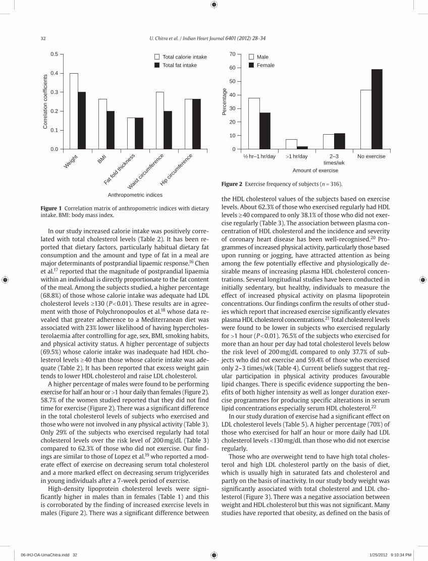
32 U. Chitra et al. / Indian Heart Journal 6401 (2012) 28–34
In our study increased calorie intake was positively corre-lated with total cholesterol levels (Table 2). It has been re-ported that dietary factors, particularly habitual dietary fat consumption and the amount and type of fat in a meal are major determinants of postprandial lipaemic response.16 Chen et al.17 reported that the magnitude of postprandial lipaemia within an individual is directly proportionate to the fat content of the meal. Among the subjects studied, a higher percentage (68.8%) of those whose calorie intake was adequate had LDL cholesterol levels ≥130 (P < 0.01). These results are in agree-ment with those of Polychronopoulos et al.18 whose data re-vealed that greater adherence to a Mediterranean diet was associated with 23% lower likelihood of having hypercholes-terolaemia after controlling for age, sex, BMI, smoking habits, and physical activity status. A higher percentage of subjects (69.5%) whose calorie intake was inadequate had HDL cho-lesterol levels ≥ 40 than those whose calorie intake was ade-quate (Table 2). It has been reported that excess weight gain tends to lower HDL cholesterol and raise LDL cholesterol.
A higher percentage of males were found to be performing exercise for half an hour or >1 hour daily than females (Figure 2). 58.7% of the women studied reported that they did not find time for exercise (Figure 2). There was a significant difference in the total cholesterol levels of subjects who exercised and those who were not involved in any physical activity (Table 3). Only 29% of the subjects who exercised regularly had total cholesterol levels over the risk level of 200 mg/dL (Table 3) compared to 62.3% of those who did not exercise. Our find-ings are similar to those of Lopez et al.19 who reported a mod-erate effect of exercise on decreasing serum total cholesterol and a more marked effect on decreasing serum triglycerides in young individuals after a 7-week period of exercise.
High-density lipoprotein cholesterol levels were signi-ficantly higher in males than in females (Table 1) and this is corroborated by the finding of increased exercise levels in males (Figure 2). There was a significant difference between
the HDL cholesterol values of the subjects based on exercise levels. About 62.3% of those who exercised regularly had HDL levels ≥ 40 compared to only 38.1% of those who did not exer-cise regularly (Table 3). The association between plasma con-centration of HDL cholesterol and the incidence and severity of coronary heart disease has been well-recognised.20 Pro-grammes of increased physical activity, particularly those based upon running or jogging, have attracted attention as being among the few potentially effective and physiologically de-sirable means of increasing plasma HDL cholesterol concen-trations. Several longitudinal studies have been conducted in initially sedentary, but healthy, individuals to measure the effect of increased physical activity on plasma lipoprotein concentrations. Our findings confirm the results of other stud-ies which report that increased exercise significantly elevates plasma HDL cholesterol concentrations.21 Total cholesterol levels were found to be lower in subjects who exercised regularly for >1 hour (P < 0.01). 76.5% of the subjects who exercised for more than an hour per day had total cholesterol levels below the risk level of 200 mg/dL compared to only 37.7% of sub-jects who did not exercise and 59.4% of those who exercised only 2–3 times/wk (Table 4). Current beliefs suggest that reg-ular participation in physical activity produces favourable lipid changes. There is specific evidence supporting the ben-efits of both higher intensity as well as longer duration exer-cise programmes for producing specific alterations in serum lipid concentrations especially serum HDL cholesterol.22
In our study duration of exercise had a significant effect on LDL cholesterol levels (Table 5). A higher percentage (70%) of those who exercised for half an hour or more daily had LDL cholesterol levels <130 mg/dL than those who did not exercise regularly.
Those who are overweight tend to have high total choles-terol and high LDL cholesterol partly on the basis of diet, which is usually high in saturated fats and cholesterol and partly on the basis of inactivity. In our study body weight was significantly associated with total cholesterol and LDL cho-lesterol (Figure 3). There was a negative association between weight and HDL cholesterol but this was not significant. Many studies have reported that obesity, as defined on the basis of
0.5
0.4
0.3
0.2
0.1
0.0
Weig
htBM
I
Fat fo
ld th
ickne
ss
Wais
t circ
umfer
ence
Hip cir
cum
feren
ce
Cor
rela
tion
coef
ficie
nts
Anthropometric indices
Total calorie intake
Total fat intake
Figure 1 Correlation matrix of anthropometric indices with dietary intake. BMI: body mass index.
70
60
50
40
30
20
10
0
Male
Female
½ hr–1 hr/day >1 hr/day 2–3times/wk
No exercise
Per
cent
age
Amount of exercise
Figure 2 Exercise frequency of subjects (n = 316).
06-IHJ-OA-UmaChitra.indd 32 1/25/2012 9:10:34 PM

U. Chitra et al. / Indian Heart Journal 6401 (2012) 28–34 33
BMI, is consistently related to increased blood pressure and unfavourable lipid profiles.23 Waist circumference, however, may be a stronger predictor than BMI for the identification of metabolic and CVD-associated risk factors.24 In our study, BMI was positively and significantly associated with systolic blood pressure (Figure 3). A positive association was observed between waist circumference and both total cholesterol and LDL cholesterol. Waist circumference was also positively cor-related with systolic and diastolic blood pressure and tri-glycerides (Figure 3). It has been reported that a large waist circumference is significantly inversely associated with HDL cholesterol levels and significantly positively associated with LDL cholesterol levels and blood pressure.25
Conclusion
In conclusion, our findings provide support for the poten-tially beneficial effects of both diet and exercise on the serum lipid profile. The most important lifestyle factors which affect the serum lipid profile are diet composition, body weight and physical activity. The modification of blood lipid levels will be beneficial especially to those who are at higher risk of coronary heart disease. Screening for these abnormalities is essential and must be followed by active and effective inter-ventions. Interventions may be more effective if they are tar-geted at specific socio-demographic sub-groups. Dietary advise to younger people should address undesirable aspects of food patterns. Combining campaigns to improve diet with efforts to increase physical activity may be needed to effectively reduce CVD risk.
Acknowledgements
This study was supported by a grant provided by the Management Committee of Kasturba Gandhi College for Women, Secunderabad, India. The study was carried out in
CARE Hospital, Banjara Hills, Hyderabad, India. The authors are grateful to all the subjects who participated in the study.
References
1. Yajnik CS. The insulin resistance epidemic in India: fetal origins, later lifestyle or both? Nutr Rev 2001;59:1–9.
2. NFHS-3. Third National Family Health Survey. International Institute for Population Sciences 2006; Mumbai, India.
3. Gidding SS, Nehgme R, Heise C, Muscar C, Linton A, Hassink S. Severe obesity associated with cardiovascular deconditioning, high prevalence of cardiovascular risk factors, diabetes mellitus/hyperinsulinemia and respiratory compromise. J Pediatr 2004;144:766–9.
4. Yusuf S, Hawken S, Ounpuu S, et al. Effect of potentially modifiable risk factors associated with myocardial infarction in 52 countries (the INTERHEART study): case-control study. Lancet 2004;364:937–52.
5. Haskell WL. Exercise-induced changes in plasma lipids and lipo-proteins. Prev Med 1984;13:23–36.
6. Pais P, Pogue J, Gerstein H, et al. Risk factors for acute myocardial infarction in Indians: a case-control study. Lancet 1996;348:358–63.
7. El-Hazmi MA, Warsy AS. Prevalence of plasma lipid abnormalities in Saudi children. Ann Saudi Med 2001;21:21–5.
8. Allain CC, Poon LS, Chan CS, Richmond W, Fu PC. Enzymatic deter-mination of total serum cholesterol. Clin Chem 1974;20:470–5.
9. Nakamura M, Taniguti Y, Yamamoto M, Hino K, Manabe M. Homogenous assay of serum LDL-cholesterol on an auto analyzer. Clin Chem 1997;43:S260–1.
10. Bucolo G, David H. Quantitative determination of serum triglyc-erides by the use of enzymes. Clin Chem 1973;19:476–82.
11. Thimmayamma BVS. A Hand Book of Schedules and Guidelines in Socioeconomic and Diet Surveys, 1987. National Institute of Nutrition, Indian Council of Medical Research, Hyderabad, India.
12. Gopalan CB, Ramasastri V, Balasubramanian SC. Nutritive Value of Indian Foods, 1989. (As revised and updated by Narasinga Rao BS, Deosthala YG, Pant KC.) National Institute of Nutrition, Indian Council of Medical Research, Hyderabad, India.
13. Stein RA, Michielli DW, Glantz MD, et al. Effects of different exer-cise training intensities on lipoprotein cholesterol fractions in healthy middle-aged men. Am Heart J 1990;119:277–83.
14. Chadha SL, Gopinath N, Katyal I, Shekhawat S. Dietary profile of adults in an urban and a rural community. Indian J Med Res 1995;101:258–67.
15. Glick M, Michel AC, Dorn J, Horwitz M, Rosenthal T, Trevisan M. Dietary cardiovascular risk factors and serum cholesterol in on old order Mennonite community. Am J Public Health 1988;88:1202–5.
16. Berry SEE. Postprandial lipaemia—the influence of diet and its link to coronary heart disease. Nutrition Bulletin 2005;30:314–22.
17. Chen YD, Skowronski R, Coulston AM, Pietarinen J, Hollenbeck CB, Reaven GM. Effect of acute variations in dietary fat and car-bohydrate intake on retinyl ester content of intestinally derived lipoproteins. J Clin Endocrinol Metab 1992;74:28–32.
18. Polychronopoulos E, Panagiotakos DB, Polystipioti A. Diet, life-style factors and hypercholesterolemia in elderly men and women from Cyprus. Lipids Health Dis 2005;4:17.
19. Lopez A, Vial R, Balart L, Arroyave G. Effect of exercise and phy-sical fitness on serum lipids and lipoproteins. Atherosclerosis 1974;20:1–9.
20. Raz I, Rosenbilt H, Kark JD. Effect of moderate exercise on serum lipids in young men with low high density lipoprotein choles-terol. Arteriosclerosis 1988;8:245–51.
21. Marrugat J, Elosua R, Covas MI, Molina L, Rubies-Prat J. Amount and intensity of physical activity, physical fitness, and serum lipids in men. The MARATHOM Investigators. Am J Epidemiol 1996;143:562–9.
Ant
hrop
omet
ric in
dice
s
Waist–hip ratio
Fat intake
Calorie intake
Skin fold (Triceps)
Weight
Waist circumference
BMI
−0.2 0 0.2 0.4 0.6 0.8Correlation coefficients
LDL cholesterol
Total cholesterol
Triglycerides
Diastolic blood pressure
HDL cholesterol
Systolic blood pressure
Figure 3 Correlation coefficients between hypertension and lipid profiles with anthropometric indices. HDL: high-density lipoprotein, LDL: low-density lipoprotein.
06-IHJ-OA-UmaChitra.indd 33 1/25/2012 9:10:34 PM

34 U. Chitra et al. / Indian Heart Journal 6401 (2012) 28–34
22. Nieman DC, Haig JL, Fairchild KS, De Guia ED, Dizon GP, Register UD. Reducing-diet and exercise training effects on serum lipids and lipoproteins in mildly obese women. Am J Clin Nutr 1990;52:640–5.
23. Hu FB, Rimm EB, Stampfer MJ, Ascherio A, Spiegelman D, Willet WC. Prospective study of major dietary patterns and risk of coro-nary heart disease in men. Am J Clin Nutr 2000;72:912–21.
24. Zhu S, Wang Z, Heshka S, Heo M, Faith MS, Heymsfield SB. Waist circumference and obesity-associated risk factors among whites in the third National Health and Nutrition Examination Survey: clinical action thresholds. Am J Clin Nutr 2002;76:743–9.
25. Vijayalakshmi P, Anitha N. Assessing the causative factors and nutritional profile of selected obese subjects. Ind J Nutr Dietet 2003;40:436–46.
Mid-term National Interventional Council Meeting 2012
Dear Colleagues,
It is a matter of great pleasure for us to invite you to attend the Mid-term National Interventional Council Meeting 2012, to be held at Le Meridian, Kochi on 27–29th April 2012.
The NIC 2012 Meeting follows decades of annual NIC meetings but in completely new format. The current NIC meeting will have as overall theme “to explore new ideas, to seek out youth participation, build new relationships beyond the bor-ders, to boldly go where no man has gone before”.
The scientific programme of NIC 2012 will cover a wide array of contemporary topics and showcase cutting edge tech-nologies in the field of interventional cardiology. The programme will have dedicated halls focused on different skill levels of participants. All through, the programme will feature several new and innovative sessions. For the advanced level inter-ventionists there will be talks delivered by key international and national speakers, focused symposiums in niche areas, “Learning with the Masters’ sessions as well as the possibility of showcasing their skills via Live Cases, Talks and demonstra-tion of “Cutting Edge Technologies” For intermediate level there will be more effective learning tools like ‘Case–in–box,’ ‘Tips and Tricks’ sessions and hands-on workshops. At the same time they will be able to showcase their skills by participat-ing in a very meaningful award session offering, international interventional fellowships at some of the most prestigious centres of the world. For the budding interventional cardiologists there will be a strong focus on basics of interventional cardiology, a “Back to Basics” programme and learning on simulators and flow-models. We will also have Joint Sessions with important international bodies, where we will be able to offer a slice of what is happening in other parts of world.
For the first time we have tried to make the process of registration completely paperless. Registration can be done on the NIC Mid-term 2012 Kochi website, http://www.nickochi2012.org. Registration is complimentary for CSI Life Members and Fellows (DM/DNB). Complimentary accommodation will be provided to the registered invited faculty. Limited rooms will be provided complimentary to CSI Life Members and Fellows on first come, first serve basis.
As a Call to Participate in Award Session we are requesting original DICOM CDs with all the clips for evaluation and selec-tion. The CDs should be accompanied with a duly filled form with information on case history, choice of hardware used, along with teaching points of the case.
The form can be down loaded from the NIC Mid-term 2012 Kochi website, http://www.nickochi2012.org/ and should be sent to at the following address:
Dr. Sundeep Mishra, 425 Mount Kailash Tower No. 2, East of Kailash, New Delhi – 110065, Mobile: 9871421390, E-mail: [email protected]
All the cases that reach the Finals will be uploaded at the NIC Website. The awards will be as follows:1. First Prize: 3 Month Fellowship/Observer ship at a reputed interventional center in US/Europe2. Second Prize: 3 Month Fellowship/Observer ship at a reputed center in Asia–Pacific3. Third Prize: 3 Month Interventional Fellowship at a reputed high-volume center in India
Timelines:CD Submission:Opens 15th Dec 2011Closes 15th Mar 2012
Registration Opens:CSI Members 1st Jan 2012Non-CSI Members 15th Jan 2012Fellows 15th Jan 2012Deadline for registration 1st Apr 2012
Conference SecretariatDr Sundeep Mishra Dr PP Mohanan Dr Rony Mathew KadavilChairman NIC Organizing Chairman Organizing [email protected] [email protected] [email protected]
Help DeskSend us your Questions, Suggestions & Comments to [email protected] : +91 999 555 5075, Fax : +91 484 401 9264
06-IHJ-OA-UmaChitra.indd 34 1/25/2012 9:10:34 PM

Contents lists available at SciVerse ScienceDirect
Indian Heart Journal
ISSN: 0019-4832 Copyright © 2012. Cardiological Society of India. All rights reserved.doi: 10.1016/S0019-4832(12)60008-X
Indian Heart Journal 6401 (2012) 35–39
* Corresponding a uthor. E-mail address: [email protected]
Original article
Association between adipocytokines and insulin resistance in Indian hypertensive patientsSujata R. Mahadik*
Research Associate, Sir Hurkisondas Nurrotumdas Medical Research Society, Mumbai, India.
A B S T R A C T
Objective: Although the relationship between obesity, hypertension (HT), and insulin resistance is well-recognised, the pathophysiological mechanism involved is relatively poorly understood. The present study aims in examining the relationship between adipocytokines and insulin resistance in Indian hypertensive patients to better understand the pathogenesis of HT.Methods: A total of 124 subjects including 41 controls, 41 obese, and 42 hypertensive patients were recruited in this cross-sectional study. Fasting adipocytokines (leptin, adiponectin, resistin) and highly sensitive C-reactive protein (hsCRP) levels were measured by enzyme-linked immunosorbent assay (ELISA). Insulin resistance (IR) index was calculated by the homeostasis model assessment (HOMA). The relation between these variables was studied by univariate and multiple logistic regression analysis.Results: Among the hypertensive patients, obese hypertensive patients exhibited significantly in-creased HOMA-IR and altered adipocytokine profile compared to the non-obese control subjects. In a stepwise multiple linear regression analysis with IR as a dependant variable, the study shows leptin as a significant predictor in hypertensive patients. Multiple logistic regression analysis re-vealed that among the adipocytokines, leptin had a strong association with HT in our population.Conclusion: Among the adipocytokine, serum leptin levels were significantly increased in hyper-tensive patients and were also associated with IR and HT. Thus, our findings suggest that leptin may be playing an important role in the development of HT in our population.
Copyright © 2012, Cardiological Society of India. All rights reserved.
K E Y W O R D S
AdipocytokinesHypertensionIndian populationInsulin resistanceObesity
Introduction
Hypertension (HT) as a component of the metabolic syndrome results from complex and multifactorial mechanism including obesity-induced metabolic and hormonal disturbances. Insulin resistance (IR) is often associated with obesity, glucose intol-erance, and dyslipidaemia. Recently, IR has also been linked to essential HT.1–4 Hyperinsulinemia which compensates for IR, is thought to cause and maintain high blood pressure (BP) by stimulating sympathetic nervous activity, proliferation of vascular smooth muscle cells, altered cation transport, and increased sodium reabsorption.3,4 Obesity is defined as an in-creased mass of adipose tissue and is also a common back-ground of the typical lifestyle-related disease, such as HT. Although the relationship between obesity, IR, and HT is
well-recognised, the pathophysiological mechanism involved remains relatively poorly understood.
In the past, adipose tissue was thought to be a passive depot for the storage of excess energy. However, recent studies have demonstrated that the adipocyte synthesises and secretes bio-logically active molecule. These molecules collectively known as adipocytokines, which include adiponectin, resistin, leptin, tumour necrosis factor-α (TNF-α), interleukin-6 (IL-6), and free fatty acids, appears to be important in the development of IR5,6 and other related diseases including HT.
The association between obesity and HT suggests that adipose mass and adipocytokines secreted by them may be important in the regulation of BP or in the pathogenesis of HT, although the mechanism underlying this are not yet evident.7–11
Among the adipocytokines, leptin is a hormone which is predominantly secreted by the adipose tissue. Recent studies have shown that leptin increases arterial BP. Although data from available animal studies clearly indicate an association
07-IHJ-OA-Sujata.indd 35 1/25/2012 9:09:44 PM

36 S.R. Mahadik / Indian Heart Journal 6401 (2012) 35–39
between leptin and HT, results of human studies have been less definitive.9–15 Though several studies have shown that adiponectin correlates negatively with BP, the results of stud-ies on the relation between adiponectin and HT have been inconsistent.7,8,16,17 Resistin, a novel cysteine-rich protein secreted by adipocytes, has been proposed to serve as a link between obesity and IR in rodents, but this has remained controversial.18–21 Very few studies have been carried out to find the role of resistin in essential HT, but results are incon-sistent.11 All these adipocytokines such as leptin, adiponectin, and resistin have been associated with HT in either cross-sectional or longitudinal studies. However, few studies directly compared the relative association of these cytokines with BP or with the presence of HT.
Thus, the role of individual adipocytokine in HT in relation to IR is still controversial. The Indian population is relatively more IR due to the high percentage of body fat and higher abdominal obesity at low body mass index (BMI) though the exact cause of increased abdominal obesity and IR in Asian Indian has not been clear.22 According to our recent study adipocytokine may play an important role in the development of IR in Indian diabetic patients.5 In addition, we also re-ported that almost 40% of the essential hypertensive patients were insulin resistant. Thus, with the increasing prevalence of IR, HT is also becoming a major health problem in India. Hence, in the present study we would like to elucidate the role of adipocytokine in relation to IR in Indian hypertensive patients.
Methods
Subjects
A total of 124 subjects aged between 30 years and 70 years comprising 41 controls, 41 obese, and 42 hypertensive patients were taken up for the study after an overnight fast. Hyperten-sive patients were further divided into 2 subgroups based on their BMI: (1) non-obese hypertensive (n = 23) (BMI ≤ 25 kg/m2) and (2) obese hypertensive (n = 19) (BMI ≥ 25 kg/m2).
Past medical history and clinical data were collected and anthropometrics measurements such as height, weight, and waist circumference were taken. Waist circumference was measured around the abdomen just above the hip bone. Body mass index was calculated from the ratio of body weight in kilograms (kg) to height in square meters and expressed as kg/m2 units. Blood pressure was measured in the seated po-sition using mercury sphygmomanometer. Average of two consecutive readings taken 5 minutes apart was recorded. All the subjects gave their informed consent after the procedure was explained to them. The Ethics Committee of Our Hospital approved the project.
Study design
Each subject’s venous blood was collected after 12–14 hours fast for estimating fasting glucose, lipid profile, insulin, high
sensitive C-reactive protein (hsCRP), and adipocytokines includ-ing leptin, adiponectin, and resistin.
Inclusion and exclusion criteria for selection of subjects
Hypertensive patients1. Hypertension was defined as systolic BP (SBP) to diastolic
BP (DBP) >140/90 mmHg or the use of antihypertensive medication.23
2. Hypertensive patients were not receiving treatment for diabetes or any other illness at the time of study.
Obese subjects1. Obesity was defined if their BMI ≥25 kg/m2 according to
the cut-off suggested for Asian Indians.24,25
Control subjects1. Controls were classified as having normal glucose tolerance
(fasting plasma glucose <6.1 mmol/L and 2 hours glucose <7.8 mmol/L).24
2. They were non-hypertensive and non-obese.3. They were confirmed to have no known disease condition
including cardiac, thyroid disease or any other acute and chronic disease in the past or any current infection/condition.They were never symptomatic.
Biochemical analysis
Fasting plasma glucose was measured by the glucose peroxidase method (Randox, USA). Serum cholesterol, high-density lipoprotein (HDL)-cholesterol, and triglyceride levels were measured by the enzymatic method (Randox USA). Fasting serum insulin was assayed by radioimmunoassay using RIA kit (BRIT Mumbai, India) incorporating 125I-labelled porcine insulin as the tracer and guinea pig antiserum. Serum hsCRP levels were measured by a highly sensitive en-zyme-linked immunosorbent assay (ELISA) (DSL, USA) which has the lower detection limit of 1.6 ng/mL with intra-assay coefficient of variation 4.25% and inter-assay coefficient of variation 5.95%. Serum leptin, adiponectin, and resistin levels were measured by ELISA (Linco Res, USA). Intra-assay and inter-assay coef ficient of variation are as follows: for leptin (1.4%, 4.6%), for adiponectin (4.4%, 6.6%), for resistin (2.75%, 6.7%).
Calculation and data analysis
Insulin resistance measured as homeostasis model assessment-IR (HOMA-IR) using following formula26:
IR (HOMA-IR) =
Fasting insulin (U/mL) × Fasting glucose (mmol/L)
22.5
07-IHJ-OA-Sujata.indd 36 1/25/2012 9:09:45 PM
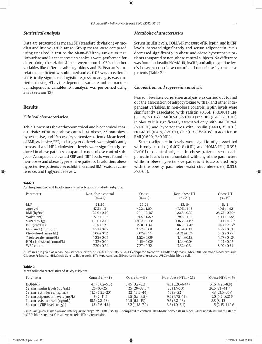
S.R. Mahadik / Indian Heart Journal 6401 (2012) 35–39 37
Statistical analysis
Data are presented as mean ± SD (standard deviation) or me-dian and inter-quartile range. Group means were compared using unpaired ‘t’ test or the Mann-Whitney rank sum test. Univariate and linear regression analysis were performed for determining the relationship between serum hsCRP and other variables like different adipocytokines and IR. Pearson’s cor-relation coefficient was obtained and P < 0.05 was considered statistically significant. Logistic regression analysis was car-ried out using HT as the dependent variable and biomarkers as independent variables. All analysis was performed using SPSS (version 15).
Results
Clinical characteristics
Table 1 presents the anthropometrical and biochemical char-acteristics of 41 non-obese control, 41 obese, 23 non-obese hypertensive, and 19 obese hypertensive patients. Mean levels of BMI, waist size, SBP, and triglyceride levels were significantly increased and HDL cholesterol levels were significantly re-duced in obese patients compared to non-obese control sub-jects. As expected elevated SBP and DBP levels were found in non-obese and obese hypertensive patients. In addition, obese hypertensive patients also exhibit increased BMI, waist circum-ference, and triglyceride levels.
Metabolic characteristics
Serum insulin levels, HOMA-IR measure of IR, leptin, and hsCRP levels increased significantly and serum adiponectin levels decreased significantly in obese and obese hypertensive pa-tients compared to non-obese control subjects. No difference was found in insulin HOMA-IR, hsCRP, and adipocytokine lev-els between non-obese control and non-obese hypertensive patients (Table 2).
Correlation and regression analysis
Pearson bivariate correlation analysis was carried out to find out the association of adipocytokine with IR and other inde-pendent variables. In non-obese controls, leptin levels were significantly associated with resistin (0.651, P < 0.001) CRP (0.354, P < 0.02), BMI (0.541, P < 0.001) and DBP (0.408, P < 0.01). In obesity it is significantly associated only with BMI (0.784, P < 0.001) and hypertensives with insulin (0.409, P < 0.01), HOMA-IR (0.419, P < 0.01), CRP (0.32, P < 0.05) in addition to BMI (0.609, P < 0.001).
Serum adiponectin levels were significantly associated with only insulin (−0.407, P < 0.01) and HOMA-IR (−0.395, P < 0.01) in control subjects. In obese patients, serum adi-ponectin levels is not associated with any of the parameters while in obese hypertensive patients it is associated only with the obesity parameter, waist circumference (−0.338, P < 0.05).
Table 1Anthropometric and biochemical characteristics of study subjects.
Parameter Non-obese control Obese Non-obese HT Obese HT (n = 41) (n = 41) (n = 23) (n = 19)
M:F 21:20 20:21 13:10 8:11Age (yr) 47.2 ± 1.31 47.2 ± 1.09 47.96 ± 1.45 49.5 ± 1.92BMI (kg/m2) 22.0 ± 0.30 29.1 ± 0.46a 22.5 ± 0.33 28.72 ± 0.69a
Waist (cm) 77.7 ± 1.19 91.5 ± 1.27a 79.5 ± 1.65 91.1 ± 1.65a
SBP (mmHg) 115.6 ± 2.45 120.2 ± 2.33a 136.7 ± 4.19a 131.1 ± 4.58b
DBP (mmHg) 75.8 ± 1.21 79.0 ± 1.39 86.7 ± 2.91c 84.2 ± 2.07b
Glucose F (mmol/L) 4.53 ± 0.08 4.57 ± 0.09 4.59 ± 0.11 4.77 ± 0.13Cholesterol (mmol/L) 5.06 ± 0.17 5.07 ± 0.14 4.71 ± 0.20 5.02 ± 0.29Triglyceride (mmol/L) 1.23 ± 0.05 1.52 ± 0.09c 1.44 ± 0.13 1.57 ± 0.12c
HDL cholesterol (mmol/L) 1.32 ± 0.04 1.15 ± 0.02c 1.24 ± 0.04 1.24 ± 0.05WBC count 7.20 ± 0.24 7.27 ± 0.32 7.62 ± 0.3 8.09 ± 0.31
All values are given as mean ± SE (standard error). aP < 0.001, bP < 0.05, cP < 0.01 compared to controls. BMI: body mass index, DBP: diastolic blood pressure, Glucose F: fasting, HDL: high-density lipoprotein, HT: hypertension, SBP: systolic blood pressure, WBC: white blood cell.
Table 2Metabolic characteristics of study subjects.
Parameter Control (n = 41) Obese (n = 41) Non-obese HT (n = 23) Obese HT (n = 19)
HOMA-IR 4.1 (3.02–5.3) 5.05 (3.9–8.2) 4.6 (3.26–6.44) 6.16 (4.25–8.9)Serum insulin levels (uU/mL) 20 (16–25) 25 (20–38.5)a 23 (17–30) 26.5 (21–44)b
Serum leptin levels (ng/mL) 11.5 (6.35–20) 22 (13.5–44)a 16 (8–22) 43 (21.5–65)a
Serum adiponectin levels (mg/L) 9 (7–11.5) 6.5 (5.2–9.5)a 9.0 (6.75–11) 7.0 (5.7–8.25)b
Serum resistin levels (ng/mL) 10.5 (7.2–13) 10.5 (6.1–13) 9.6 (6.8–13) 8.8 (8–13)Serum hsCRP levels (mg/L) 1.8 (0.6–4.8) 3.2 (1.38–7.2) 3.3 (1.0–6.1) 5 (2.15–11.2)a
Values are given as median and inter-quartile range. aP < 0.001, bP < 0.01, compared to controls. HOMA-IR: homeostasis model assessment-insulin resistance, hsCRP: high sensitive C-reactive protein, HT: hypertension.
07-IHJ-OA-Sujata.indd 37 1/25/2012 9:09:45 PM

38 S.R. Mahadik / Indian Heart Journal 6401 (2012) 35–39
Serum resistin levels were significantly associated with leptin (0.651, P < 0.001), CRP (0.495, P < 0.001), BMI (0.333, P < 0.05), SBP (0.336, P < 0.05), and DBP (0.416, P < 0.01) in control subjects, while in obese patients it is associated only with waist circumference (0.379, P < 0.01), and in hyperten-sive patients it is associated with CRP (0.407, P < 0.01) and BMI (0.337, P < 0.05). Interestingly, serum resistin levels were significantly associated with HOMA-IR in non-obese hyper-tensive patients (0.485, P < 0.02).
Findings from the above bivariate correlation analysis were further explored using the stepwise multiple linear regres-sion analysis with HOMA-IR as a dependent variable. It was found that only adiponectin is the significant predictor of IR in non-obese control subjects (β: −0.177, SE (β): 0.066, P < 0.01), and leptin in hypertensive patients (β: 0.037, SE (β): 0.018, P < 0.04). It was also observed that serum resistin levels are a significant predictor of SBP in non-obese control subjects (β: 1.306, SE (β): −0.585, P < 0.03).
Multiple logistic regression analysis using HT as the de-pendent variable revealed that among the adipocytokine, leptin had a strong association with HT in our population (odds ratio [OR] 1.048, confidence interval [CI] 1.016–1.081), P < 0.003).
Discussion
With an increasing prevalence of IR, HT is becoming a major health problem in India. According to our recent study adipocy-tokine may be playing an important role in the development of IR among Indian diabetic patients. Hence, in the present study we have elucidated the association of adipocytokines with IR in Indian hypertensive patients.
The main findings of the study are:1. Hypertensive patients exhibit affected lipid metabolism
with increased triglyceride levels.2. Homeostasis model assessment-IR and serum leptin levels
increased significantly and adiponectin levels decreased sig-nificantly in obese hypertensive patients even after normal-ising with BMI.
3. Interestingly in stepwise multiple linear regression analy-sis serum resistin levels were significantly associated with SBP in controls. In addition we also found that adiponectin is an important predictor of IR in controls and leptin in hy-pertensive patients. Multiple logistic regression analysis revealed leptin to be the important predictor of HT in our population.Disturbances of glucose and lipid metabolism are known
to be related to HT, since it has been suggested that hyper-insulinemia enhances hepatic very low-density lipoprotein (VLDL) synthesis and contribute to increased plasma trigly-ceride levels.7 Thus, our results are consistent with these previous reports, which suggest that essential hypertension often accompanies IR and resultant disturbances of lipid metabolism, increasing the risk of arteriovascular disease in essential HT.
Although till date several studies have been carried out to elucidate the role of different adipocytokines in hypertensives,
the results obtained from different studies were highly controversial.8–12,16 Among the adipocytokines an association between HT and adiponectin levels has been reported by several groups.7,8,16,17 Although no association of adiponectin was found with BP and IR in our hypertensives, it is signifi-cantly associated with IR in controls exhibiting a physiologi-cal relationship. We also found that among adipocytokines, adiponectin level decreased significantly independent of the changes in BMI in obese hypertensive patients. The mecha-nism by which adiponectin levels are lowered in patients re-mains to be clarified. Although there are several mechanisms that could account for the relationship, it is possible firstly, that the activation of rennin-angiotensin system (RAS) may be responsible, by hypoadiponectinemia resulting in an increase in fat mass and BP17 or secondly, inflammation may contrib-ute to the pathogenesis of HT. Adiponectin is said to have beneficial and protective effects including antiatherogenic and anti-inflammatory properties.
Resistin is an adipocyte derived peptide that might play a role in obesity and IR; however, its role in humans is largely unknown. In our previous study, we found that it may be playing an indirect role in the pathogenesis of T2DM (type 2 diabetes mellitus) through its association with leptin in our population.5
Studies by Furuhashi11 suggest that circulating resistin lev-els are not related to IR in essential hypertensive patients, al-though disturbances of lipid metabolism may be associated with IR. In our study we found that there was no difference in serum levels and no association with BP in hypertensives but are significantly associated with SBP in control subjects. Additional studies are needed to elucidate the regulatory and biological function of resistin in humans.
In this study, we have selected hypertensive patients ran-domly from the clinic and in addition the groups we exam-ined are relatively small. It is likely that with a large number of subjects, the statistical analysis could show better signifi-cance; this was the limitation of the study.
The result of earlier studies on the role of leptin in human HT is not consistent. Some studies report significantly higher leptin levels in essential HT than in control26,27 or a significant correlation between HT and BP.26–28 However, these results were not confirmed by others.29,30
In the present study, serum leptin levels increased signifi-cantly in obese hypertensive patients. This increase was inde-pendent of changes in BMI. Though leptin is not significantly associated with BP, in stepwise multiple linear regression analysis it is the only adipocytokine significantly associated with IR in our hypertensives. According to our previous study, 40% of the hypertensive patients exhibit IR. It can be specu-lated that insulin and leptin interact and modulate each other’s effects and contribute to HT is an effect of tubular sodium handling.14 Leptin has several effects such as stimulation of the RAS and sympathetic nervous system which may affect BP levels in humans.12,13,31
Moreover, in multiple logistic regression analysis leptin is an important predictor of HT in our population suggesting that leptin may be playing an important role in the development of HT in our population. Taken together, leptin does indeed
07-IHJ-OA-Sujata.indd 38 1/25/2012 9:09:45 PM

S.R. Mahadik / Indian Heart Journal 6401 (2012) 35–39 39
have peripheral physiological effects that suggest it may be a link in the triad of obesity, IR, and HT.
To summarise, findings from our study shows that a cor-relation exists between leptin and IR; moreover, in logistic regression analysis after adjusting for confounding variables, increasing leptin levels were associated with high-risk of the development of HT. Thus, leptin may be playing an important role in development of HT through its association with IR in our population. Detailed study on the role of adipocytokines may provide new insights into the development of safe and effective pharmaceutical treatment of essential hypertensive patients with IR.
Acknowledgments
The Sir H.N. Medical Research Society supported this work. The author wishes to thank Dr. S.S. Deo and Dr. S.D. Mehatalia for their kind support and research staff, R. Mokal, S. Mithbawkar, K. Mistry and P. Thakur for the technical and social help received by them in carrying out the study.
References
1. Modan M, Halkin H, Almong S, et al. Hyperinsulinemia: a link between hypertension obesity and glucose tolerance. J Clin Invest 1985;75:809–17.
2. Reaven GM. Banting lecture 1988: role of IR in human disease. Diabetes 1988;37:1595–1607.
3. DeFronzo RA. IR: a multifaceted syndrome responsible for NIDDM, obesity, hypertension, dyslipidemia, and atherosclerotic cardio-vascular disease. Diabetes Care 1991;14:173–92.
4. Agata J, Miyazaki Y, Takada M, Masuda A, Miura T. Association of IR and hyperinsulinemia with disturbed lipid metabolism in patients with essential hypertension. Hypertens Res 1998;21:57–62.
5. Mahadik SR, Deo SS, Mehtalia SD. Association of adiposity, in-flammation and atherosclerosis: the role of adipocytokines and CRP in Asian Indian subjects. Metab Syndr Relat Disord 2008;6:121–8.
6. Matsuzawa S. Therapy insight: adipocytokines in metabolic syn-drome and related cardiovascular disease. Nat Clin Pract Cardiovasc Med 2006;3:35–42.
7. Murakami H, Nobuyuki URA, Furuhashi M, Shimamoto K. Role of adiponectin in insulin resistant hypertension and atherosclerosis. Hypertens Res 2003;26:705–10.
8. Sung SH, Chuang SY, Sheu WH, Lee WJ, Chou P, Chen CH. Adiponectin, but not leptin or high sensitivity C-reactive protein is associated with blood pressure independently of general and abdominal adiposity. Hypertens Res 2008;31:633–40.
9. Imatoh T, Miyazaki M, Momose Y, et al. Hyperleptinemia is asso-ciated with hypertension in Japanese males. Acta Med Okayama 2008;62:169–74.
10. Almeida-Pititto B, Gimeno SGA, Freire RD, Ribeiro-Filho FF, Ferreira SRG. Leptin is not associated independently with hyper-tension in Japanese-Brazilian women. Brazilian J Med Biol Res 2006;39:99–105.
11. Furuhashi M, Ura N, Higashiura K, Murakami H, Shimamoto K. Circulating resistin levels in essential hypertension. Clin Endocrinol 2003;59:507–10.
12. Haynes W. Role of leptin in obesity-related hypertension. Exp Physiol Symp Rep 2005;90:683–8.
13. Bravo PE, Morse S, Borne DM, Aguilar EA, Reisin E. Leptin and hypertension in obesity. Vascular health and risk management 2006;2:163–9.
14. Stenvinkel P. Leptin and blood pressure—is there a link? Nephrol Dial Transplant 2000;15:1115–7.
15. Ogawa Y, Masuzaki H, Aizawa M, et al. Blood pressure elevation in transgenic mice overexpressing leptin, the obese gene product. J Hypertens 1998;16:7.
16. Imatoh T, Miyazaki M, Momose Y, Une H, Tanihara S. Adiponectin levels associated with the development of hypertension: a prospec-tive study. Hypertens Res 2008;31:229–33.
17. Patel JV, Lim HS, Hughes EA, Lip GYH. Adiponectin and hyperten-sion: a putative link between adipocyte function and atheroscle-rotic risk? J Hum Hypertens 2007;21:1–4.
18. Steppan CM, Bailey ST, Bhat S, et al. The hormone resistin links obesity to diabetes. Nature 2001;409:307–12.
19. Nagaev I, Smith U. IR and Type 2 diabetes are not related to resistin expression in human fat cell or skeletal muscle. Biochem Biophys Res Commun 2001;295:561–4.
20. Janke J, Engeli S, Gorzellniate K, et al. Resistin gene expression in human adipocytes is not related to IR. Obes Res 2002;10:1–5.
21. Savage DB, Sewter CP, Klenk ES, et al. Resistin/Fizz3 expression in relation to obesity and Peroxisome proliferator activated recep-tor γ action in humans. Diabetes 2001;50:2199–202.
22. Mahadik SR, Deo SS, Mehatalia SD. Increased prevalence of metabolic syndrome in non-obese Asian Indian: an urban-rural comparison. Metab Syndr Relat Disord 2007;5:140–50.
23. Chobanian AV, George L, Bakris GL, et al. The seventh report of the joint national committee on prevention, detection, evaluation, and treatment of high blood pressure: the JNC 7 report. JAMA 2003;289:2560–72.
24. World Health Organization (1999). Definition, diagnosis and classifiation of diabetes mellitus and its complications. Report of a WHO consultation. Part 1: diagnosis and classification of dia-betes mellitus (WHO/NCD/CS/99.2) World Health Organization, Geneva.
25. World Health Organization. The Asia-Pacific perspective: rede-fining obesity and its treatment. Geneva, Switzerland: World Health Organization 2000.
26. Matthews DR, Husker JP, Rudenski AS, et al. Homeostasis model assessment: IR and beta cell function from fasting plasma glu-cose and insulin concentrations in man. Diabetologia 1985;28:412–9.
27. Agata J, Masuda A, Takada M, Murakami H, Higashiura K, Miyazaki Y. High plasma immunoreactive leptin level in essen-tial hypertension. Am J Hypertens 1997;10:1171–4.
28. Sheu WH, Lee WJ, Chen YT. High plasma leptin concentrations in hypertensive men but not in hypertensive women. J Hypertens 1999;17:1289–95.
29. Hirose H, Saito I, Tsujioka M, et al. The obese gene product, lep-tin: possible role in obesity-related hypertension in adolescents. J Hypertens 1998;16:2007–12.
30. Kokot F, Adamczak M, Wiecek A, Cieplok J. Does leptin play a role in the pathogenesis of essential hypertension? Kidney Blood Press Res 1999;22:154–60.
31. Uckaya G, Ozata M, Sonmez A, et al. Plasma leptin levels strongly correlate with plasma rennin activity in patients with essential hypertension. Horm Metab Res 1999;31:435–8.
07-IHJ-OA-Sujata.indd 39 1/25/2012 9:09:45 PM

Contents lists available at SciVerse ScienceDirect
Indian Heart Journal
ISSN: 0019-4832 Copyright © 2012. Cardiological Society of India. All rights reserved.doi: 10.1016/S0019-4832(12)60009-1
Indian Heart Journal 6401 (2012) 40–42
* Corresponding a uthor. E-mail address: [email protected]
Original article
Vertical P-wave axis: the electrocardiographic synonym for pulmonary emphysema and its severityLovely Chhabra1*, Pooja Sareen2, Daniel Perli2, Indu Srinivasan2, David H. Spodick3
Senior Resident and Voluntary Research Scholar, Resident, Department of Internal Medicine, Professor and Director of Clinical Research Program, Department of Cardiovascular Medicine, Saint Vincent Hospital, University of Massachusetts Medical School, Worcester, MA – , USA.
A B S T R A C T
Background: The correlation between vertical P-wave axis (P-axis > 60°) and pulmonary emphysema was investigated on a very large controlled series to see if P-axis verticalisation as lone criterion can be effectively used to screen emphysema in general population. Correlation between degrees of P-axis verticalisation and the severity of the obstructive lung disease (as per global initiative for chronic obstructive lung disease [GOLD] criteria) was also studied to see if this criterion can be used for gross quantification of the chronic obstructive pulmonary disease (COPD) in routine clinical practice.Materials and methods: Around 6500 unselected, routine electrocardiograms (ECGs) were reviewed which yielded 600 ECGs with vertical P-axis in sinus rhythm. 635 ECGs from the same continuum were selected with P-axis ≤60° matched for patient’s age and sex serving as controls. Charts were reviewed for the diagnosis of COPD and emphysema based on medical history, pulmonary function tests, and imaging studies.Results: Prevalence of emphysema in patients with vertical P-axis was strikingly higher than in the control group: 85% vs 4.4%. The sensitivity and specificity of vertical P-axis for diagnosing emphy-sema was 94.76% and 86.47%, respectively. Vertical P-axis and forced expiratory volume (FEV1) were inversely correlated (Pearson correlation coefficient = –0.683). Prevalence of severe COPD was strikingly higher in patients with P-axis > 75° as compared to the group with P-axis 60°–75°: 96.3% vs 4.6%. Close to 80% of the emphysema patients with P-axis > 85° had very severe disease (FEV1 < 30%).Conclusion: P-axis verticalisation is highly effective for screening emphysema and degree of verti-calisation provides a gross quantification of the disease.
Copyright © 2012, Cardiological Society of India. All rights reserved.
K E Y W O R D S
Chronic obstructive lung diseaseElectrocardiogram (ECG)Pulmonary emphysemaPulmonary function test
Introduction
Emphysema of any pathogenesis, nearly always due to chronic obstructive pulmonary disease (COPD) and rarely due to alpha-1 antitrypsin deficiency (AAD) produces a state of abnormal lung hyperinflation and has been shown to carry an association with a vertical frontal P-wave axis1,3,9 by several studies done on small patient populations. A vertical P-wave axis (>60°) during a sinus rhythm can be easily determined by a simple glance at the electrocardiogram (ECG); an entirely
inverted or dominantly negative P-wave in lead aVL and al-ternatively a P-wave in lead III larger than the P-wave in lead II was observed. Our study aims to investigate the reproduc-ibility of the relationship between vertical P-axis and emphy-sema on a very large controlled series primarily to determine if verticalisation of the frontal P-wave vector as a lone criterion can be used as a standard guideline for screening emphysema in a general population. In addition, we wanted to investigate the correlation between the degrees of P-axis verticalisation and the severity of the obstructive lung disease as per the global initiative for chronic obstructive lung disease (GOLD) criteria to see if this criterion can be used for the gross quan-tification of the COPD in routine clinical practice.
08-IHJ-OA-Lovely chhabra.indd 40 1/25/2012 9:08:43 PM

L. Chhabra et al. / Indian Heart Journal 6401 (2012) 40–42 41
Materials and methods
Unselected, consecutive routine ECGs of 6500 patients were reviewed from the hospital TraceMaster Vue system yielding 600 ECGs with P-wave axis >60° in normal sinus rhythm (NSR) after exclusion criteria. Around 635 patients from the same ECG continuum were selected with P-wave axis <60° in sinus rhythm matched for age and sex serving as the control group (Table 1). Inclusion criteria were age >45 years,1–4,7 NSR and availability of past medical history, imaging studies and/or pulmonary function tests. Patients <45 years old were ex-cluded since vertical P-wave axis is a normal finding in healthy children and in many young adults. Any rhythm other than sinus was also excluded. Charts were then reviewed for the diagnosis of the COPD and emphysema based on the medical history, pulmonary function tests and imaging studies in-cluding chest radiographical and computed tomography (CT) scans. Diagnosis of emphysema or lung hyperinflation was pri-marily based upon the radiological evidence. The radiologist reporting of the imaging studies (including chest radiographs and CT-scans) was mainly used for the diagnosis of any em-physematous changes. The severity of the chronic obstructive lung disease was determined based upon the forced expiratory volume (FEV1) value. A direct comparison of the FEV1 value was done with the electrocardiographic P-wave axis in those patients whose pulmonary function tests (PFTs) were available within the last 6 months (from the date of the ECG study) determining any statistical correlation. Statistical analysis was done by using Statistical Package for Social Sciences (SPSS Inc., Chicago, IL, version 15.0 for Windows). The means and stan-dard deviations of the P-wave axis and age were calculated and the data were checked for skewness by Kolmogorov Smirnov tests of normality. Mann–Whitney U test was applied for comparison of the 2 groups as the data for P-wave axis were skewed. Qualitative or categorical variables were described as frequencies and proportions. Proportions were compared using χ2/Fisher’s exact test whichever was appropriately appli-cable. P values <0.05 were considered statistically significant.
Results
The patients in the groups with P-wave axis >60° and P-wave axis ≤60° were well matched for age with mean age of
68.2 years and 67.7 years respectively (P = 0.601). The preva-lence of smoking history (over 20 pack years) was found to be 78.7% in patients with vertical P-wave axis as compared to 36.9% in the control group (P < 0.001). Patients with vertical P-wave axis also had strikingly higher prevalence of emphy-sema than the control group: 85% vs 4.4% (Table 1). Sensitivity and specificity of the P-wave axis >60° for diagnosing em-physema in the adult age group (>45 years) was 94.8% and 86.5%, respectively (Table 2). The positive and negative pre-dictive values of this lone criterion were found to be 84.2% and 95.6% respectively (Table 2). The mean P-wave axis in emphysema patients was found to be 69.7° as compared to 36.1° in patients without emphysema (Table 3). The degree of P-wave verticalisation was found to have a strong inverse re-lationship with FEV1; Pearson correlation coefficient being –0.683; P < 0.001) (Table 4). The prevalence of severe obstruc-tive lung disease as defined by GOLD criteria (FEV1 < 50%) was strikingly higher in emphysema patients with a high de-gree of verticalisation (P-axis >75°) as compared to those with a lower vertical P value (P-axis between 60° and 75°): 96.3% vs 4.6% (Table 5). Interestingly, 80% of emphysema pa-tients in our cohort with P-axis >85° were found to have very severe or terminal disease (FEV1 < 30%).
Table 2Sensitivity, specificity, positive predictive value and negative predic-tive value of each vertical P-wave axis for emphysema (n = 1235).
P-wave Sensitivity Specificity Positive Negative axis (%) (%) predictive predictive value (%) value (%)
>60° 94.76 86.47 84.19 95.59≥70° 91.88 93.82 87.38 95.59≥80° 77.40 99.18 94.68 95.59
Table 1P-wave axis: comparison of patient groups with P-wave axis >60° versus ≤60° (n = 1235).
Variable P-wave axis P value
>60° (n = 600) ≤60° (n = 635)
Age (yr) 68.21 ± 13.24 67.78 ± 13.9 0.601Gender 0.071 Female 335 (55.8%) 322 (50.7%) Male 265 (44.2%) 313 (49.3%) Smoking 78.7% 36.9% <0.001Emphysema 85% 4.4% <0.001P-wave axis (°) 71 ± 8.13 31.6 ± 17.48 <0.001
Data are presented as mean ± standard deviation, n (%) or %
Table 3Comparison of patients with and without emphysema (n = 1235).
Variable Emphysema P value Yes (n = 538) No (n = 697)
Age (yr) 69.20 ± 13 66.96 ± 14.29 <0.001P-wave axis 69.71 ± 11.87 36.1 ± 20.57 <0.001
Data are presented as mean ± standard deviation.
Table 4Correlation between vertical P-wave axis and forced expiratory volume 1 (n = 191).
P-wave axis FEV1
P-wave axis Pearson correlation 1 –0.683* Significant (2-tailed) 0 n 1235 191
FEV1 Pearson correlation –0.683* 1 Significant (2-tailed) 0 n 191 191
*Correlation is significant at the 0.01 level (2-tailed).Around 191 emphysema patients with vertical P-axis had available pul-monary function tests within the last six months of the electrocardio-gram study. FEV1: forced expiratory volume.
08-IHJ-OA-Lovely chhabra.indd 41 1/25/2012 9:08:44 PM

42 L. Chhabra et al. / Indian Heart Journal 6401 (2012) 40–42
Discussion
Previous studies on this subject have found a close correlation between P-wave verticalisation and COPD/emphysema.1–8 A plausible mechanism for P-axis verticalisation in lung hyper-inflation is that the right atrium is firmly attached to the dia-phragm by a dense pericardial ligament around the inferior vena cava.9 With progressive flattening of the diaphragm, the right atrium is distorted/displaced inferiorly causing a signif-icant rightward deviation (verticalisation) of the P-wave axis. In contrast, in pure restrictive (fibrotic) lung disease, the P-wave axis tends to be horizontal or leftward, correlating with the de-gree of diaphragmatic elevation.10 Our retrospective study uti-lises an unselected large patient population to find a definite correlation of the vertical P-wave axis and emphysema. The results demonstrated that this lone criterion can be used as an effective screen for pulmonary hyperinflation, using a few simple parameters. The degree of P-wave verticalisation has a strong inverse relationship with FEV1 which quickly provides a gross quantification of severity of the obstructive lung disease. The degree of P-wave verticalisation might also have a sig-nificant correlation with the radiological severity of the em-physematous changes/lung hyperinflation which is the basic mechanism for a vertical P-wave axis; however, the radiolog-ical quantification was not performed in our study and would be an interesting subject for future studies.
Limitations
P-wave verticalisation can only be used to detect emphysema in patients who have an ECG with NSR. It is of course not un-common to see a high prevalence of atrial arrhythmias like multifocal tachycardia in patients with obstructive lung dis-ease and in such cases; this principle cannot be applied due to a variable P-wave axis. The other theoretical factors which can possibly affect P-wave axis like height, body habitus, co-existence of restrictive lung disease, etc. are not taken into account to avoid the complexity of interpretation while using vertical P-wave axis as a lone diagnostic criterion.
Conclusion
Despite previous work on this subject in various studies since 1950s, most internists and even specialists (including cardio-logists or pulmonologists) are not aware about the practical utility of this relationship. Our study is the largest controlled series so far on this subject which aimed to determine a defi-nite correlation of vertical P-axis with emphysema and se-verity of obstructive lung disease as per GOLD criteria. P-axis verticalisation can serve as a very effective electrocardiographic screening tool for emphysema in the general population. In fact, this quick bedside screening modality is inexpensive, and at the same time highly sensitive and specific. We propose its inclusion as one of the standard diagnostic guidelines for pulmonary emphysema in patients with age >45 years and emphasise its use in routine clinical practice. The degree of P-wave verticalisation also provides a quick gross quantifica-tion of the chronic obstructive lung disease.
References
1. Spodick DH. Electrocardiographic studies in pulmonary disease: I. Electrocardiographic abnormalities in diffuse lung disease. Circulation 1959;20:1067–72.
2. Littman D. The electrocardiographic findings in pulmonary em-physema. Am J Cardiol 1960;5:339–48.
3. Spodick DH, Hauger-Klevene JH, Tyler MJ. The electrocardiogram in pulmonary emphysema: relationship of characteristic electro-cardiographic findings to severity of disease as measured by the degree of airway obstruction. Am Rev Respir Dis 1963;88:14–9.
4. Spodick DH. Vectorcardiogram in pulmonary emphysema: its re-lationship to scalar electrocardiographic findings. Am Rev Respir Dis 1968;98:634–9.
5. Thomas AJ, Apiyasawat S, Spodick DH. Electrocardiographic detection of emphysema. Am J Cardiol 2011;107:1090–2.
6. Baljepally R, Spodick DH. Electrocardiographic screening for emphysema: the frontal plane P axis. Clin Cardiol 1999;22:226–8.
7. Calatayud JB, Abad JM, Khoi NB, Stanbro WJ, Silver HM. P wave changes in pulmonary disease. Am Heart J 1970;79:445–53.
8. Zuckerman R, Cabrera CE, Fishleder BL, Sodi-Pallares D. The elec-trocardiogram in chronic cor pulmonale. Am Heart J 1948;35:421–5.
9. Shah NS, Koller SM, Janover ML, Spodick DH. Diaphragm levels as determinants of P axis in restrictive vs. obstructive pulmonary disease. Chest 1995;107:697–700.
10. Zambrano SS, Moussavi MS, Spodick DH. QRS duration in chronic obstructive lung disease. J Electrocardiogr 1974;7:35–6.
11. Spodick DH. Pulmonary emphysema. Am J Geriatr Cardiol 2007;16:390.
12. Spodick DH. Pulmonary emphysema: classical, quasi-diagnostic ECG. Am J Geriatr Cardiol 2006;15:193.
13. Spodick DH. Electrocardiology teacher analysis and review–diagnostic ECG in pulmonary emphysema. Am J Geriatr Cardiol 1997;6:75.
14. Vijayakrishnan R, Spodick DH. Himalayan P waves in the setting of severe hypoxaemia and emphysema. Can J Cardiol 2010;26:136.
15. Chhabra L, Spodick DH. Transient super Himalayan P-waves in severe pulmonary emphysema. J Electrocardiol 2011;45:26–7.
Table 5Chronic obstructive pulmonary disease prevalence as per severity and degree of P-axis verticalisation (n = 191).
P-axis (°) Total
60°–75° >75°
FEV1 (%) <50 (severe and very 5 (4.6%) 79 (96.3%) 84 (44%) severe COPD) ≥50 (mild and 104 (95.4%) 3 (3.7%) 107 (56%) moderate COPD)Total 109 (100%) 82 (100%) 191 (100%)
Data are presented as n (%) or (%). COPD: chronic obstructive pulmonary disease, FEV1: forced expiratory volume.
08-IHJ-OA-Lovely chhabra.indd 42 1/25/2012 9:08:44 PM

Contents lists available at SciVerse ScienceDirect
Indian Heart Journal
ISSN: 0019-4832 Copyright © 2012. Cardiological Society of India. All rights reserved.doi: 10.1016/S0019-4832(12)60010-8
Indian Heart Journal 6401 (2012) 43–49
* Corresponding a uthor. E-mail address: [email protected]
Original article
Correlation of myocardial perfusion SPECT with invasive and computed tomography coronary angiogramS. Shelley1, M. Indirani1, I. Sathyamurthy3*, K. Subramanian3, N. Priti2, K. Harshad2, D. Padma2
Senior Consultant, Resident, Nuclear Medicine Division, Consultant Cardiologist, Apollo Main Hospitals, Chennai – .
A B S T R A C T
Background: The consequences of atherosclerosis can be detected by multislice computed tomogra-phy (MSCT), invasive coronary angiogram (CAG) and the resultant myocardial ischaemia by myo-cardial perfusion single photon emission computed tomography (MPS). In this study an attempt is made to compare MSCT with MPS and also to compare the MSCT findings with that of invasive CAG in patients suspected to have coronary artery disease (CAD).Materials and methods: A total of 99 patients suspected to have CAD underwent both MSCT and MPS with 99mTc sestamibi. The MSCT studies were classified as having no CAD, significant CAD (>50% diameter stenosis), and insignificant CAD (<50% diameter stenosis). Myocardial perfusion single photon emission computed tomography was reported as normal and reversible ischaemia. In a subgroup of 33 patient invasive CAG was done.Results: In 99 patients, 396 coronaries were evaluated with MSCT and MPS. Coronary artery cal-cium scoring (CACS) in these patient ranged from 0 to 2200. No CAD was noted in 128 (32%) coro-naries but MPS was found abnormal in 9 (7%) coronaries. Insignificant CAD was noted in 169 (43%) coronaries amongst which reversible ischaemia was noted in 23 (14%). Significant CAD was noted in 99 (25%) coronaries of which only 54 (55%) were MPS positive for reversible ischaemia. The MSCT has a negative predictive value (NPV) of 97%. When MSCT was normal, MPS was almost normal, but the reverse was not true. That is when MPS was normal MSCT was not always normal but showed lesion of insignificant obstruction.
In the subset of 33 patients, who underwent invasive angiogram, 132 coronaries were evalu-ated. Coronary angiogram showed 48 coronaries (36%) to have significant CAD (>50% diameter stenosis). Multislice computed tomography correlated well in 46 (84%) with P value of <0.001 (χ2-test) but for 9 (16%) showing overestimation due to increased CACS (>800). Myocardial per-fusion single photon emission computed tomography was normal in 15 (27%) coronaries.Conclusion: Myocardial perfusion single photon emission computed tomography provides func-tional information of the anatomical lesions and MSCT provides anatomical information. Both are two different diagnostic modalities. The MSCT has high NPV in patients with less likelihood for CAD. When compared with CAG, the correlation with MSCT was good and is useful where the calcium score is low.
Copyright © 2012, Cardiological Society of India. All rights reserved.
K E Y W O R D S
Coronary artery diseaseIschaemiaMSCT99mTc sestamibi
Introduction
It is a known fact that coronary artery disease (CAD) tends to occur at an early age in Indians. It is believed to be more se-vere and extensive in this group and follows a malignant
course.1 Cross-sectional studies in Indians also documented a prevalence of CAD which is several fold higher than the prev-alence in developed countries.2 Projections based on the Global Burden of Disease study3 estimated that by year 2020 the burden of atherosclerotic cardiovascular disease in India will surpass any other geographical region in the world. Studies in Indian migrants in various parts of world have documented increased predisposition to CAD in comparison with the native population in these regions.
09-IHJ-OA-Shelley.indd 43 1/25/2012 9:08:01 PM

44 S. Shelley et al. / Indian Heart Journal 6401 (2012) 43–49
The choice of treatment in patients with CAD stems from integration of both the extent and the severity of anatomic disease and the functional significance of the coronary le-sions. Coronary arteries were studied by conventional coro-nary angiogram (CAG) invasively whereas the presence or absence of ischaemia is studied non-invasively with myocar-dial perfusion SPECT (MPS). Various imaging modalities are available to assess these with advantages and limitations. Multislice computed tomography (MSCT) allows good qual-ity images non-invasively. Earlier studies comparing SPECT and MSCT shows some discrepancies between the anatomic extend of CAD and resultant ischaemia4 which is multifacto-rial. This leads to subjecting the patient for invasive CAG. The decision regarding revascularisation goes by the patient’s symptoms and the severity of inducible ischaemia.5 This lim-ited accuracy of MSCT in predicting physiologic significance would lead to an added non-invasive imaging with MPS, be-fore considering revascularisation. Therefore we studied the relationship between the severity of anatomic CAD based on 64 slice MSCT, invasive CAG and the perfusion abnormalities by MPS.
Materials and methods
Study population
This prospective study included 99 consecutive patients between October 2005 and March 2007 who had either signs or symptoms suggestive of CAD are asymptomatic patients with risk factors, referred by physician for MPS and 64 slice MSCT as a part of work up of CAD. Patients with elevated serum creatinine (>1.5 mg/dL) and known case of obstructive CAD were excluded. Asymptomatic patients were studied if they had one or more risk factors for CAD which included diabetes mellitus (fasting blood levels of >126 mg/dL), hyper-tension (systolic pressure of >140 mmHg, diastolic pressure of 90 mmHg), dyslipidaemia (serum total cholesterol of 200 mg/dL or serum low-density lipoprotein (LDL) cholesterol of >130 mg/dL or serum triglyceride >150 mg/dL), history of smoking, body mass index (BMI > 25 kg/m2) and strong family history of CAD or sudden coronary death.
Evaluation by myocardial perfusion single photon emission computed tomography
After informed consent, patients were administered with eight mCi of Tc-99m sestamibi intravenously at rest. The SPECT imaging was done 60 minutes post-injection by Siemen’s ECAM, a dual head variable angle gamma camera with low-energy parallel hole collimator. After 4 hours of rest injection 24 mCi of Tc-99m sestamibi was administered at target heart rate for patients age (85% of maximal heart rate) of Bruce or modified Bruce protocol on tread mill exercise test. Exercise end points included physical exhaustion, angina, dysrhyth-mias, and exercise induced hypotension. Anti-ischaemic med-ications were discontinued 48 hours before MPS. Heart rate
and blood pressure were recorded at the beginning and at regular intervals during the treadmill test. Twelve lead elec-trocardiograms (ECGs) were recorded at intervals of 3 minutes and up to 6 minutes post-exercise. Stress Tc-99m sestamibi SPECT was initiated 60 minutes post-injection of the isotope.
The MPS studies were performed on dual headed scintilla-tion camera using 76° acquisitions, 64 projections at 20 s per projection. For Tc-99m sestamibi a 15% window centred on the 140KeV peak was used, and images were obtained in supine position. Gated SPECT was performed obtaining 8 frames/cycle for 180 with continuous step and shoot method from 45 right anterior oblique to 45 left posterior oblique. Images were acquired using 64 × 64 pixel matrix. Siemens cardiac SPECT reconstruction software programme was used to recon-struct the images. A standard filtered back-projection using Butterworth filter order five was utilised. Short axis (axial), horizontal long axis (coronal) and vertical long axis (sagittal) images were obtained.
The images were interpreted by two experienced inde-pendent observers without the knowledge of MSCT results. A perfusion abnormality showing complete filling up was con-sidered to represent ischaemia. A perfusion abnormality that remained unchanged was regarded as fixed defect or scar and was excluded from the study. Where there was no demonstra-ble perfusion abnormality, the study was regarded as normal.
Evaluation by multislice computed tomography
An informed consent was taken. Patient with history of se-vere allergic reaction to contrast material and those with im-paired renal function (serum creatinine level of >1.5 mg/dL) were not included in the study. To ensure the diagnostic image quality, target heart rate of <65 beats/min was attained during image acquisition. The patient’s heart rate was mea-sured during a breath holding test to determine whether the administration of a beta-blocker was necessary; and wherever indicated patient received iv 5–20 mg of metoprolol before the MSCT unless there was contraindication to its use.
To determine contrast agent transit time a total of 15 mL of the contrast, immediately followed by 40 mL of saline was injected at the flow rate of 5 mL/sec. Scanning was initiated 10 seconds after the start of the contrast injection. Axial images were acquired at the level of the aortic root (10/mm collimation) at intervals of 2 seconds by Toshiba 64 slice CT and were instantly displayed. Scanning was terminated when sufficient contrast enhancement of the aortic root was de-tected. The time interval from the initiation of the contrast injection to the peak opacification of the ascending aorta represented the contrast transit time.
Images were acquired in helical mode during injection of 60–100 mL of the contrast, with the exact amount depending on the duration of scanning, followed by 40 mL of saline at a rate of 4–5 mL/sec.
Images were reconstructed using retrospective electrocar-diographic gating and the half scan reconstruction technique. Images were typically reconstructed with 0.75 mm section thickness and 0.4 mm overlap at 64 slice multidetector.
09-IHJ-OA-Shelley.indd 44 1/25/2012 9:08:02 PM

S. Shelley et al. / Indian Heart Journal 6401 (2012) 43–49 45
The images were interpreted by two experienced inde-pendent observes without the knowledge of the results of MPS and invasive coronary angiogram. The narrowing of lu-minal diameter of <50% was considered as insignificant CAD and >50% as significant.
Evaluation by invasive coronary artery disease
After informed consent, 33 of the 99 patients were taken up for invasive coronary angiogram (Philips Medical flat imag-ing system) using either transfemoral or transradial approach in various projections at the discretion of the cardiologist. All angiograms were reported by two different experienced car-diologists who were not aware of the MSCT and MPS results. The narrowing of luminal diameter of <50% was considered as insignificant CAD and >50% as significant.
Statistical analysis
All the variables were expressed as mean±standard devia-tion. The observations were expressed in terms of sensitivity, specificity, positive predictive value (PPV), negative predic-tive value (NPV) and accuracy. A χ2-test was used to assess the association between the two variables. A P value <0.05 was considered statistically significant.
Results
Of the 99 patients there were 79 males and 20 females, mean age of 55.4 ± 9.4 years. Clinical characteristics of the study population are given in the Table 1.
Myocardial perfusion SPECT
All our patients were stressed on a treadmill. The end point was either 85% of maximum targeted heart rate, angina or
ECG changes. Abnormal perfusion findings were seen in 86 (22%) coronaries.
Multislice computed tomography
The CACS of these patients (n = 99) ranges from 0 to 2000. According to MSCT evaluation of the 396 coronaries, signifi-cant CAD was observed in 99 (25%) coronaries. Insignificant CAD was noted in 169 (43%) coronaries and no CAD was seen in 128 (32%) coronaries.
Invasive coronary angiogram
A subset of 33 patients underwent invasive coronary angi-ogram. Vessel wise 132 coronaries were evaluated. Significant CAD was observed in 48 (36%) coronaries, 45 (34%) coronaries showed insignificant CAD and the rest 39 (30%) coronaries were normal.
Results of myocardial perfusion SPECT versus multislice computed tomography
Insignificant CAD was noted in 169 (43%) coronaries on MSCT of which, perfusion abnormalities were noted in 23 (14%) vessels. Of the 99 (25%) coronaries with significant CAD by MSCT, only 54 (55%) vessels showed perfusion abnormalities by MPS and 45 (45%) coronaries showed no perfusion abnor-malities in spite of significant CAD in MSCT. In 128 (32%) cor-onaries though there was no CAD, MPS was abnormal in 9 (7%). Whenever MSCT was negative, MPS was almost nega-tive. Whereas when MPS was negative, CAD could not be excluded. The NPV for MSCT was very high at 97% whereas MSCT could not predict ischaemic changes in a significant number of coronaries. The results of 396 coronaries evalu-ated on MPS and clinical trial application (CTA) are given in Table 2.
Results of myocardial perfusion SPECT versus multislice computed tomography versus coronary angiogram
Of the 132 coronaries, CAG and MSCT showed significant stenosis in 48 (36%) and 55 (42%) coronaries, respectively.
Table 1Patients demographic data.
Gender (male/female) 79/20
Age 55.4 ± 9.4
Risk factors for CAD Diabetes mellitus 64 (64.6%) Hypertension 47 (59.5%) Hyperlipidaemia 90 (90.9%) History of smoking 35 (35.4%) Family history of CAD 38 (38.3%) BMI (>25 kg/m2) 68 (68.6%)
Symptoms Asymptomatic 41 (41.4%) Dyspnoea 7 (07.5%) Atypical chest pain 35 (36.0%) Angina pectoris 15 (15.1%)
BMI: body mass index, CAD: coronary artery disease.
Table 2Results of 396 coronaries evaluated on myocardial perfusion SPECT and clinical trial application.
MPSDiagnosis MSCT
Negative Positive
Normal 128 (32%) 119 (93%) 9 (7%)Insignificant CAD 169 (43%) 146 (86%) 23 (14%)Significant CAD 99 (25%) 45 (45%) 54 (55%)
CAD: coronary artery disease, MPS: myocardial perfusion SPECT, MSCT: multislice computed tomography.
09-IHJ-OA-Shelley.indd 45 1/25/2012 9:08:02 PM

46 S. Shelley et al. / Indian Heart Journal 6401 (2012) 43–49
MPS showed ischaemia in 40 (73%) of 55 coronaries on MSCT but CAG showed 40 (83%) of 48 coronaries.
The sensitivity and specificity of MPS was 87% and 80%, respectively when compared with CAG.
The sensitivity, specificity of MSCT was 96% and 89%, re-spectively when compared with CAG. When the observations of MSCT was correlated with CAG (vessel to vessel), 46 of the 55 coronaries with significant CAD matched with CAG, still 11 did not correlate, that was due to the overestimated re-porting of lumen narrowing on MSCT because of high CACS (>800). The accuracy rate of MSCT was high at 92%. The com-parative results of 132 coronaries evaluated on MPS, CTA and CAG are given in Tables 3–5.
Discussion
In the present study MPS was compared with MSCT and both these non-invasive imaging modalities compared with that of invasive CAG on a vessel to vessel basis.
Myocardial perfusion SPECT versus multislice computed tomography
The incidence of abnormal MPS increased with detection of significant CAD in MSCT. But all were not haemodynamically significant. Forty-five percent of the coronaries in the group detected to have significant CAD and 86% (Table 2) of the cor-onaries in the group detected to have insignificant CAD on MSCT showed no perfusion abnormalities on MPS. Luminal narrowing does not always result in ischaemia. When MPS was normal, it did not exclude CAD because MSCT showed evidence for CAD. The mismatch in the results obtained shows that these two investigations were two different modalities com-plementing each other. Our observations are in accordance to the earlier report by Schuijf et al.6 who had studied 114 patients with intermediate likelihood of CAD. They showed 55% of the coronaries (obstructive and non-obstructive) were
Table 4Sensitivity, specificity, and accuracy of myocardial perfusion SPECT.
Gold standard test CAG/MPS (%)
Sensitivity 87Specificity 80Accuracy 83
CAG: coronary angiogram, MPS: myocardial perfusion SPECT.
Table 5Sensitivity, specificity, positive predictive value, negative predictive value, and accuracy of CTA.
Gold standard test CAG/MSCT (%)
Sensitivity 96Specificity 89PPV 85NPV 97Accuracy 92
CAG: coronary angiogram, MSCT: multislice computed tomography, NPV: negative predictive value, PPV: positive predictive value.
Table 3Results of 132 coronaries evaluated on myocardial perfusion SPECT, multislice computed tomography and coronary angiogram.
Diagnosis CAG MPS MSCT MPS
Positive Negative Positive Negative
Normal 39 (30%) 6 (15%) 33 (85%) 38 (29%) 6 (19%) 32 (81%)Insignificant CAD 45 (34%) 11 (24%) 34 (75%) 39 (30%) 11 (28%) 28 (72%)Significant CAD 48 (36%) 40 (83%) 8 (17%) 55 (41%) 40 (73%) 15 (26%)
CAD: coronary artery disease, CAG: coronary angiogram, MPS: myocardial perfusion SPECT, MSCT: multislice computed tomography.
normal on MPS. Only 45% of patients with abnormal MSCT had perfusion abnormalities on MPS. The authors concluded that both provided complementary information on CAD. Simi-lar observations were reported by Hacker et al.4 and Ripler et al.7 These authors concluded that combination of MSCT and MPS were non-invasive procedures to study comprehen-sively the anatomical and functional status of the coronary arteries. Giovanni Lucignani8 in his review discussed in depth about the role of MSCT and MPS and also with other modali-ties and was of the similar opinion. Studies published9 by au-thors on CACS for atherosclerosis burden on MSCT showed similar observations concluding that normal MPS does not exclude CAD but was of great importance in management and risk stratification.
Myocardial perfusion SPECT versus coronary angiogram
From our study the sensitivity and specificity of MPS to de-tect flow limiting stenosis were 87% and 80%, respectively (Table 4). This was consistent with the ACC/AHA/ASNC re-vised guidelines10 which showed the sensitivity and specifi-city from 33 studies to be 87% and 73%, respectively. The low specificity is due to factors affecting endothelial function, platelets, other coagulation factors, oestrogen levels, sympa-thetic tone and inflammatory processes that may affect the functional integrity of coronary plaques11 and these factors govern the pathophysiology of ischaemia (Figure 1).
Coronary angiogram versus multislice computed tomography
Multislice computed tomography was analysed with invasive CAG, 46 (84%) of the coronaries with invasive CAG (P value
09-IHJ-OA-Shelley.indd 46 1/25/2012 9:08:02 PM

S. Shelley et al. / Indian Heart Journal 6401 (2012) 43–49 47
of <0.0001) (Figure 2), Nine (16%) of the coronaries did not correlate and the degree of narrowing was over estimated (Table 3). The overestimated luminal narrowing was due to the high CACS (>800) indicating the limitations of MSCT, but the accuracy was high.
We observed that the sensitivity, specificity, PPV, NPV, and accuracy rates for MSCT was 96%, 89%, 85%, 97%, and 92%, respectively with respect to invasive CAG (Table 5).
The NPV of MSCT was high. These findings were sustained by the previous reports by Schuijf et al. and Hacker et al.4,6,12
Halon et al. studied 111 patients with 40-slice CT and compared with invasive CAG. They estimated a sensitivity, specificity and NPV of 84%, 87%, and 61%, respectively. Their NPV was very low.13,14 They concluded on the contrary to the above observations that MSCT would not replace the invasive coronary angiography in a routine clinical setting. Bax et al.15 in a pooled analysis of nine studies comparing MSCT and CAG showed a more sensitivity and specificity of 91% and 96%; however they concluded that this level of accuracy does not alter the value of MPS in the evaluation of CAD.
A
B C
Figure 1 (A) Myocardial perfusion SPECT (MPS): Significant provocable ischaemia of apex, septum and posterolateral wall. Mild provocable ischaemia of anterior wall. (B) CT angiography (CTA): Coronary artery calcium scoring (CACS) PF 389, calcified plaques in proximal left anterior descending (LAD) causing 80–90%. Soft and calcified plaques in second OM causing 70–80% stenosis. (C) Coronary artery (CA): LAD proximal 95% occlusion and 100% occlusion after Dl, OM2 90% occlusion. OM: obtuse marginal.
09-IHJ-OA-Shelley.indd 47 1/25/2012 9:08:02 PM

48 S. Shelley et al. / Indian Heart Journal 6401 (2012) 43–49
Figure 2 (A) Myocardial perfusion SPECT (MPS): Provocable ischaemia of anterior wall and apicoinferior wall. (B) CT angiography (CTA): Coronary artery calcium scoring (CACS) of 9, soft plaque in proximal LAD. (C) Coronary artery (CA): normal. CAG: coronary angiogram, LAD: left anterior descending, LMCA: left main coronary artery.
A
B CLMCA Soft plaque
LAD
Conclusion
Myocardial perfusion single photon emission computed tom-ography and MSCT are two different non-invasive modalities. Myocardial perfusion single photon emission computed tom-ography shows haemodynamic significance of the anatomi-cal lesion detected on MSCT and CAG thereby plays a larger confirmatory role in planning revascularisation. The MPS also plays a major role in patients whose calcium scores are high as the MSCT has got low specificity in them.
In patients with less likelihood for CAD, a normal MSCT can exclude CAD. The presence of CAD on MSCT indicates the need for MPS to assess the haemodynamic significance of the observed lesions. This additional information would help in planning treatment strategies.
A normal MPS excludes ischaemia but not CAD. Inclu-sion of MSCT would provide information on the burden of
atherosclerosis in the absence ischaemia. Abnormal MPS and MSCT warrant invasive coronary angiogram MSCT and MPS are non-invasive procedures and provide complementary informa-tion. MSCT and MPS can be repeated as and when necessary and are helpful in early detection of atherosclerotic burden be-fore onset of symptoms, that is the preclinical atherosclerosis.
References
1. Enas EA, Yusuf S, Mehta JL, et al. Prevalence of coronary artery disease in Asian Indians. Am J Cardiol 1992;70:945–9.
2. Begom R, Singh RB. Prevalence of coronary artery diseases and its risk factors in the urban population of south and north India. Acta Cardiol 1995;50:227–40.
3. Bulatao, RA, Stephend PW. Global estimates and projection of mortality by cause. Washington DC: Population, Health and Nutrition Department, World Bank, preworking paper 1992:1007.
09-IHJ-OA-Shelley.indd 48 1/25/2012 9:08:07 PM

S. Shelley et al. / Indian Heart Journal 6401 (2012) 43–49 49
4. Hacker M, Jacobs T, Muttherisen F, et al. Comparison of spiral multidetector CT angiography and myocardial perfusion imaging in the noninvasive detection of functionally relevant coronary artery lesions; first clinical experiences. J Nucl Med 2005;46:1294–300.
5. Hachamovitch R, Hayes SW, Friedman JD, Cohen I, Berman DS. Comparison of the short term survival benefit associated with revascularisation compared with medical therapy in patients with no prior coronary artery disease undergoing stress myocar-dial perfusion single photon emission computed tomography. Circulation 2003;107:2000–7.
6. Schuijf JD, Wijns W, Lukema JW, et al. Relationship between non invasive coronary angiography with multisclie computed tomography and myocardial perfusion imaging. J Am Coll Cardiol 2006;48:2508–14.
7. Ripler S, Roguin A, Keidar Z, et al. Integrated SPECT/CT for the as-sessment of hemodynamically significant coronary artery le-sions; first clinical experiences. J Nuc Med 2005;46:1294–3000.
8. Giovanni Lucignani. The emergence of MRI and MSCT cardiac imaging; nuclear cardiology is not the only actor on stage. Eur J Nucl Med Mol Imaging 2007;34:787–93.
9. Schuijf JD, Wijns W, Jukema JW, et al. A Comparative regional analysis of coronary atherosclerosis and calcium score on multi-slice CT versus myocardial perfusion on SPECT. J Nucl Med 2006;47:1749–55.
10. Klock FJ, Bairde MG, Lorell BH, et al. ACC/AHA/ASNA guidelines for the clinical use of cardiac radionuclide imaging—executive summary. A report of American College of Cardiology/American Heart Association Task Force on practice guidelines (ACC/AHA/ASNC Committee to revise the guidelines for the clinical use of cardiac radionuclide imaging). J Am Coll Cardiol 2003;42:1318–33.
11. Nathan D, Rozanski A, Gransar H, et al. Metablic syndrome and diabetics are associated with an increased likelihood of induci-ble myocardial ischemia among patients with subclinical athero-sclerosis. Diabetics Care 2005;28:1445–50.
12. Hacker M, Jakobs T, Hack N, et al. Combined use of 64 slice com-puted tomography angiography and gated myocardial perfusion SPECT for the detection of functionally relevant coronary artery stenosis—first results in a clinical setting concerning patient with stable angina. Nuklear Medizin 2007;46:29–35.
13. Hoffmann U, Ferencik M, Cury RC, Pena AJ. Coronary CT angiog-raphy. J Nucl Med 2006;47:797–806.
14. Halon DA, Gasper T, Adawi S, et al. Uses and limitations of 40 slice multidetector row spiral computed tomography for diag-nosing coronary lesions in unselected patients referred for rou-tine invasive coronary angiography. Cardiology 2006;108:200–9.
15. Bax JJ, Schuijf JD. Which patients should be referred for non invasive angiography with multislice CT. Int J Cardiol 2007;114:1–3.
Fellowship grant 2012
“ACADEMY of Cardiology at Mumbai invites applications from D.M. or D.N.B. (Cardiology) candidates of less than 35 years age for Fellowship grants for 1 year for 2012. Grant is provided for training at National/International centers of excellence in Non-invasive/Invasive cardiology. Apply with CV 2 letters of support from HOD to Academy of Cardiology At Mumbai, 102, Kirti Manor, S.V. Road, Santacruz (W), Mumbai 400054 by January 31, 2012.”
Dr. D.B. PahlajaniChairman
09-IHJ-OA-Shelley.indd 49 1/25/2012 9:08:13 PM

Contents lists available at SciVerse ScienceDirect
Indian Heart Journal
ISSN: 0019-4832 Copyright © 2012. Cardiological Society of India. All rights reserved.doi: 10.1016/S0019-4832(12)60011-X
Indian Heart Journal 6401 (2012) 50–53
* Corresponding a uthor. E-mail address: [email protected]
Original article
International patients with congenital heart disease: what brings them to India?Sunita Maheshwari1*, B.A. Animasahun2, O.F. Njokanma3
Narayana Hrudayalaya Institute of Cardiac Sciences, Bengaluru, Lecturer, Professor of Paediatrics, Department of Paediatrics and Child Health, Lagos State University College of Medicine, Ikeja, Lagos, Nigeria.
A B S T R A C T
Background: Factors that have led to the increasing popularity of medical travel include the high cost of healthcare, long wait times for certain procedures, the ease and affordability of interna-tional travel, and improvements in both technology and standards of care in many countries.Aim: The present study aims to elaborate the factors that attract international cardiac patients to India, to document the proportion of the admissions into the paediatric cardiac ward who are in-ternational patients, and to identify the sources of funding of the international patients.Methods: This was a prospective, cross-sectional, and analytical study carried out between May 2009 and October 2009 in the paediatric cardiac care unit of a large tertiary care cardiac centre in India paediatric wards. Structured questionnaires were administered.Results: A total of 1372 patients were admitted during the study period, of which 155 (11.3%) were patients from countries outside India. Majority of the patients were from Malaysia (45%), Nigeria (23%), and Tanzania (15%). The age ranged from 1 month to 39 years with an average of 61 months. The male to female ratio was 1:1.4 and the majority of subjects (72.5%) were in social classes 3 and 4. cheaper cost and better expertise was the prominent reason for choosing India. More than half of the respondents were either sponsored by the government or self-funded. For patients from Nigeria 53% (9) were sponsored by self (parent), 29% (5) by non-governmental organisations (NGO), 12% (2) by the parent employer, and 6% (1) by the government.Conclusion: There is a need for local development of facilities and training of personnel in special-ised areas of healthcare to provide succour for a significant number of nationals who might other-wise have suffered and possibly have even died of their ailment. There is also the added advantage that such facilities would save foreign currency and help boost our economy.
Copyright © 2012, Cardiological Society of India. All rights reserved.
K E Y W O R D S
International patientsMedical tourism
Introduction
Medical tourism can be broadly defined as a provision of ‘cost-effective’ private medical care in collaboration with the tour-ism industry for patients needing surgical and other forms of specialised treatment. This process is being facilitated both by the corporate sector involved in medical care as well as by the tourism industry—both private and public.1
The concept of medical tourism is not new. The first recorded instance of medical tourism dates back thousands of years to when Greek pilgrims travelled from all over the Mediterranean to the small territory in the Saronic Gulf called
Epidauria. This territory was the sanctuary of the healing god Asklepios. Epidauria became the original travel destination for medical tourism. Spa towns and sanitariums may also be considered an early form of medical tourism. In the 18th cen-tury in England, patients visited spas because they were places with supposedly health-giving mineral waters, treating dis-eases from gout to liver disorders and bronchitis.2
Factors that have led to the increasing popularity of medical travel include the high cost of healthcare, long wait times for certain pro cedures, the ease and affordability of international travel, and improvements in both technology and standards of care in many countries.3
Over 50 countries have identified medical tourism as a national industry.2 The countries where medical tourism is being actively promoted include Greece, South Africa, Jordan,
10-IHJ-OA-Sunita.indd 50 1/25/2012 9:06:28 PM

S. Maheshwari et al. / Indian Heart Journal 6401 (2012) 50–53 51
India, Malaysia, Philippines, and Singapore. Although India is a recent entrant into medical tourism, the Indian government predicts that India’s $17-billion-a-year healthcare industry could grow 13% in each of the next 6 years, boosted by medical tourism, which industry watchers say is growing at 30% an-nually. Price advantage is a major selling point. Thus the slogan, ‘First World Treatment at Third World Prices’. The cost differen-tial across the board is huge; only a 10th and sometimes even a 16th of the cost in the West. India has a lot of hospitals offer-ing world class treatments in nearly every medical sector.1
Not only is India one of the world’s oldest medical tourism destinations where global tourists have traditionally come for Ayurveda and other therapies, but it has also now become one of the world’s most popular medical tourism spot.1
The Indian healthcare market is 15 billion and growing at over 30% every year. Indian private hospitals are increasingly finding a mention in the travel itineraries of foreigners, with the trend of medical tourism catching up in the country. If industry estimates are to be believed, the size of the medical tourism industry stands at 1200–1500 crore ( 12–15 billion). A recent CII-McKinsey study on Indian healthcare states that medical tourism alone can contribute 5000–10,000 crore ( 50–100 billion) additional revenue for tertiary hospitals by 2012, and will account for 3–5% of the total healthcare delivery market.1 This is a huge, untapped market, not just for thera-peutic medical tourism like Ayurveda, but also for curative treatment. India can lead the world in medical and health tourism, since we have a tremendous advantage with a large pool of skilled manpower and technological edge.1
Our study attempted to analyse the reasons why patients outside of India seek cardiac care, especially paediatric cardiac care, within India.
Objectives
The present study aims:1. To study the factors that attract international cardiac
patients to India;2. To document the proportion of the admissions of interna-
tional patients into the paediatric cardiac ward;3. To identify the sources of funding of the international
patients.
Materials and methods
This was a prospective, cross-sectional and analytical study carried out between May 2009 and October 2009 in the Paediatric Cardiac care unit of a large tertiary care cardiac centre in India. The subjects were international patients ad-mitted into the paediatric wards.
Ethical clearance for the study was obtained from the Ethics Com mittee of the Hospital and informed consent was sought from parents or caregivers of potential subjects before enrolment into the study.
Structured questionnaires were administered to consecu-tive parents or caregivers of the patients after the aim of the
study was explained to them. The questionnaires were filled by the participants and returned immediately. Patients whose caregivers were not willing to fill the questionnaire were excluded from the study.
Social class classification was performed according to Oyedeji.4 The data was analysed using Microsoft Excel Program.
Results
A total of 1372 patients were admitted during the study pe-riod, of which 155 (11.3%) were patients from countries outside India. Of these, 72 questionnaires were either not appropri-ately filled or incompletely filled. Hence, a total of 73 ques-tionnaires were analysed and the study group was formed.
The general characteristics of subjects and controls includ-ing country of origin, age, gender, and social class distribu-tions are shown in Table 1. Majority of the patients were from Malaysia (45%), Nigeria (23%), and Tanzania (15%). The age ranged from 1 month to 39 years with an average of 61 months. The male to female ratio was 1:1.4 and the majority of subjects (72.5%) were in social classes 3 and 4.
Table 2 shows that the majority of the patients were re-ferred by their local doctor while Table 3 shows sources of funding of the respondents. More than half of the respond-ents were either sponsored by the government or self-funded. For patients from Nigeria, 53% (9) were sponsored by self (parent), 29% (5) by non-governmental organisations (NGO), 12% (2) by the parent employer, and 6% (1) by the govern-ment. Table 4 shows the number of respondents with regrets
Table 1General characteristics of study subjects.
Number of subjects (%)
Country Malaysia 33 (45) Nigeria 17 (23) Tanzania 11 (15) Bangladesh 8 (11) Oman 2 (3) Pakistan 1 (1.4) Maldives 1 (1.4)
Age (mo) <6 4 (5.4) 6–12 12 (16.4) >12–23 16 (22.0) 24–59 23 (31.5) 60–120 10 (13.7) >120 8 (10.9)
Gender Male 30 (41) Female 43 (59)
Social class distribution of the subjectsSocial class 1 1 (1.4) 2 8 (10.9) 3 31 (42.5) 4 22 (30.1) 5 11 (15.1)
10-IHJ-OA-Sunita.indd 51 1/25/2012 9:06:29 PM

52 S. Maheshwari et al. / Indian Heart Journal 6401 (2012) 50–53
versus those that will recommend the Indian Hospital to oth-ers. Up to one-quarter of the respondents expressed regret and would not recommend India to their colleagues. How-ever, two-thirds would still recommend the Indian Hospital to others for treatment.
Figures 1 and 2 show the availability of comparable health-care services in respondent’s country of origin and their awareness of alternative countries where comparable medi-cal services is obtainable. More than 50% of the respondents have comparable healthcare services in their country of ori-gin; these were mostly subjects from Malaysia while >70% are aware of alternatives.
Figure 3 shows reasons why India was chosen for treatment among those with alternatives and without alternatives. In both categories cheaper cost and better expertise was the prominent reason for choosing India.
Figure 4 shows average cost of transportation of respond-ents to India. More than 70% of the respondents spent >1000 dollars on transportation.
More than 90% wanted the same facility in their country while up to one-fifth of the respondents would still prefer to come to India even if the facility is available in their country. However, up to 29% of this group will still prefer to travel to India for their surgery even if the facility is available in their country.
Discussion
The present study was designed to determine why interna-tional patients travel to India to obtain their cardiac treatment. Our study revealed that one of every 10 of the paediatric car-diac admissions at a major cardiac centre in India were inter-national patients. This confirms the position of India among countries commonly visited in search of high quality special-ised treatment.
Table 2Introduction to Indian Hospital.
Means of introduction Number of patients (%)
Non-government organisation 10 (13.7)Local doctor 52 (71.2)Friends 10 (13.7)Internet 1 (1.4)
Table 3Sources of funding.
Source Number of subjects (%)
Self 19 (26)Employer 2 (2.7)Loan 5 (6.8)Government 41 (56.2)Self and loan 1 (1.4)Non-government organisation 7 (9.6)
Table 4Number of respondents with regrets versus those that will recom-mend the Indian Hospital to others.
With regret No regret n (%) n (%)
Will recommend Indian Hospital 13 (72) 52 (94)Will not recommend Indian Hospital 5 (28) 3 (6)Total 18 (100) 55 (100)
30
25
6
22
10
1 1 1 1 1 1
28
20
15
10
5
0Cheaper
costBetter
expertiseTourism Easier
accessAll
Alternative
No alternative
Figure 3 Reason for choosing India among those with alternatives and those without alternatives.
Yes
No
73%
27%
Figure 2 Awareness of alternative countries where comparable medical services is obtainable.
Yes
No
Not sure
53%
3%
44%
Figure 1 Availability of comparable healthcare services in countries of origin.
10-IHJ-OA-Sunita.indd 52 1/25/2012 9:06:29 PM

S. Maheshwari et al. / Indian Heart Journal 6401 (2012) 50–53 53
Almost half of the international patients did not partici-pate in the study principally because they could not commu-nicate in English. The study shows that up to 45% of the international patients were from Malaysia. This is surprising since comparable medical and surgical treatments are also available in that country, and Malaysia is actually one of the countries known for medical tourism. The Malaysians may be in India for treatment due to various other reasons. There was a claim of delays in intervention as a result of long wait-ing lists and also the paucity of centres equipped for provi-sion of paediatric cardiac surgery. Added to the physical proximity to India and therefore to the relatively low trans-portation cost, the decision to go to India may not have been too difficult.
On the other hand, the percentage of respondents from Africa (38%) should be considered high because unlike Malaysia, Africa is geographically and culturally far from India. However, there were other more compelling factors in opera-tion. These include the non-availability of similar specialised care in the home country and a relative cost advantage over the other advanced countries such as Europe and America. For a procedure like repair of a ventricular septal defect for in-stance, the cost of travel, surgery, and a 3-week stay in India would cost around $7500. This is just about 10% of an esti-mated cost of open heart surgery in the United Kingdom.
There is also the ‘bandwagon’ effect. This was generated by the first few successful visits after which going to India for cardiac surgery has increasingly become a viable option. The influence of the ‘bandwagon’ effect also extends to the medi-cal practitioners themselves in as much as the Indian option was introduced to the patients by their primary physician in a high percentage of cases.
A high proportion of the patients were sponsored by the government, which included all the patients from Malaysia and a few African patients. This was remarkable as it suggests
the approval of the Malaysian government to seek treatment in another country despite its local availability. The reason for this is not immediately clear but may corroborate claims of delays in treatment as a result of long waiting lists in the home country and of the relative lack of facilities for paediatric cardiac surgery.
This study also demonstrated that >90% of the respond-ents desire a similar, affordable, and available standard of care in their country of origin. More importantly, the major-ity of them would not have chosen to go to India if they had competent services in their own home country. It must be understood that the respondents are among the few who ei-ther could afford to go to India on their own or who were for-tunate to receive sponsorship. The vast majority who are not quite as fortunate are left at home perhaps to die from their conditions. For example, over a period of 2 years, in the insti-tution of one of the authors (BAA) only 8% (20 of the 250 patients) of the patients diagnosed with congenital heart dis-ease necessitating surgery have been able to visit India for intervention. Thus, there can be no alternative to local devel-opment of facilities and training of personnel in specialised areas of healthcare. It is true that the investments in estab-lishing such a centre are enormous and possibly beyond the reach of some resource-challenged countries. The rewards are, however, almost incalculable in terms of providing succour for a significant number of nationals who might otherwise have suffered and possibly even have died of their ailment. There is also the added advantage that the country with such facilities would not only save foreign currency which might otherwise have been consumed, but also benefit immensely from medical tourism and be in a position to improve services even further. These rewards should be the focus for a country like Nigeria endowed with so much natural resources and with nationals who are excelling in specialised areas of healthcare all over the world.
Beside the problem of high medical costs, there are other groups of patients who cannot wait for India. These include babies requiring emergency attention soon after birth and patients who may be too ill to be accepted by regular airlines. Local development of facilities will certainly benefit such patients.
References
1. Shaikh ZM, Khan G. A Case Study on Medical Tourism in Hyderabad City. Indian Medical Cyber lecture, 2009. Accessed August 2, 2010 http://www.cyberlectures.indmedica.com/show/235/1.
2. Gahlinger PM. The Medical Tourism Travel Guide: Your Complete Reference to Top-Quality, Low-Cost Dental, Cosmetic, Medical Care and Surgery Overseas. City of publication. Sunrise River Press, 2008.
3. Laurie Goering, “For big surgery, Delhi is dealing...” The Chicago Tribune, March 28, 2008.
4. Oyedeji GA. Socio-economic and cultural background of hospi-talized children in Ilesha. Nig J Paediatr 1985;12:111–7.
60
50
40
30
20
10
0
Average cost of transportation
0 <500 500–1000 1000–1500 1500–2000 >2000
6 57
3
52
Figure 4 Average cost of transportation of respondents to India in dollars.
10-IHJ-OA-Sunita.indd 53 1/25/2012 9:06:29 PM

ISSN: 0019-4832 Copyright © 2012. Cardiological Society of India. All rights reserved.doi: 10.1016/S0019-4832(12)60012-1
Contents lists available at SciVerse ScienceDirect
Indian Heart Journal
Indian Heart Journal 6401 (2012) 54–59
* Corresponding a uthor. E-mail address: [email protected]
Review article
Platelet adenosine diphosphate receptor antagonists: ticlopidine to ticagrelor—a long continuing journeyUpendra Kaul1*, Aijaz H. Mansoor2
Executive Director and Dean Cardiology, Junior Consultant, Fortis Escorts Heart Institute and Fortis Flt Lt Rajan Dhall Hospital, Vasant Kunj, New Delhi, India.
A B S T R A C T
Platelet aggregation plays a central role in the pathogenesis of atherothrombosis. Platelet adenosine diphosphate (ADP) receptor antagonists (ticlopidine, clopidogrel, prasugrel, and ticagrelor) are a major advance in the treatment of atherothrombotic diseases, especially acute coronary syndromes (ACS). Ticlopidine was the first thienopyridine introduced into clinical practice, but its potentially serious haematological side-effects limited its use and it was quickly eclipsed by clopidogrel. Clinical trials established aspirin plus clopidogrel as the standard dual anti-platelet therapy in patients with ACS and patients undergoing percutaneous coronary intervention (PCI) with stenting. Clopidogrel was found to have pharmacokinetic and pharmacodynamic limitations. Prasugrel is the next approved thienopyridine that has shown superior efficacy in ACS patients undergoing PCI in comparison to clopidogrel, although at the cost of a higher bleeding risk. Ticagrelor is the latest non-thienopyridine ADP receptor blocker that is potent, effective, reversible, and relatively safer as compared to clopidogrel.
Both prasugrel and ticagrelor are more potent than clopidogrel. The data so far suggests that tica-grelor has a wider applicability in usage in patients with ACS as compared to prasugrel. Prasugrel how-ever seems to be better tolerated. Search is on for newer more potent but safer anti-platelet agents.
Copyright © 2012, Cardiological Society of India. All rights reserved.
K E Y W O R D S
Anti-platelet therapyPrasugrelTicagrelor
Introduction
Platelets play a central role in the pathogenesis of athero-thrombosis. Aspirin is the basic standard anti-platelet agent. Aspirin (acetylsalicylic acid) targets cyclo-oxygenase (COX-1), inhibiting thromboxane A2 formation and inducing a func-tional permanent inhibition in platelets. The limited role of thromboxane A2 in platelet activation explains why aspirin therapy, which effectively inhibits release of thromboxane A2 by platelets is insufficient in high-risk conditions such as acute coronary syndromes (ACS) or percutaneous coronary inter-vention (PCI). The platelet P2Y12 receptor, one of two adenos-ine diphosphate (ADP) receptors on platelets, plays a central and unique role in platelet activation through amplifying the effects of numerous platelet agonists. Platelet adenosine di-phosphate receptor inhibitors are a class of agents that pro-vide additional anti-aggregatory property to prevent initial
platelet activation (Table 1). This mechanism has represented a major advance in the treatment of athero-thrombotic dis-eases especially ACS. Intravenous GP IIb/IIIa receptor antago-nists (abciximab, eptifibatide, and tirofiban) target the final common pathway of platelet aggregation.
Ticlopidine
The first thienopyridine agent to be introduced in the clinical arena was ticlopidine. It was initially evaluated and found
Table 1Platelet adenosine diphosphate receptor antagonists.
Thienopyridines Ticlopidine Clopidogrel Prasugrel
Non-thienopyridines Ticagrelor
11-IHJ-RA-Upendra.indd 54 1/25/2012 9:05:31 PM

U. Kaul and A.H. Mansoor / Indian Heart Journal 6401 (2012) 54–59 55
effective in the long-term management of ischaemic stroke and claudication. Its use was later extended to the prevention of cardiac thrombotic events. In a placebo-controlled trial of ticlopidine in unstable angina, there was a statistically sig-nificant 46% reduction in the risk of vascular death or myo-cardial infarction (MI) at 6 months.1 It was the combination of aspirin and ticlopidine which facilitated the widespread use of coronary stenting.2 However, the major shortfall of ticlopidine turned out to be the idiosyncratic and severe haematological illness associated with its clinical use. The potentially serious side-effects like leukopenia and thrombotic thrombocytopenic purpura requiring frequent monitoring was a drawback and this agent was replaced by clopidogrel, which showed better haematological and gastrointestinal tolerance besides being a once a day therapy.3
Clopidogrel
Clopidogrel monotherapy was shown to be modestly superior to aspirin monotherapy in preventing recurrent ischaemic events in patients with peripheral vascular disease, ischae-mic strokes, and recent myocardial infarction in the CAPRIE study.4 However, it did not replace aspirin because of its higher cost and was promoted as an alternative to aspirin in patients who could not tolerate it. Thereafter trials in patients with ACS and those undergoing coronary stenting showed that a combination of aspirin and clopidogrel was superior to aspi-rin alone during the 1 year follow-up of the treatment and it significantly improved the outcomes.5,6 The combination of aspirin and clopidogrel became a standard of treatment in managing patients with acute coronary syndrome with or without STEMI.7,8 Likewise patients with coronary stents es-pecially drug-eluting stents are recommended this combina-tion for at least a year.
The secondary analysis of HORIZONS AMI trial9 concluded that among patients undergoing primary PCI for STEMI, a 600 mg dose of clopidogrel was superior to a 300 mg loading dose. Likewise the recently published CURRENT/Optimal Anti-platelet Strategy for Interventions (CURRENT-OASIS-7)10 trial, clopidogrel given as a 600 mg loading dose followed by 150 mg daily for 7 days and 75 mg daily thereafter was compared with the conventional doses in patients with STEMI or NSTE-ACS. Overall, the higher dose regimen was no more effective than the conventional dose regimen, with a similar 30 day rate of the composite endpoint of cardiovascular death, MI, or stroke (4.2% vs 4.4%, respectively; hazard ratio [HR] 0.94; 0.83–1.06; P = 0.30), but was associated with increased 30 day rates of major bleeding.
However, the same trial showed that doubling the loading and maintenance dose of clopidogrel for 1 week in ACS patients undergoing planned PCI significantly reduces stent thrombosis and cardiovascular events, largely driven by reductions in MI, without a significant increase in major bleeding.
The drawbacks of clopidogrel are shown in Table 2.11 The two-step activation process involving a series of cytochrome P-450 (CYP) isoenzymes, is susceptible to the interference of genetic polymorphisms and drug–drug interactions.12,13 Proton
pump inhibitors that inhibit CYP2C19, particularly omepra-zole, decrease clopidogrel-induced platelet inhibition ex vivo, but there is currently no conclusive clinical evidence that co-administration of clopidogrel and proton pump inhibitors increases the risk of ischaemic events in addition, clopidog-rel (and prasugrel) absorption is regulated by P-glycoprotein (encoded by ABCB1), which is an ATP-dependent efflux pump that transports various molecules across extracellular and in-tracellular membranes. It is expressed, among other places, on intestinal epithelial cells, where increased expression or function can affect the bioavailability of drugs that are sub-strates. Patients with a poor response to clopidogrel have an increased risk of coronary thrombosis.14,15 The increased risk of bleeding due to prolonged persistence of its effect is an-other concern when patients need urgent coronary artery bypass grafts (CABG).
Prasugrel
This new member of the class is more effective than ticlopidine and clopidogrel at inhibiting the ADP receptor largely because it is more efficiently metabolised so more active metabolite is delivered to the platelet.
It is more rapid its onset of action and has a stronger in-hibitory effect than clopidogrel.16 As compared with clopi-dogrel, prasugrel shows lower variability in platelet response and no measurable vulnerability to genetic variation in CYP is-coenzymes. It was shown to have superior efficacy in re-ducing the ischaemic events in the TRITON-TIMI-38 clinical trial done in patients with acute coronary syndrome with moderate to high-risk.17 However, this superior efficacy was associated with a higher bleeding risk. The study showed that prasugrel significantly reduced the risks of recurrent myocar-dial infarction and stent thrombosis as compared to clopidogrel. The benefits were particularly sizeable in patients with dia-betes or ST-segment elevation. The benefits appeared to be continued over the 15-month trial period. The study pushes the standard for the appropriate duration of therapy beyond 12 months and it appears that an indefinite duration of dual anti-platelet therapy may be warranted after an acute coro-nary syndrome.18
The bleeding risk among those patients needing early CABG was 4 times higher than in the clopidogrel group. Thus, it is pru-dent to know coronary anatomy in non-STEMI (ST-segment elevation myocardial infarction) patients before initiating prasugrel. The finding of increased bleeding rates among pa-tients undergoing CABG also raises the concern of increased
Table 2Limitations of clopidogrel.
• Delayed onset of action• Requires metabolic biotransformation to active metabolite• High interpatient variability in pharmacokinetics and
pharmacodynamics (resistance/non-responders)• Modest inhibition of platelet response ex vivo• Irreversible P2Y12 receptor binding
11-IHJ-RA-Upendra.indd 55 1/25/2012 9:05:31 PM

56 U. Kaul and A.H. Mansoor / Indian Heart Journal 6401 (2012) 54–59
bleeding in patients needing non-cardiac surgery who have been on prasugrel in the past 7 days.
Recent concerns regarding the risk of thrombosis with drug-eluting stents have captured the attention of interven-tional cardiologists.19 It is felt that using prasugrel in patients undergoing complex stenting procedures may reduce the rates of stent thrombosis. The finding of an approximately 50% re-duction in the rate of stent thrombosis held true in both the DES and BMS arm (TRITON-TIMI-38) both in early and late stent thrombosis. However, it is the patients with ACS who are at a greater risk for stent thrombosis and related events than patients undergoing elective PCI.20 Therefore, caution is needed in recommending prasugrel routinely after elective PCI.
Prasugrel thus represents an advance in anti-platelet ther-apy for ACS. TRITON-TIMI-38 supports its use in patients with such syndromes when there is a very high probability of PCI such as in STEMI and in patients with non-STEMI after coro-nary angiography. Its use in other situations where clopidog-rel is used at present is not recommended. Table 3 lists the patients who are potential candidates for prasugrel therapy.
The bleeding risks were seen to be higher in patients >75-year-old, with low body mass index, and history of stroke or transient ischaemic attacks.
Therefore, it would be best to avoid prasugrel in such pa-tients. A reduction of the dose of prasugrel in these patients would probably reduce the bleeding risk. It is suggested that a dose of 5 mg instead of 10 mg as the maintenance dose may be more appropriate. This aspect is being studied in on-going clinical trials.
Concerns regarding bleeding led to several risk mitigation strategies: US FDA has put a boxed warning underscoring the increased risk of bleeding for patients >75 years of age or older and patients undergoing CABG. A statement in the label emphasises that choosing a therapy requires balancing the reduction in the risk of thrombotic event against the bleed-ing risk. Excess neoplasms which was another issue which had come out while analysing the data did not seem to be concerning after going through the details and it is felt that this possibly was a false positive finding of a very marginal statistical support. Studies conducted by the sponsor to eval-uate tumour progression possibility of prasugrel in human colon, prostate, and lung have come out to be negative.
The use of prasugrel in patients of ACS in patients not in-tended for early invasive strategy is not recommended and is the subject of another on-going study TRIOLOGY.
The advantage of prasugrel over clopidogrel appears to be the prevention of non-fatal MIs, many of which would not have immediate overt clinical consequences. The cost of this
prevention is excessive bleeding an important adverse effect but one that is transient and does not result in increase in strokes or deaths. The benefits of prasugrel over clopidogrel in patients with diabetes mellitus presenting as ACS were really spectacular and at no higher bleeding cost. Diabetics who constituted one-third of the patients of the TIMI Triton study have shown an absolute difference of 4.6% lower net clinical benefit (HR 0.74, P = 0.001).
Ticagrelor
Ticagrelor belongs to a new chemical class cyclopentyltriazol-opyrimidine (CPTP) that evolved in the process of developing an orally active mimetic of adenosine triphosphate (ATP), the natural antagonist at the P2Y12 receptor. It is an orally active drug that binds reversibly to P2Y12, with a stronger and more rapid anti-platelet effect than clopidogrel.21,22 The PLATO study showed that as compared to clopidogrel, ticagrelor was asso-ciated with a 16% relative risk reduction with regard to the primary end point—a composite of death from cardiovascular causes, myocardial infarction and stroke—but no significant increase in the overall risk of major bleeding.23
The recommended dose is 180-mg loading dose, then 90 mg twice daily. It has not been determined whether continuing ticagrelor beyond 1 year (when clopidogrel is often discon-tinued) will lead to continued accrual of benefit. This issue will be addressed in the on-going phase 3 PEGASUS-TIMI 54 study which will compare the efficacy and safety of the PLATO maintenance regimen of ticagrelor (90 mg twice daily) and a lower dose regimen of ticagrelor (60 mg twice daily) with placebo in higher risk patients with a history of MI 1–3 years previously. The PLATO-INVASIVE study24 (pre-specified invasively-treated subgroup of PLATO study) revealed a sta-tistically significant reduction of ischaemic events including stent thrombosis without an increase in major bleeding.
The major trials of platelet ADP receptor antagonists in pa-tients with ACS have been CURE, TRITON-TIMI-38, and PLATO. In CURE as well as the TRITON-TIMI-38 trial, a stronger plate-let inhibition was associated with increased risk of bleeding. On the other hand PLATO has shown that the potent ticagrelor is not associated with increased incidence of major bleeding. Ticagrelor was safer than clopidogrel in patients undergoing CABG, although non-CABG-related bleeding was more fre-quent. Perhaps the reversibility in the mechanism of action of ticagrelor comes into play.25 While clopidogrel and prasug-rel showed no mortality benefit in association with a stronger anti-platelet effect, ticagrelor did confer a 22% mortality re-duction despite being a potent anti-platelet agent.
The emerging concept is that agents with increased anti-platelet effect without an increase in bleeding complications may reduce the overall mortality. This interesting hypothesis needs to be confirmed in future investigations. In addition, the subset analysis of the PLATO study has shown that the bene-fits of ticagrelor over clopidogrel are consistent even in patients of ACS not intended for early invasive strategy.26 The inci-dence of total major bleeding (P = 0.08) and non-CABG-related bleeding (P = 0.10) was numerically higher with ticagrelor as
Table 3Target population for prasugrel.
• Patients undergoing PCI for STE myocardial infarction• Patients at risk of stent thrombosis and patients after stent
thrombosis• Diabetics undergoing PCI• Patients with the presence of genetic variants related to
non-responsiveness to clopidogrel
PCI: percutaneous coronary intervention, STE: ST-segment elevation.
11-IHJ-RA-Upendra.indd 56 1/25/2012 9:05:31 PM

U. Kaul and A.H. Mansoor / Indian Heart Journal 6401 (2012) 54–59 57
compared to clopidogrel. However, in patients of ACS under-going CABG within 7 days of the intake of last dose ticagrelor was associated with significantly lower total (9.7% vs 4.7%, P < 0.01) and cardiovascular (CV) death (7.9% vs 4.1% P < 0.01) without an increase in major bleeding as compared to clopi-dogrel.27 This makes ticagrelor a drug of choice in wider indi-cations in the treatment of ACS.
The newer side-effects seen with ticagrelor (dyspnoea, brad-yarrhythmia, increased serum creatinine and uric acid levels) were not seen in the trials of clopidogrel and prasugrel. These effects are important and need to be pursued further since they would have negative effects on the quality of life.
In addition, there was also a trend towards increase in the risk of haemorrhagic strokes especially if unclassified strokes are included in the category of haemorrhagic strokes. The sig-nificance of these findings is not very clear at present.
With the availability of three platelet ADP receptor block-ers, it may be possible to individualise anti-platelet therapy. If patients on clopidogrel or prasugrel require CABG it may be reasonable to switch them over to ticagrelor 5–7 days before surgery. Likewise ticagrelor may be preferred in non-STEMI ACS patients whose coronary anatomy is not known. It is also to be noted that the rapidly reversible effects of ticagrelor makes careful surveillance of patients’ compliance mandatory. While as prasugrel should be avoided in the elderly, the underweight or those having a history of previous stroke or TIA (TRITON-TIMI-38), ticagrelor should be discouraged in patients who have chronic obstructive pulmonary disease, hyperuricemia, renal failure, brady-arrhythmias, or a history of syncope, as per the PLATO trial. Its use in patients who have high-risk for bleeding and multiple risk factors should be avoided. The pe-culiar side-effects of ticagrelor would require a watch in the post-marketing surveillance studies.
An important message for those involved in the anti-platelet drugs research is that increasing potency of anti-platelet agents does not always imply increased bleeding risk. Search for newer agents of this group must continue.
What do the guidelines say regarding the anti-platelet therapy in acute coronary syndromes?
American College of Cardiology Foundation (ACCF)/American Heart Association (AHA)/Society for Cardiovascular Angiography and Interventions (SCAI) guidelines 2011 on PCI have made the following class I recommendations28: Patients already taking daily aspirin therapy should take 81–325 mg before PCI. Patients not on aspirin therapy should be given non-enteric aspirin 325 mg before PCI; after PCI, the use of aspirin should be continued indefinitely. A loading dose of a P2Y12 receptor inhibitor should be given to patients undergoing PCI with stenting.
The different options include:a. Clopidogrel 600 mg (ACS and non-ACS patients) b. Prasugrel 60 mg (ACS patients) c. Ticagrelor 180 mg (ACS patients).
The loading dose of clopidogrel for patients undergoing PCI after fibrinolytic therapy should be 300 mg within 24 hours
and 600 mg >24 hours after receiving fibrinolytic therapy. Patients should be counseled on the need for and risks of DAPT before placement of intracoronary stents, especially DES, and alternative therapies should be pursued if patients are unwilling or unable to comply with the recommended dura-tion of DAPT. The duration of P2Y12 inhibitor therapy after stent implantation should generally be as follows: In patients re-ceiving a stent (BMS or DES) during PCI for ACS, P2Y12 inhibi-tor therapy should be given for at least 12 months. Options include:a. Clopidogrel 75 mg dailyb. Prasugrel 10 mg dailyc. Ticagrelor 90 mg twice daily.
The European Society of Cardiology makes the following class I recommendations for anti-platelet use in NSTE-ACS.
Aspirin should be given to all patients without contraindi-cations at an initial loading dose of 150–300 mg, and at a maintenance dose of 75–100 mg daily long-term regardless of treatment strategy. A P2Y12 inhibitor should be added to aspi-rin as soon as possible and maintained >12 months, unless there are contraindications such as excessive risk of bleeding. A proton pump inhibitor (preferably not omeprazole) in com-bination with DAPT is recommended in patients with a history of gastrointestinal haemorrhage or peptic ulcer, and appropri-ate for patients with multiple other risk factors (Helicobacter pylori infection, age ≥65 years, concurrent use of anticoagulants or steroids). Ticagrelor (180-mg loading dose, 90 mg twice daily) is recommended for all patients at moderate to high-risk of ischaemic events (e.g. elevated troponins), regardless of initial treatment strategy and including those pre-treated with clopidogrel (which should be discontinued when tica-grelor is commenced).
Prasugrel (60-mg loading dose, 10-mg daily dose) is rec-ommended for P2Y12-inhibitor-naïve patients (especially di-abetics) in whom coronary anatomy is known and who are proceeding to PCI unless there is a high-risk of life-threatening bleeding or other contraindications.
Clopidogrel (300-mg loading dose, 75-mg daily dose) is recommended for patients who cannot receive ticagrelor or prasugrel. A 600-mg loading dose of clopidogrel (or a supple-mentary 300-mg dose at PCI following an initial 300-mg loading dose) is recommended for patients scheduled for an invasive strategy when ticagrelor or prasugrel is not an option. A higher maintenance dose of clopidogrel 150 mg daily should be considered for the first 7 days in patients managed with PCI and without increased risk of bleeding.
For our athero-thrombosis prone population these new additions are welcome. A careful balance between efficacy and risk however, would always be an important issue before making any recommendations. Table 4 shows a comparison of the efficacy and safety endpoints of the major trials of platelet ADP receptor inhibitors. P2Y12 inhibitors have transformed the efficacy of pharmacotherapy for ACS and PCI, and further research is on-going.
11-IHJ-RA-Upendra.indd 57 1/25/2012 9:05:31 PM
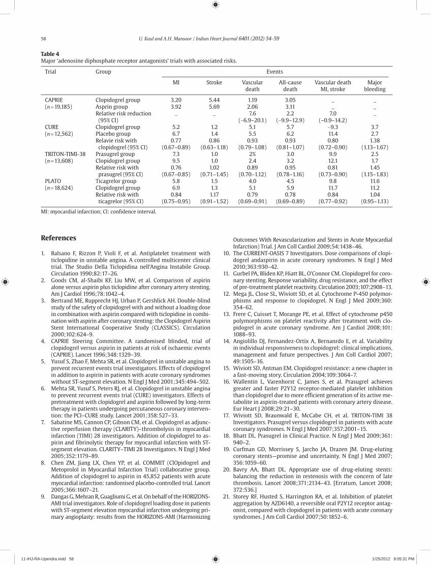
58 U. Kaul and A.H. Mansoor / Indian Heart Journal 6401 (2012) 54–59
Table 4Major ‘adenosine diphosphate receptor antagonists’ trials with associated risks.
Trial Group Events
MI Stroke Vascular All-cause Vascular death Major death death MI, stroke bleeding
CAPRIE Clopidogrel group 3.20 5.44 1.19 3.05 _ _(n = 19,185) Asprin group 3.92 5.69 2.06 3.11 _ _ Relative risk reduction _ _ 7.6 2.2 7.0 _ (95% CI) (−6.9–20.1) (−9.9–12.9) (–0.9–14.2)CURE Clopidogrel group 5.2 1.2 5.1 5.7 −9.3 3.7(n = 12,562) Placebo group 6.7 1.4 5.5 6.2 11.4 2.7 Relavie risk with 0.77 0.86 0.93 0.93 0.80 1.38 clopidogrel (95% CI) (0.67–0.89) (0.63–1.18) (0.79–1.08) (0.81–1.07) (0.72–0.90) (1.13–1.67)TRITON-TIMI-38 Prasugrel group 7.3 1.0 2% 3.0 9.9 2.5(n = 13,608) Clopidogrel group 9.5 1.0 2.4 3.2 12.1 1.7 Relative risk with 0.76 1.02 0.89 0.95 0.81 1.45 prasugrel (95% CI) (0.67–0.85) (0.71–1.45) (0.70–1.12) (0.78–1.16) (0.73–0.90) (1.15–1.83)PLATO Ticagrelor group 5.8 1.5 4.0 4.5 9.8 11.6(n = 18,624) Clopidogrel group 6.9 1.3 5.1 5.9 11.7 11.2 Relative risk with 0.84 1.17 0.79 0.78 0.84 1.04 ticagrelor (95% CI) (0.75–0.95) (0.91–1.52) (0.69–0.91) (0.69–0.89) (0.77–0.92) (0.95–1.13)
MI: myocardial infarction; CI: confidence interval.
References
1. Balsano F, Rizzon P, Violi F, et al. Antiplatelet treatment with ticlopidine in unstable angina. A controlled multicenter clinical trial. The Studio Della Ticlopidina nell’Angina Instabile Group. Circulation 1990;82:17–26.
2. Goods CM, al-Shaibi KF, Liu MW, et al. Comparison of aspirin alone versus aspirin plus ticlopidine after coronary artery stenting. Am J Cardiol 1996;78:1042–4.
3. Bertrand ME, Rupprecht HJ, Urban P, Gershlick AH. Double-blind study of the safety of clopidogrel with and without a loading dose in combination with aspirin compared with ticlopidine in combi-nation with aspirin after coronary stenting: the Clopidogrel Aspirin Stent International Cooperative Study (CLASSICS). Circulation 2000;102:624–9.
4. CAPRIE Steering Committee. A randomised blinded, trial of clopidogrel versus aspirin in patients at risk of ischaemic events (CAPRIE). Lancet 1996;348:1329–39.
5. Yusuf S, Zhao F, Mehta SR, et al. Clopidogrel in unstable angina to prevent recurrent events trial investigators. Effects of clopidogrel in addition to aspirin in patients with acute coronary syndromes without ST-segment elevation. N Engl J Med 2001;345:494–502.
6. Mehta SR, Yusuf S, Peters RJ, et al. Clopidogrel in unstable angina to prevent recurrent events trial (CURE) investigators. Effects of pretreatment with clopidogrel and aspirin followed by long-term therapy in patients undergoing percutaneous coronary interven-tion: the PCI–CURE study. Lancet 2001;358:527–33.
7. Sabatine MS, Cannon CP, Gibson CM, et al. Clopidogrel as adjunc-tive reperfusion therapy (CLARITY)–thrombolysis in myocardial infarction (TIMI) 28 investigators. Addition of clopidogrel to as-pirin and fibrinolytic therapy for myocardial infarction with ST-segment elevation. CLARITY–TIMI 28 Investigators. N Engl J Med 2005;352:1179–89.
8. Chen ZM, Jiang LX, Chen YP, et al. COMMIT (ClOpidogrel and Metoprolol in Myocardial Infarction Trial) collaborative group. Addition of clopidogrel to aspirin in 45,852 patients with acute myocardial infarction: randomised placebo-controlled trial. Lancet 2005;366:1607–21.
9. Dangas G, Mehran R, Guagliumi G, et al. On behalf of the HORIZONS-AMI trial investigators. Role of clopidogrel loading dose in patients with ST-segment elevation myocardial infarction undergoing pri-mary angioplasty: results from the HORIZONS-AMI (Harmonizing
Outcomes With Revascularization and Stents in Acute Myocardial Infarction) Trial. J Am Coll Cardiol 2009;54:1438–46.
10. The CURRENT-OASIS 7 Investigators. Dose comparisons of clopi-dogrel andaspirin in acute coronary syndromes. N Engl J Med 2010;363:930–42.
11. Gurbel PA, Bliden KP, Hiatt BL, O’Connor CM. Clopidogrel for coro-nary stenting. Response variability, drug resistance, and the effect of pre-treatment platelet reactivity. Circulation 2003;107:2908–13.
12. Mega JL, Close SL, Wiviott SD, et al. Cytochrome P-450 polymor-phisms and response to clopidogrel. N Engl J Med 2009;360:354–62.
13. Frere C, Cuisset T, Morange PE, et al. Effect of cytochrome p450 polymorphisms on platelet reactivity after treatment with clo-pidogrel in acute coronary syndrome. Am J Cardiol 2008;101:1088–93.
14. Angiolillo DJ, Fernandez-Ortix A, Bernanrdo E, et al. Variability in individual responsiveness to clopidogrel: clinical implications, management and future perspectives. J Am Coll Cardiol 2007;49:1505–16.
15. Wiviott SD, Antman EM. Clopidogrel resistance: a new chapter in a fast-moving story. Circulation 2004;109:3064–7.
16. Wallentin L, Varenhorst C, James S, et al. Prasugrel achieves greater and faster P2Y12 receptor-mediated platelet inhibition than clopidogrel due to more efficient generation of its active me-tabolite in aspirin-treated patients with coronary artery disease. Eur Heart J 2008;29:21–30.
17. Wiviott SD, Braunwald E, McCabe CH, et al. TRITON-TIMI 38 Investigators. Prasugrel versus clopidogrel in patients with acute coronary syndromes. N Engl J Med 2007;357:2001–15.
18. Bhatt DL. Prasugrel in Clinical Practice. N Engl J Med 2009;361:940–2.
19. Curfman GD, Morrissey S, Jarcho JA, Drazen JM. Drug-eluting coronary stents—promise and uncertainty. N Engl J Med 2007;356:1059–60.
20. Bavry AA, Bhatt DL. Appropriate use of drug-eluting stents: balancing the reduction in restenosis with the concern of late thrombosis. Lancet 2008;371:2134–43. [Erratum, Lancet 2008;372:536.]
21. Storey RF, Husted S, Harrington RA, et al. Inhibition of platelet aggregation by AZD6140, a reversible oral P2Y12 receptor antag-onist, compared with clopidogrel in patients with acute coronary syndromes. J Am Coll Cardiol 2007;50:1852–6.
11-IHJ-RA-Upendra.indd 58 1/25/2012 9:05:31 PM

U. Kaul and A.H. Mansoor / Indian Heart Journal 6401 (2012) 54–59 59
22. Husted S, Emanuelsson H, Heptinstall S, Sandset PM, Wickens M, Peters G. Pharmacodynamics, pharmacokinetics, and safety of the oral reversible P2Y12 antagonist AZD6140 with aspirin in patients with atherosclerosis: a double-blind comparison to clopidogrel with aspirin. Eur Heart J 2006;27:1038–47.
23. Wallentin L, Becker RC, Budaj A, et al, for the PLATO Investigators. Ticagrelor versus clopidogrel in patients with acute coronary syndromes. N Engl J Med 2009;361:1045–57.
24. Cannon C, Harrington R, James S, et al, the PLATelet inhibition and patient Outcomes (PLATO) investigators. Ticagrelor compared with clopidogrel in acute coronary syndromes patients with a planned invasive strategy (PLATO): a ran domized double-blind study. Lancet 2010;375:283–93.
25. Schömig A. Ticagrelor—is there need for a new player in the antiplatelet—therapy field? N Engl J Med 2009;361. DOI: 10.1056/NEJMoa0904327.
26. Ticagrelor versus clopidogrel in patients with acute coronary syndrome intended for non invasive management: sub study from
prospective randomized platelet inhibition and outcomes (PLATO) trial.
27. Held C, Asenblad N, Bassand JP, et al. Ticagrelor versus clopidogrel in patients with acute coronary syndrome undergoing coronary artery bypass surgery: results from PLATO trial. L Am Coll Cardiol 2011,57:672-84.
28. Levine GN, Bates ER, Blankenship JC, et al. 2011 ACCF/AHA/SCAI Guideline for Percutaneous Coronary Intervention. A Report of the American College of Cardiology Foundation/American Heart Association Task Force on Practice Guidelines and the Society for Cardiovascular Angiography and Interventions. Circulation 2011;124:e574–651.
29. Hamm CW, Bassand JP, Agewall S, et al. ESC Guidelines for the management of acute coronary syndromes in patients presenting without persistent ST-segment elevation. The Task Force for the management of acute coronary syndromes (ACS) in patients pre-senting without persistent ST-segment elevation of the European Society of Cardiology (ESC). Eur Heart J 2011.
11-IHJ-RA-Upendra.indd 59 1/25/2012 9:05:31 PM

Contents lists available at SciVerse ScienceDirect
Indian Heart Journal
ISSN: 0019-4832 Copyright © 2012. Cardiological Society of India. All rights reserved.doi: 10.1016/S0019-4832(12)60013-3
* Corresponding a uthor. E-mail address: [email protected]
Review article
Pulmonary hypertension—“state of the art” management in 2012Anita Saxena*
*Professor, Department of Cardiology, All India Institute of Medical Sciences, New Delhi – .
A B S T R A C T
Pulmonary artery hypertension (PAH) is a pathological condition of small pulmonary arteries, characterised by vascular proliferation and remodelling. The pulmonary artery pressure and pul-monary vascular resistance progressively rise, leading to right heart failure and death. Pulmonary artery hypertension may be secondary to various conditions, or it may be idiopathic where no underlying cause is identifiable. Earlier, only symptomatic treatment was available for such pa-tients which did not change the natural history of the disease. However, over the years, improve-ment in understanding the pathogenesis has resulted in the development of targeted approaches to the treatment of PAH. Survival advantage has also been shown with some of the pharmacologic agents. This review article discusses the current management strategy for PAH with special emphasis on an idiopathic variety, in an Indian context.
Copyright © 2012, Cardiological Society of India. All rights reserved.
K E Y W O R D S
DrugsIdiopathic pulmonary hypertensionManagementPulmonary hypertension
Introduction
Pulmonary artery hypertension (PAH) is defined as the sus-tained increase in mean pulmonary arterial pressure to a level ≥25 mmHg at rest, as assessed by right heart catheteri-sation.1 It is a progressive disease involving small pulmonary arteries and is characterised by vascular proliferation and re-modelling. The pulmonary vascular resistance (PVR) gradually rises, often leading to right heart failure and death. Pulmonary artery hypertension is classified based on the underlying di-agnosis. If no known risk factors are identified, it is called pri-mary PAH or more recently, idiopathic PAH (iPAH). The diagnostic classification of PAH is given in Table 1, as updated in 2008 by an expert working group (Dana Point, 2008).2 Although there are various conditions producing PAH, the histopathological features and response to various drugs tend to be similar for most. Treatment of the underlying con-dition like left heart disease or pulmonary parenchymal disease is most rewarding if PAH is secondary to one of these causes. Over the last 10 years, rapid advancements in understanding
the pathogenesis and therefore treatment options have taken place, most of these being for group 1. This article will focus primarily on the management of iPAH.
Epidemiology
The reported prevalence of PAH is 15 per million, females constitute 50–75% of those affected.3,4 Primary or iPAH is a rare disorder, the frequency is estimated at 1–2 cases/million people.5 A study from Scotland, however, cited the incidence as 2.5 cases/million/yr in men and 4.0 cases/million/yr in women.6 Any age group can be affected, but most often iPAH occurs in the third decade of life in women and fourth decade of life in men. No ethnic predisposition has been reported. The average duration between symptom and diag-nosis of PAH is >2 years.3 It was further confirmed by the National Institute of Health Registry, where the mean inter-val from symptoms onset to diagnosis was 2 years, it was shorter in cases with family history of pulmonary artery hypertension (PAH).7 The familial PAH shows an autosomal inheritance with 10–20% penetrance. There is genetic antici-pation in familial PAH, i.e. the onset of disease occurs at an
Indian Heart Journal 6401 (2012) 60–73
12-IHJ-RA-Anita saxena.indd 60 1/25/2012 9:04:19 PM

A. Saxena / Indian Heart Journal 6401 (2012) 60–73 61
earlier age in successive generations.8,9 Group 1 of Dana Point classification also includes drugs and toxins associated with development of PAH; the most notorious of these include the appetite suppressants fenfluramine and dexfenfluramine. More recently, methamphetamine is being indicated as the cause of drug-related PAH.10,11
Pathogenesis
The detailed pathophysiology of PAH is beyond the scope of this article. Suffice it to say that the pathogenesis of PAH is multifactorial (Table 2) and is not completely understood. The normal pulmonary circulation is very compliant and can accommodate the entire cardiac output at one fifth perfusion
pressures when compared with systemic circulation. This is accomplished by dilation of the existing vasculature and re-cruitment of unused vasculature. These changes maximise gas exchange surface area with no or little increase in per-fusion pressure. Several humoral mediators, such as nitric oxide (NO) and prostacyclin, which are produced locally, con-tribute to the maintenance of a compliant vasculature.
Pulmonary artery hypertension is caused by disturbance of one or more of these protective mechanisms. Three factors are considered important in raising PVR. These are vasocon-striction, remodelling of the pulmonary vessel wall and thrombosis in situ.12 Vasoconstriction may not be very impor-tant as only a small fraction of iPAH cases respond favourably to vasodilator therapy. High PVR causes hypertrophy of the right ventricle and in late stages the cardiac output falls. Hypoxaemia may occur secondary to ventilation perfusion mismatch and/or right to left shunting through a patent fo-ramen ovale.
Endothelial dysfunction seems to play a key role in the pathogenesis of PAH. An imbalance between the ‘bad’ vaso-constrictive/proliferative endothelin system and the ‘good’ vasodilatory/anti-proliferative NO and prostacyclin system is a key contributor to the initiation and progression of dis-ease.12 Platelets also play an important role as procoagulant and by releasing serotonin (vasoconstrictor), platelet derived growth factor (PDGF), and vascular endothelial growth factor (VEGF).13
Recent data suggests a primary defect in certain potassium channels in some patients with PAH.14 Dysfunctional voltage-gated potassium channels may explain the vasoconstriction and smooth muscle cell proliferation accompanying PAH. In PAH, the classic histological finding is the plexiform lesion. Activation and expression of adhesion molecules by endothe-lial cells results in a procoagulant state, resulting in thrombin deposition and platelet adhesion. All these abnormalities in the pulmonary vasculature of patients with PAH result in unregulated vasoconstriction, smooth muscle cell prolif-eration out of proportion to apoptosis, and microvascular thrombosis.15
Table 1Updated clinical classification of pulmonary hypertension (Dana Point, 2008).
1. Pulmonary arterial hypertension (PAH)1.1 Idiopathic PAH1.2 Heritable
1.2.1 BMPR21.2.2 ALK1, endoglin (with or without hereditary
haemorrhagic telangiectasia)1.2.3 Unknown
1.3 Drug- and toxin-induced1.4 Associated with
1.4.1 Connective tissue diseases1.4.2 HIV infection1.4.3 Portal hypertension1.4.4 Congenital heart diseases1.4.5 Schistosomiasis1.4.6 Chronic haemolytic anaemia
1.5 Persistent pulmonary hypertension of the newborn1’ Pulmonary veno-occlusive disease (PVOD) and/or pulmonary capillary haemangiomatosis (PCH)
2. Pulmonary hypertension owing to left heart disease2.1 Systolic dysfunction2.2 Diastolic dysfunction2.3 Valvular disease
3. Pulmonary hypertension owing to lung diseases and/or hypoxia3.1 Chronic obstructive pulmonary disease3.2 Interstitial lung disease3.3 Other pulmonary diseases with mixed restrictive and
obstructive pattern3.4 Sleep-disordered breathing3.5 Alveolar hypoventilation disorders3.6 Chronic exposure to high altitude3.7 Developmental abnormalities
4. Chronic thromboembolic pulmonary hypertension (CTEPH)5. Pulmonary hypertension with unclear multifactorial
mechanisms5.1 Haematologic disorders: myeloproliferative disorders,
splenectomy5.2 Systemic disorders: sarcoidosis, pulmonary langerhans
cell histiocytosis: lymphangioleiomyomatosis, neurofibromatosis, vasculitis
5.3 Metabolic disorders: glycogen storage disease, Gaucher disease, thyroid disorders
5.4 Others: tumoural obstruction, fibrosing mediastinitis, chronic renal failure on dialysis
Table 2Pathophysiological mechanisms for pulmonary artery hypertension.
Site Abnormality Effects
Endothelial Decrease NO, PGI2 Thrombosis Increase Endothelin-1 Platelet aggregation Increase thromboxane A2 Smooth muscle proliferation Increase VEGF
Platelets Increase serotonin Platelet aggregation Increase PDGF and VEGF Smooth muscle proliferation Vasoconstriction
Smooth muscle Dysfunctional Vasoconstriction cells K+ channels Increase angiopoietin Smooth muscle proliferation
NO: nitric oxide, PDGF: platelet derived growth factor, PGI2: prostaglan-din I2, VEGF: vascular endothelial growth factor.
12-IHJ-RA-Anita saxena.indd 61 1/25/2012 9:04:20 PM

62 A. Saxena / Indian Heart Journal 6401 (2012) 60–73
Natural history
Idiopathic PAH is a progressive disease and before the intro-duction of various therapeutic drugs, the survival was dismal. The median survival in 1980s was reported as 2.8 years in 194 patients with iPAH after the diagnosis was made.16 The actuarial survival rates were 68–77% at 1 year, 40–56% at 3 years and 22–38% at 5 years in two different studies.16,17 At that time most patient received conventional therapy including oral anticoagulants, diuretics, oxygen, cardiac glycosides, and calcium channel blockers. Poor prognosis was associated with New York Heart Association (NYHA) functional class III or IV, right heat failure, elevated right atrial pressure, decreased cardiac output or low mixed venous saturation.
Functional class III and IV continue to have extremely poor survival even in the current era as shown in data from registries.18
Over the years, however, improvement in understanding of the disease process has resulted in development of tar-geted approaches to treatment of PAH. Survival advantage has also been reported with various pharmacologic agents as will be described later.
Assessment of therapeutic strategies
Once the patient has been diagnosed with PAH and initiated on specific therapy, he or she should be assessed to monitor response to therapy at 3–6 months intervals depending on the severity of disease. Although a general assessment of the patient will provide important information whether the patient has improved symptomatically or in activities of daily living, objective evaluation is very important. As PAH is a rapidly progressive disease, long-term survival cannot be used for assessing the efficacy of a new treatment. Therefore, the commonly used end points are 6 minutes’ walk test (6MWT, the distance walked over a 6-minute period) and NYHA/WHO (World Health Organization) functional class.19 WHO functional class is described in detail in Table 3. Pulmonary artery pressure (PAP) (derived by echo-Doppler and/or by right heart catheterisation) and cardiac output are also used for assessment of efficacy of the treatment. Assess-ment by serum biomarkers and right heart function may also be helpful.20
Treatment of pulmonary artery hypertension
Regardless of the aetiology of PAH, general supportive care is similar for all patients. The supportive care aims at improv-ing symptoms and quality of life, preventing progression of the disease, and improving mortality. Unfortunately very few large randomised controlled trials (RCT) have been con-ducted for determining the best treatment options for pa-tients with PAH, most of these have been restricted to those with iPAH. Extrapolation to other populations with PAH is based mainly on expert recommendation rather than sound evidence-based medicine.21–23
General measures
General measures include strategies to avoid circumstances that may aggravate the disease. Any external stimulus that increases oxygen demand may worsen PAH and result in right heart failure. Non-steroidal anti-inflammatory drugs (NSAIDs) are best avoided due to their effect on fluid balance and renal function.
Physical activity
Heavy physical activity can be potentially dangerous in pa-tients with PAH and may trigger dyspnoea, chest pain or syn-cope. Therefore physical activity limited to a symptom free level is advised. This prevents deconditioning and muscular involution. Patients in functional class III or IV need to be more careful as life-threatening syncope may be induced on exre-cise.24 Bathing in hot water should be avoided as it can precipi-tate cutaneous vasodilatation thereby reducing cardiac output.
Pregnancy and contraception
Pregnancy results in increase cardiac output and blood vol-ume by as much as 40%. In PAH patients, the ability of right heart to compensate for the increased cardiovascular demand is limited. Hence, pregnancy poses an extreme risk to the health of women with PAH and is contraindicated. Maternal mortal-ity can be as high as 30–50% according to some reports, with most deaths occurring in the immediate post-partum period due to refractory right heart failure, sudden cardiac death, or thromboembolism.25–27
A safe and effective method of contraception must be rec-ommended in women of child bearing age. Since, safety of hormonal contraception is questionable due to its prothrom-botic potential, mechanical contraception (intrauterine device) or surgical sterilisation is generally recommended. In some
Table 3World Health Organization functional classification.
WHO functional Clinical descriptionclalss
I Patients with PH with no limitation of usual physical activity; ordinary physical activity does not cause dyspnoea, chest pain or pre-syncope
II Patients with PH with mild limitation of physical activity; no discomfort at rest but normal physical activity causes dyspnoea, fatigue, chest pain or pre-syncope
III Patients with PH with marked limitation of physical activity; no discomfort at rest but < ordinary activity causes dyspnoea, fatigue, chest pain or pre-syncope
IV Patients with PH who are unable to perform any physical activity; dyspnoea and/or fatigue are present at rest and symptoms are worsened with any physical activity. Syncope may occur with exertion
WHO: World Health Organization.
12-IHJ-RA-Anita saxena.indd 62 1/25/2012 9:04:20 PM

A. Saxena / Indian Heart Journal 6401 (2012) 60–73 63
centres, oral contraception with progesterone derivatives or low dose oestrogen has been given to women with PAH who are on oral anticoagulants with no history of thromboembo-lism.28 Similarly, post-menopausal women with intolerable menopausal symptoms can also receive hormone therapy in conjunction with anticoagulation.
High altitude
Hypoxia aggravates vasoconstriction in PAH patients. Therefore such patients must avoid living at high altitudes (>1500–2000 m). Commercial airplanes are generally pressurised to an altitude between 1600 m and 2500 m. Some of these patients may need supplemental oxygen when travelling by airplanes.
Prevention of infections
Respiratory infections can be fatal in patients with PAH. Pneumonia is the cause of death in 7% of cases.23 Pulmonary infections must be promptly recognised and treated with an-tibiotics. Regular vaccination against influenza and pneumo-coccal pneumonia is recommended. Persistent fever can also occur in patients on continuous intravenous epoprostenol infusion due to catheter related infection and sepsis.
Haemoglobin/haematocrit
Anaemia is not uncommon in patients with PAH and if de-tected, should be promptly treated. Conversely, some of the patients with long-standing hypoxia secondary to right to left shunt may develop polycythaemia. Phlebotomy is indicated only in symptomatic cases that have a high haematocrit (>65%) and volume replacement with saline or dextrose is recommended during phlebotomy.29
Anaesthesia and surgery
Patients with PAH are at a high-risk during anaesthesia and surgery. The risk is higher in those who are in functional class III or IV. Epidural anaesthesia may be better than general an-aesthesia. Anticoagulant treatment should be stopped for the shortest possible time.
Pharmacologic treatment anticoagulants
The rationale for anticoagulant therapy is based on the pres-ence of traditional risk factors for venous thromboembolism, like heart failure, sedentary lifestyle and a thrombophilic predisposition. Anticoagulant therapy has been widely used for PAH, although the evidence supporting this approach has not come from well-designed trials. In a retrospective study by Fuster and colleagues, 7 patients who were given couma-din had better survival than 37 patients who were not given
oral anticoagulant.30 In another small prospective study, Rich et al. showed that patients who did not respond to calcium channel blockers and who were treated with anticoagulants had improved survival as compared with those who were not anticoagulated.31 Three year survival improved from 21% to 49% in the series by Fuster et al. and from 31% to 47% in the series from Rich et al. Warfarin is the most widely used drug and the target international normalised ratio (INR) is gener-ally kept around 2.0. Some authors from European centres prefer to have INR between 2.0 and 3.0. The usage of antico-agulants in some of the recent trials for newer drugs has been in 51–86% of patients. Highest prevalence of anticoagu-lants use was in trials involving iPAH patients in NYHA/WHO functional class III and IV. Thus, while there is consistent evi-dence that anticoagulation improves outcomes in those with PAH, there is as of yet no RCT showing a benefit.
Diuretics
Diuretics improve symptoms and signs of right heart failure in decompensated patients of PAH. On the other hand, too much diuresis can cause hypovolaemia and further reduce cardiac output by decreasing right ventricular preload.32 No trials have been performed for diuretics, but clear symptom-atic benefit has been shown in patients of PAH who were decompensated and have fluid retention. Some form of diu-retic therapy is prescribed to 49–70% of patients in well-studied trials.23 Furosemide or spironolactone is generally used. The dosages are modified according to the individual case. Serum electrolytes and renal functions should be carefully monitored in patients receiving diuretic therapy.
Oxygen therapy
Mild degree of hypoxaemia at rest is a common finding in PAH. This occurs as a consequence of low mixed venous oxy-gen saturation caused by low cardiac output. The ventilation perfusion matching is minimally altered and therefore does not contribute significantly to hypoxaemia. A small number of cases have severe hypoxaemia caused by a right to left shunt through a patent foramen ovale. No consistent data are available on the effect of long-term oxygen therapy in PAH. However, since hypoxaemia may aggravate PAH by increas-ing pulmonary vasoconstriction, supplemental oxygen ther-apy should be considered in patients with severe hypoxaemia at rest (PaO2 < 55 mmHg). Improvement in PAH with low flow supplemental oxygen has been reported in some cases. The goal of oxygen therapy is to maintain arterial saturation at >90%. Oxygen therapy can also improve quality of life by reducing dyspnoea and by increasing exercise capacity.
Cardiac glycosides
In cases with right heart failure secondary to PAH, myocar-dial contractility is likely to be impaired and inotropic agents
12-IHJ-RA-Anita saxena.indd 63 1/25/2012 9:04:20 PM

64 A. Saxena / Indian Heart Journal 6401 (2012) 60–73
may help. Short-term intravenous digoxin has been reported to increase cardiac output by a modest degree.33 Long-term data on effect of digoxin in PAH is not available. In the rare patient of PAH with supraventricular tachyarrhythmia (like atrial flutter or fibrillation), digoxin should be used to slow the ventricular rate. It must be noted that digoxin toxicity is more likely in PAH patients especially if hypoxaemia and/or hypokalaemia are also present. Patients with end stage PAH have been treated with intravenous dobutamine in most expert centres.32
Specific therapies
Based on pathogenesis of PAH, there are three major classes of specific therapies. These include prostanoids, endothelial receptor antagonists (ETRA) and phosphodiesterase-5 (PDE5) inhibitors. However, calcium channel blockers have been used to treat PAH patients for a long time before specific drugs became available. Prior to initiating any of these drugs, all efforts must be made to look for any underlying cause as the cause of PAH. If discovered it should be treated promptly.
Several articles have published expert opinions regarding treatment, which is based on WHO functional class and risk assessment.34
Calcium channel blockers
Since, vasoconstriction of pulmonary arterioles is often one of the several abnormalities responsible for PAH, vasodilator therapy has long been used in the treatment of PAH. Calcium channel blockers like nifedipine and diltiazem are widely used oral vasodilators, but only a small percentage of patients actually respond to these drugs. Calcium channel blockers do not have the selectivity for the pulmonary vascular bed and may cause systemic hypotension in some cases.
Vasodilator challenge
Calcium channel blockers should be used only in cases where a clear acute vasodilator response is demonstrated haemody-namically. Acute vasodilator testing is performed using short acting agents like intravenous epoprostenol, adenosine or in-haled NO. A decrease of at least 20% in PAP from the baseline value and a decrease of >30% in PVR with an increased or un-changed cardiac output is usually predictive of a favourable response.31–35 Some workers accept a decrease in mean PAP by >10 mmHg, to a value <40 mmHg with no reduction in cardiac output.36 Generally only 10–15% of iPAH patients show a positive acute vasoreactive response and about half of them will continue to respond to calcium channel blockers over a long period of time.1 In a recent study, Sitbon and colleagues showed that of 12.6% patients with iPAH who displayed acute pulmonary vasoreactivity, only 54% had a sustained, long-term benefit with oral calcium channel
blockers as monotherapy.37 Hence overall <7% of 557 patients with iPAH were long-term responders. Long-term therapy with a calcium channel blocker is not recommended in those not showing acute response to vasodilator testing in the catheterisation lab. Further, these drugs should not be used as a screening agent for pulmonary vascular responsiveness in patients with PAH because severe adverse reactions can occur.
Calcium channel blockers are indicated in class II and III patients who show response on acute vasodilator testing. These drugs are much cheaper compared to some of the newer drugs. The choice of agent depends on the underlying heart rate. Those with heart rate of >80 beats/min are better given diltiazem and those with slower heart rates are given nifed-ipine. Verapamil is not used due to its negative inotropic ef-fect. Amlodepin has been used, but its long-term efficacy is not known. The dosages used are quite high; 90–180 mg/day for nifedipine (up to 240 mg/day) and 240–720 mg/day for diltiazem (up to 900 mg/day). Systemic hypotension and limb oedema can occur; concomitant diuretics can alleviate lower limb oedema.
Prostanoid therapy
Prostacyclin, a prostanoid, is released from the endothelial cell membranes of the vascular intimae and its levels are de-creased in iPAH. Based on this rationale, prostacyclin is being used in patients with PAH. The precise mechanism of action of prostacyclin in PAH is likely to be multifactorial. It may in-clude relaxation of vascular smooth muscle cell, inhibition of platelet aggregation, healing of endothelial injury, inhibition of smooth cell proliferation, facilitating reverse remodelling of pulmonary vascular changes and so on. Stable analogues of prostacyclin have been synthesised and are in use for patients with PAH over the last several years.
Epoprostenol
Intravenous epoprostenol was first used to treat iPAH in early 1980s.38 Since then, it has proved to be lifesaving in a large number of patients with PAH of varied aetiology. In 1990, Rubin et al. reported the first randomised unblinded trial with intravenous epoprostenol in 25 patients with iPAH.39 An improvement in exercise capacity as well as haemodynamics was shown after 2 months in those treated with intravenous epoprostenol. Second prospective randomised open trial re-ported improvement in 6MWT by 32 m, in the epoprostenol group after 12 weeks of therapy.40 Survival advantage was also demonstrated in this trial in the group of patients on epoprostenol. Following these studies, intravenous epopros-tenol was approved in USA and France for treatment of iPAH for those in functional class III or IV. In another study by Barst et al., the survival with epoprostenol was 87% at 1 year, 63% at 3 years and 54% at 5 years compared to 77%, 41%, and 27%, respectively in historic controls.41 There are other reports of improved survival with intravenous epoprostenol.42–44
12-IHJ-RA-Anita saxena.indd 64 1/25/2012 9:04:20 PM

A. Saxena / Indian Heart Journal 6401 (2012) 60–73 65
Intravenous epoprostenol is typically reserved for individ-uals with severe PAH. To date it is the only medication that has a mortality benefit.40
Intravenous epoprostenol is now considered as a first line therapy in the western world. In a survey collected from 19 centres in the US, it was shown that more than two-thirds of patients with iPAH who were treated with intravenous epo-prostenol could be taken off the list for lung transplanta-tion.45 This drug may be useful even in patients who do not show an acute haemodynamic response to it in the catheteri-sation lab.
Epoprostenol is dispensed as freeze-dried preparation that needs to be dissolved together with an alkaline buffer (glycine) and is given intravenously. Since, it has a very short half-life (3–5 minutes), it has to be given as a continuous infusion. Fresh solution has to be prepared every 24 hours due to the unstable nature of the drug. Epoprostenol is delivered by in-fusion pumps through long-indwelling venous catheters. The initial dose is 2–4 ng/kg/min and increased gradually. The target dose is 10–15 ng/kg/min for the first 2–4 weeks and then periodic dose increases are done to maximise efficacy. The optimal dose usually varies from 20–40 ng/kg/min in majority of patients. Dose adjustments are made based on clinical symptoms, distance walked during 6MWT and occur-rence of side effects.
Common side effects include headache, flushing, jaw pain, diarrhoea, nausea, vomiting, and rarely ascites. Most of these side effects are generally mild and dose related. More serious complications are related to the delivery system and include exit site infections, bleeding, bacteraemia and sepsis. The incidence of catheter related sepsis has been reported to be 0.1–0.4 per patient-year.40 Severe catheter related sepsis has resulted in death in some cases. Delivery system mal-function can be the other serious complication resulting in sudden, sometimes fatal decompensation due to interrup-tion of drug supply. Patients suspected of veno-occlusive dis-ease or pulmonary capillary haemangiomatosis which may masquerade as iPAH can develop fatal pulmonary oedema if given epoprostenol.
Despite favourable results from intravenous epoprostenol for cases with PAH, it is far from ideal treatment, primarily because of its route of delivery. It is very expensive over long-term use and is not curative. Currently this drug is not avail-able in India for general use.
Treprostinil
The difficulties in delivering intravenous epoprostenol have stimulated interest in developing stable analogues of this compound which can be administered using less complex delivery systems. Treprostinil is one such analogue which has chemical stability at room temperature and can be given both intravenously and subcutaneously. It also has a longer half-life and needs to be prepared once in 48 hours.
The largest multicentre RCT was reported by Simonneau et al. involving 470 patients with PAH.46 There was statisti-cally significant improvement in median exercise capacity,
haemodynamics and clinical events compared to placebo group after 12 weeks of treatment with subcutaneous trepro-stinil. The effect was dose related.
The drug can be given through small subcutaneous cathe-ters similar to those utilised for insulin administration in diabetics. The management of this system is much simpler compared to epoprostenol infusion system. Common side effects include headache, jaw pain, diarrhoea, nausea, and rash. Infusion site pain is seen in 85% of cases. Food and Drug Administration (FDA) has approved this drug in 2002 for class II, III and IV patients with PAH. It is not available in India.
Sodium beraprost
Beraprost sodium is the first biologically stable and orally ac-tive prostacyclin analogue. It is absorbed rapidly, peak con-centration is reached after 30 minutes and the elimination half-life is 35–40 minutes after a single oral dose. Initial trials have shown an improvement in 6MWT, functional class and survival.47,48 In the RCT reported by Galie et al, there was sig-nificant improvement in exercise capacity at 12 weeks.49 However, the data from Barst et al showed no difference in 6MWT distance with oral beraprost compared to placebo at 12 months follow-up, although improvement was observed at 3 and 6 months.50 Beraprost sodium is approved in Japan and South Korea but not in USA and Europe. This drug is also not available in India.
Iloprost
Iloprost is a chemically stable prostacyclin analogue available for intravenous, oral, and aerosol usage. Inhaled therapy may provide selectivity of the haemodynamic effects to the lung vasculature, avoiding systemic side effects. Iloprost has a half-life of 20–25 minutes and can be delivered by inhaler, the aerosolised particles should be small enough (diameter 0.5–3 um) to ensure alveolar deposition. Since, it has a short du-ration of action, it must be inhaled 6–12 times a day.51 With jet inhalers, the duration of each inhalation takes about 15 minutes.
In a randomised, multicentre, placebo-controlled study of 207 patients (all in functional class III or IV) with PAH, the 6MWT improved by 36 m at 12 weeks in those on iloprost.52 In the subgroup with iPAH, the improvement in 6MWT was even greater (59 m). The haemodynamic values were also much improved. In another study of 76 patients with inhaled iloprost as monotherapy, the 1 year, 3 years, and 5 years sur-vival free of lung transplantation and addition or switch to other drugs were 53%, 20%, and 13%, respectively.53 Thus, the role of iloprost as monotherapy is limited.
Side effects include cough, flushing, and headache. The short-term results are encouraging; however, long-term efficacy needs to be established. Inhaled iloprost is approved in Europe, Australia and New Zealand. It is not available in India.
12-IHJ-RA-Anita saxena.indd 65 1/25/2012 9:04:20 PM

66 A. Saxena / Indian Heart Journal 6401 (2012) 60–73
Endothelin receptor antagonists
Increased levels of endothelin-1 have been found in the pul-monary arteries of PAH patients. Endothelin-1 is a potent va-soconstrictor and mitogen for smooth muscle cells. Levels of endothelin-1 have been related to disease severity and sur-vival.54,55 The effects of endothelin-1 are mediated through two types of receptors ETA and ETB. ETA receptors cause sus-tained vasoconstriction and proliferation of vascular smooth muscles and ETB receptors induce production of NO and prostacyclin by the endothelial cells, thereby producing va-sodilatation.56 Therefore use of endothelin-1 receptor antag-onists that can block either ETA or both ETA and ETB receptors will help patients with PAH.
Bosentan
Bosentan is an orally active dual (ETA and ETB) endothelin re-ceptor antagonist. It is the first molecule of this class of drugs to be synthesised. Bosentan has been evaluated in two RCT. In the larger BREATHE-1 study, 213 patients with PAH who were in functional class III or IV were randomised to receive placebo or bosentan.57 The initial dose used was 62.5 mg twice daily for 4 weeks; thereafter either 125 mg or 250 mg twice daily dose was given for at least another 12 weeks. The mean effect of treatment on the 6MWT was a gain of 44 m in the overall study population and 52 m among patients with iPAH. Improvement was also seen in the time to clinical worsening. No dose-response effect on the efficacy could be ascertained. Similar response was seen in another RCT.58 Sustained improvement at 6 months and 1 year in NYHA functional class and haemodynamic parameters was re-ported by Sitbon et al. in an open label extension study with bosentan.59
Survival analysis of iPAH patients treated with oral bosen-tan as first line therapy in 177 patients has been published in 2005.60 The dose used was 125 mg twice daily in majority and the mean follow-up period was 2.1 ± 0.5 years. Kaplan-Meier survival estimates were 96% at 1 year, 89% at 2 years as compared to the predicted survival of 69% at 1 year and 57% at 2 years with no specific treatment. According to the analy-sis by McLaughlin, treatment with bosentan was considered superior to epoprostenol in 139 iPAH patients in WHO func-tional class III.61 Oral bosentan has been proposed as a transi-tion therapy in patients having severe side effects due to epoprostenol therapy. An uncontrolled study in children be-tween 4 and 17 years of age with PAH (BREATHE-3) has con-firmed the beneficial effects of bosentan as seen in adults. There was significant improvement in haemodynamics at 12 weeks in the 18 children given either bosentan alone or with epoprostenol.62
Bosentan has also been used in mildly symptomatic adult patients with PAH in the EARLY trial.63 Although a sig-nificant fall was seen in PVR, there was no significant change in 6MWT distance. Currently, this drug is perhaps best re-served for patients with WHO or NYHA functional class III or more.
Bosentan is metabolised in the liver and may increase hepatic aminotransferase levels. In the BREATHE-1 study, increases in hepatic aminotransferases were seen in 10% of the subjects, and were found to be dose dependent and reversible after dose reduction or discontinuation.57 Elevation in liver enzymes to >8 times the upper limit of normal range occurred in 3% in those receiving 125 mg twice a day dose. Liver function tests must be performed at least once a month in those receiving bosentan. This drug is contraindicated in pregnancy due to its potential teratogenic effect. It is approved for use in NYHA/WHO functional class III and IV PAH patients in USA, Canada and Europe. The drug has become available in India but is quite expensive for long-term use for most Indian patients.
Sitaxsentan and ambrisentan
These drugs are specific ETA receptor blockers and can be given orally once daily. Sitaxsentan was used in one RCT on 178 patients with PAH in NYHA class II, III or IV.64 The drug was given over a period of 12 weeks. Improvement in exer-cise capacity, haemodynamics and clinical events was seen. The incidence of abnormal liver function test was 0% for 100 mg once daily and 9.5% for 300 mg once daily dose. Ambrisentan is currently being tested. It was shown to im-prove exercise capacity in ARIES I and ARIES II study.65
Although hepatotoxicity seems to be less common in pa-tients taking selective ETA receptors antagonists, cases of acute hepatitis have been described. Hence liver functions should be constantly monitored.
There is currently no data to establish the benefit of one ETRA over the other. Patients may prefer Ambrisentan due to its daily dosing and lower incidence of hepatotoxicity.
Type 5 phosphodiesterase inhibitors
Sildenafil
Inhibition of type 5 phosphodiesterase leads to increased concentration of cyclic guanosine monophosphate (cGMP) in the smooth muscles of the vessels leading to vasodilatation. Sildenafil is an orally active, potent selective inhibitor of cGMP-phosphodiesterase type 5, increasing the intracellular concentration of cGMP. Since, this enzyme is present in high concentration in pulmonary vasculature, sildenafil can reduce PAP in patients with PAH.66 Sildenafil proved to be a potent pulmonary selective vasodilator when used for treating PAH in animal experiments with very little fall in systemic pres-sure.67 An acute vasodilatory effect on the pulmonary circu-lation was seen in patients with iPAH when combined with inhaled iloprost in the study by Wilkens and colleagues.68 The fall in PVR was dose dependent in this study using intra-venous sildenafil during right heart catheterisation. In an-other study, addition of sildenafil improved exercise capacity and pulmonary haemodynamics in patients who were dete-riorating inspite of on-going inhaled iloprost therapy.69 These encouraging findings led to several RCT. Some of the trials
12-IHJ-RA-Anita saxena.indd 66 1/25/2012 9:04:20 PM

A. Saxena / Indian Heart Journal 6401 (2012) 60–73 67
with sildenafil are summarised in Table 4. An observational study from India showed improvement in 6MWT distance and fall in right ventricular systolic pressure in 14 cases with severe PAH treated with sildenafil.70 The initial randomised controlled cross over trial carried out in 22 patients showed improvement in treadmill time, quality of life and cardiac out-put.71 Similar study by Bharani and colleagues also reported significant improvement in exercise capacity, but no signifi-cant improvement was found in the NYHA functional class.72 Similar results are reported in 20 patients in another study, also from India.73 A much larger, double-blind, placebo-controlled randomised trial is reported in the New England Journal of Medicine in 2005.74 In this multicentre Sildenafil Use in Pulmonary Arterial Hypertension (SUPER) study, 278 symptomatic patients with PAH (idiopathic associated with connective tissue disease or with operated left-to-right shunts) were enrolled. These were randomised to placebo or sildenafil (20 mg, 40 mg, or 80 mg) orally 3 times a day for 12 weeks. At the end of 12 weeks, 6MWT distance improved in all sildenafil groups. The mean placebo corrected treatment effects were 45 m (99% CI 21–70), 46 m (99% CI 20–72) and 50 m (99% CI 23–77) for 20 mg, 40 mg and 80 mg of sildenafil, respectively (P < 0.001 for all comparisons). The WHO func-tional class also improved in more patients on sidenafil than on placebo. Two hundred and twenty patients completed 1 year of treatment with sildenafil monotherapy and the im-provement in 6MWT distance from baseline at 1 year was re-ported as 51 m. The PAP reduced and cardiac index improved with sildenafil. The drug was well tolerated with minor side effects. This study was not powered to assess mortality and there was no evidence of a dose-response relationship asso-ciated with the primary end points in the 12 weeks study. It is possible that the lower dose of 20 mg 3 times a day is good enough for complete inhibition of phosphodiesterase type 5. In the long-term, a smaller dose may not be desirable when using in combination with bosentan as bosentan decreases the plasma level of sildenafil.75 SUPER-1 study was later ex-tended as SUPER-2 study in which SUPER-1 subjects taking 80 mg sildenafil 3 times a day for at least 3 years were fol-lowed; the majority of these cases were in WHO functional class II–III.76 One hundred and seventy patients completed both studies and were followed for a median of 1242 days. Sixty percent of these patients either improved or maintained
their functional class. Additional specific therapies were required in 3%, 10%, and 18%, at 1 year, 2 years, and 3 years, respectively. Those with a baseline 6MWT distance of <325 m and did not improve in SUPER-1 during the first 12 weeks fared worse in terms of survival.
One study has compared bosentan with sildenafil.77 Both drugs seem to be equally effective with a trend towards better improvement with sildenafil when 6MWT distance was compared.
Food and drug administration approved sildenafil for pa-tients with PAH in any functional class only at a dose of 20 mg 3 times a day. Sildenafil can be recommended as first line drug in patients with PAH in India, where other modes of treatment like prostenoid therapy, endothelin antagonists and lung transplantation are either not available or are pro-hibitively expensive.
Recently intravenous preparation of sildenafil has become available and can be used for hospitalised patients who were on chronic oral sildenafil but are unable to take it orally.
In a recent open label study, the pharmacokinetic and pharmacodynamic effects of 10 mg of IV sildenafil were com-pared to 20 mg of oral sildenafil and were found to be simi-lar.78 tadalafil is another phosphodiesterase inhibitor which can be used once daily, data is emerging on the use of this drug.79 It has recently been used in Public Health Institutional Review, Submission Tracking (PHIRST) trial (PAH and re-sponse to tadalafil). This trial was a 16-week, randomised, double-blind, placebo-controlled multicentre trial which studied the efficacy and tolerability of 4 doses of tadalafil on 405 PAH subjects with and without background bosentan therapy.80 40 mg of tadalafil had significantly reduced the in-cidence of clinical worsening and improved time to clinical worsening when compared to placebo. This drug improved exercise capacity in a dose dependent manner, but only the 40mg dose met the pre-specified value for statistical signifi-cance of <0.01. Haemodynamics also improved with 40 mg of tadalafil, although this data was available for 93 patients only. Better results were noticeable in tadalafil naive patients.
Presently there is little to choose between tradalafil and sildenafil. Although once daily dosing with tadalafil is more appealing, most of clinical experience is with sildenafil.
Nitrates are contraindicated in patients treated with phos-phodiesterase inhibitors.
Table 4Studies on use of sildenafil in pulmonary artery hypertension.
Author/yr Type of trial No. Duration Improvement in
6MWT/Ex time Func Cl PAP
Kothari, 200270 Observational 14 7.3 ± 2.4 mo Y Y YSastry, 200471 RCT 22 6 wk Y Y NBharani, 200372 RCT 9 2 wk Y N YSingh, 200673 RCT 2 6 wk Y Y YGalie, 200574 RCT 278 12 wk Y Y Y (SUPER)
Func Cl: Functional class (NYHA/WHO/others), N: No, PAP: pulmonary artery pressure, RCT: randomised controlled trial, Y: Yes.
12-IHJ-RA-Anita saxena.indd 67 1/25/2012 9:04:20 PM

68 A. Saxena / Indian Heart Journal 6401 (2012) 60–73
The side effect profile for phosphodiesterase inhibitors includes headache, flushing, nasal congestion, dyspepsia, and myalgias, these effects are primarily due to the drug’s vasodila-tory effects.
Investigational therapies
Nitric oxide
Inhaled NO is a potent selective pulmonary vasodilator that directly relaxes vascular smooth muscle through stimulation of soluble guanylate cyclase and increased production of cGMP. Nitric oxide lowers pulmonary pressures, improves haemodynamics and has been used in critical care setting.81 However, its use has been limited by concerns regarding the toxicity of the gas and by the lack of a practical delivery system for long-term use.
L-arginine
L-arginine is essential in the production of NO and has been used for reducing PAP in some studies. Mehta et al. have re-ported a reduction in PAP and PVR with L-arginine.82 These beneficial results have not been seen in other studies.
Vasoactive intestinal peptide
Vasoactive intestinal peptide inhibits platelet activation and proliferation of smooth muscle cells acting as potent pulmo-nary vasodilator. In a small study, inhalation of this agent led to significant fall in PAP with functional improvement in 8 patients with iPAH.83
Imitinib mesylate
Imitinib is a tyrosine kinase receptor blocker which prevents phosphorylation of PDGF and inhibits smooth cell prolifera-tion. An animal study has reported its beneficial effect in reducing PAP in rat models with PAH.84 Clinical improvement with increase in 6MWT distance was seen in one case which did not respond to iloprost, bosentan, and sildenafil.85 Expe-rience with this drug is limited to a few cases only in other reports also. An additional study evaluating the long-term safety and efficacy of imatinib in PAH is on-going
Riociguat
Since, the beneficial effects of phosphodiesterase inhibitors are mediated through soluble guanylate cyclase (sGC) and the second messenger cGMP, augmenting the NO pathway by direct stimulation of sGC can be another approach. Riociguat is such a drug that activates sGC independently of NO.86 This drug has been used in a phase II study in PAH patients. The
drug appears promising as it improved 6MWT distance and haemodynamics. It was well tolerated.87 Phase III trials are currently on-going.
Dichloracetate
Dichloroacetate is another drug which has been used in ex-perimental animals. It prevents and reverses monocrotaline-induced PAH in rats. It has been shown to partially regress established PAH in mice. A phase I trial of this drug is cur-rently underway in patients with PAH who are in functional class III–IV and are stable on other specific drugs.88
Other drugs
Statins may be useful by repressing vascular smooth muscle cell proliferation secondary to PDGF, as seen in rats with hy-poxic PAH.89 However, this drug has not been used in human beings. Nikorandil has been used as potassium channel abnormalities are known in patients with PAH.90
Meta-analyses of drug therapy
Meta-analyses for drug treatment in PAH have been pub-lished. One was reported in 2007 which included 16 RCT and 1962 patients.91 According to their results, limited bene-fits were achieved for clinical end points, but no significant survival advantage could be demonstrated. A more recent meta-analysis addressed the all-cause mortality and survival.92 Twenty-three RCT were included, majority of patients were in WHO class III. A reduction of 43% in overall mortality was demonstrable in patients randomised to active drug treat-ment as compared to those randomised to the placebo arm (data from 21 trials). The benefit was seen with prostanoids, endothelin receptor antagonists and phosphodiesterase type 5 inhibitors. Besides reduction in all-cause mortality, the func-tional class and haemodynamic parameters also improved in those on drugs.
Combination therapy
Current therapies for PAH target the three known intracellular pathways; NO, prostacyclin and endothelin, which are known to be abnormal in patients with PAH. Therefore, it appears logical to use a combination of two or more drugs which act on different pathways especially in cases with incomplete re-sponse to a single agent. This is similar to the situation in sys-temic hypertension where a combination of drugs acting on different pathophysiological mechanisms results in better control of blood pressure than monotherapy. It is also possible that one pathway may play a more dominant role than others in pathogenesis of PAH and one drug may be more effective than the others. One can start with one drug and add the second in those with incomplete response. Alternatively two drugs
12-IHJ-RA-Anita saxena.indd 68 1/25/2012 9:04:21 PM

A. Saxena / Indian Heart Journal 6401 (2012) 60–73 69
combination may be used to begin with and a third drug added later in those with insufficient clinical and haemody-namic response. At present, the choice of the drug or the order in which drugs should be used is not clear due to lack of large trials addressing this issue. Combination therapy is also recom-mended by “Consensus statement on the management of pul-monary hypertension in clinical practice in UK and Ireland”.93
The use of initial or “upfront” combination therapy has been suggested for WHO class IV PAH patients in recent guide-lines.94 However, there is limited data on the comparative ef-ficacy of this strategy. The combination of sildenafil and inhaled iloprost was seen to cause more potent pulmonary vasodila-tation than either agent alone.68 Similarly, patients who were deteriorating on iloprost therapy improved with addition of sildenafil.95 Similarly adjunctive therapy with bosentan and intravenous prostacyclin produced favourable, though statis-tically non-significant, outcomes in a small study of 33 pa-tients.96 On the other hand, another report indicated that addition of long-term treatment with sildenafil had minimal effects on functional class and right heart failure in 13 patients already receiving vasodilators for PAH (calcium channel blockers, epoprostenol or bosentan).97 There is no randomised trial addressing the issue of combination therapy in PAH. Non-randomised data from Hoeper et al. suggests that an ap-proach using bosentan, sildenafil and iloprost in combination yields acceptable long-term results.98 These authors called it a goal-oriented therapeutic strategy. The study included 123 patients with PAH. All of these were started on bosentan up to a maximum dose of 125 mg twice a day to achieve the therapeutic goals of: (a) a 6MWT distance >380 m; (b) a peak oxygen uptake >10.4 mL/min/kg; and (c) a peak systolic blood pressure during exercise of >120 mmHg. In those, not able to achieve these goals, sildenafil was added in a maximum dose of 50 mg 3 times a day. Similarly inhaled iloprost was given to the group which still did not achieve the therapeutic goals on combination of bosentan and sildenafil. Survival with this goal-oriented combination therapy was much better as compared to historic controls. A two drug combination was required in 43% and a three drug combination, in 16% of pa-tients. Six patients did not achieve the desired goals even with triple drug regimen and were switched over to intrave-nous iloprost (5) or underwent lung transplantation (1). The combinations were well tolerated.
The strategy of sequential addition of drugs appears to be promising, although additional studies, preferably RCTs are needed to confirm these results. In countries like India, one needs to explore the benefits of combining calcium chan-nel blockers with sildenafil, since bosentan and prostacyclines are either not available or are too expensive.
Interventional procedures
Balloon atrial septostomy
Observational and epidemiologic studies have shown that Eisenmenger’s syndrome patients and patients with iPAH who have a patent foramen ovale had a better prognosis than
those without intracardiac shunting.99,100 Because of these observations atrial septostomy has been used for patients with disabling right heart failure. Blade balloon atrial septo-stomy was first reported by Rich in 1983 in patients with re-fractory PAH.101 The presence of an atrial septal defect allows right to left shunting which increases systemic output result-ing in an increased systemic oxygen transport despite a fall in systemic arterial oxygen saturation. The shunt at atrial level also allows for the decompression of the right atrium and right ventricle with improvement in signs and symptoms of right heart failure. A study in children with iPAH has demon-strated a significant long-lasting benefit with clinical, haemo-dynamic and survival advantage following blade-balloon atrial septostomy.102 In adults, a large series is reported by Sandoval and colleagues from Mexico.103 The atrial septum is first punc-tured percutaneously. The septal defect is then gradually di-lated using balloon catheters with incremental diameter of 8–14 mm. Unfortunately, the procedure related mortality is high, varying from 5% to 15%. This high mortality may be due to the patient profile as this procedure is often done in severely ill cases as a palliative bridge to lung transplantation. Accord-ing to Sitbon et al., a mean right atrial pressure >20 mmHg, PVR >55 units/m2, and a predicted 1 year survival <40% are significant predictors of procedure related deaths.28 Currently the indications for atrial septostomy may be recurrent syn-cope or right heart failure despite maximal medical therapy including intravenous prostacyclin’s. This procedure may have more relevance in India since treatment with other modes of therapy, i.e. drugs and lung transplantation has limited scope. The data from the All India Institute of Medical Sciences by Kothari et al. reports improvement in clinical sta-tus and haemodynamic variables in 11 patients with severe PAH.104 However, the procedure related mortality was high at 18.2%. Authors suggested that the procedure was high-risk in very sick patients and hence balloon atrial septostomy may be advocated early in the course of the disease. Atrial septo-stomy should be performed only in specialised centres to reduce procedure related risks.
Lung transplantation
For patients with PAH who cannot be managed medically, lung transplantation remains the ultimate alternative. Heart-lung, single lung and double lung transplantations have been performed successfully in patients with iPAH.105,106 These modes of treatment have been assessed in uncontrolled se-ries since RCTs are unethical. Survival is reported to be 65%, 55%, and 44% at 1 year, 3 years, and 5 years, respectively.107,108 Lung transplantation is indicated in PAH patients with ad-vanced functional class III and IV. The problem of donor shortage makes this mode of therapy relatively difficult even in western countries. Bilateral lung transplantation is gener-ally preferred due to less postoperative complications. Func-tional recovery is generally good after lung transplantation, but long-term survival has been limited by the high preva-lence of chronic allograft rejection.103 No relapse of PAH has been reported.
12-IHJ-RA-Anita saxena.indd 69 1/25/2012 9:04:21 PM

70 A. Saxena / Indian Heart Journal 6401 (2012) 60–73
The choice of whether to perform heart-lung or bilateral lung transplantation is largely dependent on centre policy and donor availability. Both techniques have a range of advan-tages and disadvantages, but overall survival rates have been reported to be similar. It is claimed that heart-lung transplan-tation is less likely to result in death from obliterative bron-chiolitis.109 Generally, heart-lung transplantation is performed when there is either significant impairment of cardiac func-tion (inotrope dependence) or PAH is secondary to a complex congenital heart disease.110
Treatment algorithm
Treatment algorithm have been published for WHO functional class III and IV patients based on the grade of recommendation and the level of evidence as derived by the clinical trials. How-ever, very little data is available for class I and II patients on the most appropriate treatment strategy. Perhaps calcium channel blockers should be used in those who show vasoreactivity in the cardiac catheterisation lab, as these drugs are relatively safer.
Before initiating treatment, all efforts should be made to diagnose the underlying condition which may be responsible for PAH in a given case.
The suggested approach includes the adoption of general measures as described earlier and initiation of therapy with oral anticoagulants (if no contraindication), diuretics (if fluid reten-tion), supplemental oxygen (if hypoxaemia) and digoxin (in those with refractory right heart failure and/or supraventricular arrhythmias). All such patients should be referred to a special-ised centre for treatment. Acute vasoreactivity testing should be undertaken and the patients with positive response should be treated with maximal tolerated dose of calcium channel block-ers. The dose should be increased in a stepwise fashion.
Non-responders who remain in functional class III or IV should be considered for treatment with bosentan or prostanoid. In India, perhaps sildenafil should be the first drug to be used in non-responders. For class IV patients, intravenous epoproste-nol is recommended as survival benefit has been demonstrated with this drug. Combination therapy is recommended for those who fail to improve on monotherapy. In children who respond to a specific treatment, the survival is usually better than in adults. On the other hand, if they fail to respond to the avail-able drugs, their survival is often shorter than that of adults.
In an earlier issue of the European Respiratory Review, a strategy of goal-oriented therapy has been recommended.111 In this review, prognostic indicators of survival are used as treatment targets. Setting of WHO functional class II rather than class III as a treatment goal is likely to improve survival. Hence, it is important to escalate therapy even if a given case has improved from class IV to III. A future treatment trend may be to start therapy upfront with double or triple combination therapy, more so for patients in the advanced stage of the disease who represent a particularly challenging population.
Balloon atrial septostomy and/or lung transplantation are indicated for refractory PAH cases or where medical treatments are unavailable. These procedures should be done in centres with expertise available. Again, balloon atrial septostomy may have a more important role in our setup, especially for cases with recurrent syncope, if their oxygen saturation is good. The suggested treatment algorithm for Indian patients is shown in the Figure 1.
Prognosis
The natural history has been significantly modified by available therapies. In a recent study of American and French cohorts,
Continue calciumchannel blockers
Transplantation (IV prostanoids)
Atrial septostomy
Combination of drugs
NoYes
Conventional treatmentAnticoagulants ± diuretics ± oxygen ± digoxin
Acute vasodilator response
Responders
Sustained response
Calcium channel blockersNifedipine, Diltiazem
Functional class
Non-responders
SildenafilTadalafilBosentan
Ambrisentan
Class IV
Class II/III
Figure 1 Treatment algorithm.
12-IHJ-RA-Anita saxena.indd 70 1/25/2012 9:04:21 PM

A. Saxena / Indian Heart Journal 6401 (2012) 60–73 71
3-years survival rates have increased to 58–72%.112,113 In gen-eral, PAH secondary to connective tissue diseases, particu-larly systemic sclerosis, have a worse prognosis than those with idiopathic PAH. Those with PAH due to congenital heart disease have a better prognosis.
Conclusion
Presently PAH is an incurable condition and is generally diag-nosed late in the course of the disease. Pulmonary artery hy-pertension is not a diagnosis per se and it is necessary to fully evaluate all such patients to rule out an underlying cause which may be amenable to successful treatment. Several effective treatments are available, but a step wise treatment strategy must be applied. All patients require a careful on-going clinical assessment to determine if the drug dosage needs to be in-creased or use of additional modality of treatment is required. Over the past several decades, our understanding of patho-physiology has improved. This has led to introduction of new drugs and regimens, which have been tested in RCTs. Increasing data is emerging for use of upfront combination therapy using a drug from each class of drug rather than sequential therapy, especially for those in WHO functional class III or IV.
The on-going research in multiple directions is likely to re-sult in emergence of advances in the diagnosis, treatment and prognosis for this devastating disease in the next decade. However, much more research is required to assess the impact of drug therapy over long-term follow-up.
References
1. Badesch DB, Champion HC, Gomez Sanchez MA, et al. Diagnosis and assessment of pulmonary arterial hypertension. J Am Coll Cardiol 2009;54:S55–66.
2. Simonneau G, Robbins IM, Beghetti M, et al. Updated clinical clas-sification of pulmonary hypertension. J Am Coll Cardiol 2009;54:S43–54.
3. Humbert M, Sitbon O, Chaouat A, et al. Pulmonary arterial hyper-tension in France: results from a national registry. Am J Respir Crit Care Med 2006;173:1023–30.
4. Badesch DB, Raskob GE, Elliott CG, et al. Pulmonary arterial hypertension: baseline characteristics from the REVEAL Registry. Chest 2010;137:376–87.
5. Gaine SP, Rubin LJ. Primary pulmonary hypertension. Lancet 1998;352:719–25.
6. Peacock AJ, Murphy NF, McMurray JJ, Caballero L, Stewart S. An epidemiological study of pulmonary arterial hypertension in Scotland. Europ Resp J 2007 March 14; [Epub ahead of print].
7. Rich S, Dantzker DR, Ayres SM, et al. Primary pulmonary hyper-tension: a national prospective study. Ann Intern Med 1987;107:216–23.
8. Runo JR, Loyd JE. Primary pulmonary hypertension. Lancet 2003;361:1533–44.
9. Loyd JE, Butler MG, Foroud TM, Conneally PM, Phillips JA, Newman JH. Genetic anticipation and abnormal gender ratio at birth in familial primary pulmonary hypertension. Am J Respir Crit Care Med 1995;152:93–7.
10. Walker AM, Langleben D, Korelitz JJ, et al. Temporal trends and drug exposures in pulmonary hypertension: an American expe-rience. Am Heart J 2006;152:521–6.
11. Chin KM, Channick RN, Rubin LJ. Is methamphetamine use asso-ciated with idiopathic pulmonary arterial hypertension? Chest 2006;130:1657–63.
12. Voelkel NF, Tuder RM, Weir EK. In: Pathophysiology of Primary Pulmonary Hypertension Rubin L, Rich S, eds. Primary pulmonary hypertension. New York: Marcel Dekker 1997:83–129.
13. Herve P, Launay JM, Scrobohaci ML, et al. Increased plasma serotonin in primary pulmonary hypertension. Am Med 1995;99:249–54.
14. Yuan JX, Aldinger AM, Juhaszova M, et al. Dysfunctional voltage-gated K+ channels in pulmonary artery smooth muscle cells of patients with primary pulmonary hypertension. Circulation 1998;98:1400–6.
15. Archer SL, Weir EK, Wilkins MR. Basic science of pulmonary arte-rial hypertension for clinicians: new concepts and experimental therapies. Circulation 2010;121:2045–66.
16. D’Alonzo GE, Barst RJ, Ayres SM, et al. Survival in patients with primary pulmonary hypertension: results from a national pro-spective registry. Ann Intern Med 1991;115:343–9.
17. McGoon MD. Prognosis and natural history. In: Primary Pulmonary Hypertension Rubin L, Rich S, eds. New York: Marcel Dekker 1997:305–17.
18. Humbert M, Sitbon O, Chaouat A, et al. Survival in patients with idiopathic, familial, and anorexigen-associated pulmonary arte-rial hypertension in the modern management era. Circulation 2010;122:156–63.
19. British Cardiac Society Guidelines and Medical Practice Committee. Recommendations on the management of pulmonary hyperten-sion in medical practice. Heart 2001;86(Suppl 1):1–13.
20. Rubin L, Simonneau G. Perspective on the optimal endpoints for pulmonary arterial hypertension trials. Curr Opin Pulm Med 2010;16(Suppl 1):S43–6.
21. Barst RJ, Gibbs JS, Ghofrani HA, et al. Updated evidence-based treatment algorithm in pulmonary arterial hypertension. J Am Coll Cardiol 2009;54:S78–84.
22. McLaughlin VV, Archer SL, Badesch DB, et al. ACCF/AHA 2009 ex-pert consensus document on pulmonary hypertension a report of the American College of Cardiology Foundation Task Force on Expert Consensus Documents and the American Heart Associa-tion developed in collaboration with the American College of Chest Physicians; American Thoracic Society, Inc.; and the Pulmonary Hypertension Association. J Am Coll Cardiol 2009;53:1573–619.
23. Galie N, Torbicki A, Barst R, et al. Guidelines on diagnosis and treatment of pulmonary arterial hypertension. The Task Force on Diagnosis and Treatment of Pulmonary Arterial Hypertension of the European Society of Cardiology. Eur Heart J 2004;25:2243–78.
24. Humbert M, Nunes H, Sitbon O, Parent F, Herve P, Simonneau G. Risk factors for pulmonary arterial hypertension. Clin Chest Med 2001;22:459–75.
25. Oakley C, Child A, Jung B, et al. Expert consensus document on management of cardiovascular diseases during pregnancy. Eur Heart J 2003;24:761–81.
26. Weiss BM, Zemp L, Seifert B, Hess OM. Outcome of pulmonary vascular disease in pregnancy: a systematic overview from 1978 through 1996. J Am Coll Cardiol 1998;31:1650–7.
27. Kahn ML. Eisenmenger’s syndrome in pregnancy. N Engl J Med 1993;329:887.
28. Sitbon O, Humbert M, Simonneau G. Primary pulmonary hyper-tension: current therapy. Prog Cardiovasc Dis 2002;45:115–28.
29. Deanfield J, Thaulow E, warnes C, et al. Management of grown up congenital heart disease. Eur Heart J 2003;24:1035–84.
30. Fuster V, Steele PM, Edwards WD, Gersh BJ, McGoon MD, Frye RL. Primary pulmonary hypertension: natural history and the im-portance of thrombosis. Circulation 1984;70:580–7.
31. Rich S, Kaufmann E, Levy PS. The effect of high doses of calcium channel blockers on survival in primary pulmonary hypertension. N Engl J Med 1992;327:76–81.
32. Naeji R, Vachiery JL. Medical therapy of pulmonary hypertension. Conventional therapies. Clin Chest Med 2001;22:517–27.
12-IHJ-RA-Anita saxena.indd 71 1/25/2012 9:04:21 PM
72 A. Saxena / Indian Heart Journal 6401 (2012) 60–73
33. Rich S, Seidlitz M, Dodin E, et al. The short term effects of digoxin in patients with right ventricular dysfunction from pulmonary hypertension. Chest 1998;114:787–92.
34. Badesch DB, Abman SH, Simonneau G, Rubin LJ, McLaughlin VV. Medical therapy for pulmonary arterial hypertension: updated ACCP evidence-based clinical practice guidelines. Chest 2007;131:1917–28.
35. Barst RJ, Maislin G, Fishman AP. Vasodilator therapy for primary pulmonary hypertension in children. Circulation 1999;99:1197–208.
36. Rich S, ed. Executive summary from the World Symposium on Primary Pulmonary Hypertension 1998, Evian, France, September 6–10, 1998, cosponsored by the World Health Organization. Retrieved April 14, 2000, from the World Wide Web: http://www.who.int/ncd/cvd/pph
37. Sitbon O, Humbert M, Jais X, et al. Long-term response to cal-cium channel blockers in idiopathic pulmonary arterial hyper-tension. Circulation 2005;111:3105–11.
38. Higenbottam T, Wheeldon D, Wells F, et al. Long term treatment of primary pulmonary hypertension with continuous intrave-nous epoprostenol (prostacyclin). Lancet 1984;1:1046–7.
39. Rubin LJ, Mendoza J, Hood M, et al. Treatment of primary pulmo-nary hypertension with continuous intravenous prostacyclin (epoprostenol). Results of a randomised trial. Ann Intern Med 1990;112:485–91.
40. Barst RJ, Rubin LJ, Long WA, et al. A comparison of intravenous epoprostenol (prostacyclin) with conventional therapy for primary pulmonary hypertension. The Primary Pulmonary Hypertension Study Group. N Engl J Med 1996;334:296–302.
41. Barst RJ, Rubin LJ, McGoon MD, et al. Survival in primary pulmo-nary hypertension with long-term continuous intravenous pros-tacyclin. Ann Intern Med 1994;121:409–15.
42. Shapiro SM, Oudiz RJ, Cao T, et al. Primary pulmonary hyperten-sion: improved long-term effects and survival with continuous intravenous epoprostenol infusion. J Am Coll Cardiolo 1997;30:343–9.
43. Sitbon O, Humbert M, Nunes H, et al. Long-term intravenous epoprostenol infusion in primary pulmonary hypertension: prognostic factors and survival. J Am Coll Cardiol 2002;40:780–8.
44. McLaughlin V, Genthner D, Panella M, et al. Reduction in pulmo-nary vascular resistance with long-term epoprostenol prostacyc-lin) therapy in primary pulmonary hypertension. N Engl J Med 1998;338:273–7.
45. Robbins IM, Christman BW, Newman JH, et al. A survey of diag-nostic practices and the use of epoprostenol in patients with pri-mary pulmonary hypertension. Chest 1998;114:1269–75.
46. Simonneau G, Barst RJ, Galie N, et al. Continuous subcutaneous infusion of treprostinil, a prostacyclin analogue, in patients with pulmonary arterial hypertension: a double blind, randomised, placebo-controlled trial. Am J Respir Crit Care Med 2002;165:800–4.
47. Vizza CD, Sciomer S, Morelli S, et al. Long term treatment of pul-monary arterial hypertension with beraprost, an oral prostacyc-lin analogue. Heart 2001;86:661–5.
48. Nagaya N, Uematsu M, Okano Y, et al. Effect of oraly active pros-tacyclin analogue on survival of outpatients with primary pul-monary hypertension. J Am Coll Cardiol 1999;34:1188–92.
49. Galie N, Humbert M, Vachiery JL, et al. Effects of beraprost so-dium, an oral prostacyclin analogue, in patients with pulmonary arterial hypertension: a randomised, double-blind, placebo-controlled trail. J Am Coll Cardiol 2002;39:1496–502.
50. Barst RJ, McGoon M, McLaughlin V, et al. Beraprost therapy for pulmonary arterial hypertension. J Am Coll Cardiol 2003;41:2119–25.
51. Hoeper MM, Schwarze M, Ehlerding S, et al. Long-term treat-ment of primary pulmonary hypertension with aerosolised iloprost, a prostacyclin analogue. N Engl J Med 2000;342:1866–70.
52. Olschewski H, Simonneau G, Galie N, et al. Inhaled iloprost in se-vere pulmonary hypertension. N Engl J Med 2002;347:322–7.
53. Opitz CF, Wensel R, Winkler J, et al. Clinical efficacy and survival with first-line inhaled iloprost therapy in patients with idio-pathic pulmonary arterial hypertension. Eur Heart J 2005;26:1895–902.
54. Giaid A, Yanagisawa M, Langleben D, et al. Expression of en-dothelin-1 in the lungs of patients with pulmonary hyperten-sion. N Engl J Med 1993;328:1732–9.
55. Galie N, Grigioni F, Bacchi-Reggiani L, et al. Relation of endothelin-1 to survival in patients with primary pulmonary hypertension. Eur J Clin Inves 1996;26:A48.
56. Benigni A, Remuzzi G. Endothelin antagonists. Lancet 1999;353:133–8.
57. Rubin LJ, Badesch DB, Barst RJ, et al. Bosentan therapy for pulmo-nary arterial hypertension. N Engl J Med 2002;346:896–903.
58. Channick RN, Simonneau G, Sitbon O, et al. Effects of the dual endothelin-receptor antagonist bosentan in patients with pul-monary hypertension: a randomised placebo controlled study. Lancet 2001;358:1119–23.
59. Sitbon O, Badesch DB, Channick RN, et al. Effects of the dual en-dothelin receptor antagonist bosentan in patients with pulmo-nary arterial hypertension: a 1-year follow-up study. Chest 2003;124:247–54.
60. McLaughlin VV, Sitbon O, Badesch DB, et al. Survival with first-line bosentan in patients with primary pulmonary hypertension. Eur Respir J 2005;25:244–9.
61. McLaughlin VV. Survival in patients with pulmonary arterial hypertension treated with first-line bosentan. Eur J Clin Invest 2006;36(Suppl 3):10–5.
62. Barst RJ, Ivy D, Dingemanse J, et al. Pharmacokinetics, safety, and efficacy of bosentan in pediatric patients with pulmonary arte-rial hypertension. Clin Pharmacol Ther 2003;73:372–82.
63. Galie N, Rubin LJ, Hoeper M, et al. Treatment of patients with mildly symptomatic pulmonary arterial hypertension with bosentan (EARLY study): a double blind, randomised controlled trial. Lancet 2008;371:2093–100.
64. Barst RJ, Langleben D, Frost A, et al. Sitaxsentan, an ETA receptor antagonist, for the treatment of pulmonary arterial hypertension. Am J Respir Crit Care Med 2004;169:441–7.
65. Galie N, Olschewski H, Oudiz RJ, et al. Ambrisentan for the treat-ment of pulmonary arterial hypertension: results of the am-brisentan in pulmonary hypertension, randomised, double-blind, placebo-controlled, multicenter, efficacy (ARIES) study 1 and 2. Circulation 2008;117:3010–9.
66. Mehta S. Sildenafil for pulmonary arterial hypertension: excit-ing, but protection required. Chest 2003;123:989–92.
67. Weimann J, Ullrich R, Hromi J, et al. Sildenafil is a pulmonary vasodilator in awake lambs with acute pulmonary hypertension. Anesthesiology 2000;92:1702–12.
68. Wilkens H, Guth A, Konig J, et al. Effect of inhaled iloprost plus oral sildenafil in patients with primary pulmonary hypertension. Circulation 2001;104:1218–22.
69. Ghofrani A, Wiedemann R, Rose F, et al. Combination therapy with oral sildenafil and inhaled nitric oxide for severe pulmo-nary hypertension. Ann Intern Med 2002;136:515–22.
70. Kothari SS, Duggal B. Chronic oral sildenafil therapy in severe pulmonary artery hypertension. Indian Heart J 2002;54:404–9.
71. Sastry BK, Narasimhan C, Reddy NK, Raju BS. Clinical efficacy of sildenafil in primary pulmonary hypertension: a randomised, placebo-controlled, double-blind, crossover study. J Am Coll Cardiol 2004;43:1149–53.
72. Bharani A, Mathew V, Sahu A, Lunia B. The efficacy and tolerabil-ity of sildenafil in patients with moderate-to-severe pulmonary hypertension. Indian Heart J 2003;55:55–9.
73. Singh TP, Rohit M, grover A, et al. A randomised, placebo-controlled, double-blind, crossover study to evaluate the efficacy of oral sildenafil therapy in severe pulmonary arterial hypertension. Am Heart J 2006;151:851. e1–5.
12-IHJ-RA-Anita saxena.indd 72 1/25/2012 9:04:21 PM

A. Saxena / Indian Heart Journal 6401 (2012) 60–73 73
74. Galie N, Ghofrani HA, Torbicki A, et al. Sildenafil citrate therapy for pulmonary arterial hypertension. N Engl J Med 2005;353:2148–57.
75. Hoeper MM, Welte T. Sildenafil citrate therapy for pulmonary ar-terial hypertension. N Engl J Med 2006;354:1091–3.
76. Rubin LJ, Badesch DB, Fleming TR, et al. Long term treatment with sildenafil citrate in pulmonary arterial hypertension: SUPER-2. Chest 2011.
77. Wilkins MR, Paul GA, Strange JW, et al. Sildenafil versus endothe-lin receptor antagonist for pulmonary hypertension (SERAPH) study. Am J Respir Crit Care Med 2005;171:1292–7.
78. Vachiery JL, Huez S, Gillies H, et al. Safety, tolerability and phar-macokinetics of an intravenous bolus of sildenafil in patients with pulmonary arterial hypertension. Br J Clin Pharmacol 2011;71:289–92.
79. Palmieri EA, Affuso F, fazio S, Lembo D. Tadalafil in primary pul-monary arterial hypertension. Ann Intern Med 2004;141:743–4.
80. Galie N, Brundage BH, Ghofrani HA, et al. Tadalafil therapy for pulmonary arterial hypertension. Circulation 2009;119:2894–903.
81. Zapol WM, Falke KJ, Hurford WE, Roberts JD Jr. Inhaling nitric oxide: a selective pulmonary vasodilator and bronchodilator. Chest 1994;105(3 Suppl):87S–91S.
82. Mehta S, Stewart DJ, Langleben D, Levy RD. Short-term pulmo-nary vasodilation with L-arginine in pulmonary hypertension. Circulation 1995;92:1539–45.
83. Petkov V, Mosgoeller W, Ziesche R, et al. Vasoactive intestinal peptide as a new drug for treatment of primary pulmonary hypertension. J Clin Invest 2003;111:1339–46.
84. Schermuly RT, Dony E, Ghofrani HA, et al. Reversal of experimental pulmonary hypertension by PDGF inhibition. J Clin Invest 2005;115:2811–21.
85. Ghofrani HA, Seeger W, Grimminger F. Imatinib for the treat-ment of pulmonary arterial hypertension. N Engl J Med 2005;353:1412–3.
86. Schermuly RT, Janssen W, Weissmann N, Stasch JP, Grimminger F, Ghofrani HA. Riociguat for the treatment of pulmonary hyper-tension. Expert Opin Invest Drugs 2011;20:567–76.
87. Ghofrani HA, Hoeper MM, Halank M, et al. Riociguat for chronic thromboembolic pulmonary hypertension and pulmonary arte-rial hypertension: a phase II study. Eur Respir J 2010;36:792–9.
88. ClinicalTrials.gov. Dichloroacetate (DCA) for the treatment of pulmonary arterial hypertension. NCT01083524. http://clinical-trials.gov/ct2/show/NCT01083524 Date last updated: March 9, 2010. Date last accessed: August 9, 2011.
89. Girgis RE, Li D, Tuder RM, Johns RA, Garcia JGN. Attenuation of hypoxic pulmonary hypertension in rats by the HMG-CoA reductase inhibitor simvastatin. J Heart Lung Transplant 2002;21:149.
90. Krick S, Platoshyn O, McDaniel SS, et al. Augmented K(+) currents and mitochondrial membrane depolarization in pulmonary artery myocyte apoptosis. Am J Physiol Lung Cell Mol Physiol 2001;281:L887–94.
91. Macchia A, Marchioli R, Marfisi R, et al. A meta-analysis of trials of pulmonary hypertension: a clinical condition looking for drugs and research methodology. Am Heart J 2007;153:1037–47.
92. Galie N, Manes A, Negro L, Palazzini M, Bacchi-Reggiani ML, Branzi A. A meta-analysis of randomised controlled trials in pul-monary arterial hypertension. Eur Heart J 2009;30:394–403.
93. National Pulmonary Hypertension Centres of the UK and Ireland. Consensus statement on the management of pulmonary hyper-tension in clinical practice in the UK and Ireland. Heart 2008;94(Suppl I):i1–i41.
94. Galié N, Hoeper MM, Humbert M, et al. Guidelines for the diagnosis and treatment of pulmonary hypertension: the Task Force for
the Diagnosis and Treatment of Pulmonary Hypertension of the European Society of Cardiology (ESC) and the European Respiratory Society (ERS), endorsed by the International Society of Heart and Lung Transplantation (ISHLT). Eur Heart J 2009;30:2493–537.
95. Ghofrani HA, Rose F, Schermuly RT, et al. Oral sildenafil as long-term adjunct therapy to inhaled iloprost in severe pulmonary arterial hypertension. J Am Coll Cardiol 2003;42:158–64.
96. Humbert M, Barst RJ, Robbins IM, et al. Combination of bosen-tan with epoprostenol in pulmonary arterial hypertension: BREATHE-2. Eur Respir J 2004;24:353–9.
97. Bhatia S, Frantz RP, Severson CJ, Durst LA, McGoon MD. Immediate and long-term hemodynamic and clinical effects of sildenafil in patients with pulmonary arterial hypertension re-ceiving vasodilator therapy. Mayo Clin Proc 2003;78:1207–13.
98. Hoeper MM, Markevych I, Spiekerkoetter E, et al. Goal-oriented treatment and combination therapy for pulmonary arterial hypertension. Eur respire J 2005;26:858–63.
99. Rozkovec A, Montanes P, Oakley CM. Factors that influence the outcome of primary pulmonary hypertension. Br Heart J 1986;55:449–58.
100. Glanville AR, Burke CM, Theodore J, et al. Primary pulmonary hypertension. Length of survival in patients referred for heart-lung transplantation. Chest 1987;91:675–81.
101. Rich S, Lam W. Atrial septostomy as palliative therapy for refrac-tory primary pulmonary hypertension. Am J Cardiol 1983;51:1560–61.
102. Kerstein D, Levy PS, Hsu DT, et al. Blade balloon atrial septo-stomy in patients with severe primary pulmonary hyperten-sion. Circulation 1995;91:2028–35.
103. Sandoval J, Rothman A, Pulido T. Atrial septostomy for pulmo-nary hypertension. Clin Chest Med 2001;22:547–60.
104. Kothari SS, Yusuf A, Juneja R, Yadav R, Naik N. Graded balloon atrial septostomy in severe pulmonary hypertension. Indian Heart J 2002;54:164–9.
105. Reitz BA, Wallwork JL, Hunt SA, et al. Heart-lung transplanta-tion: successful therapy for patients with pulmonary vascular disease. N Engl J Med 1982;306:557–64.
106. Levine SM, Gibbons WJ, Bryan CL, et al. Single lung transplanta-tion for primary pulmonary hypertension. Chest 1990;98:1107–15.
107. Hosenpud JD, Bennett LE, Keck BM, et al. The registry of the in-ternational Society for Heart and Lung Transplantation: 18th of-ficial report-2001. J Heart Lung Transplant 2001;20:805–15.
108. Trulock EP. Lung transplantation for primary pulmonary hyper-tension. Clin Chest Med 2001:22:583–93.
109. Fadel E, Mercier O, Mussot S, et al. Long-term outcome of dou-ble lung and heart–lung transplantation for pulmonary hypertension:a comparative retrospective study of 219 patients. Eur J Cardiothoracic Surg 2010;38:277–84.
110. Toyoda Y, Thacker J, Santos R, et al. Long-term outcome of lung and heart-lung transplantation for idiopathic pulmonary arte-rial hypertension. Ann Thorac Surg 2008;86:1116–22.
111. Sitbon O, Galié N. Treat-to-target strategies in pulmonary arte-rial hypertension: the importance of using multiple goals. Eur Respir Rev 2010;19:272–8.
112. Humbert M, Sitbon O, Chaouat A, et al. Survival in patients with idiopathic, familial, and anorexigen-associated pulmonary arte-rial hypertension in the modern management era. Circulation 2010;122:156–63.
113. Benza RL, Miller DP, Gomberg-Maitland M, et al. Predicting sur-vival in pulmonary arterial hypertension: insights from the Registry to Evaluate Early and Long-Term Pulmonary Arterial Hypertension Disease Management (REVEAL). Circulation 2010;122:164–72.
12-IHJ-RA-Anita saxena.indd 73 1/25/2012 9:04:21 PM

Contents lists available at SciVerse ScienceDirect
Indian Heart Journal
ISSN: 0019-4832 Copyright © 2012. Cardiological Society of India. All rights reserved.doi: 10.1016/S0019-4832(12)60014-5
Indian Heart Journal 6401 (2012) 74–76
* Corresponding a uthor. E-mail address: [email protected]
Case report
Thrombolytic therapy in prosthetic valve thrombosis during early pregnancyB.C. Srinivas1, Nagaraja Moorthy2*, Arora Kuldeep3, Harsha Jeevan3, Danalakshmi Chandrasekaran4, C.N. Manjunath5
Professor, Resident, Assistant Professor, Chief Echocardiographer, Director, Professor and Head, Department of Cardiology, Sri Jayadeva Institute of Cardiovascular Sciences and Research, Bengaluru.
K E Y W O R D S
PregnancyProsthetic valveThrombolysisThrombosis
Introduction
Regardless of the improvements in the design of prosthetic heart valves and the use of anticoagulation, systemic embo-lism and valve thrombosis remain the most dreaded compli-cations of mechanical heart valve replacement. The safety of thrombosis during pregnancy is not clear. Thrombosis of left-sided prosthetic valves is an uncommon yet a potentially se-rious complication. Thrombolytic therapy has been proposed as an alternative to surgical methods in treating this condition.
Case report
A 25-year-old woman, primigravida in first trimester of pregnancy (10 weeks) presented with history of progressive dyspnoea of 2 days duration. Her dyspnoea had progressed rapidly over 2 days to New York Heart Association (NYHA) class IV symptoms at admission. Her past medical records re-vealed that she had undergone percutaneous balloon mitral valvotomy at the age of 8 years and closed mitral valvotomy at the age 11 years for severe rheumatic mitral stenosis. At
19 years of age she underwent mitral valve replacement (MVR) for progressive dyspnoea and mitral valve restenosis. The mitral valve was replaced with St Jude bileaflet mechanical prosthetic valve. She was on regular oral anticoagulant (war-farin) with an acceptable international normalised ratio (INR). As soon as the pregnancy was anticipated, oral anticoagulant was switched over from warfarin to heparin. She was on regu-lar follow-up with acceptable levels of anticoagulation (60–80 seconds aPTT [activated partial thromboplastin time]).
On admission, physical examination revealed tachypnoea (48 breaths/min) with tachycardia (158 beats/min) and arterial hypotension (70/50 mmHg). She had no neurological deficits. Breath sounds at lung bases were severely decreased with moist rales in the lower two-thirds bilaterally. Cardiovascular system examination showed raised jugular venous pulse with muffled prosthetic valve click with grade II mid-diastolic mur-mur at the apex.
Together with the clinical scenario and examination find-ings, clinical diagnosis of prosthetic mitral valve thrombosis with pulmonary oedema was made.
Electrocardiogram (ECG) revealed sinus tachycardia and chest radiography revealed mild cardiomegaly with interstitial pulmonary oedema. Blood analysis revealed haemoglobin of 10.2 g/dL. All other routine biochemical parameters were normal.
A B S T R A C T
Regardless of the improvements in the design of prosthetic heart valves and the use of anticoagula-tion, systemic embolism and valve thrombosis remains the most dreaded complications of mechani-cal heart valve replacement. A course of thrombolytic therapy may be considered as a first-line therapy for prosthetic heart valve thrombosis. The safety of thrombolysis in early pregnancy is not known. We describe a primigravida with mitral valve replacement status presenting with acute prosthetic valve thrombosis and treated successfully with intravenous streptokinase.
Copyright © 2012, Cardiological Society of India. All rights reserved.
13-IHJ-CR-Srinivas.indd 74 1/25/2012 9:03:01 PM

B.C. Srinivas et al. / Indian Heart Journal 6401 (2012) 74–76 75
On admission, transthoracic echocardiograms (TTEs) were performed which showed a normal left ventricular systolic function and a significantly enlarged left atrium (56 mm). The mechanical mitral valve prosthesis leaflet motion was restricted with turbulent flow (Figure 1A, Video 1, 2) and esti-mated mitral inflow mean gradient of 26.7 mmHg (Figure 1B). A large hyperechoic mass measuring 4.5 × 4.0 cm2 attached to valve and extending into the left atrium compatible with the recently formed thrombus was noted. The pulmonary artery systolic pressure was 82 mmHg. Besides, radiographic fluor-oscopy also revealed a reduced opening of the mitral valve prosthesis.
Due to the patient’s poor clinical status, the decision for acute thrombolytic treatment versus immediate valve sur-gery was made in agreement with our cardiac surgeons. The patient’s informed consent was obtained. Thrombolytic ther-apy was considered using streptokinase with 250,000 IU over 30 minutes infusion and 100,000 IU/hr infusion was contin-ued for the next 24 hours. The other supportive measures like inotropic and diuretic support were instituted. Imme-diately at the end of the thrombolytic infusion, the patient’s clinical status improved significantly. The repeat echocardi-ography after 24 hours showed gained opening of mitral valve disc prosthesis with significantly reduced gradient across the mitral valve (Figure 2A). The repeat mean inflow gradient across the mitral valve was 6 mmHg (Figure 2B). There was clearance of left atrial thrombus. Thereafter, anticoagu-lation using unfractionated heparin with strict monitoring of aPTT values was performed. During the second trimester of
pregnancy heparin was switched over to warfarin with fre-quent monitoring of INR. Foetal well-being was monitored at frequent intervals throughout the pregnancy with foetal echo-cardiography. A week prior to delivery, warfarin was again changed over to unfractionated heparin. She gave birth to a full-term normal male baby through vaginal delivery. The baby was apparently healthy with no congenital defects. At 1 year follow-up both baby and mother are healthy.
Discussion
The most dreaded complications following mechanical pros-thetic valve replacement are dehiscence/disruption/dysfunc-tion, infection, embolism, and thrombosis. Thrombosis of a mechanical prosthetic valve is a particularly pernicious com-plication because it is often associated with embolism and/or life-threatening deterioration in the patient’s clinical status.1 Thrombotic prosthetic valve occlusion is an uncommon but a serious complication that has been reported to occur in 0.5–8% of the left-sided mechanical prosthetic valves and in up to 20% of tricuspid prostheses.2–4
Lengyel and Laszlo concluded that anticoagulation was in-adequate in 82% of patients with prosthetic valve thrombo-sis.5 It is believed that pregnancy-related changes exaggerate the blood coagulation reaction in mothers, which leaves them more vulnerable to thrombosis. Therefore, a more strict con-trol of anticoagulation therapy is necessary for those patients who have used a mechanical valve.
A B
Figure 1 (A) Transthoracic echocardiogram showing turbulence across mitral valve prosthesis with large left atrial thrombus. (B) Continuous wave Doppler recording across prosthetic mitral valve (mean gradient 26.7 mmHg).
A B
Figure 2 (A) Post-thrombolysis reduced turbulence across mitral valve prosthesis. (B) Post-thrombolysis continuous wave Doppler recording across prosthetic mitral valve showing significantly reduced gradient (mean gradient 6 mmHg).
13-IHJ-CR-Srinivas.indd 75 1/25/2012 9:03:01 PM

76 B.C. Srinivas et al. / Indian Heart Journal 6401 (2012) 74–76
In general, prosthetic valve thrombosis (PVT) develops more frequently at the mitral valve position than at the aortic valve position. This tendency is more exacerbated by preg-nancy. There is limited data available on the safety of throm-bolysis for PVT during pregnancy.6
Prosthetic valve thrombosis should be kept in mind for patients with a history of new or worsening symptoms. The clinical presentation of PVT may vary from dyspnoea, em-bolic events, and symptoms of cardiac insufficiency, to cardio-genic shock and pulmonary oedema.
The differentiation between pannus and thrombus forma-tion as the underlying aetiology of valve dysfunction is essen-tial. Doppler echocardiography is currently the non-invasive method of choice for evaluating prosthetic valve function; however, it cannot provide any further information concern-ing the nature of the obstruction. Transoesophageal echocar-diogram (TEE) has been shown to have a diagnostic accuracy superior to that of TTE.7
The ideal management of PVT is still controversial. Accord-ing to American Heart Association (AHA) recommendations, surgery is the preferred treatment for left-sided PVT.8 Many authors recommend against using thrombolytic therapy (TT) in patients with left heart prostheses, as it carries a high-risk of precipitating cerebral or peripheral embolism and the rethrombosis rate is higher.9
Fibrinolytic therapy has emerged as a promising alterna-tive to surgery, particularly in critically ill patients. Success rates ranging from 75% to 88% have been described.10 In a re-view of 200 articles on thrombolysis in PVT of left chambers, Lengyel et al. found an initial success rate of 82%, with a thromboembolism rate of 12% and mortality rate of 10%.11
Pregnancy due to its physiological changes is a procoagu-lant state. The rate of cardiac valve prosthesis thrombosis, deep venous thrombosis, and pulmonary embolism are all in-creased. Thrombolytic therapy with tissue plasminogen acti-vator (rt-PA) is an approved therapy for ischaemic stroke, myocardial infarction, pulmonary embolism, and thrombosis of cardiac valve prosthesis. However, there are no data avail-able from randomised controlled trials in pregnant patients.
The safety of thrombolytic therapy in pregnancy is still not known. However, permanent sequelae have not been ob-served in children born after maternal thrombolytic therapy or in foetuses aborted for reasons unrelated to thrombolytic therapy.12 The complication rate of thrombolytic treatment does not seem higher in pregnant women than in the non-pregnant population, and complications occur mostly when thrombolytic therapy is administered intrapartum and if given concomitantly with heparin or oral anticoagulants.13
Our patient having developed PVT in spite of satisfactory anti coagulation level may be precipitated by the hypercoagu-lable state of pregnancy. She received successful thromboly-sis with streptokinase and had a normal delivery at full-term.
Conclusion
Left-sided PVT is potentially a life-threatening medical emer-gency. Since, pregnancy is a prothrombotic state more strict control of anticoagulation therapy and frequent monitoring is necessary for those patients who have used a mechanical valve. Thrombolytic therapy can be a safe alternative to sur-gery even during pregnancy.
Conflict of interest
None.
References
1. Vongpatanasin W, Hillis LD, Lange RA. Prosthetic heart valves. N Engl J Med 1996;335:407–16.
2. Edmunds LH. Thromboembolic complications of current cardiac valvular prosthesis. Ann Thorac Surg 1982;34:96–106.
3. Kontos GH, Schaff HV, Orszulak TA, et al. Thrombotic obstruction of disc valves: clinical recognition and surgical management. Ann Thorac Surg 1989;48:60–5.
4. Thorburn CW, Morgan JJ, Shanahan MX, Chang VP. Long-term results of tricuspid valve replacement and the problem of pros-thetic valve thrombosis. Am J Cardiol 1983;51:1128–32.
5. Lengyel M, Laszlo V. The role of thrombolysis in the management of left-sided prosthetic valve thrombosis: a study of 85 cases di-agnosed by transesophageal echocardiography. J Heart Valve Dis 2001;10:636–49.
6. Anbarasan C, Kumar VS, Latchumanadas K, et al. Successful thrombolysis of prosthetic mitral valve thrombosis in early preg-nancy. J Heart Valve Dis 2001;10:393–5.
7. Barbetseas J, Nagueh SF, Pitsavas C, Toutouzas PK, Quinones MA, Zoghbi WA. Differentiating thrombus from pannus formation in obstructed mechanical prosthetic valves: an evaluation of clini-cal, transthoracic and transesophageal echocardiographic param-eters. J Am Coll Cardiol 1998;32:1410–7.
8. Bonow RO, Carabello B, de Leon AC, et al. Guidelines for the management of patients with valvular heart disease: a report of the American College of Cardiology/American Heart Association Task Force on Practice Guidelines (Committee on Management of Patients with Valvular Heart Disease). Circulation 1998;98:1949–84.
9. Roudaut R, Labbe T, Lorient-Roudaut MF, et al. Mechanical car-diac valve thrombosis. Is fibrinolysis justified. Circulation 1992;86:II8–15.
10. Silber H, Khan SS, Matloff JM, Chaux A, DeRobertis M, Gray R. The St. Jude valve. Thrombolysis as the first line of therapy for cardiac valve thrombosis. Circulation 1993;87:30–7.
11. Lengyel M, Fuster V, Keltai M, et al. Guidelines for management of left-sided prosthetic valve thrombosis: a role for thrombolytic therapy. J Am Coll Cardiol 1997;30:1521–6.
12. Leonhardt G, Gaul C, Nietsch HH, et al. Thrombolytic therapy in pregnancy. J Thromb Thrombolysis 2006;21:271–6.
13. Turrentine MA, Braems G, Ramirez MM. Use of thrombolytics for the treatment of thromboembolic disease during pregnancy. Obstet Gynecol Surg 1995;50:534–41.
13-IHJ-CR-Srinivas.indd 76 1/25/2012 9:03:02 PM

Contents lists available at SciVerse ScienceDirect
Indian Heart Journal
ISSN: 0019-4832 Copyright © 2012. Cardiological Society of India. All rights reserved.doi: 10.1016/S0019-4832(12)60015-7
Indian Heart Journal 6401 (2012) 77–79
* Corresponding a uthor. E-mail address: [email protected]
Case report
Submitral aneurysm of the left ventricleD.K. Baruah1*, P.V. Naresh Kumar2, G.S.P. Reddy3, V. Ramesh Babu4
Director of Catheterisation Laboratory, Chief Cardiothoracic Surgeon, Chief Anaesthesiologist, Apollo Hospitals, Visakhapatnam, Andhra Pradesh, Senior Consultant Cardiologist, Citi Heart Care Center and Apollo Hospitals, Visakhapatnam, Andhra Pradesh.
K E Y W O R D S
AneurysmCardiacMitral valve
Introduction
Submitral aneurysm of the left ventricle (LV) though rela-tively unknown is a widely recognised cardiac pathology of varied aetiology. Reported for the first time in Nigeria and other African nations, this disease is more prevalent among the black Africans.1 While this racial predilection is still prev-alent, cases have been described in patients of other races from different parts of the world including India. Patients with submitral aneurysm exhibit varied clinical manifestations,2–4 and are reported to have poor surgical outcome. We report a young female with a large submitral aneurysm of the LV who presented in cardiogenic shock.
Case report
A 27-year-old female presented with a history of severe retro-sternal chest pain with progressive breathlessness of 2-week duration and a low-grade intermittent fever-associated with these symptoms. She was referred with the diagnosis of peri-cardial effusion causing cardiac tamponade, and already on anti-tubercular drugs. On general examination, heart rate was 110/min, blood pressure 86/60 mmHg, with cold and clammy extremities. Cardiovascular examination revealed soft heart sounds without any audible added sounds. She had low
haemoglobin count (8.6 g/dL), high total counts (17,600/mm3 of blood) with polymorphonuclear leucocytosis, and positive C-reactive protein. Resting electrocardiogram (ECG) showed sinus rhythm with low-voltage complexes, and chest radio-graph revealed grossly enlarged cardiac silhouette with a car-diothoracic ratio of 18/25.5 cm, straight left cardiac border and normal pulmonary vasculature. Transthoracic echocardi-ogram (TTE) revealed moderate pericardial effusion without cardiac tamponade with preserved LV function. Of signifi-cance was the presence of an aneurysmal dilatation of the LV in its posterior area adjacent to the mitral valve with an ostial communication between the aneurysm and this cardiac chamber. Transoesoph ageal echocardiography (TEE) confirmed the findings (Figure 1A). Computed tomography (CT)-angiogram showed a large aneurysm of the LV arising from the posterolateral wall with a wide neck situated close to the mitral valve. There was moderate pericardial and right pleural effusion with a mild degree of mediastinal lymphad-enopathy. Coronary arteries were reported as normal. Since, cardiac catheterisation could not be performed due to rapid deterioration of the patient’s condition, surgical treatment was opted for.
Surgery was performed under cardiopulmonary bypass. During surgery, the inspection after median sternotomy re-vealed inflamed pericardium with thick exudates. A large an-eurysm was visible involving the posterolateral wall of the LV, measuring 10 × 8 cm with part of the posterior wall adherent to the pericardium (Figure 2A). The neck of the aneurysm was wide measuring 5.5 cm with distinctly visible chordae
A B S T R A C T
Submitral aneurysm is a rare cardiac pathology of uncertain origin with varied clinical manifesta-tions. Recent studies have revealed a congenital basis of this pathology, although genetic link has been suspected because of the racial predilection. The other suggested aetiologies are infection and inflammation. The case reported here is that of a young female with a large submitral aneu-rysm presenting in a state of cardiogenic shock. In addition, the presence of raised inflammatory parameters indicates that the cause of origin of this aneurysm is related to inflammation.
Copyright © 2012, Cardiological Society of India. All rights reserved.
14-IHJ-CR-Baruah.indd 77 1/25/2012 9:01:03 PM

78 D.K. Baruah et al. / Indian Heart Journal 6401 (2012) 77–79
and papillary muscles through it (Figure 2B). The aneurysm was repaired using a Dacron patch with excision of the excess tissue (Figure 1B). The patient came out of the cardiopulmo-nary bypass, and was able to maintain good haemodynamics during the first postoperative day. However, she developed hypotension on the third postoperative day with a gradual decrease in urine output. Her condition continued to deterio-rate despite maximum inotropic support, and the patient expired on the fifth postoperative day.
Microscopic examination of the recovered tissue from the aneurysm showed variable myocyte hypertrophy and interstitial fibrosis. There was marked interstitial infiltrate comprising lymphocytes, histiocytes, and high proportion of eosinophils without any granuloma or caseous necrosis. All these features were in favour of hypersensitive or eosinophilic myocarditis.
Discussion
Submitral aneurysms are rare and have been described for the first time by Abrahams et al. among the black African
population.1 They are diseases of obscure origin with rare and varied causes. Genetic cause has been suggested because of racial predilection.5 Submitral aneurysm associated with Takayasu’s arteritis6 and tubercular pericarditis7 has been re-ported, suggesting the role of infection and inflammation in the pathogenesis of this disease. On the other hand, reports of non-infectious and non-traumatic aneurysms support the notion that the submitral aneurysms result from a congenital defect in the mitral valve ring. This fact is supported by the findings that submitral aneurysms occur only underneath the posterior leaflet of the mitral valve and below the inter-mediate portion of the left aortic sinus.8 This is contrary to the occurrence of sinus of Valsalva aneurysms, which can involve any of the sinuses. Additionally, there are foetal echo-cardiographic evidences to confirm the congenital origin of many of these aneurysms.9
The basic pathology in these lesions has been described as a disjunction between the LV musculature and the left atrium-mitral valve region due to the disturbance of complex embryogenesis, which ties up the left atrium, LV and the mitral valve ensuring electrical isolation.10 In a recent study, Nayak et al. described a submitral membranous curtain as the potential anatomical basis of these aneurysms.10 This membrane, which extends along varying lengths of the pos-terior annulus beyond the posteromedial commissure, forms an area of potential weakness. Infection of this inherently strong area may predispose to aneurysm formation.11 The ex-tent of the involvement in the aneurysmal process can vary from a small area to the entire region of the posterior mitral annulus.11 Based on this, DuToit et al. classified submitral aneurysm into 3 types, namely Type I—single localised neck; Type II—multiple necks (separate distinct openings); Type III—involvement of the entire mitral annulus.11
Patients with submitral aneurysms may be asymptomatic or present with mitral insufficiency with or without LV dys-function. They may present with myocardial ischaemia sec-ondary to the compression of left main artery,2 left circumflex artery,12 thromboembolism,13 and arrhythmias.3,14
The case reported here is a large submitral aneurysm pre-sented in a state of cardiogenic shock. This could be due to
A B
Figure 1 (A) Transoesophageal echocardiogram showing a large submitral aneurysm. (∗) indicates the aneurysm; (#) indicates the left ven-tricle; small arrows indicate the mitral valve. (B) Transoesophageal echocardiogram immediately after surgery showing the Dacron patch (thick arrow) used during surgery to repair the aneurysm.
A B
Figure 2 (A) Intra-operative picture image of the submitral aneu-rysm (black star) after opening the pericardium. (B) Intra-operative picture image after exposing the neck of the aneurysm (small black arrows), showing the papillary muscle with its chordae within the aneurysm.
14-IHJ-CR-Baruah.indd 78 1/25/2012 9:01:04 PM

D.K. Baruah et al. / Indian Heart Journal 6401 (2012) 77–79 79
the inadequate stroke volume secondary to stealing of a large part of it into the aneurysm. In addition, inflammatory nature of the origin of the aneurysm was evident by the elevated inflammatory parameters.
References
1. Abrahams DG, Barton CJ, Cockshott WP, Edington GM, Weaver CJ. Annular Subvalvular left ventricular aneurysms. Quart J Med 1962;31:345–9.
2. Skoularigis J, Sareli P. Submitral left ventricular aneurysm com-pressing the left, main coronary artery. Cathet Cardiovasc Diagn 1997;40:173–5.
3. Jetley V, Duggal JS, Singh C, et al. Submitral aneurysm of the left ventricle. MJAFI 2004;60:399–401.
4. Geukens R, Van de Werf F, Ector H, Stalpaert G, De Geest H. Ventricular tachycardia as a complication of annular subvalvular ventricular aneurysm in a Caucasian woman. Eur Heart J 1987;8:431–4.
5. Szarnicki RJ, De Level MR, Stark J. Calcified left ventricular aneu-rysm in a 6 year old Caucasian boy. Br Heart J 1981;45:464–6.
6. Rose AG, Folb J, Sinclair Smith CC, Schneider JW. Idiopathic an-nular submitral aneurysm associated with Takayasu’s aortitis. Arch Pathol Lab Med 1995;119:831–5.
7. Lintermans JP. Calcified subvalvular left ventricular aneurysm: an unusual case in a 4-year-old child. Pediatr Radiol 1976;4:193–6.
8. Esposito F, Renzulli A, Festa M, et al. Submitral left ventricular aneurysm. Report of 2 surgical cases. Tex Heart Inst J 1996;23:51–3.
9. Chesler E, Mitha AS, Edwards JE. Congenital aneurysms adjacent to the anuli of the aortic and/or mitral valves. Chest 1982;82:334–7.
10. Nayak VM, Victor S. Sub-mitral membranous curtain: a potential anatomical basis for congenital sub-mitral aneurysms. Ind J Thorac Cardiovasc Surg 2006;22:205.
11. DuToit HJ, Von Oppell UO, Hewitson J, Lawrenson J, Davies J. Left ventricular subvalvar mitral aneurysms. Interac Cardiovas Thorac Surg 2003;2:547–51.
12. Cheng TO. Submitral aneurysm is not a false aneurysm. Circulation 1999;100:211–4.
13. Kontoziz L, Skoularigis J, Skudicky D, Sareli P. Submitral aneu-rysm. Circulation 1998;98:1698.
14. Chi NH, Yu HY, Chang CI, Ling FY, Wang SS. Clinical surgical expe-rience of congenital submitral left ventricular aneurysm. Thorac Cardiov Surg 2004;52:115–6.
14-IHJ-CR-Baruah.indd 79 1/25/2012 9:01:05 PM

ISSN: 0019-4832 Copyright © 2012. Cardiological Society of India. All rights reserved.doi: 10.1016/S0019-4832(12)60016-9
Contents lists available at SciVerse ScienceDirect
Indian Heart Journal
Indian Heart Journal 6401 (2012) 80–83
* Corresponding a uthor. E-mail address: [email protected]
Case report
Left main coronary artery bifurcation angioplasty and stenting after aortic valve replacement: a case reportSanjeeb Roy1*, Ajeet Bana2, Rajeev Gupta3, Rakesh Chittora4, Sameer Sharma4, Navneet Mehta5
Senior Consultant, Department of Cardiology, Director and Chief Cardiac Surgeon, Department of Cardiothoracic Surgery, Head, Department of Internal Medicine, Director, Research, Senior Consultant, Department of Cardiac Surgery, Senior Consultant and Head, Department of Cardiac Anaesthesia, Fortis Escorts Hospital, Jaipur – .
K E Y W O R D S
Aortic valve replacement (AVR)Left main coronary artery (LMCA)
diseaseLMCA stentingOpen heart surgeryPercutaneous coronary intervention (PCI)
Introduction
Left main coronary artery (LMCA) involvement has been re-ported after cardiac coronary catheterisation1,2 or after cannu-lation for cardioplegia during open heart surgery. It has also been reported after aortic valve replacement (AVR)3–7 which involves the ostium. In such a condition repeat surgical inter-vention is of a very high-risk. Percu taneous coronary inter-vention (PCI) appears most suitable, albeit keeping in mind that standby surgical back-up may not be feasible. We report an unusual case of tight left main bifurcation stenosis in a left dominant system, shortly following AVR that was treated successfully with PCI and stenting.
Case report
A 43-year-old young lady presented to hospital triage with chest discomfort and perspiration. She had closed mitral
valvotomy (CMV) in 1994 and AVR with tilting disc in June 2007 in another place. She had presented to the same facility with unstable angina (Braunwald class III B2) in October 2007 and was managed with nitrates, beta-blockers, clopidogrel, aspirin, atorvastatin, and oral anticoagulants. Her coronary an-giogram showed tight left main bifurcation stenosis in left dominant system. She was advised early revascularisation for which she was transferred to this facility on inotropic support.
Examination revealed a small built short stature lady, with on-going chest discomfort and perspiration. She had moist skin with pulse rate of 100/min and blood pressure of 90/60 mmHg on inotropic support. She had soft heart sounds and a pros-thetic sound with short ejection systolic murmur over aortic area. Auscultation of chest revealed normal respiratory sounds and other systemic examination were unremarkable. Electro-cardiogram (ECG) showed left ventricular hypertrophy (LVH) with ST depression in anterolateral leads. Chest radiograph showed normal cardiac silhouette with sternal wires and had no signs of pulmonary venous congestion. Echo revealed hypertrophied left ventricle and a prosthetic aortic valve nor-mally functioning (peak-to-peak gradient of 25 mmHg, no regurgitation) and mild mitral stenosis (mitral valve area of
A B S T R A C T
A 43-year-old young lady had closed mitral valvotomy (CMV) in 1994 and aortic valve replace-ment (AVR) in June 2007. Shortly thereafter, she presented with unstable angina in October 2007 with on-going pain and haemodynamic instability. Coronary angiogram showed tight left main bifurcation stenosis in a left dominant system. Having had open heart surgery (AVR) recently, and being on oral anticoagulation, with on-going ischaemia and unstable haemodynamics, percutane-ous coronary intervention (PCI) was considered the most suitable option. She underwent success-ful PCI with two drug-eluting stents (T-stenting) to left main bifurcation through transradial approach and intra-aortic balloon support. Clinically she remained symptom free and coronary angiogram after 5 months and 15 months of follow-up showed patent stents. This case demon-strates the acute effectiveness of PCI for the treatment of critical left main disease following open heart surgery in patients who are not appropriate surgical candidates.
Copyright © 2012, Cardiological Society of India. All rights reserved.
15-IHJ-CR-Sanjeeb Roy.indd 80 1/25/2012 8:59:39 PM

S. Roy et al. / Indian Heart Journal 6401 (2012) 80–83 81
2.3 cm2) but no regurgitation. There were no regional wall motion abnormalities. The prothrombin time international normalised ratio (INR) was 2.7 and troponins (qualitative bedside assay) were negative on admission.
Due to continuing ischaemic symptoms and haemody-namic instability, physician, interventionist, cardiac surgeon, discussed with patient and her family members the possible modes of management and interventions. The option of re-peat surgical intervention at such a short interval carried unacceptably high peri-operative mortality. Ultimately the decision was made to proceed with PCI.
In catheterisation laboratory, intra-aortic balloon pump (IABP), catheter was inserted through right femoral access, using 8F catheter (Data scope, Fairfield, NJ, USA), in view of haemodynamic instability and on-going ischaemia. Heparin (unfractionated) bolus dose and Abxicimab (bolus plus infu-sion) were administered according to body weight. Left fem-oral access was kept free for percutaneous cardio-pulmonary support if need arises. Under conscious sedation, after radial access, 6F sheath (Terumo Corp, Tokyo, Japan) was inserted. The 6F sheath was exchanged for 7Fr sheath (Cordis Corp., Miami, Florida, USA) to facilitate use of 7Fr Guide and hence, make easier simultaneous use of two balloons or stents. Left main coronary artery was then engaged with a 7Fr Launcher
Judkins left catheter (Medtronic Inc. Minneapolis, MN, USA). Check angiogram showed tight LMCA bifurcation lesion (Figures 1A and B). The LMCA lesion was crossed with 0.014,190 cm Allstar wire (Abbott Vascular, Santa Clara, CA, USA) into left anterior descending artery (LAD) and another 0.014,190 cm Allstar wire into left circumflex artery (LCX). The LMCA-LAD lesion was then dilated with 2.5 × 13 Fortis balloon (Kaneka Corporation, Osaka, Japan) at 12 atmosphere (atm) and stented with 3.5 × 18 Cypher select plus (Cordis Corp., Miami, Florida, USA) at 14 atm. Check angiogram showed pinching of LCX ostia. The wires in LAD and the jailed wire in LCX were exchanged. The LCX ostium was then dilated with 2.5 × 13 Fortis balloon. The Cypher select stent in LMCA-LAD was then dilated with 4 × 8 non-compliant Fortis balloon at 18–26 atm. After kissing balloon dilatation with 4 × 8 Fortis non-compliant balloon and 3 × 15 semi-compliant Xtram-Way balloon (Blue Medical Helmond, Netherlands), LCX ostia showed residual stenosis. T stenting was done to LCX branch with 3.5 × 15 Xience V (Abbott Vascular, Santa Clara, CA, USA) at 12 atm. Final kissing balloon dila tation was then done with 3.5 × 12 Fortis and 3.5 × 15 NC Mercury (Abbott Vascular Instruments, Deutschland GmbH, Germany) non-compliant balloons in LMCA-LAD and LMCA-LCX at 20 atm. Final angi-ogram showed no residual stenosis (Figures 2A and B).
A B
Figure 1 (A) Anterior–posterior view and (B) left anterior oblique caudal—spider view: distal left main bifurcation tight stenosis before angio-plasty (tight blocks shown by arrow).
A B
Figure 2 (A) Anterior–posterior view and (B) left anterior oblique caudal—spider view: left main after angioplasty and T stenting (shown by arrow for comparison).
15-IHJ-CR-Sanjeeb Roy.indd 81 1/25/2012 8:59:40 PM
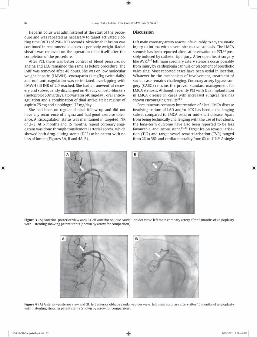
82 S. Roy et al. / Indian Heart Journal 6401 (2012) 80–83
Heparin bolus was administered at the start of the proce-dure and was repeated as necessary to target activated clot-ting time (ACT) of 250–300 seconds. Abxicimab infusion was continued in recommended doses as per body weight. Radial sheath was removed on the operation table itself after the completion of the procedure.
After PCI, there was better control of blood pressure, no angina and ECG remained the same as before procedure. The IABP was removed after 48 hours. She was on low molecular weight heparin (LMWH)—enoxaparin (1 mg/kg twice daily) and oral anticoagulation was re-initiated, overlapping with LMWH till INR of 2.0 reached. She had an uneventful recov-ery and subsequently discharged on 4th day on beta-blockers (metoprolol 50 mg/day), atorvastatin (40 mg/day), oral antico-agulation and a combination of dual anti-platelet regime of aspirin 75 mg and clopidogrel 75 mg/day.
She had been on regular clinical follow-up and did not have any recurrence of angina and had good exercise toler-ance. Anticoagula tion status was maintained in targeted INR of 2–3. At 5 months and 15 months, repeat coronary angi-ogram was done through transfemoral arterial access, which showed both drug-eluting stents (DES) to be patent with no loss of lumen (Figures 3A, B and 4A, B).
Discussion
Left main coronary artery reacts unfavourably to any traumatic injury to intima with severe obstructive stenosis. The LMCA stenosis has been reported after catheterisation or PCI,1,2 pos-sibly induced by catheter tip injury. After open heart surgery like AVR,3–6 left main coronary artery stenosis occur possibly from injury by cardioplegia cannula or placement of prosthetic valve ring. Most reported cases have been ostial in location. Whatever be the mechanism of involvement, treatment of such a case remains challenging. Coronary artery bypass sur-gery (CABG) remains the proven standard management for LMCA stenosis. Although recently PCI with DES implantation in LMCA disease in cases with increased surgical risk has shown encouraging results.8,9
Percutaneous coronary intervention of distal LMCA disease involving ostium of LAD and/or LCX has been a challenging subset compared to LMCA ostia or mid-shaft disease. Apart from being technically challenging with the use of two stents, the long-term outcome have also been reported to be less favourable, and inconsistent.10–12 Target lesion revascularisa-tion (TLR) and target vessel revascularisation (TVR) ranged from 2% to 38% and cardiac mortality from 0% to 11%.10 A single
A B
Figure 3 (A) Anterior–posterior view and (B) left anterior oblique caudal—spider view: left main coronary artery after 5 months of angioplasty with T stenting showing patent stents (shown by arrow for comparison).
A B
Figure 4 (A) Anterior–posterior view and (B) left anterior oblique caudal—spider view: left main coronary artery after 15 months of angioplasty with T stenting showing patent stents (shown by arrow for comparison).
15-IHJ-CR-Sanjeeb Roy.indd 82 1/25/2012 8:59:40 PM

S. Roy et al. / Indian Heart Journal 6401 (2012) 80–83 83
centre retrospective observational study reported 5% incidence of in-hospital complication with use of DES in unprotected LMCA disease. Event-free survival has been 66% at 28 months.8 Inconsistency in PCI results of LMCA disease in various series has been attributed to variability in baseline and angiographic characteristics like location of left main disease (ostial, main stem, or bifurcation), various bifurcation strategies amongst operators in current available reports, lack of commercially available DES of sufficient size to match large diameter left main segments, and uncertainty regarding the duration of clopidogrel therapy.8,10,12 The results for LMCA stenting have been more unfavourable when involving bifurcation, requiring two stents. Rest enosis in such cases, has a predilection for os-tium of circumflex10 and can result in sudden death. However, the role of surveillance angiography is controversial.10
Complexity of this case was not only because of involve-ment of left main bifurcation of left dominant system, but also the clinical scenario in toto. Clinical instability in the form of on-going ischaemia and lower pressure indicated higher risk for procedural outcome. Use of IABP, in such cases im-proves the outcome. Percutaneous cardio-pulmonary support was considered as an added option to supplement haemody-namic support if need arises.
Stenting to left main in this particular case was done through transradial approach, using two DES. Approach of provisional T stenting was preferred over simultaneous kiss-ing stents in view of better long-term results with former. Final kissing balloon inflation was done at high pressure using non-compliant balloons. Intravascular ultrasound (IVUS) though recommended to ensure stent expansion and apposi-tion and assess peri-stent dissection10 was not done in this case due to its unavailability at that time. Repeat angiography after 5 months and 15 months showed gratifying results.
Recently published SYNTAX study failed to demonstrate non-inferiority of PCI over CABG in 1 year of follow-up of patients with three-vessel or LMCA disease.13 Mostly this was because of need for repeat revascularisation, and hence major adverse cardiac or cerebrovascular events were signifi-cantly higher in the PCI group. Rates of death and myocardial infarction were similar between the two groups, but stroke was significantly more likely to occur with CABG. The CABG was concluded to be the standard of care for patients with three-vessel or LMCA disease with lower rates of the com-bined end point of major adverse cardiac or cerebrovascular events at 1 year. This case however, demonstrates the acute
effectiveness of PCI for the treatment of critical left main dis-ease following open heart surgery like AVR. The PCI should be reserved for cases like these who are not attractive surgi-cal candidates and perhaps those with low SYNTAX score. Short-term angiographic follow-up at the end of 15 months shows good result.
References
1. Graf RH, Verani MS. Left main coronary artery stenosis: a possi-ble complication of transluminal coronary angioplasty. Catheter Cardiovasc Diagn 1984;10:163–6.
2. Waller BF, Finkerton CA, Foster LN. Morphologic evidence of ac-celerated left main coronary artery stenosis: a late complication of percutaneous transluminal balloon angioplasty of the proximal left anterior descending coronary artery. JACC 1987;9:1019–23.
3. Pennington DG, Dincer B, Bashiti H, et al. Coronary artery steno-sis following aortic valve replacement and intermittent intracor-onary cardioplegia. Ann Thorac Surg 1982;33:576–84.
4. Prachar H, Muhlbauer J, Pollack H, Enenkel W. Iatrogenic left main coronary artery stenosis following aortic valve replace-ment. Eur Heart J 1988;9:1151–4.
5. Molina JE. Coronary stenosis following aortic valve replacement. Ann Thorac Surg 1983;35:473–4.
6. Barner HB, Fiore AC. Update on left coronary ostial stenosis: comparison with left main coronary artery stenosis. Ann Thorac Surg 1997;64:282–3.
7. Worthley MI, Burgess J, Traboulsi M. Bilateral coronary ostial stenoses post-Bentall procedure: management options in the DES era. J Invasive Cardiol 2005;17:680–2.
8. Wood FO, Saylors EK, Schneider JE, et al. Unprotected left main disease managed with drug-eluting stents: long-term outcome of 100 patients with increased surgical risk. Catheter Cardiovasc Interv 2008;71:533–8.
9. Chieffo A, Colombo A. Treatment of unprotected left main coro-nary artery disease with drug-eluting stents: is it time for a ran-domized trial? Nat Clin Prac Cardiovasc Med 2005;2:396–400.
10. Teirstein PS. Unproteted left main intervention: patient selec-tion, operator technique and clinical outcomes. JACC Cardiovasc Interv 2008;1:5–13.
11. Price MJ, Cristea E, Sawhney N, et al. Serial angiographic follow-up of sirolimus-eluting stents for unprotected left main coronary artery revascularization. JACC 2006;47:871–7.
12. Biondi-Zoccai GCL, Lotrionte M, Moretti C, et al. A collaborative systematic review and meta-analysis of 1278 patients undergo-ing percutaneous drug-eluting stenting for unprotected left main coronary artery disease. Am Heart J 2008;155:274–83.
13. Serruys PW, Morice MC, Kappetein AP, et al, for the SYNTAX Investigators. Percutaneous coronary interventions versus coro-nary artery bypass grafting for severe coronary artery disease. N Engl J Med 2009;360:961–72.
15-IHJ-CR-Sanjeeb Roy.indd 83 1/25/2012 8:59:41 PM

Contents lists available at SciVerse ScienceDirect
Indian Heart Journal
ISSN: 0019-4832 Copyright © 2012. Cardiological Society of India. All rights reserved.doi: 10.1016/S0019-4832(12)60017-0
Indian Heart Journal 6401 (2012) 84–87
* Corresponding a uthor. E-mail address: [email protected]
Case report
A case of arrhythmogenic right ventricular cardiomyopathy—Naxos diseaseR.R. Saravanan1, V. Amuthan2, R.A. Janarthanan3, S. Balasubramanian4, S. Naina Mohamed4
Resident, Professor and Head, Professor, Assistant Professor Department of Cardiology, Madurai Medical College, Madurai, Tamilnadu, India.
A B S T R A C T
We present a case of arrhythmogenic right ventricular cardiomyopathy (ARVC)—Naxos disease. The patient is 21-year-old male with no history of previous heart disease admitted in a private hospi-tal for rhythm disorder in heart. The condition was diagnosed as ventricular tachycardia (VT) and was treated with cardioversion. The patient was referred to our hospital for further evaluation. On examination patient had palmoplantar keratoderma, wooly hair, and dystrophic nails. The cardio-vascular system examination was clinically normal. His electrocardiogram showed epsilon wave in lead V1; echocardiography showed hypo-echogenic tissues in the right ventricular (RV) apex and free wall; magnetic resonance imaging (MRI) investigation revealed fibrofatty replacement of RV free wall and dyskinetic RV wall with diastolic outbulging.
Copyright © 2012, Cardiological Society of India. All rights reserved.
K E Y W O R D S
CMRIElectrocardiographyNaxos diseasePalmoplantar keratodermaVentricular tachycardia
Introduction
Arrhythmogenic right ventricular dysplasia (ARVD) is a car-diomyopathy characterised by fibrofatty degeneration of the myocardium with progressive myocardial dysfunction, elec-trical instability, and sudden death. It is an inherited disorder associated with arrhythmias and sudden death. Right ven-tricular (RV) features predominate but left ventricular (LV) involvement can also arise with disease progression and may be the dominant presentation. Arrhythmogenic RV cardiomyo-pathy exhibits age-related expression. Arrhythmogenic RV dysplasia is familial in 30–50% cases and is typically inher-ited in a dominant fashion although recessive forms are also recognised. The Naxos disease is a recessive mutation in the gene encoding plakoglobin characterised by arrhythmogenic right ventricular cardiomyopathy (ARVC), a non-epidermo-lytic palmoplantar keratoderma, and wooly hair. Most fre-quently involved areas of RV are posterior base, apex, and infundibulum; these are collectively called as the triangle of dysplasia. Magnetic resonance imaging (MRI), ECHO, electro-cardiography (ECG), and SAEG are helpful in the diagnosis. Epsilon wave or small high frequency deflection found in the terminal portion of QRS in leads V1–V3 may be present.
We present a case of ARVC–Naxos disease with ECG, echocar-diographic, and cardiac magnetic resonance imaging (CMRI) features of ARVC.
Case history
Our case is a 21-year-old male with no history of previous heart disease, who presented with ventricular tachycardia (VT) of left bundle branch block (LBBB) morphology (Figure 1A) and got treated with dilated cardiomyopathy (DC) cardi-oversion. The patient was referred to the Government Rajaji Hospital, Madurai for further investigation and management. His ECG during sinus rhythm showed epsilon wave in lead V1 (Figures 1B and C). On examination he had wooly hair (Figure 2) and palmoplantar keratoderma (Figure 3). His cardiovas-cular system was clinically normal. We proceeded with MRI of heart which showed fibro-fatty replacement of RV free wall (Figure 4). Dyskinetic RV wall with diastolic outbulging was also seen. Echocardiogram showed dyskinesia and di-astolic outbulging in the RV apex and free wall with normal RV systolic function (Figure 5). No obvious demonstrable pa-thology was seen in the LV. Cardiac valves and pericardium were normal.
Patient was treated with amiodarone and discharged. Since, he developed another episode of VT with a slightly different
16-IHJ-CR-RRSaravanan.indd 84 1/25/2012 8:57:25 PM

R.R. Saravanan et al. / Indian Heart Journal 6401 (2012) 84–87 85
A
B
C
Figure 1 (A) Electrocardiogram showing ventricular tachycardia with left bundle branch block morphology, superior axis. (B) Epsilon wave after reversion to sinus rhythm. (C) Epsilon wave.
16-IHJ-CR-RRSaravanan.indd 85 1/25/2012 8:57:26 PM

86 R.R. Saravanan et al. / Indian Heart Journal 6401 (2012) 84–87
LBBB morphology reverted with DC cardioversion, a detailed electrophysiologic evaluation was planned.
Discussion
Arrhythmogenic right ventricular cardiomyopathy is pre-dominantly a genetically determined heart muscle disorder characterised pathologically by fibrofatty replacement of the RV myocardium. In the early stage of the disease structural changes may be absent or subtle and confined to the localised region of RV typically the inflow tract, outflow tract or the apex of the RV collectively called as the triangle of dysplasia.
The disease expression is variable. During the early con-cealed phase individuals are often asymptomatic but none-theless and at a risk of sudden cardiac death notably during exertion.1 In the overt electrical phase individuals present with symptomatic arrhythmias and RV morphological abnor-malities are readily discernible by conventional imaging. In our case, the patient entered the overt electrical phase and presented with VT of LBBB morphology which arose from the RV free wall. Morphological abnormalities demonstrated with CMRI were fibrofatty replacement of RV free wall and dyskinetic RV wall with diastolic outbulging.2 Later, diffuse disease results in biventricular failure where ventricular arrhythmias may or may not be present.
According to the Original Task Force Criteria our case satis-fied the following three major criteria needed to diagnose ARVC3,4:1. Dyskinetic RV wall with diastolic outbulging.2. Epsilon wave in Lead V1.3. Sustained VT of LBBB morphology with superior axis.
A recessive form of Naxos disease was first described in families originating from the Greek island of Naxos. Wooly hair appears from birth whereas palmoplantar keratoderma5–7 develops during the first year of life when infants start to use their hands and feet. The cardiomyopathy clinically manifests by adolescence and shows 100% penetrance.
This type of ARVD is caused by a mutation of the plakoglobin gene, the product of which is a component of desmosomes and adherens junctions. Desmosomes are major cell adhesion junctions prominent in epidermis and cardiac tissue that are important for rigidity and strength of cells under conditions of mechanical stress desmosomal dysfunction and leads to detachment of myocytes at intercalated discs with progressive myocyte apoptosis. As the regeneration of cardiac myocytes is limited fibrofatty replacement takes place and provides the anatomic basis for progressive cardiac failure, arrhythmias, and sudden death.
Figure 2 Woolly hair.
Figure 3 Plantar-keratoderma.
Figure 4 Cardiac magnetic resonance imaging showing dysplastic right ventricular and diastolic outbulging.
Figure 5 Two-dimensional echo apical four chamber showing right ventricular free wall infiltration.
16-IHJ-CR-RRSaravanan.indd 86 1/25/2012 8:57:27 PM

R.R. Saravanan et al. / Indian Heart Journal 6401 (2012) 84–87 87
The symptomatic presentation is usually with syncope and/or VT of LBBB morphology, as in our case. During a mean follow-up period of 10 years, 50% of patients develop pro-gressive heart disease involving the right or both ventricles. Progression appears to be stepwise in some cases associated with arrhythmic storm or sudden cardiac death. Symptoms of right heart failure appear in the final stages when the right or both ventricles are severely affected.
Young age, malignant family history, QRS dispersion ≥40 ms,8 T wave inversion beyond V1, LV involvement, VT, syncope or previous cardiac arrest are considered as the major determinants of adverse prognosis and impending sudden death. Different anti-arrhythmics like sodium blockers, sotalol, and amiodarone are used for treatment. Implantable cardioverter defibrillator has been proven to be lifesaving in patients who develop symptoms/or structural progression of disease particularly before age 35 years.9,10
Familial occurrence of a combination of DC with pal-moplantar keratoderma and curly hair was first reported in India by Rao et al. in 1996 with no ECG evidence of epsilon wave and VT, and echocardiographic evidence of RV involvement.11
In the present case, the diagnosis of Naxos disease is to be considered with the presence of woolly hair, palmoplantar keratoderma presenting with VT of LBBB morphology with global or regional dysfunction, and structural alterations of RV.
References
1. Corroda D, Thiene G, Nava A, et al. Sudden death in young com-petitive athletes clinicopathologic correlations in 22 cases. Am J Med 1990;89:588–96.
2. Tandri H, Saranathan M, Rodriguez ER, et al. Non invasive detec-tion of myocardial fibrosis in arrhythmogenic right ventricular cardiomyopathy using delayed enhancement magnetic reso-nance imaging. J Am Coll Cardiol 2005;45:98–103.
3. McKenna WJ, Thiene G, Nava A, et al. Diagnosis of right ventricu-lar dysplasia/cardiomyopathy. Task Force of the working group myocardial and pericardial disease of the European Society of Cardiology and of the Scientific Council on Cardiomyopathies of the International Society and Federation of Cardiology. Br Heart J 1994;71:215–8.
4. Richardson P, McKenna, Bristow M, et al. Report of the 1995 WHO/ISFC Task Force on the definition of and classification of cardiomyopathies. Circulation 1996;93:841–2.
5. Protonatarios N, Tsatsopoulou A, Anastasakis A, et al. Genotype-Phenotype assessment in autosomal recessive arrhythmogenic right ventricular cardiomyopathy (Naxos disease) caused by de-letion in plakoglobin. J Am Coll Cardiol 2001;38:1477–84.
6. Mc Koy G, Protonotarios N, Crosby A, et al. Identification of a dele-tion in plakoglobin in arrhythmogenic right ventricular cardiomy-opathy with palmoplantar keratoderma and woolly hair (Naxos disease). Lancet 2000;355:2119–24.
7. Carvajal-Huerta L. Epidermolytic palmoplantar keratoderma with woolly hair and dilated cardiomyopathy. J Am Acad Dermatol 1998;39:418–21.
8. Turrini P, Corrado D, Basso C, et al. Dispersion of ventricular depolarization-repolarization a non invasive marker of risk stratification in arrhythmogenic right ventricular cardiomyopa-thy. Circulation 2001;103:3075–80.
9. Corrado D, Leoni L, Link MS, et al. Implantable cardioverter defibrillator therapy for prevention of sudden death in patients with arrhythmogenic right ventricular cardiomyopathy/dyspla-sia. Circulation 2003;108:3084–91.
10. Gatzoulis K, Protonotarios N, Anastasakis A, et al. Implantable defibrillator therapy in Naxos Disease. Pacing Clin Elecrophysiol 2000;23:1176–8.
11. Rao HB, Reddy IS, Chandra KS. Familial occurrence of a rare com-bination of dilated cardiomyopathy with palmoplantar kerato-derma and curly hair. Indian Heart J 1996;48:161–2.
Book review
Percutaneous mitral valvotomy, 1e. Harikrishnan S. Publisher: Jaypee Brothers Medical Publishers Pvt. Ltd, New Delhi. Publication: 2012. Pages: 474. Paper Back. Price: $50. ISBN: 978-93-5025-561-2.
Although there has been a lot of literature on mitral valvotomy both from within India and abroad, the new book on percutaneous mitral valvotomy edited by Dr. Hari Krishnan from Sree Chitra Tirunal Institute of Medical Sciences and Technology, Thiruvananthapuram is a valuable addition to our knowledge. The book is a nearly complete encyclopaedia on per-cutaneous balloon mitral valvuloplasty (PBMV) discussing all its aspects covering anatomy, physiology, pathology, procedure details, and all the new gadgets. The technical details of the procedure are well discussed, and most importantly, it is written in common language and can be read through quickly. With excellent illustrations, the book has contributing authors from all over the world. However, Dr. Allen Cribier’s name is surprisingly missing. I strongly recom-mend this book for all the interventional cardiologists dealing with valvular heart disease.
Contributed byDr. K. Sarat Chandra
Honorary EditorIndian Heart Journaldoi: 10.1016/S0019-4832(12)60042-X
16-IHJ-CR-RRSaravanan.indd 87 1/25/2012 8:57:28 PM
Contents lists available at SciVerse ScienceDirect
Indian Heart Journal
ISSN: 0019-4832 Copyright © 2012. Cardiological Society of India. All rights reserved.doi: 10.1016/S0019-4832(12)60018-2
Indian Heart Journal 6401 (2012) 88–89
*Corresponding author.E-mail address: [email protected]
Case report
Percutaneous balloon pericardiotomy for the treatment of recurrent malignant pericardial effusionAniket Puri1*, Nitin Agarwal2, S.K. Dwivedi3, V.S. Narain3
Associate Professor, Senior Resident, Professor, Department of Cardiology, CSM Medical University, Lucknow.
A B S T R A C T
Malignant disease with pericardial metastasis is one of the most common causes of recurrent pericardial effusion (PE) with tamponade. While surgical pericardiotomy in these patients is very morbid and may not be a viable option, a palliative treatment percutaneously with percutaneous balloon pericardiotomy (PBP) can be a preferred treatment. We report herewith a case of PBP technique done using our day-to-day catheterisation laboratory equipment.
Copyright © 2012, Cardiological Society of India. All rights reserved.
K E Y W O R D S
Pericardial effusionPericardiotomy
Introduction
Malignant disease with pericardial metastasis is one of the most common causes of pericardial effusion (PE) with tam-ponade. Recurrence rates are high after pericardiocentesis and in critically ill patients it is desirable to avoid the risk of general anaesthesia and the discomfort of surgical pericardi-otomy. We report a case of malignant angiocarcinoma of lung presented with malignant PE with tamponade with recurrent filling, which was successfully treated percutaneously with percutaneous balloon pericardiotomy (PBP).
Case summary
A 45-year-old female was referred to our department for re-current PE with tamponade. The patient was a known case of malignant adenocarcinoma of the right lung with recurrent PE. Her pericardial fluid cytology was positive for malignant cells and she was planned for chemotherapy. The patient un-derwent emergency percutaneous pericardiocentesis at our
centre via sub-xiphoid approach to relieve the tamponade and a 6 French pigtail catheter was left in the pericardial cavity for drainage. She was having repeated refilling of the pericardial cavity during the hospital stay and thus, PBP was planned for her after discussions with her family.
The procedure was done under local anaesthesia. We used a percutaneously sub-xiphoid approach to access the peri-cardial cavity. A 0.035″ angiography guide wire was passed into the pericardial cavity. A TYSHAK II® (NuMED, New York, USA) percutaneous transluminal angioplasty (PTA) balloon catheter of 18 mm size was tried over the wire, but could not be negotiated due to lack of support. The angiography guide wire was then exchanged with stiff coiled-tip 0.25″–175 cm stainless steel Inuoe balloon guide wire with coil tip (Toray Medical, Tokyo) (Figure 1) and local site was dilated with 10 French dilator. The balloon catheter was then manipulated over the coiled-tip guide wire into the pericardial cavity. The balloon was partially inflated and withdrawn slowly until a waist appeared over the balloon which confirmed the posi-tion of the parietal pericardium (Figure 2). After optimal position, the balloon was inflated with 20cc contrast for 1 minute until the waisting disappeared (Figure 3). The ab-sence of the waist was confirmed again, by second inflation and the balloon and coiled-tip catheter were removed subse-quently. The patient remained haemodynamically stable
17-IHJ-CR-Aniket Puri.indd 88 1/25/2012 8:56:02 PM

A. Puri et al. / Indian Heart Journal 6401 (2012) 88–89 89
throughout the procedure. Echocardiogram done immediately and 24 hours after the procedure, revealed no PE. Patient re-mained effusion-free in the 1 month follow-up.
Discussion
Patients with large malignant PEs are often in a symptomatic, preterminal stage. The survival of this subgroup is most closely related to the extent of disease and the tumour type. In cancerous patients with symptomatic large PE, the stan-dard treatment consisted of pericardiocentesis and drainage. Palacios et al. reported on the effectiveness of PBP,1 and in a multicentre study of 50 patients by Ziskind et al. the success rate of PBP was 95%.2 The recurrence rate after pericardio-centesis may be as high as 62%.3 Recurrence suggests the need for repeated hospitalisation, with its attendant cost, morbidity, and mortality. More recently Swanson et al. showed that balloon pericardiotomy as the initial management of symptomatic malignant PEs, allows a definitive procedure to be performed at presentation and that reaccumulation rates between balloon pericardiotomy and simple aspiration (7.4% vs 14.3%) and complication rates (7.4% vs 7.1%) were not statistically different.4 Therefore, primary therapy with a peri-cardial window creation should be considered.
Many individual studies and case reports across the world have reported successful percutaneous pericardiotomy; how-ever, the use of coiled-tip balance middle weight (BMW) guide wire with springe tip in performing percutaneous peri-cardiotomy successfully is reported for the first time. The technique is relatively simple, safe, and can be performed with local anaesthesia and with minimal discomfort even in critically ill patients.
References
1. Palacios I, Tuzcu E, Sizkind A, et al. Percutaneous balloon pericar-dial window for patients with malignant pericardial effusion and tamponade. Cathet Cardiovasc Diagn 1991;22:244–9.
2. Ziskind AA, Pearce AC, Lemon C, et al. Percutaneous balloon peri-cardiotomy for the treatment of cardiac tamponade and large pericardial effusion: description of technique and report of the first 50 cases. J Am Coll Cardiol 1993;21:1–5.
3. Laham RJ, Cohen DJ, Kuntz RE, et al. Pericardial effusion in pa-tients with cancer: outcome with contemporary management strategies. Heart 1996;75:67–71.
4. Swanson N, Mirza I, Wijesinghe N, Devlin G. Primary percutane-ous balloon pericardiotomy for malignant pericardial effusion. Catheter Cardiovasc Interv 2008;71:504–7.
Figure 1 Balloon catheter in pericardial cavity over coiled-tip guide wire.
Figure 2 Inflated balloon showing waist at pericardial junction.
Figure 3 Near loss of waist and full inflation of balloon.
17-IHJ-CR-Aniket Puri.indd 89 1/25/2012 8:56:03 PM

Contents lists available at SciVerse ScienceDirect
Indian Heart Journal
ISSN: 0019-4832 Copyright © 2012. Cardiological Society of India. All rights reserved.doi: 10.1016/S0019-4832(12)60019-4
Indian Heart Journal 6401 (2012) 90–92
*Corresponding author.E-mail address: [email protected]
Case report
Isolated left ventricular non-compaction in association with ventricular tachycardiaRajesh Vijayvergiya1*, Mukesh Yadav2, Anand Subramaniyan2
Associate Professor, Senior Resident, Department of Cardiology, Post Graduate Institute of Medical Education and Research (PGIMER), Chandigarh – , India.
A B S T R A C T
A 32-year-old young male was found to have non-sustained, repetitive, monomorphic ventricular tachycardia of right bundle branch morphology during routine pre-anaesthetic evaluation for or-thopaedic surgery. Echocardiography and left ventricular angiogram were suggestive of isolated non-compaction of left ventricular apex with systolic dysfunction. He was successfully managed with anti-arrhythmic drugs and had an uneventful 9-month follow-up. The index case is an unu-sual association of asymptomatic, non-sustained ventricular tachycardia with isolated ventricular non-compaction.
Copyright © 2012, Cardiological Society of India. All rights reserved.
K E Y W O R D S
CardiomyopathyNon-compaction of left ventricleVentricular tachycardia
Introduction
Isolated ventricular non-compaction (IVNC) is a rare cardio-myopathy characterised by the loss of compaction of myo-cardial fibre meshwork during intrauterine life. This is possibly due to an arrest of intrauterine compaction of the myocardial fibres in the absence of any other structural heart disease.1 It is characterised by the presence of deep inter-trabecular recesses in the hypertrophied, frequently hypoki-netic segment of ventricular myocardium.2,3 The clinical manifestation includes triad of ventricular systolic dysfunc-tion, thromboembolism and malignant tachy-arrhythmias.4 We hereby report a case of IVNC of left ventricle (LV) pre-sented with non-sustained ventricular tachycardia (VT), which was effectively treated with anti-arrhythmic drugs.
Case report
A 32-year-old male, manual labourer by occupation was referred to our outpatient clinic in July 2009, for his electro-cardiogram (ECG) evaluation prior to right knee joint
arthroscopy for osteo-chondritis. He did not have any cardiac symptoms like palpitation, dyspnoea, angina, or syncope. He was non-smoker, normotensive, and non-diabetic. There was no family history of sudden cardiac death in any of the family members. General physical examination revealed irregular, normal volume radial pulse. Rest of his general and systemic examination was within normal limits. The ECG revealed repetitive runs of non-sustained, monomorphic VT of right bundle branch block (RBBB) morphology (Figures 1A and B). Echocardiogram features of LV apical IVNC as characterised by hyper-trabeculation and inter-trabecular recesses and >2:1 ratio of non-compacted to compacted layer of left ventricular wall,3 was present as evident in Figure 2. There was no clot at left ventricular apex. Coronary angiography revealed normal epicardial coronaries. Left ventriculogram revealed spongi-form appearance of apex and diaphragmatic surface (Figure 3) suggestive of localised non-compaction of LV as described by us in our previous publication.5 The LV ejection fraction was 30%. The pulmonary artery pressure was 25/10 mmHg (mean 15 mmHg). A diagnosis of IVNC-LV type with systolic dys-function and haemodynamically stable non-sustained, mon-omorphic VT of LV origin was made. His tachycardia could be controlled with amiodarone 400 mg twice a day tablets (Figure 1C). There were no runs of VT on 24-hour Holter monitoring on day 10 of admission. He was discharged on tablets—amiodarone 200 mg, ramipril 5 mg, and aspirin
18-IHJ-CR-Rajesh Vijayvergiya.indd 90 1/25/2012 8:55:31 PM

R. Vijayvergiya et al. / Indian Heart Journal 6401 (2012) 90–92 91
150 mg per day. An implantable cardioverter defibrillator (ICD) could not be implanted because of financial constraint. He is doing well and had an uneventful 9-month follow-up. The ECG did not show any runs of VT or ventricular prema-ture complexes, echocardiogram showed the unchanged LV ejection fraction of 30%.
Discussion
Out of the clinical triad of IVNC,4 the index case had two manifestations in the form of LV systolic dysfunction and
non-sustained VT. It is uncommon to encounter IVNC as an underlying structural abnormality in a patient with VT.6 Nevertheless, ventricular arrhythmia is the frequent compli-cation in IVNC patients on long-term follow-up.7,8 The inci-dence is variably reported as 4.6% in 238 IVNC patients at 6 months of follow-up by Fazio Ge et al7 and 41% in 34 IVNC patients at 44 months of follow-up by Oechslin et al.8 The precise mechanism of ventricular arrhythmia is not clear. A relatively decreased perfusion and ischaemia-related fibrosis at sub-endocardial non-compacted area has been postulated as the cause of electrical inhomogeneity and micro-reentry resulting into ventricular tachycardia.6,9 Steffel et al. have
A
B
C
Figure 1 (A, B) Electrocardiogram showing repetitive runs of non-sustained ventricular tachycardia of right bundle branch block morphology. (C) Tachycardia controlled with the administration of amiodarone and sotalol.
18-IHJ-CR-Rajesh Vijayvergiya.indd 91 1/25/2012 8:55:31 PM

92 R. Vijayvergiya et al. / Indian Heart Journal 6401 (2012) 90–92
tried to risk stratify these cases by VT induction during electro-physiological studies.10 The medical treatment of VT includes beta-blockers and anti-arrhythmic drugs like amio-darone, which was effective in the index case. There are reports of ICD8/cardiac resynchronisation therapy implantation,11 radiofrequency ablation,6 and even cardiac transplantation8,12 in IVNC cases with malignant ventricular tachy-arrhythmias. The long-term prognosis of these patients is variable de-pending on the New York Heart Association (NYHA) func-tional class, LV systolic dysfunction, presence of malignant ventricular arrhythmias, and cerebral thromboembolism.4,10
Nevertheless, our case had an uneventful 9-month follow-up on medical treatment only.
In conclusion, IVNC may be the underlying cause for ven-tricular tachycardia in a young individual, evaluated for idio-pathic VT. A careful echocardiographic evaluation can diagnose the underlying unclassified cardiomyopathy. As the long-term prognosis is variable depending upon the NYHA functional class, LV systolic dysfunction, and associated arrhythmias, the management should be individualised depending upon the risk stratification.
References
1. Freedom RM, Yoo SJ, Perrin D, Taylor G, Petersen S, Anderson RH. The morphological spectrum of ventricular noncompaction. Cardiol Young 2005;15:345–64.
2. Richardson P, McKenna W, Bristow M, et al. Report of the 1995 World Health Organization/International Society and Federation of Cardiology Task Force on the definition and classification of cardiomyopathies. Circulation 1996;93:841–2.
3. Jenni R, Oechslin E, Schneider J, Attenhofer Jost C, Kaufmann PA. Echocardiographic and patho-anatomic characteristics of isolated left ventricular noncompaction: a step towards classification as a distinct cardiomyopathy. Heart 2001;86:666–71.
4. Lofiego C, Biagini E, Pasquale F, et al. Wide spectrum of presenta-tion and variable outcomes of isolated left ventricular noncom-paction. Heart 2007;93:65–71.
5. Vijayvergiya R, Jha A, Pandian RP, Sharma R, Grover A. Isolated left ventricular noncompaction in association with rheumatic mitral stenosis. Int J Cardiol 2008;123:e54–6.
6. Derval N, Jais P, O’Neill MD, Haissaguerre M. Apparent idiopathic ventricular tachycardia associated with isolated ventricular non-compaction. Heart Rhythm 2009;6:385–8.
7. Fazio G, Corrado G, Novo G, et al. Ventricular tachycardia in non-compaction of left ventricle: is this a frequent complication? Pacing Clin Electrophysiol 2007;30:544–6.
8. Oechslin EN, Attenhofer Jost CH, Rojas JR, Kaufmann PA, Jenni R. Long-term follow-up of 34 adults with isolated left ventricular noncompaction: a distinct cardiomyopathy with poor prognosis. J Am Coll Cardiol 2000;36:493–500.
9. Chin TK, Perloff JK, Williams RG, Jue K, Mohrmann R. Isolated noncompaction of left ventricular myocardium. A study of eight cases. Circulation 1990;82:507–13.
10. Steffel J, Kobza R, Namdar M, et al. Electrophysiological findings in patients with isolated left ventricular non-compaction. Europace 2009;11:1193–200.
11. Oginosawa Y, Nogami A, Soejima K, et al. Effect of cardiac resyn-chronization therapy in isolated ventricular noncompaction in adults: follow-up of four cases. J Cardiovasc Electrophysiol 2008;19:935–8.
12. Duru F, Candinas R. Noncompaction of ventricular myocardium and arrhythmias. J Cardiovasc Electrophysiol 2000;11:493.
Figure 2 Echocardiogram in modified apical four chamber view with anterior tilt shows non-compaction of left ventricular apex. LV: left ventricle, RA: right atrial, RV: right ventricle.
Figure 3 Left ventricular angiogram in right anterior oblique 30° show-ing the spongiform appearance of apex and diaphragmatic surface.
18-IHJ-CR-Rajesh Vijayvergiya.indd 92 1/25/2012 8:55:32 PM

Contents lists available at SciVerse ScienceDirect
Indian Heart Journal
ISSN: 0019-4832 Copyright © 2012. Cardiological Society of India. All rights reserved.doi: 10.1016/S0019-4832(12)60020-0
Indian Heart Journal 6401 (2012) 93–94
* Corresponding a uthor. E-mail address: [email protected]
Case report
Parachute tricuspid valve in an asymptomatic adultJagdish C. Mohan1, Chandra Shekhar2, Vipul Mohan3, Bimalpreet Kaur3, Shivesh Kumar Singh3
Chairman, Consultant, Resident, Department of Cardiology, Ridge Heart Centre, Sunder Lal Jain Hospital, Ashok Vihar-III, New Delhi – .
A B S T R A C T
Parachute tricuspid valve is a rare anomaly usually reported in small children. This report de-scribes transthoracic and trans-oesophageal echocardiographic (TEE) features of parachute mitral valve in an adult patient.
Copyright © 2012, Cardiological Society of India. All rights reserved.
K E Y W O R D S
Parachute tricuspid valveTrans-oesophageal echocardiography (TEE)
Introduction
The malformation of an atrioventricular valve in which the tension apparatus springs from a single papillary muscle or muscle group has been labelled as the parachute valve. It may or may not have any haemodynamic significance.
Parachute tricuspid valve is a rare anomaly with <10 cases reported in the literature since 1972 when it was first described at necropsy in a patient with cor triatriatum.1 The initial three cases were described at necropsy.1–3 The first clinical descrip-tion of the anomaly without right-sided obstructive lesions was made in 2006.4
Case report
This report describes a relatively asymptomatic 30-year-old individual who was incidentally found to have a systolic mur-mur. A complete transthoracic and trans-oesophageal echocar-diographic examination revealed a 12 mm secundum atrial septal defect with a large left-to-right shunt, pulmonary artery pressures of 45/20 mmHg and a typical parachute tricuspid valve with a single short papillary muscle (Figures 1 and 2) which was attached to all the three tricuspid leaflets showing
leaflet crowding during diastole and doming although with no significant obstruction (Figure 3).
Discussion
In our case, there was a single papillary muscle group giving rise to all the shortened and thickened chordae tendineae
Figure 1 Transthoracic apical four chamber view in end-diastole showing septal and anterior tricuspid leaflets attached to a single short papillary muscle (arrow). LA: left atrial, LV: left ventricle, RA: right atrial; RV: right ventricle.
19-IHJ-CR-JCMohan.indd 93 1/25/2012 8:54:41 PM

94 J.C. Mohan et al. / Indian Heart Journal 6401 (2012) 93–94
supporting the tricuspid valve. Previously, a single case with associated atrial septal defect and parachute tricuspid valve has been reported.4
Sometimes, the normal number of papillary muscles is seen, but one muscle is much larger than its peers and shows some characteristic features. This is known as a parachute-like asymmetric valve.5 Detailed echocardiographic examination in this case did not reveal the presence of any other papillary muscle.
References
1. Maitre Azcarate MJ. Parachute deformity of the tricuspid valve. Thorax 1980;35:240.
2. Milo S, Stark J, Macartney FJ, Anderson RH. Parachute deformity of the tricuspid valve. Thorax 1979;34:543–6.
3. Ariza S, Cintado C, Castillo JA, et al. Parachute tricuspid valve asso-ciated with Fallot’s tetralogy. Arch Mal Coeur Vaiss 1979;72:317–20.
4. Marwah A, Suresh PV, Shah S, Misri A, Maheshwari S. Parachute tricuspid valve. Eur J Echocardiogr 2006;7:226–7.
5. Purvis JA, Barr SH. Parachute-like asymmetric tricuspid valve in an asymptomatic adult. Eur J Echocardiogr 2010;11:E23.
Figure 2 Zoomed picture of Figure 1. RA: right atrial, RV: right ventricle.
Figure 3 Trans-oesophageal echocardiographic view at 98#. Anterior and posterior tricuspid leaflets are attached to a single short papil-lary muscle (arrow). Note a small secundum atrial septal defect. RA: right atrial.
19-IHJ-CR-JCMohan.indd 94 1/25/2012 8:54:42 PM

Contents lists available at SciVerse ScienceDirect
Indian Heart Journal
ISSN: 0019-4832 Copyright © 2012. Cardiological Society of India. All rights reserved.doi: 10.1016/S0019-4832(12)60021-2
Indian Heart Journal 6401 (2012) 95–98
*Corresponding author.E-mail address: [email protected]
Case report
Tetralogy of Fallot with Holt-Oram syndromeVikas Kumar1, Vikas Agrawal2*, Dharmendra Jain2, Om Shankar2
Senior Resident, Assistant Professor, Department of Cardiology, Institute of Medical Sciences, Banaras Hindu University, Varanasi – .
A B S T R A C T
Holt-Oram syndrome (HOS) is characterised by mild to severe congenital cardiac defects and skeletal abnormalities of the upper limb. This syndrome is also referred to as Hand-Heart syn-drome. The most common cardiac disorder is an ostium secundum detected an atrial septal defect (ASD), followed by ventricular septal defect (VSD) and ostium primum ASD. We report a case of HOS with tetralogy of Fallot (TOF). This association is very rare and is hardly reported in the literature.
Copyright © 2012, Cardiological Society of India. All rights reserved.
K E Y W O R D S
ArrhythmiasAtrial septal defectHolt-Oram syndromeTetralogy of Fallot
Introduction
Holt-Oram syndrome (HOS) was first reported in 1960 by Mary Clayton Holt and Samuel Oram, who detected an atrial septal defect (ASD) in members of four generations of a family. Detected an ASD which was associated with a con-genital anomaly of the thumbs which lay in the same plane as the fingers.
The most common congenital heart defect is the ostium secundum ASD, seen in 60% of patients with HOS followed by the ventricular septal defect (VSD). The other associated findings include hand malformations and conduction distur-bances, originally described by Holt and Oram. The other complex congenital cardiac malformations, like VSD with in-fundibular pulmonary stenosis, complete atrio-ventricular (AV) canal defect, mitral valve prolapse, hypoplastic left heart syndrome, coarctation of aorta, subaortic stenosis, patent ductus arteriosus, etc. have rarely been reported in HOS.
Recently, we came across a case of hand malformation with cyanotic congenital heart disease, which was later iden-tified as tetralogy of Fallot (TOF). This association is rarely described in patients with HOS.
Case report
A 2-year-old female child presented with fatiguability, bluish discolouration (cyanosis), and growth retardation, since 6 months of age (Figure 1). Patient also had right hand mal-formation in the form of radial deviation at wrist and cubitus valgus deformity of right upper limb and hypoplastic right
Figure 1 Cyanosis of lips while crying.
20-IHJ-CR-Vikas kumar.indd 95 1/25/2012 8:51:08 PM

96 V. Kumar et al. / Indian Heart Journal 6401 (2012) 95–98
thumb being at the same plane as fingers (distally located than the opposite thumb) (Figure 2). One of her family mem-bers had congenital cardiac malformation. Her father’s sister had medium size perimembranous VSD, but she had no anomaly related to either of the hands. None of the other family members are affected.
General examination
Patient had cyanosis, which increased on crying and did not improved on 100% oxygen inhalation. Grade II clubbing was also present.
Cardiovascular system examination
Apex beat was within midclavicular line at the fifth intercos-tal space. A systolic thrill was present at the upper left inter-costal space. Grade I left parasternal heave was present. Ejection systolic murmur was heard at mid and upper left in-tercostal space. A single second heart sound (S2) was heard.
Musculoskeletal system examination
Right thumb was hypoplastic and located distally in the plane of fingers, in comparison to the left thumb. Right hand was deviated radially at the wrist. Rest of the systemic examination did not reveal any abnormality.
Investigations
Haemoglobin (Hb)—19.8 g/dL, total count—10,100/mm3, differ-ential count—N40 L52 E3 M5, platelet count—1,14,000/mm3, haematocrit—60.9%, mean corpuscular volume (MCV)—75.0 fl, red blood cells (RBC) count—8.13 × 10⁶/mm3.
A
B
C
Figure 2 (A, B) Showing right hand deformities (radial deviation of wrist and cubitus valgus deformity of the right elbow). (C) Left hand-deformity.
Figure 3 Forearm and hand radiograph showing hypoplastic radius and triphalangeal thumb.
Figure 4 Chest radiograph showing boot-shaped heart and pulmonary oligemia.
20-IHJ-CR-Vikas kumar.indd 96 1/25/2012 8:51:09 PM

V. Kumar et al. / Indian Heart Journal 6401 (2012) 95–98 97
Radiograph right hand: triphalangeal thumb, distally lo-cated, hypoplastic radius, radial deviation of the right wrist (Figure 3).
Chest radiograph posterior–anterior view: No cardiomeg-aly (cardiothoracic ratio <50%), boot-shaped heart (Coeur en sabot), main pulmonary artery inconspicuous (concave), de-creased pulmonary vascularity (Figure 4).
Electrocardiogram (ECG): Right ventricular hypertrophy, right axis deviation, no arrhythmia detected (Figure 5).
Two-dimensional echocardiography with colour Doppler: Situs solitus, levocardia, large perimembranous VSD with 40% aortic override with right to left shunt, hypoplastic and confluent main, right, and left pulmonary artery, predomi-nantly infundibular pulmonary stenosis with peak gradient
Figure 5 Right ventricular hypertrophy; right axis deviation.
A B C
Figure 6 (A) Parasternal long axis view showing overriding of aorta and large perimembranous ventricular septal defect. (B) Parasternal short axis view showing hypoplastic and confluent PA. (C) Continuous Doppler showing infundibular pulmonary stenosis with pressure gradient of 80 mmHg. AO: ascending aorta, PA: pulmonary artery, RA: right atrial, RV: right ventricle.
of 80 mmHg, interatrial septum is intact, no patent ductus arteriosus or any other defect seen (Figure 6).
We concluded from the above findings that this was a case of TOF associated with HOS.
Discussion
Holt-Oram syndrome is a rare syndrome with one in 1,00,000 live births affected.1 About 300 cases have been published re-vealing a wide spectrum of clinical signs. Association of TOF is very rare. We could find only one study which included 18 patients of HOS, conducted in China, which showed some association between these two findings.2
20-IHJ-CR-Vikas kumar.indd 97 1/25/2012 8:51:11 PM

98 V. Kumar et al. / Indian Heart Journal 6401 (2012) 95–98
Holt-Oram syndrome is an autosomal dominant disorder. The underlying genetic defect was found on the long arm of chromosome 12. Mutation in the TBX-3 and TBX-5 genes lead to a wide range of phenotypes typical of HOS.1,3
The review of orthopaedic sign in the literature remains poor.4 Frequent signs are radial ray abnormalities, absent or abnormal radius, upper limb-transverse elements missing, and various thumb anomalies. Occasional signs are scoliosis, abnormal ribs, hypertelorism, scapula anomalies, and pectus excavatum, which were absent in our patient.
Arrhythmias have also been reported in HOS. Most of the patients who present with arrhythmias have anatomical heart anomalies. Around 39% of the patients with HOS show no anatomical heart anomalies but only ECG abnormalities. Electrocardiogram abnormalities include right bundle branch block (RBBB), wandering pacemaker, sinus node dysfunction, atrial fibrillation/flutter, paroxysmal supraventricular tachy-cardia (PSVT), and Wolff-Parkinson-White syndrome (WPW).
Ostium secundum ASD is the most common in patients with HOS5 followed by VSD. The severity of the cardiac spec-trum is not always proportional to that of the upper limb deformity. Anomalies of the great vessels have also been as-sociated with HOS (vena cava, subclavian artery, coarctation
of aorta, and hypoplastic aortic arch). But, it is worth empha-sising that the spectrum of this syndrome is still growing as there are miles to go because this is a relatively rare syn-drome with only about 300 cases been reported till date worldwide. Hence, it is rather too premature to say that the above association (HOS with TOF) is really a rare association. The whole aim of publishing this case is to add on to the present spectrum of this syndrome.
References
1. Basson CT, Cowley GS, Solomon SD, et al. The clinical and genetic spectrum of the Holt-Oram Syndrome (Hand-Heart Syndrome). N Engl J Med 1994;330:885–91.
2. Zhang KZ, Sun QB, Cheng TO. Holt-Oram Syndrome in China: a collective review of 18 cases. Am Heart J 1986;111:572–7.
3. Li QY, Newbury-Ecob RA, Terrett JA, et al. Holt-Oram syndrome is caused by mutations in TBX5, a member of the Brachyury (T) gene family. Nat Genet 1997;15:21–9.
4. Weber M, Wenz W, van Riel A, et al. The Holt-Oram syndrome. Review of the literature and current orthopaedic treatment con-cepts. Z Orthop Ihre Grenzgeb 1997;135:368–75.
5. Sletten LJ, Pierpont ME. Variation in severity of cardiac disease in Holt-Oram syndrome. Am J Med Genet 1996;65:128–32.
Obituary
Dr. A.K. Thakur, Patna
Dr. A.K. Thakur, the doyen of cardiology in the state of Bihar died on 19th December 2011. He is considered the father of modern cardiology in Bihar. He is closely associated with the establishment and development of Indira Gandhi Institute of Cardiology, Patna. He was instru-mental in developing the cardiology training programme at the same institute. Born in 1937, Dr. Thakur did his MBBS and MD at Patna and received Cardiology training at Manchester, UK. After his retirement from government service he established a Private Hospital for Cardiology called the “Heart Hospital” and continued his services to public. He actively participated in CSI activities throughout. He is survived by wife, two children and two grandchildren.
Compiled byDr. Ajay Kumar Sinha
Patna
20-IHJ-CR-Vikas kumar.indd 98 1/25/2012 8:51:12 PM

Contents lists available at SciVerse ScienceDirect
Indian Heart Journal
ISSN: 0019-4832 Copyright © 2012. Cardiological Society of India. All rights reserved.doi: 10.1016/S0019-4832(12)60022-4
Indian Heart Journal 6401 (2012) 99–102
*Corresponding author.E-mail address: [email protected], [email protected]
Case report
Magnetic resonance imaging findings in apical ballooning syndrome or takotsubo cardiomyopathyJambhekar Kedar1, Tarun Pandey1*, Chhavi Kaushik3, Sanjaya Viswamitra2, Behzad Molavi4
Assistant Professor, Associate Professor, Clinical Instructor, Department of Radiology, Assistant Professor, Department of Cardiology, University of Arkansas for Medical Sciences, West Markham, Little Rock, AR. .
A B S T R A C T
Cardiac magnetic resonance imaging (CMRI) plays an important role in the diagnosis and follow-up of apical ballooning syndrome (takotsubo syndrome), a recently described cardiac condition characterised by transient dyskinesia of the left ventricle secondary to an acute emotional event. We present the CMRI findings in a 53-year-old female diagnosed with apical ballooning syndrome and discuss its value in the diagnosis and follow-up of this condition.
Copyright © 2012, Cardiological Society of India. All rights reserved.
K E Y W O R D S
Acute coronary syndromeApical ballooning syndromeCardiac MRI (CMRI)
Introduction
Transient left ventricular (LV) apical ballooning syndrome is a cardiac syndrome of unknown aetiology usually seen in postmenopausal women following severe emotional or physical stress. It is characterised by transient LV dyskinesia, electrocardiogram (ECG) that may mimic acute myocardial infarction (MI), slightly increased myocardial enzymes in the absence of obstructive coronary artery disease. Our re-port highlights how cardiac magnetic resonance imaging (CMRI) can be a useful non-invasive tool and can potentially avoid cardiac catheterisation in an appropriate clinical setting.
Case report
A 53-year-old white woman, with no prior cardiac problems, presented to the emergency room (ER) with chest uneasiness and orthopnoea and was found to have ST elevation of >0.5 mm in leads V2–V6 on ECG (Figure 1), deep T inversions in V2–V6, and T inversions in inferior leads. The differential considerations included acute myocardial ischaemia, acute
dilatation of the left ventricle and myocardial contusion. Elevated troponin of 2.14 was noted.
Cardiac catheterisation, revealed normal coronary arteries. A LV angiogram suggested a poor ejection fraction with basal hypercontractility and apical akinesia.
With these findings, it was considered that the patient had an anterior wall MI with spontaneous thrombolysis. An echocardiogram done at the same time also showed mark-edly decreased ejection fraction of 10–15%.
A cardiac MR study was ordered to rule out MI and was performed using a phased-array body matrix coil with bright blood cine (2 and 4 chamber and short axis views) and delayed enhanced (DE) sequences after gadolinium adminis-tration. Cardiac magnetic resonance imaging showed poor ejection fraction measuring <20%, LV apical ballooning and normal contractility of the ventricular base (Figure 2). No ab-normal myocardial enhancement was noted on the DE se-quence (Figure 3). These findings were suggestive of apical ballooning syndrome and also explained the findings on car-diac catheterisation. Specific stressors on review of the pa-tient’s history included recent deaths of her husband and sister.
The patient was managed as non-ischaemic cardiomyopa-thy in the hospital and was started on angiotensin-converting enzyme inhibitors (ACEI), β-blockers and Lasix. At the time of discharge the patient’s vitals were stable.
A follow-up CMRI 3 months later showed completely normal LV contractility as well as marked improvement of
21-IHJ-CR-Jambhekar.indd 99 1/25/2012 8:49:58 PM

100 J. Kedar et al. / Indian Heart Journal 6401 (2012) 99–102
I
II
III
V1
II
V5
aVR
aVL
aVF
V1
V2
V3 V6
V4
V5
Figure 1 The electrocardiogram of the patient at the time of presentation shows sinus tachycardia and ST elevation in the precordial leads V2–V6 of >0.5 mm as well as wide spread T wave abnormalities.
Diastole
A
Systole Postcontrast
B C
Figure 2 Acute stage. (A) Cine True FISP imaging steady-state precession sequence in two chamber-during diastole and four chamber-during systole. (B) Show non-contractility and ballooning of the left ventricular apex. (C) Four chamber delayed postcontrast image shows no abnormal myocardial enhancement during the acute episode.
ejection fraction which increased from 20% to 70%. The LV apical ballooning also showed complete reversal (Figure 3).
Based on the clinical presentation, the diagnostic work up and the magnetic resonance imaging (MRI) findings, the final diagnosis of apical ballooning syndrome was made.
Discussion
Takotsubo cardiomyopathy is a syndrome characterised by the acute onset of chest pain and a completely reversible
regional contractile myocardial dysfunction. First described in the Japanese literature, the apical ballooning was named after the bottle used for trapping octopus with a round bot-tom and a narrow neck.1
The characteristics needed for diagnosis include:(a) Transient akinesis or dyskinesis of the LV apex and midven-
tricular segments with regional wall motion abnormalities extending beyond a single epicardial vascular distribution,
(b) Absence of significant obstructive coronary disease,(c) New EKG abnormalities (either ST-segment elevation or
T wave inversion) and
21-IHJ-CR-Jambhekar.indd 100 1/25/2012 8:49:59 PM

J. Kedar et al. / Indian Heart Journal 6401 (2012) 99–102 101
(d) Absence of recent significant head trauma, intracranial bleeding, pheochromocytoma, myocarditis and hyper-trophic cardiomyopathy.2
Though the cause of this syndrome is unknown, it is con-sistently observed after intense emotional or physical stress, with strong predominance among postmenopausal women. During the course of the acute phase, complications like pul-monary oedema, cardiogenic shock and fatal arrhythmias may be seen.3
No definite explanation exists regarding the mechanism for this syndrome. The most widely proposed theory relates to the role of catecholamine secondary to increased sympa-thetic activity related to endogenous (emotional) or exoge-nous stresses (trauma or surgical procedure) with elevated levels of catecholamine as seen in these patients.4 Oxidation of catecholamine results in formation of highly toxic sub-stances and free radicals causing intracellular calcium over-load and myocardial cell damage. The distinctive contractile pattern may be explained by an enhanced responsiveness of apical myocardium to sympathetic stimulation. Alternatively, a base-to-apex gradient could result in regional differences in myocardial blood flow in the setting of catecholamine-mediated epicardial or microvascular vasoconstriction.5
Another theory suggests that microvascular dysfunction contributes substantially towards the development of this syndrome though it is unclear whether this is the primary or secondary mechanism involved in the development of this syndrome. Furthermore, the underlying cause of the micro-vascular dysfunction is unknown.6
Other mechanisms thought responsible for this condition include epimyocardial vasospasm, though the wall motion ab-normality does not correspond to a single vascular territory.4
Though the typical appearance of this syndrome is well visualised by echocardiography and ventriculogram during coronary angiography, coronary angiograms can exclude sig-nificant coronary artery disease.7 The CMRI definitively rules out the presence of an underlying infarction which is the most important differential diagnostic consideration in such
patients. This is because it has been conclusively shown that delayed hyper-enhancement seen on MRI (DE-MRI) is exclusively associated with myocyte necrosis.8, 9 The utility of CMRI in apical ballooning was recently investigated by Mitchell et al.10 Though, there were concerns about resolu-tion of CMRI to detect changes at microscopic level, excellent correlation has been shown between DE-MRI and histopa-thology even for small infarcts.8,9,11,12
It is important to note that regions subjected to severe but reversible ischaemic injuries do not hyper-enhance on DE-MRI, even in the presence of myocardial stunning. Similarly, regions within areas at risk but outside of areas of infarction do not exhibit hyper-enhancement.13 This explains why in patients with apical ballooning there is an absolute lack of hyper-enhancement of the myocardium because it essen-tially is a reversible stunned myocardium. The DE-MRI is by far the most accurate modality which can make the distinc-tion between infracted and ischaemic myocardium. It can suggest presence of other conditions like myocarditis which can mimic apical ballooning syndrome in terms of clinical presentation.13
Cardiac magnetic resonance imaging by demonstrating the characteristic wall motion abnormality extending beyond a single vascular territory also argues against MI as an under-lying aetiological factor.
Moreover, the combined use of DE-MRI and morphologi-cal assessment can predict those patients who are most likely to experience improvement in LV ejection fraction.13–15 Since, DE-MRI showed no hyper-enhancement of the myocardium, it can be predicted that on follow-up complete reversal of the morphological abnormalities should be observed, as was seen in our case (Figure 3).
There are no established guidelines for treatment of this condition. However, patients are evaluated and treated ini-tially in a manner similar to an acute MI.
In conclusion, apical ballooning syndrome is a unique car-diac syndrome which has characteristic clinical and imaging findings and in the appropriate clinical setting, performing
Diastole
A
Systole Postcontrast
B C
Figure 3 Three months after the first scan. (A) Cine True fast imaging with steady-state precession sequence in the two chamber-diastolic and four chamber-systolic. (B) Shows normal contractility of the left ventricular apex with complete resolution of the apical ballooning. (C) Four chamber delayed postcontrast sequence again shows no abnormal myocardial enhancement.
21-IHJ-CR-Jambhekar.indd 101 1/25/2012 8:50:00 PM

102 J. Kedar et al. / Indian Heart Journal 6401 (2012) 99–102
CMRI early in the work up may prove valuable in the patient management and may avoid invasive procedures.
References
1. Sato H, Tateishi H, Uchida T, Dote K, Ishihara M. Takatsubo-like left ventricular dysfunction due to multivessel coronary spasm. In: Clinical Aspect of Myocardial Injury: From Ischemia to Heart Failure Kodama K, Haze K, Hori M, eds. Tokyo: Kagokuhyoronsha Publishing Co. 1990:56–64 (in Japanese).
2. Bybee KA, Kara T, Prasad A, et al. Systematic Review: Transient Left Ventricular Apical Ballooning: a Syndrome That Mimics ST-Segment Elevation Myocardial Infarction. Ann Intern Med 2004;141:858–65.
3. Akashi YJ, Nakazawa K, Sakakibara M, Miyake F, Koike H, Sasaka K. The clinical features of takotsubo cardiomyopathy. QJM 2003;96:563–73.
4. Gianni M, Dentali F, Grandi AM, Sumner G, Hiralal R, Lonn E. Apical ballooning syndrome or takotsubo cardiomyopathy: a systematic review. Eur Heart J 2006;27:1523–9.
5. Ako J, Takenaka K, Uno K, et al. Reversible left ventricular systolic dysfunction: reversibility of coronary microvascular abnormal-ity. Jpn Heart J 2001;42:355–63.
6. Sadamatsu K, Tashiro H, Maehira N, Yamamoto K. Coronary mi-crovascular abnormality in the reversible systolic dysfunction observed after noncardiac disease. Jpn Circ J 2000;64:789–92.
7. Nef HM, Mollmann H, Elsasser A. Takotsubo cardiomyopathy (apical ballooning). Heart 2007;93:1309–15.
8. Wagner A, Mahrholdt H, Holly TA, et al. Contrast-enhanced MRI and routine single photon emission computed tomography (SPECT) perfusion imaging for detection of subendocardial myo-cardial infarcts: an imaging study. Lancet 2003;361:374–9.
9. Amado LC, Gerber BL, Gupta SN, et al. Accurate and objective inf-arct sizing by contrast-enhanced magnetic resonance imaging in a canine myocardial infarction model. J Am Coll Cardiol 2004;44:2383–9.
10. Mitchell JH, Hadden TB, Wilson JM, et al. Clinical features and usefulness of cardiac magnetic resonance imaging in assessing myocardial viability and prognosis in Takotsubo cardiomyopathy (transient left ventricular apical ballooning syndrome). Am J Cardiol 2007;100:296–301.
11. Kim RJ, Fieno DS, Parrish TB, et al. Relationship of MRI delayed contrast enhancement to irreversible injury, infarct age, and contractile function. Circulation 1999;100:1992–2002.
12. Fieno DS, Kim RJ, Chen EL, et al. Contrast-enhanced magnetic resonance imaging of myocardium at risk: distinction between reversible and irreversible injury throughout infarct healing. J Am Coll Cardiol 2000;36:1985–91.
13. Weinsaft JW, Klem I, Judd RM. MRI for the assessment of myocar-dial viability. Magn Reson Imaging Clin N Am 2007;15:505–25.
14. Beek AM, Kuhl HP, Bondarenko O, et al. Delayed contrast-enhanced magnetic resonance imaging for the prediction of re-gional functional improvement after acute myocardial infarction. J Am Coll Cardiol 2003;42:895–901.
15. Tarantini G, Razzolini R, Cacciavillani L, et al. Influence of trans-murality, infarct size, and severe microvascular obstruction on left ventricular remodeling and function after primary coronary angioplasty. Am J Cardiol 2006;98:1033–40.
21-IHJ-CR-Jambhekar.indd 102 1/25/2012 8:50:00 PM

Contents lists available at SciVerse ScienceDirect
Indian Heart Journal
ISSN: 0019-4832 Copyright © 2012. Cardiological Society of India. All rights reserved.doi: 10.1016/S0019-4832(12)60023-6
Indian Heart Journal 6401 (2012) 103–105
* Corresponding a uthor. E-mail address: [email protected]
Case report
Recurrent and rapidly occurring pericardial tamponade in Erdheim Chester diseaseOommen K. George1, M.S.K. Subhendu2*
Professor, Assistant Professor, Department of Cardiology, Christian Medical College, Vellore, India.
A B S T R A C T
Erdheim Chester disease is a very rare histiocytic disorder characterised by tissue infiltration by lipid laden histiocytes. The most common presentation is bone pains typically involving the long bones. Over time almost 50% of the patients develop extraosseous involvement. The prognosis depends on the extent and distribution of the extraskeletal manifestations. Cardiovascular involve-ment is seen in up to 40% of the patients and the most common manifestations are periaortic fibrosis and pericardial involvement. Respiratory distress, extensive pulmonary fibrosis, and cardiac failure are the most common causes of death in these patients. Cardiac tamponade has also been docu-mented to cause death in these patients. We describe a patient of Erdheim Chester disease who presented with recurrent and very rapidly occurring cardiac tamponade in a short duration of time and benefited from timely recognition and management.
Copyright © 2012, Cardiological Society of India. All rights reserved.
K E Y W O R D S
ChesterErdheimHistiocytosisTamponade
Introduction
Erdheim Chester disease is a rare histiocytic disorder involving multiple systems with <400 cases been reported worldwide. Bony involvement is most common manifestation, although cardiovascular involvement is also frequently seen and is one of the common causes of death. Patients can present with re-current, rapid, and life-threatening pericardial tamponade. Timely recognition of the disease and appropriate therapy can be lifesaving in these patients.
Case history
A 52-year-old man was seen in the emergency department with complaints of steadily progressing dyspnoea on exer-tion of 5–6 days duration. Clinical examination revealed tachycardia with a heart rate of 122 bpm, blood pressure of 100/64 mmHg, and muffled heart sounds. Chest radiography
showed cardiomegaly and an urgent echocardiography done showed a large pericardial effusion (PE) with echocardio-graphic features of cardiac tamponade (Figures 1 and 2). An immediate pericardiocentesis was done and 2500 mL of fluid was drained. Fluid analysis was suggestive of an exudative effusion and cytological examination showed the presence of mesothelial cells and more lymphocytes than neutrophils. The past history of the patient revealed that he was diagnosed to have diabetes insipidus and then subsequently found to have Erdheim Chester disease one year back. He had pre-sented then with polyuria, polydipsia, and bilateral proptosis and had subsequently developed bone pains involving both the legs. A bone scintigraphy showed areas of increased tracer uptake. Diagnosis was confirmed from a tissue specimen taken from the retro-orbital space which showed the typical histological picture and immunohistochemistry.
He gave history of dyspnoea a month back and was also found to have a large PE which needed pericardiocentesis. He underwent three pericardiocentesis during this period because of rapid reaccumulation. Each pericardial aspiration had yielded 2000–2500 mL of fluid. Following this he was referred to our centre. By day 4 of the present admission the
22-IHJ-CR-Oommen George.indd 103 1/25/2012 8:48:48 PM

104 O.K. George and M.S.K. Subhendu / Indian Heart Journal 6401 (2012) 103–105
patient had developed another large effusion needing drain-age. A catheter was positioned in the pericardial cavity for frequent fluid aspiration. Almost 1000–1500 mL of fluid was aspirated every day. Investigations carried out showed no other apparent cause of PE and the features suggested that the recurrent PEs were due to Erdheim Chester disease involving the pericardium. The patient was already on in-terferon therapy but that did not prevent the pericardial in-volvement. Considering the life-threatening, recurrent, and rapid reaccumulation of fluid in the pericardial cavity, we decided that the patient should undergo a pericardiectomy. He underwent a surgery on day 10 of admission. Postopera-tive period was uneventful and the patient was discharged from hospital with an advice to continue interferon therapy. The patient came back for review after 6 months. He was asymptomatic with New York Heart Association (NYHA) func-tional class I.
Discussion
Erdheim Chester disease is a very rare form of histiocytosis with no specific aetiology. Histiocytosis is a group of rare disor-ders characterised by tissue infiltration by macrophages, mono-cytes or dendritic cells. It has further been broadly divided into Langerhan cell histiocytosis (LCH) and non-langerhan cell histiocytosis.1,2 The LCH is characterised by the infiltra-tion of the tissues by the characteristic dendritic cells seen in the epidermis. The disease has a wide range of presentation and was earlier described as separate entities: (1) Letterer-Siwe disease; (2) Hand-Schüller-Christian disease; and (3) oeosinophilic granuloma. They were thought to be different disorders but have now been considered to be various forms of LCH presentation.2,3 Although LCH involves multiple organ systems, the most common involvement is that of the bony system which is characterised by osteolytic lesions. The other organ systems involved are the skin, posterior pituitary gland, lymph nodes, rarely the gastrointestinal tract, nervous system, lungs, and the liver. The diagnosis of LCH is confirmed by the characteristic histologic picture and the presence of CD1a or CD207 positive histiocytic cells.1 Electron microscopy shows
the presence of Birbeck granules in the cells but is rarely used currently to diagnose the disease.
Non-LCH is an even more uncommon disease which has been termed as Erdheim Chester disease after the two people who described it first. It is a very rare disorder with a total of <400 cases reported worldwide. This disease is characterised by a mononuclear infiltrate consisting of lipid laden, foamy histiocytes with surrounding fibrosis (Figure 3). The histio-cytes are constantly positive for CD68 and negative for CD1a (Figure 4) and S100, and ultrastructural studies show no Birbeck granules in contrast to the findings in LCH.4,5 The clinical manifestations range from localised form to a dissem-inated and life-threatening disease. Bone pain is the most frequent symptom. The disease is characterised by the involve-ment of the long bones, unlike the flat bone involvement in LCH. The bony involvement is in the form of osteosclerotic lesions involving the metaphyseal and diaphyseal region and sparing the epiphyses. This pattern of involvement represents the almost pathognomonic radiologic picture seen in this disorder.6 The extraskeletal manifestations include exoph-thalmos, diabetes insipidus, interstitial lung disease, bilateral adrenal enlargement, retroperitoneal fibrosis, renal impairment,
Figure 1 Chest radiograph taken in the emergency department showed a large cardiac shadow without any significant lung pathology.
Figure 2 Urgent echocardiogram showed a very large pericardial effusion with typical features of tamponade. Patient had similar echocardiographic features 5 times within a span of 30–40 days necessitating urgent pericardiocentesis.
Figure 3 Xanthogranulomatous reaction with foamy histiocyte (arrow head).
22-IHJ-CR-Oommen George.indd 104 1/25/2012 8:48:48 PM

O.K. George and M.S.K. Subhendu / Indian Heart Journal 6401 (2012) 103–105 105
testis infiltration, central nervous system, and cardiovascular involvement. The diagnosis is based on the almost pathogno-monic radiological picture mentioned and the typical histo-logic picture with typical immunohistochemical staining. There is also a characteristically abnormal increased uptake of technetium-99 in the long bones of the lower limbs.7
Cardiovascular involvement is one of the most common causes of death and morbidity in these patients. Periaortic fibrosis and pericardial involvement are the two most com-mon cardiovascular pathologies. Periaortic fibrosis leads to the so called “coated aorta” which is due to extensive fibrosis in the adventitial layer of the aorta which may vary from short segment to the involvement of the whole aorta. Peri-cardial involvement varies from only pericardial thickening to PEs with occasional cardiac tamponade. In a large series of 184 cases, 72 (39%) had cardiovascular involvement and of these 74 patients, 54 (75%) had involvement of the heart. Pericardial infiltration was seen in 32 (44%) of the patients (leading to tamponade in 5 cases), myocardial infiltration in 22 cases (31%), 40 (56%) patients had a periaortic fibrosis, and 19 (26%) patients had heart failure. There were also a small number of patients who had valvular lesions, right atrial thickening, and even acute myocardial infarction. Among the 58 patients (81%) with follow-up data, 35 (60%) died. Death was due to the cardiovascular involvement in 31% of the cases.8
Since, this is a very rare disorder, there is no consensus on the definitive therapy. Various modalities used include cortico-steroids, immunomodulators, radiotherapy, and interferon-α. Our patient was also started on interferon-α; however, that ob-viously did not preclude cardiovascular involvement, although the duration since, the initiation of therapy was less than a year. Importantly the reaccumulation of effusions was very rapid causing tamponade, and was life-threatening. Patient had to undergo repeated pericardiocentesis necessitating pericardiectomy, which proved to be lifesaving in this case.
Conclusion
Erdheim Chester disease is a rare disorder with frequent cardiovascular involvement. Pericardial involvement is fre-quently seen and has caused death by pericardial tamponade in these patients. While a definitive therapeutic approach is still not defined, various modalities include corticosteroids, radiotherapy, and interferon-α. However, these therapies may not prevent a cardiovascular involvement. Rapidly accumu-lating PEs may be life-threatening. Prompt recognition of the aetiology and appropriate management can be lifesaving. A surgical pericardiectomy can be an option in such cases and the patients seem to do well after surgery.
References
1. Windebank K, Nanduri V. Langerhans cell histiocytosis. Arch Dis Child 2009;94:904–8.
2. Azouz EM, Saigal G, Rodriguez MM, et al. Langerhans’ cell histio-cytosis: pathology, imaging and treatment of skeletal involve-ment. Pediatr Radiol 2005;35:103–15.
3. Egeler RM, D’Angio GJ. Langerhans cell histiocytosis. J Pediatr 1995;127:1–11.
4. Simiele N, Novoa F, Rodriguez N. Erdheim-Chester disease and Langerhans histiocytosis. A fortuitous association? Ann Med Int 2004;21:27–30.
5. Sheu SY, Wenzel RR, Kersting C, et al. Erdheim-Chester disease: case report with multisystemic manifestations including testes, thyroid, and lymph nodes, and a review of literature. J Clin Pathol 2004;57:1225–8.
6. Breuil V, Brocq O, Pellegrino C, et al. Erdheim-Chester disease: typical radiological bone features for a rare xanthogranulomato-sis. Ann Rheum Dis 2002;61:199–200.
7. Spyridonidis TJ, Giannakenas C, Barla P, et al. Erdheim–Chester disease: a rare syndrome with a characteristic bone scintigraphy pattern. Ann Nucl Med 2008;22:323–6.
8. Haroche J, Amoura Z, Dion E, et al. Cardiovascular involvement, an overlooked feature of Erdheim-Chester Disease: report of 6 new cases and a literature review. Medicine 2004;83:371–92.
Figure 4 Immunohistochemistry is negative for CD1a.
22-IHJ-CR-Oommen George.indd 105 1/25/2012 8:48:49 PM

Contents lists available at SciVerse ScienceDirect
Indian Heart Journal
ISSN: 0019-4832 Copyright © 2012. Cardiological Society of India. All rights reserved.doi: 10.1016/S0019-4832(12)60024-8
Indian Heart Journal 6401 (2012) 106–107
* Corresponding a uthor. E-mail address: [email protected]
Case report
Systemic lupus erythematosus presenting as cardiac tamponade—a case reportMohan Ashok Kumar1, I. Sathyamurthy2*, K. Jayanthi3, Ramakrishnan4, Ramasubramanian5
Resident, Director and Interventional Cardiologist, Consultant Cardiologist, Consultant Rheumatologist, Consultant Infectious Diseases, Department of Cardiology, Apollo Main Hospitals, Chennai – .
A B S T R A C T
Although pericarditis and pericardial effusion (PE) are some of the common manifestations of sys-temic lupus erythematosus (SLE), the occurrence of cardiac tamponade is quite rare. We present herewith a young girl with cardiac tamponade presenting as initial manifestation of SLE.
Copyright © 2012, Cardiological Society of India. All rights reserved.
K E Y W O R D S
Cardiac tamponadeSystemic lupus erythematosus (SLE)
Introduction
Although pericarditis is one of the most common manifesta-tions of systemic lupus erythematosus (SLE) with cardiac in-volvement, there have been very few case reports of cardiac tamponade as an initial presenting feature of SLE. Here, we present a previously well 17-year-old girl from a West African nation presenting with cardiac tamponade as the initial man-ifestation of SLE.
A 17-year-old female of Nigerian National, currently study-ing at a university in the US presented with the primary com-plaint of the New York Heart Association (NYHA) functional class II dyspnoea for 2 years, which had gradually progressed to a class III dyspnoea on arrival here. She also experienced pleuritic type of retrosternal chest pain, generalised fatigue, and chronic low-grade fever off and on for several months.
She had been fully immunised as a child and did not have a previous history of rheumatic fever or exposure to an active case of tuberculosis. She had been treated for “intercostal muscle strain” with Naproxen for several weeks prior to the visit to our hospital.
On Examination, she was febrile, tachycardia with a pulse rate of 120/min and a blood pressure of 100/80 mmHg. There was pulsus paradoxus, jugular venous distension, impalpable apical impulse, muffled heart sounds, a doubtful gallop, hepa-tomegaly, and normal respiratory system findings.
An electrocardiogram (ECG) showed PR segment depres-sion as well as diffuse ST segment elevation in all the leads. A chest radiograph showed a “bottle shaped heart” with mas-sive cardiomegaly. The echocardiogram showed a large peri-cardial effusion (PE) with diastolic collapse of both right atrium and right ventricle suggestive of cardiac tamponade, thickened pericardium, and adhesions.
The blood reports showed anaemia, elevated white blood cell count with predominant neutrophilia, elevated erythro-cyte sedimentation rate (ESR), and increased activated par-tial thromboplastin time (aPTT). The patient was taken up for emergency pericardiocentesis, which was however, aborted due to the inability to introduce and negotiate the guide wire into the pericardial cavity because of thickened pericardium with significant adhesions. Emergency surgical pericardiec-tomy with window procedure was performed and 1000 mL of sanguino-purulent pericardial fluid was drained. The peri-cardial fluid contained 1.5 million red blood cells and 4000 white blood cells/mm3 (56% polymorphs, 44% lymphocytes). The pericardial fluid sugar content was 12 mg%, protein was 6.9 g%, and lactate dehydrogenase (LDH) was 21,400. Gram-stain and acid-fast stain revealed no organisms. There was no evidence of malignancy by cytology.
After surgical pericardiectomy, she was started on treatment with non-steroidal anti-inflammatory drugs, and broad spec-trum antibiotics for presumed bacterial pericarditis. Although the patient improved symptomatically, she continued to be febrile. Pericardial fluid culture was sterile.
Further investigations were carried out to determine the aetiology. Mantoux testing showed no reaction to tuberculin.
23-IHJ-CR-MAKumar.indd 106 1/25/2012 8:47:53 PM

M.A. Kumar et al. / Indian Heart Journal 6401 (2012) 106–107 107
C-reactive protein was raised, anti-nuclear antibody (ANA) was 4+ positive in 1:40 dilution. Rheumatoid factor was negative. The pericardial biopsy revealed features of chronic non-specific inflammation, with no evidence of any granulomas and negative for acid-fast bacilli. Pericardial fluid polymerase chain reaction (PCR) for tuberculosis and blood cultures was negative. Anti-double stranded deoxyribonucleic acid (ds-DNA) antibodies was positive in 1:10 dilution. Antiphospholipid antibody (APLA) and lupus anticoagulant were positive. The patient had C4 hypocomplementemia, raised aPTT, raised reticulocyte count, positive direct coomb’s test, and positive anti-cardiolipin antibody.
The patient’s ANA and anti-ds-DNA positivity together with serositis and features of immune haemolytic anaemia satis-fied 4 out of the 11 American Rheumatism Association criteria for diagnosing SLE. In this patient, there was also an associ-ated secondary APLA syndrome.
The antibiotics were stopped and the patient was started on intravenous methyl prednisolone 15 mg/kg over 8 hours for 3 days and later changed over to oral prednisolone 90 mg/day (tapering dose) along with non-steroidal anti-inflammatory drugs (NSAID) and hydroxychloroquine.
The patient became afebril and a repeat echocardiogram prior to discharge showed only trace PE with pericardial thick-ness of only 2–3 mm.
Discussion
Pericarditis is one of the most common manifestations of SLE accounting for 60% with cardiac involvement. But cardiac tam-ponade is very rare with only 12 cases reported until 1987 and a report of four cases in 2000 from Brazil.1 The incidence of cardiac tamponade as an initial presenting feature is ex-tremely rare and has been reported only in four adult cases
and two children so far.1–6 Drug-induced lupus syndromes with hydralazine7 and procainamide were sometimes reported to present initially as cardiac tamponade. Patients with lupus-induced PE require high doses of prednisolone therapy after pericardiocentesis with concomitant use of hydroxychloro-quine to reduce recurrences of serositis in SLE.
This case is reported for the rarity of cardiac tamponade as an initial manifestation of SLE and for the fact that effusion significant enough to warrant a pericardial window, which is extremely rare in SLE.
Conclusion
The possibility of SLE presenting with tamponade should be considered.
References
1. Marcia BC, Francisco MA. Cardiac tamponade in systemic lupus erythematosis—report of 4 cases. Arquivos Brazialieros Cardiologia 2000;75:446–8.
2. Rudra T, Evans PA, O’Brien EN. Systemic lupus erythematosis presenting with cardiac tamponade due to haemorrhagic effu-sion. Postgrad Med J 1987;63:567–8.
3. Carrol N, Barret JA. Systemic lupus erythematosis presenting with cardiac tamponade. Br Heart J 1984;51:542–3.
4. Naohiko I, Tatsuko S. Systemic lupus erythematosis presenting as cardiac tamponade with Lupus pneumonitis—a case report. Jpn J Med 1989;28:362–5.
5. Lerer RJ. Cardiac tamponade as an initial finding in systemic lupus erythematosis. Am J Dis Child 1972;124:436–7.
6. Sanjeev G, Lata K. Cardiac tamponade as an initial manifestation in early childhood. Ann Rheum Dis 1992;51:279–80.
7. Carey RM, Coleman M, Feder A. Pericardial tamponade—a major presenting manifestation of hydralazine induced lupus syndrome. Am J Med 1973;54:84–5.
23-IHJ-CR-MAKumar.indd 107 1/25/2012 8:47:55 PM

Contents lists available at SciVerse ScienceDirect
Indian Heart Journal
ISSN: 0019-4832 Copyright © 2012. Cardiological Society of India. All rights reserved.
Indian Heart Journal 6401 (2012) 108–117
Journal review
1. Long term outcome of patients with isolated thin discrete subaortic stenosis treated by balloon dilation: a 25 year study. De Lezo JS, Romero M, Segura J, et al. Circulation 2011;124:1461–8. doi: 10.1016/S0019-4832(12)60025-X
This 25 year follow-up, retrospective study describes results of percutaneous transluminal balloon tearing of isolated thin discrete subaortic stenosis (DSS) in 76 patients. The age of patients at the time of dilation ranged from 2–67 years (mean 19 ± 16 years). The inclusion for the study was restricted to only those patients who had isolated DSS where the subaortic membrane was thin (<3 mm thickness, without fibromuscular component) along with significant subaortic gradient or symptoms or ECG signs of left ventricular strain. Following balloon dilation, the left ventricular gradient decreased from 70 ± 27 mmHg to 18 ± 12 mmHg, (P < 0.001). No significant post procedure aortic regurgitation was observed. After a mean follow-up time of 16 ± 6 years, 11 patients (15%) developed restenosis, 3 patients (4%) progressed to muscular obstructive disease and one patient (1.3%) developed a new distant obstructive membrane. Twelve (16%) patients were redilated at a mean of 5 ± 3 years and 4 patients (5%) under-went surgery at a mean of 3 ± 2 years after their first treatment. Fifty-eight (77%) patients remained alive and free of redilation or surgery at follow-up. Larger annulus diameter and thinner membranes were independent factors associated with better long-term results. Authors concluded that most patients (77%) with isolated thin DSS treated with transluminal balloon tearing of the membrane had sustained relief at subsequent follow-up without restenosis, need for surgery, progression to muscular obstructive disease, or an increase in the degree of aortic regurgitation.
Perspective
Discrete subaortic stenosis is a rare cardiac lesion which was considered to be a mystery due to many uncertainties regarding its origin, pathogenesis, natural history, treatment options, and recurrence after treatment. Although DSS has classically been considered a congenital malformation, there is evidence for an acquired subaortic membrane during life. Though first described in 1985, balloon dilatation has not been widely used, perhaps due to variable results in literature. The thickness of the mem-brane and its proximity to the aortic valve and/or mitral valve are some of the factors which may influence the result of balloon tearing. The findings of this study in which patients have been followed up for a long period of time, may help in better selec-tion of patients who can benefit from an interventional procedure as a first line management strategy. Though the recurrence rate is noticeable, it is similar to that observed in surgical patients and both are probably influenced by the progressive nature of the disease. If restenosis of the membrane develops, balloon dilation can be repeated successfully in most patients. With this strategy, surgery for a thin membrane may be delayed or even avoided.
2. A comparison of Blalock-Taussig shunts with and without closure of the ductus arteriosus in neonates with pul monary atresia. Zahorec M, Hrubsova Z, Skrak P, Poruban R, Nosal M, Kovacikova L. Ann Thorac Surg 2011;92:653–9. doi: 10.1016/S0019-4832(12)60026-1
There is no consensus whether the patent ductus arteriosus (PDA) should be ligated or not at the time of performing modified Blalock-Taussig (BT) shunt in neonates with pulmonary atresia. This retrospective observational study from National Institute of Cardiovascular Diseases, Bratislava, Slovakia, aimed to address this issue. Between January 1997 and October 2010, 62 neonates (mean age 6.9 ± 5.5 days) underwent modified BT shunt through mid sternotomy approach. Of these patients 31 neonates under-went closure of PDA while in remaining half it was left open. The decision regarding closing or leaving PDA during surgery was based on institutional protocol in majority and on operating surgeon’s preference, with nine different operators involved. Primary outcomes analysed were mortality, resuscitation events, and the need for reintervention within the first 48 postoperative hours.
24-IHJ-JS.indd 108 1/25/2012 8:47:24 PM

Indian Heart Journal 6401 (2012) 108–117 109
Time to extubation, maximum vasoactive-inotropic score, and length of hospital stay were selected as the secondary outcome variables.
Compared with patients in whom the PDA was left open, patients with a surgically closed arterial duct had a higher inci-dence of resuscitation events (29.0% vs 0%, P = 0.0012), reinterventions (35.5% vs 3.2%, P = 0.0013), and higher early hospital mortality (9.7% vs 0%, P = 0.038). Time to extubation and length of hospital stay did not differ between the 2 groups (P = 0.16 and 0.73, respectively). A trend towards a higher maximum vasoactive-inotropic score in the group with a closed duct was observed (median 13.5 vs 10, P = 0.10). Authors concluded that in newborns with pulmonary atresia, ductal closure during modified BT shunt procedure is associated with increased incidence of resuscitation events, need for reintervention, and increased mortality during the early postoperative period.
Perspective
Systemic to pulmonary shunts have proven to be highly effective for the palliation of neonates with cyanotic congenital heart disease who are not candidates for early complete repair. Despite improvement in surgical techniques, operative mortality of modified BT shunt among neonates remains at 3–14%.
Though ductal patency may be lifesaving in case of early shunt obstruction or postoperative increase of pulmonary vascular resis-tance, it may also adversely influence the early postoperative haemodynamics due to increased diastolic run-off, volume overload, and low cardiac output state. Furthermore, this poses the risk of accompanying coronary steal and consequent impairment of myocardial contractility. According to the authors this could be avoided in the index study by choosing a smaller (3–3.5 mm) shunt. In contrast, in patients with closed PDA, episodes of hypoxaemia with or without documented shunt thrombosis are well known and were also observed by authors, leading to higher reintervention rate in this group of neonates. Though most surgeons in India refrain from closing PDA at the time of shunt surgery in neonates, this is the first study addressing the issue systematically. Although retrospective, the study provides data to support the approach of not closing PDA at the time of BT shunt in neonates.
3. Efficacy and safety of Bosentan for pulmonary arterial hypertension in adults with congenital heart disease. Monfredi O, Griffiths L, Clarke B, Mahadevan VS. Am J Cardiol 2011;108:1483–8. doi: 10.1016/S0019-4832(12)60027-3
The dual endothelin receptor antagonist, Bosentan, has been shown to be well tolerated and effective in improving pulmonary arterial hypertension (PAH) symptoms in patients with Eisenmenger syndrome but data from longer-term studies are lacking. The aim of this retrospective study was to analyse the long-term efficacy and safety of Bosentan in adults with PAH secondary to congenital heart disease (PAH-CHD). Prospectively collected data between October 2007 and June 2010 from adult patients with PAH-CHD (with and without Down’s syndrome) initiated on Bosentan has been analysed. Parameters measured before Bosentan initiation (62.5 mg 2 times/day for 4 weeks titrated to 125 mg 2 times/day) and at each follow-up (1 month and 3, 6, 9, 12, 18, and 24 months) included exercise capacity (6-minutes walk distance [6MWD]), pre-test oxygen saturation, liver enzymes, and haemoglobin. Thirty-nine patients with PAH-CHD (10 with Down’s syndrome) who had received >1 dose of bosentan (mean duration of therapy 2.1 ± 1.5 years) were included in the analysis. A significant (P < 0.0001) average improvement in 6MWD of 54 m over a 2-year period in patients with PAH-CHD without Down’s syndrome was observed. Male patients had a 6MWD of 33 m greater than females (P < 0.01). This improvement in 6MWD was not observed in patients with Down’s syndrome. In all patients, oxygen saturation, liver enzymes, and haemoglobin levels remained stable. None of the patients discontinued Bosentan due to adverse events. Authors concluded that patients with PAH-CHD without Down’s syndrome gain long-term symptomatic benefits in exercise capacity after Bosentan treatment. Men seem to benefit more than women. Bosentan appears to be well tolerated in patients with PAH-CHD with or without Down’s syndrome.
Perspective
Bosentan is the only endothelin receptor antagonist currently recommended for patients with Eisenmenger syndrome. However, there is limited data regarding sustained beneficial effects of bosentan in these patients. This study from University of Manchester, United Kingdom addresses this issue in adults with PAH-CHD. Sustained benefits and that too without significant side effects seen in the study are promising considering otherwise unfavourable natural history of CHD-PAH. However, it must be noted that the average age of patients was 38.7 ± 13.3 years. The functional class at baseline or on follow-up is not mentioned in the study. Though difficult to hypothesise the underlying mechanism, males did better than females in terms of 6MWD im-provement. In concordance with other studies, there was no change in oxygen saturation. Data from this study and the earlier BREATH 5 trial prompts increasing use of bosentan in all patients with Eisenmenger syndrome. However, current European Society of Cardiology (ESC) guidelines limit use of bosentan in patients with World Health Organization (WHO) functional class III or more. These guidelines assume even more significance in Indian context since bosentan is a rather expensive drug.
24-IHJ-JS.indd 109 1/25/2012 8:47:24 PM

110 Indian Heart Journal 6401 (2012) 108–117
4. Pulse Oximetry screening for congenital heart defects in newborn infants (PulseOx): a test accuracy study. Ewer AK, Middletonn LJ, Furmston AT, et al, on behalf of the PulseOx Study Group. Lancet 2011;378:785–94.doi: 10.1016/S0019-4832(12)60028-5
Antenatal ultrasonography and postnatal clinical examination are the current standard methods of screening for congenital heart disease (CHD); however, life-threatening defects often are not detected. This large multicentric prospective study from UK assesses the accuracy of pulse oximetry as a screening test for congenital heart defects.
In six maternity units in the UK, 20,055 asymptomatic newborn babies (gestation >34 weeks) were screened with pulse oxi-metry before discharge. Infants who did not achieve predetermined oxygen saturation thresholds underwent echocardiogra-phy while all others were followed up to 12 months of age. The main outcome was the sensitivity and specificity of pulse oximetry for detection of critical congenital heart defects (causing death or requiring invasive intervention before 28 days of life) or major CHD (causing death or requiring invasive intervention within 12 months of age). Fifty-three babies had major CHD (24 critical), a prevalence of 2.6 per 1000 live births. Analyses were done on all babies for whom a pulse oximetry reading was obtained. Sensitivity of pulse oximetry was 75% (95% CI 53.29–90.23) for critical cases and 49.06% (35.06–63.16) for all major congenital heart defects. Congenital heart defects were already suspected after antenatal ultrasonography in 35 cases and if these cases are excluded the sensitivity of pulse oximetry further reduced to 58.33% (27.67–84.83) for critical cases and 28.57% (14.64–46.30) for major defects. There were 169 (0.8%) false positive results (specificity 99.16%, 99.02–99.28) of which 6 cases were significant, but not major congenital heart defects, and 40 were other illnesses that required urgent medical intervention.
Pulse oximetry is a safe and feasible test that adds value to existing screening. It identifies cases of critical congenital heart defects that go undetected with antenatal ultrasonography with an additional advantage of early detection of other diseases.
Perspective
It is important to detect major life-threatening congenital heart defects in time as sudden deterioration may occur in newborn babies with critical or major congenital heart defects. This is the largest study of its kind, by the National Institute of Health Research, United Kingdom and aims at assessing efficacy of pulse oximetry as a screening tool for congenital heart defects. In accordance with other recent studies, the results support the routine practice of pulse oximetry in neonates. The sensitivity of pulse oximetry is consistently proven to be from 50% to 80% which may not be acceptable for screening of potentially fatal major congenital heart defects. Furthermore, additional cost might explain current hesitation for widespread use of pulse oxi-metry of all newborns. In developing countries like India, where substantial numbers of deliveries are not supervised and occur outside the hospital the implementation remains an important issue. Furthermore, accuracy would be different in neonatal population where respiratory causes of desaturation are not uncommon. For pulse oximetry to be of utility, it is imperative to perform this test properly, preferably by using a plethysmograph which displays arterial waveform, lest errors of measurement may occur.
Contributed bySaurabh Kumar Gupta, Anita Saxena
Department of Cardiology, All India Institute of Medical Sciences,
New Delhi – 110029, India.
1. Rivaroxaban versus warfarin in nonvalvular atrial fibrillation. Manesh R, Patel MD, Kenneth W, et al and the ROCKET AF Steering Committee for the ROCKET AF Investigators. N Engl J Med 2011;365:883–91. doi: 10.1016/S0019-4832(12)60029-7
Background: The use of warfarin reduces the rate of ischaemic stroke in patients with atrial fibrillation (AF) but requires fre-quent monitoring and dose adjustment. Rivaroxaban, an oral factor Xa inhibitor, may provide more consistent and predictable anti-coagulation than warfarin.Methods: In a double-blind trial, the authors randomly assigned 14,264 patients with non-valvular AF who were at increased risk for stroke to receive either rivaroxaban (at a daily dose of 20 mg) or dose-adjusted warfarin. The per-protocol, as-treated primary analysis was designed to determine whether rivaroxaban was non-inferior to warfarin for the primary end point of stroke or systemic embolism.Results: In the primary analysis, the primary end point occurred in 188 patients in the rivaroxaban group (1.7% per year) and in 241 in the warfarin group (2.2% per year) (hazard ratio in the rivaroxaban group, 0.79; 95% confidence interval [CI], 0.66–0.96; P < 0.001 for non-inferiority). In the intention-to-treat analysis, the primary end point occurred in 269 patients in the rivaroxa-ban group (2.1% per year) and in 306 patients in the warfarin group (2.4% per year) (hazard ratio, 0.88; 95% CI, 0.74–1.03; P < 0.001 for non-inferiority; P = 0.12 for superiority). Major and non-major clinically relevant bleeding occurred in 1475 patients in the rivaroxaban group (14.9% per year) and in 1449 in the warfarin group (14.5% per year) (hazard ratio, 1.03; 95% CI, 0.96–1.11;
24-IHJ-JS.indd 110 1/25/2012 8:47:24 PM

Indian Heart Journal 6401 (2012) 108–117 111
P = 0.44), with significant reductions in intracranial haemorrhage (0.5% vs 0.7%, P = 0.02) and fatal bleeding (0.2% vs 0.5%, P = 0.003) in the rivaroxaban group.Conclusion: In patients with AF, rivaroxaban was non-inferior to warfarin for the prevention of stroke or systemic embolism. There was no significant between-group difference in the risk of major bleeding, although intracranial and fatal bleeding oc-curred less frequently in the rivaroxaban group.
2. Apixaban versus warfarin in patients with atrial fibrillation. Granger CB, Alexander JH, McMurray JJ, ARISTOTLE Committees and Investigators. N Engl J Med 2011;365:981–92. doi: 10.1016/S0019-4832(12)60030-3
Background: Vitamin K antagonists are highly effective in preventing stroke in patients with atrial fibrillation (AF), but have several limitations. Apixaban is a novel oral direct factor Xa inhibitor that has been shown to reduce the risk of stroke in a similar population in comparison with aspirin.Methods: In this randomised, double-blind trial, we compared apixaban (at a dose of 5 mg twice-daily) with warfarin (target international normalised ratio [INR], 2.0–3.0) in 18,201 patients with AF and at least one additional risk factor for stroke. The primary outcome was ischaemic or haemorrhagic stroke or systemic embolism. The trial was designed to test for non-inferiority, with key secondary objectives of testing for superiority with respect to the primary outcome and to the rates of major bleeding and death from any cause.Results: The median duration of follow-up was 1.8 years. The rate of the primary outcome was 1.27% per year in the apixaban group, as compared with 1.6% per year in the warfarin group (hazard ratio with apixaban, 0.79; 95% confidence interval [CI], 0.66–0.95; P < 0.001 for non-inferiority; P = 0.01 for superiority). The rate of major bleeding was 2.13% per year in the apixaban group, as compared with 3.09% per year in the warfarin group (hazard ratio, 0.69; 95% CI, 0.60–0.80; P < 0.001), and the rates of death from any cause were 3.52% and 3.94%, respectively (hazard ratio, 0.89; 95% CI, 0.80–0.99; P = 0.047). The rate of haemor-rhagic stroke was 0.24% per year in the apixaban group, as compared with 0.47% per year in the warfarin group (hazard ratio, 0.51; 95% CI, 0.35–0.75; P < 0.001), and the rate of ischaemic or uncertain type of stroke was 0.97% per year in the apixaban group and 1.05% per year in the warfarin group (hazard ratio, 0.92; 95% CI, 0.74–1.13; P = 0.42).Conclusion: In patients with AF, apixaban was superior to warfarin in preventing stroke or systemic embolism, caused less bleeding, and resulted in lower mortality.
Perspective
Atrial fibrillation is the most prevalent sustained arrhythmia affecting aging population. One of the most important aims of managing AF is to prevent embolic stroke. Oral anti-coagulation with vitamin K antagonist has been shown to be very effective in achieving this goal and has been used for long time now. Although very effective, this needs frequent monitoring and dose adjustment. Besides the drug and food interactions are very common, making the dose adjustments very important. Bleeding complications related to oral anti-coagulation in addition to the above mentioned difficulties make physicians extremely cau-tious while prescribing these very important drugs especially elderly in India. With limited high quality pathology laboratory set up in the semi-urban and rural India physicians many a time refrain from prescribing oral anti-coagulants for AF. Advent of oral Xa inhibitors mark a new era in management of patients requiring oral anti-coagulation. These drugs are given in fixed dose, have shorter half-lives and have limited interactions with other drugs and food. The two recent studies ROCKET AF and ARISTOTLE done with rivaroxaban and apixaban indicate the potential of these drugs. Both studies included large number of patients treated with either of the drug and followed up for approximately 2 years. Most importantly, both these studies used the established therapy, i.e. warfarin as the comparator drug. This puts both the studies on a very high pedestal. ROCKET AF study showed non-inferiority of rivaroxaban to warfarin in preventing stokes and systemic embolism. Incidence of bleeding was same although intracranial bleeding and fatal bleeding was less with rivaroxaban. In ARISTOTLE, apixaban reduced strokes and bleeding compared to warfarin when tested in >18,000 patients for mean 1.8 years. Both these studies usher in a new era of drugs for stoke prevention in AF. The beauty of these drugs lies in fixed drug dose requiring no monitoring of the drug effect as against warfarin. Frequent international normalised ratio (INR) monitoring with dose adjustment has been the main hurdle in using warfarin especially in countries like ours. The only question for us would be the cost of the new drugs as the therapy is long-term. If the industry can offer factor Xa inhibitors at a cost which is feasible for long-term use in most of the patients; stoke preven-tion in AF can change from dream to reality. However, a word of caution is that these drugs are predominantly excreted through kidneys and therefore one has to be careful while prescribing to patients with chronic kidney disease (CKD).
Contributed byNiteen Deshpande
Consultant Cardiologist,Spandan Hospital, Nagpur.
24-IHJ-JS.indd 111 1/25/2012 8:47:24 PM

112 Indian Heart Journal 6401 (2012) 108–117
1. Dronedarone in high-risk permanent atrial fibrillation. Connolly SJ, Camm AJ, Halperin JL, et al, PALLAS Investigators. N Engl J Med 2011;365:2268–76. doi: 10.1016/S0019-4832(12)60031-5
Dronedarone is a new anti-arrhythmic drug developed for the management of atrial fibrillation (AF) and in major trials like Euridis/Adonis and Athena has been shown to reduce AF recurrence, slow the ventricular rate and reduce cardiovascular outcomes and deaths in patients with paroxysmal and persistent AF. In the background of these studies, PALLAS trial was designed with the hypothesis that patients with high-risk permanent AF would similarly benefit from dronedarone. This trial included patients aged ≥65 years with permanent AF of ≥6 months duration and at least one risk factor. Patients were ran-domised to receive dronedarone (400 mg twice daily) or placebo and both the groups were well matched with clinical charac-teristics and risk factors which included coronary artery disease (CAD) (40.8% vs 41.2%), previous stroke or transient ischaemic attack (26.9% vs 28.3%), symptomatic heart failure (14.4% vs 14.8%), a left ventricular ejection fraction of 40% or less (21.3% vs 20.7%), peripheral arterial disease (PAD) (11.6% vs 13.2%); or the combination of an age of ≥75 years, hypertension, and diabetes (18.2% vs 17.1%). CHADS2 score of ≥2 was seen in 88.1% in the drug arm and 89.3% in the placebo arm. The first co primary out-come was stroke, myocardial infarction, systemic embolism, or death from cardiovascular causes. The second co primary outcome was unplanned hospitalisation for a cardiovascular cause or death. The trial was prematurely terminated for safety reasons after a year of enrolment when 3236 patients had undergone randomisation. The first co primary outcome occurred in 43 patients receiving dronedarone and 19 receiving placebo (hazard ratio, 2.29; 95% confidence interval [CI], 1.34–3.94; P = 0.002). There were 21 deaths from cardiovascular causes in the dronedarone group and 10 in the placebo group (hazard ratio, 2.11; 95% CI, 1.00–4.49; P = 0.046), including death from arrhythmia in 13 patients and 4 patients, respectively (hazard ratio, 3.26; 95% CI, 1.06–10.00; P = 0.03). Stroke occurred in 23 patients in the dronedarone group and 10 in the placebo group (hazard ratio, 2.32; 95% CI, 1.11–4.88; P = 0.02). Hospitalisation for heart failure occurred in 43 patients in the dronedarone group and 24 in the placebo group (hazard ratio, 1.81; 95% CI, 1.10–2.99; P = 0.02). The occurrence of co primary end point dou-bled in the dronedarone arm predominantly due to increase in rates of stroke and death from cardiovascular causes. ANDROMEDA trial had earlier shown increased cardiovascular mortality with this drug in patients with severe LV systolic dys-function. PALLAS reproduced this trend and conclusively showed that dronedarone increased rates of heart failure, stroke, and death from cardiovascular causes in permanent AF patients with high-risk for major vascular events and should not be used in this group of patients.
2. Prevention of ventricular fibrillation episodes in Brugada syndrome by catheter ablation over the anterior right ventricular outflow tract epicardium. Nademanee K, Veerakul G, Chandanamattha P, et al. Circulation 2011;123:1270–9.doi: 10.1016/S0019-4832(12)60032-7
Brugada syndrome is an autosomally transmitted channellopathy and one of the important causes of sudden death in the young with apparently normal hearts. The electrophysiological mechanisms resulting in the abnormal electrocardiogram (ECG) pattern in this syndrome are unclear. Implantable cardioverter defibrillator (ICD) is the only effective treatment to prevent sud-den death in these patients but is difficult solution in the very young, in low-income communities and in those with frequent ventricular fibrillation (VF) episodes necessitating frequent ICD discharges. Haissaguerre et al. earlier showed the efficacy of radiofrequency ablation in these patients by targeting PVC’s originating from right ventricular outflow tract (RVOT). This paper describes endocardial and epicardial electroanatomic mapping in 9 patients with type I Brugada syndrome having recurrent VF episodes necessitating ICD discharges. It demonstrates the presence of abnormal arrythmogenic substrate exclusively in the anterior RVOT epicardium in the form of low amplitude (<1 mv) fractionated electrograms (>130 ms duration) and polyphasic late potentials (>100 ms) after QRS complex on surface ECG) which cause delayed depolarization to cause arrhythmias. Such abnormal electrograms were not seen in the endocardial surface of the same region of RVOT. Ablation at these epicardial sites rendered ventricular tachycardia (VT)/VF non-inducible (7 of 9 patients [78%]; 95% confidence interval [CI], 0.40–0.97, P = 0.015) and normalisation of the Brugada ECG pattern in 89% (95% CI, 0.52–0.99; P = 0.008). Long-term follow-up (20 ± 6 months) showed no recurrent VT/VF in all patients off medication (except 1 patient on amiodarone). This paper effectively proves the existence of a localised electrical substrate in patients with Brugada syndrome and presents the potential feasibility of radiofre-quency ablation in these patients having recurrent ICD shocks or storms of ventricular arrhythmias.
3. Endocardial radiofrequency ablation for hypertrophic obstructive cardiomyopathy acute results and 6 months’ follow-up in 19 patients. Lawrenz T, Borchert B, Leuner C, et al. J Am Coll Cardiol 2011;57:572–6. doi: 10.1016/S0019-4832(12)60033-9
Endocardial radiofrequency (RF) ablation of septal hypertrophy (ERASH) is a new approach to reduce left ventricular outflow tract (LVOT) gradient in hypertrophic obstructive cardiomyopathy (HOCM) by inducing a discrete septal contraction disorder. The purpose of this study was to study the safety and efficacy of this procedure for LVOT gradient reduction in HOCM. Nineteen severely symptomatic patients despite medications with LVOT gradients of ≥50 mmHg at rest or after provocation were enrolled.
24-IHJ-JS.indd 112 1/25/2012 8:47:24 PM

Indian Heart Journal 6401 (2012) 108–117 113
The mean age was 60.7 ± 12 years. In 8 patients previous transcoronary ablation of septal hypertrophy (TASH) had been ineffec-tive. The RF energy was delivered to the most proximal parts of the septum in the immediate vicinity of LVOT. A region of 2 cm was ablated with a therapeutic end point of achieving a gradient of <50 mmHg. In 9 patients, the left ventricular septum was ablated, and in 10 patients, the right ventricular septum was ablated. Follow-up examinations (echocardiography, 6-minute walk test, bicycle ergometry) were performed 3 days and 6 months after ERASH. The procedure resulted in reduction of 62% in resting gradients and 60% in provoked gradients. It was associated with improvement in New York Heart Association (NYHA) class (from 3.0 ± 0.0 to 1.6 ± 0.7) and 6-minute walk test (from 412.9 ± 129 m to 471.2 ± 139 m) all of which were sustained at 6 months. In this series 21% had atrioventricular (AV) block requiring permanent pacemaker implantation and 1 patient had cardiac tamponade. It is possible that conduction system damage could have resulted from deep lesions caused by high RF en-ergy (40–70 W), delivered by irrigated tip ablation catheter particularly when right ventricular side of the septum was ablated. The ERASH is a promising therapy for obstructive HOCM; however, further refinements in procedure are necessary before this procedure can be offered on a larger scale.
4. Use of secondary prevention drugs for cardiovascular disease in the community in high-income, middle-income, and low-income countries (the PURE Study): a prospective epidemiological survey. Yusuf S, Shofi qul Islam SQ, Chow CK, et al, Prospective Urban Rural Epidemiology (PURE) Study Investigators. Lancet 2011;378:1231–43. doi: 10.1016/S0019-4832(12)60034-0
A large burden of cardiovascular disease exists in the low-income and middle-income countries, however the use of proven medications for secondary prevention these countries is unknown. Prospective Urban Rural Epidemiology (PURE) study was designed to study the use of proven effective secondary prevention drugs (antiplatelet drugs, β-blockers, angiotensin-converting-enzyme [ACE] inhibitors or angiotensin-receptor blockers [ARBs] and statins) in individuals with a history of coronary heart disease (CHD) or stroke. Individuals aged 35–70 years from rural and urban communities from countries of varied economies were recruited. Standardised questionnaires were administered by telephonic or personal interview to collect data on presence of cardiovascular disease and use of proven medications for secondary prevention and anti-hypertensive drugs. Totally 153,996 adults from 628 (348 urban and 280 rural) communities in 17 countries with incomes classified as high (three countries), upper middle (seven), lower middle (three), or low (four) were enrolled between January 2003 and December 2009. Around 5650 participants had a self-reported CHD event (median duration 5 years) and 2292 had stroke (median duration 4 years). Use of medications by patients with cardiovascular disease were antiplatelet drugs (25.3%), β-blockers (17.4%), ACE inhibitors or ARBs (19.5%), or statins (14.6%). Use was highest in high-income countries (antiplatelet drugs 62.0%, β-blockers 40.0%, ACE inhibitors or ARBs 49.8%, and statins 66.5%), lowest in low-income countries (8.8%, 9.7%, 5.2%, and 3.3%, respectively), and decreased in line with reduction of country economic status (P trend <0.0001 for every drug type). Fewest patients received no drugs in high-income countries (11.2%), compared with 45.1% in upper middle-income countries, 69.3% in lower middle-income countries, and 80.2% in low-income countries. Drug use was higher in urban than rural areas (antiplatelet drugs 28.7% urban vs 21.3% rural, β-blockers 23.5% vs 15.6%, ACE inhibitors or ARBs 22.8% vs 15.5%, and statins 19.9% vs 11.6%; all P < 0.0001), with greatest variation in poorest countries (P interaction <0.0001) for urban vs rural differences by country economic status). Two-third of the under usage of medications was related to the stage of economic development of the country and 1/3rd to individ-ual factors which included younger people, lower level of education, female gender, smokers, non-hypertensive, non-diabetic, and non-obese individuals. Globally there a significant under usage of medications for secondary prophylaxis and this is more striking in low-income countries. Systematic programmes to ensure increased and appropriate use of proven drugs for secondary prevention are needed in most countries.
Perspective
Our knowledge regarding the usage of medications by patients with CAD or strokes is limited to data available from published clinical trials. This paper is unique in illustrating the real life situation prevalent globally where there is a serious under usage of proven medications including inexpensive drugs for secondary prophylaxis for CAD and stroke. This trend is seen more in low and low middle-income countries. The data is robust and clearly shows correlation of usage of medications with economic sta-tus of a country. The data strongly argues for a concerted community level approach to address this issue which is the most cost effective strategy to improve cardiovascular outcomes in patients with established cardiovascular disease.
Contributed byHygriv Rao
Department of Cardiology, CARE Hospitals,
Hyderabad – 500034, India.
24-IHJ-JS.indd 113 1/25/2012 8:47:24 PM

114 Indian Heart Journal 6401 (2012) 108–117
Four year follow up of SYNTAX trial; optimal revascularization strategy in patients with three vessel disease and/or left main disease. Holmes D, Cannon LA, Stathla E, et al. TCT 2011 Presentation. doi: 10.1016/S0019-4832(12)60035-2
Background: The Syntax trial was designed to compare contemporary surgical and percutaneous techniques in patients with three vessel and/or left main disease. Syntax trial used a scoring system based on coronary angiogram which quantifies lesion complexity. Based on this score patients were classified into 3 groups, those with score <22 (low-risk), 23–32 (intermediate-risk) and ≥33 (high-risk).Methods: A total of 1800 patients with de novo three vessel and/or left main disease were randomised 1:1 to coronary artery bypass grafts (CABG) or percutaneous coronary intervention (PCI) with taxus express paclitaxel eluting stent. Design was a pro-spective randomised one involving 85 centres worldwide. Primary end point was a composite of major adverse cardiac and cerebrovascular events; MACCE (death from any cause, stroke, myocardial infarction [MI], or repeat revascularization).Results: At 1 year PCI failed to meet the prespecified margin of non-inferiority against CABG, after the primary end point of major adverse cardiac and cerebrovascular events (MACCE) occurred significantly more in the PCI arm than in the CABG arm, driven predominantly by a higher rate of repeat procedures in the PCI arm. For the harder end points of death, stroke and MI rates were identical between the two. This trend continued in the 2- and 3-years results.
Four-year follow-up results of SYNTAX have shown a continuing failure of non-inferiority of PCI compared to CABG. The results have made it clear that even in the so called intermediate-risk group (Syntax score 22–32) CABG is superior to PCI. This is not only driven by an increased rate of repeat procedures in the PCI group but by a difference in hard end points of death, MI, and cerebrovasular accidents (14.9% for CABG vs 17.3% for PCI). As expected, the rate of revascularization was significantly lower in the CABG arm (10.9% vs 20.7%).Conclusion: Four-year MACCE rates in the overall randomised cohort were significantly higher for PCI than CABG. There was a significant increase of cardiac death, MI and repeat revascularisation in PCI vs CABG-treated patients. However, composite safety (death/stroke/MI) remains not significantly different between arms at 4 years (P = 0.07). MACCE rates at 4 years were not significantly different for patients with a low baseline SYNTAX Score; for patients with intermediate or high SYNTAX scores, MACCE was increased at 4 years in patients treated with PCI. The 4-years SYNTAX results suggest that PCI may be an acceptable alternative revascularisation method to CABG when treating patients with less complex (lower SYNTAX score) disease including lunar module (LM) disease.
Perspective
The CABG as compared to first generation paclitaxel eluting stent is the preferred method of revascularisation in triple vessel disease with complex anatomy. In less complex anatomy with low syntax score, PCI is an acceptable alternative. The results with new generation stents like everolimus eluting stents (Xiance V/Promus) which have lower major adverse cardiac events (MACE) could be different.
Contributed byDheeaj Gandotra, Upendra Kaul
Fortis Escorts Heart Institute and Research Centre, New Delhi, India.
RadIal Vs FemorAL (RIVAL) trial for coronary angiography and intervention in patients with acute coronary syndromes. Jolly SS, Yusuf S, Cairns J, et al. Lancet 2011;377:1409–20. doi: 10.1016/S0019-4832(12)60036-4
Background: It has become clear in a number of recent trials that bleeding following a percutaneous coronary intervention (PCI) is as major a risk factor for subsequent mortality as a myocardial infarction (MI) and this risk has been seen to persist even after 1 year after the procedure. This has prompted an intense interest in promotion of so-called “bleeding avoiding strategies”. One of the potential bleeding avoiding strategies is a shift of vascular access site from femoral to radial. This has been shown in smaller studies to be associated with less bleeding but with concern about its feasibility in different clinical situations where speed is an essence. This trial was designed to compare the use of two vascular access sites in the setting of acute coronary syndromes (ACS).Design and methods:• The RadIal Vs femorAL access for coronary intervention (RIVAL) trial was a randomised, parallel group, multicentre trial.
Patients with ACS were randomly assigned (1:1) to radial or femoral artery access.• The trial assessed the impact of the vascular access site on a quadruple primary end point of a composite of death, MI, stroke,
and non-CABG (coronary artery bypass grafts) related major bleeding at 30-days. The individual components of the quadruple formed the major secondary end points.
• As it is widely accepted that there is a prominent learning curve for performing a radial PCI, it was deemed necessary that the participants involved in the trial be comfortable with both routes and should have had performed at least 50 radial PCIs in the preceding year.
24-IHJ-JS.indd 114 1/25/2012 8:47:24 PM

Indian Heart Journal 6401 (2012) 108–117 115
Concomitant medications:• All the patients were required to be receiving all the current guideline directed medications. Consequently 99.3% were on aspirin,
and 96% on clopidogrel. Amongst the adjuvant anti-thrombotic therapy 33% received unfractionated heparin, 3% received bivalirudin, and 10.9% received fondaparinux; GpIIb-IIIa inhibitors were given to 25% of patients.
Principle findings:• A total of 7021 patients were enrolled, 3507 to radial access and 3514 to femoral access.• About 28% presented with ST-segment elevation myocardial infarction (STEMI), 22% had diabetes, and 67% underwent PCI,
while 8% underwent CABG.• The success rates were similar and high in both the arms (95.3%). The operators were fairly high volume and trained with
a median annual PCI rate of 300 out of which approximately 40% were radial PCIs.• The first important finding was a relatively high cross-over rate. The rate was 7.6% in the radial arm compared to 2% in the
femoral arm and this difference was statistically significant (P < 0.0001).• There was no difference in the primary quadruple end point of death, MI, stroke, and non-CABG related major bleeding
(3.7% for radial vs 4% for femoral access).• The secondary end point of death, MI, or stroke were similar in the two arms (3.2% vs 3.2%).• As there was a very high interest in the safety end point of bleeding it was analysed in detail. There was no difference in
bleeding if non-CABG major bleeding was taken as a whole (0.7% for radial vs 0.9% for femoral). However, as expected major vascular access site complications (1.4% vs 3.7%; P < 0.0001) and ACUITY non-CABG major bleeding (1.9% vs 4.5%, P < 0.0001) were significantly lower in the radial access arm.
• Another area of concern with the radial access is the speed. The study showed no significant difference in the overall PCI time between the two arms (35 minutes vs 34 minutes). However, the total flouroscopy time was significantly different between the two (9.3 minutes vs 8 minutes, P < 0.0001).
• The most significant parameter where radial access scored impressively over femoral access was patient satisfaction. Patient preferred access site for next procedure was almost twice as high with radial access (90% vs 49%, P < 0.0001).
Interpretation
Where do the results of this study leave us? The results are fairly predictable. In experienced hands both sites lead to high and equal success rates. There is no increase in the total procedure time with radial access although there is a significant increase in total fluoroscopy time making cumulative radiation exposure an important safety issue for operators who opt for a very high or exclusive radial practice over a prolonged period of time. Although overall bleeding is same but vascular site bleeding is an im-portant issue and radial access clearly establishes it’s superiority over femoral access. The issue which probably will drive the future increase in use of radial access as shown in this study is patient satisfaction. There is a substantial difference of having to lie down for hours with pressure and sheaths in areas which may be discomforting and embarrassing for many patients and being able to move about immediately with a small band tied to the arm.
Perspective
Like many other “this or that” situations the message here is also “this and that rather than this or that”. Interventional cardio-logists should become proficient in both the accesses and select the access on an individual basis keeping in mind the speed, bleeding risk and of course the patient preference. Contrary to the expectation that radial route will be safer for patients, this study failed to show increased safety for radial route. The main reason for this is that the non-access bleeds were nearly equal in both the arms. This shows that our search for further means of reducing bleeding complications should continue.
Contributed byAbid Hussain, Upendra KaulFortis Escorts Heart Institute,
New Delhi, India.
1. Rivaroxaban in patients with a recent acute coronary syndrome. Mega JL, Braunwald E, Wiviott SD, ATLAS ACS 2–TIMI 51 Investigators. N Engl J Med 2012;366:9–19. doi: 10.1016/S0019-4832(12)60037-6
Background: Acute coronary syndromes (ACS) arise from coronary atherosclerosis with superimposed thrombosis. Since fac-tor Xa plays a central role in thrombosis, the inhibition of factor Xa with low-dose rivaroxaban might improve cardiovascular outcomes in patients with a recent ACS.
24-IHJ-JS.indd 115 1/25/2012 8:47:24 PM

116 Indian Heart Journal 6401 (2012) 108–117
Methods: In this double-blind, placebo-controlled trial, we randomly assigned 15,526 patients with a recent ACS to receive twice-daily doses of either 2.5 mg or 5 mg of rivaroxaban or placebo for a mean of 13 months and up to 31 months. The primary efficacy end point was a composite of death from cardiovascular causes, myocardial infarction (MI), or stroke.Results: Rivaroxaban significantly reduced the primary efficacy end point, as compared with placebo, with respective rates of 8.9% and 10.7% (hazard ratio in the rivaroxaban group, 0.84; 95% confidence interval [CI], 0.74–0.96; P = 0.008), with significant improvement for both the twice-daily 2.5-mg dose (9.1% vs 10.7%, P = 0.02) and the twice-daily 5-mg dose (8.8% vs 10.7%, P = 0.03). The twice-daily 2.5-mg dose of rivaroxaban reduced the rates of death from cardiovascular causes (2.7% vs 4.1%, P = 0.002) and from any cause (2.9% vs 4.5%, P = 0.002), a survival benefit that was not seen with the twice-daily 5-mg dose. As compared with placebo, rivaroxaban increased the rates of major bleeding not related to coronary artery bypass grafting (2.1% vs 0.6%, P < 0.001) and intracranial haemorrhage (0.6% vs 0.2%, P = 0.009), without a significant increase in fatal bleeding (0.3% vs 0.2%, P = 0.66) or other adverse events. The twice-daily 2.5-mg dose resulted in fewer fatal bleeding events than the twice-daily 5-mg dose (0.1% vs 0.4%, P = 0.04).Conclusion: In patients with a recent ACS, rivaroxaban reduced the risk of the composite end point of death from cardiovascular causes, MI, or stroke. Rivaroxaban increased the risk of major bleeding and intracranial haemorrhage but not the risk of fatal bleeding.
Perspective
Since coronary artery disease especially in the form of ACS is a prothrombotic state, it makes great logic to use long-term oral anti-coagulation. However, in the presence of dual antiplatelet therapy, the so called triple therapy it is fraught with high-risk of bleeding complications but in settings like a patient with atrial fibrillation who undergoes stenting, it becomes mandatory to use triple therapy for variable periods of time. Earlier meta-analysis revealed improved cardiovascular outcomes with addition of warfarin to aspirin in ACS patients but with increased bleeding complications. The ATLAS ACS TIMI 51 trial is a further extension to use triple therapy in patients with recent ACS in general. Rivaroxaban is an oral anti-coagulant that directly and selectively inhibits factor Xa. Aspirin was used in 98% of the patients and thienopyridin in 93% of patients. Rivaroxaban added to dual antiplatelet therapy fol-lowing ACS significantly reduced the primary end point of cardiovascular death, myocardial infarction and stroke at a mean follow-up of 13 months. The lower dose (2.5 mg twice daily) used in one arm also showed a survival benefit. Although there was increased incidence of major bleeding including intracranial haemorrhage, there was no fatal bleeding with use of add-on rivaroxaban. The result was irrespective of the mode of presentation (ST-segment elevation myocardial infarction [STEMI], non-STEMI [NSTEMI] or unstable angina). Index event was a STEMI in 50.3% of the patients, an NSTEMI in 25.6% and unstable angina in 24.0%. The mean time from the index event to randomisation was 4.7 days. The mean duration of treatment was 13.1 months. Rivaroxaban reduced the risk of stent thrombosis as well. The benefit with rivaroxaban was consistent among the sub-groups except for patients with a history of stroke or transient ischaemic attack. There was no evidence of increased liver toxicity. Earlier in the ATLAS ACS TIMI 46 and APPRAISE 1 trials, rivaroxaban and apixaban showed trends towards a reduction in cardiovascular events. However, further studies are re-quired to have this reproducible result in varied subset of ACS patients with other co-morbid conditions. We need a large trial which compares rivaroxaban to warfarin in the presence of dual antiplatelet therapy in post ACS patients before this new idea is accepted.
2. Effect of two intensive statin regimens on progression of coronary disease. Nicholls SJ, Ballantyne CM, Barter PJ. N Engl J Med 2011;365:2078–87. doi: 10.1016/S0019-4832(12)60038-8
Background: Statins reduce adverse cardiovascular outcomes and slow the progression of coronary atherosclerosis in propor-tion to their ability to reduce low-density lipoprotein (LDL) cholesterol. However, few studies have either assessed the ability of intensive statin treatments to achieve disease regression or compared alternative approaches to maximal statin administration.Methods: We performed serial intravascular ultrasonography in 1039 patients with coronary disease, at baseline and after 104 weeks of treatment with either atorvastatin, 80 mg daily, or rosuvastatin, 40 mg daily, to compare the effect of these two inten-sive statin regimens on the progression of coronary atherosclerosis, as well as to assess their safety and side-effect profiles.Results: After 104 weeks of therapy, the rosuvastatin group had lower levels of LDL cholesterol than the atorvastatin group (62.6 mg/dL vs 70.2 mg/dL [1.62 mmol/L vs 1.82 mmol/L], P < 0.001), and higher levels of high-density lipoprotein (HDL) choles-terol (50.4 mg/dL vs 48.6 mg/dL [1.30 mmol/L vs 1.26 mmol/L], P = 0.01). The primary efficacy end point, percent atheroma volume (PAV), decreased by 0.99% (95% confidence interval [CI], −1.19 to −0.63) with atorvastatin and by 1.22% (95% CI, −1.52 to −0.90) with rosuvastatin (P = 0.17). The effect on the secondary efficacy end point, normalised total atheroma volume (TAV), was more favourable with rosuvastatin than with atorvastatin: −6.39 mm3 (95% CI, −7.52 to −5.12), as compared with −4.42 mm3 (95% CI, −5.98 to −3.26) (P = 0.01). Both agents induced regression in the majority of patients: 63.2% with atorvastatin and 68.5% with rosuvas-tatin for PAV (P = 0.07) and 64.7% and 71.3%, respectively, for TAV (P = 0.02). Both agents had acceptable side-effect profiles, with a low incidence of laboratory abnormalities and cardiovascular events.Conclusion: Maximal doses of rosuvastatin and atorvastatin resulted in significant regression of coronary atherosclerosis. Despite the lower level of LDL cholesterol and the higher level of HDL cholesterol achieved with rosuvastatin, a similar degree of regression of PAV was observed in the two treatment groups.
24-IHJ-JS.indd 116 1/25/2012 8:47:25 PM

Indian Heart Journal 6401 (2012) 108–117 117
Perspective
The favourable effects of statins in reducing cardiovascular events are well established and have been shown to be effective ir-respective of baseline LDL cholesterol levels. In addition to the pleotropic effects, statins have been well demonstrated to re-duce the rate of progression of coronary atheroma and in some studies even regression. The LDL cholesterol has directly been correlated with amount of atherosclerosis burden in coronary arteries. Rosuvastatin has been shown to be more potent and ef-fective than atorvastatin in reducing LDL cholesterol and increasing HDL cholesterol.
The SATURN trial was done to study and establish the effects of high dose statins on progression of coronary atherosclerosis. This trial revealed that both rosuvastatin and atorvastatin in high doses are equally effective in regressing plaque burden (percent atheroma volume) in coronary arteries when used for over 2 years.
The improvement in lipid profile (reducing LDL cholesterol level and increasing HDL cholesterol levels) was seen more with rosuvastatin which may have resulted in significant but modest reduction in total atheroma volume in coronary arteries. The frequency of cardiovascular events was low (approximately 7% over 2 years) and similar with the use of both regimens.
The overall regression of atherosclerosis was observed in approximately 65% of the study patients over 2 years of statin therapy and was equal in both groups. This implies that approximately one third of patients had disease progression despite the use of high dose statin therapy. This leaves a scope for a novel therapeutic agent which may be more effective either by im-proving lipid profile or by a different pathway to modify disease activity.
The overall incidence of adverse effects was similarly low with both regimen of statin. Elevation of serum transaminases was slightly higher in atorvastatin arm whereas protenuria was slightly higher with rosuvastatin therapy. Overall incidence of diabetes was not affected by use of either statin over 2 years in the study.
The SATURN trial goes on to demonstrate that it is the dose of the statin which is more important than the nature of the sta-tin in achieving the treatment goal. Naturally a less powerful statin has to be given in appropriate doses to achieve a given target when compared to a more powerful statin.
Contributed byDeepak Kumar Saha, K. Sarat Chandra
Department of Cardiology, Nizam’s Institute of Medical Sciences,
Hyderabad, India.
Cardiac magnetic resonance imaging pericardial late gadolinium enhancement and elevated inflammatory markers can predict the reversibility of constrictive pericarditis after antiinflammatory medical therapy: a pilot study. Feng D, Glockner J, Kim K, et al. Circulation 2011;124:1830–7. doi: 10.1016/S0019-4832(12)60039-X
The diagnosis of constrictive pericarditis has been an enigma since long and continues to present as a diagnostic challenge. Once the diagnosis has been made it is even more challenging to decide which patient benefits from therapy. It is in this context that this study looked at 89 patients over a 7 years period with documented constriction by cardiac magnetic resonance imaging (MRI) with gadolinium. Twenty-nine patients received anti-inflammatory medications after CMR. A subset of 14 patients had resolution of constrictive pericarditis (defined by improvement by one New York Heart Association [NYHA] grade), whereas 15 patients had persistent disease after 13 months. The group with reversible constrictive pericarditis had greater pericardial thickness (4 ± 1 mm vs 2 ± 1 mm, P = 0.001), had increased intensity of gadolinium (93% vs 33%), higher C-reactive protein (59 ± 52 mg/L vs 12 ± 14 mg/L, P = 0.04), respectively and erythrocyte sedimentation rate (ESR) (49 ± 25 mm/hr vs 15 ± 16 mm/hr, P = 0.04) compared to the per-sistent group. Anti-inflammatory therapy was associated with a reduction in C-reactive protein, ESR, and pericardial late gado-linium enhancement (LGE) in the group with reversible constrictive pericarditis, but not in the persistent disease group.
Perspective
Constrictive pericarditis is challenging both in terms of diagnosis and response to treatment. This small but focused study shows the adjunctive role of cardiac MRI in making the diagnosis, plan therapy and prognostication. This study demonstrated an asso-ciation between systemic inflammatory markers and gadolinium enhancement with improvement in NYHA functional class fol-lowing anti-inflammatory therapy among patients with clinical evidence of constrictive pericarditis. Further large studies will help in establishing this technique as the gold standard in differentiating reversible from persistent constrictive pericarditis.
Contributed byJohann Christopher
Department of Cardiology, CARE Hospitals,Hyderabad – 500034, India.
24-IHJ-JS.indd 117 1/25/2012 8:47:25 PM

Contents lists available at SciVerse ScienceDirect
Indian Heart Journal
ISSN: 0019-4832 Copyright © 2012. Cardiological Society of India. All rights reserved.doi: 10.1016/S0019-4832(12)60040-6
Indian Heart Journal 6401 (2012) 118–119
* Corresponding a uthor. E-mail address: [email protected]
Arrhythmia graphics
What is your diagnosis?
A 17-year-old boy presented with history of recurrent palpi-tations of sudden onset and offset. No structural heart dis-ease was observed on the echocardiogram (ECG) done during the episode (Figure 1).
Commentary
Electrocardiogram (Figure 1) shows a regular tachycardia (QRS 120 ms) with the rate being 150/min. P-waves are not
seen. The morphology is atypical right bundle branch block (RBBB) combined with left axis deviation. The suggested di-agnosis is fascicular ventricular tachycardia (VT) of left pos-terior fascicular origin. In patients with no structural heart disease, tachycardia with RBBB having left axis deviation with a relatively narrow QRS, especially with a sharp initial part of QRS, suggests it to be of a fascicular VT arising from near the left posterior fascicle. The sharp initial deflection of the QRS complex is due to the involvement of the fast con-ducting fascicles and distinguishes the tachycardia from
I
II
III
aVR
aVL
aVF
C1
C2
C3
C4
C5
C6
II
Figure 1 Electrocardiogram.
25-What is your diagnosis.indd 118 1/25/2012 8:46:19 PM

S. Bohora / Indian Heart Journal 6401 (2012) 118–119 119
myocardial VT. Tachycardia responds well to calcium channel blockers. Electrophysiology study and radiofrequency abla-tion gives permanent cure. Electrophysiologic tracing is shown in Figure 2. Atrial electrograms in the high right atrium distal (HRAD) channel are less than the ventricular beats (VA dissociation) suggesting it to be a VT. Quadripolar catheters at the his bundle region (the distal electrode pairs [HBED] and proximal electrode pairs [HBEP]), right ventricular region (right atrial appendage [RAA] and right ventricular
pacing [RVP]) and right atrial appendage (HRAD and high right atrial proximal [HRAP]).
Contributed by Dr. Shomu Bohora* Assistant Professor,
UN Mehta Institute of Cardiology & Research Center,Ahmedabad, India.
Figure 2 Electrophysiologic tracing during tachycardia. HBED: His bundle electrogram distal, HBEP: his bundle electrogram proximal, HRAP: high right atrial proximal, RAD: right axis deviation, RVA: right ventricular apex, RVP: right ventricular pressure.
I
II
III
aVR
aVL
aVF
V1
V2
V3
V4
V5
V6
HRAP
HBED
HBEP
RVA
RVP
330 ms
SRAD
25-What is your diagnosis.indd 119 1/25/2012 8:46:22 PM

Instructions to authors
Editorial policies
The Indian Heart Journal commits to high ethical and scien-tific standards. Submitted manuscripts are considered with the understanding that they have not been published previ-ously in print or electronic format (except in abstract or poster form) and are not under consideration by another publication or electronic medium.
Statements and opinions expressed in the articles pub-lished in the Journal are those of the authors and not neces-sarily of the Editor. Neither the Editor nor the Publisher guarantees, warrants, or endorses any product or service advertised in the Journal.
Submitted manuscripts are reviewed by two or more refe-rees, who determine which articles will be considered for publication based on their scientific merit, originality, valid-ity of the material presented and readability. Accepted arti-cles are edited, without altering the meaning, to improve clarity and understanding. Decision about provisional or final acceptance is communicated within 8–12 weeks.
Manuscript submission
The Indian Heart Journal accepts online submissions in elec-tronic format. All new manuscripts must be submitted through Indian Heart Journal online and review Website (http://ees.elsevier.com/ihj). Please follow the following steps to submit your manuscript:1. Open the homepage of the Journal’s Website http://ees.
elsevier.com/ihj2. Register yourself for free by clicking on register on the top
and create a user profile with a desired username and mandatory details. On submission of the information, you will receive an e-mail confirming your registration along with the “Password”.
3. Click “Log In” on the main navigation menu at the top of the journal screen to open the login page.
4. Enter your username and password in the appropriate fields (e-mailed to you at the time of registration).
5. Click “Author Log in”, this takes you to the “Author Main Menu”.
Types of manuscripts
The following categories of articles are accepted by the Indian Heart Journal: 1. Original Research Article: Original, in-depth clinical
research that represents new and significant contribu-tions to medical science. The entire manuscript should not exceed 5000 words and should have maximum 50 references.
2. Review Article: These are comprehensive review articles on topics of current clinical interest in Cardiology targeting specialists. The entire manuscript should not exceed 10,000 words with not more than 50 references. Following types of articles can be submitted under this category:• Newer drugs• New technologies• Review of a current concept
Please note that generally review articles are by invita-tion only. But unsolicited review articles will be consid-ered for publication on merit basis.
3. Case Report: Brief patient reports of special interest with a teaching angle. Manuscript should be limited to 1200 words with maximum 2 illustrations and a maximum of 15 references.
4. Arrhythmia Graphics: This will include any ECG and/or EP tracing which is interesting and makes a teaching point. The graphics need to be limited to 4 and should be followed by a brief commentary to describe the illustra-tion. The commentary should focus on the graphic/s rather than on the clinical presentation/management/outcome.
5. Cardiovascular Images: A short summary of the case fol-lowed by good quality illustrations (ECG, homodynamic tracings, chest X-ray, angiograms, etc.).
6. Letter to the Editor: The letters related to an article published in the journal within the preceding 20 weeks. A letter must not exceed 500 words, not have more than 3 authors and 5 references. There should be no tables or figures. Authors of the original article cited in the letter are invited to reply.
7. Interesting Photograph: These are unsolicited photographs, not necessarily related to the mandate of the Journal, pub-lished as space fillers. There are no restrictions on subject matter, but photographs of familiar people are generally not published. A digital picture of 300 dpi or higher reso-lution in eps, tif, or jpg format needs to be submitted.
Besides these, following may be submitted with the editors for publication in the journal:1. Book Review: A critical appraisal of selected books on
cardiovascular medicine and surgery. Potential authors or publishers may submit books, as well as a list of suggested reviewers, to the editorial office.
2. Calendar of Conferences: Information regarding future important meetings and courses are published in each issue. Course directors/organizers are advised to send full information about educational events to the Editor.
Covering letter
The covering letter should explain any deviation from the standard IMRAD (Introduction, Methods, Results, and
26-IHJ-IA.indd 120 1/25/2012 12:44:19 PM

Instructions to authors
Discussion) format and should outline the importance and uniqueness of the work. It should include the signed declara-tion from all authors on: 1. The category of manuscript (original research, review arti-
cles, case reports etc.)2. Statement that the material has not been previously pub-
lished or submitted elsewhere for publication (This restric-tion does not apply to abstracts published in connection with scientific meetings.)
3. Transfer of copyright to the Indian Heart Journal upon the acceptance of the manuscript for publication
4. All authors have reviewed the article and agreed with its contents
5. Information about any conflicts of interest of any of the authors
6. Sources of research support, if any, including funding, equipment, and drugsThe covering letter should also include the mailing ad-
dress, telephone and fax numbers, and e-mail address of the corresponding author.
Manuscript preparation
The manuscripts should comply with the following guide-lines. Title page (Page 1) should contain: • Title and key words for the article • Brief abstract of the article (For Original articles, review ar-
ticles and case reports)• Name(s) of author(s), their academic qualifications and
current affiliations • Name, mailing and e-mail addresses of the corresponding
author • Acknowledgement of financial support, if any.
The title represents the subject matter of the manuscript. A subtitle can be added if necessary. The title should be brief and comprehensive. A structured abstract not more than 250 words long, should be provided with 3–5 key words. Key words should be the listed terms in the medical subject’s headings (MeSH) of the Index Medicus, to help in easy indexing.
The manuscript should be well organized and written in simple and correct English under appropriate headings. Preferably, no statement should be supported by more than three references. The abbreviations and acronyms should be spelled out when they occur first time.
The Introduction should address the subject of the paper. The Methods section should describe in adequate detail the laboratory or study methods followed and state the statisti-cal procedures employed in the research. This section should also identify the ethical guidelines followed by the investiga-tors with regard to the population, patient samples or animal specimens used. The Results section should be concise and include pertinent findings and necessary tables and figures. The Discussion should contain conclusions based on the major findings of the study, a review of the relevant literature, clinical application of the conclusions and future research im-plications. Following the Discussion, Acknowledgements of
important contributors and funding agencies may be given. The editorial office must receive written, signed consent from each contributor recognized in the Acknowledgements because the statement can imply endorsement of data and conclusions.
References
At the end of the article, a list of references should be included. In general, the number of references should not exceed 50 for original and review articles and 15 for case reports. The authors are responsible for the accuracy and completeness of the references and their citations in the text. 1. References should be numbered consecutively in the order
in which they are first mentioned in the text. 2. References in text, tables and legends should be identified
by superscript Arabic numerals at the end of the sentence outside any punctuation. If several different studies or papers are cited within one sentence, the number should be placed where it will accurately identify the correct study.
3. The names of authors in the text should concur with the reference list.
4. References cited only in tables or in legends to figures should be numbered in accordance with a sequence established by the first identification in the text of the particular table or illustration.
5. Abstracts as references may be used; “unpublished obser-vations” and “personal communications” may not be used as references, although references to written, not oral, communications may be inserted (in parentheses) in the text.
6. Papers accepted but not yet published may be included as references by adding “In press” after the journal name. Information from manuscripts submitted but not yet accepted should be cited in the text as “unpublished observations” (in parentheses).
7. In general: • All authors/editors should be listed unless the number
exceeds three, when you should give three followed by “et al.”
• Full page number for first page and as many digits as are changed for final page need to be mentioned.
For more details and examples of correct forms of refer-ences, please refer to ICMJE guidelines sheet at http://www.nlm.nih.gov/bsd/uniform_requirements.html.
Tables
1. Number tables in the order of their find it citation in the text. Each table should be cited in the text.
2. Titles should be brief and a short or abbreviated heading for each column should be given. Explanatory matter should be placed in footnotes and not in the heading.
3. Abbreviations in each table should be explained in foot-notes.
26-IHJ-IA.indd 121 1/25/2012 12:44:19 PM

Instructions to authors
4. The data presented in a table should not be repeated in the text or figure.
Figures and graphics
1. For graphics, a digital picture of 300 dpi or higher resolu-tion in eps, tif, or jpg format should be submitted.
2. Figures should be numbered consecutively according to the order in which they have been first cited in the text, if there is more than 1 figure. Each figure should be cited in the text.
3. Each figure/illustration should be provided with a suitable legend, that includes enough information to permit its interpretation without reference to the text.
4. All photomicrographs should indicate the magnification of the prints.
5. When symbols, arrows, numbers or letters are used to identify parts of the illustrations, each one should be explained clearly in the legend.
Units
Units of measurement should be given in metric units. All bioclinical measurements should be given in conventional units, with Systeme International d’unites (SI) units given in parenthesis. Generic rather than trade names of drugs should be used.
Checklist
• Covering letter mentioning.• Manuscript category.• Single-journal submission affirmation.• Conflict of interest statement (if appropriate).• Sources of funding, equipment, drugs.• Copyright transfer/author declaration statement.• Informed patient consent statement.• Funding agency’s role in data interpretation.
26-IHJ-IA.indd 122 1/25/2012 12:44:19 PM