Septal-lateral annular cinching abolishes acute ischemic mitral regurgitation
10
2002;123:881-888 J Thorac Cardiovasc Surg Daughters, Paul Dagum, Neil B. Ingels, Jr and D. Craig Miller Tomasz A. Timek, David T. Lai, Frederick Tibayan, David Liang, George T. Septal-lateral annular cinching abolishes acute ischemic mitral regurgitation http://jtcs.ctsnetjournals.org/cgi/content/full/123/5/881 located on the World Wide Web at: The online version of this article, along with updated information and services, is 2002 American Association for Thoracic Surgery Association for Thoracic Surgery and the Western Thoracic Surgical Association. Copyright © is the official publication of the American The Journal of Thoracic and Cardiovascular Surgery on June 3, 2013 jtcs.ctsnetjournals.org Downloaded from
-
Upload
independent -
Category
Documents
-
view
0 -
download
0
Transcript of Septal-lateral annular cinching abolishes acute ischemic mitral regurgitation

2002;123:881-888 J Thorac Cardiovasc SurgDaughters, Paul Dagum, Neil B. Ingels, Jr and D. Craig Miller
Tomasz A. Timek, David T. Lai, Frederick Tibayan, David Liang, George T. Septal-lateral annular cinching abolishes acute ischemic mitral regurgitation
http://jtcs.ctsnetjournals.org/cgi/content/full/123/5/881located on the World Wide Web at:
The online version of this article, along with updated information and services, is
2002 American Association for Thoracic Surgery Association for Thoracic Surgery and the Western Thoracic Surgical Association. Copyright ©
is the official publication of the AmericanThe Journal of Thoracic and Cardiovascular Surgery
on June 3, 2013 jtcs.ctsnetjournals.orgDownloaded from

Septal-lateral annular cinching abolishes acute ischemicmitral regurgitationTomasz A. Timek, MDa
David T. Lai, FRACSa
Frederick Tibayan, MDa
David Liang, MD, PhDb
George T. Daughters, MSa,c
Paul Dagum, MD, PhDa
Neil B. Ingels, Jr, PhDa,c
D. Craig Miller, MDa
Objective: Ring annuloplasty prevents acute ischemic mitral regurgitation in sheep,but it also abolishes normal mitral annular and posterior leaflet dynamics. Weinvestigated a novel surgical approach of simple septal-lateral annular cinching withsutures to treat acute ischemic mitral regurgitation.
Methods: Nine adult sheep underwent implantation of multiple radiopaque markerson the left ventricle, mitral anulus, and mitral leaflets. A septal-lateral transannularsuture was anchored to the midseptal mitral anulus and externalized to a tourniquetthrough the midlateral mitral anulus and left ventricular wall. Open-chest animalswere studied immediately postoperatively. Acute ischemic mitral regurgitation wasinduced by means of proximal left circumflex artery snare occlusion, and 3 pro-gressive steps of septal-lateral annular cinching (each 2-3 mm suture tightening for5 seconds) were performed with the transannular suture. Biplane videofluoroscopyfor 3-dimensional marker coordinates and transesophageal echocardiography wereperformed continuously before and during left circumflex ischemia and septal-lateral annular cinching.
Results: Acute left circumflex ischemia caused ischemic mitral regurgitation(�0.5 � 0.4 [baseline] vs �2.0 � 0.7 [ischemia]; P � .005; scale, �0-4), whichdecreased progressively with each step of septal-lateral annular cinching and waseliminated during the third step (ischemic mitral regurgitation, �0.6 � 0.5; P � notsignificant vs baseline). The third step of septal-lateral annular cinching decreasedthe septal-lateral diameter by 6.0 � 2.6 mm (P � .005); however, mitral anulus areareduction (8.5% � 1.0% and 6.9% � 1.9% for ischemic mitral regurgitation andseptal-lateral annular cinching step 3, respectively; P � .006) and posterior leafletexcursion (50° � 9° and 44° � 11° for regurgitation and annular cinching step 3,respectively; P � .002) throughout the cardiac cycle were affected only mildly.Normal mitral annular 3-dimensional shape was maintained with septal-lateralannular cinching.
Conclusions: Isolated 22% � 10% reduction in mitral annular septal-lateral dimen-sion abolished acute ischemic mitral regurgitation in normal sheep hearts whileallowing near-normal mitral annular and posterior leaflet dynamic motion. Septal-lateral annular cinching may represent a simple method for the surgical treatment ofischemic mitral regurgitation, either as an adjunctive technique or alone, whichhelps preserve physiologic annular and leaflet function.
From the Department of CardiovascularSurgerya and the Division of Cardiovascu-lar Medicine,b Stanford University Schoolof Medicine, Stanford, Calif, and the Lab-oratory of Cardiovascular Physiology andBiophysics,c Research Institute of the PaloAlto Medical Foundation, Palo Alto, Calif.
Supported by grants HL-29589 and HL-67025 from the National Heart, Lung, andBlood Institute. Drs Timek, Tibayan, Da-gum, and Lai are Carl and Leah McConnellCardiovascular Surgical Research Fellows.Drs Timek, Tibayan, and Dagum were sup-ported by National Heart, Lung, and BloodInstitute Individual Research Service AwardsHL-10452, HL-67563, and HL-10000, re-spectively. Dr Timek was also a recipient ofthe Thoracic Surgery Foundation ResearchFellowship Award. Dr Lai was supportedby a fellowship from the American HeartAssociation, Western States Affiliate.
Read at the Eighty-first Annual Meeting ofThe American Association for ThoracicSurgery, San Diego, Calif, May 6-9, 2001.
Received for publication May 15, 2001;revisions requested July 13, 2001; revisionsreceived Dec 4, 2001; accepted for publi-cation Dec 12, 2001.
Address for reprints: D. Craig Miller, MD,Falk Cardiovascular Research Center, Stan-ford University School of Medicine, Stan-ford, CA 94305-5247 (E-mail: [email protected]).
J Thorac Cardiovasc Surg 2002;123:881-8
Copyright © 2002 by The American Asso-ciation for Thoracic Surgery
0022-5223/2002 $35.00�0 12/6/122296
doi:10.1067/mtc.2002.122296
Timek et al Surgery for Acquired Cardiovascular Disease
The Journal of Thoracic and Cardiovascular Surgery ● Volume 123, Number 5 881
ACD
on June 3, 2013 jtcs.ctsnetjournals.orgDownloaded from
Ischemic mitral regurgitation (IMR) continues tofrustrate surgeons because neither its mechanismnor an ideal surgical therapy has been clearly de-fined. MR caused by ischemic heart disease is as-sociated with a far less satisfactory prognosis thanother forms of mitral disease.1-3 Although mitral
valve repair, usually consisting of simple ring annuloplasty,has been associated with more encouraging results in thischallenging patient cohort,4,5 the superiority of valve repairover valve replacement has yet to be firmly established.6,7
Unfortunately, only approximately 50% of patients are stillalive at 5 years, primarily because of left ventricular (LV)dysfunction present at the time of the operation caused byprevious LV infarction and ischemia. Mitral ring annulo-plasty has been demonstrated to effectively correct IMR inclinical7-9 and experimental10 studies, most likely by facil-itating leaflet coaptation through reduction of the annularseptal-lateral (SL; or clinically termed anteroposterior) di-mension.10 On the other hand, ovine experiments haveshown that ring annuloplasty, whether semirigid or flexible,abolishes normal mitral annular dynamics11 and freezes theposterior mitral leaflet.12 Limited posterior leaflet excursionafter implantation of a rigid annuloplasty ring has also beenreported in a porcine model13 and clinically is seen oftenafter any type of annuloplasty.
We developed a novel technique of SL annular reductionusing a simple transannular suture to enhance leaflet coap-tation while avoiding the deleterious effects of ring annu-loplasty on annular and leaflet dynamic motion. By usingradiopaque marker technology, we carried out a preliminaryinvestigation of the efficacy of septal-lateral annular cinch-ing (SLAC) in an open-chest sheep preparation during acuteposterolateral ischemia.
MethodsSurgical PreparationNine adult sheep were used in the study. The operative procedurefor marker implantation has been described previously in detail.14
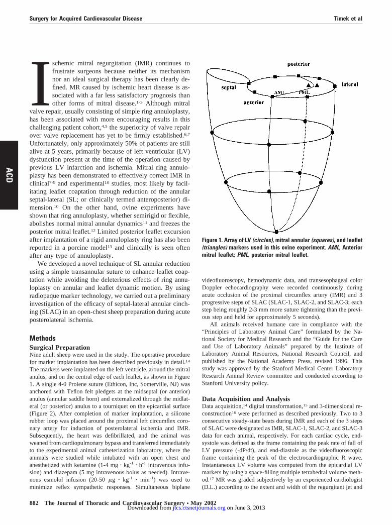
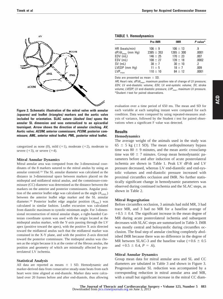
The markers were implanted on the left ventricle, around the mitralanulus, and on the central edge of each leaflet, as shown in Figure1. A single 4-0 Prolene suture (Ethicon, Inc, Somerville, NJ) wasanchored with Teflon felt pledgets at the midseptal (or anterior)anulus (annular saddle horn) and externalized through the midlat-eral (or posterior) anulus to a tourniquet on the epicardial surface(Figure 2). After completion of marker implantation, a siliconerubber loop was placed around the proximal left circumflex coro-nary artery for induction of posterolateral ischemia and IMR.Subsequently, the heart was defibrillated, and the animal wasweaned from cardiopulmonary bypass and transferred immediatelyto the experimental animal catheterization laboratory, where theanimals were studied while intubated with an open chest andanesthetized with ketamine (1-4 mg � kg-1 � h-1 intravenous infu-sion) and diazepam (5 mg intravenous bolus as needed). Intrave-nous esmolol infusion (20-50 �g � kg-1 � min-1) was used tominimize reflex sympathetic responses. Simultaneous biplane
videofluoroscopy, hemodynamic data, and transesophageal colorDoppler echocardiography were recorded continuously duringacute occlusion of the proximal circumflex artery (IMR) and 3progressive steps of SLAC (SLAC-1, SLAC-2, and SLAC-3; eachstep being roughly 2-3 mm more suture tightening than the previ-ous step and held for approximately 5 seconds).
All animals received humane care in compliance with the“Principles of Laboratory Animal Care” formulated by the Na-tional Society for Medical Research and the “Guide for the Careand Use of Laboratory Animals” prepared by the Institute ofLaboratory Animal Resources, National Research Council, andpublished by the National Academy Press, revised 1996. Thisstudy was approved by the Stanford Medical Center LaboratoryResearch Animal Review committee and conducted according toStanford University policy.
Data Acquisition and AnalysisData acquisition,14 digital transformation,15 and 3-dimensional re-construction16 were performed as described previously. Two to 3consecutive steady-state beats during IMR and each of the 3 stepsof SLAC were designated as IMR, SLAC-1, SLAC-2, and SLAC-3data for each animal, respectively. For each cardiac cycle, end-systole was defined as the frame containing the peak rate of fall ofLV pressure (-dP/dt), and end-diastole as the videofluoroscopicframe containing the peak of the electrocardiographic R wave.Instantaneous LV volume was computed from the epicardial LVmarkers by using a space-filling multiple tetrahedral volume meth-od.17 MR was graded subjectively by an experienced cardiologist(D.L.) according to the extent and width of the regurgitant jet and
Figure 1. Array of LV (circles), mitral annular (squares), and leaflet(triangles) markers used in this ovine experiment. AML, Anteriormitral leaflet; PML, posterior mitral leaflet.
Surgery for Acquired Cardiovascular Disease Timek et al
882 The Journal of Thoracic and Cardiovascular Surgery ● May 2002
ACD
on June 3, 2013 jtcs.ctsnetjournals.orgDownloaded from
categorized as none (0), mild (�1), moderate (�2), moderate tosevere (�3), or severe (�4).
Mitral Annular DynamicsMitral annular area was computed from the 3-dimensional coor-dinates of the 8 markers sutured to the mitral anulus by using anannular centroid.10 The SL annular diameter was calculated as thedistance in 3-dimensional space between markers placed on themidseptal and midlateral mitral anulus, and the commissure-com-missure (CC) diameter was determined as the distance between themarkers on the anterior and posterior commissures. Angular posi-tion of the anterior leaflet edge was calculated as the angle (�AML)between the anterior leaflet edge marker and the SL annulardiameter.18 Posterior leaflet edge angular position (�PML) wascalculated in similar fashion. Leaflet excursion was calculatedfrom diastolic maximum to systolic minimum angle. For 3-dimen-sional reconstruction of mitral annular shape, a right-handed Car-tesian coordinate system was used with the origin located at themidseptal anulus marker, with the Y-axis passing through the LVapex (positive toward the apex), with the positive X axis directedtoward the midlateral anulus such that the midlateral marker wascontained in the X-Y plane, and with the positive Z-axis directedtoward the posterior commissure. The midseptal anulus was cho-sen as the origin because it is at the center of the fibrous anulus, theposition and geometry of which are minimally affected by pos-terolateral LV ischemia.
Statistical AnalysisAll data are reported as means � 1 SD. Hemodynamic andmarker-derived data from consecutive steady-state beats from eachheart were time aligned at end-diastole. Marker data were calcu-lated over 20 frames before and after end-diastole, thus allowing
evaluation over a time period of 650 ms. The mean and SD foreach variable at each sampling instant were computed for eachcondition. Data were compared by using repeated-measures anal-ysis of variance, followed by the Student t test for paired obser-vations when a significant F value was detected.
ResultsHemodynamicsThe average weight of the animals used in the study was65 � 5 kg (�1 SD). The mean cardiopulmonary bypasstime was 80 � 9 minutes, and the mean aortic crossclamptime was 60 � 7 minutes. Group mean hemodynamic pa-rameters before and after induction of acute posterolateralischemia are shown in Table 1. Peak LV dP/dt and LVpressure decreased, whereas LV end-diastolic and end-sys-tolic volumes and end-diastolic pressure increased withproximal circumflex occlusion and IMR. No further statis-tically significant change in hemodynamic parameters wasobserved during continued ischemia and the SLAC steps, asshown in Table 2.
Mitral RegurgitationBefore circumflex occlusion, 3 animals had mild MR, 3 hadtrace MR, and 3 had no MR for a baseline average of�0.5 � 0.4. The significant increase in the mean degree ofMR during acute posterolateral ischemia and subsequentdecreases with SLAC steps are summarized in Table 3. IMRwas mostly central and holosystolic during circumflex oc-clusion. The final step of annular cinching completely abol-ished IMR because there was no difference in the degree ofMR between SLAC-3 and the baseline value (�0.6 � 0.5and �0.5 � 0.4, P � .6).
Mitral Annular DynamicsGroup mean data for mitral annular area and SL and CCdiameters are tabulated in Table 3 and shown in Figure 3.Progressive annular SL reduction was accompanied by acorresponding reduction in mitral annular area and MR,with a small but significant increase in the mitral CC diam-
Figure 2. Schematic illustration of the mitral valve with annular(squares) and leaflet (triangles) markers and the aortic valveincluded for orientation. SLAC suture (dashed line) spans theannular SL dimension and was externalized to an epicardialtourniquet. Arrow shows the direction of annular cinching. AV,Aortic valve; ACOM, anterior commissure; PCOM, posterior com-missure; AML , anterior mitral leaflet; PML , posterior mitral leaflet.
TABLE 1. HemodynamicsPre-IMR IMR P value*
HR (beats/min) 106 � 9 106 � 13 .9dP/dtmax (mm Hg) 2385 � 353 1265 � 300 .0001EDV (mL) 146 � 25 170 � 23 .007ESV (mL) 108 � 27 139 � 18 .0002SV (mL) 38 � 7 30 � 10 .2LVEDP (mm Hg) 11 � 5 18 � 7 .009LVPmax 110 � 10 84 � 12 .0001
Data are presented as mean � SD.HR, Heart rate; dP/dtmax , maximum positive rate of change of LV pressure;EDV, LV end-diastolic volume; ESV, LV end-systolic volume; SV, strokevolume; LVEDP, LV end-diastolic pressure; LVPmax , maximum LV pressure.*Student t test for paired observations.
Timek et al Surgery for Acquired Cardiovascular Disease
The Journal of Thoracic and Cardiovascular Surgery ● Volume 123, Number 5 883
ACD
on June 3, 2013 jtcs.ctsnetjournals.orgDownloaded from
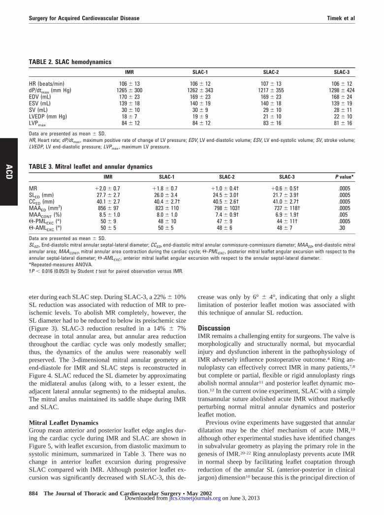
eter during each SLAC step. During SLAC-3, a 22% � 10%SL reduction was associated with reduction of MR to pre-ischemic levels. To abolish MR completely, however, theSL diameter had to be reduced to below its preischemic size(Figure 3). SLAC-3 reduction resulted in a 14% � 7%decrease in total annular area, but annular area reductionthroughout the cardiac cycle was only modestly smaller;thus, the dynamics of the anulus were reasonably wellpreserved. The 3-dimensional mitral annular geometry atend-diastole for IMR and SLAC steps is reconstructed inFigure 4. SLAC reduced the SL diameter by approximatingthe midlateral anulus (along with, to a lesser extent, theadjacent lateral annular segments) to the midseptal anulus.The mitral anulus maintained its saddle shape during IMRand SLAC.
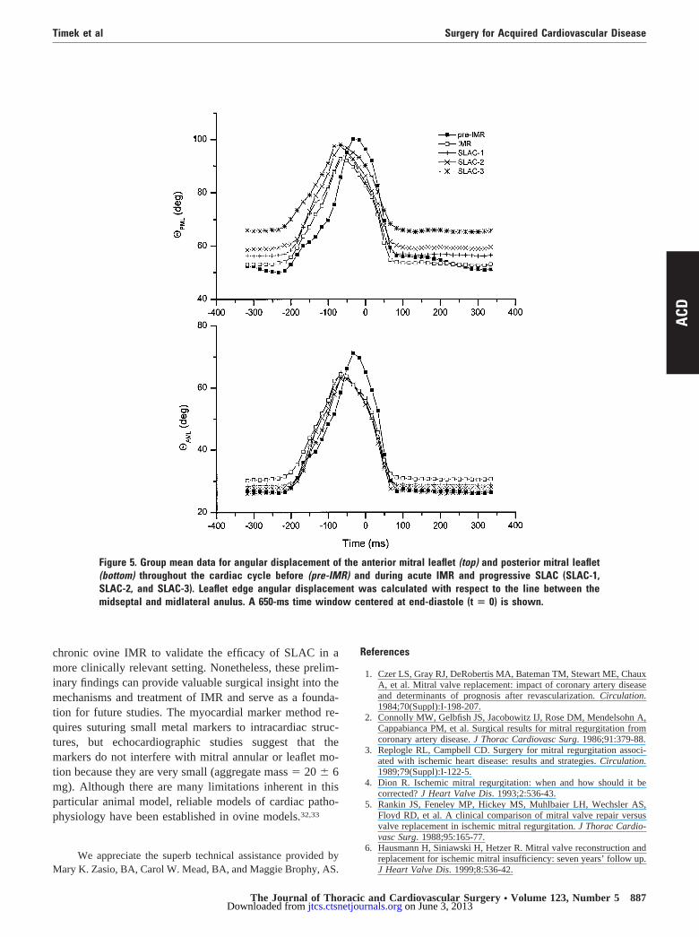
Mitral Leaflet DynamicsGroup mean anterior and posterior leaflet edge angles dur-ing the cardiac cycle during IMR and SLAC are shown inFigure 5, with leaflet excursion, from diastolic maximum tosystolic minimum, summarized in Table 3. There was nochange in anterior leaflet excursion during progressiveSLAC compared with IMR. Although posterior leaflet ex-cursion was significantly decreased with SLAC-3, this de-
crease was only by 6° � 4°, indicating that only a slightlimitation of posterior leaflet motion was associated withthis technique of annular SL reduction.
DiscussionIMR remains a challenging entity for surgeons. The valve ismorphologically and structurally normal, but myocardialinjury and dysfunction inherent in the pathophysiology ofIMR adversely influence postoperative outcome.4 Ring an-nuloplasty can effectively correct IMR in many patients,7,8
but complete or partial, flexible or rigid annuloplasty ringsabolish normal annular11 and posterior leaflet dynamic mo-tion.12 In the current ovine experiment, SLAC with a simpletransannular suture abolished acute IMR without markedlyperturbing normal mitral annular dynamics and posteriorleaflet motion.
Previous ovine experiments have suggested that annulardilatation may be the chief mechanism of acute IMR,19
although other experimental studies have identified changesin subvalvular geometry as playing the primary role in thegenesis of IMR.20-22 Ring annuloplasty prevents acute IMRin normal sheep by facilitating leaflet coaptation throughreduction of the annular SL (anterior-posterior in clinicaljargon) dimension10 because this is the principal direction of
TABLE 2. SLAC hemodynamicsIMR SLAC-1 SLAC-2 SLAC-3
HR (beats/min) 106 � 13 106 � 12 107 � 13 106 � 12dP/dtmax (mm Hg) 1265 � 300 1262 � 343 1217 � 355 1298 � 424EDV (mL) 170 � 23 169 � 23 169 � 23 168 � 24ESV (mL) 139 � 18 140 � 19 140 � 18 139 � 19SV (mL) 30 � 10 30 � 9 29 � 10 28 � 11LVEDP (mm Hg) 18 � 7 19 � 9 21 � 10 22 � 10LVPmax 84 � 12 84 � 12 83 � 16 81 � 16
Data are presented as mean � SD.HR, Heart rate; dP/dtmax , maximum positive rate of change of LV pressure; EDV, LV end-diastolic volume; ESV, LV end-systolic volume; SV, stroke volume;LVEDP, LV end-diastolic pressure; LVPmax , maximum LV pressure.
TABLE 3. Mitral leaflet and annular dynamicsIMR SLAC-1 SLAC-2 SLAC-3 P value*
MR �2.0 � 0.7 �1.8 � 0.7 �1.0 � 0.4† �0.6 � 0.5† .0005SLED (mm) 27.7 � 2.7 26.0 � 3.4 24.5 � 3.0† 21.7 � 3.9† .0005CCED (mm) 40.1 � 2.7 40.4 � 2.7† 40.5 � 2.6† 41.0 � 2.7† .0005MAAED (mm2) 856 � 97 823 � 110 798 � 103† 737 � 118† .0005MAACONT (%) 8.5 � 1.0 8.0 � 1.0 7.4 � 0.9† 6.9 � 1.9† .005�-PMLEXC (°) 50 � 9 48 � 10 47 � 9 44 � 11† .0005�-AMLEXC (°) 50 � 5 50 � 5 48 � 6 48 � 7 .30
Data are presented as mean � SD.SLED , End-diastolic mitral annular septal-lateral diameter; CCED, end-diastolic mitral annular commissure-commissure diameter; MAAED, end-diastolic mitralannular area; MAACONT, mitral annular area contraction during the cardiac cycle; �-PMLEXC , posterior mitral leaflet angular excursion with respect to theannular septal-lateral diameter; �-AMLEXC , anterior mitral leaflet angular excursion with respect to the annular septal-lateral diameter.*Repeated-measures ANOVA.†P � 0.016 (0.05/3) by Student t test for paired observation versus IMR.
Surgery for Acquired Cardiovascular Disease Timek et al
884 The Journal of Thoracic and Cardiovascular Surgery ● May 2002
ACD
on June 3, 2013 jtcs.ctsnetjournals.orgDownloaded from
annular enlargement during acute left circumflex arteryischemia.19,23,24 Annular enlargement caused by CC diam-eter increase, on the other hand, does not seem to lead toMR,25 and in the current experiment a slight increase in CCdiameter was actually observed with SLAC. Conversely,ring annuloplasty has also been shown to attenuate apicalleaflet tethering in acute ovine IMR,26 suggesting an influ-ence on the subvalvular apparatus. It is possible that SLACabolished IMR by altering subvalvular geometry, althoughannular SL reduction leading to improved leaflet coaptationis more likely the predominant mechanism. Central MRduring myocardial ischemia, as seen in the current study, ismore likely to be associated with annular dilatation,27 and itis therefore not surprising that reduction of the annular SLdimension would correct this type of IMR. Clinically, sur-
gical therapy designed to increase leaflet coaptation, such asimplantation of an undersized ring annuloplasty, is usuallyeffective in ameliorating MR in patients with advanceddilated cardiomyopathy, either idiopathic or ischemic.28
Furthermore, the extent of annular SL reduction may be adeterminant of operative success in patients with IMR un-dergoing valve repair.8 Any technique that reduces the sizeof the mitral anulus, however, also changes the 3-dimen-sional geometric relationships between the anulus and sub-valvular apparatus because these structures are tightly cou-pled.29 Perhaps restoration of this perturbed relationshippartially accounts for the efficacy of SLAC in this ovinemodel of acute IMR.
SLAC effectively abolished IMR, but this novel tech-nique only mildly altered normal mitral annular geometry
Figure 3. Group mean data for SL annular diameter (in millimeters, top), mitral annular area (in square millimeters,center), and CC annular diameter (in millimeters, bottom) throughout the cardiac cycle before (pre-IMR) and duringacute IMR and progressive SLAC (SLAC-1, SLAC-2, and SLAC-3). A 650-ms time interval centered at end-diastole(t � 0) is illustrated for all 4 groups.
Timek et al Surgery for Acquired Cardiovascular Disease
The Journal of Thoracic and Cardiovascular Surgery ● Volume 123, Number 5 885
ACD
on June 3, 2013 jtcs.ctsnetjournals.orgDownloaded from

and dynamic motion. The 6-mm annular SL reduction withSLAC-3 is comparable with the degree of annular reductionneeded to prevent IMR with either a flexible or semirigidannuloplasty ring,10 but SLAC-3 reduced end-diastolic an-nular area by only 14%, which is considerably less than the30% to 35% annular area reduction associated with annu-loplasty rings.11 Even though annular area was significantlysmaller with SLAC, the magnitude of this decrease wasmodest; perhaps it is smaller total annular size reductionthat permits continued dynamic motion of the anulus inSLAC. Annular flexibility serves a dual role by aiding LVfilling in diastole and by facilitating leaflet coaptation in latediastole-early systole by virtue of its sphincteric action.30
Therefore, preservation of annular flexibility may havephysiologic advantages, yet ring annuloplasty generallyminimizes dynamic area change.11 Although SLAC sub-stantially decreased annular SL diameter and mitral area, the3-dimensional saddle shape of the anulus remained intact,with elevation of the midseptal anulus (or saddle horn)above the annular plane. Recent finite-element analysis ofannular shape suggests that this saddle-shaped configurationmay have important implications for reducing systolic stresson the valve leaflets.31
Perturbed posterior leaflet motion has been observedafter implantation of an annuloplasty ring in animal mod-els12,13 and is frequently observed clinically on postopera-tive echocardiography. Indeed, annuloplasty rings, whetherflexible or semirigid, freeze the motion of the posterior
leaflet, effectively converting the mitral valve into a singleleaflet valve.12 Although SLAC inhibited posterior leafletexcursion modestly, the posterior leaflet remained mobile.Whether maintained posterior leaflet motion offers an ad-vantage in terms of effectiveness or durability of valverepair remains to be determined. This could possibly dis-tribute systolic closing stresses more favorably in a bileafletvalve, but further studies are needed to answer this question.
This experiment assessed a novel technique to reducemitral annular SL dimension to enhance leaflet coaptationand correct IMR in an ovine model of acute ischemia.Progressive SLAC decreased MR because the SL diameterwas cinched smaller, yet annular dynamics and posteriorleaflet motion were only modestly affected. SLAC poten-tially represents an expedient and simple surgical methodfor the treatment of IMR, either alone or as an adjunctivetechnique.
Although SLAC was effective in abolishing IMR in thisexperiment, this model of acute IMR is distinctly differentthan the clinical situation consisting of chronic MR and LVdilatation and systolic dysfunction, which makes clinicalextrapolation difficult. The above findings can only be in-terpreted in the setting of acute LV ischemia in a normalsheep heart under open-chest conditions. These observa-tions cannot be applied to patients with chronic IMR underclosed-chest conditions in which subvalvular geometric per-turbations may play a more predominant role in the patho-genesis of MR. We are currently exploring a protocol of
Figure 4. Left, Group mean 3-dimensional reconstruction of the mitral anulus at end-diastole during acute IMR andsubsequent progressive SLAC (SLAC-1, SLAC-2, and SLAC-3). Drop lines are shown for IMR and SLAC-3 data pointsin the apical-basal and SL planes as solid (IMR) and dashed (SLAC-3) lines. Right, Rotated view of eachreconstruction to the approximate level of the annular plane viewed from the lateral to septal anulus to illustratethe 3-dimensional shape of the anulus. ACOM, Anterior commissure; PCOM, posterior commissure.
Surgery for Acquired Cardiovascular Disease Timek et al
886 The Journal of Thoracic and Cardiovascular Surgery ● May 2002
ACD
on June 3, 2013 jtcs.ctsnetjournals.orgDownloaded from
chronic ovine IMR to validate the efficacy of SLAC in amore clinically relevant setting. Nonetheless, these prelim-inary findings can provide valuable surgical insight into themechanisms and treatment of IMR and serve as a founda-tion for future studies. The myocardial marker method re-quires suturing small metal markers to intracardiac struc-tures, but echocardiographic studies suggest that themarkers do not interfere with mitral annular or leaflet mo-tion because they are very small (aggregate mass � 20 � 6mg). Although there are many limitations inherent in thisparticular animal model, reliable models of cardiac patho-physiology have been established in ovine models.32,33
We appreciate the superb technical assistance provided byMary K. Zasio, BA, Carol W. Mead, BA, and Maggie Brophy, AS.
References
1. Czer LS, Gray RJ, DeRobertis MA, Bateman TM, Stewart ME, ChauxA, et al. Mitral valve replacement: impact of coronary artery diseaseand determinants of prognosis after revascularization. Circulation.1984;70(Suppl):I-198-207.
2. Connolly MW, Gelbfish JS, Jacobowitz IJ, Rose DM, Mendelsohn A,Cappabianca PM, et al. Surgical results for mitral regurgitation fromcoronary artery disease. J Thorac Cardiovasc Surg. 1986;91:379-88.
3. Replogle RL, Campbell CD. Surgery for mitral regurgitation associ-ated with ischemic heart disease: results and strategies. Circulation.1989;79(Suppl):I-122-5.
4. Dion R. Ischemic mitral regurgitation: when and how should it becorrected? J Heart Valve Dis. 1993;2:536-43.
5. Rankin JS, Feneley MP, Hickey MS, Muhlbaier LH, Wechsler AS,Floyd RD, et al. A clinical comparison of mitral valve repair versusvalve replacement in ischemic mitral regurgitation. J Thorac Cardio-vasc Surg. 1988;95:165-77.
6. Hausmann H, Siniawski H, Hetzer R. Mitral valve reconstruction andreplacement for ischemic mitral insufficiency: seven years’ follow up.J Heart Valve Dis. 1999;8:536-42.
Figure 5. Group mean data for angular displacement of the anterior mitral leaflet (top) and posterior mitral leaflet(bottom) throughout the cardiac cycle before (pre-IMR) and during acute IMR and progressive SLAC (SLAC-1,SLAC-2, and SLAC-3). Leaflet edge angular displacement was calculated with respect to the line between themidseptal and midlateral anulus. A 650-ms time window centered at end-diastole (t � 0) is shown.
Timek et al Surgery for Acquired Cardiovascular Disease
The Journal of Thoracic and Cardiovascular Surgery ● Volume 123, Number 5 887
ACD
on June 3, 2013 jtcs.ctsnetjournals.orgDownloaded from
7. Cohn LH, Rizzo RJ, Adams DH, Couper GS, Sullivan TE, Collins JJ,et al. The effect of pathophysiology on the surgical treatment ofischemic mitral regurgitation: operative and late risks of repair versusreplacement. Eur J Cardiothorac Surg. 1995;9:568-74.
8. Czer LS, Maurer G, Trento A, DeRobertis M, Nessim S, BlancheC, et al. Comparative efficacy of ring and suture annuloplasty forischemic mitral regurgitation. Circulation. 1992;86(Suppl):II-46-52.
9. Grossi EA, Lapietra A, Galloway AC, Ribakove GH, Culliford AT,Sposito RA, et al. Late results of isolated mitral annuloplasty forischemic mitral insufficiency [abstract]. Circulation. 2000;102(Suppl):II-491.
10. Timek T, Glasson JR, Dagum P, Green GR, Nistal JF, Komeda M, etal. Ring annuloplasty prevents delayed leaflet coaptation and mitralregurgitation during acute left ventricular ischemia. J Thorac Cardio-vasc Surg. 2000;119:774-83.
11. Glasson JR, Green GR, Nistal JF, Dagum P, Komeda M, DaughtersGT, et al. Mitral annular size and shape in sheep with annuloplastyrings. J Thorac Cardiovasc Surg. 1999;117:302-9.
12. Green GR, Dagum P, Glasson JR, Nistal JF, Daughters GT, Ingels NBJr, et al. Restricted posterior leaflet motion after mitral ring annulo-plasty. Ann Thorac Surg. 1999;68:2100-6.
13. van Rijk-Zwikker GL, Mast F, Schipperheyn JJ, Huysmans HA,Bruschke AV. Comparison of rigid and flexible rings for annuloplastyof the porcine mitral valve. Circulation. 1990;82(Suppl):IV-58-64.
14. Glasson JR, Komeda M, Daughters GT, Foppiano LE, Bolger AF, TyeTL, et al. Most ovine mitral annular 3-D size reduction occurs beforeventricular systole and is abolished with ventricular pacing. Circula-tion. 1997;96(Suppl):II-115-123.
15. Niczyporuk MA, Miller DC. Automatic tracking and digitization ofmultiple radiopaque myocardial markers. Comput Biomed Res. 1991;24:129-42.
16. Daughters GT, Sanders WJ, Miller DC, Schwarzkopf A, Mead CW,Ingels NBJ. A comparison of two analytical systems for 3-D recon-struction from biplane videoradiograms. IEEE Comput Cardiol. 1989;15:79-82.
17. Moon MR, DeAnda A, Daughters GT, Ingels NBJ, Miller DC. Ex-perimental evaluation of different chordal preservation methods dur-ing mitral valve replacement. Ann Thorac Surg. 1994;58:931-44.
18. Karlsson MO, Glasson JR, Bolger AF, Daughters GT, Komeda M,Foppiano LE, et al. Mitral valve opening in the ovine heart. Am JPhysiol. 1998;274:H552-63.
19. Glasson JR, Komeda M, Daughters GT, Bolger AF, Karlsson MO,Foppiano LE, et al. Early systolic mitral leaflet “loitering” duringacute ischemic mitral regurgitation. J Thorac Cardiovasc Surg. 1998;116:193-205.
20. Gorman JH 3rd, Jackson BM, Gorman RC, Kelley ST, Gikakis N,Edmunds LH, Jr. Papillary muscle discoordination rather than in-creased annular area facilitates mitral regurgitation after acute poste-rior myocardial infarction. Circulation. 1997;96(Suppl):II-124-7.
21. He S, Fontaine AA, Schwammenthal E, Yoganathan AP, Levine RA.Integrated mechanism for functional mitral regurgitation: leaflet re-striction versus coapting force: in vitro studies. Circulation. 1997;96:1826-34.
22. Otsuji Y, Nathan N, Handschumacher MD, Coulter S, Liel-Cohen N,D’Ambra M, et al. Mechanism of ischemic mitral regurgitation: in-traoperative evaluation of leaflet tethering geometry by three-dimen-sional geometry [abstract]. Circulation. 1997;96(Suppl):I-156.
23. Kay GL, Kay JH, Zubiate P, Yokoyama T, Mendez M. Mitral valverepair for mitral regurgitation secondary to coronary artery disease.Circulation. 1986;74(Suppl):I-88-98.
24. Gorman JH 3rd, Gorman RC, Jackson BM, Hiramatsu Y, Gikakis N,Kelley ST, et al. Distortions of the mitral valve in acute ischemicmitral regurgitation. Ann Thorac Surg. 1997;64:1026-31.
25. Green GR, Dagum P, Glasson JR, Daughters GT, Bolger AF, Foppi-ano LE, et al. Mitral annular dilatation and papillary muscle disloca-tion without mitral regurgitation in sheep. Circulation. 1999;100(Suppl):II-95-102.
26. Lai DTM, Timek T, Green GR, Glasson JR, Daughters GT, Liang D,et al. The effects of ring annuloplasty on mitral leaflet geometry duringacute left ventricular ischemia. J Thorac Cardiovasc Surg. 2000;120:966-75.
27. Izumi S, Miyatake K, Beppu S, Park YD, Nagata S, Kinoshita N, et al.Mechanism of mitral regurgitation in patients with myocardial infarc-tion: a study using real-time two-dimensional Doppler flow imagingand echocardiography. Circulation. 1987;76:777-85.
28. Bolling SF, Pagani FD, Deeb GM, Bach DS. Intermediate-term out-come of mitral reconstruction in cardiomyopathy. J Thorac Cardio-vasc Surg. 1998;115:381-8.
29. Dagum P, Timek T, Green GR, Lai D, Daughters GT, Liang D, et al.Coordinate-free analysis of mitral valve dynamics and ischemichearts. Circulation. 2000;102(Suppl):III-62-9.
30. Tsakiris AG, Von Bernuth G, Rastelli GC, Bourgeois MJ, Titus JL,Wood EH. Size and motion of the mitral valve annulus in anesthetizedintact dogs. J Appl Physiol. 1971;30:611-8.
31. Salgo IS GJ, Gorman RC, Jackson BM, Bowen F, Plappert TT, StJohn Sutton MG, et al. Structural implication of mitral annular geom-etry and the saddle shape: a finite element analysis [abstract]. Circu-lation. 2000;102(Suppl):II-631.
32. Markovitz SE, Ratcliffe MB, Bavaria JE, Kreiner G, Iozzo RV,Hargrove WC, et al. Large animal model of left ventricular aneurysm.Ann Thorac Surg. 1989;48:838-45.
33. Llaneras MR, Nance ML, Streicher JT, Lima JA, Savino JS, BogenDK, et al. Large animal model of ischemic mitral regurgitation. AnnThorac Surg. 1994;57:432-9.
DiscussionDr Irving L. Kron (Charlottesville, Va). Dr Timek, the mech-
anism you have created obviously is an acutely ischemic model,and this is a very interesting approach. I know your laboratoryknows more about IMR than probably anyone on earth. The typicalpatient, obviously, has some retraction of the posterior leaflets,some scarring, annular dilatation, and such. Would this techniqueor some adaptation of it work in those situations, do you think?
Dr Timek. Thank you for that question. That is a very goodpoint. This is an acute model in healthy, normal sheep, andtherefore it does not reflect the clinical situation, where chronicchanges and volume overload are present. However, this modelgives us some insight into the mechanisms of IMR. We arecurrently working on a model of chronic ovine IMR, and we willtry to investigate this method in that setting, which will be moreclinically pertinent.
Surgery for Acquired Cardiovascular Disease Timek et al
888 The Journal of Thoracic and Cardiovascular Surgery ● May 2002
ACD
on June 3, 2013 jtcs.ctsnetjournals.orgDownloaded from

2002;123:881-888 J Thorac Cardiovasc SurgDaughters, Paul Dagum, Neil B. Ingels, Jr and D. Craig Miller
Tomasz A. Timek, David T. Lai, Frederick Tibayan, David Liang, George T. Septal-lateral annular cinching abolishes acute ischemic mitral regurgitation
Continuing Medical Education Activities
http://cme.ctsnetjournals.org/cgi/hierarchy/ctsnetcme_node;JTCSSubscribers to the Journal can earn continuing medical education credits via the Web at
Subscription Information
http://jtcs.ctsnetjournals.org/cgi/content/full/123/5/881#BIBLThis article cites 29 articles, 17 of which you can access for free at:
Citations
http://jtcs.ctsnetjournals.org/cgi/content/full/123/5/881#otherarticlesThis article has been cited by 29 HighWire-hosted articles:
Subspecialty Collections
http://jtcs.ctsnetjournals.org/cgi/collection/valve_disease Valve disease
This article, along with others on similar topics, appears in the following collection(s):
Permissions and Licensing
http://www.elsevier.com/wps/find/obtainpermissionform.cws_home/obtainpermissionformreceipt, is available at: An on-line permission request form, which should be fulfilled within 10 working days of
. http://www.elsevier.com/wps/find/supportfaq.cws_home/permissionusematerialcan be found online at: General information about reproducing this article in parts (figures, tables) or in its entirety
on June 3, 2013 jtcs.ctsnetjournals.orgDownloaded from


![Medial septal [beta]-amyloid 1-40 injections alter septo-hippocampal anatomy and function](https://static.fdokumen.com/doc/165x107/6329e172e9556f820801538c/medial-septal-beta-amyloid-1-40-injections-alter-septo-hippocampal-anatomy-and.jpg)