
OralCard: A bioinformatic tool for the study of oral proteome
Upload
independentCategory
view
0download
0
RESEARCH ARTICLE Open Access
MicroRNAs and cardiac sarcoplasmic reticulumcalcium ATPase-2 in human myocardial infarction:expression and bioinformatic analysisEmanuela Boštjančič, Nina Zidar and Damjan Glavač*
Abstract
Background: Cardiac sarco(endo)plasmic reticulum calcium ATPase-2 (SERCA2) plays one of the central roles inmyocardial contractility. Both, SERCA2 mRNA and protein are reduced in myocardial infarction (MI), but thecorrelation has not been always observed. MicroRNAs (miRNAs) act by targeting 3'-UTR mRNA, causing translationalrepression in physiological and pathological conditions, including cardiovascular diseases. One of the aims of ourstudy was to identify miRNAs that could influence SERCA2 expression in human MI.
Results: The protein SERCA2 was decreased and 43 miRNAs were deregulated in infarcted myocardium comparedto corresponding remote myocardium, analyzed by western blot and microRNA microarrays, respectively. All thesamples were stored as FFPE tissue and in RNAlater. miRNAs binding prediction to SERCA2 including four predictionalgorithms (TargetScan, PicTar, miRanda and mirTarget2) identified 213 putative miRNAs. TAM and miRNApathannotation of deregulated miRNAs identified 18 functional and 21 diseased states related to heart diseases, andassociation of the half of the deregulated miRNAs to SERCA2. Free-energy of binding and flanking regions (RNA22,RNAfold) was calculated for 10 up-regulated miRNAs from microarray analysis (miR-122, miR-320a/b/c/d, miR-574-3p/-5p, miR-199a, miR-140, and miR-483), and nine miRNAs deregulated from microarray analysis were used forvalidation with qPCR (miR-21, miR-122, miR-126, miR-1, miR-133, miR-125a/b, and miR-98). Based on qPCR results, thecomparison between FFPE and RNAlater stored tissue samples, between Sybr Green and TaqMan approaches, aswell as between different reference genes were also performed.
Conclusion: Combing all the results, we identified certain miRNAs as potential regulators of SERCA2; however,further functional studies are needed for verification. Using qPCR, we confirmed deregulation of nine miRNAs inhuman MI, and show that qPCR normalization strategy is important for the outcome of miRNA expression analysisin human MI.
Keywords: Human myocardial infarction, Expression, SERCA2, miRNA, Bioinformatics
BackgroundA growing body of evidence has revealed that dysfunc-tion of 3'- and 5'-untranslated regions (UTRs) is linkedto the pathophysiology of variety of diseases. Both UTRsare important as regulatory elements of mRNAtranslation [1]. One of the negative regulators of mRNAtranslation are small non-coding RNAs, microRNAs(miRNAs) that bind to 3'-UTR of target mRNA. Bybinding to target mRNA, miRNAs have been found to
regulate a variety of physiological functions and contrib-ute to various diseases, including ischemic heart diseases[2]. It has been suggested that some parts of the con-served regions of the 3'-UTRs of SERCA2 might repre-sent post-transcription binding sites for miRNAs [3].SERCA2 is muscle-specific sarco(endo)plasmic reticulum
calcium ATPase-2, which promotes cardiac relaxation andcontractility. During every heartbeat, a pulse of calcium isreleased from the sarcoplasmic reticulum (SR) into thecytoplasm of cardiac muscle cells through a calciumrelease channel (RyR, ryanodine receptor), thus triggeringmuscle contraction. During relaxation, SERCA2 re-sequesters the calcium back into the internal sarcoplasmic
* Correspondence: [email protected] of Molecular Genetics, Institute of Pathology, Korytkova 2,Faculty of Medicine, Ljubljana, Slovenia
© 2012 Boštjančič et al.; licensee BioMed Central Ltd. This is an Open Access article distributed under the terms of the CreativeCommons Attribution License (http://creativecommons.org/licenses/by/2.0), which permits unrestricted use, distribution, andreproduction in any medium, provided the original work is properly cited.
Boštjančič et al. BMC Genomics 2012, 13:552http://www.biomedcentral.com/1471-2164/13/552

reticulum storage pool, thus priming the next releaseof calcium [4]. SERCA2 pre-mRNA is edited dependingon the cellular type. Due to different alternative splicing,the last exons of SERCA2 pre-mRNA produce differentmRNAs, which differ at the 3´-UTR regions and encodefor SERCA2a, SERCA2b and SERCA2c isoforms. In theheart, SERCA is mainly represented by the SERCA2a iso-form, which is expressed in the heart and slow-twitchskeletal muscle [3,5]. SERCA2b, ubiquitously expressed inall tissues, may at least partially replace SERCA2a func-tion [6].Decreased expression of the SERCA2 is one of the key
features of cardiac myocyte dysfunction in both experi-mental and human heart failure as well as in myocardialinfarction MI [3,4,6,7]. It is believed that this decreasecontributes to abnormal contractility in post-infarctionrat myocytes and that increased expression of SERCA2could improve myocardial contractility in cardiovasculardiseases [8,9]. It has been shown that the main regula-tors of SERCA2 protein and cardiac contractility arephospholamban (PLN) in the ventricles and sarcolipin(SLN) in the atria [3]. A simultaneous decrease inSERCA2 mRNA and protein levels is not alwaysobserved [7], and there remain gaps in understandingregulation of SERCA2 by PLN and SLN in the heart [4].Since functionally critical genes that are spatially
expressed are stringently regulated by miRNAs [10], wasthe aim of our study to test for miRNA and SERCA2 ex-pression in infarcted myocardium compared to corre-sponding remote myocardium of patients with MI, anddefined by target prediction algorithms those miRNAswith a potential influence on SERCA2 mRNA 3'-UTR.Differentially expressed miRNAs were further investi-gated using bioinformatic approaches as possible regula-tors of SERCA2 protein regulators (PLN, SLN). We alsocompared the miR-26b and RNU6B as reference genes(RGs), both of which were used as endogenous controlsin our previous study [11]. Two different approacheswere used (TaqMan and Sybr Green), as well as two dif-ferent types of tissues (RNAlater, FFPE).
ResultsWestern blotIn 4 out of 6 tested samples of infarcted tissues, itwas possible to extract enough proteins in a definedvolume to get comparable result using western blot(20 μg). In all four tested tissue samples we observeddecreased SERCA2 protein expression in theinfarcted tissue (I) when compared to correspondingremote (R) myocardium. The GAPDH protein, usedas endogenous control, showed no change in the ex-pression between infarcted tissue and correspondingremote myocardium. The results are summarized inFigure 1.
MicroRNA microarray resultsMicroarray data are deposited to the Gene ExpressionOmnibus public depository (GSE17304). MicroRNAmicroarray expression analysis revealed 43 differentiallyexpressed miRNAs in infarcted tissue when compared tocorresponding remote myocardium. Ten of the 43 differ-entially expressed miRNAs showed up-regulation. Theresults are summarized in Table 1. Unsupervised hier-archical clustering was performed using all miRNAsbeing differentially expressed in microarray analysis. Theresults are summarized in Figure 2. Since SERCA2 pro-tein showed down-regulation in infarcted tissue com-pared to corresponding remote myocardium, theresulting 10 miRNAs, which showed up-regulation, weresubject to further bioinformatics analysis.We further compared our results to our previous
study based on microarray analysis [11], where themiRNA expression in infarcted tissue was calculatedrelative to the miRNA expression in healthy humanhearts (independent groups of samples). Of 43 differen-tially expressed miRNAs in infarcted tissue compared toremote myocardium, 20 miRNAs showed similar expres-sion also when infarcted tissue was compared to healthyhuman hearts, 4 were specifically differentially expressedin the present study, and 19 showed higher difference inexpression in the present study. The results are summar-ized in Table 1.
Bioinformatic analysis of miRNAsmiRNA predictionUsing four different available prediction algorithms,namely TargetScan [12], PicTar [13], mirTarget2included in miRDB [14], and miRanda [15] included inMicroCosm Targets (former miRBase) [16] and inmicrorna.org [17], 213 miRNAs were predicted forbinding to 3'-UTR mRNA SERCA2. However, targetprediction programs did not distinguish between iso-forms SERCA2a and SERCA2b. Of 213 predicted
Figure 1 Western blot results of SERCA2 expression in theinfarcted tissue and corresponding remote myocardium.SERCA2 (~100 kDa) is under-expressed in the infarcted tissue whencompared to corresponding remote myocardium. GAPDH (~37 kDa),used as internal control, showed similar expression in infarcted andremote myocardium of patients with myocardial infarction. Legend:I, infarcted tissue, R, corresponding remote myocardium.
Boštjančič et al. BMC Genomics 2012, 13:552 Page 2 of 14http://www.biomedcentral.com/1471-2164/13/552

Table 1 Differentially expressed miRNAs in infarcted tissue compared to corresponding remote myocardium, theirclassification (from miRBase) and prediction of targeting SERCA2 (PicTar, TargetScan, miRanda, mirTarget2)
miRNA averagelog2
average log2* predicted miRNA Genefamily
Clustered(chr: miR; location)
let-7b 0.04 −0.32 Yes let-7 22: let-7a-3, miR-4763; intronic
let-7c −0.87 −0.88 Yes let-7 21: miR-99a; intronic
let-7d −1.34 −1.33 Yes let-7 9: let-7a-1, let-7f-1; intronic
let-7g −1.50 −1.67 Yes let-7 3: as single gene; intronic
miR-1 −3.28 −1.89 No miR-1 18: miR-133a-1; intronic
20: miR-133a-2; intronic
miR-122 3.28 ND No miR-122 18: miR-3591; intergenic
miR-125a-5p −1.53 −1.46 No miR-125 19: let-7e, miR-99b; intronic
miR-125b −0.44 −0.47 No miR-125 11: as single gene; intronic
miR-126 −2.00 −0.50 No miR-126 9: as single gene; intronic
miR-133a −1.52 ND No miR-133 18: miR-1-2; intronic
20: miR-1-1; intronic
miR-133b −1.53 ND No miR-133 6: miR-206; intronic
miR-140-3p 0.32 ND No miR-140 16: as single gene; intronic
miR-143 −0.26 ND No miR-143 5: miR-145; intergenic
miR-145 −0.79 ND No miR-145 5: miR-143; intergenic
miR-150 −1.63 −1.60 No miR-150 19: as single gene; intergenic
miR-16 −0.03 ND No miR-15 13: miR-15a; intronic
3: miR-15a; intronic
miR-195 −0.51 −1.37 Yes miR-15 17: miR-497; intronic
miR-197 −0.57 ND No miR-197 1: as single gene; intergenic
miR-199a-3p 0.39 ND No miR-199 19: as single gene; intronic
1: miR-199a-2, miR-3120, miR-214; intronic
miR-19b −2.36 ND Yes miR-19 13: miR-17, miR-18a, miR-19a,miR-20a, miR-92a-1; intergenic
X: miR-106a, miR-18b, miR-20b,miR-92a-2, miR-363; intergenic
miR-21 −0.05 ND No miR-21 17: as single gene; intergenic
miR-23a −1.71 −0.20 No miR-23 19: miR-27a, miR-24-2; intergenic
miR-26a −1.50 −0.54 No miR-26 3: as single gene; intronic
12: as single gene; intronic
miR-27a −1.97 −1.60 No miR-27 19: miR-23a, miR-24-2; intergenic
miR-27b −2.18 −1.19 No miR-27 9: miR-23b, miR-24-1, miR-3074; intronic
miR-29a −1.40 ND Yes miR-29 7: miR-29b-1; intergenic
miR-29c −2.28 ND No miR-29 1: miR-29b-2; intergenic
miR-30a −2.23 −1.25 Yes miR-30 6: as single gene; intronic
miR-30b −3.03 −2.01 Yes miR-30 8: miR-30d; intergenic
miR-30c −2.70 −2.08 Yes miR-30 1: miR-30e; intronic
6: as single gene; intronic
miR-30d −1.31 ND Yes miR-30 8: miR-30b; intergenic
miR-30e −2.71 ND Yes miR-30 1: miR-30c-1; intronic
miR-320a 2.37 0.98 No miR-320 8: as single gene; intergenic
miR-320b 2.24 0.98 No miR-320 1: two copies as single gene; one intronic
and one intergenic
Boštjančič et al. BMC Genomics 2012, 13:552 Page 3 of 14http://www.biomedcentral.com/1471-2164/13/552

Table 1 Differentially expressed miRNAs in infarcted tissue compared to corresponding remote myocardium, theirclassification (from miRBase) and prediction of targeting SERCA2 (PicTar, TargetScan, miRanda, mirTarget2) (Continued)
miR-320c 2.27 0.98 No miR-320 18: two copies as single gene; intronic
miR-320d 2.13 0.98 No miR-320 13: as single gene; intergenic
X: as single gene; intergenic
miR-378 −1.66 ND No miR-378 5. as single gene; intronic
miR-483-5p 2.92 ND No miR-483 11: as single gene; intronic
miR-499-5p −2.94 −3.36 Yes miR-499 20: miR-499a; intronic
miR-574-3p 2.25 2.46 Yes miR-574 4: as single gene; intronic
miR-574-5p 2.17 3.18 No miR-574
miR-98 −1.76 −3.10 Yes let-7 X: let-7f; intronic
Legend: *, results from previous microarray study [11], where infarcted tissue was compared to healthy adult hearts (independent groups of samples); ND, notdeterminate; chr, chromosome.
Figure 2 Hierarchical clustering analysis of the log2 value of differentially expressed miRNAs in myocardial infarction. Dual colourexperiments and swap-dye were performed, using Cy3 and Cy5 to compare miRNAs expression in infarcted tissue and corresponding remotemyocardium. Statistical analysis and hierarchical clustering was performed using Acuity 4.0 analytical software. The results are displayed in a heatmap. The legend on the right indicates the miRNA shown in the corresponding row, all of which are human specific. The bar code on thebottom shows the colour scale of the log2 value. Each column represents data from one microarray. The heat map was constructed of 43miRNAs dysregulated in all of the microarrays performed.
Boštjančič et al. BMC Genomics 2012, 13:552 Page 4 of 14http://www.biomedcentral.com/1471-2164/13/552

miRNAs, 136 corresponded to an isoform SERCA2b and74 to an isoform SERCA2a. Significantly, TargetScan5.1gave no results for isoform SERCA2b, and mirTarget2predicted only one miRNAs to target SERCA2b. In thecase of SERCA2a, all of the used algorithms predictedmiRNAs (Additional file 1: Table S1). Of all 213 miRNAspredicted to target SERCA2, 15 were differentiallyexpressed in human MI (Table 1).
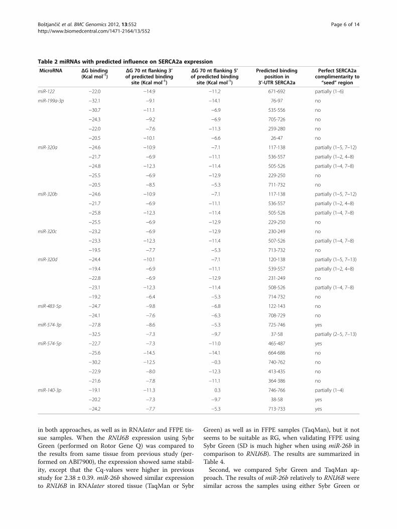
Free-energy analysisIt has been recently demonstrated with the majorityof miRNA-validated targets that miRNAs preferentiallybind to 3'-UTR sites that do not have complex sec-ondary structures and are located in accessible regionsof the mRNA based on favourable thermodynamics.Ten miRNAs were used for further in silico validation:one predicted by using above programs with elevatedexpression in microarray analysis (hsa-miR-574-3p;miRBase accession number: MIMAT0003239) andnine up-regulated in microarray analysis but not pre-dicted by the algorithms used (hsa-miR-122, hsa-miR-199a-3p, hsa-miR-140-3p, hsa-miR-320a, hsa-miR-320b, hsa-miR-320c, hsa-miR-320d, hsa-miR-483-5p,hsa-miR-574-5p; miRBase accession numbers:MIMAT0000421, MIMAT0000232, MIMAT0004597,MIMAT0000510, MIMAT0005792, MIMAT0005793,MIMAT0006764, MIMAT0004761, MIMAT0004795,respectively). Although is believed that SERCA2a isthe major isoform in the heart, our further analysisfocused on both isoform SERCA2a and SERCA2b,since primary antibody used in our western blot ana-lysis did not distinguish between isoform SERCA2aand SERCA2b. All results for SERCA2a and SER-CA2b are summarized in Table 2 and Table 3,respectively.Using criteria postulated by Zhao et al. (2005) [18], we
predicted binding sites in 3´-UTR of SERCA2b and SER-CA2a mRNA for up-regulated miRNAs. Using RNA22algorithm [19], some miRNAs were predicted to haveover 10 potential binding sites either in SERCA2a orSERCA2b. In case of SERCA2b, 13 binding sites werepredicted for hsa-miR-320a/b, 12 for hsa-miR-320c/d, 15for hsa-miR-574-5p and 20 for hsa-miR-122. In case ofSERCA2a, 16 binding sites were predicted for hsa-miR-574-5p. We therefore presented those with the highestdifference (at least 10 kcal/mol) between the potentialbinding site and the 70 nt flanking 3' and 5' of predictedbinding site based on calculation using RNAfold [20].Large proportion of binding sites is located in certainpositions in 3'-UTRs of corresponding SERCA2a andSERCA2b. In SERCA2a, there are 7 “hot-spot” locationsfor miRNA binding and in SERCA2b there are 9 “hot-spot” locations. For most of predicted binding sites thereis more than one predicted miRNA (cooperativity), and
this is further attenuated by more than one binding sitefor one miRNA (multiplicity). Most of predicted miR-NAs have at least one binding site with perfect compli-mentarity to seed region.
miRNA annotation and functional classificationAnalysis using miRBase revealed that differentiallyexpressed miRNAs belongs to 25 miRNA Gene family,and at least one gene copy of 25 differentially expressedmiRNAs is clustered. Further, the 4 clusters are fullyrepresented among differentially expressed miRNAs.The results are summarized in Table 1.TAM tool [21] was used to annotate differentially
expressed miRNAs in the mean of function and diseaseassociations. Among 35 defined functions for differen-tially expressed miRNAs, 18 are related to heart diseases(Additional file 2: Table S2). Further, differentiallyexpressed miRNAs are related to 136 different diseaseoutcomes, 21 are related to heart diseases (Additionalfile 3: Table S3).Finally, miRNApath [22] revealed that calcium signal-
ling pathway includes 175 genes, and 468 miRNAs regu-lating these genes. Of these, 95 miRNAs are associatedwith SERCA2, 19 miRNAs with PLN, and 21 miRNAswith SLN. Of differentially expressed miRNAs in ourstudy, 18 are according to miRNApath related toSERCA2, none is related to PLN, and three are relatedto SLN. In summary, only miR-30a/e and miR-145 aredifferentially expressed and related to SERCA2 as well asto its regulator SLN (data not shown).
Quantitative real-time PCRUsing two different qPCR technologies, we validated theexpression of nine miRNAs. Sybr Green technology wasused to validate: miR-122, which was specificallyexpressed on present microarrays compared to our pre-vious study [11] and it showed the highest up-regulation; miR-21 and miR-126, which are in additionto muscle-specific miR-1 and miR-133 the most com-mon miRNAs involved in heart diseases; miR-125a/b,which are according to TAM tool involved in myocardialremodelling after MI. TaqMan based approach wasused to validate miR-98, which was one of the fewmiRNAs overlapping target prediction and is accordingto TAM tool involved in hypertrophy; and miR-1 andmiR-133a/b, muscle-specific miRNAs.In addition to microarray validation, the qPCR meth-
odology was used to test several presumptions. First,miR-26b was tested as RG in comparison to RNU6B.Both were used as RGs in our previous study [11], usingmiR-26b as RG in TaqMan based approach and RNU6Bas RG in Sybr Green approach. In present study, bothwere used in TaqMan as well as in Sybr Green technol-ogy. The expression of RNU6B showed relative stability
Boštjančič et al. BMC Genomics 2012, 13:552 Page 5 of 14http://www.biomedcentral.com/1471-2164/13/552
in both approaches, as well as in RNAlater and FFPE tis-sue samples. When the RNU6B expression using SybrGreen (performed on Rotor Gene Q) was compared tothe results from same tissue from previous study (per-formed on ABI7900), the expression showed same stabil-ity, except that the Cq-values were higher in previousstudy for 2.38 ± 0.39. miR-26b showed similar expressionto RNU6B in RNAlater stored tissue (TaqMan or Sybr
Green) as well as in FFPE samples (TaqMan), but it notseems to be suitable as RG, when validating FFPE usingSybr Green (SD is much higher when using miR-26b incomparison to RNU6B). The results are summarized inTable 4.Second, we compared Sybr Green and TaqMan ap-
proach. The results of miR-26b relatively to RNU6B weresimilar across the samples using either Sybr Green or
Table 2 miRNAs with predicted influence on SERCA2a expression
MicroRNA ΔG binding(Kcal mol-1)
ΔG 70 nt flanking 3’of predicted binding
site (Kcal mol-1)
ΔG 70 nt flanking 5’of predicted binding
site (Kcal mol-1)
Predicted bindingposition in
3’-UTR SERCA2a
Perfect SERCA2acomplimentarity to
“seed” region
miR-122 −22.0 −14.9 −11.2 671-692 partially (1–6)
miR-199a-3p −32.1 −9.1 −14.1 76-97 no
−30.7 −11.1 −6.9 535-556 no
−24.3 −9.2 −6.9 705-726 no
−22.0 −7.6 −11.3 259-280 no
−20.5 −10.1 −6.6 26-47 no
miR-320a −24.6 −10.9 −7.1 117-138 partially (1–5, 7–12)
−21.7 −6.9 −11.1 536-557 partially (1–2, 4–8)
−24.8 −12.3 −11.4 505-526 partially (1–4, 7–8)
−25.5 −6.9 −12.9 229-250 no
−20.5 −8.5 −5.3 711-732 no
miR-320b −24.6 −10.9 −7.1 117-138 partially (1–5, 7–12)
−21.7 −6.9 −11.1 536-557 partially (1–2, 4–8)
−25.8 −12.3 −11.4 505-526 partially (1–4, 7–8)
−25.5 −6.9 −12.9 229-250 no
miR-320c −23.2 −6.9 −12.9 230-249 no
−23.3 −12.3 −11.4 507-526 partially (1–4, 7–8)
−19.5 −7.7 −5.3 713-732 no
miR-320d −24.4 −10.1 −7.1 120-138 partially (1–5, 7–13)
−19.4 −6.9 −11.1 539-557 partially (1–2, 4–8)
−22.8 −6.9 −12.9 231-249 no
−23.1 −12.3 −11.4 508-526 partially (1–4, 7–8)
−19.2 −6.4 −5.3 714-732 no
miR-483-5p −24.7 −9.8 −6.8 122-143 no
−24.1 −7.6 −6.3 708-729 no
miR-574-3p −27.8 −8.6 −5.3 725-746 yes
−32.5 −7.3 −9.7 37-58 partially (2–5, 7–13)
miR-574-5p −22.7 −7.3 −11.0 465-487 yes
−25.6 −14.5 −14.1 664-686 no
−30.2 −12.5 −0.3 740-762 no
−22.9 −8.0 −12.3 413-435 no
−21.6 −7.8 −11.1 364-386 no
miR-140-3p −19.1 −11.3 0.3 746-766 partially (1–4)
−20.2 −7.3 −9.7 38-58 yes
−24.2 −7.7 −5.3 713-733 yes
Boštjančič et al. BMC Genomics 2012, 13:552 Page 6 of 14http://www.biomedcentral.com/1471-2164/13/552

TaqMan based approach, either FFPE or RNAlaterstored tissue samples (data not shown).Third, the comparison between RNAlater and FFPE
tissue samples has been performed. The results are
summarized in Table 5. Using both technologies (Taq-Man and Sybr Green), we confirmed most of the micro-array results using RNU6B as RG, as well as using miR-26b (only in case of TaqMan approach). Both was true
Table 3 miRNAs with predicted influence on SERCA2b expression
MicroRNA ΔG binding(Kcal mol-1)
ΔG 70 nt flanking 3’of predicted binding
site (Kcal mol-1)
ΔG 70 nt flanking 5’of predicted binding
site (Kcal mol-1)
Predicted bindingposition in
3’-UTR SERCA2b
Perfect SERCA2bcomplimentarityto “seed” region
miR-122 −24.9 −9.9 −8.7 313-334 no
−23.7 −3.0 −8.2 788-809 no
−21.5 −0.2 −9.5 496-517 no
−19.3 −8.2 −7.5 253-274 no
−19.3 −3.5 −5.2 476-497 no
−19.3 −6.3 −9.0 458-479 no
−18.1 −4.3 −8.3 151-172 no
miR-199a-3p −22.0 −4.9 −9.5 380-401 yes
−21.0 −3.1 −11.1 487-508 yes
−18.6 −8.3 −3.9 59-80 yes
−18.1 −7.5 −8.3 251-272 no
miR-320a −21.2 −9.9 −9.3 405-426 no
−21.8 −9.0 0.3 798-819 no
−21.2 −4.7 −9.8 65-86 partially (1–4)
−23.7 −8.2 −8.2 687-708 no
−21.8 −9.1 −11.9 123-144 partially (1–6)
miR-320b −21.2 −9.9 −9.3 405-426 no
−21.8 −9.0 0.3 798-819 no
−21.2 −4.7 −9.8 65-86 partially (1–4)
−23.7 −8.2 −8.2 687-708 no
−21.8 −9.1 −11.9 123-144 partially (1–6)
miR-320c −19.1 −8.0 0.3 800-819 no
−24.0 −12.4 −8.2 689-708 no
−21.1 −11.4 −9.2 650-669 no
miR-320d −23.4 −13.2 −8.2 690-708 no
−24.3 −12.2 −9.0 709-727 partially (1–4, 6–7)
miR-483-5p −23.8 −13.1 −9.0 705-726 no
−21.8 −4.7 −8.5 63-84 no
−22.7 −9.1 −9.3 121-142 no
−20.3 −8.2 −3.0 789-810 Partially (1, 3–6, 8)
miR-574-3p −22.9 −8.2 −6.5 347-368 No
−21.2 −2.7 −11.3 96-117 no
miR-574-5p −21.7 −8.6 −8.0 263-285 No
−23.4 −4.9 −3.5 479-501 No
−18.2 −3.3 −3.5 12-34 No
−20.5 −9.0 0.3 797-819 Partially (1–5, 7)
miR-140-3p −19.9 −8.2 −6.5 348-368 yes
−20.5 −9.9 −3.5 484-504 No
−19.7 −8.7 0.3 799-819 no
Boštjančič et al. BMC Genomics 2012, 13:552 Page 7 of 14http://www.biomedcentral.com/1471-2164/13/552
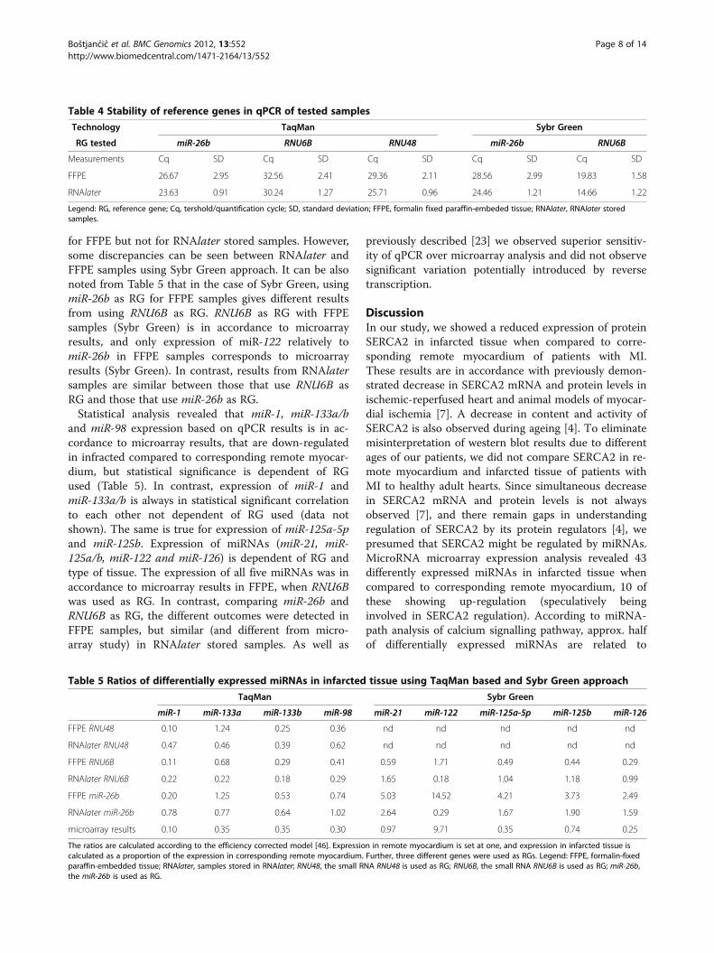
for FFPE but not for RNAlater stored samples. However,some discrepancies can be seen between RNAlater andFFPE samples using Sybr Green approach. It can be alsonoted from Table 5 that in the case of Sybr Green, usingmiR-26b as RG for FFPE samples gives different resultsfrom using RNU6B as RG. RNU6B as RG with FFPEsamples (Sybr Green) is in accordance to microarrayresults, and only expression of miR-122 relatively tomiR-26b in FFPE samples corresponds to microarrayresults (Sybr Green). In contrast, results from RNAlatersamples are similar between those that use RNU6B asRG and those that use miR-26b as RG.Statistical analysis revealed that miR-1, miR-133a/b
and miR-98 expression based on qPCR results is in ac-cordance to microarray results, that are down-regulatedin infracted compared to corresponding remote myocar-dium, but statistical significance is dependent of RGused (Table 5). In contrast, expression of miR-1 andmiR-133a/b is always in statistical significant correlationto each other not dependent of RG used (data notshown). The same is true for expression of miR-125a-5pand miR-125b. Expression of miRNAs (miR-21, miR-125a/b, miR-122 and miR-126) is dependent of RG andtype of tissue. The expression of all five miRNAs was inaccordance to microarray results in FFPE, when RNU6Bwas used as RG. In contrast, comparing miR-26b andRNU6B as RG, the different outcomes were detected inFFPE samples, but similar (and different from micro-array study) in RNAlater stored samples. As well as
previously described [23] we observed superior sensitiv-ity of qPCR over microarray analysis and did not observesignificant variation potentially introduced by reversetranscription.
DiscussionIn our study, we showed a reduced expression of proteinSERCA2 in infarcted tissue when compared to corre-sponding remote myocardium of patients with MI.These results are in accordance with previously demon-strated decrease in SERCA2 mRNA and protein levels inischemic-reperfused heart and animal models of myocar-dial ischemia [7]. A decrease in content and activity ofSERCA2 is also observed during ageing [4]. To eliminatemisinterpretation of western blot results due to differentages of our patients, we did not compare SERCA2 in re-mote myocardium and infarcted tissue of patients withMI to healthy adult hearts. Since simultaneous decreasein SERCA2 mRNA and protein levels is not alwaysobserved [7], and there remain gaps in understandingregulation of SERCA2 by its protein regulators [4], wepresumed that SERCA2 might be regulated by miRNAs.MicroRNA microarray expression analysis revealed 43differently expressed miRNAs in infarcted tissue whencompared to corresponding remote myocardium, 10 ofthese showing up-regulation (speculatively beinginvolved in SERCA2 regulation). According to miRNA-path analysis of calcium signalling pathway, approx. halfof differentially expressed miRNAs are related to
Table 4 Stability of reference genes in qPCR of tested samples
Technology TaqMan Sybr Green
RG tested miR-26b RNU6B RNU48 miR-26b RNU6B
Measurements Cq SD Cq SD Cq SD Cq SD Cq SD
FFPE 26.67 2.95 32.56 2.41 29.36 2.11 28.56 2.99 19.83 1.58
RNAlater 23.63 0.91 30.24 1.27 25.71 0.96 24.46 1.21 14.66 1.22
Legend: RG, reference gene; Cq, tershold/quantification cycle; SD, standard deviation; FFPE, formalin fixed paraffin-embeded tissue; RNAlater, RNAlater storedsamples.
Table 5 Ratios of differentially expressed miRNAs in infarcted tissue using TaqMan based and Sybr Green approach
TaqMan Sybr Green
miR-1 miR-133a miR-133b miR-98 miR-21 miR-122 miR-125a-5p miR-125b miR-126
FFPE RNU48 0.10 1.24 0.25 0.36 nd nd nd nd nd
RNAlater RNU48 0.47 0.46 0.39 0.62 nd nd nd nd nd
FFPE RNU6B 0.11 0.68 0.29 0.41 0.59 1.71 0.49 0.44 0.29
RNAlater RNU6B 0.22 0.22 0.18 0.29 1.65 0.18 1.04 1.18 0.99
FFPE miR-26b 0.20 1.25 0.53 0.74 5.03 14.52 4.21 3.73 2.49
RNAlater miR-26b 0.78 0.77 0.64 1.02 2.64 0.29 1.67 1.90 1.59
microarray results 0.10 0.35 0.35 0.30 0.97 9.71 0.35 0.74 0.25
The ratios are calculated according to the efficiency corrected model [46]. Expression in remote myocardium is set at one, and expression in infarcted tissue iscalculated as a proportion of the expression in corresponding remote myocardium. Further, three different genes were used as RGs. Legend: FFPE, formalin-fixedparaffin-embedded tissue; RNAlater, samples stored in RNAlater; RNU48, the small RNA RNU48 is used as RG; RNU6B, the small RNA RNU6B is used as RG; miR-26b,the miR-26b is used as RG.
Boštjančič et al. BMC Genomics 2012, 13:552 Page 8 of 14http://www.biomedcentral.com/1471-2164/13/552

SERCA2 but not to its regulatory proteins. Compared toour previous study based on microarray analysis [11],where the miRNA expression in infracted tissue was cal-culated relative to the miRNA expression in healthyhuman hearts (independent groups of samples), only halfof miRNAs showed similar expression in the presentstudy, suggesting specifically expressed miRNAs ininfracted tissue as well as in remote myocardium com-pared to healthy adult hearts. This is in accordance torecent findings that some miRNAs in the non-infarctedarea might also participate in the pathophysiology re-sponse to MI [24].SERCA2 has two major isoforms, SERCA2a and SER-
CA2b, both of which are expressed in heart from earlydevelopmental stages, although it is believed that SER-CA2a is the major isoform expressed in the heart [5].Using four different available prediction algorithms, 213miRNAs were predicted for binding to 3'-UTR mRNASERCA2; however, used algorithms did not distinguishbetween SERCA2 isoforms. Of predicted miRNAs, twotimes more corresponded to an isoform SERCA2b thanto an isoform SERCA2a, and only approx. 10% were dif-ferentially expressed in infarcted tissue compared to re-mote myocardium. Finally, only one of predicted anddifferentially expressed was up-regulated (miR-574-3p).miR-574-3p was also predicted to target ATP2A2 in
our previous study, and has been elevated in infarctedcompared to healthy adult hearts [11]. We thereforeused this and other up-regulated miRNAs to search forpotential binding using algorithm RNA22. The inter-action of miRNA:mRNA usually occurs via non-strictbase pairing, and the most important should be match-ing to the 1–8 nucleotides from the 5´ end of miRNA(“seed” region). miRNA-mRNA binding may also occurin other ways [25]. Some features of miRNA:mRNAbinding have been implicated in bioinformatics and ex-perimental approaches, but most of the target sites iden-tified by available tools are based on seed regioncomplimentarity, therefore might be biased against theone class of binding sites. Since the majority of a givenmRNA sequence is highly structured and local RNA ac-cessibility of the binding site may be a critical feature ofmiRNA target recognition [18], we determined the free-energy (ΔG) of the miRNA:mRNA binding. We furthertested all 10 up-regulated miRNAs for free-energy of 70nt of 5´ and 3´ of the predicted binding sites to testwhether the predicted binding sites are located in a re-gion of very high free energy, suggesting a locally access-ible site. Complex RNA secondary structures mayprevent miRNA/mRNA interactions and may have in-hibitory effects on miRNA:mRNA interactions. Repres-sion of mRNAs may be also increased by multiplicity(several binding sites in a transcript for a single miRNA)and cooperativity (several miRNAs bind to a single
transcript) [25]. In line with that, several miRNAs werepredicted for binding to the same positions of 3´-UTRSERCA2 (7 predicted miRNAs binding positions forSERCA2a, and 9 predicted miRNAs binding positionsfor SERCA2b), and for the majority of the predictedmiRNAs there was more than one putative binding site.Most of predicted miRNAs have at least one binding sitewith perfect complimentarity to seed region.As previously described, some of the up-regulated and
in silico validated miRNAs have been already related tocardiovascular diseases [26]. In addition to miR-122,which was specifically expressed on present microarrayscompared to our previous study [11] and is according torecent report down-regulated in plasma of patients withMI [27], miR-574 and miR-140 have not been yetdescribed as involved in cardiovascular pathology. Otherup-regulated and in silico validated miRNAs were miR-199a, four members of miR-320 family and miR-483.miR-199a was described as involved in the maintenanceof cell size in cardiomyocytes [28], and as a master regu-lator of a hypoxia-triggered pathway [29]. miR-320 wasshown to be involved in the regulation of cardiac ische-mia/reperpusion injury through targeting heat-shockprotein 20: over-expression enhanced cardiomyocyteapoptosis, whereas knockdown was cytoprotective [30].miR-483 was described as in vitro regulator of angiogen-esis through serum response factor [31].Further analysis using miRBase revealed that 43 differ-
entially expressed miRNAs belong to 25 miRNA Genefamily, and that half of differentially expressed miRNAsis clustered. Further, the 4 clusters are fully representedamong differentially expressed miRNAs. This is expectedsince many genetically clustered and co-transcribedmiRNAs are often expressed at different levels [26].TAM tool was used to annotate 43 differentiallyexpressed miRNAs; more than half of the defined func-tions are related to heart pathology and physiology, aswell as to 21 disease outcomes related to heart path-ology. Based on annotation we used nine miRNAs tovalidate microarray results, using two different qPCRtechnologies.Most of the validated miRNAs have been already
described as being involved in post-infarct remodelling[32]. Using TaqMan based technology; the miR-1 andmiR-133 were used to compare present study to our-previous research [33] and confirmed up-regulation ofmiR-1 in remote myocardium. However, to the best ofour knowledge, this is the first report of miR-98 down-regulation in human MI, which has been shown to beinvolved in regulation of cardiac hypertrophy [34], andits role in MI was proposed also through inflammation,although the verification still lacks [35]. Using SybrGreen the differential expression was shown for miR-21,which is deregulated in numerous cardiac diseases
Boštjančič et al. BMC Genomics 2012, 13:552 Page 9 of 14http://www.biomedcentral.com/1471-2164/13/552

[24,36-38], although the role of miR-21 in cardiac path-ology is controversial at present. miR-21 is broadlyexpressed in multiple tissue, expression in the heart cellsis developmental and age-dependent [36], there is differ-ence in expression between infracted regions and borderzone [36] as well as in cell type (cardiomyocytes, fibro-blasts, etc.) [39]. We also showed down-regulation ofmiR-126, which plays an important role in ischemicangiogenesis [40], miR-125a/b, which are involved(according to TAM) in myocardial remodelling after MI,although their target genes in cardiovascular pathologyare not known at the present.In addition, we showed that the expression analysis
using TaqMan or Sybr Green gave similar results. Wealso showed that analysis of RNAlater and FFPE withTaqMan gives the same results, which was also partiallyexamined in our previous study [33]. In addition, ana-lysis of RNAlater and FFPE with Sybr Green is compar-able. However, some discrepancies can be seen betweenRNAlater and FFPE samples, and this can be due tosampling error. FFPE tissues are microscopically exam-ined before cutting for subsequent RNA isolation, butthis cannot be performed when using RNAlater storedsamples. Further, normalization strategy in qPCR experi-ments (often used for validation of microarray results) isin addition to the recent study [41] also important forthe outcome of miRNA expression analysis in thehuman MI. Based on our experience, the best RG wouldbe RNU6B, especially when using Sybr Green. In accord-ance to our previous studies [11,33], miR-26b is as goodRG, but only when using TaqMan technology.
ConclusionIt has been recently shown, that miRNAs are importantregulators of calcium handling subunits, and might bealso involved in regulation of calcium pumps [42,43].Moreover, a correlation between SERCA2 expressionand miRNAs has been demonstrated [44], and in ac-cordance to that study is also our observation thatmore predicted binding sites are in SERCA2b than inSERCA2a. Although is hard to predict, which miRNAis involved in SERCA2 regulation, since the differen-tially expressed miRNA can be also from non-cardio-myocytes, we identified some good candidate miRNAs,which could be involved in the SERCA2 regulation(miR-199a for SERCA2b, miR-140 for both isoforms,and miR-574 for SERCA2a). The best candidate is miR-574-3p, which was also predicted to target SERCA2 inour previous study [11]. In contrast to SERCA2, whichwas down-regulated, both miR-574-3p and -5p showedup-regulation in infarcted tissue compared to corre-sponding remote myocardium as well as to healthyhuman hearts. There was also high difference in free-energy of binding and flanking region; seed region is
also predicted to be involved in pairing. However, theseare theoretical results based on expression patterns ofmiRNAs and SERCA2 and computational algorithms,which need to be further confirmed in vitro and/orin vivo [45]. These results could be a starting point for usand others for further validation and verification experi-ments regarding miRNAs and SERCA2 interaction.In addition, we showed that the expression analysis
using TaqMan or Sybr Green gave similar results. Wealso showed that analysis of RNAlater and FFPE usingeither technology is comparable. Finally, we defined thatnormalization strategy is important for the outcome ofmiRNA expression analysis in the human MI.
MethodsPatients and tissue samplesOur study included autopsy samples of infarcted hearttissue and border zone, and the corresponding remotemyocardium from 6 patients with myocardial infarction(MI), who died one week after MI. All autopsies wereperformed within 24 h after death. MI was diagnosedclinically by symptoms and/or electrocardiographicchanges, and confirmed by elevated plasma levels ofmarkers of cardiac necrosis. The duration of MI at thetime of death was estimated on the basis of histologicalchanges and clinical data. The rupture of free-wall wasobserved in 2 patients. Among patients with MI, therewere 3 males and 3 females, aged 56–83 years (71.83 ±10.83). Diabetes and arterial hypertension were recordedin 3 and 3 patients, respectively. Three patients hadreceived reperfusion treatment and 2 had documentedarrhythmias.For all six patients with MI the paired tissue samples
were from heart ventricle (infarcted tissue and remotemyocardium) and were immediately stored in RNAlater(Ambion) as well as formalin-fixed paraffin-embedded(FFPE).The investigation conforms to the principles outlined
in the Helsinki Declaration. The Ethics Review Board ofthe National Medical Ethics Committee (NMEC) of theRepublic of Slovenia granted approval for this research(39/01/08).
Protein extractionRNAlater stored tissue samples were homogenized inRIPA buffer (50 mM Tris HCl (Merck), pH 8; 150 mMNaCl (Merck); 1% (v/v) NP40 (USB); 0.5% sodium deox-ycholate (Merck); 0.1% SDS (Sigma)) containing proteaseinhibitors in a final concentration of 1 mM EDTA (Cal-biochem), pH 8, 1 mM sodium orthovanadate (SantaCruz), 10 mM aprotinin (Sigma), 10 μg/ml leupeptin(Sigma), 10 μg/ml antiapin (Sigma), 1 μg/ml pepstatin(Sigma), 1 mM PMSF (Fluka). Homogenization of tissuewas performed by Polytron PT3000 (Kinematica AG),
Boštjančič et al. BMC Genomics 2012, 13:552 Page 10 of 14http://www.biomedcentral.com/1471-2164/13/552

followed by incubation for 30 min on ice and centrifuga-tion at 10.000xg and 4°C for 15 min. The supernatantwas transferred and stored at −70°C. The concentrationof proteins in the tissue homogenate was measured bythe BCA method (Nanodrop-1000, Thermo Scientific)using the BCA protein assay (Pierce) according to themanufacturer’s protocol.
Western blotTwenty μg of proteins of tissue homogenate was electro-phoresed at 150 V and 4°C for 40 min through SDS-polyacrilamide gel (10% Tris-HEPES-SDS, Pierce). Afterelectrophoresis was completed and equilibration of gelhad been carried out, the proteins were electroblottedonto a 0.45 μm nitrocellulose membrane (Pierce) for1.5 h at 40 V and 4°C. The membrane was blocked with5% (w/v) blocking reagent (Amersham) in TTBS buffer(20 mM Tris (Merck), 150 mM NaCl (Merck), pH 7.4,0.05% (v/v) Tween-20 (Sigma)) overnight at 4°C. Afterblocking, the membrane was incubated with mouse anti-SERCA2 (Santa Cruz, sc-73022) for 2 h at roomtemperature, diluted 1:100 in 5% blocking reagent(Amersham) in TTBS buffer, and mouse anti-GAPDH(Santa Cruz, sc-69778) for 2 h at room temperature,diluted 1:300 in 5% blocking reagent (Amersham) inTTBS buffer. GAPDH was used as internal control gene.Three washes for 5 min each time were performed inTTBS buffer and the membrane was incubated withhorseradish peroxidase-conjugated anti-mouse (SantaCruz Biotehnology) for 2 h at room temperature, diluted1:2000 in 5% blocking reagent (Amersham) in TTBSbuffer. After three more washes, ECL detection by ECLWestern Blotting Substrate (Pierce) and Hyperfilm ECL(Amersham) was performed according to the manufac-turer’s protocol.
miRNA isolation and analysis of extracted miRNARNA isolation from RNAlater stored tissue samplesAutopsy samples stored in RNAlater (Ambion) wereused for RNA extraction according to manufacturerprotocol. Total RNA isolation was performed using amiRNeasy kit (Qiagen) according to the manufacturer’sprotocol. All the reagents were from Qiagen, exceptwhere otherwise indicated. Briefly, the tissue washomogenized by Polytron PT3000 (Kinematica AG) in1 ml of QIAzol Lysis Reagent, following addition of200 μl of chloroform and centrifugation for 15 min at12.000xg. After addition of 100% ethanol (Merck, USA)to the aqueous phase the mixture was transferred to anRNeasy Mini spin column. Three washing steps werefurther performed (using buffers RWT and RPE) aswell as on-column DNase digestion (RNase-Free DNaseSet). The RNA was eluted in 50 μl of nuclease-freewater.
RNA isolation from FFPE tissue samplesThree to eight 10-μm sections were used for the isola-tion procedure. Total RNA isolation was performedusing a miRNeasy FFPE kit (Qiagen) according to themanufacturer’s protocol. All the reagents were from Qia-gen, except where otherwise indicated. Briefly, 1 ml ofXylene (Merck) was added for de-paraffinization, fol-lowed by brief vortexing and centrifugation. After theethanol-washing step (using 1 ml of 100% ethanol), pel-lets were air-dried and digestion with Proteinase K in0.25 ml digestion mix was performed at 55°C for15 min. Further, 15 min incubation at 80°C in order topartially reverse formaldehyde modification of nucleicacid was performed. After adding 0.5 ml of RBC bufferand gDNA elimination step, 1.75 ml of 100% ethanol(Merck) was added to the eluted samples and the mix-ture was transferred to an RNeasy MiniElute spin col-umn. After two washing steps (using buffer RPE), theRNA was eluted in 30 μl of nuclease-free water.
Concentration and integrity measurements of extractedRNAThe concentration of the extracted RNA was measuredusing NanoDrop-1000 (Thermo Scientific) and testedfor UV/vis ratios. The A260/A230 nm intensity rationeeds to be above 1.0 and A260/A280 ratio needs to beabove 1.8. The integrity and presence of small RNAs(<200 nucleotides) was analyzed on a Bioanalyzer 2100(Agilent) using the high resolution Small RNA Assay.
microRNA microarray analysisExpression analysis was performed using hybridizationto μParafloW microfluidics micorarrays (LC Sciences) inthe Sanger miRBase database Release 10.1 (719 predictedmature human miRNA probes, MRA-1001), and spike-in/perfectly matched and mismatched probes for qualitycontrol. Each miRNA probe was represented 5 times ona single microarray. The control probes were spiked intothe RNA samples before labelling. Five to ten μg of RNAfrom heart samples was used for miRNA microarray.Using size-fractionation, small RNA (<300 nt) enrich-ment was performed, following extension with a poly(A)tail, to which the an oligonucleotide tag was added forsubsequent labelling. The two sets of RNA sequenceswere labelled with different affinity tags to allow simul-taneous detection of two samples using dual colour label-ling (Cy3 and Cy5) and dye-reversal. Three microRNAmicroarrays were performed, using RNA samples from3 infarcted tissues and compared to 3 corresponding re-mote myocardium.To detect small amounts of miRNA, standard data
analysis was performed (calculation of signal intensities,determination of detectable transcripts, and calculationof differential ratios). The signal was amplified and the
Boštjančič et al. BMC Genomics 2012, 13:552 Page 11 of 14http://www.biomedcentral.com/1471-2164/13/552
background was subtracted using the local regressionmethod. Spot signals normalization was carried outusing a cyclic LOWESS (Locally-weighted regression)method (resulting in so-called processed data). ThemiRNAs were treated as detected, when the signals ofthe repeating probes were above detection level in atleast 3 of 5 probes for each miRNA. For the dual colourexperiment, the ratio of the two sets of detected signals(Log2 transformed, balanced) and p-values of the t-testwere calculated; differentially detected signals were thosewith less than 0.01 of p-values. All data processes arecarried out using in-house developed computer pro-grams by LC sciences. Data adjustment includes data fil-tering, Log2 transformation, and gene centring andnormalization. miRNAs with intensity values below athreshold value (data filtering) were removed and log2transformation was used to convert expression valuesinto a linear scale for statistical comparison. Gene centringand normalization transform the Log2 values using themean and the standard deviation of individual genes acrossall samples.Further microarray analysis using processed data and
the Acuity 4.0 program defined which miRNAs were dif-ferently expressed in all of the microarrays performed(infarcted tissue compared to corresponding remotemyocardium). MiRNA expression heat map was con-structed by unsupervised hierarchical clustering to revealthe relationship between differentially expressed miRNAs.
Bioinformatics analysis and miRNA predictionSERCA2:miRNA predictionTo investigate SERCA2 mRNA as a target for miRNAregulation, 3'-UTRs mRNA sequences of correspondingSERCA2a and SERCA2b were analyzed by various com-putational algorithms, which utilize distinct parametersto predict the probability of a functional miRNA-bindingsite within a given mRNA target. We used the followingbioinformatic algorithms to predict miRNA target sites:PicTar [13], TargetScan5.1 [12], mirTarget2 included inmiRDB [14], and miRanda [15] included in databasesmiRBase (MicroCosm Targets) [16] and in microrna.org[17].
SERCA2a:miRNA free-energy binding analysisTo further investigate the putative miRNAs (up-regu-lated miRNAs in infracted tissue) that may repress theexpression of mRNA SERCA2, we performed in silicofree-energy validation. We determined the free-energy(ΔG) of the miRNA:mRNA binding, using the RNA22algorithm [19], and ΔG 70 nt flanking the 5' and 3' sidesof the predicted miRNA binding sites, using RNAfold[20]. According to Zhao et al. (2005) [18] we tested thefollowing presumptions to verify the miRNA as potentialregulator of SERCA2: (i) if miRNAs seed regions are
involved in binding; (ii) we calculated the free-energyof binding SERCA2a:miRNAs and 70 nt flanking the 5'and 3' sides. Sequences for 3-UTR SERCA2a andSERCA2b were downloaded from NCBI, accessionnumbers NM_001681.3 and NM_170665.3, respectively,and from Ensembl, transcript ID ENST00000313432 andENST00000308664, respectively.
miRNA annotation and functional classificationUsing miRBase [16] the differentially expressed miRNAswere searched for genomic location, clusters and miRNAGene family. TAM tool [21] was used to annotate differ-entially expressed miRNAs in the mean of function anddisease associations, and miRNApath [22] was used tosearch for miRNAs that may be involved in calcium sig-nalling pathway, and could regulate two other proteinsthat regulate SERCA2 expression, phosphlamban (PLN)and sarcolipin (SLN).
Quantitative real-time PCR (qPCR)Some miRNAs differentially expressed in miRNA micro-array expression analysis were validated using the miS-cript system (Qiagen) or TaqMan based technology(Applied Biosystems). All the reagents were from Qiagenor Applied Biosystems, respectively, except where other-wise indicated. qPCR was carried out using the RotorGene Q (Qiagen) or ABI 7900 Real-Time PCR System(Applied Biosystems).Prior to qPCR analysis two pools of RNA samples
were created, obtained from RNAlater stored and FFPEtissue samples. After reverse transcription, the cDNAwas diluted in five steps, ranging from 3-fold dilution to243-fold dilution, and the probes were tested for PCR ef-ficiency. All the qPCR efficiency reactions were per-formed in triplicate.
miScript System (Qiagen)No-reverse transcriptase (no-RT) control included allthe qPCR reagents without the reverse transcription(RT) step. If the product was amplified, it indicatedthat there was still some genomic DNA contamination.Using DNA digestion step, the RNA obtained fromheart tissue samples was treated with DNAse I (RNase-Free DNase Set, Qiagen) prior to qPCR, if the productwas amplified during no-RT control. Up to 1 μg (5 μl)of RNA contaminated with genomic DNA was used,adding 2 μl 10x DNase buffer (RDD buffer), 10 unitsRNase inhibitor, 0.5 Kunitz units DNase I and RNase-free water to obtained 20 μl reaction mixtures. After30 min incubation step at 37°C, 2 μl 140 mM EDTA(Sigma) was added and incubate for 5 min at 65°C toinactivate DNase I. Concentration was measured againusing NanoDrop-1000. An miScript reverse transcrip-tion kit was used for RT. Briefly, a 15 μl reaction
Boštjančič et al. BMC Genomics 2012, 13:552 Page 12 of 14http://www.biomedcentral.com/1471-2164/13/552

master mix was created, containing 100 ng of totalRNA, 3 μl 5x miScript RT buffer, 0.75 μl miScript re-verse transcriptase mix and 10 units (0.33 μl) RNaseinhibitor. After incubation for 60 min at 37°C and5 min at 95°C, the cDNA was diluted 10-fold, and 3 μlwas used for each qPCR reaction. Ten μl PCR mastermix contained 5 μl 2x QuantiTect SYBR Green PCRMaster Mix, 1 μl 10x miScript universal primer and1 μl 10x miScript Primer Assay. All the qPCR reactionswere performed in duplicate as following: initial de-naturation at 95°C for 15 min, and 40 cycles for 15 s at94°C (denaturation), for 30 s at 55°C (primers anneal-ing), and for 30 s at 70°C (elongation). Following amp-lification, melting curves were acquired on the SYBRchannel using a ramping rate of 1°C/60 s for 60–95°C.MicroRNAs, miR-122, miR-125a-5p, miR-125b, miR-126, miR-21 were tested relatively to RNU6B and miR-26b. RNU6B and miR-26b were tested as the RGs. Thesignal was collected at the endpoint of every cycle.
TaqMan based technologyLooped primers for specific miRNA RT were utilizedfollowing the manufacturer’s protocol. RNU6B, RNU48and miR-26b were used as RGs, according to the man-ufacturer’s protocol. MicroRNAs, miR-1, miR-133a,miR-133b, and miR-98 were tested relatively toRNU6B, RNU48 and miR-26b. Briefly, the 10 μl RT re-action master mix was performed with 10 ng of totalRNA sample, 0.71 μl of MultiScribe Reverse Tran-scriptase (50 U/μl), 1 μl of Reverse Transcription Buf-fer (10x), 0.1 μl of dNTP (100 mM), 0.19 μl RNAaseinhibitor (20 U/μl), and 2 μl of RT primer (5x). The re-action conditions were: at 16°C for 30 min, at 42°C for30 min, at 85°C for 5 min. qPCR was carried out in20 μl PCR master mix containing 10 μl TaqMan 2xUniversal PCR Master Mix, 1 μl TaqMan assay, 9 μl RTproducts diluted 7-fold. The qPCR reactions were per-formed in duplicates or triplicates as following: initialdenaturation at 95°C for 10 min, and 40 cycles for 15 sat 95°C (denaturation), for 60 s at 60°C (primersannealing and elongation). The signal was collected atthe endpoint of every cycle.
Statistical analysisFor miRNA expression analysis using qPCR, the calcu-lation based on efficiency correction was used [46].Statistical analysis was performed using nonparametricWillcoxon Rank test and Spermans Rho correlation co-efficient, both of which are included in SPSS statisticalsoftware (v20), with a cut off point at p < 0.05. Whenthe differences in ratio of tested groups reached orwere below p = 0.05, the difference in miRNA expres-sion was considered as statistical significant.
Additional files
Additional file 1: Table S1. Predicted miRNAs to target SERCA2according to the used programs.
Additional file 2: Table S2. Annotation of differentially expressedmiRNAs using TAM tool – functions.
Additional file 3: Table S3. Annotation of differentially expressedmiRNAs using TAM tool - disease association (HMDD) related to heartdiseases, development and physiology.
AbbreviationsBCA: Bicinchoninic acid; ECL: Enhanced chemiluminiscence; FFPE: Formalin-fixed paraffin-embedded; ΔG: Free-energy; GAPDH: Glyceraldehyde-3-phosphate dehydrogenase; miRNA: microRNA; mRNA: Messenger RNA;MI: Myocardial infarction; nt: Nucleotide; PLN: Phospholamban;qPCR: Quantitative PCR; RG: Reference gene; RIPA: Radioimmunoprecipitationassay; RT: Reverse transcription; SERCA2: Sarcoplasimic reticulum calciumATPase; SLN: Sarcolipin; TTBS: Tris-tween buffered saline; UTR: Untranslatedregion.
Competing interestsThe authors declare that they have no competing interests.
Authors’ contributionsEB participated in the design of the study, analysis and interpretation of data;NZ estimated the duration of myocardial infarction at the time of death onthe basis of histological changes and clinical data; DG have been involved indrafting the manuscript, revising it critically and have given final approval ofthe version to be published. All authors read and approved the finalmanuscript.
AcknowledgementsThe Slovenian Research Agency (ARRS) (Program P3-054) supported thiswork, which was a part of the PhD thesis of candidate Emanuela Bostjančič,BSc of Microbiology.
Received: 22 February 2012 Accepted: 13 September 2012Published: 15 October 2012
References1. Chatterjee S, Pal JK: Role of 5'- and 3'-untranslated regions of mRNAs in
human diseases. Biol Cell 2009, 101:251–262.2. Silvestri P, Di Russo C, Rigattieri S, Fedele S, Todaro D, Ferraiuolo G,
Altamura G, Loschiavo P: MicroRNAs and ischemic heart disease: towardsa better comprehension of pathogenesis, new diagnostic tools and newtherapeutic targets. Recent Pat Cardiovasc Drug Discov 2009, 4:109–118.
3. Vandecaetsbeek I, Raeymaekers L, Wuytack F, Vangheluwe P: Factorscontrolling the activity of the SERCA2a pump in the normal and failingheart. Biofactors 2009, 35:484–499.
4. Periasamy M, Bhupathy P, Babu GJ: Regulation of sarcoplasmic reticulumCa2+ ATPase pump expression and its relevance to cardiac musclephysiology and pathology. Cardiovasc Res 2008, 77:265–273.
5. Gorza L, Vettore S, Volpe P, Sorrentino V, Samuel JL, Anger M, Lompré AM:Cardiac myocytes differ in mRNA composition for sarcoplasmicreticulum Ca2+ channels and Ca2+ pumps. Ann NY Acad Sci 1995,752:141–148.
6. Prasad V, Okunade GW, Miller ML, Shull GE: Phenotypes of SERCA andPMCA knockout mice. Biochem Biophys Res Commun 2004, 322:1192–1203.
7. Sallinen P, Mänttäri S, Leskinen H, Ilves M, Ruskoaho H, Saarela S: Timecourse of changes in the expression of DHPR, RyR(2), and SERCA2 aftermyocardial infarction in the rat left ventricle. Mol Cell Biochem 2007,303:97–103.
8. Ahlers BA, Song J, Wang J, Zhang XQ, Carl LL, Tadros GM, Rothblum LI,Cheung JY: Effects of sarcoplasmic reticulum Ca2+-ATPase overexpressionin postinfarction rat myocytes. J Appl Physiol 2005, 98:2169–2176.
9. Niwano K, Arai M, Koitabashi N, Watanabe A, Ikeda Y, Miyoshi H,Kurabayashi M: Lentiviral vector-mediated SERCA2 gene transfer protectsagainst heart failure and left ventricular remodeling after myocardialinfarction in rats. Mol Ther 2008, 16:1026–1032.
Boštjančič et al. BMC Genomics 2012, 13:552 Page 13 of 14http://www.biomedcentral.com/1471-2164/13/552

10. Cheng C, Bhardway N, Gerstein M: The relationship between evolution ofmicroRNA targets and the length of their UTRs. BMC Genomics 2009,10:431.
11. Bostjancic E, Zidar N, Glavac D: MicroRNA microarray expression profilingin human myocardial infarction. Dis Markers 2009, 27:255–268.
12. Lewis BP, Shih IH, Jones-Rhoades MW, Bartel DP, Burge CB: Prediction ofmammalian microRNA targets. Cell 2003, 115:787–798.
13. Krek A, Grün D, Poy MN, Wolf R, Rosenberg L, Epstein EJ, MacMenamin P,Da Piedade I, Gunsalus KC, Stoffel M, Rajewsky N: Combinatorial microRNAtarget predictions. Nat Genet 2005, 37:495–500.
14. Wang X: miRDB: a microRNA target prediction and functional annotationdatabase with a wiki interface. RNA 2008, 14:1012–1017.
15. Enright AJ, John B, Gaul U, Tuschl T, Sander C, Marks DS: MicroRNA targetsin Drosophila. Genome Biol 2003, 5:R1.
16. Griffiths-Jones S, Grocock RJ, Van Dongen S, Bateman A, Enright AJ:miRBase: microRNA sequences, targets and gene nomenclature. NucleicAcids Res 2006, 34:D140–D144.
17. Betel D, Wilson M, Gabow A, Marks DS, Sander C: The microRNA.orgresource: targets and expression. Nucleic Acids Res 2008, 36:D149–D153.
18. Zhao Y, Samal E, Srivastava D: Serum response factor regulates a muscle-specific microRNA that targets Hand2 during cardiogenesis. Nature 2005,436:214–220.
19. Miranda KC, Huynh T, Tay Y, Ang YS, Tam WL, Thomson AM, Lim B,Rigoutsos I: A pattern-based method for the identification of MicroRNAbinding sites and their corresponding heteroduplexes. Cell 2006,126:1203–1217.
20. Zuker M: Mfold web server for nucleic acid folding and hybridizationprediction. Nucleic Acid Res 2003, 31:3406–3415.
21. Lu M, Shi B, Wang J, Cao Q, Cui Q: TAM: a method for enrichment anddepletionanalysis of a microRNA category in a list of microRNAs.BMC Bioinforma 2010, 11:419.
22. Chiromatzo AO, Oliveira TY, Pereira G, Costa AY, Montesco CA, Gras DE,Yosetake F, Vilar JB, Cervato M, Prado PR, Cardenas RG, Cerri R, Borges RL,Lemos RN, Alvarenga SM, Perallis VR, Pinheiro DG, Silva IT, Brandão RM,Cunha MA, Giuliatti S, Jr Silva WA: miRNApath: a database of miRNAs,target genes and metabolic pathways. Genet Mol Res 2007, 6:859–865.
23. Chen Y, Gelfond JAL, McManus LM, Shireman PK: Reproducibility ofquantitative RT-PCR array in miRNA expression profiling and comparisonwith microarray analysis. BMC Genomics 2009, 10:407.
24. Dong S, Cheng Y, Yang J, Li J, Liu X, Wang X, Wang D, Krall TJ, Delphin ES,Zhang C: MicroRNA expression signature and the role of microRNA-21 inthe early phase of acute myocardial infarction. J Biol Chem 2009,284:29514–29525.
25. Barnes MR, Deharo S, Grocock RJ, Brown JR, Sanseau P: The micro RNAtarget paradigm: a fundamental and polymorphic control layer ofcellular expression. Expert Opin Biol Ther 2007, 7:1387–1399.
26. Small EM, Frost RJ, Olson EN: MicroRNAs add a new dimension tocardiovascular disease. Circulation 2010, 121:1022–1032.
27. D'Alessandra Y, Devanna P, Limana F, Straino S, Di Carlo A, Brambilla PG,Rubino M, Carena MC, Spazzafumo L, De Simone M, Micheli B, Biglioli P,Achilli F, Martelli F, Maggiolini S, Marenzi G, Pompilio G, Capogrossi MC:Circulating microRNAs are new and sensitive biomarkers of myocardialinfarction. Eur Heart J 2010, 31:2765–2773.
28. Song XW, Li Q, Lin L, Wang XC, Li DF, Wang GK, Ren AJ, Wang YR, Qin YW,Yuan WJ, Jing Q: MicroRNAs are dynamically regulated in hypertrophichearts, and miR-199a is essential for the maintenance of cell size incardiomyocytes. J Cell Physiol 2010, 225:437–443.
29. Rane S, He M, Sayed D, Vashistha H, Malhotra A, Sadoshima J, Vatner DE,Vatner SF, Abdellatif M: Downregulation of miR-199a derepresseshypoxia-inducible factor-1alpha and Sirtuin 1 and recapitulates hypoxiapreconditioning in cardiac myocytes. Circ Res 2009, 104:879–886.
30. Ren XP, Wu J, Wang X, Sartor MA, Qian J, Jones K, Nicolaou P, Pritchard TJ,Fan GC: MicroRNA-320 is involved in the regulation of cardiac ischemia/reperfusion injury by targeting heat-shock protein 20. Circulation 2009,119:2357–2366.
31. Qiao Y, Ma N, Wang X, Hui Y, Li F, Xiang Y, Zhou J, Zou C, Jin J, Lv G, Jin H,Gao X: MiR-483-5p controls angiogenesis in vitro and targets serumresponse factor. FEBS Lett 2011, 585:3095–3100.
32. Zhu H, Fan GC: Role of MicroRNAs in the Reperfused Myocardiumtowards Post-Infarct Remodeling. Cardiovasc Res 2012, 94:284–92.
33. Bostjancic E, Zidar N, Stajner D, Glavac D: MicroRNA miR-1 is up-regulatedin remote myocardium in patients with myocardial infarction. Folia Biol(Praha) 2010, 56:27–31.
34. Yang Y, Ago T, Zhai P, Abdellatif M, Sadoshima J: Thioredoxin 1 negativelyregulates angiotensin II-induced cardiac hypertrophy throughupregulation of miR-98/let-7. Circ Res 2011, 108:305–313.
35. Zhu W, Yang L, Shan H, Zhang Y, Zhou R, Su Z, Du Z: MicroRNA expressionanalysis: clinical advantage of propranolol reveals key microRNAs inmyocardial infarction. PLoS One 2011, 6:e14736.
36. Cheng Y, Liu X, Zhang S, Lin Y, Yang J, Zhang C: MicroRNA-21 protectsagainst the H(2)O(2)-induced injury on cardiac myocytes via its targetgene PDCD4. J Mol Cell Cardiol 2009, 47:5–14.
37. Roy S, Khanna S, Hussain SR, Biswas S, Azad A, Rink C, Gnyawali S, Shilo S,Nuovo GJ, Sen CK: MicroRNA expression in response to murinemyocardial infarction: miR-21 regulates fibroblast metalloprotease-2 viaphosphatase and tensin homologue. Cardiovasc Res 2009, 82:21–29.
38. Cheng Y, Zhu P, Yang J, Liu X, Dong S, Wang X, Chun B, Zhuang J, ZhangC: Ischaemic preconditioning-regulated miR-21 protects heart againstischaemia/reperfusion injury via anti-apoptosis through its targetPDCD4. Cardiovasc Res 2010, 87:431–439.
39. Cheng Y, Zhang C: MicroRNA-21 in cardiovascular disease. J CardiovascTransl Res 2010, 3:251–255.
40. Chen JJ, Zhou SH: Mesenchymal stem cells overexpressing MiR-126enhance ischemic angiogenesis via the AKT/ERK-related pathway.Cardiol J 2011, 18:675–681.
41. Brattelid T, Aarnes EK, Helgeland E, Guvaåg S, Eichele H, Jonassen AK:Normalization strategy is critical for the outcome of miRNA expressionanalyses in the rat heart. Physiol Genomics 2011, 43:604–610.
42. Terentyev D, Belevych AE, Terentyeva R, Martin MM, Malana GE, Kuhn DE,Abdellatif M, Feldman DS, Elton TS, Györke S: miR-1 overexpressionenhances Ca(2+) release and promotes cardiac arrhythmogenesis bytargeting PP2A regulatory subunit B56alpha and causing CaMKII-dependent hyperphosphorylation of RyR2. Circ Res 2009, 104:514–521.
43. Belevych AE, Sansom SE, Terentyeva R, Ho HT, Nishijima Y, Martin MM,Jindal HK, Rochira JA, Kunitomo Y, Abdellatif M, Carnes CA, Elton TS, GyörkeS, Terentyev D: MicroRNA-1 and −133 increase arrhythmogenesis in heartfailure by dissociating phosphatase activity from RyR2 complex. PLoSOne 2011, 6:e28324.
44. Salomonis N, Schlieve CR, Pereira L, Wahlquist C, Colas A, Zambon AC,Vranizan K, Spindler MJ, Pico AR, Cline MS, Clark TA, Williams A, Blume JE,Samal E, Mercola M, Merrill BJ, Conklin BR: Alternative splicing regulatesmouse embryonic stem cell pluripotency and differentiation. Proc NatlAcad Sci U S A 2010, 107:10514–10519.
45. Van Rooij E: The art of microRNA research. Circ Res 2011, 108:219–234.46. Pfaffl MW: A new mathematical model for relative quantification in real-
time RT-PCR. Nucleic Acids Res 2001, 29:e45.
doi:10.1186/1471-2164-13-552Cite this article as: Boštjančič et al.: MicroRNAs and cardiac sarcoplasmicreticulum calcium ATPase-2 in human myocardial infarction: expressionand bioinformatic analysis. BMC Genomics 2012 13:552.
Submit your next manuscript to BioMed Centraland take full advantage of:
• Convenient online submission
• Thorough peer review
• No space constraints or color figure charges
• Immediate publication on acceptance
• Inclusion in PubMed, CAS, Scopus and Google Scholar
• Research which is freely available for redistribution
Submit your manuscript at www.biomedcentral.com/submit
Boštjančič et al. BMC Genomics 2012, 13:552 Page 14 of 14http://www.biomedcentral.com/1471-2164/13/552
Copyright © 2022 FDOKUMEN