
No Ancient DNA Damage in Actinobacteria from the Neanderthal Bone
Upload
khangminh22Category
view
0download
0

ColofonISBN: 978-94-6295-034-4
Lay-out: Maikel D. WoutersCover design: Maarten Ebbelinghaus Printed by: Proefschriftmaken.nl || Uitgeverij BOXPress Published by: Uitgeverij BOXPress, ‘s-Hertogenbosch
The research described in this thesis has been financially supported by the Dutch Cancer Society and an Erasmus MC mRace grant and was performed at the De-partment of Genetics of the Erasmus Medical Center, Rotterdam, the Netherlands. The department is part of the Medical Genetics Centre South-west Holland.
© Maikel Wouters, 2014, Rotterdam, the NetherlandsAll rights reserved. No part of this thesis may be reproduced or transmitted in any form or by any means, electronic or mechanical, including photocopying, record-ing, or any information storage or retrieval system, without permission in writing from the author, or when appropriate, from the publishers of the publications.

MicroRNAs, the DNA Damage Response and Cancer
MicroRNAs, de DNA Schade Respons en Kanker
Proefschrift
ter verkrijging van de graad van doctor aan de
Erasmus Universiteit Rotterdam
op gezag van de rector magnificus Prof.dr. H.A.P. Pols
en volgens besluit van het College voor Promoties
De openbare verdediging zal plaatsvinden op
vrijdag 12 december om 13.30 uur door
Maikel Daniël Wouters
geboren te Capelle aan den IJssel

Promotiecommissie
Promotor: Prof.dr. J.H.J. Hoeijmakers
Overige leden: Dr. E.A. Wiemer Prof.dr. G. Jenster Dr. H. Vrieling
Copromotor: Dr. J. Pothof

Voor mijn vader, voor de man die hij was en nu is.

Although I consider our political world to be the best of which we have any historical know-ledge, we should beware of attributing this fact to democracy or to freedom. Freedom is not a supplier who delivers goods to our door. Democracy does not ensure that anything is accomplished — certainly not an economic miracle. The most we can say of democracy or freedom is that they give our personal abilities a little more influence on our well-being.
Karl Popper
Thinking must never submit itself, neither to a dogma, nor to a party, nor to a passion, nor to an interest, nor to a preconceived idea, nor to whatever it may be, if not to facts themsel-ves, because, for it, to submit would be to cease to be.
Henri Poincaré
Let us understand what our own selfish genes are up to, because we may then at least have a chance to upset their designs, something that no other species has ever aspired to do.
Richard Dawkins
Table of Contents
Chapter 1 General introduction to microRNAs, the DNA damage response and cancer 11 Chapter 2 DNA damage responsive microRNAs misexpressed in human cancer modulate therapy sensitivity 38 Chapter 3 MicroRNA-processing RNA-binding proteins regulate DNA damage-induced microRNAs and are misexpressed in breast cancer 66 Chapter 4 MicroRNA-24 differentially regulates MDC1 isoforms upon ionizing radiation 102 Chapter 5 DNA damage-induced microRNA Let-7 influences both cancer and aging-associated processes 126 Chapter 6 General discussion of microRNAs, the DNA damage response and cancer 145
Appendix Summary 162 Nederlandse samenvatting 164 List of abbreviations 166 Curriculum Vitae 168 PhD portfolio 170 Acknowledgements 172


Chapter 1
General introduction tomicroRNAs, the DNA damage response and cancer
An adaptation of the review published in Mutation Research 2011 Dec 1;717(1-2):54-66 by
Maikel D. Wouters1, Dik van Gent1, Jan H. J. Hoeijmakers1, J. Pothof1*
1 Department of Genetics, Erasmus University Medical Center, Rotterdam, The Netherlands


11
The DNA damage responseDNA encodes all developmental instructions and heritable traits of an organism, organized in discrete units called genes. According to the central dogma of gene expression, genes are transcribed into mRNAs, which are translated into proteins that perform physical roles in a cell. However, genes can also express non-protein-coding RNAs that influence ex-pression levels of protein-coding genes. Both protein-coding and non-protein-coding genes determine structure, function and fate of each cell and thereby shape the whole organism. It is therefore of vital importance that DNA integrity is maintained to secure functional gene transcription. In addition, DNA integrity is also required to faithfully replicate the genome to enable cellular proliferation, organismal development and reproduction.
However, DNA is continuously damaged by various endogenous and exogenous agents, such as Reactive Oxygen Species (ROS) from cellular metabolism and ultraviolet light (UV) from the sun, respectively. DNA damage comes in many forms; for example, the backbone of the DNA molecule can be broken, both strains of the DNA helix can be cova-lently linked or the bases of the nucleotides composing the DNA sequence can be chemi-cally linked or altered. DNA lesions may obstruct correct gene transcription and DNA repli-cation. Accumulating DNA damage decreases cellular viability and proliferation, supporting aging. DNA lesions can also lead to changes in the DNA sequence that cause cancer, such as mutations, DNA deletions/insertions or chromosomal rearrangements [1]. Hence, it is of crucial significance that cells counteract the detrimental effects of DNA damage to pre-vent both cancer and premature aging. Therefore, DNA damage induces a potent specific cellular response that maximizes cell survival and minimizes the risk of accumulation of e.g. mutations. This response encompasses DNA repair mechanisms, cell cycle control pathways and, when damage is excessive, cellular senescence and cell death pathways (Figure 1). This combined cellular response upon DNA damage is called the DNA damage response (DDR) and is highly conserved across all organisms [2, 3].
Logically, DNA repair is crucial within the DDR. Every kind of damage is dealt with by several DNA repair mechanisms. Base Excision Repair (BER) and Nucleotide Exci-sion Repair (NER) excise chemically altered nucleotide bases. NER mostly deals with the category of helix-distorting and transcription-blocking lesions, such as pyrimidine dimers introduced by UV. BER repairs damage to a single base caused by methylation, oxidation, alkylation, hydrolysis or deamination. Double stranded DNA breaks (DSB) are repaired by non-homologous end joining (NHEJ) in all phases of the cell cycle, but is especially import-ant before the cell has replicated its DNA, since at that point there is no template available for repair by homologous recombination (HR). When such a template is available in the S- or G2 phase of the cell cycle, DSBs can also be repaired by HR using the homologous sequence on the sister chromatid. Although the repair mechanism for very toxic DNA inter-strand crosslinks is not exactly known, the Fanconi pathway and components of HR and NER are involved. Finally, mismatch repair protects the integrity of the genetic code by removing mis-incorporated nucleotides during DNA synthesis or replication errors in DNA repeats leading to shorter or expanded microsatellites [2, 4].
Next to activating DNA repair pathways, elaborate signal transduction routes, the so-called DNA damage checkpoints, are activated to temporarily halt cell proliferation al-lowing the cell time to repair the damage. In addition, these signaling pathways induce apoptosis or cellular senescence upon excessive damage. The initial steps in these sig-naling cascades comprise activation of the serine/threonine-specific kinases ATR and ATM. ATR primarily responds to stalled replication forks and single-stranded DNA bound by sensor protein RPA. ATM is activated by sensor complex MRN that binds DSBs. Both
1
General introduction

12
DNA-damaging chemicals, ionizing radiation (IR) and replication fork stalling can result in DSBs or single-stranded DNA. ATM and ATR phosphorylate multiple target proteins to elicit downstream DDR pathways, e.g. induction of cell cycle arrest, apoptosis or cellular senescence [3, 5].
Histone H2AX is among the first proteins that is phosphorylated by ATM and ATR. Phosphorylated H2AX (γH2AX) opens the chromatin of the damaged chromosome to en-able DNA repair and further histone modifications. Phosphorylation of H2AX is a clear marker of DNA damage by forming large focal structures known as nuclear foci at the site of damage. γH2AX recruits various DDR proteins into these DNA damage-associated foci [6]. The main proteins immediately recruited to DNA damage are MDC1, 53BP1 and BRCA1 [6, 7] and foci formation of these proteins is a measure for the activation of the DNA damage checkpoint response [8]. MDC1 is an adaptor protein that binds to γH2AX and fa-cilitates cell cycle checkpoints and DNA repair by recruiting multiple proteins. For example, MDC1 recruits E3 ubiquitin ligase RNF8 that facilitates ubiquitination of histone H2A. Ubiq-uitination and phosphorylation of histones are necessary to recruit BRCA1 to DNA lesions and to reveal methylated histone residues that recruit 53BP1 to damage [6, 7, 9]. MDC1 is required for sustained retention of 53BP1 and BRCA into DNA damage-associated foci. Both 53BP1 and BRCA1 facilitate the DNA damage checkpoint [3, 10], while BRCA1 also mediates HR [11]. Foci formation of MDC1, 53BP1 and BRCA1 facilitates an efficient DDR by increasing the retention time of ATM, ATR and other, more transiently bound proteins such as P53 and DNA damage kinases CHK1 and CHK2 to support efficient activation of downstream pathways [8].
To illustrate, ATR and ATM respectively activate CHK1 and CHK2 to regulate cell cycle arrest via two distinct branches [3, 12]. First, a rapid, but transient cell cycle ar-rest by CHK1/2-dependent phosphorylation and subsequent proteasomal degradation of CDC25a, a protein phosphatase which is required for activation of CDK activity [13, 14]. Second, a relatively slow response, in which CHK1/2-dependent phosphorylation of P53 results in protein stabilization and subsequent transcriptional induction of cell cycle arrest genes (e.g. p21waf1), thereby inducing a prolonged cell cycle arrest [15, 16]. When sufficient
Figure 1: Schematic overview of DNA damaging agents, DNA da-mage, the cellular response upon DNA damage and cancer. Genotoxic agents induce DNA damage, which activates the DNA damage response, a barrier against carcinogenesis. The DNA damage response consists of va-rious processes, such as DNA repair and cell cycle control, which can be controlled by microRNAs. Defects in the DNA damage response in tumors are used by DNA damaging cancer treatments to treat cancer. However, tumors can develop therapy resistan-ce by activating or inhibiting various processes within the DNA damage response. MicroRNAs can control DNA damage response processes and the-refore can influence therapy resistan-ce. Note that therapy resistance can also be mediated via other processes.
cell cycle control
DNA damage responseDNA repair
replication tolerance
cancer
therapy resistance
genotoxic agents
cancer treatments:e.g. gamma irradiation &
chemotherapy
intrinsic e.g. ROS
extrinsic e.g. UV
DNA damage
apoptosis
MicroRNAs
Figure 1
1
Chapter 1

13
DNA lesions are repaired, cells continue to proliferate by inactivating the DNA damage checkpoints, a process called checkpoint recovery [5]. If damage is too extensive, P53 is the main protein that induces apoptosis or cellular senescence to prevent pre-malignant cells to proliferate [17].
The DNA damage response and cancerThe DDR minimizes the risk of accumulation of mutations e.g. and therefore is an active barrier against tumorigenesis. Pre-malignant cells, for example in a state of oncogene-in-duced senescence, have to overcome this barrier in order to progress into more malignant states [18, 19]. Most cancer cells studied to date have at least one defect in the DDR, such as sensitivity/resistance towards genotoxic stress or an altered cell cycle profile upon DNA damaging treatments, although the exact underlying molecular defect is not always known. Oncogenic stress or DNA damage activates the key tumor suppressor P53, which prevents tumor growth. However, p53 is inactivated in 30% of all sporadic cancer cases [17], un-derlining the importance of the DDR in cancer avoidance. In addition to sporadic tumors, hereditary cancer predisposition syndromes are often the result of a germ line mutation in a DDR gene. Example are, e.g. ataxia telangiectasia (ATM), Nijmegen breakage syndrome (NBS1), hereditary breast cancer (BRCA1/BRCA2), Li-Fraumeni syndrome (P53) and mu-tations in various genes causing Xeroderma pigmentosum (NER genes), Fanconi’s anemia (Fanconi pathway genes) or hereditary colon cancer (mismatch repair genes) respectively [2].
DNA damage plays a dual role in cancer: on one hand it causes mutations and/or gross chromosomal rearrangements, which drive the carcinogenesis process. On the other hand it is employed therapeutically to combat cancer (figure 1). Many cytotoxic drug treatments rely on the use of genotoxic agents, such as IR and DNA-crosslinking platinum compounds, to induce cell death in cancer cells. Some specific defects in DDR pathways in combination with a high proliferation rate may sensitize tumor cells for subsequent geno-toxic treatment. However, not all cancer types respond equally well to cytotoxic drug treat-ment. Moreover, malignancies often show initial sensitivity to cytotoxic drugs, but ultimately these tumors become resistant to the drugs applied. Drug resistance can be acquired in various ways, e.g. by limiting/inactivating the amount of chemotherapeutic agents within the cell, by inactivating cell death pathways or activating cell survival pathways. Unfortu-nately, there is still insufficient knowledge about the molecular mechanisms underlying this drug unresponsiveness [20-23].
Frequently observed defects in DNA damage induced signaling pathways in tu-mors underline an important role for regulation of the DDR to prevent cancer [18, 19]. Reg-ulation of gene expression levels and coordination of specific DDR processes are tightly controlled and critical for maintaining DNA integrity [3, 5]. Regulation of gene expression in the DDR has mainly been studied at the transcriptional and post-translational levels. In the last decade however, a novel important level of gene expression control has emerged: Post-transcriptional gene regulation by non-coding RNAs (ncRNA). Specifically, this thesis focuses on post-transcriptional gene regulation by microRNAs as a novel level of gene expression in the DDR.
Post-transcriptional gene regulation by microRNAsNinety percent of the mammalian genome can be transcribed [24, 25], but only 2% codes for protein [26]. Recent progress in high throughput RNA sequencing shows that a pleth-
1
General introduction
14
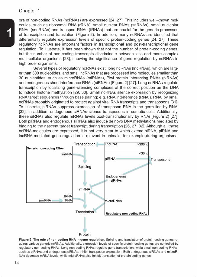
ora of non-coding RNAs (ncRNAs) are expressed [24, 27]. This includes well-known mol-ecules, such as ribosomal RNA (rRNA), small nuclear RNAs (snRNAs), small nucleolar RNAs (snoRNAs) and transport RNAs (tRNAs) that are crucial for the generic processes of transcription and translation (Figure 2). In addition, many ncRNAs are identified that differentially regulate expression levels of specific protein-coding genes [24, 27]. These regulatory ncRNAs are important factors in transcriptional and post-transcriptional gene regulation. To illustrate, it has been shown that not the number of protein-coding genes, but the number of non-coding transcripts discriminate between less and more complex multi-cellular organisms [28], showing the significance of gene regulation by ncRNAs in high order organisms. Several types of regulatory ncRNAs exist: long ncRNAs (lncRNAs), which are larg-er than 300 nucleotides, and small ncRNAs that are processed into molecules smaller than 30 nucleotides, such as microRNAs (miRNAs), Piwi protein interacting RNAs (piRNAs) and endogenous short interference RNAs (siRNAs) (Figure 2) [27]. Long ncRNAs regulate transcription by localizing gene-silencing complexes at the correct position on the DNA to induce histone methylation [29, 30]. Small ncRNAs silence expression by recognizing RNA target sequences through base pairing; e.g. RNA interference (RNAi). RNAi by small ncRNAs probably originated to protect against viral RNA transcripts and transposons [31]. To illustrate, piRNAs suppress expression of transposon RNA in the germ line by RNAi [32]. In addition, endogenous siRNAs silence transposons in somatic cells. Additionally, these siRNAs also regulate mRNAs levels post-transcriptionally by RNAi (Figure 2) [27]. Both piRNAs and endogenous siRNAs also induce de novo DNA methylations mediated by binding to the nascent target transcript during transcription [26, 27, 32]. Although all these ncRNA molecules are expressed, it is not very clear to which extend siRNA, piRNA and lncRNA-mediated gene regulation is relevant in animals, for example during organismal
Figure 2: The role of non-coding RNA in gene regulation. Splicing and translation of protein-coding genes re-quires various generic ncRNAs. Additionally, expression levels of specific protein-coding genes are controlled by regulatory non-coding RNAs. Long non-coding RNAs regulate gene transcription, while small non-coding RNAs, such as piRNAs and endogenous siRNAs, inhibit transposon expression. Both endogenous siRNAs and microR-NAs decrease mRNA levels, while microRNAs also inhibit translation of protein coding genes.
1
Chapter 1
tRNA
piRNA
EndogeneoussiRNAs
LncRNA
snRNA
rRNAsnoRNA
Splicing
Transcription
mRNA
Translation
Protein
Transposons
microRNAs
Generic non-coding RNAs
Regulatory non-coding RNAs
>300nt
<30nt
Figure 2

15
development, within normal cellular physiology and in diseases such as cancer. In contrast, microRNAs are well-known factors in post-transcriptional gene regula-tion in healthy and diseased cells. MicroRNAs are ~22 nucleotides long ncRNAs that neg-atively regulate protein expression levels by binding to the 3’untranslated-region (3’UTR) of target mRNAs [33], thereby decreasing protein translation mainly through mRNA deg-radation [34]. MicroRNAs are expressed in metazoans [35-40], plants [41] and alga [42, 43], which indicates that microRNAs probably evolved before multi-cellularity as a common feature of all eukaryotic cells [42, 43]. Since their discovery [37, 44-47], more than 2500 human microRNAs (miRBase, September 2013) have been identified in humans and most of these microRNAs are conserved in mammals [48, 49]. Presumably more than half of all protein-coding genes are regulated by microRNAs [50]. Consequently, one microRNA can regulate multiple genes, but one gene can also be regulated by multiple microRNAs. This implies that a single microRNA can regulate various cellular processes simultaneously or various genes acting in the same pathway [51], adding a whole new layer of gene regula-tion.
MicroRNA biogenesis, processing and target regulationMicroRNAs can be expressed under control of their own promoter, but a majority of microR-NA genes are co-expressed with protein-coding transcripts [52]. Similar to protein-coding genes, microRNAs are transcribed by RNA polymerase II into a primary microRNA (pri-mi-croRNA) transcript, which is also poly-adenylated and 5’ capped (Figure 3). This transcript contains one or multiple hairpin structures that are cleaved by the Drosha-DGCR8 complex to generate a precursor microRNA (pre-microRNA) [53-55]. In this complex, DGCR8 rec-ognizes sequences flanking the hairpin, which are essential for Drosha cleavage [56, 57]. After nuclear processing, pre-microRNAs are exported into the cytosol by exportin-5 [58]. Here, the Dicer-TRBP complex further processes pre-microRNAs into double stranded, 22-nucleotides-long RNA molecules by removing the pre-microRNA terminal loop [59, 60]. This loop contains binding motifs for multiple RNA binding proteins (RBPs) that influence cleavage by Dicer [61, 62]. Upon removal of the terminal loop, one of the 2 strands of the resulting microRNA duplex is degraded, based on its thermodynamic stability, while the other is incorporated into the RNA-Induced Silencing Complex (RISC) [63, 64]. In this complex, one of the four Argonaute proteins (AGO1-4) mediates the interaction between the microRNA and the 3’UTR of its target gene [65, 66] (Figure 3). MicroRNAs bind their target genes at a MicroRNA Responsive Elements (MRE). Nucleotides 2-8 within a mature microRNA, called the seed sequence, need to bind perfectly within the MRE for microRNA action to function. MicroRNAs that share this seed region belong to the same microRNA family and have highly overlapping gene targets (Box1) [67]. Two distinct molecular mechanisms mediate microRNA-mediated gene repres-sion [68]: Inhibition of translation [69, 70] and mRNA degradation [34, 71, 72]. Inhibition of translation is achieved by blocking translation initiation [73-76] and shielding mRNAs from translation within P-bodies [77]. These sub-cellular structures recruit microRNAs and their targets, but expel ribosomes [65, 66]. However, the main process that occurs within P-bodies is microRNA-mediated mRNA degradation [78, 79]. Degradation is achieved in two ways: destabilization by poly(A) tail removal (de-adinylation) [65, 66] or endonucleotic cleavage of the mRNA by AGO2 [80]. MicroRNAs that are fully complementary to their MREs induce cleavage of their targets within the target sequence [41, 65], which is a common mechanism in plants. In contrast, the majority of mammalian MREs are not fully complementary to mature microRNA sequences. Only the seed sequence and additional 3′-compensatory sites of the microRNA are complementary to the mRNA target [67]. These
1
General introduction

16
mRNAs are destabilized by microRNA-mediated de-adinylation that results in 5’cap remov-al and exonuclease mediated mRNA decay [81-83]. MicroRNA-mediated post-transcriptional gene regulation has a crucial role in de-velopment and differentiation [84]. Therefore microRNAs have been found to play a role in various diseases such as pathologies in brain [85-88], heart [89] and liver [90] as well as viral infections [91]. MicroRNAs regulate cell proliferation, cell cycle control and apoptosis [92, 93]. Therefore, it is not difficult to imagine that microRNAs are also causally involved in cancer, a main subject in this thesis.
MicroRNAs and cancerA frequently observed phenotype in cancer cells is a global decrease of microRNA levels [94]. This phenotype was mimicked in a lung tumor mouse model by conditionally inacti-vating Dicer, which resulted in enhanced tumor development [95]. Furthermore, reduced Dicer expression is observed in human lung cancer and correlates with poor survival [96] indicating that microRNAs protect against carcinogenesis. Next to global changes in mi-croRNA expression, silencing or induction of specific microRNAs has also been document-ed in human tumors. MicroRNA profiling studies show extensive differential regulation of microRNAs in tumors originating from various tissues [97-103], which can be used for classification on tissue origin, cancer type, progression and survival prognosis, sometimes even better than mRNA profiling [94, 104-107]. Some microRNA expression changes are causal events in tumor formation and can be regarded as tumor-suppressive or oncogenic microRNAs [92, 108-111]: microRNAs act as tumor-suppressors, when they control the
Pol IImicroRNA
Pol IImicroRNA
Ago1-4AAAAAAAAA
3’UTR Ago1-4
Pol IImicroRNA
Pol IImicroRNA
Pol IImicroRNA
PolII
primary microRNA
DicerTRBP
DicerTRBPTRBP
Ago1-43’UTR Ago1-43’UTR
mRNA degradation Translation inhibition
RISC RISC
precursor microRNA
Ago1-4 Ago1-4
Ago1-4Ago1-4
DroshaDGCR8
Figure 3
Figure 3: Schematic overview of the microRNA biogenesis pathway. MicroRNAs are transcribed by RNA Polymerase II into primary microRNAs (pri-mi-croRNA). These transcripts are cleaved into precurs-or microRNA (pre-microRNA) hairpin molecules by Drosha-DGCR8 and exported into the cytosol by ex-portin-5. MicroRNAs are processed by Dicer-TRBP into a double stranded, 22 nucleotides long RNA mo-lecules. One strand is incorporated into the RNA In-duced Silencing Complex (RISC) in which the Argo-naute (AGO1-4) proteins mediate binding between microRNA and 3’UTR of the target gene. This leads predominantly to mRNA degradation.
1
Chapter 1

17
expression of proto-oncogenes, or function as oncogenic microRNAs, when they influence gene expression levels of tumor-suppressive genes.
In cancers, microRNA levels are modulated by genetic and epigenetic alterations [112, 113]: both promoter methylation [114, 115] as well as genetic alterations (duplication, translocation or deletion) are frequently observed [113]. For example, in chronic lympho-cytic leukemia miR-15 and miR-16 are located in a frequently deleted region in 13q14 [116]. This genomic area does not contain any obvious protein-coding genes and further studies identified miR-15 and miR-16 to be causally involved in human cancer as the first microRNA tumor suppressors [117]. These microRNAs regulate various proto-oncogenes such as the cell cycle control gene CDC25a and anti-apoptotic gene BCL2 [117, 118]. Examples of other tumor-suppressive microRNAs that are often decreased in cancer are Let-7 and miR-34, which control expression of oncogenes such as proliferation factor Ras and BCL2, respectively [119, 120]. Examples of frequently occurring oncogenic microRNAs are miR-221, miR-222, the miR-17-92 cluster and miR-21. These microRNAs are induced in various human cancers and negatively regulate important tumor suppressors that control cell proliferation such as PTEN (miR-221/miR-222) [121], P27 (miR-221/miR-222) [122], E2F1 (miR-17-92 cluster) [123] and apoptotic and metastatic factor PDCD4 (miR-21) [124, 125]. In conclusion, it is clear that mis-expression of microRNAs plays an important role in carcinogenesis.
MicroRNAs and the DNA damage ResponseThe DDR has long been known to be regulated at the post-translational and transcription-al levels. However, post-transcriptional regulation by microRNAs also influences aspects of the DDR. One of the first clues that implicated microRNA-mediated gene silencing in the DDR was knockdown of the microRNA biogenesis pathway (Dicer), which resulted in increased sensitivity to the DNA-damaging agents cisplatin and UV [50, 118, 126] and altered cell cycle control after UV damage [118]. These results showed that microRNA biogenesis factors are involved in the DDR and implied that microRNAs are required for a proper DDR.
Box 1. MicroRNA nomenclatureMicroRNAs are assigned and annotated by miRBase at the Sanger Centre [202] according to the sequence of the mature, 22 nucleotide long microRNA. MicroRNA annotations start with defining the species they are iden-tified in (e.g. hsa-miR-16 for humans or dre-miR-16 for zebrafish). All microRNAs, except for microRNA Let-7 and other early-discovered microRNAs in C. elegans or Drosophilia, are annotated by numbers indicating their order of discovery. MicroRNAs with the same number contain identical nucleotides at position 2 to 8, their seed sequence, determining their complementary mRNA targets.The same microRNA can be transcribed from different sites in the genome. These microRNA isoforms are annotated with different letters (e.g. miR-34a and miR-34b) when they have different nucleotides outside the seed sequence. If the mature microRNA sequence between two isoforms is identical, extra numbers are added as appendix (e.g. miR-125b-1 and miR-125b-2). These microRNAs might differ from each other in the loop of the pre-microRNA or within sequences of the pri-microRNA, which may determine differential processing of the microRNA isoforms by the microRNA processing pathway.After dicer processing, microRNAs form a duplex with the complementary RNA molecule that originates from the opposite side of the pre-microRNA stem loop. When the mature microRNA is incorporated in the RISC com-plex, the less stable sequence is degraded. However, some microRNA-complementary duplex molecules are also stable and functional. These complementary microRNAs are annotated with a star (e.g. miR-29c*) and are functionally different from the dominant microRNA, because they have a different seed sequence. Additionally, sometimes both sides of the stem are equally stable and therefore it is unclear which microRNA is dominant. In this case, microRNAs are annotated according to the arm of the stem loop within the pre-microRNA from which they originated (e.g. miR-125a-3p or miR-125a-5p).
1
General introduction

18
However, microRNA-processing factors Drosha, Dicer and Ago2 were recently also shown to have a non-canonical, microRNA-independent role in the DDR of plants and animals. These proteins are also required to generate small ncRNAs that are derived from sequences flanking DSBs. These site-specific ncRNAs are necessary for DNA repair and foci formation of ATM, MDC1, 53BP1, but not γH2AX, at sites of damage. Although the exact mechanism has to be unraveled, microRNA-processing factors and DSB-specific ncRNAs might modulate the DDR as co-factors at the site of damage by recruiting proteins that function downstream of phosphorylated H2AX [127, 128].
Nevertheless, the canonical role of Drosha and Dicer is to produce mature mi-croRNAs that could regulate DDR genes and many microRNAs are differentially regulated upon DNA damage. MicroRNA profiling revealed modulation of various microRNAs after treatment with several genotoxic agents such as UV, hydrogen peroxide, IR, cisplatin and etoposide (topoisomerase II inhibitor leading to DSBs) [118, 129, 130]. Currently, it is hard to extract a common microRNA response from these studies, since different microRNA ar-ray platforms, cell lines, DNA damages, dosages and time points were used, which may all influence the outcome. A standardized approach will be necessary to extract information on specific microRNA profiles indicative for the general DNA damage response or genotoxic agent, dose or cell type specific response.
Interestingly, it has been shown that the expression level of a substantial subset of microRNAs rapidly changed after UV damage: within a few hours after DNA damage microRNA levels either increased or decreased, while at 24 hours their expression levels had (almost) returned to the pre-damage situation [118]. This response was faster than the transcriptional activation of p53 target genes such as p21waf1, but slower than post-trans-lational protein modifications. Thus, it was postulated that microRNAs act in time between the protein modifications (such as phosphorylation and ubiquitination), which are rapid, and the gene transcriptional events, which are relatively slow (Figure 4). These intermediate kinetics of microRNA expression changes after DNA damage was also observed for the let-7 family of microRNAs in cells treated with IR [131] and is thus likely not a UV-specific phenomenon. A clear example of this regulation at three levels is cell cycle control and DDR protein CDC25a. This protein is a known ATM and ATR target and is phosphorylat-ed and degraded within minutes after DNA damage, thereby directly inhibiting cell cycle progression [132]. Furthermore, CDC25a is transcriptionally repressed by P53-dependent p21waf1 induction as early as 9 hours after DNA damage [15]. However, in the intermedi-ate timeframe CDC25a mRNA is also reduced for which the UV-inducible miR-16 is found responsible [118, 133]. This timing perfectly fits in between rapid post-translational and slow transcriptional regulation of CDC25a and indicates a role for microRNAs at the inter-mediate timeframe in the DDR.
DDR-dependent regulation of microRNA expression and efficacyCurrently, not much is known about the mechanistics of differential regulation of microRNA expression after DNA damage or other stimuli. Similar to normal protein-coding genes, mi-croRNA genes are transcribed by RNA polymerase II from their own promoters or as part of protein-coding transcripts where they reside in the introns [33]. Although some transcripts are small and could be induced relatively quickly by DNA damage inducible transcription factors, others have much longer primary transcripts or are co-regulated with their “host” gene. For example, miR-34a, which is transcriptionally induced by P53 after DNA damage and has a primary transcript of ~30 kilobases [120, 134-139] , is regulated at later time points [118].
Recently however, it has been shown that microRNAs themselves are subject to
1
Chapter 1

19
post-transcriptional regulation. Various RBPs selectively control processing of specific mi-croRNAs by influencing the Drosha-DGCR8 and DICER-TRBP complexes [61, 140, 141]. A main example is Lin28, which inhibits pre-let-7 processing by binding to its terminal hairpin loop [142, 143]. This interaction recruits Zcchc11 (Tutase4), which prevents Dicer processing and targets pre-let-7 for degradation [141, 144-146]. This type of microRNA regulation is relatively fast, because it bypasses transcription and might therefore be in-volved in the quick regulation of microRNAs after DNA damage (Figure 4). An example of post-transcriptional regulation of microRNA levels upon DNA damage involves P53. This protein was shown to stimulate the maturation of a number of microRNAs after DNA dam-age through association with RNA helicase p68 from the Drosha complex, thereby induc-ing increased pri-microRNA processing [147]. A second mechanism involves regulation of microRNA-processing by Drosha co-factor KHSRP [61]. This protein is phosphorylated by ATM after DSB induction by the radiomimetic drug neocorzinostatin, thereby inducing mat-uration of various DNA damage responsive microRNAs, again via increased processing of specific pri-microRNAs [148]. Both examples show a direct and intimate relation between the DDR and microRNA-processing and further provide evidence for the model presented in figure 4.
MicroRNA efficacy can also be regulated without modulating microRNA process-ing. Some microRNAs are ubiquitously expressed, while their predicted target transcripts are not regulated. This can be explained by RBPs that bind to 3’UTRs and modulate the accessibility of an MRE [149]. For example, the human dead end 1 (Dnd1) and Pumilio-1 (PUM1) proteins modulate regulation of cell cycle control protein P27Kip1 by the UV-re-sponsive microRNAs miR-221/222 [118, 150, 151]. Additionally, Dnd1 protects nucleotide mismatch factor MSH2 against miR-21- mediated regulation [152]. Another RNA binding
Figure 4: Model of three levels of gene regulation after DNA damage induction in time. Fast, but transient post-translational protein modifications are directly (seconds/minutes) induced upon DNA damage. Transcriptio-nal regulation changes are relatively slowly (hours/days) induced, but are usually more stable. Post-transcriptional gene regulation by microRNAs acts in the intermediate timeframe. MicroRNAs are themselves also post-tran-scriptionally regulated, which is activated by fast, but transient post-translational protein modifications.
Figure 4
time after DNA damage
modulation of microRNA processing
by DDR
???
P53post-transcriptional regulation by microRNAs
Dicer
transcriptional regulation
pUb
UbUbUb
transient protein modifications
DNA damage
ATM
1
General introduction

20
protein, RBM38, is induced by p53 upon doxorubicin-induced genotoxic stress and binds mRNA of p53 target genes, such as p21 and PCNA, relieving them from microRNA-medi-ated repression [153]. These examples show that RBPs play an important role in microR-NA-mediated gene regulation upon DNA damage.
MicroRNAs and their DDR target genesDNA damage responsive microRNAs are likely to regulate many cellular processes and pathways. This part of the introduction mainly focuses on those microRNAs that have a known direct role in regulating DDR genes, are regulated by such a gene or after DNA damage. In table 1 we summarize known microRNA–DDR gene interactions and the most prominent interactions are described hereafter. As mentioned earlier, the tumor-suppressive miR-16, but also miR-21, target CD-C25a expression [118, 154]. In addition, many other cell cycle control gene transcripts are regulated by miR-16, such as CDC27 and CDK6, negatively regulating G1 to S cell cycle progression [155]. Other confirmed target genes are the anti-apoptotic BCL2 gene [117] and WIP1 [156], which dephosphorylates key DNA damage checkpoint proteins such as ATM, Chk1/2 and P53, thereby inactivating them, which enables cell cycle recovery after genotoxic stress [157-159]. Mir-21 does not only regulate aspects of the cell cycle control, but also targets genes that function in apoptosis, proliferation and DNA repair. Besides CDC25a, miR-21 regulates programmed cell death 4 (PDCD4) protein [124], proliferation and survival factor PTEN [160] and DNA repair factor MSH2 [161]. All these proteins are important tumor-suppressors and potentially explain the profound oncogenic capabilities of miR-21 [111]. Another well-studied microRNA is the DNA damage inducible, P53-dependent miR-34a [162]. This microRNA recapitulates P53-dependent effects on cell cycle control and apoptosis after DNA damage [120, 134-139, 163]. MiR-34a is implicated in cell cycle control by regulating genes that code for G1/S checkpoint regulators, such as E2F, Cy-clin-E2, CDK4 and CDK6 [120, 134, 135]. In addition, miR-34a also targets Bcl2, which regulates apoptosis [120, 136, 138, 139, 163], and MDM4 which is a negative regulator of p53 [164]. Besides miR-34a, several other microRNAs have been identified as p53-tran-scriptional targets after DNA damage, e.g. miR-192 and miR-215, although it is not known if they regulate any DDR genes [165, 166]. In contrast, p53-dependent microRNAs miR-22 and Let-7a are well characterized. MiR-22 levels increase by UV-radiation and doxorubicin treatment. It targets cell cycle and proliferation genes p21 and PTEN and protects cells against UV radiation [167]. As mentioned before, the let-7 family of microRNAs is regulated upon IR treatment [131], which also depends on P53 [168]. Potential target genes were identified by gene expression arrays: overexpression of let-7 resulted in the regulation of DNA damage checkpoint and repair genes BRCA1, BRCA2, CHK1 and FANCD2 [169]. Whether these genes are direct let-7 targets remains to be elucidated although some of them have predicted microRNA target sites. Confirmed targets of let-7 are apoptosis ef-fector caspase 3 and apoptotic factor BCL-xL [10, 170] suggesting that let-7 regulates cell death induction upon DNA damage [171]. P53 regulates microRNAs levels, but is also regulated by microRNAs. Multiple microRNAs, such as miR-25, miR-30d, miR-504 and miR-125b target P53 and regulate P53-dependent stress responses, such as apoptosis [172-174]. For example, miR-125b levels decrease in human primary fibroblasts and neuroblastoma cells after 24 hours of
Table 1, next pages: DDR microRNAs and their DDR targets. Included are microRNAs that are regulated by a genotoxic stimuli and/or regulate DDR genes. Additionally, the role of target genes within the DDR is also noted. Furthermore, included are also the effects of several microRNAs on genotoxic cancer treatment.
1
Chapter 1

21
TAB
LE 1
. Mic
roR
NAs
with
a r
ole
in th
e D
DR
mic
roR
NA
regu
late
d by
Che
mot
oxic
resi
stan
ce/s
ensi
tatio
nD
DR
targ
ets
Rol
e in
DD
RR
efer
ence
hsa-
miR
-15/
16up
regu
late
d up
on U
V ra
diat
ion
and
post
-tran
scrip
tiona
llyse
nsiti
ze b
reas
t and
gas
tric
canc
er c
ells
Bcl2
Apop
tosi
s11
7, 1
18,
upre
gula
ted
by P
53 u
pon
doxo
rubi
cin
treat
men
tfo
r dox
orub
icin
, cis
plat
in a
nd e
topo
side
CD
C25
aC
ell c
ycle
con
trol
155,
156
, C
DC
27C
ell c
ycle
con
trol
193,
194
CD
K6C
ell c
ycle
con
trol
Wip
1C
ell c
ycle
reco
very
hsa-
miR
-21
upre
gula
ted
upon
UV
radi
atio
n, T
GF-
β ac
tivity
and
neg
ativ
ely
prot
ects
diff
eren
t can
cers
aga
inst
mul
tiple
C
DC
25a
Cel
l cyc
le c
ontro
l11
8, 1
52,
regu
late
d by
FO
XO3a
DN
A-da
mag
ing
chem
othe
rape
utic
sPD
CD
4Ap
opto
sis
154,
160
MSH
2N
ucle
otid
e m
ism
atch
repa
ir16
1, 1
93,
PTEN
Cel
l pro
lifer
atio
n an
d su
rviv
al19
7-20
0,20
3
hsa-
miR
-34
trans
crip
tion
upre
gula
ted
by P
53 u
pon
UV
radi
atio
n an
d m
odul
ates
sen
sitiv
ity o
f diff
eren
t can
cer
Cyc
lin-E
2 C
ell c
ycle
con
trol
118,
120
,do
xoru
bici
n tre
atm
ent
cells
to d
iffer
ent D
NA-
dam
agin
g tre
atm
ents
CD
K4C
ell c
ycle
con
trol
134-
139
CD
K6C
ell c
ycle
con
trol
162-
164,
Bcl2
Apop
tosi
s19
5, 1
96M
DM
2C
ontro
l of P
53M
DM
4C
ontro
l of P
53
hsa-
miR
-192
tra
nscr
iptio
n up
regu
late
d by
P53
unkn
own
unkn
own
Cel
l cyc
le c
ontro
l16
5, 1
66
hsa-
miR
-215
tra
nscr
iptio
n up
regu
late
d by
P53
unkn
own
unkn
own
Cel
l cyc
le c
ontro
l16
5, 1
66
has-
miR
-22
upre
gula
ted
upon
UV
radi
atio
n an
d do
xoru
bici
n tre
atm
ent
prot
ects
cel
ls a
gain
st U
V tre
atm
ent
P21
Apop
tosi
s/ce
ll cy
cle
cont
rol
167,
204
depe
nden
t on
P53
PTEN
Cel
l pro
lifer
atio
n an
d su
rviv
al
hsa-
Let-7
dow
nreg
ulat
ed u
pon
UV,
ioni
zing
radi
atio
n, e
topo
side
and
mod
ulat
es s
ensi
tivity
of d
iffer
ent c
ells
to
Cas
pase
-3Ap
opto
sis
118,
119
,hy
drog
en p
erox
ide.
Pos
t-tra
nscr
iptia
lly d
epen
dent
on
P53
seve
ral D
NA
dam
agin
g tre
atm
ents
Bcl-x
LAp
opto
sis
131,
16
8-17
1
hsa-
miR
-25
unkn
own
redu
ces
apop
tosi
s an
d ce
ll cy
cle
arre
stP5
3Ap
opto
sis/
cell
cycl
e co
ntro
l17
2 a
fter e
topo
side
trea
tmen
t
hsa-
miR
-30d
unkn
own
redu
ces
apop
tosi
s an
d ce
ll cy
cle
arre
st
P53
Apop
tosi
s/ce
ll cy
cle
cont
rol
172
afte
r eto
posi
de tr
eatm
ent
hsa-
miR
-504
un
know
nre
duce
s ap
opto
sis
and
cell
cycl
e ar
rest
P5
3Ap
opto
sis/
cell
cycl
e co
ntro
l17
3af
ter e
topo
side
trea
tmen
t
hsa-
miR
-125
b do
wnr
egul
ated
upo
n et
opos
ide
treat
men
t, up
regu
late
d up
onde
crea
sed
apop
tosi
s af
ter i
oniz
ing
P53
Apop
tosi
s/ce
ll cy
cle
cont
rol
174,
205
UV
treat
men
t, de
pend
ent o
n AT
M a
nd N
FКB
irrad
iatio
n an
d ca
mpt
othe
cin
treat
men
tp3
8αSt
ress
sig
nalli
ngin
zeb
rafis
h an
d pr
otec
ts c
ells
aga
inst
UV
1
General introduction

22
TAB
LE 1
, con
tinue
d. M
icro
RN
As w
ith a
role
in th
e D
DR
mic
roR
NA
regu
late
d by
Che
mot
oxic
resi
stan
ce/s
ensi
tatio
nD
DR
targ
ets
Rol
e in
DD
RR
efer
ence
hsa-
miR
-375
unkn
own
Red
uced
apo
ptos
is in
AG
S ce
lls u
pon
P53
Apop
tosi
s/ce
ll cy
cle
cont
rol
206
etop
osid
e an
d Io
nizi
ng R
adia
tion
treat
men
t
hsa-
miR
-421
tra
nscr
iptio
n re
gula
ted
by n
-Myc
sens
itize
HEL
A ce
lls fo
r ion
izin
g ra
diat
ion
ATM
DN
A da
mag
e ch
eckp
oint
176
hsa-
miR
-101
un
know
nse
nsiti
ze x
enog
rafte
d hu
man
lung
and
AT
MD
NA
dam
age
chec
kpoi
nt17
7gl
iom
a ca
ncer
cel
ls fo
r ion
izin
g ra
diat
ion
DN
A-PK
DN
A da
mag
e ch
eckp
oint
hsa-
miR
-18a
unkn
own
sens
itize
bre
ast c
ance
r cel
ls fo
r ion
izin
g AT
MD
NA
dam
age
chec
kpoi
nt17
5ra
diat
ion
hsa-
miR
-18b
trans
crip
tion
regu
late
d by
c-m
yc, u
preg
ulat
ed u
pon
UV
radi
atio
nun
know
nE2
F1C
ell p
rolif
erat
ion
118,
123
hsa-
miR
-138
unkn
own
sens
itize
s U
2OS
cells
for d
iffer
ent
H2A
XD
NA
dam
age
chec
kpoi
nt17
9D
NA
dam
agin
g ag
ents
hsa-
miR
-24
upre
gula
ted
upon
term
inal
ly d
iffer
enta
tion
and
UV
radi
atio
nse
nsiti
ze te
rmin
ally
diff
eren
tiate
d bl
ood
H2A
X D
NA
dam
age
chec
kpoi
nt11
8, c
ells
for b
leom
icin
e an
d ci
spla
tinP1
6C
ell c
ycle
con
trol
180-
183,
P2
7C
ell c
ycle
con
trol
207
hsa-
miR
-182
dow
nreg
ulat
ed u
pon
ioni
zing
radi
atio
nse
nsiti
ze x
enog
rafte
d hu
man
bre
ast
BRC
A1D
NA
repa
ir18
4ca
ncer
cel
ls fo
r ion
izin
g ra
diat
ion
hsa-
miR
-210
upre
gula
ted
in h
ypox
ic c
ondi
tions
unkn
own
Rad
52D
NA
repa
ir18
5
hsa-
miR
-373
upre
gula
ted
in h
ypox
ic c
ondi
tions
unkn
own
Rad
52D
NA
repa
ir18
5R
ad23
BD
NA
repa
ir
hsa-
miR
-96
unkn
own
sens
itize
xen
ogra
fted
hum
an b
reas
t R
EV1
DN
A re
pair
186
canc
er c
ells
for i
oniz
ing
radi
atio
nR
AD51
DN
A re
pair
has-
miR
-148
aun
know
nun
know
nC
DC
25a
Cel
l cyc
le c
ontro
l20
8
hsa-
miR
-449
a/b
trans
crip
tion
regu
late
d by
E2F
1un
know
nC
DC
25a
Cel
l cyc
le c
ontro
l20
9C
DK6
Cel
l cyc
le c
ontro
l
1
Chapter 1

23
TAB
LE 1
con
tinue
d. M
icro
RN
As w
ith a
rol
e in
the
DD
R
mic
roR
NA
regu
late
d by
Che
mot
oxic
resi
stan
ce/s
ensi
tatio
nD
DR
targ
ets
Rol
e in
DD
RR
efer
ence
hsa-
miR
-221
upre
gula
ted
upon
UV
radi
atio
nun
know
nPT
ENC
ell p
rolif
erat
ion
and
surv
ival
118,
121
, P2
7C
ell c
ycle
con
trol
122
hsa-
miR
-195
unkn
own
unkn
own
Wee
1C
ell c
ycle
con
trol
210
hsa-
miR
-100
unkn
own
sens
itize
xen
ogra
fted
hum
an lu
ng c
ance
rPL
K1C
ell c
ycle
con
trol
201,
211
cells
for I
R
hsa-
miR
-155
unkn
own
unkn
own
hMLH
1N
ucle
otid
e m
ism
atch
repa
ir21
2hM
SH2
Nuc
leot
ide
mis
mat
ch re
pair
hMSH
6N
ucle
otid
e m
ism
atch
repa
ir
hsa-
miR
-182
-5p
unkn
own
sens
itize
s br
east
can
cer c
ells
for P
ARP
CH
K1D
NA
dam
age
chec
kpoi
nt21
3in
hibi
tion
P27
Cel
l cyc
le c
ontro
l
hsa-
miR
-186
unkn
own
unkn
own
CD
K2C
ell c
ycle
con
trol
214
CD
K6C
ell c
ycle
con
trol
Cyc
linD
1C
ell c
ycle
con
trol
CU
1276
unkn
own
sens
itize
s Bu
rkitt
lym
phom
a ce
ll lin
e to
RPA
1ss
DN
A se
nsor
215
non-
anno
tate
d tR
NA-
etop
osid
ede
rived
mic
roR
NA
hsa-
miR
-31-
5pun
know
nun
know
nhM
LH1
Nuc
leot
ide
mis
mat
ch re
pair
216
hsa-
miR
-335
Dow
nreg
ulat
ion
upon
Ioni
zin
Rad
iatio
n de
pend
s on
ATM
sens
itize
s H
eLa
cells
for I
oniz
ing
Rad
iatio
nC
tIPD
NA
repa
ir21
7
hsa-
miR
-129
unkn
own
unkn
own
Bcl2
Apop
tosi
s21
8
hsa-
miR
-23a
upre
gula
tion
upon
UV
treat
men
tun
know
nTo
p1re
plic
atio
n21
9
hsa-
miR
-185
dow
nreg
ulat
ed u
pon
ioni
zing
radi
atio
nin
crea
ses
apop
tosi
s in
786
-O c
ells
upo
nAT
RD
NA
dam
age
chec
kpoi
nt22
0io
nizi
ng ra
diat
ion
hsa-
miR
-302
unkn
own
unkn
own
P21
Cel
l cyc
le c
ontro
l/apo
ptos
is22
1
hsa-
miR
-107
unkn
own
sens
itize
s br
east
can
cer c
ells
for P
ARP
Rad
51D
NA
repa
ir18
8in
hibi
tion
hsa-
miR
-222
unkn
own
sens
itize
s br
east
can
cer c
ells
for P
ARP
Rad
51D
NA
repa
ir18
8in
hibi
tion
1
General introduction

24
etoposide treatment thereby inducing P53 expression levels [174]. The DNA damage sig-naling protein ATM is another DDR gene that is regulated by microRNAs: miR-421, miR-18a and miR-101 [175-177]. The latter microRNA also targets DNA-PK [177], which regu-lates checkpoint responses and induces NHEJ repair [178]. Overexpression of miR-421, miR-101 and miR-18a results in reduced survival after IR [175-177], but it is not clear whether these microRNAs are regulated after DNA damage and thereby influence ATM activity. ATM phosphorylates histone H2AX which is also targeted by microRNAs: MiR-138 and miR-24 [179, 180]. MiR-138 sensitizes cells for several DNA damaging treatments [179], but it is unknown if this microRNA is regulated upon DNA damage. MiR-24 is regu-lated upon UV-radiation [118] and during terminal differentiation of various hematopoietic cells resulting in decreased DNA repair, increased chromosome breakage and sensitivity to genotoxic agents when compared to its progenitor counterpart [180]. Additionally, various cell cycle promoting proteins are regulated by miR-24, such as P16, E2F2 and MYC, sug-gesting that miR-24 has an anti-proliferative effect independent of P53 [181-183]. MiR-24 also regulates p53-independent cellular proliferation via S-phase enzyme DHFR, a target of chemotherapeutic drug methotrexate [183]. Finally, recent articles show that multiple microRNAs regulate DNA repair proteins. MiR-182, a microRNA that is reduced after IR, regulates the DNA repair and cell cycle checkpoint protein BRCA1 [184]. MicroRNAs MiR-210 and miR-373 are both induced in hypoxic conditions and target Rad52 and Rad23B. Rad52 functions in HR, while Rad23B has a role in NER, although it is unknown if these microRNAs decrease DNA repair capac-ity and sensitize cells for DNA damage [185]. In contrast, miR-96, miR-107 and miR-222 sensitize cells for cisplatin treatment and reduced NHEJ-capacity through regulation of HR-protein Rad51 [186, 187].
MicroRNA target prediction of DDR genesAlthough the examples mentioned above show that microRNAs play roles in many aspects of the DDR (including cell cycle regulation, apoptosis and DNA repair), it does currently not provide a coherent view of the function of microRNA-mediated gene silencing in the DDR and it is not yet clear how important this level of regulation is for these processes. It is to be expected that many new DNA damage responsive microRNAs and their targets will be found in the years ahead and that it will become clear how central this response is to the various aspects of the DDR.
As a first approximation, microRNA targets can be predicted using in silico meth-ods. MicroRNA target identification algorithms have already predicted various microRNA–gene interactions which are described above. To give an overview which additional DDR genes are potential microRNA target genes, we previously analyzed the 3’UTR of 142 genes that are implicated in the DDR using two different microRNA target prediction algo-rithms (Miranda and Targetscan) [188]. These prediction programs are based on different parameters: Targetscan is partially based on wet-lab evidence and does not only monitor perfect binding of the microRNA seed region to its predicted target gene, but also assesses additional binding (most notably around nucleotides 13-19), AU-rich elements flanking the microRNA-binding site, the position of the target site within the 3’UTR and conservation of these target sites across at least mammals but often also other vertebrates [50, 189-191]. The Miranda algorithm focuses on base complementarity and minimum free energy between mRNA and microRNA, but allows mismatches between the seed region and the 3’UTR [191, 192].
We found 74 (52%) DDR genes that contain conserved microRNA target sites (at least across mammals) predicted in their 3’UTR by either target Targetscan, Miranda
1
Chapter 1

25
or both [188]. This supports estimations that 50 to 60% of all genes are regulated by mi-croRNAs and suggests that post-transcriptional microRNA regulation of DDR genes is not over- or underrepresented compared to other cellular processes [50]. Using these predic-tion data for enrichment calculations within DDR sub-pathways, a one-sided Fisher-Ex-act test suggests that cell cycle control and DNA damage responsive ubiquitin machinery are overrepresented (p<0.01) with genes containing microRNA target sites indicating that these regulatory pathways themselves are possibly extensively regulated. Within the DNA repair pathways, the upstream branch of transcription-coupled nucleotide excision repair (TC-NER) is also enriched (p<0.05), while in contrast downstream NER, but also base ex-cision repair and mismatch repair are underrepresented (p<0.05), probably because these processes may be regulated in a different manner, if regulated at all.
Since it is possible that some of these predicted target genes are post-transcrip-tionally regulated in specific cell types and not after DNA damage (because the regulatory microRNA is not regulated after DNA damage, but in a cell type specific manner), we also indicated which microRNAs are UV-responsive and can have an effect on DDR gene ex-pression levels after UV.
MicroRNA-mediated gene silencing is probably mainly achieved by mRNA degra-dation instead of translation inhibition [34] implying that microRNA expression levels can directly be linked to mRNA levels to identify valid microRNA-mRNA relationships. For ex-ample, CDC25a and RAD23B mRNA are reduced upon UV [133], while their predicted mi-croRNA regulators are induced: miR-30a, miR-16 and miR-23ab are all predicted to target RAD23B and miR-16 was shown to regulate CDC25a upon UV treatment in fibroblasts [118]. It is therefore likely that these predictions can be used to build gene expression net-works to elucidate various aspects of the DDR.
DDR microRNAs, cancer and chemotherapy sensitivityDefects in the DDR and mis-expression of microRNAs are both causally involved in carcinogenesis [18, 19, 95, 111]. Therefore, it is an obvious question whether microRNAs that are involved in the DDR play a role in cancer. As mentioned before, one hallmark of cancer cells is the general lowering of microRNA expression levels [94]. Mimicking a general low microRNA expression phenotype by conditionally inactivating the microRNA processing protein Dicer in a lung cancer mouse model results in accelerated cancer development, indicating that the decreased overall levels of microRNAs accelerate tumor-igenesis [95]. Although Dicer silencing does not generate obvious cancer cell phenotypes such as decreased apoptosis or increased cell proliferation (rather slowing), dicer-defi-cient cells do exhibit a DDR defect [50, 118, 128]. This suggests that Drosha/Dicer activity and overall microRNA expression levels should be maintained to elicit a proper DDR as a barrier against cancer development.
Moreover, misexpression of various individual microRNAs has been found to play a causal role in the process of carcinogenesis. Interestingly, UV-responsive microRNAs [118] show a considerable overlap with frequently mis-expressed microRNAs in human solid tumors [103], suggesting that DNA damage responsive microRNAs can play a role in cancer etiology. Indeed, some DNA damage responsive microRNAs have been character-ized as tumor-suppressors or oncogenes, e.g. miR-16 [117], miR-21 [111], miR-221 [121, 122], members of the Let-7 family [119] and miR-17-92 cluster [123]. For these cancer-as-sociated and DNA damage responsive microRNAs it is not known whether the microRNA itself is (epi-) genetically altered or that DDR pathways that control their expression are defective resulting in their mis-expression.
As described before, DNA damage and subsequent DNA aberrations are driving
1
General introduction

26
carcinogenesis, but genotoxic agents are also used to battle cancer. Not much is known about the underlying mechanisms that induce sensitivity or resistance to the applied geno-toxic drugs, or how these tumors acquire resistance after being sensitive initially. It is con-ceivable that mis-expression of some microRNAs that are important for the DDR plays a role in establishing genotoxic drug responsiveness. A few examples indicate indeed that mis-expression of microRNAs is associated with drug responsiveness [131, 193-196]. For example, members of the let-7 family of microRNAs are rapidly silenced upon IR in A549 lung cancer cells and experimental induction modifies IR sensitivity [131].
Other well-known DNA damage responsive microRNAs also influence sensitivi-ty to genotoxic drug treatment. MiR-16 expression in multidrug-resistant gastric cancer cells is inversely correlated with resistance against various DNA-damaging drug therapies [194]. Furthermore, overexpression of DNA damage inducible miR-21 [118, 197], which is a causal oncogenic event [111], induces cytotoxic drug resistance in various cancer cell types [161, 193, 198-200] and has a negative predictive value for clinical outcome of ad-juvant therapy in pancreatic cancer [200]. In addition, restoration of P53-driven miR-34a expression in different cancer cells leads to renewed sensitivity towards chemotherapeutic treatments [135, 195, 196].
Multiple other microRNAs also support inhibition of tumor growth induced by geno-toxic drug treatment. Xenografted breast cancer cells are sensitized for IR by miR-182, which targets HR-repair protein BRCA1. The same cells are sensitized for cisplatin treat-ment by miR-96 that targets HR-repair protein Rad51 and translesion synthesis protein Rev1 [184, 186]. Furthermore, xenografted lung cancer cells are sensitized for IR using miR-100, which controls Polo-like Kinase 1 expression [201]. Finally, miR-101 also sen-sitizes a human glioma cell line to IR by down-regulating the DDR genes DNA-PKcs and ATM. Xenograft tumors of this glioma cell line in the mouse brain were subsequently infect-ed with a miR-101 overexpression lentiviral vector. Interestingly, miR-101 overexpression did not alter the tumor cell proliferation rate compared to control lentiviral vector delivery, but sensitized these human xenografts to IR and enhanced organism survival [177], show-ing the potential for therapeutic approaches using microRNA expression constructs.
In conclusion, multiple DNA damage responsive microRNAs and/or microRNAs that target DDR genes play a role in cancer and can modulate the cellular sensitivity to DNA-damaging agents, which will be of significant interest for future research on cancer treatment and/or prognosis.
Scope and aim of this thesisThe impact of microRNA-mediated gene regulation in the DDR and its role in cancer etiol-ogy is just beginning to surface. It is crucial to address the role of DNA damage responsive microRNAs in cancer development and/or genotoxic drug sensitivity/resistance to under-stand the impact of microRNAs in cancer. This knowledge will contribute to our under-standing of the regulation of important gene expression networks after DNA damage and the subsequent alteration of various cellular processes such as cell cycle control and cell death. Moreover, it might point to potential prognostic markers for genotoxic drug sensitivity and may support or even create a basis for new genotoxic cancer therapy.
To contribute to these issues, we addressed the following questions within this thesis. First, which microRNAs are regulated upon DNA damage? Second, how are these DDR microRNAs regulated? Third, which DDR genes do these microRNAs regulate? And finally, what is the role of these microRNAs in cancer and genotoxic therapy sensitivity?
1
Chapter 1

27
To answer these questions we used different approaches. In chapter 2 and 3, we used expression arrays to profile microRNA expression upon DNA damage. In chapter 2, the wildtype microRNA response after DNA damage was studied in human primary epithe-lial cells from breast and lung origin. We applied different doses of IR and cisplatin to iden-tify a common DNA-damage microRNA response. These microRNAs were compared with microRNAs that were found deregulated in cancer. We identified several DDR microRNAs that were deregulated in cancer to influence the response on genotoxic cancer therapy.
In chapter 3, we studied the regulation of microRNA levels upon IR and UV-in-duced DNA damage in human sarcoma cells. DNA damage-dependent microRNA regula-tion was investigated upon knockdown of several RBPs that are phosphorylayed by ATM or ATR and function in microRNA processing. We showed that the regulation of multiple IR and UV-responsive microRNAs depends on these proteins.
In chapter 4 and 5, we investigated which DDR processes and genes are regulat-ed by DNA damage responsive microRNAs. In chapter 4, we thoroughly studied the role of miR-24 in the DNA damage response. MiR-24 expression is induced multiple days after IR and we showed that miR-24 directly regulates MDC1, an adaptor protein in the DNA dam-age checkpoint response. MDC1 expression is silenced multiple days after DNA damage by miR-24 and miR-24 modulates several processes that function downstream of MDC1. Additionally, we discovered an alternative isoform of MDC1 that is differentially regulated by miR-24. The crucial functions of MDC1 might still be enabled by this alternative isoform when the canonical isoform is silenced in cells with high miR-24 levels.
In chapter 5, we investigated the role of microRNA Let-7 in ageing, cancer and upon DNA damage. We found that Let-7 is regulated within aging and is induced directly upon DNA damage. Let-7 regulation and overexpression showed different effects depend-ing on genotoxic agent and cell type. Let-7 influences the cell cycle and regulated apop-tosis, supporting its role in cancer. Let-7 might also regulate multiple genes that regulate growth, proliferation and metabolism. These genes have important roles in aging and lon-gevity and therefore Let-7 might play a role in aging by modulation the response to DNA damage.
Finaly, the discussion in chapter 6 summarizes the main conclusions of this thesis. It connects the findings of different chapters and links their conclusions with previously elu-cidated knowledge. It offers a final overview of the importance of microRNAs in the DDR, cancer and genotoxic cancer therapy.
1
General introduction
28
References1. Hoeijmakers, J.H., DNA damage, aging, and cancer. N Engl J Med, 2009. 361(15): p. 1475-85.2. Hoeijmakers, J.H., Genome maintenance mechanisms for preventing cancer. Nature, 2001. 411(6835):
p. 366-74.3. Harper, J.W. and S.J. Elledge, The DNA damage response: ten years after. Mol Cell, 2007. 28(5): p.
739-45.4. Ciccia, A. and S.J. Elledge, The DNA damage response: making it safe to play with knives. Mol Cell.
40(2): p. 179-204.5. Bartek, J. and J. Lukas, DNA damage checkpoints: from initiation to recovery or adaptation. Curr Opin
Cell Biol, 2007. 19(2): p. 238-45.6. Stewart, G.S., Solving the RIDDLE of 53BP1 recruitment to sites of damage. Cell Cycle, 2009. 8(10): p.
1532-8.7. Jungmichel, S. and M. Stucki, MDC1: The art of keeping things in focus. Chromosoma, 2010. 119(4): p.
337-49.8. Bekker-Jensen, S. and N. Mailand, Assembly and function of DNA double-strand break repair foci in
mammalian cells. DNA Repair (Amst), 2010. 9(12): p. 1219-28.9. Coster, G. and M. Goldberg, The cellular response to DNA damage: a focus on MDC1 and its interact-
ing proteins. Nucleus, 2010. 1(2): p. 166-78.10. Tsang, W.P. and T.T. Kwok, Let-7a microRNA suppresses therapeutics-induced cancer cell death by
targeting caspase-3. Apoptosis, 2008. 13(10): p. 1215-22.11. Murphy, C.G. and M.E. Moynahan, BRCA gene structure and function in tumor suppression: a re-
pair-centric perspective. Cancer J, 2010. 16(1): p. 39-47.12. Fernandez-Capetillo, O., et al., H2AX: the histone guardian of the genome. DNA Repair (Amst), 2004.
3(8-9): p. 959-67.13. Falck, J., et al., The ATM-Chk2-Cdc25A checkpoint pathway guards against radioresistant DNA synthe-
sis. Nature, 2001. 410(6830): p. 842-7.14. Bartek, J. and J. Lukas, Mammalian G1- and S-phase checkpoints in response to DNA damage. Curr
Opin Cell Biol, 2001. 13(6): p. 738-47.15. Vigneron, A., et al., The cell cycle inhibitor p21waf1 binds to the myc and cdc25A promoters upon DNA
damage and induces transcriptional repression. J Biol Chem, 2006. 281(46): p. 34742-50.16. Bartek, J. and J. Lukas, Chk1 and Chk2 kinases in checkpoint control and cancer. Cancer Cell, 2003.
3(5): p. 421-9.17. Vousden, K.H. and X. Lu, Live or let die: the cell’s response to p53. Nat Rev Cancer, 2002. 2(8): p. 594-
604.18. Bartkova, J., et al., DNA damage response as a candidate anti-cancer barrier in early human tumori-
genesis. Nature, 2005. 434(7035): p. 864-70.19. Gorgoulis, V.G., et al., Activation of the DNA damage checkpoint and genomic instability in human pre-
cancerous lesions. Nature, 2005. 434(7035): p. 907-13.20. Bouwman, P. and J. Jonkers, The effects of deregulated DNA damage signalling on cancer chemother-
apy response and resistance. Nat Rev Cancer, 2012. 12(9): p. 587-98.21. Lord, C.J. and A. Ashworth, The DNA damage response and cancer therapy. Nature, 2012. 481(7381):
p. 287-94.22. Igney, F.H. and P.H. Krammer, Death and anti-death: tumour resistance to apoptosis. Nat Rev Cancer,
2002. 2(4): p. 277-88.23. Gordon, R.R. and P.S. Nelson, Cellular senescence and cancer chemotherapy resistance. Drug Resist
Updat, 2012. 15(1-2): p. 123-31.24. Carninci, P., RNA dust: where are the genes? DNA Res, 2010. 17(2): p. 51-9.25. Carninci, P., et al., The transcriptional landscape of the mammalian genome. Science, 2005. 309(5740):
p. 1559-63.26. Mattick, J.S. and I.V. Makunin, Non-coding RNA. Hum Mol Genet, 2006. 15 Spec No 1: p. R17-29.27. Costa, F.F., Non-coding RNAs: Meet thy masters. Bioessays, 2010. 32(7): p. 599-608.28. Taft, R.J., M. Pheasant, and J.S. Mattick, The relationship between non-protein-coding DNA and eu-
karyotic complexity. Bioessays, 2007. 29(3): p. 288-99.29. Khalil, A.M., et al., Many human large intergenic noncoding RNAs associate with chromatin-modifying
complexes and affect gene expression. Proc Natl Acad Sci U S A, 2009. 106(28): p. 11667-72.30. Wang, K.C. and H.Y. Chang, Molecular mechanisms of long noncoding RNAs. Mol Cell, 2011. 43(6): p.
904-14.31. Mattick, J.S. and I.V. Makunin, Small regulatory RNAs in mammals. Hum Mol Genet, 2005. 14 Spec No
1
Chapter 1

29
1: p. R121-32.32. Siomi, M.C., et al., PIWI-interacting small RNAs: the vanguard of genome defence. Nat Rev Mol Cell
Biol, 2011. 12(4): p. 246-58.33. Bartel, D.P., MicroRNAs: genomics, biogenesis, mechanism, and function. Cell, 2004. 116(2): p. 281-
97.34. Guo, H., et al., Mammalian microRNAs predominantly act to decrease target mRNA levels. Nature,
2010. 466(7308): p. 835-40.35. Lau, N.C., et al., An abundant class of tiny RNAs with probable regulatory roles in Caenorhabditis ele-
gans. Science, 2001. 294(5543): p. 858-62.36. Lee, R.C. and V. Ambros, An extensive class of small RNAs in Caenorhabditis elegans. Science, 2001.
294(5543): p. 862-4.37. Lagos-Quintana, M., et al., Identification of novel genes coding for small expressed RNAs. Science,
2001. 294(5543): p. 853-8.38. Lim, L.P., et al., Vertebrate microRNA genes. Science, 2003. 299(5612): p. 1540.39. Lagos-Quintana, M., et al., Identification of tissue-specific microRNAs from mouse. Curr Biol, 2002.
12(9): p. 735-9.40. Grimson, A., et al., Early origins and evolution of microRNAs and Piwi-interacting RNAs in animals.
Nature, 2008. 455(7217): p. 1193-7.41. Jones-Rhoades, M.W., D.P. Bartel, and B. Bartel, MicroRNAS and their regulatory roles in plants. Annu
Rev Plant Biol, 2006. 57: p. 19-53.42. Molnar, A., et al., miRNAs control gene expression in the single-cell alga Chlamydomonas reinhardtii.
Nature, 2007. 447(7148): p. 1126-9.43. Zhao, T., et al., A complex system of small RNAs in the unicellular green alga Chlamydomonas rein-
hardtii. Genes Dev, 2007. 21(10): p. 1190-203.44. Lee, R.C., R.L. Feinbaum, and V. Ambros, The C. elegans heterochronic gene lin-4 encodes small
RNAs with antisense complementarity to lin-14. Cell, 1993. 75(5): p. 843-54.45. Wightman, B., I. Ha, and G. Ruvkun, Posttranscriptional regulation of the heterochronic gene lin-14 by
lin-4 mediates temporal pattern formation in C. elegans. Cell, 1993. 75(5): p. 855-62.46. Reinhart, B.J., et al., The 21-nucleotide let-7 RNA regulates developmental timing in Caenorhabditis
elegans. Nature, 2000. 403(6772): p. 901-6.47. Pasquinelli, A.E., et al., Conservation of the sequence and temporal expression of let-7 heterochronic
regulatory RNA. Nature, 2000. 408(6808): p. 86-9.48. Bentwich, I., et al., Identification of hundreds of conserved and nonconserved human microRNAs. Nat
Genet, 2005. 37(7): p. 766-70.49. Berezikov, E., et al., Phylogenetic shadowing and computational identification of human microRNA
genes. Cell, 2005. 120(1): p. 21-4.50. Friedman, R.C., et al., Most mammalian mRNAs are conserved targets of microRNAs. Genome Res,
2009. 19(1): p. 92-105.51. Cui, Q., et al., Principles of microRNA regulation of a human cellular signaling network. Mol Syst Biol,
2006. 2: p. 46.52. Rodriguez, A., et al., Identification of mammalian microRNA host genes and transcription units. Ge-
nome Res, 2004. 14(10A): p. 1902-10.53. Lee, Y., et al., The nuclear RNase III Drosha initiates microRNA processing. Nature, 2003. 425(6956):
p. 415-9.54. Denli, A.M., et al., Processing of primary microRNAs by the Microprocessor complex. Nature, 2004.
432(7014): p. 231-5.55. Gregory, R.I., et al., The Microprocessor complex mediates the genesis of microRNAs. Nature, 2004.
432(7014): p. 235-40.56. Zeng, Y., R. Yi, and B.R. Cullen, Recognition and cleavage of primary microRNA precursors by the
nuclear processing enzyme Drosha. Embo J, 2005. 24(1): p. 138-48.57. Zeng, Y. and B.R. Cullen, Efficient processing of primary microRNA hairpins by Drosha requires flanking
nonstructured RNA sequences. J Biol Chem, 2005. 280(30): p. 27595-603.58. Yi, R., et al., Exportin-5 mediates the nuclear export of pre-microRNAs and short hairpin RNAs. Genes
Dev, 2003. 17(24): p. 3011-6.59. Bernstein, E., et al., Role for a bidentate ribonuclease in the initiation step of RNA interference. Nature,
2001. 409(6818): p. 363-6.60. Hutvagner, G., et al., A cellular function for the RNA-interference enzyme Dicer in the maturation of the
let-7 small temporal RNA. Science, 2001. 293(5531): p. 834-8.61. Trabucchi, M., et al., The RNA-binding protein KSRP promotes the biogenesis of a subset of microR-
1
General introduction

30
NAs. Nature, 2009. 459(7249): p. 1010-4.62. Newman, M.A., J.M. Thomson, and S.M. Hammond, Lin-28 interaction with the Let-7 precursor loop
mediates regulated microRNA processing. Rna, 2008. 14(8): p. 1539-49.63. Schwarz, D.S., et al., Asymmetry in the assembly of the RNAi enzyme complex. Cell, 2003. 115(2): p.
199-208.64. Khvorova, A., A. Reynolds, and S.D. Jayasena, Functional siRNAs and miRNAs exhibit strand bias.
Cell, 2003. 115(2): p. 209-16.65. Filipowicz, W., S.N. Bhattacharyya, and N. Sonenberg, Mechanisms of post-transcriptional regulation
by microRNAs: are the answers in sight? Nat Rev Genet, 2008. 9(2): p. 102-14.66. Liu, J., Control of protein synthesis and mRNA degradation by microRNAs. Curr Opin Cell Biol, 2008.
20(2): p. 214-21.67. Bartel, D.P., MicroRNAs: target recognition and regulatory functions. Cell, 2009. 136(2): p. 215-33.68. Chekulaeva, M., et al., miRNA repression involves GW182-mediated recruitment of CCR4-NOT through
conserved W-containing motifs. Nat Struct Mol Biol, 2011. 18(11): p. 1218-26.69. Bazzini, A.A., M.T. Lee, and A.J. Giraldez, Ribosome profiling shows that miR-430 reduces translation
before causing mRNA decay in zebrafish. Science, 2012. 336(6078): p. 233-7.70. Djuranovic, S., A. Nahvi, and R. Green, miRNA-mediated gene silencing by translational repression
followed by mRNA deadenylation and decay. Science, 2012. 336(6078): p. 237-40.71. Lim, L.P., et al., Microarray analysis shows that some microRNAs downregulate large numbers of target
mRNAs. Nature, 2005. 433(7027): p. 769-73.72. Bagga, S., et al., Regulation by let-7 and lin-4 miRNAs results in target mRNA degradation. Cell, 2005.
122(4): p. 553-63.73. Kiriakidou, M., et al., An mRNA m7G cap binding-like motif within human Ago2 represses translation.
Cell, 2007. 129(6): p. 1141-51.74. Mathonnet, G., et al., MicroRNA inhibition of translation initiation in vitro by targeting the cap-binding
complex eIF4F. Science, 2007. 317(5845): p. 1764-7.75. Wakiyama, M., et al., Let-7 microRNA-mediated mRNA deadenylation and translational repression in a
mammalian cell-free system. Genes Dev, 2007. 21(15): p. 1857-62.76. Chendrimada, T.P., et al., MicroRNA silencing through RISC recruitment of eIF6. Nature, 2007.
447(7146): p. 823-8.77. Liu, J., et al., MicroRNA-dependent localization of targeted mRNAs to mammalian P-bodies. Nat Cell
Biol, 2005. 7(7): p. 719-23.78. Parker, R. and U. Sheth, P bodies and the control of mRNA translation and degradation. Mol Cell, 2007.
25(5): p. 635-46.79. Eulalio, A., I. Behm-Ansmant, and E. Izaurralde, P bodies: at the crossroads of post-transcriptional
pathways. Nat Rev Mol Cell Biol, 2007. 8(1): p. 9-22.80. Meister, G., et al., Human Argonaute2 mediates RNA cleavage targeted by miRNAs and siRNAs. Mol
Cell, 2004. 15(2): p. 185-97.81. Wu, L., J. Fan, and J.G. Belasco, MicroRNAs direct rapid deadenylation of mRNA. Proc Natl Acad Sci
U S A, 2006. 103(11): p. 4034-9.82. Giraldez, A.J., et al., Zebrafish MiR-430 promotes deadenylation and clearance of maternal mRNAs.
Science, 2006. 312(5770): p. 75-9.83. Behm-Ansmant, I., et al., mRNA degradation by miRNAs and GW182 requires both CCR4:NOT dead-
enylase and DCP1:DCP2 decapping complexes. Genes Dev, 2006. 20(14): p. 1885-98.84. Kloosterman, W.P. and R.H. Plasterk, The diverse functions of microRNAs in animal development and
disease. Dev Cell, 2006. 11(4): p. 441-50.85. Wang, W.X., et al., The expression of microRNA miR-107 decreases early in Alzheimer’s disease and
may accelerate disease progression through regulation of beta-site amyloid precursor protein-cleaving enzyme 1. J Neurosci, 2008. 28(5): p. 1213-23.
86. Kim, J., et al., A MicroRNA feedback circuit in midbrain dopamine neurons. Science, 2007. 317(5842): p. 1220-4.
87. Lin, S.L., S.J. Chang, and S.Y. Ying, First in vivo evidence of microRNA-induced fragile X mental retar-dation syndrome. Mol Psychiatry, 2006. 11(7): p. 616-7.
88. Perkins, D.O., et al., microRNA expression in the prefrontal cortex of individuals with schizophrenia and schizoaffective disorder. Genome Biol, 2007. 8(2): p. R27.
89. Care, A., et al., MicroRNA-133 controls cardiac hypertrophy. Nat Med, 2007. 13(5): p. 613-8.90. Chang, J., et al., miR-122, a mammalian liver-specific microRNA, is processed from hcr mRNA and may
downregulate the high affinity cationic amino acid transporter CAT-1. RNA Biol, 2004. 1(2): p. 106-13.91. Pedersen, I.M., et al., Interferon modulation of cellular microRNAs as an antiviral mechanism. Nature,
1
Chapter 1

31
2007. 449(7164): p. 919-22.92. Croce, C.M., Causes and consequences of microRNA dysregulation in cancer. Nat Rev Genet, 2009.
10(10): p. 704-14.93. Garofalo, M., G. Condorelli, and C.M. Croce, MicroRNAs in diseases and drug response. Curr Opin
Pharmacol, 2008. 8(5): p. 661-7.94. Lu, J., et al., MicroRNA expression profiles classify human cancers. Nature, 2005. 435(7043): p. 834-8.95. Kumar, M.S., et al., Impaired microRNA processing enhances cellular transformation and tumorigene-
sis. Nat Genet, 2007. 39(5): p. 673-7.96. Karube, Y., et al., Reduced expression of Dicer associated with poor prognosis in lung cancer patients.
Cancer Sci, 2005. 96(2): p. 111-5.97. Iorio, M.V., et al., MicroRNA gene expression deregulation in human breast cancer. Cancer Res, 2005.
65(16): p. 7065-70.98. Calin, G.A., et al., MicroRNA profiling reveals distinct signatures in B cell chronic lymphocytic leuke-
mias. Proc Natl Acad Sci U S A, 2004. 101(32): p. 11755-60.99. Yang, H., et al., MicroRNA expression profiling in human ovarian cancer: miR-214 induces cell survival
and cisplatin resistance by targeting PTEN. Cancer Res, 2008. 68(2): p. 425-33.100. Visone, R., et al., Specific microRNAs are downregulated in human thyroid anaplastic carcinomas.
Oncogene, 2007. 26(54): p. 7590-5.101. Ozen, M., et al., Widespread deregulation of microRNA expression in human prostate cancer. Onco-
gene, 2008. 27(12): p. 1788-93.102. Volinia, S., et al., A microRNA expression signature of human solid tumors defines cancer gene targets.
Proc Natl Acad Sci U S A, 2006. 103(7): p. 2257-61.103. Calin, G.A. and C.M. Croce, MicroRNA signatures in human cancers. Nat Rev Cancer, 2006. 6(11): p.
857-66.104. Rosenfeld, N., et al., MicroRNAs accurately identify cancer tissue origin. Nat Biotechnol, 2008. 26(4):
p. 462-9.105. Raponi, M., et al., MicroRNA classifiers for predicting prognosis of squamous cell lung cancer. Cancer
Res, 2009. 69(14): p. 5776-83.106. Foekens, J.A., et al., Four miRNAs associated with aggressiveness of lymph node-negative, estrogen
receptor-positive human breast cancer. Proc Natl Acad Sci U S A, 2008. 105(35): p. 13021-6.107. Schetter, A.J., et al., MicroRNA expression profiles associated with prognosis and therapeutic outcome
in colon adenocarcinoma. Jama, 2008. 299(4): p. 425-36.108. Medina, P.P. and F.J. Slack, microRNAs and cancer: an overview. Cell Cycle, 2008. 7(16): p. 2485-92.109. Esquela-Kerscher, A. and F.J. Slack, Oncomirs - microRNAs with a role in cancer. Nat Rev Cancer,
2006. 6(4): p. 259-69.110. Calin, G.A., MicroRNAs and cancer: what we know and what we still have to learn. Genome Med, 2009.
1(8): p. 78.111. Medina, P.P., M. Nolde, and F.J. Slack, OncomiR addiction in an in vivo model of microRNA-21-induced
pre-B-cell lymphoma. Nature, 2010.112. Davalos, V. and M. Esteller, MicroRNAs and cancer epigenetics: a macrorevolution. Curr Opin Oncol,
2010. 22(1): p. 35-45.113. Calin, G.A., et al., Human microRNA genes are frequently located at fragile sites and genomic regions
involved in cancers. Proc Natl Acad Sci U S A, 2004. 101(9): p. 2999-3004.114. Saito, Y., et al., Specific activation of microRNA-127 with downregulation of the proto-oncogene BCL6
by chromatin-modifying drugs in human cancer cells. Cancer Cell, 2006. 9(6): p. 435-43.115. Lujambio, A., et al., A microRNA DNA methylation signature for human cancer metastasis. Proc Natl
Acad Sci U S A, 2008. 105(36): p. 13556-61.116. Calin, G.A., et al., Frequent deletions and down-regulation of micro- RNA genes miR15 and miR16 at
13q14 in chronic lymphocytic leukemia. Proc Natl Acad Sci U S A, 2002. 99(24): p. 15524-9.117. Cimmino, A., et al., miR-15 and miR-16 induce apoptosis by targeting BCL2. Proc Natl Acad Sci U S A,
2005. 102(39): p. 13944-9.118. Pothof, J., et al., MicroRNA-mediated gene silencing modulates the UV-induced DNA-damage re-
sponse. Embo J, 2009.119. Johnson, S.M., et al., RAS is regulated by the let-7 microRNA family. Cell, 2005. 120(5): p. 635-47.120. Bommer, G.T., et al., p53-mediated activation of miRNA34 candidate tumor-suppressor genes. Curr
Biol, 2007. 17(15): p. 1298-307.121. Garofalo, M., et al., miR-221&222 regulate TRAIL resistance and enhance tumorigenicity through
PTEN and TIMP3 downregulation. Cancer Cell, 2009. 16(6): p. 498-509.122. le Sage, C., et al., Regulation of the p27(Kip1) tumor suppressor by miR-221 and miR-222 promotes
1
General introduction

32
cancer cell proliferation. Embo J, 2007. 26(15): p. 3699-708.123. O’Donnell, K.A., et al., c-Myc-regulated microRNAs modulate E2F1 expression. Nature, 2005.
435(7043): p. 839-43.124. Asangani, I.A., et al., MicroRNA-21 (miR-21) post-transcriptionally downregulates tumor suppressor
Pdcd4 and stimulates invasion, intravasation and metastasis in colorectal cancer. Oncogene, 2008. 27(15): p. 2128-36.
125. Frankel, L.B., et al., Programmed cell death 4 (PDCD4) is an important functional target of the microR-NA miR-21 in breast cancer cells. J Biol Chem, 2008. 283(2): p. 1026-33.
126. Huang, Y., et al., Phospho-DeltaNp63alpha is a key regulator of the cisplatin-induced microRNAome in cancer cells. Cell Death Differ, 2011. 18(7): p. 1220-30.
127. Wei, W., et al., A role for small RNAs in DNA double-strand break repair. Cell, 2012. 149(1): p. 101-12.128. Francia, S., et al., Site-specific DICER and DROSHA RNA products control the DNA-damage response.
Nature, 2012. 488(7410): p. 231-5.129. Simone, N.L., et al., Ionizing radiation-induced oxidative stress alters miRNA expression. PLoS One,
2009. 4(7): p. e6377.130. Galluzzi, L., et al., miR-181a and miR-630 Regulate Cisplatin-Induced Cancer Cell Death. Cancer Res,
2010. 70(5): p. 1793-1803.131. Weidhaas, J.B., et al., MicroRNAs as potential agents to alter resistance to cytotoxic anticancer therapy.
Cancer Res, 2007. 67(23): p. 11111-6.132. Mailand, N., et al., Rapid destruction of human Cdc25A in response to DNA damage. Science, 2000.
288(5470): p. 1425-9.133. Garinis, G.A., et al., Transcriptome analysis reveals cyclobutane pyrimidine dimers as a major source
of UV-induced DNA breaks. Embo J, 2005. 24(22): p. 3952-62.134. He, L., et al., A microRNA component of the p53 tumour suppressor network. Nature, 2007. 447(7148):
p. 1130-4.135. Tazawa, H., et al., Tumor-suppressive miR-34a induces senescence-like growth arrest through mod-
ulation of the E2F pathway in human colon cancer cells. Proc Natl Acad Sci U S A, 2007. 104(39): p. 15472-7.
136. Tarasov, V., et al., Differential regulation of microRNAs by p53 revealed by massively parallel sequenc-ing: miR-34a is a p53 target that induces apoptosis and G1-arrest. Cell Cycle, 2007. 6(13): p. 1586-93.
137. Corney, D.C., et al., MicroRNA-34b and MicroRNA-34c are targets of p53 and cooperate in control of cell proliferation and adhesion-independent growth. Cancer Res, 2007. 67(18): p. 8433-8.
138. Chang, T.C., et al., Transactivation of miR-34a by p53 broadly influences gene expression and pro-motes apoptosis. Mol Cell, 2007. 26(5): p. 745-52.
139. Raver-Shapira, N., et al., Transcriptional activation of miR-34a contributes to p53-mediated apoptosis. Mol Cell, 2007. 26(5): p. 731-43.
140. Katoh, T., et al., Selective stabilization of mammalian microRNAs by 3’ adenylation mediated by the cytoplasmic poly(A) polymerase GLD-2. Genes Dev, 2009. 23(4): p. 433-8.
141. Heo, I., et al., Lin28 mediates the terminal uridylation of let-7 precursor MicroRNA. Mol Cell, 2008. 32(2): p. 276-84.
142. Viswanathan, S.R., G.Q. Daley, and R.I. Gregory, Selective blockade of microRNA processing by Lin28. Science, 2008. 320(5872): p. 97-100.
143. Rybak, A., et al., A feedback loop comprising lin-28 and let-7 controls pre-let-7 maturation during neural stem-cell commitment. Nat Cell Biol, 2008. 10(8): p. 987-93.
144. Heo, I., et al., TUT4 in concert with Lin28 suppresses microRNA biogenesis through pre-microRNA uridylation. Cell, 2009. 138(4): p. 696-708.
145. Hagan, J.P., E. Piskounova, and R.I. Gregory, Lin28 recruits the TUTase Zcchc11 to inhibit let-7 matu-ration in mouse embryonic stem cells. Nat Struct Mol Biol, 2009. 16(10): p. 1021-5.
146. Lehrbach, N.J., et al., LIN-28 and the poly(U) polymerase PUP-2 regulate let-7 microRNA processing in Caenorhabditis elegans. Nat Struct Mol Biol, 2009. 16(10): p. 1016-20.
147. Suzuki, H.I., et al., Modulation of microRNA processing by p53. Nature, 2009. 460(7254): p. 529-33.148. Zhang, X., et al., The ATM Kinase Induces MicroRNA Biogenesis in the DNA Damage Response. Mol
Cell, 2011. 41(4): p. 371-83.149. van Kouwenhove, M., M. Kedde, and R. Agami, MicroRNA regulation by RNA-binding proteins and its
implications for cancer. Nat Rev Cancer. 11(9): p. 644-56.150. Kedde, M., et al., RNA-binding protein Dnd1 inhibits microRNA access to target mRNA. Cell, 2007.
131(7): p. 1273-86.151. Kedde, M., et al., A Pumilio-induced RNA structure switch in p27-3’ UTR controls miR-221 and miR-222
accessibility. Nat Cell Biol, 2010. 12(10): p. 1014-20.
1
Chapter 1

33
152. Bhandari, A., et al., The Grainyhead transcription factor Grhl3/Get1 suppresses miR-21 expression and tumorigenesis in skin: modulation of the miR-21 target MSH2 by RNA-binding protein DND1. Onco-gene, 2013. 32(12): p. 1497-507.
153. Leveille, N., et al., Selective inhibition of microRNA accessibility by RBM38 is required for p53 activity. Nat Commun, 2011. 2: p. 513.
154. Wang, P., et al., microRNA-21 negatively regulates Cdc25A and cell cycle progression in colon cancer cells. Cancer Res, 2009. 69(20): p. 8157-65.
155. Linsley, P.S., et al., Transcripts targeted by the microRNA-16 family cooperatively regulate cell cycle progression. Mol Cell Biol, 2007. 27(6): p. 2240-52.
156. Zhang, X., et al., Oncogenic Wip1 Phosphatase is Inhibited by miR-16 in the DNA Damage Signaling Pathway. Cancer Res, 2010.
157. Shreeram, S., et al., Wip1 phosphatase modulates ATM-dependent signaling pathways. Mol Cell, 2006. 23(5): p. 757-64.
158. Lu, X., B. Nannenga, and L.A. Donehower, PPM1D dephosphorylates Chk1 and p53 and abrogates cell cycle checkpoints. Genes Dev, 2005. 19(10): p. 1162-74.
159. Fujimoto, H., et al., Regulation of the antioncogenic Chk2 kinase by the oncogenic Wip1 phosphatase. Cell Death Differ, 2006. 13(7): p. 1170-80.
160. Liu, Z.L., et al., MicroRNA-21 (miR-21) expression promotes growth, metastasis, and chemo- or radio-resistance in non-small cell lung cancer cells by targeting PTEN. Mol Cell Biochem, 2012.
161. Valeri, N., et al., MicroRNA-21 induces resistance to 5-fluorouracil by down-regulating human DNA MutS homolog 2 (hMSH2). Proc Natl Acad Sci U S A, 2010. 107(49): p. 21098-103.
162. He, L., et al., microRNAs join the p53 network--another piece in the tumour-suppression puzzle. Nat Rev Cancer, 2007. 7(11): p. 819-22.
163. Welch, C., Y. Chen, and R.L. Stallings, MicroRNA-34a functions as a potential tumor suppressor by inducing apoptosis in neuroblastoma cells. Oncogene, 2007. 26(34): p. 5017-22.
164. Mandke, P., et al., MicroRNA-34a Modulates MDM4 Expression via a Target Site in the Open Reading Frame. PLoS One, 2012. 7(8): p. e42034.
165. Braun, C.J., et al., p53-Responsive micrornas 192 and 215 are capable of inducing cell cycle arrest. Cancer Res, 2008. 68(24): p. 10094-104.
166. Georges, S.A., et al., Coordinated regulation of cell cycle transcripts by p53-Inducible microRNAs, miR-192 and miR-215. Cancer Res, 2008. 68(24): p. 10105-12.
167. Tsuchiya, N., et al., Tumor suppressor miR-22 determines p53-dependent cellular fate through post-transcriptional regulation of p21. Cancer Res, 2011. 71(13): p. 4628-39.
168. Saleh, A.D., et al., Cellular stress induced alterations in microRNA let-7a and let-7b expression are dependent on p53. PLoS One, 2011. 6(10): p. e24429.
169. Johnson, C.D., et al., The let-7 microRNA represses cell proliferation pathways in human cells. Cancer Res, 2007. 67(16): p. 7713-22.
170. Shimizu, S., et al., The let-7 family of microRNAs inhibits Bcl-xL expression and potentiates sorafenib-in-duced apoptosis in human hepatocellular carcinoma. J Hepatol, 2010. 52(5): p. 698-704.
171. Wang, S., et al., Let-7/miR-98 regulate Fas and Fas-mediated apoptosis. Genes Immun, 2011. 12(2): p. 149-54.
172. Kumar, M., et al., Negative regulation of the tumor suppressor p53 gene by microRNAs. Oncogene, 2011. 30(7): p. 843-53.
173. Hu, W., et al., Negative regulation of tumor suppressor p53 by microRNA miR-504. Mol Cell, 2010. 38(5): p. 689-99.
174. Le, M.T., et al., MicroRNA-125b is a novel negative regulator of p53. Genes Dev, 2009. 23(7): p. 862-76.175. Song, L., et al., miR-18a impairs DNA damage response through downregulation of ataxia telangiecta-
sia mutated (ATM) kinase. PLoS One, 2011. 6(9): p. e25454.176. Hu, H., et al., ATM is down-regulated by N-Myc-regulated microRNA-421. Proc Natl Acad Sci U S A,
2010. 107(4): p. 1506-11.177. Yan, D., et al., Targeting DNA-PKcs and ATM with miR-101 sensitizes tumors to radiation. PLoS One,
2010. 5(7): p. e11397.178. Mahaney, B.L., K. Meek, and S.P. Lees-Miller, Repair of ionizing radiation-induced DNA double-strand
breaks by non-homologous end-joining. Biochem J, 2009. 417(3): p. 639-50.179. Wang, Y., et al., MicroRNA-138 modulates DNA damage response by repressing histone H2AX expres-
sion. Mol Cancer Res, 2011. 9(8): p. 1100-11.180. Lal, A., et al., miR-24-mediated downregulation of H2AX suppresses DNA repair in terminally differen-
tiated blood cells. Nat Struct Mol Biol, 2009. 16(5): p. 492-8.181. Lal, A., et al., p16(INK4a) translation suppressed by miR-24. PLoS One, 2008. 3(3): p. e1864.
1
General introduction

34
182. Lal, A., et al., miR-24 Inhibits cell proliferation by targeting E2F2, MYC, and other cell-cycle genes via binding to “seedless” 3’UTR microRNA recognition elements. Mol Cell, 2009. 35(5): p. 610-25.
183. Mishra, P.J., et al., MiR-24 tumor suppressor activity is regulated independent of p53 and through a target site polymorphism. PLoS One, 2009. 4(12): p. e8445.
184. Moskwa, P., et al., miR-182-mediated downregulation of BRCA1 impacts DNA repair and sensitivity to PARP inhibitors. Mol Cell, 2011. 41(2): p. 210-20.
185. Crosby, M.E., et al., MicroRNA regulation of DNA repair gene expression in hypoxic stress. Cancer Res, 2009. 69(3): p. 1221-9.
186. Wang, Y., et al., MiR-96 Downregulates REV1 and RAD51 to Promote Cellular Sensitivity to Cisplatin and PARP Inhibition. Cancer Res, 2012. 72(16): p. 4037-46.
187. Neijenhuis, S., et al., Identification of miRNA modulators to PARP inhibitor response. DNA Repair (Amst), 2013. 12(6): p. 394-402.
188. Wouters, M.D., et al., MicroRNAs, the DNA damage response and cancer. Mutat Res, 2011.189. Lewis, B.P., C.B. Burge, and D.P. Bartel, Conserved seed pairing, often flanked by adenosines, indi-
cates that thousands of human genes are microRNA targets. Cell, 2005. 120(1): p. 15-20.190. Grimson, A., et al., MicroRNA targeting specificity in mammals: determinants beyond seed pairing. Mol
Cell, 2007. 27(1): p. 91-105.191. Saito, T. and P. Saetrom, MicroRNAs - targeting and target prediction. N Biotechnol, 2010. 27(3): p.
243-249.192. Griffiths-Jones, S., et al., miRBase: tools for microRNA genomics. Nucleic Acids Res, 2008. 36(Data-
base issue): p. D154-8.193. Blower, P.E., et al., MicroRNAs modulate the chemosensitivity of tumor cells. Mol Cancer Ther, 2008.
7(1): p. 1-9.194. Xia, L., et al., miR-15b and miR-16 modulate multidrug resistance by targeting BCL2 in human gastric
cancer cells. Int J Cancer, 2008. 123(2): p. 372-9.195. Fujita, Y., et al., Effects of miR-34a on cell growth and chemoresistance in prostate cancer PC3 cells.
Biochem Biophys Res Commun, 2008. 377(1): p. 114-9.196. Ji, Q., et al., Restoration of tumor suppressor miR-34 inhibits human p53-mutant gastric cancer tumor-
spheres. BMC Cancer, 2008. 8: p. 266.197. Wang, K. and P.F. Li, Foxo3a regulates apoptosis by negatively targeting miR-21. J Biol Chem, 2010.
285(22): p. 16958-66.198. Shi, L., et al., MiR-21 protected human glioblastoma U87MG cells from chemotherapeutic drug temo-
zolomide induced apoptosis by decreasing Bax/Bcl-2 ratio and caspase-3 activity. Brain Res, 2010.199. Li, Y., et al., MicroRNA-21 targets LRRFIP1 and contributes to VM-26 resistance in glioblastoma multi-
forme. Brain Res, 2009. 1286: p. 13-8.200. Hwang, J.H., et al., Identification of microRNA-21 as a biomarker for chemoresistance and clinical out-
come following adjuvant therapy in resectable pancreatic cancer. PLoS One, 2010. 5(5): p. e10630.201. Li, B.H., et al., Reduced miR-100 expression in cervical cancer and precursors and its carcinogenic
effect through targeting PLK1 protein. Eur J Cancer, 2011. 47(14): p. 2166-74.202. Kozomara, A. and S. Griffiths-Jones, miRBase: integrating microRNA annotation and deep-sequencing
data. Nucleic Acids Res, 2010. 39(Database issue): p. D152-7.203. Yu, Y., et al., Context-dependent bidirectional regulation of the MutS homolog 2 by transforming growth
factor beta contributes to chemoresistance in breast cancer cells. Mol Cancer Res, 2010. 8(12): p. 1633-42.
204. Tan, G., Y. Shi, and Z.H. Wu, MicroRNA-22 promotes cell survival upon UV radiation by repressing PTEN. Biochem Biophys Res Commun, 2012. 417(1): p. 546-51.
205. Tan, G., et al., NF-kappaB-dependent MicroRNA-125b Up-regulation Promotes Cell Survival by Target-ing p38alpha upon Ultraviolet Radiation. J Biol Chem, 2012. 287(39): p. 33036-47.
206. Liu, Y., et al., miR-375 targets the p53 gene to regulate cellular response to ionizing radiation and etopo-side in gastric cancer cells. DNA Repair (Amst), 2013.
207. Giglio, S., et al., MicroRNA miR-24 promotes cell proliferation by targeting the CDKs inhibitors p27(Kip1) and p16(INK4a.). J Cell Physiol, 2013. 228(10): p. 2015-23.
208. Liffers, S.T., et al., MicroRNA-148a is down-regulated in human pancreatic ductal adenocarcinomas and regulates cell survival by targeting CDC25B. Lab Invest, 2011. 91(10): p. 1472-9.
209. Yang, X., et al., miR-449a and miR-449b are direct transcriptional targets of E2F1 and negatively reg-ulate pRb-E2F1 activity through a feedback loop by targeting CDK6 and CDC25A. Genes Dev, 2009. 23(20): p. 2388-93.
210. Bhattacharya, A., et al., Regulation of cell cycle checkpoint kinase WEE1 by miR-195 in malignant melanoma. Oncogene, 2012.
1
Chapter 1

35
211. Feng, B., R. Wang, and L.B. Chen, MiR-100 resensitizes docetaxel-resistant human lung adenocarci-noma cells (SPC-A1) to docetaxel by targeting Plk1. Cancer Lett, 2012. 317(2): p. 184-91.
212. Valeri, N., et al., Modulation of mismatch repair and genomic stability by miR-155. Proc Natl Acad Sci U S A, 2010. 107(15): p. 6982-7.
213. Krishnan, K., et al., MicroRNA-182-5p targets a network of genes involved in DNA repair. Rna, 2013. 19(2): p. 230-42.
214. Cai, J., et al., miR-186 downregulation correlates with poor survival in lung adenocarcinoma, where it interferes with cell-cycle regulation. Cancer Res, 2013. 73(2): p. 756-66.
215. Maute, R.L., et al., tRNA-derived microRNA modulates proliferation and the DNA damage response and is down-regulated in B cell lymphoma. Proc Natl Acad Sci U S A, 2013. 110(4): p. 1404-9.
216. Zhong, Z., et al., MicroRNA-31-5p modulates cell cycle by targeting human mutL homolog 1 in human cancer cells. Tumour Biol, 2013. 34(3): p. 1959-65.
217. Martin, N.T., et al., ATM-dependent MiR-335 targets CtIP and modulates the DNA damage response. PLoS Genet, 2013. 9(5): p. e1003505.
218. Karaayvaz, M., H. Zhai, and J. Ju, miR-129 promotes apoptosis and enhances chemosensitivity to 5-fluorouracil in colorectal cancer. Cell Death Dis, 2013. 4: p. e659.
219. Guo, Z., et al., MiR-23a regulates DNA damage repair and apoptosis in UVB-irradiated HaCaT cells. J Dermatol Sci, 2013. 69(1): p. 68-76.
220. Wang, J., et al., Repression of ATR pathway by miR-185 enhances radiation-induced apoptosis and proliferation inhibition. Cell Death Dis, 2013. 4: p. e699.
221. Dolezalova, D., et al., MicroRNAs regulate p21(Waf1/Cip1) protein expression and the DNA damage response in human embryonic stem cells. Stem Cells, 2012. 30(7): p. 1362-72.
1
General introduction

36

37
Chapter 2
DNA damage responsive microRNAs misexpressed in human cancer modulate therapy sensitivity
Published in Molecular Oncology 2014 May;8(3):458-68
Marijn T.M. van Jaarsveld1, Maikel D. Wouters2, Antonius W.M. Boersma1, Marcel Smid1, Wilfred F.J. van IJcken3, Ron H.J. Mathijssen1, Jan H.J.
Hoeijmakers2, John W. M. Martens1, Steven van Laere4, Erik A.C. Wiemer1, Joris Pothof2
1. Department of Medical Oncology, Erasmus MC Cancer Institute, Erasmus University Medical Center, Rotterdam, The Netherlands2. Department of Genetics, Erasmus University Medical Center, Rotterdam, The Netherlands3. Center for Biomics, Erasmus University Medical Center, Rotterdam, The Netherlands4. Translational Cancer Research Unit, Oncology Center, GZA hospitals St-Augustinus, Antwerp, Belgium.

38
AbstractThe DNA damage response (DDR) is activated upon DNA damage and prevents accumu-lation of mutations and chromosomal rearrangements, both driving carcinogenesis. Tumor cells often have defects in the DDR, which in combination with continuous cell proliferation are exploited by genotoxic cancer therapies. Most cancers, overcome initial sensitivity and develop drug resistance, e.g. by modulation of the DDR. Not much is known, however, about DNA damage responsive microRNAs in cancer therapy resistance. Therefore, we mapped temporal microRNA expression changes in primary breast epithelial cells upon low and high dose exposure to the DNA damaging agents ionizing radiation and cisplatin. A third of all DDR microRNAs commonly regulated across all treatments was also misexpres-sed in breast cancer, indicating a DDR defect. We repeated this approach in primary lung epithelial cells and non-small cell lung cancer samples and found that more than 40% of all DDR microRNAs was deregulated in non-small cell lung cancer. Strikingly, the microRNA response upon genotoxic stress in primary breast and lung epithelial cells was markedly different, although the biological outcome of DNA damage signaling (cell death/senescen-ce or survival) was similar. Several DDR microRNAs deregulated in cancer modulated sen-sitivity to anti-cancer agents. In addition we were able to distinguish between microRNAs that induced resistance by potentially inducing quiescence (miR-296-5p and miR-382) or enhancing DNA repair or increased DNA damage tolerance (miR-21). In conclusion, we provide evidence that DNA damage responsive microRNAs are frequently misexpressed in human cancer and can modulate chemotherapy sensitivity.
2
Chapter 2

39
IntroductionThe DNA damage response (DDR) maintains genome stability by protecting the genome against mutations and chromosomal rearrangements, the driving events of carcinogenesis. The DDR comprises DNA repair, cell cycle arrest to allow the cell time to repair the dama-ge and, if DNA damage is beyond repair, induction of apoptosis or cellular senescence (permanent cell cycle arrest) [1]. The DDR forms an active barrier against tumorigenesis. Consequently, most, if not all, pre-malignant cells obtain at least one defect in the DDR, which allows them to progress into more malignant states [2-4]. Defects in the DDR com-bined with increased proliferation of tumor cells are exploited by DNA damaging cancer therapy to selectively target tumor cells. For instance, ionizing radiation (IR), alkylating agents, anthracyclines and platinum-containing compounds induce DNA damage and are used to treat a wide-range of cancers. Therapy resistance, which can be either innate or acquired, is a major impediment for the successful treatment of cancer. Cellular resistance to genotoxic agents can be achieved by impaired delivery, uptake or retention of drugs [5]. In addition, cancer cells modulate therapy sensitivity by altering DDR pathways involved in DNA repair [6, 7] apoptosis and senescence [8, 9]. Therefore, studying the DDR and its regulation can result in new leads to combat therapy resistance [10]. MicroRNAs (miRNAs) are an important class of regulatory factors. These small non-coding RNA molecules regulate gene expression by translational repression and/or mRNA degradation [11]. Most, if not all, tumors display deregulated miRNA expression compared to normal tissue [12]. Moreover, emerging evidence indicates important roles for miRNAs in the DDR and in anti-cancer therapy [13, 14]. Based on these data, we hypo-thesized that miRNAs involved in the DDR might be altered in human cancer due to DDR defects, which in turn could modulate chemotherapy sensitivity. We used a large-scale miRNA genomics approach to screen miRNA expression in both primary cell lines exposed to genotoxic stress and human tumors to identify these relationships.
Materials & MethodsCell culture and ReagentsHuman Mammary Epithelial progenitor Cells (HMEpC) and Human Small Airway Epithelial progenitor Cells (HSAEpC) were obtained from Promocell (Germany) and cultured according to the supplier’s instructions. Cells were expanded up to population doubling 5, after which they were aliquotted and frozen in culture medium (Pro-mocell) containing 10% DMSO and 5% Human Serum Albumin (HSA, Octalbine). All experiments were carried out with cells cultured from a freshly thawed vial that were expanded till population doubling 10. The HMEpC/HSAEpC retained their proliferation characteristics up to 16 population doublings. Cancer cell lines H226 (CRL-5826), H460 (HTB-177), MDA-MB-231 (HTB-26), MDA-MB-157 (HTB-24) and SK-BR-3 (HTB-30) were obtained from the ATCC. Cisplatin, doxorubicin and paclitaxel were obtained from Pharmachemie, the Netherlands.
Clonal survival assayClonal survival assays were performed after exposure of cells to a continuous treatment of cisplatin or a pulse of IR. A total of 1*104 primary epithelial cells, or 250 - 1250 lung and breast cancer cells were seeded in 3 cm dishes and cultured at 3% O2, conditions that approximate tissue oxygen levels. After 16 hours, cells were treated with IR or cisplatin. Untreated cells formed 100-200 colonies after 7 (cancer cell lines) or 10 days (epithelial cell lines). Colonies were fixed with 50% methanol, 7% acetic acid and stained with 0.1% Coomassie-blue before counting.
Western blotting15-20 mg of total protein was subjected to SDS-PAGE/Western blotting. Specific proteins were detected with antibodies against PARP1 (SA-249; Biomol) and p53 (SC-126, Santa Cruz). Goat-anti-mouse conjugated to HRP
2
DDR miRNAs regulate chemotherapy sensitivity

40
(Santa Cruz, SC-2005) was used as secondary antibody.
FACSHuman primary epithelial cells were incubated for 15 minutes with 1µM Bromodeoxyuridine (BRDU), trypsinized and washed with PBS before fixation with 70% ethanol. After proteolytic treatment with pepsine and HCl, cells were incubated with FITC-conjugated antibody against BRDU (BD Biosciences, #3447583) and finally propidium iodide (PI) (1 μg/mL, Sigma), after which the cell cycle profile was analyzed. To analyze IL6/IL8 levels, medium supernatant was collected at the indicated times and incubated with human IL6/IL8 capture beads (Cytometric Bead Array (CBA) flex set, BD Biosciences), according to manufactu-rer’s instructions. These beads have unique fluorescence intensities, allowing simultaneous detection of IL6/IL8.
MiRNA profilingMiRNA RT-qPCR expression analysis using microfluidic cards (A&B TLDA arrays, Applied Biosystems) was per-formed on 8 normal breast tissue samples and 84 breast tumor tissue samples as described before [15]. The represented breast cancer subtypes are listed in Supplemental table 1. MiRNA microarray analysis was performed on (i) Untreated and cisplatin/IR treated HMEpC and HSAE-pC cells (4 replicates for each condition), (ii) 18 lung tumor tissue samples and 14 adjacent ‘normal’ lung tissue samples (the represented non-small cell lung cancer (NSCLC) subtypes are listed in Supplemental table 1), and (iii) 52 breast cancer cell lines [16] and 12 lung cancer cell lines. Total RNA was isolated with RNA Bee (BioCon-nect, the Netherlands). One µg RNA was hybridized with an LNATM-based probeset (Exiqon) version 10 (all cell lines) or version 7 (lung tissue), spotted in duplicate on Nexterion E slides [17]. Version 10 probeset contains 1344 probes capable of detecting 725 human miRNAs, version 7 contains 576 probes of which 328 recognize human miRNAs. Slides were scanned and quantified with Imagene software. After background subtraction, expression values were quantile normalized. Low expressed miRNAs and obvious outliers were removed from probeset version 10, while low expressed miRNAs from probeset 7 were removed after statistical analysis. The resulting datasets consisted of (i) 696 (ii) 328 and (iii) 425 human miRNAs.
TransfectionsBreast and lung cancer cell lines (50% confluent) were transfected with miRNA mimics, scrambled control mi-mic or fluorescent transfection control mimic (Dharmacon, miRIDIAN mimics; 50 nM final concentration) using Dharmafect 1 according to supplier’s protocol. After 48h cells were exposed to anticancer drugs for 24h after which cell viability was tested by MTT assay [18]. For clonal survival assays, cells were cultured at 3% O2 to approximate tissue oxygen levels. Cells were seeded in 6 well plates at 80% confluency, and transfected two times at 6h intervals with the appropriate miRNA mimics using RNAiMax (Invitrogen) according to the supplier’s protocol. After the second transfection overnight (16h), cells were trypsinized and seeded as described above for clonal assays or cells were splitted and seeded on glass slides. The latter cells were treated the following day with cisplatin and fixed for immune fluorescence staining [19] with 53BP1 antibody (H-300; Santa Cruz Biotechnology) and DAPI staining.
Statistical analysisSignificant Analysis of Micro-Arrays (SAM) statistical analysis was carried out to identify differentially expressed miRNAs in the array datasets [20]. For HMEpC and HSAEpC cells, SAM analysis was performed to compare miR-NA expression levels in treated versus untreated cells for each time point. Differentially expressed miRNAs that showed a fold change <1.5 fold were excluded, resulting in datasets of 122 HMEpC and 123 HSAEpC regulated miRNAs. SAM was also performed to compare breast and lung tumors with their corresponding normal tissue. A flow chart depicting the various statistical and experimental steps is presented in Supplemental figure 1. Statistics and heatmap generation have been performed using TM4 microarray software suite [21]. Correlation between miRNA expression levels and drug sensitivity in the NCI60 panel was assessed using Pearson correlation statis-tics and Benjamini-Hochberg’s FDR was used to control for multiple hypothesis testing (Supplementary table 2). Paired sample T-tests were performed on MTT data of minimally three independent experiments, to test whether differences between miRNA mimic or scrambled mimic transfected cells were significant (Figure 5).
2
Chapter 2

41
ResultsCharacterization of the DDR in primary epithelial mammary cellsTo identify miRNAs that are regulated upon DNA damage (DDR miRNAs) and misexpres-sed in human tumors we focused on normal breast tissue and breast cancer. Because established breast cancer cell lines likely have defects in the DDR, most notably loss of p53 expression, and therefore might not - or aberrantly - express specific DDR miRNAs upon genotoxic stress treatment, we used primary human mammary epithelial progenitor cells (HMEpCs) to monitor DDR miRNAs. HMEpCs were characterized by their ability to form mammospheres, which is associated with stemness [22] and displayed cellular senescen-ce after approximately 16 population doublings. Treatment with cisplatin resulted in p53 upregulation (Supplemental figure 2A), indicating that HMEpCs are wild-type primary cells that exhibit a normal p53 response. The severity of DNA damage determines the outcome of DDR signaling, i.e. repair and survival or apoptosis/senescence. Since there is evidence in the literature that all these branches within the DDR can be defective in cancer, conditions were established to identify miRNAs that are regulated upon DNA damage in general, independent of biological outcome. Therefore, we used two genotoxic therapeutic agents (cisplatin and IR) for which we determined a dose that allows for cellular recovery after DNA damage and a dose that induces primarily cell death or senescence. To define the conditions for recovery we used a clonal survival assay, which determines the capacity of individual cells to recover and form colonies after DNA damage. We chose 0.25 µM cisplatin or 1.5 Gy IR, both resulting in a 50% reduction of HMEpC colony formation (Supplemental figure 2B), thus 50% of all initially damaged cells can still grow out into a colony within 10 days. As expected, these conditions activate the DDR and induce cell cycle arrest (Supplemental figure 2B). Subsequently, using an MTT assay, we established a higher dose at which the cells undergo apoptosis after cisplatin treatment. Treatment with 15 µM cisplatin for 48h led to a 50% reduction of HMEpC viability (Supplemental figure 3A), which was accompanied by decreased cellular PARP1 levels (Supplemental figure 3B), a measure for apoptosis. Note that at this dose all cells will eventually undergo apoptosis. In contrast to cisplatin, a high dose of IR did not lead to apoptosis but to cellular senescence, which does not affect cellular viability as measured by the MTT assay (Supplemental figure 3C) [23-25]. Therefore, we determined the dose at which HMEpCs display a total, permanent stop of cell division, which was at 6 Gy or higher (Supplemental figure 3D). Cellular senescence is associated with the secretion of cytokines in the medium, such as IL-6 and IL-8 [25]. In line with this, we found that IR enhanced the secretion of IL-6 and IL-8 (Supplemental figure 3E). Thus, we show that HMEpCs have at least a p53 proficient DDR, which can result in cell cycle arrest, apoptosis and senescence depending on the type of DNA damage and we have determined doses to be used in our profiling studies.
Regulation of DDR miRNAs and their differential expression in breast cancerIn order to characterize the miRNA response to DNA damage, HMEpCs were treated with the low and high dose of cisplatin and IR. Based on miRNA kinetics after DNA damage treatment [17, 26], we isolated total RNA at 6h, 12h, and 24h after start of treatment for each genotoxic treatment and dose. MiRNA profiling was performed using miRNA arrays containing Locked Nucleic Acid-based capture probes against 725 human miRNAs. Re-producibility between biological replicates (n=4) was analyzed before normalization, which demonstrates that the correlation between replicates was higher than between non-repli-cates (Supplemental figure 4). After normalization, we identified condition-specific regula-
2
DDR miRNAs regulate chemotherapy sensitivity

42
Figure 1
0.25 μ
M
15 μM
1.5 G
y
6 Gy
0.25 μ
M
15 μM
1.5 G
y
6 Gy
0.25 μ
M
15 μM
1.5 G
y
6 Gy
24H12H6H
hsa-miR-518a-3phsa-miR-615-3phsa-miR-26bhsa-miR-369-5phsa-miR-518c*hsa-miR-557hsa-miR-518bhsa-miR-374b*hsa-miR-34bhsa-miR-185*hsa-miR-380*hsa-miR-921hsa-miR-183*hsa-miR-183hsa-miR-541*hsa-miR-625hsa-miR-877hsa-miR-299-3phsa-miR-551bhsa-miR-371-3phsa-miR-369-3phsa-miR-491-3phsa-miR-7-2*hsa-miR-939hsa-miR-16hsa-miR-23bhsa-miR-509-3-5phsa-let-7ahsa-miR-499-3phsa-miR-26ahsa-miR-886-5phsa-miR-888*hsa-miR-141hsa-miR-140-5phsa-miR-125a-3phsa-miR-29b-1*hsa-miR-498hsa-miR-506hsa-miR-296-5phsa-miR-379hsa-let-7f-1*hsa-miR-608hsa-miR-324-3phsa-miR-193a-3phsa-miR-668hsa-miR-516bhsa-miR-25*hsa-miR-516a-5phsa-miR-194*hsa-miR-513a-5phsa-miR-1hsa-miR-342-5phsa-miR-890hsa-miR-93hsa-miR-876-3phsa-miR-526b*hsa-miR-107
hsa-miR-612
hsa-miR-371-5phsa-miR-509-5p
-1.75 +1.75
Fold Change
Figure 1: MiRNAs regulated in four DNA damaging conditions. HMEpC cells were treated with the indicated doses of cisplatin (µM) and IR (Gy). Each miRNA was identified by SAM statistics, but significant miRNAs with a fold-change lower than 1.5 fold were excluded. Shown are standard heatmaps, which depict the average fold change. To calculate the average fold change, we divided the average expression value after damage (n=4) by the average control value (n=4) at the same time point. To give the best representation of differences in expression, we chose arbitrary colors to depict fold changes between -1.75 en +1.75. A white color indicates a fold change of 1.00.
2
Chapter 2

43
tion of miRNAs dependent on type of genotoxic stress (Supplemental figure 5A) and dose (Supplemental figure 5B/C) or miRNAs that exhibited an opposite regulation after IR and cisplatin treatment (Supplemental figure 5D). As explained, we focused on general miRNA responders to DNA damage, i.e. significantly regulated miRNAs across all genotoxic condi-tions per time point. We identified several general DDR miRNAs and most were regulated at 6h and 12h after treatment (Figure 1), which is in agreement with published miRNA expression kinetics after DNA damage [17, 26]. In conclusion, we have identified several miRNAs that can be characterized as general responders to genotoxic cancer treatments in HMEpCs. We next examined whether these general genotoxic stress-responsive miRNAs were differentially expressed as part of a defective DDR in breast tumors compared to nor-mal breast tissue. To that end, we used a miRNA expression dataset consisting of 84 breast tumor samples and 8 normal breast tissue samples [15] in which 439 human miRNAs were found expressed by RT-PCR. Subsequently, we selected only those miRNAs present on both the micro-array and RT-PCR platforms and found that 31% of our general responder miRNA set (10 out of 32) was differentially expressed between normal breast tissue and cancer (Figure 2). The fact that almost one third of general DNA damage responsive miR-NAs in wild-type HMEpCs are deregulated in human breast cancer indicates that these tumors have a defect in the DDR.
DNA damage responsive miRNA expression in epithelial small airway progenitor cells and non-small cell lung cancerTo test whether this approach could also be applied to other epithelial cells, tissues and corresponding tumors, we used primary human small airway epithelial progenitor cells (HSAEpCs), healthy lung tissue and non-small cell lung cancer (NSCLC) samples . First, we established treatment doses of cisplatin and IR as described for HMEpCs. Since HSAE-pCs did not form colonies, we used an adapted clonal survival assay in which we counted the number of surviving cells. Similar to HMEpCs treatment 0.25 µM cisplatin and 1.5 Gy IR resulted in a 50% cell number reduction (Supplemental figure 2C). These doses were sufficient to induce cell cycle arrest (Supplemental figure 2C). A high dose of 40 µM cispla-tin, which was >2 fold higher than HMEpCs, resulted in 50% reduction in viability as seen in a MTT assay (Supplemental figure 3A) and was accompanied by p53 activation and apoptosis activation as seen by reduction of PARP1 levels (Supplemental figure 2A/3B). Together, this indicates that HSAEpCs have a wild-type p53 response. IR at a dose of 6 Gy completely abolished cell division in the HSAEpC (Supplemental figure 3D) and induced IL-6 and IL-8 secretion indicative of cellular senescence (Supplemental figure 3E). Using these established doses, we performed miRNA profiling in HSAEpCs iden-tical as described for HMEpCs. Most miRNAs were regulated 6h and 12h after all 4 tre-atments (Figure 3), which was similar to HMEpCs. We also revealed miRNAs in HSAE-pC which were treatment or dose-specific (Supplemental figure 5E/F) and were different from miRNAs regulated under similar conditions in HMEpCs. In line with the p53 activa-tion observed upon DNA damage, p53 target miRNA miR-34b was found upregulated in both HMEpCs and HSAEpCs (Supplemental figure 6A). We identified several miRNAs that showed similar expression patterns in both HSAEpC and HMEpC after DNA damage (Supplemental figure 6B), however, most differentially expressed miRNAs characterized as general DNA damage responders were specific for either HSAEpC or HMEpC (Supple-mental Figure 6A). These observations indicate that similar types of DNA damage do indu-ce overlapping but also clearly different miRNA expression alterations in distinct epithelial cell types, although the biological outcome is identical, suggesting that cell type or cellular
2
DDR miRNAs regulate chemotherapy sensitivity

44
Figu
re 2
1022
111
Dere
gula
ted
mic
roRN
As
in b
reas
t can
cer
DNA
dam
age
resp
onsi
ve
mic
roRN
As in
HM
EpC
hsa-
miR
-93
hsa-
miR
-183
hsa-
miR
-369
-3p
hsa-
miR
-23b
hsa-
miR
-141
hsa-
let-7
ahs
a-m
iR-1
83*
hsa-
miR
-34b
hsa-
miR
-193
a-3p
hsa-
miR
-296
-5p
normal
breast
tissu
es
brea
st c
ance
r tis
sues
-4.0
+4.0
fold
cha
nge
Figu
re 2
: Ove
rlap
betw
een
gene
ral m
iRN
A re
spon
ders
to D
NA
dam
age
in p
rimar
y br
east
epi
thel
ial c
ells
and
miR
NA
s de
regu
late
d in
bre
ast t
umor
s. A
tota
l of 4
12
miR
NAs
wer
e de
tect
ed in
bot
h da
tase
ts, o
f whi
ch 1
21 m
iRN
As w
ere
diffe
rent
ially
exp
ress
ed in
bre
ast t
umor
s co
mpa
red
to h
ealth
y tis
sue
and
32 m
iRN
As w
ere
sim
ilarly
re
gula
ted
in a
ll D
NA
dam
agin
g co
nditi
ons
in H
MEp
C. E
xpre
ssio
n va
lues
of t
he 1
0 ov
erla
ppin
g m
iRN
As in
hea
lthy
tissu
e an
d br
east
tum
or s
ampl
es a
re d
epic
ted
in th
e he
atm
ap. F
old
chan
ges
indi
cate
the
diffe
renc
e in
exp
ress
ion
com
pare
d to
the
aver
age
of n
orm
al b
reas
t tis
sue
sam
ples
.
2
Chapter 2

45
Figure 3
0.25 μ
M
40 μM
1.5 G
y
6 Gy
0.25 μ
M
40 μM
1.5 G
y
6 Gy
0.25 μ
M
40 μM
1.5 G
y
6 Gy
hsa-miR-888*
hsa-miR-151-3phsa-miR-382hsa-miR-369-5phsa-miR-297hsa-miR-326hsa-miR-1908hsa-miR-599hsa-miR-485-3phsa-miR-330-3phsa-miR-10a*hsa-miR-551bhsa-let-7e*hsa-miR-129*hsa-miR-16hsa-miR-138-1*
hsa-miR-583hsa-miR-383hsa-miR-410hsa-miR-506hsa-miR-508-3phsa-miR-507hsa-miR-509-3phsa-miR-379
hsa-miR-15ahsa-miR-125a-5phsa-miR-550hsa-miR-499-3phsa-miR-1307
hsa-miR-574-5phsa-miR-509-5phsa-miR-518c*
hsa-miR-886-3phsa-miR-582-3phsa-miR-222hsa-miR-708hsa-let-7ehsa-miR-630hsa-miR-25*hsa-miR-541*hsa-miR-193a-5phsa-miR-576-3phsa-miR-628-3phsa-miR-549hsa-miR-493hsa-miR-206hsa-miR-627
hsa-miR-497hsa-miR-491-5phsa-miR-634hsa-miR-365hsa-miR-22hsa-miR-29c*hsa-miR-886-5phsa-miR-767-5p
hsa-miR-518a-5phsa-miR-497*hsa-miR-374a*hsa-miR-193a-3phsa-miR-21
hsa-miR-22*hsa-miR-149*
24H12H6H
-1.75 +1.75
Fold Change
Figure 3: MiRNAs regulated in four DNA damaging conditions. HSAEpC cells were treated with the indicated doses of cisplatin and IR. Each miRNA was identified by SAM statistics, but significant miRNAs with a fold chan-ge lower than 1.5 fold were excluded. Shown are standard heatmaps, which depict the average fold change. To calculate the average fold change, we divided the average expression value after damage (n=4) by the average control value (n=4) at the same time point. To give the best representation of differences in expression, we selec-ted arbitrary colors to depict fold changes between -1.75 en +1.75. A white color indicates a fold change of 1.00.
2
DDR miRNAs regulate chemotherapy sensitivity

46
origin is intrinsically a major determinant in the miRNA response to DNA damage. To determine whether general DNA damage responsive miRNAs in HSAEpCs were misexpressed in human NSCLC, we used the miRNA expression profiles of 14 normal lung and 18 NSCLC samples and selected those present on both platforms. Next, we searched for miRNAs overlapping between general DNA damage responders in HSAEpCs and the set of 42 miRNAs differentially expressed in NSCLC and identified a 43% overlap (6 out of 14) (Figure 4). These findings, together with the HMEpC data, suggest that deregulation of DDR miRNAs in breast and lung tumors are indicative of DDR defects.
DNA damage responsive miRNAs alter sensitivity towards DNA damaging treatmentsIt is conceivable that DDR miRNAs that are deregulated in cancer modulate the response of tumors to genotoxic anti-cancer therapy. To test this hypothesis, we first correlated the expression levels of our 10 general DNA damage responsive miRNAs that were misex-pressed in breast cancer with the response to chemotherapeutics using the data available for the NCI60 panel [27], a well-defined set of cancer cell lines for which the sensitivity for multiple chemotherapeutics is known. We have assessed the response to cisplatin and doxorubicin, which induces double strand DNA breaks, and to paclitaxel as a control to mo-nitor general stress sensitivity. Paclitaxel is not genotoxic [28], but affects microtubule dy-namics, thereby inhibiting mitosis, and also stimulates reactive oxygen species production [17, 29]. In this analysis, we found that expression levels of 3 out of 9 (33%) miRNAs that could be analyzed showed a correlation (p<0.05) with cisplatin resistance. This percentage was higher than all other analyzed breast tumor miRNAs that were not generally regulated after DNA damage (21%) (Supplemental table 2). Notably 44% of DDR miRNAs correlated with doxorubicin resistance compared to 20% of the non-DDR tumor miRNAs. Furthermo-re, we found that 33% of DDR miRNAs associated with paclitaxel resistance versus 10% of Figure 4
6 836Deregulated microRNAs in NSCLC
DNA damage responsive microRNAs in HSAEpC
hsa-miR-22
hsa-miR-382
hsa-miR-493
NSCLC tissuesnormal lung tissues
hsa-miR-16hsa-miR-125a
-4.0 +4.0
fold change
hsa-miR-21
Figure 4: Overlap between general miRNA responders to DNA damage in primary lung epithelial cells and miRNAs deregulated in NSCLC. A total of 158 miRNAs were detected in both datasets, of which 42 miRNAs were differentially expressed in lung tumors compared to healthy tissue and 14 miRNAs were similarly regulated in all DNA damaging conditions in HSAEpC. Expression values of the 6 overlapping miRNAs in healthy tissue and lung tumor samples are depicted in the heatmap. Fold changes indicate the difference in expression compared to the average of normal lung tissue samples.
2
Chapter 2

47
non-DDR miRNAs, indicating that these miRNAs control general stress resistance. In addi-tion, these analyses provide evidence that several DDR miRNAs deregulated in cancer are associated with anti-cancer therapy sensitivity. In order to demonstrate that general DDR miRNAs misexpressed in breast cancer affect viability of breast cancer cells upon exposure to chemotherapeutic agents we over-expressed a selection of overlapping miRNAs (miR-296-5p, miR-193a-3p, miR-183, let-7a) in breast cancer cell lines that have a low basal expression of these specific miRNAs (Sup-plemental table 3). MiRNA overexpressing cells were treated with an IC50 dose of cisplatin, doxorubicin or paclitaxel based on the sensitivity of mock-transfected cells (Supplemen-tal table 4). After 24 hours of continuous exposure we measured the viability of exposed cells compared to mock-treated cells by MTT assay. MiR-296-5p and miR-193a-3p over-expression consistently induced resistance to cisplatin, whereas miR-183 overexpression induced sensitivity. MiR-296-5p overexpression also led to resistance for doxorubicin and paclitaxel, while let-7a overexpression did not modulate chemotherapy sensitivity (Figure 5A). Next, we examined whether overexpression of general DDR miRNAs from HSAEpCs deregulated in NSCLC (miR-16, miR-21, and miR-382) in lung cancer cells can modulate chemotherapy sensitivity. MiR-382 and miR-21 overexpression had no effect or resulted in resistance, while miR-16 promoted sensitivity to cisplatin and doxorubicin (Figure 5A). Thus, we demonstrate that several general DDR miRNAs deregulated in human cancer are capable of modulating chemotherapy sensitivity. Drug tolerant cells (including cisplatin resistant tumor cells) may temporarily acqui-re a quiescent phenotype and later recover cell proliferation and cause tumor relapse [30]. Cells that undergo quiescence in response to genotoxic stress treatment are detected by the MTT assay as viable, but will not (or very slowly) grow out into colonies. Resistance associated with colony outgrowth is a sign of enhanced DNA repair or increased drug clea-rance. Therefore, we performed clonal survival assays to determine whether resistance in the MTT assay is associated with colony outgrowth. We omitted miR-193a-3p in our analysis, because its overexpression already abolished colony formation in non-genotoxic treated cells.. We observed that only miR-21 overexpression led to increased cisplatin re-sistance as indicated by increased colony formation. In contrast, miR-296-5p and miR-382 transfected cells, which appear resistant to cisplatin in the MTT assay, were as sensitive as control cells (Figure 5B). In addition, miR-21 also enhanced resistance to IR. Together, this suggests that miR-296-5p and miR-382 induce drug resistance by undergoing quiescence whereas miR-21 induces drug resistance via improved repair, drug clearance or enhanced DNA damage tolerance. In line with this, increased miR-21 levels resulted in a decreased number of 53BP1 foci (Figure 5C), which is a marker for unrepaired double strand DNA breaks (DSBs) [31], suggesting (in combination with the enhanced colony formation) that its mode of action involves improved repair or drug clearance. In toto, our data indicate that DDR miRNAs are partially cell type specific, are frequently found deregulated in human cancer and can modulate sensitivity towards genotoxic therapeutic anti-cancer treatments.
2
DDR miRNAs regulate chemotherapy sensitivity

48
Figure 5
0.1
1.0
10
100
0 1 2 3 4 5 6 7Gy
% s
urvi
val
Control
miR-21
B
0.1
1.0
10
100
0.0 0.2 0.4 0.6 0.8 1.0 1.2 1.4 1.6
% s
urvi
val
Control
miR-21
μM cisplatin
% s
urvi
val
A
0
20
40
60
80
100
Cisplatin Paclitaxel Doxorubicin
ControlmiR-296-5pmiR-193a-3p
% v
iabi
lity
0
20
40
60
80
% v
iabi
lity
20
40
60
80
100
Cisplatin Paclitaxel Doxorubicin
ControlmiR-183
% v
iabi
lity
0
% v
iabi
lity
ControlmiR-382
20
40
60
80
100
Cisplatin Paclitaxel Doxorubicin
ControlLet-7a
% v
iabi
lity
1.0
10
100
0 2 4 6 8
% s
urvi
val
Gy
ControlmiR-3820.1
1.0
10
100
0.0 0.5 1.0 1.5 2.0
% s
urvi
val
μM cisplatin
Control
miR-382
1.0
10
100
0 1 2 3 4 5
% s
urvi
val
Gy
Control
miR-296-5p
0.1
1.0
10
100
0.0 0.5 1.0 1.5 2.0 2.5
% s
urvi
val
μM cisplatin
Control
miR-296-5p
C Control miR-21
53BP1
DAPI
8h 5μM cisplatin0
20
40
60
80
100
120ControlMiR-21
% fo
ci re
lativ
e to
con
trol
Cisplatin Paclitaxel Doxorubicin
**
**
*
*
*
*
*
0
**
ControlmiR-16miR-21
*
*
0
20
40
60
80
Cisplatin Paclitaxel Doxorubicin
Figure 5: MiRNA overexpression modulates the response to DNA damaging agents in breast and lung cancer cell lines. MiRNAs were overexpressed in cell lines that had a low basal expression of the respective miRNA. For miR-193a-3p and miR-296-5p breast cancer cell line MDA-MB-157 was selected (A, upper left panel). For miR-183 breast cancer cell line MDA-MB-231 was used (A, upper middle panel). For let-7a breast cancer cell line SK-BR-3 was used (A, upper right panel). For miR-16 and miR-21 lung cancer cell line H460 was selected (A, lower left panel) and for miR-382 lung cancer cell line H226 (A, lower middle panel) was used. A) Drugs were added 48 hours after transfection with a scrambled control or a miRNA mimic. An MTT assay was carried out after 24 hours of continuous exposure (for dose: see Supplemental table 4). The viability of drug treated cells is depicted relative to the viability of untreated cells which is arbitrarily set at 100%. Shown is a representative ex-periment (average values ± S.D., n=3). Paired sample t-tests were used to assess whether differences between independent experiments were consistent. Asterisks indicate statistical significance but only in those experiments in which the difference between viability of control mimic and miRNA mimic transfectants was consistent in three independent experiments, *p<0.05, ** p<0.01. B) Clonogenic survival assay of cells transfected with selected miRNAs. Colonies were counted one week after treatment with a pulse of IR or continuous exposure of cisplatin. The amount of colonies under untreated conditions was set at 100%. For each miRNA transfection the same cell-lines were used as in A. A representative experiment is shown (n=2) and depicted are average values ± S.D. (n=3). C) 53BP1 immune fluorescence staining of H460 cells transfected with miR-21 and treated for 8h with 5μM cisplatin. DAPI stains the nuclei. A representative experiment is shown (n=2). 53BP1 foci were quantified in >200 nuclei for both conditions. The graph depicts the average amount of foci per nucleus normalized to the control.
2
Chapter 2

49
DiscussionIn this study, we used primary wild-type epithelial cells from breast and lung to delineate DDR miRNAs, reasoning that intrinsic DDR defects in cancer cell lines might not give rise to a wild-type DDR miRNA expression profile, which is required to distinguish deregulated DDR miRNAs in human cancer. We have demonstrated that miRNA expression is altered in primary epithelial cells derived from breast and lung tissue in response to DNA damage in a cell type specific manner. Subsequently, we have shown that these DDR miRNAs are frequently deregulated in breast cancer or NSCLC and can modulate the sensitivity to ge-notoxic, but also non-genotoxic, anti-cancer drugs. In separate studies, it has been established that miRNAs play an important role in the DDR [13, 17] and exposure to DNA damage results in specific miRNA expression changes [17, 26, 32, 33]. In addition, most, if not all, human tumors exhibit defects in the DDR [2-4] and deregulated miRNA expression [12, 34]. Finally, multiple reports point to-wards an important role for miRNAs in chemotherapy sensitivity [14, 35-38]. The careful setup of this study allowed us to combine all these observations to provide evidence for our hypothesis that miRNAs under control by the DDR might be altered in human cancer due to DDR defects, which in turn could modulate chemotherapy sensitivity. Some of these DDR miRNAs, e.g. miR-16 and miR-21, were already known to be regulated upon DNA damage and to play a role in therapy resistance [39-41]. Additional miRNAs identified by us have also been associated with the DDR or drug resistance (miR-183, miR-193a-3p) [42, 43], which confirms our approach to identify putative modulators of drug resistance. MiR-21 was found overexpressed in our cancer datasets and has been reported to be a frequently upregulated oncogenic miRNA in various solid cancers [44]. Of all miRNAs conferring drug resistance, miR-21 overexpression unexpectedly led to increased colony formation after both IR and cisplatin and to decreased numbers of 53BP1 foci. DSBs as induced by IR or caused by cisplatin-induced DNA crosslinks regressing into DSBs during DNA replication, need to be repaired to complete S-phase and continue cell division. Thus a large increase in colony outgrowth as seen when miR-21 is overexpressed is most logically explained by improved DNA repair, enhanced drug clearance or by increased DNA damage tolerance. The observation that miR-21 overexpression also increases colony outgrowth after IR, a direct DNA damaging insult that is not inhibited by drug clearance mechanisms, suggests that miR-21 facilitates DNA damage repair and/or tolerance. MiR-21-mediated induction of drug resistance has been shown before and linked to cell survival pathway induction [41, 45-47]. We propose that besides activation of described cellular survival pathways, miR-21 also enhances DNA repair assisting in drug tolerance. We focused on general miRNA responders upon genotoxic stress, i.e. cisplatin, a DNA crosslinking agent generating DSBs during DNA replication and IR, which also ge-nerates DSBs. Therefore, these general miRNA responders could also be regarded DSB responsive miRNAs. Additional comparative studies are required to determine whether the-se miRNAs are in fact general genotoxic or DSB specific responsive miRNAs. There are several lines of evidence that DSB repair is defective in breast cancer and NSCLC [48-50] and therefore cisplatin and IR are likely well-chosen genotoxic anti-cancer treatments for our study. Depending on the specific DDR defect in other tumor categories it can be envis-aged however, that different genotoxic agents inducing bulky or alkylated adducts, should be used in an approach as taken here. Systematic miRNA profiling of tumors before, during and after genotoxic chemotherapy or treatment (IR) and of circulating miRNA in the perip-heral blood may – in conjunction with objective clinical response data and knowledge of their involvement in therapy resistance – reveal tumor and/or serum biomarkers that predict
2
DDR miRNAs regulate chemotherapy sensitivity
50
whether a tumor is responsive for a given anti-cancer therapy. In addition, the therapeutic targeting of these miRNAs - either by overexpression or inhibition - could sensitize tumors to therapy and enhance the progression free and overall survival. Surprisingly, we observed that different miRNAs were regulated after identical ge-notoxic treatment in HMEpC and HSAEpC. While both cell lines were treated at equitoxic doses, which in both cell lines led to identical biological outcomes (repair/recovery; apop-tosis/senescence), the miRNA response was markedly different. If we assume that these DDR miRNAs in each of the examined cell lines contribute to the observed cellular res-ponses, it would mean that a distinct set of cell-type specific miRNAs drives a part of the DDR in each of these cell types. Importantly, our findings suggest that at least part of the DDR is different in the two epithelial cell lines at the level of miRNA expression, although the biological outcome upon similar genotoxic insult is similar. This has implications for the approach taken here. Not only should one use primary wild-type cells in screens aiming at identifying miRNAs in cancer etiology and behaviour, but also cell types that resemble tumor origin as close as possible. In conclusion, we have shown that DDR miRNAs are frequently deregulated in human cancer and can modulate the cellular response to (ge-notoxic) anti-cancer treatments. Moreover, these miRNAs could be promising targets to sensitize tumors to therapy.
AcknowledgementsWe thank Sabine van Steenbergen-Langeveld for performing measurements of IL6/IL8 in medium supernatant and Christel Kockx for printing microarray slides and providing technical assistance with the miRNA array hybri-dization.
Author contributionsM.v.J and M.D.W designed and performed experiments, analyzed all data and wrote manuscript; A.B generated miRNA array data of lung cancer samples; W.v.IJ spotted miRNA arrays; M.S was involved in statistical analyses; R.M, J.H and J.M commented on manuscript; J.M provided breast cancer cell lines; S.v.L provided miRNA RT qPCR data of breast cancer samples; E.W and J.P conceived study, designed and supervised experiments and wrote manuscript. All authors have read and agreed with the manuscript.
Author informationMicro-array data is available through the public repository database ArrayExpress at accession code: GSE47526. The authors declare no competing financial interests. Correspondence and requests for materials should be ad-dressed to J.P ([email protected]) or E.W ([email protected]).
2
Chapter 2
51
References1. Hoeijmakers, J.H., DNA damage, aging, and cancer. N Engl J Med, 2009. 361(15): p. 1475-85.2. Hanahan, D. and R.A. Weinberg, Hallmarks of cancer: the next generation. Cell, 2011. 144(5): p. 646- 74.3. Bartkova, J., et al., DNA damage response as a candidate anti-cancer barrier in early human tumori- genesis. Nature, 2005. 434(7035): p. 864-70.4. Gorgoulis, V.G., et al., Activation of the DNA damage checkpoint and genomic instability in human precancerous lesions. Nature, 2005. 434(7035): p. 907-13.5. Gottesman, M.M., Mechanisms of cancer drug resistance. Annu Rev Med, 2002. 53: p. 615-27.6. Bouwman, P. and J. Jonkers, The effects of deregulated DNA damage signalling on cancer chemothe- rapy response and resistance. Nat Rev Cancer, 2012. 12(9): p. 587-98.7. Lord, C.J. and A. Ashworth, The DNA damage response and cancer therapy. Nature, 2012. 481(7381):p.287-94.8. Igney, F.H. and P.H. Krammer, Death and anti-death: tumour resistance to apoptosis. Nat Rev Cancer, 2002. 2(4): p. 277-88.9. Gordon, R.R. and P.S. Nelson, Cellular senescence and cancer chemotherapy resistance. Drug Re- sist Updat, 2012. 15(1-2): p. 123-31.10. Bartek, J. and J. Lukas, DNA damage checkpoints: from initiation to recovery or adaptation. Curr Opin Cell Biol, 2007. 19(2): p. 238-45.11. Bartel, D.P., MicroRNAs: target recognition and regulatory functions. Cell, 2009. 136(2): p. 215-33.12. Farazi, T.A., et al., miRNAs in human cancer. J Pathol, 2011. 223(2): p. 102-15.13. Wouters, M.D., et al., MicroRNAs, the DNA damage response and cancer. Mutat Res, 2011. 717(1-2): p.54-66.14. Wiemer, E.A.C., Role of MicroRNAs in Anticancer Drug Resistance, in MicroRNAs in Cancer Transla- tional Research, W.C.S. Cho, Editor. 2011, Springer Verlag. p. 449-483.15. Van der Auwera, I., et al., Integrated miRNA and mRNA expression profiling of the inflammatory breast cancer subtype. Br J Cancer, 2010. 103(4): p. 532-41.16. Riaz, M., et al., MicroRNA expression profiling of 51 human breast cancer cell lines reveals subtype and driver mutation-specific miRNAs. Breast Cancer Res, 2013. 15(2): p. R33.17. Pothof, J., et al., MicroRNA-mediated gene silencing modulates the UV-induced DNA-damage re- sponse. Embo J, 2009. 28(14): p. 2090-9.18. Carmichael, J., et al., Evaluation of a tetrazolium-based semiautomated colorimetric assay: assess ment of chemosensitivity testing. Cancer Res, 1987. 47(4): p. 936-42.19. Rademakers, S., et al., Xeroderma pigmentosum group A protein loads as a separate factor onto DNA lesions. Mol Cell Biol, 2003. 23(16): p. 5755-67.20. Tusher, V.G., R. Tibshirani, and G. Chu, Significance Analysis of Microarrays Applied to the Ionizing Radiation Response. Proc Natl Acad Sci U S A, 2001. 98(9): p. 5116-21.21. Saeed, A.I., et al., TM4: a free, open-source system for microarray data management and analysis. Biotechniques, 2003. 34(2): p. 374-8.22. Dontu, G., et al., In vitro propagation and transcriptional profiling of human mammary stem/progenitor cells. Genes Dev, 2003. 17(10): p. 1253-70.23. Sabin, R.J. and R.M. Anderson, Cellular Senescence - its role in cancer and the response to ionizing radiation. Genome Integr, 2011. 2(1): p. 7.24. Bunnak, J., et al., Gamma irradiation effects on cultured cells: investigated by the MTT method. J Radiat Res, 1994. 35(4): p. 205-12.25. Coppe, J.P., et al., Senescence-associated secretory phenotypes reveal cell-nonautonomous func- tions of oncogenic RAS and the p53 tumor suppressor. PLoS Biol, 2008. 6(12): p. 2853-68.26. Zhang, X., et al., The ATM kinase induces microRNA biogenesis in the DNA damage response. Mol Cell, 2011. 41(4): p. 371-83.27. Blower, P.E., et al., MicroRNAs modulate the chemosensitivity of tumor cells. Mol Cancer Ther, 2008. 7(1):p. 1-9.28. Danesi, C.C., et al., Mutagenic evaluation of combined paclitaxel and cisplatin treatment in somatic cells of Drosophila melanogaster. Mutat Res, 2010. 696(2): p. 139-43.29. Alexandre, J., et al., Novel action of paclitaxel against cancer cells: bystander effect mediated by re- active oxygen species. Cancer Res, 2007. 67(8): p. 3512-7.30. Sharma, S.V., et al., A chromatin-mediated reversible drug-tolerant state in cancer cell subpopula- tions. Cell, 2010. 141(1): p. 69-80.31. Rodier, F., et al., DNA-SCARS: distinct nuclear structures that sustain damage-induced senescence
2
DDR miRNAs regulate chemotherapy sensitivity

52
growth arrest and inflammatory cytokine secretion. J Cell Sci, 2011. 124(Pt 1): p. 68-81.32. Simone, N.L., et al., Ionizing radiation-induced oxidative stress alters miRNA expression. PLoS ONE, 2009. 4(7): p. e6377.33. Suzuki, H.I., et al., Modulation of microRNA processing by p53. Nature, 2009. 460(7254): p. 529-33.34. Garzon, R., G.A. Calin, and C.M. Croce, MicroRNAs in Cancer. Annu Rev Med, 2009. 60: p. 167-79.35. Allen, K.E. and G.J. Weiss, Resistance may not be futile: microRNA biomarkers for chemoresistance and potential therapeutics. Mol Cancer Ther, 2010. 9(12): p. 3126-36.36. Giovannetti, E., et al., Molecular mechanisms underlying the role of microRNAs (miRNAs) in antican- cer drug resistance and implications for clinical practice. Crit Rev Oncol Hematol, 2012. 81(2): p. 103- 22.37. Haenisch, S. and I. Cascorbi, miRNAs as mediators of drug resistance. Epigenomics, 2012. 4(4): p. 369-81.38. van Jaarsveld, M.T., et al., miR-141 regulates KEAP1 and modulates cisplatin sensitivity in ovarian cancer cells. Oncogene, 2013. 32(36): p. 4284-93.39. Xia, L., et al., miR-15b and miR-16 modulate multidrug resistance by targeting BCL2 in human gastric cancer cells. Int J Cancer, 2008. 123(2): p. 372-9.40. Li, Y., et al., MicroRNA-21 targets LRRFIP1 and contributes to VM-26 resistance in glioblastoma multi- forme. Brain Res, 2009. 1286: p. 13-8.41. Shi, L., et al., MiR-21 protected human glioblastoma U87MG cells from chemotherapeutic drug temo- zolomide induced apoptosis by decreasing Bax/Bcl-2 ratio and caspase-3 activity. Brain Res, 2010. 1352: p.255-64.42. Dai, Y., et al., MicroRNA expression profiles of head and neck squamous cell carcinoma with doce- taxel-induced multidrug resistance. Head Neck, 2011. 33(6): p. 786-91.43. Ma, K., et al., DNA methylation-regulated miR-193a-3p dictates resistance of hepatocellular carcino- ma to 5-fluorouracil via repression of SRSF2 expression. J Biol Chem, 2012. 287(8): p. 5639-49.44. Volinia, S., et al., A microRNA expression signature of human solid tumors defines cancer gene tar- gets. Proc Natl Acad Sci U S A, 2006. 103(7): p. 2257-61.45. Meng, F., et al., Involvement of human micro-RNA in growth and response to chemotherapy in human cholangiocarcinoma cell lines. Gastroenterology, 2006. 130(7): p. 2113-29.46. Meng, F., et al., MicroRNA-21 regulates expression of the PTEN tumor suppressor gene in human hepatocellular cancer. Gastroenterology, 2007. 133(2): p. 647-58.47. Giovannetti, E., et al., MicroRNA-21 in pancreatic cancer: correlation with clinical outcome and phar- macologic aspects underlying its role in the modulation of gemcitabine activity. Cancer Res, 2010. 70(11): p.4528-38.48. Postel-Vinay, S., et al., The potential of exploiting DNA-repair defects for optimizing lung cancer treat ment. Nat Rev Clin Oncol, 2012. 9(3): p. 144-55.49. Ashworth, A., A synthetic lethal therapeutic approach: poly(ADP) ribose polymerase inhibitors for the treatment of cancers deficient in DNA double-strand break repair. J Clin Oncol, 2008. 26(22): p. 3785-90.50. Tseng, R.C., et al., Lung cancer susceptibility and prognosis associated with polymorphisms in the nonhomologous end-joining pathway genes: a multiple genotype-phenotype study. Cancer, 2009. 115(13): p.2939-48.
2
Chapter 2
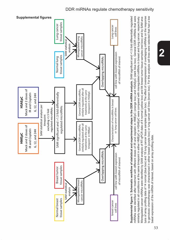
53
Gen
eral
DD
R m
icro
RNA
s;
sim
ilarly
regu
late
d af
ter a
ll tr
eatm
ents
at t
he s
ame
timep
oint
in
HM
EpC
Gen
eral
DD
R m
icro
RNA
s;
sim
ilarly
regu
late
d af
ter a
ll tr
eatm
ents
at t
he s
ame
timep
oint
in H
SAEp
C
HM
EpC
Moc
kan
d2
dose
s of
IR a
ndCi
splatin
6, 1
2, a
nd24
H
HSA
EpC
Moc
kan
d2
dose
s of
IR a
ndCi
splatin
6, 1
2, a
nd24
H
SAM
sig
ni�c
ant a
nd >
1.5
fold
di�
eren
tially
re
gula
ted
mic
roRN
As
SAM
stati
stica
lana
lysis
per
tim
epoi
nt
Ove
rlapp
ing
mic
roRN
AsO
verla
ppin
g m
icro
RNAs
Char
acte
rizat
ion
of m
icro
RNA
s in
bre
ast
or l
ung
canc
er c
ell l
ines
Nor
mal
bre
ast
tissu
e sa
mpl
es
Brea
st tu
mor
tis
sue
sam
ples
N
orm
al lu
ng
tissu
e sa
mpl
es
Lung
can
cer
tissu
e sa
mpl
es
SAM
stati
stica
lan
alys
is SA
M st
atisti
cal
anal
ysis
Mis
regu
late
d br
east
ca
ncer
mic
roRN
As
Mis
regu
late
d lu
ng
canc
er m
icro
RNA
s Lung
can
cer
cell
lines
brea
st c
ance
r ce
ll lin
es
excl
udin
g <1
.5 fo
ld
regu
late
d m
icro
RNAs
cell
line
with
low
est e
xpre
ssio
n of
mic
roRN
A o
f int
eres
tce
ll lin
e w
ith lo
wes
t exp
ress
ion
of m
icro
RNA
of i
nter
est
Sup
plem
enta
l fig
ure
1
Supp
lem
enta
l figu
re 1
: Sch
emat
ic w
orkfl
ow o
f sta
tistic
al a
nd e
xper
imen
tal s
teps
in th
e D
DR
miR
NA
anal
ysis
. SAM
sig
nific
ant a
nd >
1.5
fold
diff
eren
tially
regu
late
d m
iRN
As w
ere
iden
tified
afte
r tre
atm
ent w
ith d
iffer
ent d
oses
of I
R a
nd c
ispl
atin
in H
MEp
C (o
rang
e bo
x) a
nd H
SAEp
C (d
ark
blue
box
). G
ener
al D
DR
miR
NAs
that
wer
e si
mila
rly re
gula
ted
in a
ll co
nditi
ons
at th
e sa
me
timep
oint
in e
ither
HM
EpC
or H
SAEp
C w
ere
com
pare
d w
ith m
isre
gula
ted
miR
NAs
in b
reas
t and
lung
can
cer,
resp
ectiv
ely.
M
isre
gula
ted
canc
er m
iRN
As w
ere
iden
tified
by
SAM
ana
lysi
s of
RT-
qPC
R d
ata
of 8
nor
mal
(yel
low
box
) and
84
brea
st tu
mor
tiss
ue s
ampl
es (r
ed b
ox) a
nd b
y SA
M a
na-
lysi
s of
arra
y pr
ofilin
g da
ta o
f 14
norm
al (b
lue
box)
and
18
lung
can
cer t
issu
e sa
mpl
es (g
reen
box
). Th
e ef
fect
of o
vere
xpre
ssio
n of
ove
rlapp
ing
miR
NAs
on
the
resp
onse
to
gen
otox
ic c
hem
othe
rapy
was
cha
ract
eriz
ed in
eith
er b
reas
t (pu
rple
box
) or l
ung
canc
er c
ell l
ines
(bro
wn
box)
. For
this
ana
lysi
s ce
ll lin
es w
ere
sele
cted
that
had
a lo
w
basa
l exp
ress
ion
of th
e m
iRN
A of
inte
rest
, bas
ed o
n ar
ray
profi
ling
data
.
Supplemental figures
2
DDR miRNAs regulate chemotherapy sensitivity
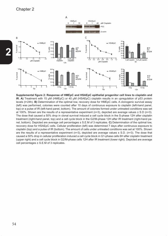
54
A
P53
β-actin
0 1 40 μM Cisplatin0 1 15HSAEpCHMEpC
Supplemental figure 2
B
HMEpC
0.1
1.0
10
100
0.0 0.5 1.0 1.5 2.0μM Cisplatin
% s
urvi
val
10
100
0 1 2 3 4 5Gy
% s
urvi
val HMEpC
0.0
10.0
20.0
30.0
40.0
50.0
0 μM 0.25 μM
G1S G2/M%
cel
ls
12h
% c
ells
12h
0.0
10.0
20.0
30.0
40.0
50.0
0 Gy 1.5 Gy
G1S G2/M
C
10
100
0 1 2 3 4Gy
% s
urvi
val HSAEpC
10
100
0.0 0.2 0.4 0.6 0.8 1.0 1.2μM Cisplatin
% s
urvi
val
HSAEpC
0.010.020.030.040.050.060.070.080.0
0 Gy 1.5 Gy
G1S G2/M%
cel
ls
12h
0.010.020.030.040.050.060.070.0
0 μM cisplatin 0.25 μM cisplatin
G1S G2/M%
cel
ls
6hcisplatincisplatin
Supplemental figure 2: Response of HMEpC and HSAEpC epithelial progenitor cell lines to cisplatin and IR. A) Treatment with 15 µM (HMEpC) or 40 µM (HSAEpC) cisplatin results in an upregulation of p53 protein levels (t=24h). B) Determination of the optimal low, recovery dose for HMEpC cells. A clonogenic survival assay (left) was performed, colonies were counted after 10 days of continuous exposure to cisplatin (left-hand panel, top) or a pulse of IR (left-hand panel, bottom). The amount of colonies formed under untreated conditions was set at 100%. Shown are the results of a representative experiment (n=3), depicted are average values ± S.D (n=3). The dose that caused a 50% drop in clonal survival induced a cell cycle block in the S-phase 12H after cisplatin treatment (right-hand panel, top) and a cell cycle block in the G2/M-phase 12H after IR treatment (right-hand pa-nel, bottom). Depicted are average cell percentages ± S.E.M of 3 replicates. C) Determination of the optimal low, recovery dose for HSAEpC cells. Cellular proliferation (left) was determined 7 days after continuous exposure to cisplatin (top) and a pulse of IR (bottom). The amount of cells under untreated conditions was set at 100%. Shown are the results of a representative experiment (n=3), depicted are average values ± S.D. (n=3). The dose that caused a 50% drop in cellular proliferation induced a cell cycle block in G1-phase cells 6H after cisplatin treatment (upper right) and a cell cycle block in G2/M-phase cells 12H after IR treatment (lower right). Depicted are average cell percentages ± S.E.M of 3 replicates.
2
Chapter 2

55
0
20
40
60
80
100
120
140
0.1 1.0 10 100 1000μM Cisplatin
% v
iabi
lity
HMEpC
HSAEpCPARP1
cleaved PARP
0 1 40 μM cisplatin0 1 15HSAEpCHMEpC
β-actin
0
20
40
60
80
100
120
0 2 4 6 8 10 12 14Gy
% c
ells
HSAEpCHMEpC
Supplemental �gure 3
A B
C
D
E
HMEpC t=24H
0
20
40
60
80
100
120
0 2 4 6 8Gy
% v
iabili
ty
HSAEpCt=24H
0
20
40
60
80
100
120
0 2 4 6 8Gy
% v
iabili
ty
05000
100001500020000250003000035000400004500050000
0 Gy 1.5 Gy 6.0 Gy
AU IL
-8 le
vels
in H
MEp
C
0
50
100
150
200
250
300
350
0 Gy 1.5 Gy 6.0 Gy
AU IL
-6 le
vels
in H
MEp
C
0
5000
10000
15000
20000
25000
30000
0 Gy 1.5 Gy 6.0 Gy
AU IL
-6 le
vels
in H
SAEp
C
0
20000
40000
60000
80000
100000
120000
140000
160000
0 Gy 1.5 Gy 6.0 Gy
AU IL
-8 le
vels
in H
SAEp
C
Supplemental figure 3: Response of HMEpC and HSAEpC epithelial progenitor cell lines to high doses of cisplatin and IR. A) Determination of the optimal high cisplatin dose for HMEpC and HSAEpC cells. Cellular viability was determined with an MTT assay after 48h of continuous exposure to cisplatin. The viability of untreated cells was set at 100%. Depicted is the average viability of four independent experiments ± S.D. B) Treatment with the IC50 dose of cisplatin dose results in a decrease of uncleaved PARP1 protein levels. C) IR does not affect viability of HMEpC and HSAEpC cells as determined by MTT assay after 24h. The viability of untreated cells was set at 100%. The standard deviation represents the variation within one experiment. D) Determination of the optimal high IR dose for HMEpC and HSAEpC cells using a cellular proliferation assay. Cellular density was determined at 48h after treatment with the indicated dose of IR. The amount of cells under untreated conditions was set at 100%. Shown are the results of a representative experiment (n=3), depicted are average values ± S.D. of 3 replicates. E) IR induced secretion of IL-8 (left) and IL-6 (right). Cell culture medium was collected at 13 days from untreated or IR exposed cells. IL-6 and IL-8 levels were determined using human IL6/IL8 capture beads by FACS. IL-6 and IL-8 levels (arbitrary units) in HMEpC (top) and HSAEpC (bottom) are depicted.
2
DDR miRNAs regulate chemotherapy sensitivity

56
0.70
0.71
0.72
0.73
0.74
0.75
0.76
non-replicates replicates
Pear
son
corr
elati
on-c
oeffi
cien
tCorrelation between (non-)replicates
Supplemental figure 4
Supplemental figure 4: Correlation of the replicates.Human miRNA expression values were used to calculate the Pearson correlation-coefficient between the replicates (n=4 for 30 conditions) and every possible combination of non-replicates. The average correlation-coefficient of re-plicates and non-replicates is depicted in the graph and a T-test showed that the difference is significant (p<0.01).
Supp
lem
enta
l figu
re 5
, nex
t 2 p
ages
: Tre
atm
ent-s
peci
fic a
nd d
ose-
spec
ific
miR
NA
s in
HM
EpC
and
HSA
EpC
. Prim
ary
epith
elia
l cel
ls w
ere
treat
ed w
ith th
e in
dica
ted
dose
s of
cis
plat
in (µ
M) a
nd IR
(Gy)
. Eac
h m
iRN
A w
as id
entifi
ed b
y SA
M s
tatis
tics,
but
sig
nific
ant m
iRN
As w
ith a
fold
-cha
nge
low
er th
an 1
.5 fo
ld w
ere
excl
uded
. Sho
wn
are
stan
dard
hea
tmap
s, w
hich
dep
ict t
he a
vera
ge fo
ld c
hang
e. T
o ca
lcul
ate
the
aver
age
fold
cha
nge,
we
divi
ded
the
aver
age
expr
essi
on v
alue
afte
r dam
age
(n=4
) by
the
aver
age
cont
rol v
alue
(n=4
). A
) Hea
t map
with
miR
NAs
that
are
spe
cific
ally
regu
late
d in
resp
onse
to e
ither
IR o
r cis
plat
in tr
eatm
ent i
n H
MEp
C a
t 6h,
12h
and
24h
of g
eno-
toxi
c st
ress
. B) H
eat m
ap w
ith m
iRN
As th
at a
re s
peci
fical
ly re
gula
ted
in re
spon
se to
eith
er a
hig
h do
se o
r a lo
w d
ose
of IR
and
cis
plat
in tr
eatm
ent i
n H
MEp
C a
t 6h,
12h
and
24
h of
gen
otox
ic s
tress
. C) H
eat m
ap w
ith m
iRN
As th
at a
re s
peci
fical
ly re
gula
ted
6h a
fter 6
Gy
and
12h
afte
r 1.5
Gy
of IR
in H
MEp
C. D
) Hea
t map
with
miR
NAs
that
sho
w
oppo
site
regu
latio
n af
ter t
reat
men
t with
a h
igh
dose
of c
ispl
atin
and
a h
igh
dose
of I
R in
HM
EpC
at 6
h, 1
2h a
nd 2
4h o
f gen
otox
ic s
tress
. E) H
eat m
ap w
ith m
iRN
As th
at
are
spec
ifica
lly re
gula
ted
in re
spon
se to
eith
er IR
or c
ispl
atin
trea
tmen
t in
HSA
EpC
at 6
h, 1
2h a
nd 2
4h o
f gen
otox
ic s
tress
. F) H
eat m
ap w
ith m
iRN
As th
at a
re s
peci
fical
ly
regu
late
d in
resp
onse
to e
ither
a h
igh
dose
or a
low
dos
e of
IR a
nd c
ispl
atin
trea
tmen
t in
HSA
EpC
at 6
h, 1
2h a
nd 2
4h o
f gen
otox
ic s
tress
. A-F
) The
fold
cha
nge
indi
cate
s th
e di
ffere
nce
in e
xpre
ssio
n co
mpa
red
to m
ock
treat
ed c
ells
at t
he s
ame
time
poin
t. To
giv
e th
e be
st re
pres
enta
tion
of d
iffer
ence
s in
exp
ress
ion,
we
chos
e ar
bitra
ry c
olor
s to
dep
ict f
old
chan
ges
betw
een
-1.7
5 en
+1.
75. A
whi
te c
olor
indi
cate
s a
fold
cha
nge
of 1
.00.
2
Chapter 2

57
Supp
lem
enta
l �gu
re 5
A
hsa-
miR
-656
hsa-
miR
-15a
hsa-
miR
-16
hsa-
miR
-584
hsa-
miR
-492
hsa-
miR
-7-2
*hs
a-m
iR-3
71-5
phs
a-m
iR-5
51a
hsa-
miR
-342
-5p
hsa-
miR
-574
hsa-
let-7
ehs
a-m
iR-9
3hs
a-m
iR-5
09-5
phs
a-m
iR-2
3b
hsa-
miR
-497
hsa-
miR
-183
hsa-
miR
-486
-3p
0.25μM 15μM1.5gy6gy
0.25μM 15μM1.5gy6gy
0.25μM 15μM1.5gy6gy
24H
12H
6H
-1.7
5+1
.75
Fold
Cha
nge
hsa-
miR
-766
hsa-
miR
-939
hsa-
miR
-1hs
a-m
iR-2
5*hs
a-m
iR-5
76-3
phs
a-m
iR-5
16b
hsa-
miR
-485
-3p
hsa-
miR
-637
hsa-
miR
-140
-3p
hsa-
let-7
dhs
a-m
iR-1
35b*
hsa-
miR
-486
-3p
hsa-
miR
-490
-3p
hsa-
miR
-499
-3p
hsa-
miR
-506
hsa-
miR
-324
-3p
hsa-
miR
-125
a-3p
hsa-
miR
-876
-3p
hsa-
miR
-509
-5p
hsa-
miR
-492
hsa-
miR
-380
*hs
a-m
iR-1
908
hsa-
miR
-518
bhs
a-m
iR-5
18a-
3phs
a-m
iR-5
41*
hsa-
miR
-16
hsa-
miR
-608
-1.7
5+1
.75
Fold
Cha
nge
24H
12H
6H
0.25 μM 15 μM
6 Gy
15 μM1.5 Gy6 Gy
15 μM
6 Gy
1.5 Gy
0.25 μM
1.5 Gy
0.25 μM
C
hsa-
miR
-93
hsa-
miR
-602
hsa-
miR
-29c
*hs
a-m
iR-5
26b*
hsa-
miR
-18a
-1.7
5+1
.75
Fold
Cha
nge
24H
12H
6H
0.25μM 15μM1.5gy6gy
0.25μM 15μM1.5gy6gy
0.25μM 15μM1.5gy6gy
D0.25 μM15 μM1.5 Gy6 Gy
0.25 μM15 μM1.5 Gy6 Gy
0.25 μM15 μM1.5 Gy6 Gy hs
a-m
iR-3
26
hsa-
miR
-498
hsa-
miR
-223
*hs
a-m
iR-5
20a-
5phs
a-m
iR-1
86hs
a-m
iR-6
62hs
a-m
iR-2
3bhs
a-m
iR-5
03hs
a-m
iR-8
75-3
phs
a-m
iR-2
23hs
a-m
iR-6
56hs
a-m
iR-1
03-2
*hs
a-m
iR-3
74a
hsa-
miR
-652
-1.7
5+1
.75
Fold
Cha
nge
24H
12H
6H
E
hsa-
miR
-150
hsa-
miR
-297
hsa-
miR
-29b
-1*
hsa-
miR
-532
-3p
hsa-
miR
-590
-5p
hsa-
miR
-506
hsa-
miR
-509
-3p
hsa-
miR
-507
hsa-
miR
-410
hsa-
miR
-379
hsa-
miR
-508
-3p
hsa-
miR
-383
hsa-
miR
-513
a-5p
hsa-
miR
-129
*hs
a-m
iR-1
85*
hsa-
miR
-221
*
hsa-
miR
-637
hsa-
let-7
e*hs
a-m
iR-5
99hs
a-m
iR-2
23hs
a-m
iR-3
69-5
phs
a-m
iR-3
82hs
a-m
iR-2
0ahs
a-m
iR-3
1
hsa-
miR
-494
U6-
snR
NA-
1hs
a-m
iR-3
0c-2
*hs
a-m
iR-1
5b
0.25 μM 40 μM1.5 Gy6 Gy
0.25 μM 40 μM1.5 Gy6 Gy
0.25 μM 40 μM1.5 Gy6 Gy hs
a-m
iR-5
18b
hsa-
miR
-765
hsa-
miR
-34b
hsa-
miR
-194
hsa-
miR
-642
hsa-
miR
-628
-3p
hsa-
miR
-549
hsa-
miR
-630
hsa-
miR
-220
bhs
a-m
iR-6
47hs
a-m
iR-8
88*
hsa-
miR
-326
hsa-
miR
-886
-5p
hsa-
miR
-424
hsa-
miR
-298
-1.7
5+1
.75
Fold
Cha
nge
24H
12H
6H
F
0.25 μM 40 μM1.5 Gy6 Gy
0.25 μM 40 μM1.5 Gy6 Gy
0.25 μM 40 μM1.5 Gy6 Gy hs
a-m
iR-4
90-3
p
hsa-
miR
-32*
hsa-
miR
-518
c*hs
a-m
iR-6
42hs
a-m
iR-1
85*
hsa-
miR
-195
hsa-
miR
-15a
hsa-
miR
-181
a*hs
a-m
iR-6
34
U6-
snR
NA-
2
-1.7
5+1
.75
Fold
Cha
nge
24H
12H
6H
B
2
DDR miRNAs regulate chemotherapy sensitivity

58
Supp
lem
enta
l �gu
re 5
A
hsa-
miR
-656
hsa-
miR
-15a
hsa-
miR
-16
hsa-
miR
-584
hsa-
miR
-492
hsa-
miR
-7-2
*hs
a-m
iR-3
71-5
phs
a-m
iR-5
51a
hsa-
miR
-342
-5p
hsa-
miR
-574
hsa-
let-7
ehs
a-m
iR-9
3hs
a-m
iR-5
09-5
phs
a-m
iR-2
3b
hsa-
miR
-497
hsa-
miR
-183
hsa-
miR
-486
-3p
0.25μM 15μM1.5gy6gy
0.25μM 15μM1.5gy6gy
0.25μM 15μM1.5gy6gy
24H
12H
6H
-1.7
5+1
.75
Fold
Cha
nge
hsa-
miR
-766
hsa-
miR
-939
hsa-
miR
-1hs
a-m
iR-2
5*hs
a-m
iR-5
76-3
phs
a-m
iR-5
16b
hsa-
miR
-485
-3p
hsa-
miR
-637
hsa-
miR
-140
-3p
hsa-
let-7
dhs
a-m
iR-1
35b*
hsa-
miR
-486
-3p
hsa-
miR
-490
-3p
hsa-
miR
-499
-3p
hsa-
miR
-506
hsa-
miR
-324
-3p
hsa-
miR
-125
a-3p
hsa-
miR
-876
-3p
hsa-
miR
-509
-5p
hsa-
miR
-492
hsa-
miR
-380
*hs
a-m
iR-1
908
hsa-
miR
-518
bhs
a-m
iR-5
18a-
3phs
a-m
iR-5
41*
hsa-
miR
-16
hsa-
miR
-608
-1.7
5+1
.75
Fold
Cha
nge
24H
12H
6H
0.25 μM 15 μM
6 Gy
15 μM1.5 Gy6 Gy
15 μM
6 Gy
1.5 Gy
0.25 μM
1.5 Gy
0.25 μM
C
hsa-
miR
-93
hsa-
miR
-602
hsa-
miR
-29c
*hs
a-m
iR-5
26b*
hsa-
miR
-18a
-1.7
5+1
.75
Fold
Cha
nge
24H
12H
6H
0.25μM 15μM1.5gy6gy
0.25μM 15μM1.5gy6gy
0.25μM 15μM1.5gy6gy
D
0.25 μM15 μM1.5 Gy6 Gy
0.25 μM15 μM1.5 Gy6 Gy
0.25 μM15 μM1.5 Gy6 Gy hs
a-m
iR-3
26
hsa-
miR
-498
hsa-
miR
-223
*hs
a-m
iR-5
20a-
5phs
a-m
iR-1
86hs
a-m
iR-6
62hs
a-m
iR-2
3bhs
a-m
iR-5
03hs
a-m
iR-8
75-3
phs
a-m
iR-2
23hs
a-m
iR-6
56hs
a-m
iR-1
03-2
*hs
a-m
iR-3
74a
hsa-
miR
-652
-1.7
5+1
.75
Fold
Cha
nge
24H
12H
6H
E
hsa-
miR
-150
hsa-
miR
-297
hsa-
miR
-29b
-1*
hsa-
miR
-532
-3p
hsa-
miR
-590
-5p
hsa-
miR
-506
hsa-
miR
-509
-3p
hsa-
miR
-507
hsa-
miR
-410
hsa-
miR
-379
hsa-
miR
-508
-3p
hsa-
miR
-383
hsa-
miR
-513
a-5p
hsa-
miR
-129
*hs
a-m
iR-1
85*
hsa-
miR
-221
*
hsa-
miR
-637
hsa-
let-7
e*hs
a-m
iR-5
99hs
a-m
iR-2
23hs
a-m
iR-3
69-5
phs
a-m
iR-3
82hs
a-m
iR-2
0ahs
a-m
iR-3
1
hsa-
miR
-494
U6-
snR
NA-
1hs
a-m
iR-3
0c-2
*hs
a-m
iR-1
5b
0.25 μM 40 μM1.5 Gy6 Gy
0.25 μM 40 μM1.5 Gy6 Gy
0.25 μM 40 μM1.5 Gy6 Gy hs
a-m
iR-5
18b
hsa-
miR
-765
hsa-
miR
-34b
hsa-
miR
-194
hsa-
miR
-642
hsa-
miR
-628
-3p
hsa-
miR
-549
hsa-
miR
-630
hsa-
miR
-220
bhs
a-m
iR-6
47hs
a-m
iR-8
88*
hsa-
miR
-326
hsa-
miR
-886
-5p
hsa-
miR
-424
hsa-
miR
-298
-1.7
5+1
.75
Fold
Cha
nge
24H
12H
6H
F
0.25 μM 40 μM1.5 Gy6 Gy
0.25 μM 40 μM1.5 Gy6 Gy
0.25 μM 40 μM1.5 Gy6 Gy hs
a-m
iR-4
90-3
p
hsa-
miR
-32*
hsa-
miR
-518
c*hs
a-m
iR-6
42hs
a-m
iR-1
85*
hsa-
miR
-195
hsa-
miR
-15a
hsa-
miR
-181
a*hs
a-m
iR-6
34
U6-
snR
NA-
2
-1.7
5+1
.75
Fold
Cha
nge
24H
12H
6H
B
2
Chapter 2

59
Supp
lem
enta
l fig
ure
6
hsa-
miR
-921
hsa-
miR
-183
*hs
a-m
iR-8
77hs
a-m
iR-2
5*hs
a-m
iR-6
68hs
a-m
iR-3
71-3
phs
a-m
iR-2
99-3
phs
a-m
iR-5
16a-
5phs
a-m
iR-5
16b
hsa-
miR
-194
*hs
a-m
iR-5
13a-
5phs
a-m
iR-1
hsa-
miR
-183
hsa-
miR
-26b
hsa-
miR
-886
-5p
hsa-
let-6
08hs
a-m
iR-2
96-5
phs
a-m
iR-5
18a-
3phs
a-m
iR-6
15-3
phs
a-m
iR-3
69-5
phs
a-le
t-7a
hsa-
miR
-26a
hsa-
miR
-29b
-1*
hsa-
miR
-506
hsa-
miR
-379
hsa-
let-7
f-1*
hsa-
miR
-324
-3p
hsa-
miR
-125
a-3p
hsa-
let-4
98hs
a-m
iR-1
40-5
phs
a-m
iR-8
88*
hsa-
miR
-499
-3p
hsa-
miR
-141
hsa-
miR
-518
c*hs
a-m
iR-5
41*
hsa-
miR
-15a
hsa-
miR
-574
-5p
hsa-
miR
-509
-5p
hsa-
miR
-549
hsa-
miR
-130
7hs
a-m
iR-5
76-3
phs
a-m
iR-5
50hs
a-m
iR-1
25a-
5phs
a-m
iR-6
27hs
a-m
iR-2
06hs
a-m
iR-4
93hs
a-m
iR-6
28-3
phs
a-m
iR-1
93a-
5phs
a-m
iR-6
30hs
a-m
iR-5
82-3
phs
a-m
iR-4
97hs
a-le
t-7e
hsa-
miR
-708
hsa-
miR
-222
hsa-
miR
-886
-3p
hsa-
miR
-767
-5p
hsa-
miR
-29c
*hs
a-m
iR-2
2hs
a-m
iR-3
65hs
a-m
iR-6
34hs
a-m
iR-4
91-5
phs
a-m
iR-2
1hs
a-m
iR-1
93a-
3phs
a-m
iR-3
74a*
hsa-
miR
-497
*0.25μM 15μM1.5gy6gy
0.25μM 40μM1.5gy6gy
HM
EpC
HSA
EpC
12H
hsa-
let-7
e*hs
a-m
iR-3
30-3
phs
a-19
3a-5
phs
a-m
iR-1
29*
hsa-
miR
-10a
*hs
a-m
iR-1
38-1
*hs
a-m
iR-1
6hs
a-m
iR-4
85-3
phs
a-m
iR-2
5*hs
a-m
iR-5
99hs
a-m
iR-5
51b
hsa-
miR
-888
*hs
a-m
iR-2
3bhs
a-m
iR-2
99-3
phs
a-m
iR-5
09-3
-5p
hsa-
miR
-371
-3p
hsa-
miR
-369
-3p
hsa-
miR
-491
-3p
hsa-
miR
-7-2
*
hsa-
miR
-939
hsa-
miR
-541
*hs
a-m
iR-6
25hs
a-m
iR-1
307
hsa-
miR
-499
-3p
hsa-
miR
-550
hsa-
miR
-379
hsa-
miR
-382
hsa-
miR
-506
hsa-
miR
-326
hsa-
miR
-190
8hs
a-m
iR-1
51-3
phs
a-m
iR-5
09-3
phs
a-m
iR-5
07hs
a-m
iR-5
08-3
phs
a-m
iR-3
83
hsa-
miR
-410
hsa-
miR
-583
hsa-
miR
-369
-5p
hsa-
miR
-149
*hs
a-m
iR-5
18c*
hsa-
miR
-374
b*hs
a-m
iR-3
80*
hsa-
miR
-185
*hs
a-m
iR-3
4bhs
a-m
iR-1
83hs
a-m
iR-9
21hs
a-m
iR-1
83*
hsa-
miR
-518
bhs
a-m
iR-5
57
0.25μM 15μM1.5gy6gy
0.25μM 40μM1.5gy6gy hs
a-m
iR-8
77
hsa-
miR
-297
HM
EpC
HSA
EpC
6H
A
-1.7
5+1
.75
Fold
Cha
nge
HM
EpC
HSA
EpC
hsa-
miR
-371
-5p
hsa-
miR
-374
b*hs
a-m
iR-3
42-5
phs
a-m
iR-8
90hs
a-m
iR-5
26b*
hsa-
miR
-518
c*hs
a-m
iR-1
hsa-
miR
-513
a-5p
hsa-
miR
-876
-3p
hsa-
miR
-516
a-5p
hsa-
miR
-107
hsa-
miR
-509
-5p
hsa-
miR
-93
hsa-
miR
-194
*hs
a-le
t-7a
hsa-
miR
-499
-3p
hsa-
miR
-369
-3p
hsa-
miR
-380
*hs
a-m
iR-6
12hs
a-m
iR-5
09-3
-5p
hsa-
miR
-130
7hs
a-m
iR-5
08-3
phs
a-m
iR-2
2*hs
a-m
iR-1
49*
hsa-
miR
-518
a-5p
hsa-
let-7
e
0.25μM 15μM1.5gy6gy
0.25μM 40μM1.5gy6gy
HM
EpC
HSA
EpC
24H
Supp
lem
enta
l figu
re 6
, Leg
enda
nex
t pag
e
2
DDR miRNAs regulate chemotherapy sensitivity

60
B 0.25μM 15μM1.5gy6gy
0.25μM40μM1.5gy6gy hs
a-m
iR-5
48b-
5phs
a-m
iR-1
93a-
5phs
a-m
iR-5
57hs
a-m
iR-2
4-1*
hsa-
miR
-26a
-2*
hsa-
miR
-300
hsa-
miR
-124
*hs
a-m
iR-3
65hs
a-m
iR-7
a-2*
hsa-
miR
-130
b*hs
a-m
iR-3
39-5
phs
a-m
iR-6
62hs
a-m
iR-3
79hs
a-m
iR-5
42-3
phs
a-m
iR-5
41*
hsa-
miR
-625
hsa-
miR
-550
hsa-
miR
-891
ahs
a-m
iR-5
86hs
a-m
iR-9
22hs
a-m
iR-4
55-3
p
hsa-
miR
-526
ahs
a-m
iR-1
30a*
hsa-
miR
-624
hsa-
miR
-181
bhs
a-m
iR-6
45hs
a-m
iR-1
81d
hsa-
miR
-532
-5p
hsa-
miR
-325
hsa-
miR
-502
-3p
hsa-
miR
-17*
hsa-
miR
-194
hsa-
miR
-186
hsa-
miR
-876
-5p
hsa-
miR
-758
hsa-
miR
-130
7hs
a-m
iR-2
18-2
*hs
a-m
iR-1
48b*
hsa-
miR
-493
hsa-
miR
-335
*
hsa-
miR
-891
b
hsa-
miR
-585
hsa-
miR
-30e
hsa-
miR
-140
-5p
hsa-
miR
-30a
*hs
a-m
iR-1
26*
hsa-
miR
-708
*H
MEp
CH
SAEp
C6H
HM
EpC
HSA
EpC
hsa-
miR
-625
hsa-
miR
-526
bhs
a-m
iR-4
23-3
phs
a-m
iR-2
18hs
a-m
iR-6
21hs
a-m
iR-1
39-3
phs
a-m
iR-4
48
hsa-
miR
-487
ahs
a-m
iR-6
26hs
a-m
iR-5
16a-
3phs
a-m
iR-1
28hs
a-m
iR-5
19e*
hsa-
miR
-889
hsa-
miR
-181
a-2*
hsa-
miR
-106
a*
hsa-
miR
-526
b*
hsa-
miR
-103
-2*
hsa-
miR
-554
hsa-
miR
-518
ehs
a-m
iR-6
60hs
a-m
iR-4
11hs
a-m
iR-3
4a*
hsa-
miR
-29c
hsa-
miR
-567
hsa-
miR
-19b
hsa-
miR
-564
hsa-
miR
-376
a*hs
a-m
iR-6
57hs
a-m
iR-7
08*
0.25μM 15μM1.5gy6gy
0.25μM40μM1.5gy6gy
24H
-1.7
5+1
.75
Fold
Cha
nge
hsa-
miR
-526
ahs
a-m
iR-4
99-3
phs
a-m
iR-7
08*
hsa-
miR
-106
b*hs
a-m
iR-1
48b
hsa-
miR
-17
hsa-
miR
-518
c*
hsa-
miR
-587
hsa-
miR
-204
hsa-
miR
-575
hsa-
miR
-25
hsa-
miR
-885
-3p
hsa-
miR
-509
-5p
hsa-
miR
-720
hsa-
miR
-422
a
hsa-
miR
-141
hsa-
miR
-24
hsa-
miR
-429
hsa-
let-7
e*hs
a-m
iR-2
22*
hsa-
miR
-519
ehs
a-m
iR-6
41
0.25μM 15μM1.5gy6gy
0.25μM40μM1.5gy6gy
HM
EpC
HSA
EpC
12H
Supp
lem
enta
l figu
re 6
, las
t 2 p
ages
: Com
paris
on o
f DD
R m
iRN
As
in H
MEp
C a
nd H
SAEp
C.
Prim
ary
epith
elia
l cel
ls w
ere
treat
ed w
ith th
e in
dica
ted
dose
s of
cis
plat
in (µ
M) a
nd IR
(Gy)
and
eac
h di
ffere
ntia
lly re
gula
ted
miR
NA
was
iden
tified
by
SAM
sta
tistic
s. S
how
n ar
e st
anda
rd h
eatm
aps,
whi
ch
depi
ct th
e av
erag
e fo
ld c
hang
e. T
o ca
lcul
ate
the
aver
age
fold
cha
nge,
we
divi
ded
the
aver
age
ex-
pres
sion
val
ue a
fter d
amag
e (n
=4) b
y th
e av
erag
e co
ntro
l val
ue (n
=4).
A) E
ach
miR
NA
was
iden
tified
by
SAM
sta
tistic
s in
eith
er H
MEp
C o
r HSA
EpC
, but
sig
nific
ant m
iRN
As w
ith a
fold
-cha
nge
low
er th
an
1.5
fold
wer
e ex
clud
ed. H
eat m
ap in
clud
es m
iRN
As th
at w
ere
regu
late
d in
all
geno
toxi
c co
nditi
ons
in
eith
er H
MEp
C o
r HSA
EpC
. Alth
ough
ther
e ar
e so
me
sim
ilarit
ies,
HM
EpC
and
HSA
EpC
mai
nly
show
di
ffere
nt m
iRN
A ex
pres
sion
pat
tern
s at
the
sam
e tim
epoi
nts.
B) M
iRN
As th
at s
how
a s
imila
r reg
ula-
tion
unde
r all
geno
toxi
c co
nditi
ons
at th
e sa
me
timep
oint
s in
bot
h H
MEp
C a
nd H
SAEp
C. I
ndic
ated
ar
e m
iRN
As th
at w
ere
iden
tified
by
perfo
rmin
g SA
M s
tatis
tics
of th
e co
mbi
ned
HM
EpC
and
HSA
EpC
ex
pres
sion
dat
a. T
he fo
ld c
hang
e in
dica
tes
the
diffe
renc
e in
exp
ress
ion
com
pare
d to
moc
k tre
ated
ce
lls a
t the
sam
e tim
e po
int.
To g
ive
the
best
repr
esen
tatio
n of
diff
eren
ces
in e
xpre
ssio
n, w
e ch
ose
arbi
trary
col
ors
to d
epic
t fol
d ch
ange
s be
twee
n -1
.75
en +
1.75
. A w
hite
col
or in
dica
tes
a fo
ld c
hang
e of
1.0
0.
2
Chapter 2

61
Supplemental table 1
Subtypes NSCLC
squamous 5large cell 6adenocarcinoma 6adenosquamous 1
Normal lung tissue 16
Subtypes breast cancer
Luminal B-like 12Luminal A-like 25Basal-like 8ERBB2-like 8Normal-like 5Unknown 26
Normal breast tissue 8
Supplemental table 1: Overview of breast and lung tumor tissue subtypes used for analysis of misexpressed miRNAs.
Supplemental table 2
Breast tumor miRNAs that are general responders to DNA damage
cpt dox pac
let-7a 0.169 0.013 0.910
miR-296-5p 0.622 0.208 0.247
miR-193a-3p 0.020 0.097 0.002
miR-183 0.047 0.048 0.502
miR-23b 0.118 0.002 0.411
miR-34b 0.448 0.188 0.030
miR-369-3p 0.869 0.357 0.504
miR-93 0.422 0.901 0.028
miR-141 0.000 0.005 0.054
33% 44% 33%
Breast tumor miRNAs that are not general responders to DNA damage
cpt dox pac
let-7b 0.091 0.002 0.682
let-7f 0.110 0.309 0.716
miR-103 0.322 0.041 0.382
miR-106b 0.126 0.460 0.870
miR-128 0.366 0.906 0.205
miR-130b 0.093 0.683 0.534
miR-133a 0.069 0.012 0.374
miR-145 0.490 0.647 0.490
miR-148a 0.776 0.472 0.956
miR-148b 0.237 0.868 0.867
miR-150 0.312 0.164 0.602
miR-15a 0.023 0.451 0.244
miR-15b 0.058 0.007 0.716
miR-18 0.803 0.831 0.053
miR-181a 0.308 0.706 0.203
miR-181c 0.156 0.972 0.433
miR-182 0.228 0.113 0.511
miR-185 0.102 0.568 0.397
miR-186 0.850 0.946 0.201
miR-18b 0.333 0.352 0.917
miR-197 0.322 0.490 0.929
miR-199a 0.380 0.493 0.008
miR-200b 0.000 0.012 0.035
miR-200c 0.000 0.046 0.105
miR-20b 0.644 0.687 0.084
miR-210 0.017 0.058 0.608
miR-212 0.065 0.629 0.728
miR-215 0.002 0.902 0.171
miR-218 0.036 0.950 0.399
miR-224 0.655 0.945 0.511
miR-25 0.626 0.012 0.792
miR-27a 0.024 0.001 0.669
miR-27b 0.013 0.013 0.219
miR-29b 0.121 0.059 0.224
miR-301 0.321 0.018 0.693
miR-324-5p 0.005 0.128 0.827
miR-330-3p 0.444 0.873 0.243
miR-339-5p 0.048 0.287 0.578
miR-340 0.068 0.013 0.683
miR-34a 0.386 0.679 0.015
miR-362-5p 0.288 0.152 0.224
miR-369-5p 0.592 0.343 0.010
miR-370 0.886 0.231 0.981
miR-371-3p 0.016 0.017 0.185
miR-372 0.074 0.955 0.106
miR-376a 0.379 0.472 0.956
miR-383 0.636 0.922 0.978
miR-424 0.820 0.359 0.393
miR-429 0.031 0.287 0.543
miR-452 0.473 0.997 0.436
miR-455-3p 0.792 0.123 0.718
miR-487b 0.193 0.659 0.583
miR-500 0.264 0.567 0.052
miR-503 0.793 0.973 0.984
miR-505 0.444 0.906 0.037
miR-518e 0.695 0.230 0.222
miR-518f 0.038 0.172 0.331
miR-96 0.465 0.820 0.030
miR-98 0.895 0.348 0.378
miR-99a 0.356 0.338 0.142
miR-99b 0.074 0.213 0.943
21% 20% 10%
Supplemental table 2: Deregulated breast tumor miRNAs that are generally regulated after DNA da-mage are more likely to be associated with drug response than tumor miRNAs not generally regula-ted after DNA damage. Depicted are the p-values of a Pearson correlation between miRNA expression levels (Blower et al., 2008) and drug sensitivity in the NCI60 pa-nel. Significant correlations (p<0.05; blue) are indicated and the percentage indicates the amount of miRNAs that showed a significant correlation.
Supplemental tables
2
DDR miRNAs regulate chemotherapy sensitivity

62
Supplemental table 3
Breast cancer cell lines
miR-296-5p hsa-let-7a miR-183 miR-193a-3pMM-MB-157 111.3 393.2 22.7 13.8MM-MB-231 281.4 683.9 5.0 19.1SK-BR-3 189.1 19.8 39.2 78.7range 92.2-370.4average 180.8 162.0 17.3 31.6
Lung cancer cell lines
miR-382 miR-16 miR-21H226 120.8 139.7 361.9H460 239.1 40.4 47.3range 120.8-1084.9average 383.3 108.3 97.1
5.1-683.9 6.5-107.95.0-42.0
32.2-268.3 9.4-361.9
Supplemental table 3: MiRNA expression levels of various breast and lung cancer cell lines. MiRNA ex-pression levels were determined in 52 breast cancer cell lines (Riaz et al., 2013) and 12 lung cancer cell lines. Indicated are the average and range of expression values in all cell lines and the expression levels in the three selected breast or lung cancer cell lines. The cell line that was selected for miRNA overexpression experiments is indicated in yellow.
Supplemental table 4
IC50 concentrations used for MTT assay of miRNA transfected cells
µM cisplatin µM doxorubicin µM paclitaxel
MDA-MB-157 100 110 8
MDA-MB-231 125 40 17
SK-BR-3 125 80 8
H226 80 450 8
H460 50 180 10
Supplemental table 4: IC50 treatment dose of cisplatin, paclitaxel and doxorubicin used for MTT assay in various breast and lung cancer cell lines.
2
Chapter 2

63
2
DDR miRNAs regulate chemotherapy sensitivity

64

65
Chapter 3
MicroRNA-processing RNA-binding proteins regulate DNA damage-induced microRNAs and
are misexpressed in breast cancer
in preparation
Maikel D. Wouters1, Kasper Derks1, Ricardo Leite1, Esther Jongste1, Wilfred F.J. van Ijcken2, Jan H.J. Hoeijmakers1, Erik A.C. Wiemer4, Steven
van Laere3, Dik C. van Gent1, Joris Pothof1
1. Department of Genetics, Erasmus University Medical Center, Rotterdam, The Netherlands2. Center for Biomics, Erasmus University Medical Center, Rotterdam, The Netherlands3. Translational Cancer Research Unit, Oncology Center, GZA hospitals St-Augustinus, Antwerp, Belgium.4. Department of Medical Oncology, Erasmus MC Cancer Institute, Erasmus University Medical Center, Rotterdam, The Netherlands
66
AbstractMicroRNAs are important factors in the cellular response after DNA damage, but not much is known about their regulation upon genotoxic insults. Recently, a RNA-binding protein, KHSRP, was characterized to regulate microRNA maturation after DNA damage. We fu-rther studied the role of KHSRP and five other microRNA-processing RNA-binding pro-teins, namely Tutase-4, SNIP1, Dhx9, Mov10 and HNRNPUL1, upon DNA damage. All these proteins were previously identified to be phosphorylated after DNA damage and we investigated how extensively they regulate mature microRNA levels upon DNA damage. For every microRNA-processing factor, we identified multiple microRNAs whose dama-ged-induced expression was dependent on these factors. Additionally, since a proper DNA damage response is important for preventing tumorigenesis, we assessed the significance of these microRNA-processing RNA-binding proteins in cancer. Therefore, we studied the expression of RNA-binding protein dependent microRNAs in various tumors. We found that KHSRP modulates the cellular outcome after DNA damage and UV-responsive microRNAs regulated by KHSRP and other microRNA-processing proteins were found mis-expressed in breast cancer samples. Our data strongly suggest that these proteins are not functional in a subset of breast cancer samples. In addition, misregulated expression of UV-respon-sive, KHSRP-dependent microRNAs in breast cancer tumors correlates with the Luminal subtype of breast tumors. Our work shows that microRNA processing is a relevant part of the cellular response on DNA damage and likely plays an important role in cancer.
3
Chapter 3

67
IntroductionDNA is damaged by various agents, such as UV light and reactive metabolic (by-)products. DNA lesions interfere with correct DNA replication and, if not repaired properly, can lead to mutations, deletions and chromosomal rearrangements that eventually may cause cancer. To counteract the deleterious effects of DNA damage, cells induce the DNA damage res-ponse (DDR) upon exposure to genotoxic agents. The DDR tightly regulates multiple cellu-lar processes, such as the cell cycle, DNA repair, apoptosis and cellular senescence. Cell cycle arrest allows time for DNA repair to restore the DNA. If DNA damage is beyond repair, the DDR can also induce apoptosis or cellular senescence, which counteracts tumorige-nesis [1-3]. Pre-malignant cells require defects in components of the DDR to proceed into more malignant states and therefore DDR defects are found in most, if not all, tumors [4, 5]. Correct regulation of the DDR is essential to prevent tumorigenesis. Upon DNA damage, the DNA damage checkpoint signaling cascade is induced to arrest the cell cycle and permit DNA repair. Signaling kinases ATM and ATR (ATM/R) are the central players of the DNA damage checkpoint activation as they phosphorylate a plethora of targets [6] that inhibit cell cycle progression and influence expression levels of many genes. For example, post-translational gene regulation by ATM/R modulates gene transcription by activating p53 [7]. However, besides post-translational and transcriptional gene regulation, emerging evidence shows that the DDR is also highly regulated post-transcriptionally by microRNAs [8]. These small non-coding RNA molecules negatively influence gene expression levels by binding to microRNA-responsive elements (MREs) in the 3’UTR of a mRNA target [9], mainly inducing their degradation [10]. MicroRNAs are transcribed by RNA polymerase II as pri-microRNAs. The Drosha-DGCR8 complex cleaves these transcripts and releases the precursor microRNAs (pre-microRNAs). These hairpin-structured molecules are exported into the cytosol, whe-re the Dicer-TRBP complex processes pre-microRNAs into double stranded, 22-nucleoti-des-long RNA molecules by removing the pre-microRNA terminal loop [11]. The strands of the resulting microRNA duplex are separated and one strand is incorporated into the RNA-Induced Silencing Complex (RISC) [11]. This complex contains one of the four Argo-naute proteins (AGO1-4), which are crucial for the interaction between a microRNA and its MRE within the 3’UTR of its target gene [12]. Not much is known about regulation of microRNA levels. Recent findings show that microRNAs can be post-transcriptionally regulated during their processing [13-15]. Various RNA-binding proteins (RBPs) selectively control processing of specific microRNAs. These proteins bind certain RNA sequences that flank the pre-microRNA hairpin or are located within the hairpin loop, controlling maturation and stability of microRNAs [16] by influencing the activity of the Drosha-DGCR8 [17], DICER-TRBP [13, 14] or RISC complex [18]. Selec-tive post-transcriptional processing of specific microRNAs by microRNA-processing RBPs could be a common mechanism to regulate mature microRNA levels after stimuli, such as DNA damage. This mechanism could play an important role, since microRNAs are rapidly regulated upon DNA damage [19-21], which can not only be explained by modulation of transcription [8], while RBPs are known factors in RNA-processing in response to DNA damage [22, 23]. Recently, a DNA damage responsive RBP was indeed identified to quickly regulate microRNA processing upon DNA damage; KHSRP, a Drosha co-factor, influences microR-NA-processing by binding to the terminal loop of specific pri-microRNAs [14]. KHSRP is directly phosphorylated and controlled by ATM after induction of DNA damage using the ra-diomimetic drug neocarzinostatin. This mechanism was the first example showing ATM-de-
3
RBPs regulate DDR miRNAs

68
pendent processing of specific DDR pri-microRNAs by a RBP [24]. Proteomic screening identified many other RBPs as ATM/R targets upon DNA damage [6]. We wondered whether other microRNA-processing RBPs are ATM/R targets and also control microRNA levels af-ter DNA damage. Since both the DDR [4, 5] and microRNA-processing by RBPs plays a role in tumorigenesis [16], we also studied the expression of RBP-dependent, UV-respon-sive microRNAs in breast cancer tumors. Our results revealed an important role for RBPs in microRNA-processing upon DNA damage and also indicated their potential influence in cancer.
Materials & MethodsCell culture and transfectionU2OS cells were cultured in a 1:1 mix of Lonza DMEM and F10 medium, supplemented with 10% FCS, penicillin (100 U/mL) and streptomycin (100 µg/mL), and incubated at 37°C with 5% CO2. For clonogenic survival assay, cells were grown at 3% O2 a week before and during transfection and the experiment. For transfection, cells were grown to 80% confluence in Ø3 cm wells and transfected twice with 100nM Dharmacon siRNAs using Invitrogen RNAimax according to manufacturer’s protocol.
Protein isolation and immunoblotU2OS cells were washed with ice-cold PBS and lyzed and collected in lysis buffer (25 mM Tris HCl, 20mM NaCl, 2mM EDTA/EGTA, 1% Triton and protease inhibitors). Proteins were separated on SDS-PAGE gel, transferred to nitrocellulose membranes and blocked in skim-milk. Rabbit anti-KHSRP (Cell signaling)(1/1000), Goat anti-Tuta-se4 (MyBioSource)(1/1000), Rabbit anti-Dhx9 (Abcam)(1/1000) and Rabbit anti-HNRNPUL1 (Sigma-Aldrich)(1/1000), anti-Tubulin, Goat anti-Ku70 (Santa Cruz)(1/2000) antibodies were used to stain proteins of interest. These proteins were visualized with GE Healthcare ECL-plus using sheep anti-mouse, sheep anti-rabbit and Donkey anti-goat HRP antibodies (Jackson Laboratories) (1/2000) and visualized on an Alliance scanner. Clonogenic survival assayFour hundred U2OS cells were seeded per Ø3 cm wells, and cultured at 3% O2. After 16 hours, cells were treated with cisplatin or one pulse of UV or IR. Untreated cells formed ~200 colonies after 7 days. Colonies were fixed and stained with 50% methanol, 7% acetic acid and 0.1% Coomassie-blue.
RNA isolation, RT-qPCR and microRNA microarray hybridisationU2OS cells were washed with RNAse-free PBS and total RNA was collected with 1mL Trizol (Invitrogen) per Ø3 cm well. RNA was extracted with chloroform, precipitated with isopropanol, washed twice with RNAse-free 70% ethanol, air dried and solubilized in RNAse free water. Total RNA was reverse transcribed with Applied Biosystems Taqman assay according to manufactures instructions. Expression levels of the RBPs of interest were measured using Applied Biosystems Taqman probes on a Biorad CFX-96 C1000 Thermal cycler. One µg total RNA was hybridized with an LNATM-based probeset (Exiqon; version 10) capable of detec-ting 725 human miRNAs. Arrays were scanned and quantified with Imagene software as described before [19]. After background subtraction, expression values were quantile normalized. MiRNA RT-qPCR expression analysis using microfluidic cards (A&B TLDA arrays, Applied Biosystems) was performed on 8 normal breast tissue samples and 84 breast tumor tissue samples as described before [25]. The represented breast cancer subtypes are listed in Supplemental table 1.
Statistical analysisLimma statistical analysis with Benjamini–Hochberg correction to adjust for false positives was carried out to iden-tify significant (P>0.05) differentially expressed miRNAs between mock and genotoxic treated cells or between control and siRNA-transfected cells. To identify RBP-dependent regulation of genotoxic microRNAs, we compa-red fold changes of significantly regulated microRNAs in control transfected cells with their DNA damage-induced fold changes in each RBP-depleted condition. Alternatively, significant genotoxic-induced fold changes for each depletion were compared with respective fold changes in control transfected cells. We only included microRNAs that minimally showed a one-third difference in DNA damage-induced fold change between control and RBP-de-
3
Chapter 3

69
pleted cells. Additionally, average genotoxic-induced expression values of control-transfected cells had to differ with average genotoxic expression values of RBP-depleted cells by a sigma of 2. Finally, low expressed microR-NAs with an average expression in all conditions below a specific cut-cut off were discarded. Hierarchical clustering and heatmap generation have been performed using TM4 microarray software suite, version 4.9.0 [26]. Pearson correlation analysis and significance has been calculated at www.socscistatis-tics.com and the Fisher exact test has been performed at www.quantpsy.org.
ResultsMany microRNAs are regulated upon DNA damage [8], but generally it is unclear how they are regulated by the DDR. RBPs could be potential factors in the DDR as they control microRNA levels. Recently KHSRP was indeed shown to regulate microRNA processing shortly upon DNA damage [24]. We investigated if KHSRP thereby influenced the DDR and has a role in cellular survival. Therefore, we analyzed the genotoxic sensitivity of KHS-RP-depleted cells in a clonogenic survival assay (Figure 1A). KHSRP-depleted cells (Figu-re 1B) were more sensitive for increasing doses of UV, Ionizing Radiation (IR) and cisplatin than control cells. This strongly suggests that KHSRP has a role in the DDR, protecting cells against a remarkably wide spectrum of DNA lesions, and shows that microRNA-pro-
Figure 1: Role of RBPs in the DDR and microRNA processing. A) Depletion of KHSRP sensitizes U2OS cells for increasing doses of cisplatin, IR and UV. The number of colonies formed in untreated conditions was set at 100% and shown are average values ± SAM (n=3). Note survival is indicated using a log-scale. B) KHSRP protein levels (75kDa) were depleted 2 days upon siRNA transfection. An a-specific band is indicated, while Tubulin is used as loading control C) The position of microRNA-processing RBPs within the microRNA processing pathway is shown. MicroRNA-processing RBPs identified from proteomic DNA-damage-induced phosphorylation screens are highlighted with a P [6, 27, 28], although actual direct phosphorylation by ATM/R has not been validated.
Figure 1
A
1
10
100
1000
0 1 2 3 4 5
% s
urvi
val
Gy IR
siControlsiKHSRP
1
10
100
1000
0 2 4 6 8 10
% s
urvi
val
J/m2UV
siControlsiKHSRP
0.1
1.0
10
100
1000
0 0.5 1.0 1.5 2.0 2.5
% s
urvi
val
µM cisplatin
siControlsiKHSRP
C
BsiC siK
HSRP
75kDa KHSRP
Tubulin
a-speci�c
KHSRP
Precurser microRNA
Primary microRNA
SMADSNIP1
MOV10
DHX9
Lin28Tutase4
HNRPUL1
DroshaDGCR8
DicerTRBP
Ago1-4Ago1-4Duplex mature microRNA
P
P
P
P
PP
3
RBPs regulate DDR miRNAs

70
cessing RBPs can play a role in the DDR. ATM regulates KHSRP-dependent differential processing of microRNAs [24]. Ho-wever, many other microRNAs are also regulated shortly after DNA damage [19, 21] and ATM-dependent, KHSRP-mediated microRNA processing cannot explain regulation of every DDR microRNA. Since many other RBPs are also identified as ATM/R targets [6], potentially including microRNA-processing factors, we set up to investigate which other DDR microRNAs could also be regulated by RBPs like KHSRP. To identify these RBPs, we studied two proteomic ATM/R substrate screens and a DDR phospho-proteomic screens [6, 27, 28]. These studies used various agents that induce single strand breaks (SSBs) and double strand breaks (DSBs), which activate ATM and ATR, respectively. These screens identified several microRNA-processing RBPs that are phosphorylated upon DNA dama-ge (supplemental table 2), including KHSRP. Furthermore, 5 other microRNA-processing RBPs were found: Tutase4 (Zcchc11), SNIP1, Dhx9, Mov10 and HNRNPUL1. Five of these 6 proteins were identified as ATM/R target in HEK293T cells 1 hour after treatment with 10gy of IR, while HNRNPUL1 was exclusively found phosphorylated in human melanoma G361 cells treated for 1 hour with radio-mimicking agent neocarzinostatin. Of these RBPs, KHSRP, SNIP1 and HNRNPUL1 are known to associate with Drosha [24, 27, 29] (Figure 1C). Both SNIP1 and KHSRP bind microRNAs and influence pri-microRNA processing [24, 29]. DHX9 and Zcchc11 associate with Lin28 and affect Dicer-mediated pre-microRNA processing [30-32], while Mov10 is associated with the Ago complex and might influence unwinding of the mature microRNA and its opposite strand [33]. Phosphorylation of these RBPs upon DNA damage suggests that they have a role in the DDR and may regulate microRNA processing and thereby influence microRNA levels upon DNA damage.
UV- and IR-induced microRNA responsesTo investigate which microRNAs depend on these microRNA-processing RBPs after geno-toxic treatment, we depleted expression levels of these RBPs in U2OS cells using siRNAs (Supplemental figure 1). Control and RBP-depleted cells were mock treated or treated with either 16J/m2 UV-radiation or 12Gy of IR (n=4) 48 hours after siRNA transfection. These treatments induce distinct types of DNA damage; generally, UV induces helix-distorting les-ions, most notably cyclobutane pyrimidine dimers and 6-4 photoproducts, while IR causes SSBs and DSBs. Our previous studies showed that microRNAs are quickly regulated upon UV, IR and cisplatin-induced DNA damage [19, 21], which might be mediated by RBP-de-pendent differential processing upon activation of the checkpoint response. To focus on these microRNAs, RNA was isolated 4 hours after start of treatment to minimize the contri-bution of differential transcription regulating microRNA levels. Total RNA was hybridized on microRNA expression arrays containing Locked Nucleic Acid-based capture probes [19] and we first analyzed the normal physiological microRNA response of control transfected cells after DNA damage. Of the 725 human mi-croRNA probes present on the array, 169 microRNAs were significantly regulated after UV radiation (Limma test with Benjamini–Hochberg correction, p<0.05)(Figure 2AB). In con-trast, only 20 microRNAs were significantly regulated upon IR (Figure 2C). The large dif-ference in the number of significantly regulated microRNAs between UV and IR treatment was also observed in every RBP-depleted condition (supplemental table 3). Additionally, another cell-line, HeLa also displayed a low number of significantly regulated microRNAs shortly after irradiation with 5Gy of IR, as identified by SAM analysis (p<0.05) (Figure 2D). Furthermore, IR-induced microRNAs in both HeLa and U2OS cells mainly increased after treatment and only microRNAs miR-603 and miR-637 were found regulated in both cell types. These results suggest that IR does not induce a thorough and common microRNA
3
Chapter 3
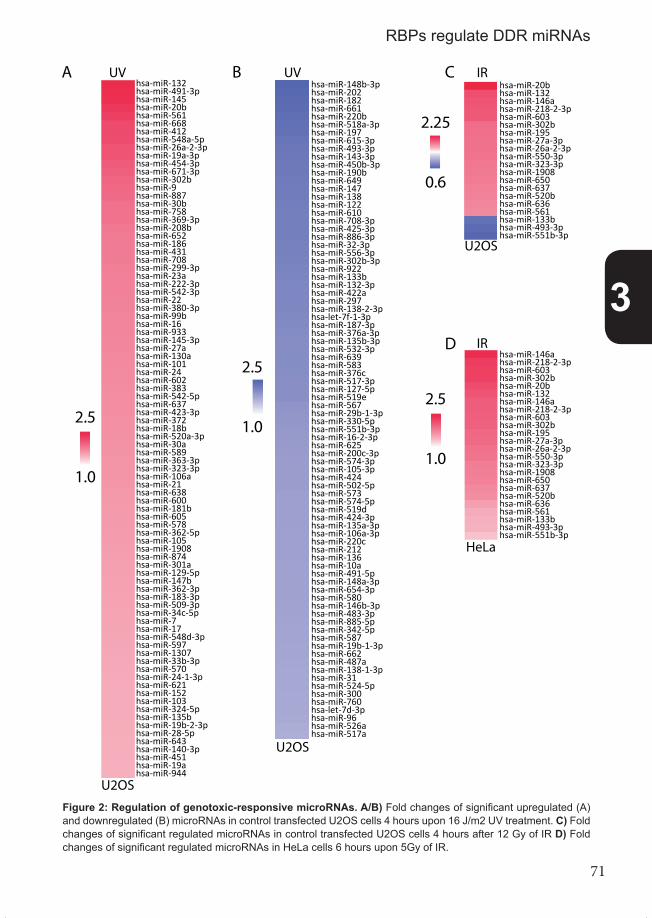
71
Figure 2
A B C
2.5
1.0
2.5
1.0
2.25
0.6
D
2.5
1.0
hsa-miR-28-5phsa-miR-643hsa-miR-140-3phsa-miR-451hsa-miR-19ahsa-miR-944
U2OS
U2OS
U2OS
HeLa
UVUV IR
IR
hsa-miR-621hsa-miR-152hsa-miR-103hsa-miR-324-5phsa-miR-135bhsa-miR-19b-2-3p
hsa-miR-548d-3phsa-miR-597hsa-miR-1307hsa-miR-33b-3phsa-miR-570hsa-miR-24-1-3p
hsa-miR-362-3phsa-miR-183-3phsa-miR-509-3phsa-miR-34c-5phsa-miR-7hsa-miR-17
hsa-miR-105hsa-miR-1908hsa-miR-874hsa-miR-301ahsa-miR-129-5phsa-miR-147b
hsa-miR-638hsa-miR-600hsa-miR-181bhsa-miR-605hsa-miR-578hsa-miR-362-5p
hsa-miR-30ahsa-miR-589hsa-miR-363-3phsa-miR-323-3phsa-miR-106ahsa-miR-21
hsa-miR-542-5phsa-miR-637hsa-miR-423-3phsa-miR-372hsa-miR-18bhsa-miR-520a-3p
hsa-miR-27ahsa-miR-130ahsa-miR-101hsa-miR-24hsa-miR-602hsa-miR-383
hsa-miR-22hsa-miR-380-3phsa-miR-99bhsa-miR-16hsa-miR-933hsa-miR-145-3p
hsa-miR-431hsa-miR-708hsa-miR-299-3phsa-miR-23ahsa-miR-222-3phsa-miR-542-3p
hsa-miR-30bhsa-miR-758hsa-miR-369-3phsa-miR-208bhsa-miR-652hsa-miR-186
hsa-miR-302bhsa-miR-9hsa-miR-887
hsa-miR-412hsa-miR-548a-5phsa-miR-26a-2-3phsa-miR-19a-3phsa-miR-454-3phsa-miR-671-3p
hsa-miR-132hsa-miR-491-3phsa-miR-145hsa-miR-20bhsa-miR-561hsa-miR-668
hsa-miR-300hsa-miR-760hsa-let-7d-3phsa-miR-96hsa-miR-526ahsa-miR-517a
hsa-miR-19b-1-3phsa-miR-662hsa-miR-487ahsa-miR-138-1-3phsa-miR-31hsa-miR-524-5p
hsa-miR-580hsa-miR-146b-3phsa-miR-483-3phsa-miR-885-5phsa-miR-342-5phsa-miR-587
hsa-miR-212hsa-miR-136hsa-miR-10ahsa-miR-491-5phsa-miR-148a-3phsa-miR-654-3p
hsa-miR-574-5phsa-miR-519dhsa-miR-424-3phsa-miR-135a-3phsa-miR-106a-3phsa-miR-220c
hsa-miR-200c-3phsa-miR-574-3phsa-miR-105-3phsa-miR-424hsa-miR-502-5phsa-miR-573
hsa-miR-567hsa-miR-29b-1-3phsa-miR-330-5phsa-miR-551b-3phsa-miR-16-2-3phsa-miR-625
hsa-miR-639hsa-miR-583hsa-miR-376chsa-miR-517-3phsa-miR-127-5phsa-miR-519e
hsa-miR-138-2-3phsa-let-7f-1-3phsa-miR-187-3phsa-miR-376a-3phsa-miR-135b-3phsa-miR-532-3p
hsa-miR-302b-3phsa-miR-922hsa-miR-133bhsa-miR-132-3phsa-miR-422ahsa-miR-297
hsa-miR-610hsa-miR-708-3phsa-miR-425-3phsa-miR-886-3phsa-miR-32-3phsa-miR-556-3p
hsa-miR-450b-3phsa-miR-190bhsa-miR-649hsa-miR-147hsa-miR-138hsa-miR-122
hsa-miR-615-3phsa-miR-493-3phsa-miR-143-3p
hsa-miR-202hsa-miR-182hsa-miR-661hsa-miR-220bhsa-miR-518a-3phsa-miR-197
hsa-miR-148b-3p
hsa-miR-561hsa-miR-133bhsa-miR-493-3phsa-miR-551b-3p
hsa-miR-323-3phsa-miR-1908hsa-miR-650hsa-miR-637hsa-miR-520bhsa-miR-636
hsa-miR-27a-3phsa-miR-26a-2-3phsa-miR-550-3p
hsa-miR-132hsa-miR-146ahsa-miR-218-2-3phsa-miR-603hsa-miR-302bhsa-miR-195
hsa-miR-20b
hsa-miR-561hsa-miR-133bhsa-miR-493-3phsa-miR-551b-3p
hsa-miR-323-3phsa-miR-1908hsa-miR-650hsa-miR-637hsa-miR-520bhsa-miR-636
hsa-miR-27a-3phsa-miR-26a-2-3phsa-miR-550-3p
hsa-miR-132hsa-miR-146ahsa-miR-218-2-3phsa-miR-603hsa-miR-302bhsa-miR-195
hsa-miR-20b
hsa-miR-146ahsa-miR-218-2-3phsa-miR-603hsa-miR-302b
Figure 2: Regulation of genotoxic-responsive microRNAs. A/B) Fold changes of significant upregulated (A) and downregulated (B) microRNAs in control transfected U2OS cells 4 hours upon 16 J/m2 UV treatment. C) Fold changes of significant regulated microRNAs in control transfected U2OS cells 4 hours after 12 Gy of IR D) Fold changes of significant regulated microRNAs in HeLa cells 6 hours upon 5Gy of IR.
3
RBPs regulate DDR miRNAs

72
response shortly after induction of DNA damage. In contrast to IR-induced microRNAs, UV did induce an extensive, systematic mi-croRNAs response. We compared UV-responsive microRNAs in U2OS with microRNAs significantly regulated 4 hours upon UV treatment in human primary dermal fibroblasts [19] and mouse primary NIH-3T3 fibroblasts (not published), both profiled using similar mi-croRNA arrays and identified using T-statistics. More than 75 microRNAs were significantly regulated in all three (supplemental figure 2A) or two of the three cells types (supplemental figure 2B). More than half of these microRNAs were regulated into the same direction in all three cell types. This shows that the UV-induced microRNA response is remarkably similar between primary fibroblasts and cancer cells and between human and mouse fibroblasts. Although both IR and UV induce ATM/R signaling, the microRNA response shortly upon UV is much more consistent than the IR-induced microRNA response. We also found significant UV-responsive microRNAs in fibroblasts and U2OS cells to overlap with microRNAs that were significantly regulated after KHSRP depletion in mock-treated U2OS cells (Supplemental figure 2C). This suggests that KHSRP might have a role within the UV-induced DDR. Half of these overlapping microRNAs are regulated in opposite directions upon UV-induction compared to KHSRP-depletion in at least two of the-se cell-types. This implies that normal or increased activity of KHSRP might stimulate the UV-induced microRNA DDR. However, UV-induced regulation of these specific microRNAs did not depend much on KHSRP in U2OS cells (Supplemental figure 3A, right panel), sug-gesting that KHSRP is required for either physiological levels of a specific set of microR-NAs, while it is required for the UV-induced response of a different set of microRNAs. Besides depletion of KHSRP, we also checked if silencing of one of the 5 other microRNA-processing RBPs influenced microRNA expression in mock-treated cells. Every depletion significantly influenced expression of several microRNAs (Supplemental figure 3, left panels), but only depletion of KHSRP and HNRNPUL1 modulated expression of a large number of microRNAs. Additionally, almost half of these KHSRP and HNRNPUL1-de-pendent microRNAs overlap (Supplemental figure 3A/F), suggesting that these proteins have common targets and are both important for the general microRNA machinery. Stri-kingly, virtually all HNRNPUL1-dependent microRNAs are regulated in opposite directions upon UV-induction (Supplemental figure 3F), like KHSRP-dependent microRNAs. Except for Mov10-dependent microRNAs, silencing of the RBPs did not much influence the UV-in-duced regulation of depletion-responsive microRNAs (Supplemental figure 3, right panels). This strongly suggests that silencing of these factors does generally not indicate which microRNAs they regulate upon UV radiation.
Regulation of DNA damage-responsive microRNAs by microRNA- processing proteinsTo identify microRNAs that are regulated by microRNA-processing RBPs upon DNA da-mage, we analyzed if significantly regulated DDR microRNAs were differently regulated in control and RBP-depleted cells (Figure 3-4, right panels). Upon IR treatment, KHSRP-de-pletion did not lead to any differential regulation (Figure 3). Only a few microRNAs were differently regulated when other RBPs were silenced. None of these microRNAs were sig-nificantly regulated in control cells after IR treatment, but they were regulated only by IR if any of the microRNA-processing RBPs were depleted. Expression of the majority of these microRNAs increased upon depletion and IR treatment, while they were not, or only weakly, regulated by depletion alone in untreated conditions (Figure 3, left panels). This suggests that regulation of these microRNAs is inhibited by RBPs shortly after IR-treatment. In contrast to IR treatment, many UV-responsive microRNAs were differentially regulated in RBP-depleted cells compared to control cells (Figure 4, right panels). These
3
Chapter 3

73
Figure 3: RBP-dependent regulation of IR-responsive microRNAs. Fold changes of microRNAs that were significantly regulated upon IR treatment were compared in control or RBP-depleted cells. MicroRNAs were dif-ferentially regulated upon IR in control versus RBP-depleted cells (right panels), while they were not strongly regulated after RBP-depletion in mock treated conditions (left panels).
RBP-dependent, UV-responsive microRNAs were not, or only weakly, regulated by depleti-on alone in untreated conditions (Figure 4, left panels). This showed that microRNA-proces-sing RBPs regulate certain microRNAs specifically upon UV-treatment. RBP-dependent, UV-responsive microRNAs could be classified in two categories. First, microRNAs with an UV-induced fold change that was weakened, abolished or even reversed in RBP-depleted cells (panel I). The other category included microRNAs that showed a sudden or stronger fold change induction upon UV treatment in RBP-depleted cells compared to control cells (panel II). This strongly suggests that all these microRNA-processing RBPs both stimulate and inhibit microRNA expression upon UV-induced DNA damage. Maturation of some microRNAs is regulated at multiple levels within the microRNA processing machinery [15, 34, 35]. UV-induced regulation of several microRNAs in figure 4, such as microRNAs Let-7a, miR-371-3p, miR-181b and miR-145, was also dependent on the presence or absence of multiple RBPs. Therefore, we analyzed the expression of all microRNAs whose UV-responsive regulation depended on at least two microRNA-proces-sing RBPs (e.g. microRNAs from panels I). Using two-sided hierarchical clustering, we tried to identify microRNAs that were similarly regulated by the same set of RBPs and RBPs that regulated the similar subset of microRNAs. The patterns of UV-induced fold changes clus-tered some of the RBP-depleted conditions together (Supplemental figure 4, upper tree). For example, Snip1 and HNRNPUL1-dependent fold changes and fold changes that are dependent on Tutase4 and DHX9. Strikingly, these RBPs function at the same level within the microRNA-processing machinery (Figure 1B). Additionally, Mov10-dependent UV-indu-ced microRNA responses were the first to separate from the other responses (Supplemen-tal figure 4), indicating that this RBP might regulate a more distinct set of microRNAs, also corresponding to its separate function within the microRNA-processing machinery (Figure 1B). Clustering based on the microRNAs did not clearly group a subset of microRNAs that is similarly regulated by the same RBPs. MicroRNA regulation by multiple RBPs in res-ponse to UV damage depends on several mechanistic combinations, which are different for every individual microRNA. This data implies that UV-induced regulation of microRNA-pro-cessing by RBPs can be quite complex.
Figure 3
A B
C D
E
0.5 2.0
siTutase
4
siTutase
4
siContro
l
IRMock
siSnip1
siSnip1
siContro
l
IRMock
siMov10
siMov10
siContro
l
IRMock
siDhx9
siDhx9
siContro
l
IRMock
siHNRNPUL1
siHNRNPUL1
siContro
l
IRMockhsa-miR-24hsa-miR-221hsa-miR-23bhsa-miR-886-3p
hsa-miR-129-3phsa-miR-551bhsa-miR-363-3p
hsa-miR-30c-2-3phsa-miR-518a-3p
hsa-miR-363-3phsa-miR-147bhsa-miR-193a-5phsa-miR-1
hsa-miR-148b
hsa-miR-1
hsa-miR-147hsa-miR-296-5phsa-miR-518bhsa-miR-337-3p
3
RBPs regulate DDR miRNAs

74
Figure 4: RBP-dependent regulation of UV-respon-sive microRNAs. Fold changes of microRNAs that were significantly re-gulated upon UV treatment were compared in control or RBP-depleted cells. Mi-croRNAs were differentially regulated upon UV in control versus RBP-depleted cells (right panels), while they were not strongly regulated after RBP-depletion in mock treated conditions (left pa-nels). Upper panels show microRNAs with UV-induced fold changes that were wea-kened, abolished or even reversed in RBP-depleted cells (panel I). The lower pa-nels show microRNAs with a sudden or stronger UV-indu-ced fold change in RBP-de-pleted cells compared to control cells (panel II).
0.5 2.0
Figure 4
A B
D
E
C
siKHSRP
siKHSRP
siContro
l
UVMock
siTutase
4
siTutase
4
siContro
l
UVMock
siMov10
siMov10
siContro
l
UVMock
siDhx9
siDhx9
siContro
l
UVMock
siSnip1
siSnip1
siContro
l
UVMock
siHNRNPUL1
siHNRNPUL1
siContro
l
UVMock
Panel I
Panel II
F
Panel I
Panel II
hsa-miR-548a-5phsa-miR-145hsa-miR-708hsa-miR-668
hsa-miR-324-5phsa-miR-520a-3phsa-miR-186
hsa-miR-133bhsa-miR-190bhsa-miR-922hsa-miR-518a-3p
hsa-miR-519ehsa-let-7d-3p
hsa-miR-202
hsa-miR-183hsa-miR-663hsa-miR-16-1-3phsa-miR-575
hsa-miR-1307hsa-miR-24-1-3phsa-miR-622
hsa-miR-125a-3phsa-let-7a
hsa-miR-450b-3phsa-miR-181bhsa-miR-520a-3phsa-miR-101
hsa-miR-371-3phsa-miR-133bhsa-miR-502-5p
hsa-miR-208bhsa-miR-145hsa-miR-668
hsa-miR-936hsa-miR-432hsa-miR-875-3phsa-miR-603
hsa-miR-138hsa-miR-524-5phsa-miR-194
hsa-miR-140-5phsa-miR-501-5phsa-miR-153hsa-miR-631hsa-miR-939
hsa-miR-493-3phsa-miR-450b-3phsa-miR-518a-3phsa-miR-886-3p
hsa-miR-519ehsa-miR-127-5phsa-miR-190b
hsa-let-7d-3phsa-miR-133bhsa-miR-324-5phsa-miR-509-3phsa-miR-887hsa-miR-671-3phsa-miR-668
hsa-let-7b-3phsa-miR-98hsa-miR-663hsa-miR-432hsa-miR-1307hsa-miR-140-5phsa-miR-193a-5phsa-miR-508-5phsa-miR-519a
hsa-miR-758hsa-miR-371-3phsa-let-7d-3phsa-miR-519e
hsa-miR-34c-5phsa-miR-186
hsa-let-7f-1-3phsa-miR-133b
hsa-miR-628-3phsa-miR-153hsa-miR-193a-5phsa-miR-30c-2-3p
hsa-miR-208bhsa-miR-363-3p
hsa-miR-556-3phsa-miR-487bhsa-miR-146b-3p
hsa-miR-373-3phsa-miR-153hsa-miR-513a-5phsa-miR-765hsa-miR-30c-2-3phsa-miR-16-1-3p
hsa-miR-520a-3phsa-miR-105hsa-miR-145hsa-miR-532-3phsa-miR-638hsa-miR-519ehsa-miR-133bhsa-miR-10ahsa-miR-491-5phsa-miR-922hsa-miR-425-3phsa-miR-450b-3phsa-miR-148b-3p
hsa-miR-124-3phsa-miR-922hsa-miR-127-5phsa-miR-132-3phsa-miR-425-3phsa-miR-135b-3phsa-miR-202hsa-miR-517-3phsa-miR-519ehsa-let-7d-3phsa-miR-133bhsa-miR-324-5phsa-miR-145hsa-miR-708hsa-miR-520a-3phsa-miR-30b
hsa-miR-556-3phsa-let-7ahsa-miR-432hsa-miR-16-1-3phsa-miR-148bhsa-miR-363-3phsa-miR-501-5phsa-miR-154hsa-miR-365hsa-miR-29c-3p
Panel I
Panel II
3
Chapter 3

75
Role of microRNA-processing, RNA-binding proteins in cancerDefects in parts of the DDR are required for pre-malignant cells to proceed into more malignant states [4, 5]. Both RBPs and microRNAs have a role in the DDR (Figure 3-4) and are also often found dysregulated in cancer [8, 16]. Hence, we wanted to know if the microRNA-processing RBPs might not be functional in tumours. Therefore, we examined if RBP-dependent microRNAs were mis-expressed in cancer by analyzing their expression within 84 breast tumor samples and 8 normal breast tissue samples (Supplemental table 1) [25]. Log-transformed RT-qPCR expression values of different sets of RBP-dependent microRNAs were analyzed using two methods. First, the samples were hierarchically clus-tered to identify subsets of cancer samples with similar expression patterns, for example using expression values of KHSRP-dependent, UV-responsive microRNAs (Figure 5). This showed that normal breast tissues have a rather homogeneous expression pattern (Figure 5, right panel) compared to the tumor samples, which differentially express many microR-NAs (Figures 5, main panel). Second, expression values of every cancer sample were compared with the averaged expression values of RBP-dependent microRNAs in normal samples. To quantitatively measure the (dis)similarity of microRNA expression between normal and cancer samples we calculated the Pearson correlation for every cancer sam-ple using these microRNA expression values (Figure 5, lower graph). To identify if microRNA-processing RBPs might be dysregulated in breast cancer, we analyzed the expression of various sets of RBP-dependent, UV-responsive microRNAs that were profiled in these breast cancer samples (Figure 5, supplemental figures 5-9). We only studied UV-responsive, instead of IR-responsive microRNAs, since these microRNAs displayed the most pronounced RBP-dependent DDR response (Compare figure 3 and 4). For example, clustering based on KHSRP-dependent, UV-responsive microRNAs resulted in subsets of breast tumor samples with expression values that positively or negatively correlated with expression in normal breast tissue samples (Figure 5, lower graph). Sam-ples with a negative correlation suggest that KHSRP or upstream factors controlling KHS-RP could be dysregulated in these tumor samples. Hierarchical clustering based on the expression profiles of other RBP-dependent, UV-responsive microRNAs did also cluster tumors samples with microRNA expression opposite to normal tissue (supplemental figures 5-9). This indicates that regulation of DDR-microRNAs, mediated by microRNA-processing RBPs, might be commonly altered in many breast cancer tumors. We wanted to elucidate if the microRNA-processing RBPs themselves -and not any upstream factors that control these RBPs- might be dysregulated in breast cancer tissue. Therefore, we also studied the tumor expression of microRNAs that were differen-tially expressed after depletion of a RBP in mock-treated conditions (Supplemental figure 3). As described above, expression values of these RBP-dependent microRNAs were also compared with their expression in normal tissue and analyzed using Pearson correlation. If tumor expression of these microRNAs is similarly correlated with normal tissue as UV-res-ponsive, RBP-dependent microRNAs are correlated, than would this strongly indicate that the RBP itself is not functional in these tumor samples. Again we identified breast tumor samples in which KHSRP-dependent microRNA expression was significantly similar with normal breast tissue (p<0.05) or the opposite; samples with significantly negative correlated expression (p<0.05) (Table 1, left), sugge-sting that KHSRP might be dysregulated in these tumor samples. Significant correlation coefficients of these samples were compared with correlation coefficients based on the expression of KHSRP-dependent, UV-responsive microRNAs in the same samples (Table
3
RBPs regulate DDR miRNAs

76
Figu
re 5
: KH
SRP-
depe
nden
t, U
V-re
spon
sive
mic
roR
NA
s cl
uste
red
brea
st c
ance
r tum
ors
with
pos
itive
and
neg
ativ
e co
rrel
ated
exp
ress
ion
com
pare
d to
nor
-m
al b
reas
t tis
sue.
Log
-tran
sfor
med
del
ta-C
t RT-
qPC
R e
xpre
ssio
n va
lues
of K
HSR
P-de
pend
ent,
UV-
resp
onsi
ve m
icro
RN
As w
ere
used
to c
lust
er b
reas
t can
cer t
issu
es
(upp
er p
anel
). Va
lues
wer
e co
mpa
red
with
nor
mal
tiss
ue (r
ight
pan
el) a
nd fo
r eve
ry s
ampl
e m
icro
RN
A ex
pres
sion
was
com
pare
d w
ith e
xpre
ssio
n in
nor
mal
tiss
ue u
sing
pe
arso
n co
rrela
tion
(low
er g
raph
). N
ote
that
eve
ry d
ata
poin
t in
this
gra
ph is
pos
ition
ed e
xact
ly u
nder
the
heat
map
dat
a of
the
sam
e ca
ncer
sam
ple.
Lum
inal
-A s
ubty
pes
are
indi
cate
d as
red
data
poin
t and
clu
ster
ed b
reas
t can
cer s
ampl
es e
nric
hed
for l
umin
al-A
sub
type
s ar
e un
derli
ned
in re
d.
P <
0.05
-0.7
5
-0.5
0
-0.2
5
0.00
0.25
0.50
0.75
Correlation
Lum
inal
-AO
ther
or u
nkno
wn
subt
ypes
Figu
re 5
P <
0.05
1.5
-1.5lo
g de
lta C
t
P <
0.05
norm
al ti
ssue
brea
st c
ance
r tiss
ue
hsa-
miR
-519
ehs
a-m
iR-9
22hs
a-m
iR-5
48b-
5p
hsa-
let-
7a
hsa-
miR
-622
hsa-
miR
-190
b
hsa-
miR
-133
b
hsa-
miR
-202
hsa-
miR
-520
d-3p
hsa-
miR
-324
-5p
hsa-
miR
-183
hsa-
miR
-16-
1*
hsa-
miR
-518
a-3p
hsa-
miR
-145
hsa-
miR
-125
a-3p
hsa-
miR
-186
hsa-
miR
-24
hsa-
miR
-708
3
Chapter 3

77
Table 1
Pearson correlation of microRNA expression in tumors compared to healthy tissue
based on KHSRP-dependent microRNAs based on UV-responsive KHSRP-dependent microRNAssignificance R R significance Cancer subtype
X4238 0.002 -0.521 -0.482 0.043 BASALX7194 0.000 -0.608 -0.076 0.764 ERBB2X824 0.014 -0.425 -0.255 0.307 NORMALX8545 0.009 -0.449 -0.570 0.014 N/DX6292 0.001 -0.569 -0.372 0.128 N/DX448 0.010 -0.443 -0.099 0.696 ERBB2X6351 0.000 -0.610 -0.048 0.850 LUMBX462 0.038 -0.364 0.305 0.218 LUMBX7401 0.021 -0.401 0.149 0.555 LUMBX8539 0.043 -0.356 -0.676 0.002 N/DX4444 0.031 -0.377 -0.382 0.118 LUMBX454 0.024 -0.392 0.044 0.862 LUMAX8571 0.003 -0.507 -0.289 0.245 N/DX8579 0.018 -0.408 -0.481 0.043 LUMBX5018 0.019 -0.406 -0.080 0.752 LUMAX408 0.037 -0.366 -0.339 0.169 LUMAX5155 0.212 -0.223 -0.485 0.041 LUMBX415 0.706 0.068 -0.494 0.037 ERBB2X4435 0.485 -0.127 -0.482 0.043 LUMAX8592 0.398 -0.152 -0.632 0.005 N/DX5336 0.398 -0.263 -0.517 0.028 LUMAX8505 0.287 0.191 0.706 0.001 N/DX618 0.836 0.037 0.584 0.011 BASALX8543 0.058 0.333 0.697 0.001 N/DX6034 0.004 0.483 -0.003 0.991 ERBB2X7101 0.029 0.381 0.078 0.757 LUMAX469 0.033 0.373 -0.030 0.906 LUMAX5134 0.034 0.370 0.300 0.227 ERBB2X8594 0.000 0.676 0.394 0.106 N/DX8596 0.012 0.432 0.063 0.804 N/DX391 0.006 0.466 0.551 0.018 LUMAX434 0.014 0.422 0.310 0.210 NORMALX4600 0.002 0.523 0.249 0.319 NORMALX5611 0.040 0.359 0.214 0.319 LUMAX7125 0.011 0.436 -0.339 0.169 LUMAX9354 0.008 0.456 -0.339 0.169 LUMA
Correlation between R-coefficients of both datasetsR-coefficient 0.517
P-value 0.001
Table 1: Correlation of KHSRP-dependent microRNA expression in tumors compared to normal tissue. Left columns annotate cancer samples, the correlation coefficient (R) and significance (p-value) of the KHS-RP-dependent microRNAs compared to the normal tissue. Right columns show the same information for UV-res-ponsive, KHSRP-dependent microRNAs. Significant values indicated in blue font and positive or negative corre-lation highlighted in green and red, respectively.
3
RBPs regulate DDR miRNAs

78
1, right), and vice versa, significant (p<0.05) correlation coefficients based on the expres-sion of KHSRP-dependent, UV-responsive microRNAs were compared with the correlation based on RBP-dependent microRNAs. Both datasets showed that in many tumor sam-ples microRNA expression was similarly negatively or positively correlated with normal tissue (Table 1). For example, 17 out of 21 tumor samples contained negatively correlated KHSRP-dependent microRNAs in both datasets, while KHSRP-dependent microRNAs in 11 of 15 tumor samples were positively correlated in both datasets (Table 1). Comparing R-coefficients using Pearson correlation indeed showed that both datasets were positively correlated (P=0.001), strongly suggesting that KHSRP itself is dysregulated in these tumor samples. We could not perform the same analysis using Tutase4-dependent microRNAs (Supplemental figure 3B), since these microRNAs were not profiled in the cancer samples. However, correlation based on Snip1, Mov10, Dhx9 and HNRNPUL1-dependent microR-NAs also showed many tumors with a similar correlation between both datasets (Suple-mental tables 4-7). This strongly suggests that Mov10 (P<0.02), Dhx9 (P<0.0001) and HNRNPUL1 (P<0.004) -and not any upstream factors that regulate them- are dysregulated in a subset of tumors. These RBPs -and their target microRNAs- could be misregulated in a specific bre-ast tumor subtype. The characteristics of many of these breast tumor samples are known [25]; They were molecularly characterized as luminal A-like (N=25), luminal B-like (N=12), normal-like (N=5), basal-like (N=8) or ERBB2-like (N=8) (Supplementary table 3). The characteristics of 26 tumor samples were not determined. Using this information we tried to investigate if a specific breast cancer subtype is over- or underrepresented in tumor sam-ples that likely have a dysregulated microRNA-processing RBP. One-sided Fisher-exact test suggested that the Luminal-B subtype is overrepresented (P<0.05) in tumors that have dysregulated expression of KHSRP-dependent microRNAs (table 1). In contrast, there is a trend (P<0.08) that Luminal-B subtype is underrepresented in tumors with a positive correlation compared to normal tissue. This indicates that KHSRP might be preferably dys-regulated in Luminal-B subtypes of breast cancer. To summarize, our results suggest that microRNA-processing RBPs do not only function in the DNA damage-induced microRNA response, but also play a role in cancer. Breast cancer expression data suggest that mi-croRNA-processing RBPs and the microRNAs they regulate are commonly dysregulated in a subset of breast cancers.
DiscussionThis study provides evidence that many RBPs regulate microRNA levels upon DNA da-mage and can be dysregulated in cancer. We studied several RBPs that have a role in microRNA processing and are phosphorylated after DNA damage. Using siRNA-mediated depletion and array profiling of mature microRNA levels, we identified multiple microRNAs whose DNA damage-induced expression depends on these microRNA-processing RBPs. Since mature microRNAs regulate expression of many genes [9] and influence processes such as DNA repair and the cell cycle [8], RBP-dependent microRNA expression can in-fluence various cellular processes [36] and potentially modulate the cellular response upon DNA damage [8]. In addition, a functional response on DNA damage is necessary to pre-vent tumorigenesis and RBPs with a role in the DDR by regulating microRNA levels could therefore also play an important role in cancer. DDR genes and processes are thoroughly regulated upon DNA damage by dif-ferential transcription and post-translational modifications [6, 7]. However, less is known
3
Chapter 3

79
about DNA damage-induced post-transcriptional gene regulation by microRNAs and RBPs. MicroRNAs levels and RBP activity are both regulated after DNA damage by DDR factors, such as ATM, p53 and BRCA1 [6, 8, 17, 24, 31], showing that both RBPs and microRNAs are part of the DDR. A striking example is KHSRP, a RBP that recently was identified with a role in the DDR as it regulates microRNA processing after DNA damage [24]. This protein binds the hairpin loop of specific pri-microRNAs and pre-microRNAs, thereby promoting their maturation [14]. Checkpoint signaling kinase ATM phosphorylates KHSRP upon DNA damage induced by radiomimetic drug neocarzinostatin. This stimulates KHSRP-depen-dent, Drosha-mediated microRNA processing, resulting in differential microRNA levels [24]. In our study we investigated the role of KHSRP after induction of DNA damage by IR and UV. Remarkably, no microRNAs were regulated by KHSRP 4 hours after IR-treat-ment, which is in contrast with neocarzinostatin treatment that also induces DSBs [24, 37]. This could be explained by additional factors that might be required after IR treatment to induce KHSRP-mediated microRNA processing. Alternatively, the high dose of neocarzi-nostatin might have induced additional types of DNA lesions, besides DSBs, which could be mainly responsible for inducing differential processing by KHSRP. For example, our study showed that KHSRP also regulates microRNA expression upon UV-induced damage (Figure 4A). Like ATM, it is very conceivable that ATR regulates KHSRP-dependent mi-croRNAs upon UV treatment, although this should be validated in further research. No significant overlap was observed between neocarzinostatin-responsive [24] and UV-responsive, KHSRP-dependent microRNAs (Figure 4A). This suggests that KHS-RP regulates distinct subsets of microRNAs depending on the type of DNA damage. Nonet-heless, KHSRP decreased cellular survival in response to various DNA damaging agents, namely UV, IR and cisplatin (Figure 1B). These agents cause distinct DNA lesions, respec-tively helix distorting lesions, DSBs and DNA crosslinks, each repaired by different specific repair mechanisms. Since KHSRP increased cellular sensitivity for all these agents, stron-gly suggests that KHSRP has a general importance for the DDR, for example by controlling cell cycle progression. Additionally, in contrast to neocarzinostatin-responsive microRNAs, UV-induced expression of microRNAs is both dependent and prevented by KHSRP, de-monstrating that KHSRP both stimulates and inhibits microRNA-processing upon UV treat-ment. Neocarzinostatin [24], UV and, to a lesser extent, IR-treatment induced expression of many microRNAs (Figure 2). Not all these microRNAs are regulated by KHSRP (Figure 4). We therefore set out to identify other microRNA-processing RBPs that are phosphory-lated upon DNA damage. We studied three DDR proteomic phosphorylation screens [6, 27, 28] and found 5 other microRNA-processing RBPs that are phosphorylated upon DNA damage (supplemental table 2). These proteins associate with core factors of the microR-NA processing machinery and influence microRNA processing (Figure 1C) [24, 27, 29-33]. We showed that DNA damage-induced regulation of various microRNAs depends on these microRNA-processing RBPs (Figure 3-4). Additionally, these proteins, like KHSRP, both stimulated and inhibited UV-induced microRNA regulation (Figure 4) which strongly sug-gests that microRNA-processing by RBPs is very versatile upon UV-induced DNA damage. Expression of several of these microRNAs, such as Let-7a, miR-371-3p, miR-181b and miR-145, even depends on multiple of these RBPs (Supplemental figure 4), showing that post-transcriptional regulation of microRNA levels can be quite complex. Additionally, these microRNAs are also regulated by other types of DNA damage [20, 38, 39]. MiR-145 pro-cessing, for example, is directly modulated by p53 in response to DSBs [17], demonstrating that we identified relevant DDR microRNAs which are dependent on the expression of multiple microRNA-processing RBPs.
3
RBPs regulate DDR miRNAs

80
Previously, the role and function of microRNA-processing RBPs within the DDR were either unknown (Mov10) or only superficially known. Tutase-4 was previously as-sociated with the regulation of DDR microRNA Let-7. Tutase-4 mediates Lin28-induced decrease of Let-7 processing [32, 40]. Both Let-7 and Tutase-4 are associated with cellular survival, proliferation and organismal lifespan [41-44], processes that are influenced by cellular stresses, such as DNA damage. In our study however, we did not find Let-7 to be regulated after DNA damage by Tutase-4, although UV-induced decrease of Let-7a expres-sion is inhibited by both KHSRP and HNRNPUL1 (Figure 4A/F). Further research should elucidate if HNRNPUL1, like KHSRP (Figure 1), has a role in cellular survival and if both RBPs influence proliferation and organismal lifespan mediated by Let-7a processing. SNIP1 was previously identified to regulate various DDR factors; SNIP1 depletion decreases phosphorylation of several ATR targets, such as kinase Chk1 and p53 upon UV treatment [45]. Since SNIP1 binds ATR, it was suggested that SNIP1 is involved in ATR-dependent phosphorylation, although this interaction is weak and probably transient. Moreover, SNIP1 and ATR do not co-localize at sites of DNA damage and therefore this transient association can also be alternatively explained by phosphorylation of SNIP1 by ATR. In addition, p53 protein levels and mRNA stability of cell cycle gene Cyclin D1 are also controlled by SNIP1 [45, 46], which might be explained by SNIP1-dependent processing of UV-responsive microRNAs. In contrast to SNIP1, both Dhx9 and HNRNPUL1 do recruit to damaged DNA [47, 48]. These proteins interact with several DNA repair factors [47-51] and checkpoint acti-vators [48, 52]. For example, Dhx9 binds BRCA1 [31, 53] and mediates BRCA1-induced differential microRNA-processing upon DNA damage [31]. Additionally, Dhx9 also binds to Lin28 [30] and helicases p68 and p72 [54], which modulate microRNA-processing induced by p53 [17]. P53 function is also controlled by HNRNPUL1 as it binds p53 and inhibits its transcriptional activity upon UV treatment [55]. However, this might also be mediated by HNRNPUL1-dependent regulation of UV-responsive microRNAs (Figure 3F). The DDR is one of the crucial factors that prevents tumorigenesis [4, 5]. MicroRNAs play an important role in both the DDR and in cancer [8]. MicroRNAs are often misregulated in cancer, which can occur at multiple levels. MicroRNA loci are regularly deleted in various cancers [56] and aberrant DNA methylation or transcription factors cause microRNA mis-regulation in many tumors [57, 58]. Generally, microRNA expression is often decreased in cancer [59]. This is often caused by defects in the microRNA biogenesis machinery [60]. To illustrate, Dicer levels are regularly decreased in tumors and mono-allelic loss of Dicer lowers microRNA levels and accelerates tumor growth [61-64]. Besides the general factors of the microRNA biogenesis machinery, misregulation of specific microRNAs by microRNA-processing RBPs could also be an important feature in cancer [16]. For example, RNA-binding protein Lin28 inhibits processing of tumor-sup-pressive microRNA Let-7 [13, 15] and is re-expressed in 15% of all tumors [65], which enables aberrant proliferation. Using the expression values of RBP-dependent microRNAs in breast cancer tissues, we were able to identify multiple breast cancer tumor samples in which the microRNA-processing RBPs were very likely to be dysregulated (Table 1, sup-plemental tables 4-7). Aberrant regulation of these microRNA-processing RBPs changed expression levels of mature microRNAs upon DNA damage (Figure 3 and 4). These DDR microRNAs can have important roles in the response on DNA damage in cancer [8] and, consequently, dysregulated microRNA-processing RBPs could thereby influence tumorige-nesis and genotoxic chemotherapy. Recently, the importance of RBPs in cancer was further emphasized by research
3
Chapter 3

81
that showed that RBPs are generally overexpressed in cancers [66]. Besides, in case of breast cancer, dysregulation of microRNA-processing RBPs might also depend on the molecular subtype of the breast tumor. For example, KHSRP might often be dysfunctio-nal in Luminal-B breast cancer (Table1). In addition, KHSRP might also be dysfunctional in Luminal-A breast tumors that were clustered using KHSRP-dependent, UV-responsive microRNAs (Figure 5). A subset of these tumors displayed at average a negatively corre-lated microRNA expression compared with normal tissue (average correlation < -0.33, un-derlined with a red line). Specifically, one-sided Fisher-exact test suggests that Luminal-A breast cancer subtype is overrepresented (P<0.0001) within this subset of clustered tumor samples. This was also observed in samples that were clustered using Mov10-depen-dent, UV-responsive microRNAs (P<0.02)(Supplemental Figure 7, underlined with a red line). However, other clustered tumor samples (P<0.01) and samples clustered on basis of SNIP1-dependent, UV-responsive microRNAs (P<0.03) suggest an underrepresentation of the Luminal-A subtype breast cancers (Supplemental Figure 6/7, underlined with green). This knowledge could be important to predict and identify cancer subtypes. Besides more insight in cancer, knowledge from this study could also support genotoxic cancer therapy. A recent, promising example shows that processing of a oncogenic DDR microRNA can be targeted by small molecules [67], which underlines the importance to understand microR-NA processing by RBPs.
3
RBPs regulate DDR miRNAs

82
References
1. Hoeijmakers, J.H., DNA damage, aging, and cancer. N Engl J Med, 2009. 361(15): p. 1475-85.2. Harper, J.W. and S.J. Elledge, The DNA damage response: ten years after. Mol Cell, 2007. 28(5): p. 739-45.3. Lukas, J., C. Lukas, and J. Bartek, Mammalian cell cycle checkpoints: signalling pathways and their organization in space and time. DNA Repair (Amst), 2004. 3(8-9): p. 997-1007.4. Bartkova, J., et al., DNA damage response as a candidate anti-cancer barrier in early human tumori- genesis. Nature, 2005. 434(7035): p. 864-70.5. Gorgoulis, V.G., et al., Activation of the DNA damage checkpoint and genomic instability in human precancerous lesions. Nature, 2005. 434(7035): p. 907-13.6. Matsuoka, S., et al., ATM and ATR substrate analysis reveals extensive protein networks responsive to DNA damage. Science, 2007. 316(5828): p. 1160-6.7. Meulmeester, E., et al., ATM-mediated phosphorylations inhibit Mdmx/Mdm2 stabilization by HAUSP in favor of p53 activation. Cell Cycle, 2005. 4(9): p. 1166-70.8. Wouters, M.D., et al., MicroRNAs, the DNA damage response and cancer. Mutat Res, 2011. 717(1-2): p.54-66.9. Bartel, D.P., MicroRNAs: target recognition and regulatory functions. Cell, 2009. 136(2): p. 215-33.10. Guo, H., et al., Mammalian microRNAs predominantly act to decrease target mRNA levels. Nature, 2010.466(7308): p. 835-40.11. Siomi, H. and M.C. Siomi, Posttranscriptional regulation of microRNA biogenesis in animals. Mol Cell, 2010. 38(3): p. 323-32.12. Filipowicz, W., S.N. Bhattacharyya, and N. Sonenberg, Mechanisms of post-transcriptional regulation by microRNAs: are the answers in sight? Nat Rev Genet, 2008. 9(2): p. 102-14.13. Newman, M.A., J.M. Thomson, and S.M. Hammond, Lin-28 interaction with the Let-7 precursor loop mediates regulated microRNA processing. RNA, 2008. 14(8): p. 1539-49.14. Trabucchi, M., et al., The RNA-binding protein KSRP promotes the biogenesis of a subset of micro- RNAs. Nature, 2009. 459(7249): p. 1010-4.15. Viswanathan, S.R., G.Q. Daley, and R.I. Gregory, Selective blockade of microRNA processing by Lin28.Science, 2008. 320(5872): p. 97-100.16. van Kouwenhove, M., M. Kedde, and R. Agami, MicroRNA regulation by RNA-binding proteins and its implications for cancer. Nat Rev Cancer, 2011. 11(9): p. 644-56.17. Suzuki, H.I., et al., Modulation of microRNA processing by p53. Nature, 2009. 460(7254): p. 529-33.18. Chendrimada, T.P., et al., MicroRNA silencing through RISC recruitment of eIF6. Nature, 2007. 447(7146):p. 823-8.19. Pothof, J., et al., MicroRNA-mediated gene silencing modulates the UV-induced DNA-damage re- sponse. EMBO J, 2009. 28(14): p. 2090-9.20. Weidhaas, J.B., et al., MicroRNAs as potential agents to alter resistance to cytotoxic anticancer thera py. Cancer Res, 2007. 67(23): p. 11111-6.21. van Jaarsveld, M.T., et al., DNA damage responsive microRNAs misexpressed in human cancer modulate therapy sensitivity. Mol Oncol, 2013.22. Paronetto, M.P., B. Minana, and J. Valcarcel, The Ewing sarcoma protein regulates DNA damage-in- duced alternative splicing. Mol Cell, 2011. 43(3): p. 353-68.23. Munoz, M.J., et al., DNA damage regulates alternative splicing through inhibition of RNA polymeraseII elongation. Cell, 2009. 137(4): p. 708-20.24. Zhang, X., et al., The ATM kinase induces microRNA biogenesis in the DNA damage response. Mol Cell,2011. 41(4): p. 371-83.25. Van der Auwera, I., et al., Integrated miRNA and mRNA expression profiling of the inflammatory breastcancer subtype. Br J Cancer, 2010. 103(4): p. 532-41.26. Saeed, A.I., et al., TM4: a free, open-source system for microarray data management and analysis. Biotechniques, 2003. 34(2): p. 374-8.27. Bensimon, A., et al., ATM-dependent and -independent dynamics of the nuclear phosphoproteome after DNA damage. Sci Signal, 2010. 3(151): p. rs3.28. Beli, P., et al., Proteomic investigations reveal a role for RNA processing factor THRAP3 in the DNA damage response. Mol Cell, 2012. 46(2): p. 212-25.29. Yu, B., et al., The FHA domain proteins DAWDLE in Arabidopsis and SNIP1 in humans act in small
3
Chapter 3

83
RNA biogenesis. Proc Natl Acad Sci U S A, 2008. 105(29): p. 10073-8.30. Qiu, C., et al., Lin28-mediated post-transcriptional regulation of Oct4 expression in human embryonic stemcells. Nucleic Acids Res, 2010. 38(4): p. 1240-8.31. Kawai, S. and A. Amano, BRCA1 regulates microRNA biogenesis via the DROSHA microprocessor complex. J Cell Biol, 2012. 197(2): p. 201-8.32. Heo, I., et al., TUT4 in concert with Lin28 suppresses microRNA biogenesis through pre-microRNA uridylation. Cell, 2009. 138(4): p. 696-708.33. Meister, G., et al., Identification of novel argonaute-associated proteins. Curr Biol, 2005. 15(23): p. 2149-55.34. Piskounova, E., et al., Determinants of microRNA processing inhibition by the developmentally regula ted RNA-binding protein Lin28. J Biol Chem, 2008. 283(31): p. 21310-4.35. Rybak, A., et al., A feedback loop comprising lin-28 and let-7 controls pre-let-7 maturation during neu- ral stem-cell commitment. Nat Cell Biol, 2008. 10(8): p. 987-93.36. Viswanathan, S.R. and G.Q. Daley, Lin28: A microRNA regulator with a macro role. Cell, 2010. 140(4): p.445-9.37. Ziv, Y., et al., Chromatin relaxation in response to DNA double-strand breaks is modulated by a novel ATM- and KAP-1 dependent pathway. Nat Cell Biol, 2006. 8(8): p. 870-6.38. Dolezalova, D., et al., MicroRNAs regulate p21(Waf1/Cip1) protein expression and the DNA damage response in human embryonic stem cells. Stem Cells, 2012. 30(7): p. 1362-72.39. Bisso, A., et al., Oncogenic miR-181a/b affect the DNA damage response in aggressive breast can- cer. Cell Cycle, 2013. 12(11): p. 1679-87.40. Hagan, J.P., E. Piskounova, and R.I. Gregory, Lin28 recruits the TUTase Zcchc11 to inhibit let-7 matu- ration in mouse embryonic stem cells. Nat Struct Mol Biol, 2009. 16(10): p. 1021-5.41. Jones, M.R., et al., Zcchc11 uridylates mature miRNAs to enhance neonatal IGF-1 expression, growth and survival. PLoS Genet, 2012. 8(11): p. e1003105.42. Toledano, H., et al., The let-7-Imp axis regulates ageing of the Drosophila testis stem-cell niche. Natu- re, 2012. 485(7400): p. 605-10.43. Tzatsos, A., et al., Lysine-specific demethylase 2B (KDM2B)-let-7-enhancer of zester homolog 2 (EZH2) pathway regulates cell cycle progression and senescence in primary cells. J Biol Chem, 2011. 286(38): p.33061-9.44. Lin, S., et al., Let-7b regulates the expression of the growth hormone receptor gene in deletion-type dwarf chickens. BMC Genomics, 2012. 13: p. 306.45. Roche, K.C., et al., Regulation of ATR-dependent pathways by the FHA domain containing protein SNIP1.Oncogene, 2007. 26(31): p. 4523-30.46. Bracken, C.P., et al., Regulation of cyclin D1 RNA stability by SNIP1. Cancer Res, 2008. 68(18): p. 7621-8.47. Mischo, H.E., et al., Actinomycin D induces histone gamma-H2AX foci and complex formation of gam- ma-H2AX with Ku70 and nuclear DNA helicase II. J Biol Chem, 2005. 280(10): p. 9586-94.48. Polo, S.E., et al., Regulation of DNA-end resection by hnRNPU-like proteins promotes DNA double-strand break signaling and repair. Mol Cell, 2012. 45(4): p. 505-16.49. Zhang, S., et al., DNA-dependent protein kinase (DNA-PK) phosphorylates nuclear DNA helicase II/ RNA helicase A and hnRNP proteins in an RNA-dependent manner. Nucleic Acids Res, 2004. 32(1): p.1-10.50. Chakraborty, P. and F. Grosse, WRN helicase unwinds Okazaki fragment-like hybrids in a reaction stimulated by the human DHX9 helicase. Nucleic Acids Res, 2010. 38(14): p. 4722-30.51. Friedemann, J., F. Grosse, and S. Zhang, Nuclear DNA helicase II (RNA helicase A) interacts with Werner syndrome helicase and stimulates its exonuclease activity. J Biol Chem, 2005. 280(35): p. 31303-13.52. Blackford, A.N., et al., A role for E1B-AP5 in ATR signaling pathways during adenovirus infection. J Virol, 2008. 82(15): p. 7640-52.53. Anderson, S.F., et al., BRCA1 protein is linked to the RNA polymerase II holoenzyme complex via RNA helicase A. Nat Genet, 1998. 19(3): p. 254-6.54. Wilson, B.J. and V. Giguere, Identification of novel pathway partners of p68 and p72 RNA helicases through Oncomine meta-analysis. BMC Genomics, 2007. 8: p. 419.55. Barral, P.M., et al., The interaction of the hnRNP family member E1B-AP5 with p53. FEBS Lett, 2005. 579(13): p. 2752-8.56. Calin, G.A., et al., Human microRNA genes are frequently located at fragile sites and genomic regions involved in cancers. Proc Natl Acad Sci U S A, 2004. 101(9): p. 2999-3004.57. Baer, C., R. Claus, and C. Plass, Genome-wide epigenetic regulation of miRNAs in cancer. Cancer
3
RBPs regulate DDR miRNAs

84
Res,2013. 73(2): p. 473-7.58. Jansson, M.D. and A.H. Lund, MicroRNA and cancer. Mol Oncol, 2012. 6(6): p. 590-610.59. Lu, J., et al., MicroRNA expression profiles classify human cancers. Nature, 2005. 435(7043): p. 834-8.60. Thomson, J.M., et al., Extensive post-transcriptional regulation of microRNAs and its implications for cancer. Genes Dev, 2006. 20(16): p. 2202-7.61. Kumar, M.S., et al., Dicer1 functions as a haploinsufficient tumor suppressor. Genes Dev, 2009. 23(23):p.2700-4.62. Kumar, M.S., et al., Impaired microRNA processing enhances cellular transformation and tumorigene sis.Nat Genet, 2007. 39(5): p. 673-7.63. Karube, Y., et al., Reduced expression of Dicer associated with poor prognosis in lung cancer pa- tients.Cancer Sci, 2005. 96(2): p. 111-5.64. Lambertz, I., et al., Monoallelic but not biallelic loss of Dicer1 promotes tumorigenesis in vivo. Cell De- ath Differ, 2010. 17(4): p. 633-41.65. Viswanathan, S.R., et al., Lin28 promotes transformation and is associated with advanced human malignancies. Nat Genet, 2009. 41(7): p. 843-8.66. Kechavarzi, B. and S.C. Janga, Dissecting the expression landscape of RNA-binding proteins in hu- man cancers. Genome Biol, 2014. 15(1): p. R14.67. Shi, Z., et al., AC1MMYR2, an inhibitor of dicer-mediated biogenesis of Oncomir miR-21, reverses epithelial-mesenchymal transition and suppresses tumor growth and progression. Cancer Res, 2013. 73(17): p. 5519-31.
3
Chapter 3

85
Supplemental figure 1: Depletion of microRNA-processing RBPs. RNA levels of indicated genes and some of their protein levels were depleted upon siRNA transfection.
Supplemental figures
0.0
0.2
0.4
0.6
0.8
1.0
1.2
fold
cha
nge siControl
siKHSRP
0.0
0.2
0.4
0.6
0.8
1.0
1.2
fold
cha
nge siControl
siTutase4
0.0
0.2
0.4
0.6
0.8
1.0
1.2
fold
cha
nge siControl
siSNIP1
0.0
0.2
0.4
0.6
0.8
1.0
1.2
fold
cha
nge siControl
siMov10
0.0
0.2
0.4
0.6
0.8
1.0
1.2
fold
cha
nge siControl
siDHX9
0.0
0.2
0.4
0.6
0.8
1.0
1.2
fold
cha
nge siControl
siHNRNPUL1
siCon
trol
siKHSRP
KHSRPTubulin
siCon
trol
siTuta
se4
Tutase4Tubulin
siCon
trol
siDhx
9
Dhx9Tubulin
siCon
trol
siHNRNPUL1
HNRNPUL1Tubulin
3
RBPs regulate DDR miRNAs
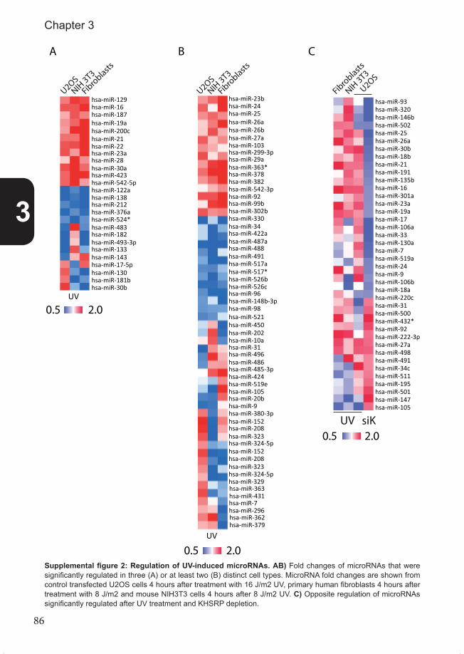
86
Supplemental figure 2: Regulation of UV-induced microRNAs. AB) Fold changes of microRNAs that were significantly regulated in three (A) or at least two (B) distinct cell types. MicroRNA fold changes are shown from control transfected U2OS cells 4 hours after treatment with 16 J/m2 UV, primary human fibroblasts 4 hours after treatment with 8 J/m2 and mouse NIH3T3 cells 4 hours after 8 J/m2 UV. C) Opposite regulation of microRNAs significantly regulated after UV treatment and KHSRP depletion.
UV
U2OSNIH
3T3
Fibroblasts
hsa-miR-129hsa-miR-16hsa-miR-187hsa-miR-19ahsa-miR-200chsa-miR-21hsa-miR-22hsa-miR-23ahsa-miR-28hsa-miR-30ahsa-miR-423hsa-miR-542-5phsa-miR-122ahsa-miR-138hsa-miR-212hsa-miR-376ahsa-miR-524*hsa-miR-483hsa-miR-182hsa-miR-493-3phsa-miR-133hsa-miR-143hsa-miR-17-5phsa-miR-130hsa-miR-181bhsa-miR-30b
U2OSNIH
3T3
Fibroblasts
hsa-miR-23bhsa-miR-24hsa-miR-25hsa-miR-26ahsa-miR-26bhsa-miR-27ahsa-miR-103hsa-miR-299-3phsa-miR-29ahsa-miR-363*hsa-miR-378hsa-miR-382hsa-miR-542-3phsa-miR-92hsa-miR-99bhsa-miR-302bhsa-miR-330hsa-miR-34hsa-miR-422ahsa-miR-487ahsa-miR-488hsa-miR-491hsa-miR-517ahsa-miR-517*hsa-miR-526bhsa-miR-526chsa-miR-96hsa-miR-148b-3phsa-miR-98hsa-miR-521hsa-miR-450hsa-miR-202hsa-miR-10ahsa-miR-31hsa-miR-496hsa-miR-486hsa-miR-485-3phsa-miR-424hsa-miR-519ehsa-miR-105hsa-miR-20bhsa-miR-9hsa-miR-380-3phsa-miR-152hsa-miR-208hsa-miR-323hsa-miR-324-5phsa-miR-152hsa-miR-208hsa-miR-323hsa-miR-324-5phsa-miR-329hsa-miR-363hsa-miR-431
UV
hsa-miR-7hsa-miR-296hsa-miR-362hsa-miR-379
Supplemental �gure 2
A B C
U2OS
NIH 3T3
Fibroblasts
UV siK
2.00.5
2.00.5
2.00.5
hsa-miR-93hsa-miR-320hsa-miR-146bhsa-miR-502hsa-miR-25hsa-miR-26ahsa-miR-30bhsa-miR-18bhsa-miR-21hsa-miR-191hsa-miR-135bhsa-miR-16hsa-miR-301ahsa-miR-23ahsa-miR-19ahsa-miR-17hsa-miR-106ahsa-miR-33hsa-miR-130ahsa-miR-7hsa-miR-519ahsa-miR-24hsa-miR-9hsa-miR-106bhsa-miR-18ahsa-miR-220chsa-miR-31hsa-miR-500hsa-miR-432*hsa-miR-92hsa-miR-222-3phsa-miR-27ahsa-miR-498hsa-miR-491hsa-miR-34chsa-miR-511hsa-miR-195hsa-miR-501hsa-miR-147hsa-miR-105
3
Chapter 3

87
Supplemental figure 3: UV-induced regulation of microRNAs that are significantly regulated upon RBP-de-pletion. MicroRNAs that were significantly regulated upon depletion of indicated RBP (left panels) were generally not much differentially regulated after UV treatment compared to control transfected cells (right panels).
Supplemental �gure 3
A B CsiK
HSRP
siKHSRP
siContro
l
UVMock
siTutase
4
siTutase
4
siContro
l
UVMock
siSnip1
siSnip1
siContro
l
UVMock
siMov10
siMov10
siContro
l
UVMock
D
EsiM
ov10
siDhx9
siContro
l
UVMock
siHNRNPUL1
siHNRNPUL1
siContro
l
UVMock
F
0.5 2.0
hsa-miR-93hsa-miR-320hsa-miR-146b-5phsa-miR-25hsa-miR-26ahsa-miR-18bhsa-miR-21hsa-miR-191hsa-miR-135bhsa-miR-16hsa-miR-301ahsa-miR-23ahsa-miR-19ahsa-miR-17hsa-miR-106ahsa-miR-130ahsa-miR-7hsa-miR-519ahsa-miR-24hsa-miR-9hsa-miR-106bhsa-miR-18ahsa-miR-30ahsa-miR-30ehsa-miR-31hsa-miR-500hsa-miR-432-3phsa-miR-27a-3phsa-miR-498hsa-miR-491hsa-miR-34chsa-miR-551bhsa-miR-195hsa-miR-501hsa-miR-147hsa-miR-557hsa-miR-584hsa-miR-608hsa-miR-671-3phsa-miR-147bhsa-miR-21-3phsa-miR-26a-2-3p
hsa-miR-30c-2-3phsa-miR-218-2-3phsa-miR-432-3p
hsa-miR-661hsa-miR-132-3phsa-miR-518e-3phsa-miR-195hsa-miR-142-5phsa-miR-491-5p
hsa-miR-195hsa-miR-181bhsa-miR-1908hsa-miR-142-5phsa-miR-26a-2-3phsa-miR-548a-5phsa-miR-1307hsa-miR-145hsa-miR-550-3phsa-miR-183-3phsa-miR-936hsa-miR-425-3p
hsa-miR-195hsa-miR-26a-2-3phsa-miR-181bhsa-miR-585hsa-miR-142-5phsa-miR-200bhsa-miR-617hsa-miR-27a-3p
hsa-miR-320hsa-miR-93hsa-miR-17hsa-miR-146-5phsa-miR-26ahsa-miR-106ahsa-miR-29ahsa-miR-19bhsa-miR-25hsa-miR-21hsa-miR-130ahsa-miR-24hsa-miR-30ahsa-miR-135bhsa-miR-182hsa-miR-27ahsa-miR-7hsa-miR-432-3phsa-miR-27a-3phsa-miR-7-2-3phsa-miR-575hsa-miR-663hsa-miR-147bhsa-let-7bhsa-miR-325hsa-miR-181bhsa-miR-491-5phsa-miR-617hsa-miR-203hsa-miR-595hsa-miR-548a-5phsa-miR-885-3phsa-miR-195hsa-miR-26a-2-3p
3
RBPs regulate DDR miRNAs

88
Supplemental figure 4: Regulation of UV-responsive microRNAs by more than one RBP. MicroRNAs were selected for clustering if their UV-induced fold change in control transfected cells weakened, abolished or rever-sed in at least two RBP-depleted conditions. These microRNAs were 2-sided, unsupervised hierarchical cluste-red. The upper side shows clustering of the RBP-depleted conditions and the left side shows clustering of the microRNAs. The right panel shows the UV-induced fold change in control transfected cells.
Supplemental �gure 4
FC upon UV
siContro
l
siSnip1
siHNRNPL1
siKHSRP
siTutase
4
siDhx9
siMov10
hsa-miR-371-3phsa-miR-520a-3phsa-miR-181bhsa-miR-105hsa-miR-7d-3phsa-miR-668hsa-miR-517-3phsa-miR-425-3phsa-miR-202hsa-miR-708hsa-miR-491-5phsa-let-7f-1-3phsa-miR-186hsa-miR-127-5p
hsa-miR-133bhsa-miR-922
hsa-miR-519ehsa-miR-671-3phsa-miR-124-3phsa-miR-34c-5phsa-miR-502-5phsa-miR-145hsa-miR-518a-3phsa-miR-190bhsa-miR-532-3phsa-miR-10ahsa-miR-30bhsa-miR-324-5phsa-miR-548a-5phsa-miR-450b-3p
3
Chapter 3

89
Sup
plem
enta
l figu
re 5
: Tut
ase4
-dep
ende
nt, U
V-r
espo
nsiv
e m
icro
RN
As
clus
tere
d br
east
can
cer
tum
ors
wit
h po
siti
ve a
nd n
egat
ive
corr
e-la
ted
expr
essi
on c
ompa
red
to n
orm
al b
reas
t ti
ssue
. Log
-tran
sfor
med
del
ta-C
t RT-
qPC
R e
xpre
ssio
n va
lues
of T
utas
e4-d
epen
dent
, UV-
resp
onsi
ve m
icro
R-
NAs
wer
e us
ed to
clu
ster
bre
ast c
ance
r tis
sues
(upp
er p
anel
). Va
lues
wer
e co
mpa
red
with
nor
mal
tiss
ue (r
ight
pan
el) a
nd fo
r eve
ry s
ampl
e m
icro
RN
A ex
pres
sion
was
co
mpa
red
with
exp
ress
ion
in n
orm
al ti
ssue
usi
ng p
ears
on c
orre
latio
n (lo
wer
gra
ph).
Not
e th
at e
very
dat
a po
int i
n th
is g
raph
is p
ositi
oned
exa
ctly
und
er th
e he
atm
ap d
ata
of th
e sa
me
canc
er s
ampl
e. L
umin
al-A
sub
type
s ar
e in
dica
ted
as re
d da
tapo
int.
-1.0
-0.50.0
0.5
1.0
Correlation
P <
0.05
P <
0.05 Lu
min
al-A
Oth
er o
r unk
now
n su
btyp
es
Supp
lem
enta
l �gu
re 5
1.5
-1.5lo
g de
lta C
t
norm
al ti
ssue
brea
st c
ance
r tiss
ue
hsa-
miR
-153
hsa-
miR
-181
ahs
a-m
iR-1
01
hsa-
miR
-140
-5p
hsa-
miR
-194
hsa-
miR
-145
hsa-
miR
-520
c-3p
hsa-
miR
-501
-5p
hsa-
miR
-502
-5p
hsa-
miR
-133
b
hsa-
miR
-432
hsa-
miR
-138
3
RBPs regulate DDR miRNAs

90
Sup
plem
enta
l figu
re 6
: Sni
p1-d
epen
dent
, UV
-res
pons
ive
mic
roR
NA
s cl
uste
red
brea
st c
ance
r tu
mor
s w
ith
posi
tive
and
neg
ativ
e co
rrel
ated
ex
pres
sion
com
pare
d to
nor
mal
bre
ast
tiss
ue.
Log-
trans
form
ed d
elta
-Ct R
T-qP
CR
exp
ress
ion
valu
es o
f Sni
p1-d
epen
dent
, UV-
resp
onsi
ve m
icro
RN
As w
ere
used
to c
lust
er b
reas
t can
cer t
issu
es (u
pper
pan
el).
Valu
es w
ere
com
pare
d w
ith n
orm
al ti
ssue
(rig
ht p
anel
) and
for e
very
sam
ple
mic
roR
NA
expr
essi
on w
as c
ompa
red
with
exp
ress
ion
in n
orm
al ti
ssue
usi
ng p
ears
on c
orre
latio
n (lo
wer
gra
ph).
Not
e th
at e
very
dat
a po
int i
n th
is g
raph
is p
ositi
oned
exa
ctly
und
er th
e he
atm
ap d
ata
of th
e sa
me
canc
er s
ampl
e. L
umin
al-A
sub
type
s ar
e in
dica
ted
as re
d da
tapo
int a
nd c
lust
ered
bre
ast c
ance
r sam
ples
dep
lete
d fo
r lum
inal
-A s
ubty
pes
are
unde
rline
d in
gre
en.
Supp
lem
enta
l �gu
re 6
P <
0.05
P <
0.05
Lum
inal
-AO
ther
or u
nkno
wn
subt
ypes
-0.7
5
-0.5
0
-0.2
5
0.00
0.25
0.50
0.75
Correlation
1.5
-1.5lo
g de
lta C
t
norm
al ti
ssue
brea
st c
ance
r tiss
ue
hsa-
miR
-140
-5p
hsa-
miR
-133
bhs
a-le
t-7b
*
hsa-
miR
-190
b
hsa-
miR
-193
a-5p
hsa-
miR
-493
hsa-
miR
-886
-3p
hsa-
miR
-519
e
hsa-
miR
-98
hsa-
miR
-518
a-3p
hsa-
miR
-508
-3p
hsa-
miR
-671
-3p
hsa-
miR
-324
-5p
hsa-
miR
-519
a3
Chapter 3

91
Supp
lem
enta
l figu
re 7
: Mov
10-d
epen
dent
, UV-
resp
onsi
ve m
icro
RN
As
clus
tere
d br
east
can
cer t
umor
s w
ith p
ositi
ve a
nd n
egat
ive
corr
elat
ed e
xpre
ssio
n co
mpa
-re
d to
nor
mal
bre
ast t
issu
e. L
og-tr
ansf
orm
ed d
elta
-Ct R
T-qP
CR
exp
ress
ion
valu
es o
f Mov
10-d
epen
dent
, UV-
resp
onsi
ve m
icro
RN
As w
ere
used
to c
lust
er b
reas
t can
cer
tissu
es (u
pper
pan
el).
Valu
es w
ere
com
pare
d w
ith n
orm
al ti
ssue
(rig
ht p
anel
) and
for e
very
sam
ple
mic
roR
NA
expr
essi
on w
as c
ompa
red
with
exp
ress
ion
in n
orm
al ti
ssue
us
ing
pear
son
corre
latio
n (lo
wer
gra
ph).
Not
e th
at e
very
dat
a po
int i
n th
is g
raph
is p
ositi
oned
exa
ctly
und
er th
e he
atm
ap d
ata
of th
e sa
me
canc
er s
ampl
e. L
umin
al-A
su
btyp
es a
re in
dica
ted
as re
d da
tapo
ints
and
clu
ster
ed b
reas
t can
cer s
ampl
es e
nric
hed
and
depl
eted
for l
umin
al-A
sub
type
s ar
e un
derli
ned
in re
d an
d gr
een,
resp
ectiv
ely.
-1.0
-0.50.0
0.5
1.0
P <
0.05
Correlation
Lum
inal
-AO
ther
or u
nkno
wn
subt
ypes
Supp
lem
enta
l �gu
re 7
P <
0.05
1.5
-1.5lo
g de
lta C
t
norm
al ti
ssue
brea
st c
ance
r tiss
ue
hsa-
miR
-363
hsa-
miR
-146
b-3p
hsa-
miR
-519
e
hsa-
miR
-193
a-5p
hsa-
miR
-34c
-5p
hsa-
miR
-186
hsa-
miR
-133
bhs
a-m
iR-7
58
hsa-
miR
-153
hsa-
miR
-628
-3p
hsa-
let-
7f-1
*hs
a-m
iR-4
87b
3
RBPs regulate DDR miRNAs

92
Supp
lem
enta
l figu
re 8
: Dhx
9-de
pend
ent,
UV-
resp
onsi
ve m
icro
RN
As
clus
tere
d br
east
can
cer t
umor
s w
ith p
ositi
ve a
nd n
egat
ive
corr
elat
ed e
xpre
ssio
n co
mpa
-re
d to
nor
mal
bre
ast t
issu
e. L
og-tr
ansf
orm
ed d
elta
-Ct R
T-qP
CR
exp
ress
ion
valu
es o
f Dhx
9-de
pend
ent,
UV-
resp
onsi
ve m
icro
RN
As w
ere
used
to c
lust
er b
reas
t can
cer
tissu
es (u
pper
pan
el).
Valu
es w
ere
com
pare
d w
ith n
orm
al ti
ssue
(rig
ht p
anel
) and
for e
very
sam
ple
mic
roR
NA
expr
essi
on w
as c
ompa
red
with
exp
ress
ion
in n
orm
al ti
ssue
us
ing
pear
son
corre
latio
n (lo
wer
gra
ph).N
ote
that
eve
ry d
ata
poin
t in
this
gra
ph is
pos
ition
ed e
xact
ly u
nder
the
heat
map
dat
a of
the
sam
e ca
ncer
sam
ple.
Lum
inal
-A s
ub-
type
s ar
e in
dica
ted
as re
d da
tapo
int.
-1.0
-0.50.0
0.5
1.0
P <
0.05
Correlation
Lum
inal
-AO
ther
or u
nkno
wn
subt
ypes
Supp
lem
enta
l �gu
re 8
P <
0.05
1.5
-1.5lo
g de
lta C
t
norm
al ti
ssue
brea
st c
ance
r tiss
ue
hsa-
miR
-10a
hsa-
miR
-491
-5p
hsa-
miR
-532
-3p
hsa-
miR
-148
bhs
a-m
iR-1
33b
hsa-
miR
-425
*hs
a-m
iR-5
19e
hsa-
miR
-16-
1*
hsa-
miR
-145
hsa-
miR
-153
hsa-
let-
30d*
hsa-
miR
-520
c-3p
hsa-
miR
-922
hsa-
miR
-638
3
Chapter 3

93
Supp
lem
enta
l figu
re 9
: HN
RN
PUL1
-dep
ende
nt, U
V-re
spon
sive
mic
roR
NA
s cl
uste
red
brea
st c
ance
r tum
ors
with
pos
itive
and
neg
ativ
e co
rrel
ated
exp
ress
ion
com
pare
d to
nor
mal
bre
ast t
issu
e. L
og-tr
ansf
orm
ed d
elta
-Ct R
T-qP
CR
exp
ress
ion
valu
es o
f HN
RN
PUL1
-dep
ende
nt, U
V-re
spon
sive
mic
roR
NAs
wer
e us
ed to
clu
ster
br
east
can
cer t
issu
es (u
pper
pan
el).
Valu
es w
ere
com
pare
d w
ith n
orm
al ti
ssue
(rig
ht p
anel
) and
for e
very
sam
ple
mic
roR
NA
expr
essi
on w
as c
ompa
red
with
exp
ress
ion
in
norm
al ti
ssue
usi
ng p
ears
on c
orre
latio
n (lo
wer
gra
ph).
Not
e th
at e
very
dat
a po
int i
n th
is g
raph
is p
ositi
oned
exa
ctly
und
er th
e he
atm
ap d
ata
of th
e sa
me
canc
er s
ampl
e.
Lum
inal
-A s
ubty
pes
are
indi
cate
d as
red
data
poin
t.
-0.7
5
-0.5
0
-0.2
5
0.00
0.25
0.50
0.75
P <
0.05Lu
min
al-A
Oth
er o
r unk
now
n su
btyp
es
Supp
lem
enta
l �gu
re 9
P <
0.05
1.5
-1.5lo
g de
lta C
t
norm
al ti
ssue
brea
st c
ance
r tiss
ue
hsa-
miR
-517
ahs
a-m
iR-5
20b
hsa-
miR
-202
hsa-
miR
-365
hsa-
miR
-154
hsa-
miR
-133
b
hsa-
miR
-432
hsa-
miR
-324
-5p
hsa-
let-
7a
hsa-
miR
-132
hsa-
let-
7b*
hsa-
miR
-145
hsa-
miR
-425
hsa-
miR
-363
hsa-
miR
-148
bhs
a-m
iR-1
6-1*
hsa-
miR
-29c
hsa-
miR
-30b
hsa-
miR
-708
hsa-
miR
-135
b*hs
a-m
iR-5
01-5
phs
a-m
iR-5
19e
hsa-
miR
-922
Correlation
3
RBPs regulate DDR miRNAs

94
Supplemental table 1: Overview of breast tumor tissue subtypes used for analysis of misexpressed miRNAs.
Supplemental tables
Supplemental table 1
Breast cancer subtypesnumber of subtypes
Luminal B-like 12Luminal A-like 25Basal-like 8ERBB2-like 8Normal-like 5Unknown 26
Normal breast tissue 8
3
Chapter 3

95
Supplemental table 2: RBPs identified in proteomic DNA-da-mage-induced phosphorylati-on screens. Three independent screens identified RBPs, which function in several, distinct pro-cesses, to be phosphorylated upon DNA damage [28] or by da-mage-induced ATM/R [6, 27].
Supplemental table 2
RNA-binding proteins identified in phosphoproteomic screens
Matsuoka S et al , Science, 2007HEK293T cells treated with 10gy of IR. RNA collected after 1h
MicroRNA processing Splicing poly-adenylation unknown/other roleKHSRP CSTF2T CSTF2 RBM34Zcchc11 (Tutase4) DDX24 RBM21 aka Tut1 RBM6SNIP1 LSM2 TSEN54 SNAPC3Mov10 PRPF38B SYNCRIPDHX9 RALY
RBM14RBM15RBM16PRPF8RBM10TSEN2U2AF1SYNCRIP
Bensimon A at al , Science Signalling, 2010Human melanoma G361 cells treated with 200 ng/mL of neocarzinostatin for 1h
MicroRNA processing Splicing poly-adenylation unknown/other roleHNRNPUL1 BUD13 SYMPK DDX42
PRPF3 DDX21RBM15 RSL1D1RBM25 G3BP1SF3A1
Other non-coding RNA processing SFRS1NCL SF3B1SF3A1 SF3B2SF3B1 CD2BP2SF3B2 CRKRS aka CDK12SFRS1 CROPSRRM1 EFTUD2SRRM2 SFRS11NCL SFRS10NOP58 SPENNOP56 SRRM1NOLC1 SRRM2RPL14 WTAPRPS6 YBX1
ZRSR2HNRPA2B1HNRPKHNRPURALYPNNEFTUD2ZRANB2
Beli P, Molecular Cell, 2012U2OS cells treated with 10gy IR or 10µM etoposide and RNA collected after 1h or 24h, respectivily
MicroRNA processing Splicing poly-adenylation unknown roleDHX9 RBM25 FIP1L1 RSL1D1
SRRM1 PAPOLA RBM6SRRM2 PABPN1 ANKHD1
Other non-coding RNA processing SFRS2IP LARP6NOLC1 YBX1 mRNA-cap binding ZFC3H1TSEN34 EIF4A3 NCBP1 CCDC111RRP1B NONO EDC4 SMG8INTS12 HNRNPA3 ZFC3H1DUS2L ARL6IP4 3'UTR binding RBM7PHAX CPSF2 SERBP1 SLC4A1APPUS10 ZNF638 RBM4 ZNF239UTP14A RBM15B CPSF2 SLC4A1APPOP1 RBM4 HNRNPA0 SLC4A1APNCL HNRNPU PABPN1 KHDRBS1
THRAP3RNA degradation U2AF1EXOSC9 PPIL1SMG5 PPP4R2WIBG CPSF2RNH1 RBM16ILF3 DDX47
SCAF1RNA modification PHAXMETTL3 CROPPUS10 SF3A1
SNW1RNA interference SNW1PRKRA PRPF6ADAR HNRNPA0
PNNPOP4ISY1SF3B2HNRNPMPRPF38B
3
RBPs regulate DDR miRNAs

96
Supplemental Table 2
number of significantly regulated microRNAs after 16J/m2 UV after 12gy IRsiControl 169 20siKHSRP 175 0
siTutase4 230 42siSNIP1 176 28
siMov10 197 11siDHX9 208 1
siHNRNPUL1 241 34
Supplemental table 3: Total number of significantly regulated microRNAs in every condition.
Supplemental table 4
Pearson correlation of microRNA expression in tumors compared to healthy tissuebased on Snip1-dependent microRNAs based on UV-responsive Snip1-dependent microRNAs
significance R R significanceX6351 0.041 -0.830 -0.175 0.550 LUMBX7181 0.015 -0.900 -0.481 0.082 BASALX7226 0.046 -0.820 -0.270 0.351 LUMBX5300 0.009 -0.920 0.282 0.328 N/DX435 0.021 -0.880 0.127 0.666 LUMAX5155 0.009 -0.920 -0.649 0.012 LUMBX6292 0.002 -0.960 -0.479 0.083 N/DX8136 0.046 -0.820 -0.139 0.636 N/DX5075 0.010 0.917 0.472 0.088 LUMAX6210 0.039 0.834 -0.107 0.716 LUMAX8573 0.022 0.875 -0.112 0.703 N/DX5611 0.721 0.188 0.592 0.026 LUMAX434 0.868 0.088 0.654 0.011 NORMALX8543 0.228 0.580 0.562 0.036 N/DX8525 0.437 0.396 -0.583 0.029 N/DX454 0.737 0.177 -0.554 0.040 BASAL
Correlation between R-coefficients of both datasetsR-coefficient 0.363
P-value 0.167
Supplemental table 4: Correlation of Snip1 microRNA expression in tumors compared to normal tissue. Left columns annotate cancer samples, the correlation coefficient (R) and significance (p-value) of the Snip1-de-pendent microRNAs compared to the normal tissue. Right columns show the same information for UV-responsive, Snip1-dependent microRNAs. Significant values indicated in blue font and positive or negative correlation high-lighted in green and red, respectively.
3
Chapter 3

97
Supplemental table 5
Pearson correlation of microRNA expression in tumors compared to healthy tissuebased on Mov10-dependent microRNAs based on UV-responsive Mov10-dependent microRNAs
significance R R significanceX8596 0.035 -0.843 -0.124 0.701 N/DX415 0.015 -0.898 -0.756 0.004 ERBB2X6548 0.034 -0.845 -0.045 0.890 N/DX7282 0.026 -0.865 0.068 0.834 BASALX6292 0.013 -0.906 -0.265 0.405 N/DX8598 0.015 -0.900 0.082 0.800 N/DX5155 0.015 -0.851 -0.695 0.012 LUMBX399 0.021 -0.878 0.024 0.800 BASALX412 0.269 -0.540 -0.580 0.048 LUMAX7181 0.100 -0.729 -0.618 0.032 BASALX435 0.166 -0.646 -0.644 0.024 LUMAX8592 0.128 -0.692 -0.601 0.039 N/DX5018 0.060 0.792 0.622 0.031 LUMAX7074 0.830 0.114 0.716 0.009 LUMAX8543 0.001 0.973 0.177 0.583 N/D
Correlation between R-coefficients of both datasetsR-coefficient 0.603
P-value 0.017
Supplemental table 5: Correlation of Mov10 microRNA expression in tumors compared to normal tissue. Left columns annotate cancer samples, the correlation coefficient (R) and significance (p-value) of the Mov10-de-pendent microRNAs compared to the normal tissue. Right columns show the same information for UV-responsive, Mov10-dependent microRNAs. Significant values indicated in blue font and positive or negative correlation high-lighted in green and red, respectively.
Supplemental table 6
Pearson correlation of microRNA expression in tumors compared to healthy tissuebased on Dhx9-dependent microRNAs based on UV-responsive Dhx9-dependent microRNAs
significance R R significanceX415 0.011 -0.955 -0.495 0.072 ERBB2X8539 0.038 -0.899 -0.529 0.052 N/DX824 0.803 -0.155 -0.568 0.034 NORMALX6324 0.273 -0.612 -0.608 0.021 N/DX4435 0.637 -0.289 -0.710 0.004 LUMAX8592 0.591 -0.327 -0.638 0.014 N/DX8543 0.030 0.913 0.585 0.028 N/DX4600 0.271 0.613 0.641 0.014 NORMALX8610 0.761 0.189 0.705 0.005 N/DX7116 0.943 0.045 0.645 0.013 ERBB2X440 0.547 0.364 0.542 0.045 NORMALX391 0.367 0.522 0.771 0.001 LUMAX6188 0.639 0.287 0.703 0.005 BASALX8505 0.628 0.296 0.717 0.004 N/D
Correlation between R-coefficients of both datasetsR-coefficient 0.826
P-value 0.000
Supplemental table 6: Correlation of Dhx9 microRNA expression in tumors compared to normal tissue. Left columns annotate cancer samples, the correlation coefficient (R) and significance (p-value) of the Dhx9-de-pendent microRNAs compared to the normal tissue. Right columns show the same information for UV-respon-sive, Dhx9-dependent microRNAs. Significant values indicated in blue font and positive or negative correlation highlighted in green and red, respectively.
3
RBPs regulate DDR miRNAs

98
Supplemental table 7
Pearson correlation of microRNA expression in tumors compared to healthy tissuebased on HNRNPUL1-dependent microRNAs based on UV-responsive HNRNPUL1-dependent microRNAs
significance R R significanceX4475 0.810 -0.053 -0.457 0.028 LUMAX6324 0.309 -0.222 -0.599 0.003 N/DX8539 0.149 -0.311 -0.596 0.003 N/DX8545 0.480 -0.155 -0.660 0.001 N/DX6210 0.459 0.163 -0.484 0.019 LUMAX8592 0.542 -0.134 -0.503 0.014 N/DX412 0.138 -0.319 -0.516 0.012 LUMAX5087 0.040 -0.432 -0.163 0.457 BASALX454 0.048 -0.416 -0.535 0.009 BASALX4444 0.041 -0.429 -0.175 0.424 LUMBX6292 0.015 -0.499 -0.076 0.730 N/DX415 0.024 -0.469 -0.411 0.051 ERBB2X6351 0.008 -0.537 -0.038 0.863 LUMBX7194 0.016 -0.497 0.076 0.729 ERBB2X824 0.024 -0.468 -0.326 0.129 NORMALX8571 0.024 -0.522 -0.261 0.229 N/DX7125 0.042 0.428 -0.228 0.295 LUMAX8594 0.029 0.455 0.534 0.009 N/DX440 0.001 0.644 0.514 0.012 NORMALX618 0.945 0.015 0.448 0.032 BASALX8505 0.989 0.003 0.557 0.006 N/DX8543 0.058 0.400 0.706 0.000 N/DX434 0.731 0.076 0.506 0.014 NORMALX4600 0.178 0.291 0.502 0.015 NORMAL
Correlation between R-coefficients of both datasetsR-coefficient 0.566
P-value 0.004
Supplemental table 7: Correlation of HNRNPUL1 microRNA expression in tumors compared to normal tissue. Left columns annotate cancer samples, the correlation coefficient (R) and significance (p-value) of the HNRNPUL1-dependent microRNAs compared to the normal tissue. Right columns show the same information for UV-responsive, HNRNPUL1-dependent microRNAs. Significant values indicated in blue font and positive or negative correlation highlighted in green and red, respectively.
3
Chapter 3

99

100

101
Chapter 4
MicroRNA-24 differentially regulates MDC1 isoforms upon ionizing radiation
in preparation
Maikel D. Wouters1, Inger Brandsma1, Serena T. Bruens1, Gisela G. G. Slaats1, Nicolaas G. J. Jaspers1, Jan H. J. Hoeijmakers1, Erik A. C. Wiemer2,
Wim Vermeulen1, Dik van Gent1, Jurgen A. F. Marteijn1, J. Pothof1
1. Department of Genetics, Erasmus University Medical Center, Rotterdam, The Netherlands2. Department of Medical Oncology, Erasmus MC Cancer Institute, Erasmus University Medical Center, Rotterdam, The Netherlands

102
AbstractMany endogenous and exogenous agents damage DNA. The initial response upon many forms of DNA damage, such as DNA double strand breaks (DSBs), include activation of elaborate signal transduction routes, the so-called DNA damage checkpoints response. These signaling pathways induce and coordinate DNA repair and cell cycle arrest. Although a lot is known about post-translational regulation of this pathway upon DNA damage, less is known about its DNA damage-dependent post-transcriptional regulation, e.g. by microR-NAs. For example, microRNA-24 sensitizes hematopoietic cells for DSB-inducing agents. We further studied the role of this microRNA in the DNA damage checkpoint response and showed that miR-24 is responsive upon ionizing radiation and modulates DNA repair, cell cycle distribution and cellular sensitivity upon ionizing radiation. This report reveals adap-tor-protein MDC1 as a target of miR-24 and also describes an alternative isoform of MDC1, which is differentially regulated by miR-24. MiR-24 alters the ratio between both isoforms, which might have important effects on DNA damage signaling of the cellular response on DNA damage.
4
Chapter 4

103
IntroductionVarious exogenous and endogenous agents damage the DNA, which can obstruct tran-scription and DNA replication. If not resolved, accumulating DNA damage decreases proli-feration and cellular viability, contributing to ageing. Additionally, DNA damage can lead to mutations, deletions or gross chromosomal aberrations that cause cancer. To counteract these detrimental effects of DNA damage, all organisms possess a highly conserved DNA damage response (DDR). This response induces signaling pathways to activate and coor-dinate several processes to counter DNA damage and its consequences. These include DNA repair mechanisms, cell cycle checkpoints that inhibit cellular proliferation to allow time for DNA repair, and apoptosis, cellular senescence or terminal differentiation in case of excessive damage [1, 2]. These processes form an active barrier against tumorigenesis [3, 4] and are tightly controlled by DDR signaling pathways, which are critical for maintaining DNA integrity [5]. One of the most toxic DNA lesions are DNA double strand breaks (DSBs), caused by ionizing radiation (IR), multiple genotoxic compounds and collapse of replication forks [1, 6]. DSBs activate the signaling kinase ATM, which initiates the DNA damage checkpoint response. This response promotes DNA repair and initiates cell cycle arrest upon DSBs [7]. ATM phosphorylates histone H2AX (γH2AX), and this phosphorylation spreads into the chromatin surrounding DSBs and can be visualized by immunofluorescence as nuclear foci. These IR-induced foci (IRIF) locally open the chromatin to facilitate additional histone modifications and recruitment of various proteins into IRIF, which stimulates efficient DSB repair and cell cycle arrest [8]. Key proteins recruited into IRIF are MDC1, 53BP1 and BRCA1. Foci formation of these proteins is a measure for the amount of DNA damage and indicates the activation of the DNA damage checkpoint [9]. MDC1 is an adaptor protein that directly binds to γH2AX and mediates cell cycle checkpoints and DNA repair by recruiting multiple other factors, such as E3 ubiquitin ligase RNF8 which facilitates UBC13-dependent ubiquitination of his-tone H2A [10]. Histone modifications reveal methylated histone residues that are neces-sary to recruit 53BP1 into foci. In contrast, BRCA1 recruitment into foci depends on H2A ubiquitination [9, 11, 12]. Both 53BP1 and BRCA1 facilitate cell cycle arrest and influence which type of DSB repair is used, either Non-Homologous End-Joining (NHEJ) or (HR) [13, 14]. NHEJ is a potentially mutagenic repair mechanism that is active in all phases of the cell cycle. It is facilitated by association of MDC1 with DNA-PK, a kinase with an essential role in NHEJ [15]. HR is mediated by BRCA1 [16, 17] and is a mechanism to repair DSBs in an error-free manner in S-phase and G2-phase cells, using information on the undamaged sister chromatid for proper repair. Regulation of the DNA damage checkpoint response has mainly been studied at the transcriptional and post-translational levels. However, post-transcriptional gene regu-lation by microRNAs has emerged as a novel layer of gene expression control [18, 19]. MicroRNAs negatively regulate gene expression levels by targeting microRNA-binding elements (MRE) in the 3’ untranslated-region (3’UTR) of target mRNAs, mainly inducing mRNA degradation [20]. Fifty percent of all DDR genes are predicted targets of microRNAs [21] and microRNAs were found to regulate multiple proteins involved in cell cycle control and apoptosis [22, 23]. MicroRNAs are differentially regulated by several genotoxic agents [24, 25], thereby regulating cell cycle and apoptosis upon DNA damage [24, 26, 27]. Mis-regulation of these processes by aberrant expression of microRNAs is implicated in cancer development [28, 29]. This strongly suggests a central role for microRNAs within both the DDR and tumorigenesis [21].
4
miR-24 regulates MDC1

104
The checkpoint response upon DSBs can also be regulated by microRNAs. Se-veral studies showed that both ATM and H2AX are targeted by multiple microRNAs, which sensitize cells for IR [30-33]. However, these studies did not show whether these microR-NAs are regulated upon DNA damage in order to modulate the checkpoint response. In contrast, miR-24, which directly represses H2AX expression [34], is upregulated in res-ponse to DNA-damaging UV-radiation and H2O2 treatment [24, 35]. This microRNA inhibits cellular proliferation [36] and is also upregulated upon terminal differentiation of several cell types [36-38], including hematopoietic cells [34, 39]. These cells are sensitized for DSB-in-ducing compounds bleomycin and etoposide by miR-24-dependent regulation of H2AX [34, 39]. MiR-24 decreases genomic integrity of these cells [34], although it is not clear if any DNA repair or cell cycle control mechanisms are affected. In contrast to hematopoietic cells, nothing is known about the role of miR-24 in res-ponse to DSBs in other cells. Moreover, miR-24 has mostly been studied in various termi-nally differentiated cells that generally have a low proliferation rate [34, 36-39]. Therefore, we studied the role of miR-24 in highly proliferative human osteosarcoma cells (U2OS) af-ter induction of DSBs by IR. We wondered if miR-24 is regulated upon IR and if it regulates the DNA damage checkpoint response and subsequent cellular processes such as DNA repair and cell cycle checkpoints in these proliferating cells. Additionally, this report descri-bes the influence of miR-24 on IRIF formation of its target H2AX and other DNA damage checkpoint response proteins, which revealed MDC1 and its newly identified, alternative isoform as surprising new targets of miR-24. These findings support an important role for miR-24 in the response to IR-induced DSBs, improving our understanding of miR-24 and its impact in the DNA damage checkpoint response and potentially cancer.
Materials and MethodsCell cultureHEK-293, HeLa and U2OS cells were cultured in a 1:1 mix of Lonza DMEM and F10 medium, supplemented with 10% FCS, penicillin (100 U/mL) and streptomycin (100 µg/mL), and incubated at 37°C with 5% CO2. U2OS cells used for clonogenic survival assay were grown at 3% O2 a week before and during the experiment. U2OS cells stably expressing fluorescent-tagged GFP-MDC1, described by [40], were GFP enriched by FACS sorting after thawing.
RNA isolation and Northern blottingCells were washed with RNAse-free PBS and total RNA was collected using 1mL Trizol (Invitrogen) per Ø3 cm well for RT-qPCR or 5mL Trizol per Ø10cm dish for Northern blotting. RNA was extracted with chloroform, precipi-tated with isopropanol, washed twice with RNAse-free 70% ethanol, air-dried and solubilized in RNAse-free water. For Northern blotting, 3-6 µg of total RNA was dissolved in RNA loading buffer (1 x TBE, 90% (v/v) formamide) and subjected to electrophoresis on a 12% poly-acrylamide gel containing 8 M urea. RNA was subse-quently transferred to a Hybond N+ membrane (Amersham, Little Chalfont, UK) using a semi-dry electroblotting method (15 min at 10 Volts followed by 30 min at 30 Volts in 0.5 x TBE buffer). RNA was cross-linked to the mem-brane using 120 mJ of UV in a Bio-Rad GS gene linker and prehybridized for 1h at 55°C in ULTRAhybTM (Ambi-on). The following oligonucleotide probe was used to detect miR-24 transcripts: 5’-CTGTTCCTGCTGAACTGAG-CC. The probe was end-labeled with [γ32P]-dATP and incubated with the membrane for 16 hours in ULTRAhyb at 52°C. Subsequently the membrane was washed twice at RT in 2 x SSC, 0.1% SDS and for 5 min at 52°C in 5 x SSC, 0.1% SDS. Membranes were exposed to a Phosphorimager screen (Molecular Dynamics, Sunnyvale, CA, USA). Afterwards, blots were stripped and re-probed with a 5S rRNA specific oligonucleotide (5’-TCTCCCATC-CAAGTACTAACCAGGCC) as a control for equal loading.
TransfectionsFor transfections, cells were grown to 50% confluence in Ø0.5 cm (HEK-293 cells) or 80% confluence in Ø3 cm wells (U2OS and HeLa cells). HEK-293 cells were co-transfected once with 1µg psiCHECK2 MDC1 3’UTR plasmid and 100nM of Dharmacon miRidian hsa-miR-24 mimic using Dharmafect #1 according to manufacturer’s
4
Chapter 4

105
protocol. Invitrogen RNAiMax was used according to manufacturer’s protocol to introduce 100nM of Dharma-con miR-24 mimic, Exiqon miRcury hsa-miR-24 LNA inhibitor or Dharmacon custom-made ON-TARGET enhan-ced antisense loading siRNA (5’-AAGGUUUCCAGGAUCCAAAUU) targeting the alternative MDC1 isoform into U2OS cells using two transfections, the second transfection O/N. One μg of the MDC1 plasmids was transfected once, 1 day after transfection of miR-24 mimics, using Invitrogen Lipofectamine 2000 according to manufacturer’s protocol. Minimally 6 h after transfections, cells were washed with PBS and the medium was replaced or the cells were trypsinized and seeded into experimental plates. For the NHEJ assay cells were transfected once with an end-joining substrate using Promega Fugene according to manufacturer’s protocol 48h after miR-24 mimic transfection. End-joining was assayed 48h after substrate transfection [46].
Clonogenic survival assayFour hundred cells were seeded in Ø3 cm wells, and cultured at 3% O2. After 16 hours, cells were treated with one indicated pulse of IR. Untreated cells formed ~200 colonies after 7 days, which was set at 100%. Colonies were fixed and stained with 50% methanol, 7% acetic acid and 0.1% Coomassie-blue. Cell cycle assays and FACS analysisFor cell cycle assays, transfected cells from one Ø3 cm well were seeded into one Ø10 cm dish and treated with IR the following day. Cells were either incubated for 15 minutes with 1μM nucleotide analogue BRDU or directly collected by trypsinization at indicated time-points. Before collection, floating mitotic cells in the medium and PBS wash were spun down and pooled with the corresponding sample. After centrifugation, cells were washed with PBS, fixed with 70% ethanol and stored overnight at 4oC. To analyze the percentage of G1, S and G2 cells, nuclei were isolated and stained for DNA-incorpora-ted BRDU. First, to degrade proteins, cell pellets were treated for 20 minutes with 0.05% pepsin in 0.1N HCl at RT followed by a wash of PBS with 0.5% Tween20 and 0.1% BSA (PBS-T+). Subsequent spins were performed at 4oC. To isolate nuclei, cell pellets were incubated for 12 minutes with 2N HCl at 37oC and neutralized with Borate buffer (pH 8.5, 4oC). Nuclei were washed with PBS-T+ and incubated for 1 hour at 4oC in the dark with 1:50 diluted anti-BRDU FITC-conjugated antibody (BD biosciences) in 100μL PBS-T+ to stain for S-Phase cells. Nuclei were washed with PBS and stained with 1 µg/mL DNA stain PI (Invitrogen) in PBS with 0.1% BSA and 0.25 mg/mL RNaseH to discriminate between G1 and G2 cells and analyzed on a BD Biosciences FACScan To determine the percentage of cells in M-phase, cells were washed with PBS and spun down at 4oC. The resulting cell pellet was incubated for 1 hour on ice with 100μL PBS and 1/200 diluted MpM-2 antibody (Millipore), which stains mitotic-specific phosphorylation. Following a PBS wash, cell pellets were incubated for 1 hour in the dark on ice with 100μL PBS and 1/50 diluted goat anti-mouse FITC-conjugated antibody (Jackson). Following a PBS wash, the pellet was resuspended in 500μL PBS with 10 µg/mL DNA stain PI (Invitrogen) and 0.25 mg/mL RNAseH and analyzed on a BD Biosciences FACScan. To analyze GFP-MDC1 levels, cells were trypsinized one day after transfection of GFP plasmids, fixed in 70% ethanol and analyzed on a BD Biosciences FACScan.
ImmunofluorescenceTransfected cells were grown on coverslips to a ~50% confluence. Thirty minutes after IR treatment, cells were washed with PBS and fixed for 15 minutes in PBS with 2% paraformaldehyde. Cells were permeabilized with 3 washes and two 10-minute incubations with PBS containing 0.1% Triton. Cells were washed and stored at 4oC in PBS containing 0.5% BSA and 0.15% glycine (PBS+). We incubated the coverslips at RT for 2 hours with the follo-wing antibodies and dilutions in 130μL PBS+: mouse anti-γH2AX (Millipore)(1/1000), mouse anti-MDC1 (Abcam)(1/1000), mouse anti-BRCA1 (Santa Cruz)(1/50) or rabbit anti-53BP1 (Santa Cruz)(1/500). Cells were washed with PBS 0.1% Triton as described above. To visualize the proteins we incubated the coverslips for 1 hour with 130μL PBS+ containing either Invitrogen goat anti-mouse Alexa green 488 IgG (1/000) or goat anti-rabbit Alexa red 594 IgG (1/800), followed by PBS 0.1% Triton washes as described above, before adding PBS. The coverslips were mounted onto microscope slides with Vectashield (Vector laboratories) containing nuclear stain DAPI and air-sealed. After overnight incubation at 4oC the cells were imaged with a Zeis LSM510meta confocal microscope.
Protein isolation and immunoblotCells were washed twice with ice-cold PBS and scraped in 70 µl PBS. Ninety µl Laemmli buffer (120 mM Tris-HCl (pH6.8), 4% SDS, 10% Glycerol) was added and samples were boiled for 5 min, homogenized with a syringe and boiled again. Samples were snap frozen in N2(l) and stored at -80oC. Proteins were separated on SDS-PAGE gel, transferred to Millipore 0.2uM PVDF membranes and blocked in skim-milk. Mouse anti-γH2AX (Millipore)(1/1000), mouse anti-MDC1 (Abcam)(1/1000), Ku70 and Orc2 antibodies were used to stain proteins of interest.
4
miR-24 regulates MDC1

106
These proteins were visualized with GE Healthcare ECL-plus using sheep anti-mouse or anti-rabbit HRP antibo-dies (Jackson Laboratories) (1/1000) and visualized on an Alliance scanner.
RT-qPCRTotal RNA was reverse transcribed with Applied Biosystems Taqman assay according to manufactures instructi-ons to measure isoform-unspecific MDC1 levels. Alternatively, Invitrogen SuperscriptII RT polymerase was used to produce cDNA to measure isoform-specific MDC1 levels using Sybergreen qPCR and the following primers: Fw-primer (5’-GTCAAGTTCCTGTGTGCCCTG) and Rv-primer (5’-CTTGCTCAGGGTCGGTCACCAC) to amplify both isoforms; Fw-primer (5’-GCAAGATGCCACCTGCTGAG) to amplify the canonical and alternative isoform in combination with either the Rv-primer (5’-GCTGGGGCTTCAGGTACTGTAGG) within the canonical part of exon 10 or Rv-primer (5’-CCACCTTTTGGATCCTGGAAACC) spanning the alternative exon-9-to-10 boundary. PCR products were amplified and expression measured on a Biorad CFX-96 C1000 Thermal cycler.
Generation of psiCHECK2 MDC1 3’UTR and Luciferase measurementThe 3’UTR of MDC1 was cloned downstream of a Renilla luciferase reporter gene of the psiCHECK2 plasmid. MDC1 3’UTRs was amplified using Finnzymes Phusion polymerase and the following primers: XhoI-Fw-primer (5’-ggattctcgagCTCCACTACCCTTTTCCCTCC) and NotI-Rv-primer (5’-ggattcgcggccgcCTGAGGTAGGAGGAT-TTCTTGAGC), resulting in a 1300bp fragment. The PCR product was purified from gel and digested with XhoI and NotI, gel-purified and ligated into psiCHECK2. The resulting plasmid was validated with restriction analysis and sequencing. This construct was transfected in Hek293 cells, which were lysed 24 hours later. Lysis and luciferase activity measurement was performed using Promega Dual-Luciferase reporter assay according to manufacturer’s protocol.
RACE assay and fusion PCROne µg of total RNA was reversed transcribed according to the instructions of Clontech RACE assay. Clontech Advantage 2 PCR kit and the following primers were used to identify and characterize the 5’ and 3’ part of the alternative MDC1 isoform: Clontech UPM primer in combination with alternative exon-9-to-10 boundary Rv-primer (5’-CCACCTTTTGGATCCTGGAAACC) and alternative exon-9-to-10 boundary Fw-primer 5’-GGTTTCCAGGAT-CCAAAAGGTGG in combination with MDC1 end-of-ORF Rv-primer (5’-GGTGGATGACATCTCCAAAGGGG), which excluded the stop-codon to enable cloning of the alternative isoform upstream of GFP. PCR products were purified from agarose gel, ligated into the pCR2.1 plasmid using Invitrogen Original TA Cloning Kit and sequenced. Multiple products of the alternative MDC1 isoform were amplified using Finnzymes Phusion polymera-se as input for a fusion PCR to join both alternative MDC1 fragments. The 5’ part of the alternative isoform was amplified using the alternative exon-9-to-10 boundary Rv-primer mentioned before and the following Fw-primers including or excluding the alternative start site: canonical ATG start-site XhoI Fw-primer (5’-ggattctcgagatcATG-GAGGACACCCAGGC), canonical ATG start-site EcoRI Fw-primer (5’-ggattgaattcagatcATGGAGGACACCCAG-GC), alternative ATG start-site XhoI Fw-primer (5’-ggattctcgagATCCATGGGGCATCCTACTTTC), alternative ATG start-site EcoRI Fw-primer (5’-ggattgaattcagATCCATGGGGCATCCTACTTTC). The 3’ part of the alternative isoform was amplified using the alternative exon-9-to-10 boundary Fw-pri-mer mentioned before and the following Rv-primers: end-of-ORF EcoRI Rv-primer (5’-ggattgaattcgGGTGGATG-ACATCTCCAAAGGGG) or XhoI Rv-primer (ggattctcgagtcaGGTGGATGACATCTCCAAAGGGG) including a stop codon. Fusion PCR of both parts was performed using Finnzymes Phusion polymerase without any primers for the first 20 cycles followed by 20 cycles using the above mentioned primers to amplify a fused 5’-XhoI-3’-EcoRI or 5’-EcoRI-3’-XhoI alternative isoform product. Fused products were purified from agarose gel, ligated either into N1 pEGFP (5’XhoI-3’EcoRI) or pCDN3.1 (5’EcoRI-3’XhoI) plasmid and checked by sequencing.
4
Chapter 4

107
ResultsMiR-24 modulates the DDR of U2OS cellsMicroRNAs have a significant role in the DDR, but their impact in specific components of the DDR, such as the DNA damage checkpoint response, is less clear. To investigate if microRNAs regulate this checkpoint response, we previously have used online predicti-on programs Targetscan and MiRanda to identify any conserved MREs in the 3’UTR of 15 checkpoint response genes [21]. Several microRNAs were identified that might target ATM, MDC1 or H2AX (table 1, chapter 6), including miR-24. This microRNA was previously shown to sensitize terminally differentiated, post-mitotic hematopoietic cells for DSB-indu-cing agents, through regulation of H2AX [34, 39]. However, it is unknown whether miR-24 also plays a role in the response to DSBs in proliferating cells. Therefore, we investigated the role of miR-24 upon DSBs induced by IR treatment in highly proliferative U2OS cells. First, we questioned if miR-24 is regulated upon IR. Northern blot analysis showed that miR-24 levels increased up to 4 fold, 24h after IR treatment, and continued to be eleva-ted multiple days after treatment (Figure 1A), demonstrating that miR-24, besides UV and H2O2 [24, 35], is also responsive for IR. Second, using a clonogenic survival assay, we showed that transient overexpression of miR-24 sensitizes U2OS cells for IR (Figure 1B). IR-sensitivity could be caused by miR-24-induced modulation of the cell cycle before tre-atment. Thus, we investigated the cell cycle distribution using incorporation of nucleotide analog BrdU to visualize S-phase cells and DNA staining to discriminate between G1-pha-se and G2/M-phase cells. This demonstrated that miR-24 did not significantly influence the cell cycle in mock treated conditions (Figure 1C). These results strongly suggest that miR-24 plays a role in the DDR in proliferating U2OS cells after induction of DSBs by IR. Since miR-24 has a role in the response upon IR, we wondered which DDR pro-cesses are modulated by miR-24 in IR-treated U2OS cells. For example, the DNA damage checkpoint response controls the cell cycle upon DNA damage. Therefore, we investigated whether miR-24 modulates cell cycle distribution after induction of DSBs using IR. This showed that IR treatment of cells transiently transfected with miR-24 led to an increased fraction of G2/M phase-cells (Figure 1C). The proportion of S-phase cells decreased to the same extent, suggesting a defect intra-S phase checkpoint, resulting in more dama-ged cells arrested in G2/M. However, a radio-resistant DNA synthesis assay [41] did not reveal an intra S-phase checkpoint defect as shown for ATM-inhibited cells (Figure 1D). Alternatively, miR-24 transfected cells may have more difficulties to exit G2 or progress through mitosis. Therefore we examined whether miR-24 influences the number of mitotic cells 2 hour upon IR treatment by staining cells with a mitosis-specific antibody. This sho-wed an increased quantity of mitotic cells after miR-24 transfection in both untreated and IR-treated conditions (Figure 1E), indicating that miR-24, independent of damage, inhibits the G2-to-M checkpoint or stalls progression through M-phase. However, the number of miR-24-transfected mitotic cells increased relatively more after DNA damage (>6-fold) than in miR-24-transfected cells without DNA damage (<2-fold). These cell cycle results indicate that miR-24 influences the cell cycle upon IR.
MiR-24-dependent regulation of MDC1 is more relevant than miR-24-de-pendent regulation of H2AX within the IR-induced DDRMiR-24-regulation upon IR, miR-24-dependent IR-sensitivity and its influence on IR-indu-ced cell cycle distribution strongly indicate that miR-24 has a role in the response to DSBs. MiR-24 regulates H2AX [34] and the sustained increase of miR-24 expression (Figure 1A) suggests that miR-24 may decrease signaling of the DNA damage checkpoint response.
4
miR-24 regulates MDC1

108
Figure 1: MiR-24 modulates the DDR. A) MiR-24 levels increased in U2OS cells after IR treatment. The upper panel shows Northern blotting of miR-24 levels 24, 48 and 96 hours after indicated doses of IR. 5S rRNA was used as loading control. The lower panel shows quantification of miR-24 levels relative to loading control. Sam-ples were normalized to untreated samples for every timepoint. B) MiR-24 reduced survival of U2OS cells after IR treatment. Increasing doses of IR resulted in decreased colony formation of miR-24 transfected cells compared to cells transfected with a control miR-mimic. For each transfection, the number of colonies formed under untreated conditions was set at 100%. Error bars represent the SEM of 3 triplicates from a representative experiment (N=3). C) MiR-24 modulated the cell cycle of U2OS cells upon IR treatment. Distribution of G1, S and G2/M-phase cells, transfected for either miR-24 or control miR-mimic, was measured 16 hours after mock or 2.5gy of IR treatment and depicted as a percentage of the total number of analyzed cells for each condition. Error bars represent SEM of 3 independent experiments. D) MiR-24 did not influence the intra-S-phase checkpoint response of U2OS cells. Increasing doses of IR resulted in decreased DNA synthesis, but less pronounced when ATM was inhibited with KUDOS55933. For each condition, the amount of DNA synthesis was determined by the ratio of [14C]Thymidine and [methyl-3H]Thymidine incorporation. The ratio of untreated conditions was set at 100%. Error bars represent the SEM of 3 triplicates of one experiment. E) MiR-24 modulated the number of mitotic U2OS cells. The number of mitotic cells, transfected with either miR-24 or control miR-mimic, was measured 2 hours after mock or IR treatment and depicted as a percentage of the total number of analyzed cells for each condition. Error bars represent SEM of 3 triplicates from a representative experiment (N=3).
Figure 1
MiR-24
5S rRNA
0gy6gy
0gy2.5gy
10gy6gy
0gy3gy
24h 48h 96h
A
24h 48h 96h
0.00.51.01.52.02.53.03.54.04.5
- 6gy - 3gy 6gy - 5gy 10gy
miR
-24
fold
cha
nge
0.0
1.0
10
100
1000
0 1 2 3 4
% s
urvi
val
Gy
ControlMiR-24
B
0
10
20
30
40
50
60
70
% o
f tot
al a
mou
nt o
f cel
ls
ControlMiR-24
G1 S G2/M- - -+ + + 2.5gy
C D
E
0
20
40
60
80
100
120
0 2.5 5 7.5 10 12.5 15 17.5 20
% D
NA
syn
thes
is
gy
ControlControl + Kudos55933MiR-24
1.251.501.752.002.252.502.753.003.253.503.75
0.000.250.50
0gy 2.5gy% m
itotic
cel
ls o
f tot
al a
mou
nt o
f cel
ls
ControlMiR-24
4
Chapter 4

109
Decreased signaling could be necessary for cell cycle recovery when DNA damage is re-paired. Therefore, we questioned whether miR-24 also regulates H2AX in U2OS cells after IR treatment. First, we were able to confirm miR-24-mediated regulation of H2AX levels (Figure 2A) in normal conditions, validating H2AX as a target of miR-24 in U2OS cells. Unfortunately, ectopic overexpression of miR-24 strongly decreased the number of viable cells upon IR treatment (results not shown). Consequently, we could only study miR-24-de-pendent effects on H2AX and the DNA damage checkpoint response shortly upon IR. The-refore, we investigated γH2AX, BRCA1 and 53BP1 IRIF formation after IR treatment. Im-mune fluorescence staining showed that miR-24 only moderately decreased IRIF of these checkpoint response factors (Figure 2B/C/D/E). This moderate decrease of IRIF formation might not be sufficient to explain the affected cell cycle and U2OS sensitivity for IR. Surprisingly however, IRIF formation based on MDC1 accumulation was strongly reduced. The number of IRIF per nucleus decreased almost 3-fold after miR-24 overex-
Figure 2 A
2.5gy
0.0gy
Control MiR-24
γH2AX
B
H2AX
ORC20 gy
Contro
l
MiR-24
Control MiR-24
53BP1
2.5gy
0.0gy
C
BRCA1
Control MiR-24
2.5gy
0gy
D E
25µm
25µm 25µm
0
20
40
60
80
100
120
yH2AX 53BP1 BRCA1
rela
tive
amou
nt o
f foc
i per
nuc
leus
(%)
ControlMiR-24
Figure 2: MiR-24 only modestly influences IRIF formation. A) Immunoblot of H2AX levels in U2OS cells, which were reduced after 4 days of miR-24 overexpression. ORC2 was used as endogenous controls. B-D) MiR-24 only moderately decreased the number of B) γH2AX C) 53BP1 and D) BRCA1 IRIF compared to control miR-mimic in U2OS cells 2 days after transfection. E) Cells were fixed 30 minutes after 2.5gy of IR and the number of foci per nucleï were counted, normalized to control, which was set at 100%. Error bars represent SEM of > 50 cells and representative experiment is shown out of 2 independent experiments.
4
miR-24 regulates MDC1

110
pression (Figure 3A). To investigate whether the effect of miR-24 on MDC1 IRIF is a direct effect influencing MDC1 expression, we performed RT-qPCR and immunoblot analysis in U2OS cells. MDC1 mRNA is downregulated upon miR-24 overexpression (Figure 3B), re-sulting in lower protein levels in both HeLa (results not shown) and U2OS cells (Figure 3C). Conversely, inhibition of miR-24 expression resulted in a modest upregulation of MDC1 protein levels (Figure 3D), showing that MDC1 regulation by miR-24 is reversible. Since endogenous expression of miR-24 increased up to four days after IR treatment in U2OS cells (Figure 1A), we investigated if endogenous MDC1 protein levels decreased in these conditions. This showed that 10 Gy of IR reduced MDC1 levels 4 days after irradiation (Fi-gure 3E), which could be rescued by silencing of miR-24 expression (Figure 3F), showing that IR-induced miR-24 upregulation is necessary for MDC1 regulation. In contrast, H2AX levels were not regulated (Figure 3E), which suggests that miR-24-dependent regulation of MDC1 is more relevant than regulation of H2AX in U2OS cells after IR. Consequently, these results indicate that miR-24-dependent effects on the DNA damage checkpoint res-
Figure 3
A
0gy
2.5gy
Control MiR-24
020406080
100120 Control
miR-24
B
0.0 0.2 0.4 0.6 0.8 1.0 1.2 1.4 Control
miR-24
Rel
ativ
e ex
pres
sion
leve
ls
MDC1Orc2
Control MiR-24C
D
MDC1
Ku70
Contro
l LNA
inhibi
tor
MiR-24 LN
A
inhibi
tor
ORC2
MDC1
H2AX
E 0gy 10gy F
MDC1
Ku70
Contro
l LNA
inhibi
tor
MiR-24 LN
A
inhibi
tor
0.0
0.2
0.4
0.6
0.8
1.2
1.0
Rel
ativ
e lu
cife
rase
leve
ls
ControlmiR-24
G
05
1015202530354045
HControlmiR-24
% G
FP p
ositi
ve c
ells
25µmre
lativ
e am
ount
of
foci
per
nuc
leus
(%)
Figure 3: MiR-24 regulates MDC1. A) MiR-24 decreased the number of MDC1 IRIF in U2OS compared to con-trol miR-mimic 2 days after transfecti-on. Cells were fixed 30 minutes after 2.5gy of IR and the number of foci per nucleï were counted, normalized to control, which was set at 100%. Error bars represent SEM of > 50 cells and representative experiment is shown out of 3 independent experiments. MiR-24 decreased MDC1 B) mRNA levels re-lative to endogenous control GAPDH and C) protein levels 2 days after trans-fection. Orc2 was used as endogenous control. D) Decrease of miR-24 levels using LNA inhibitors increased MDC1 protein levels 2 days after transfection using Ku70 as endogenous control. E) Western blot of decreased MDC1, but increased H2AX levels, in U2OS cells 4 days upon 10gy of IR treatment. F) MDC1 levels rescued by decreasing miR-24 levels in U2OS cells 4 days upon 10gy of IR treatment. Orc2 and Ku70 used as endogenous control. G) The 3’UTR of MDC1 is not responsive to 1 day of miR-24 overexpression in HEK-393T cells. Renilla luciferase ex-pression was normalized to firefly luci-ferase expression, which was used as transfection control, and these values were normalized to the control miR-mi-mic. Error bars represent the SD (n=3) H) U2OS cells stably expressing GFP-MDC1 decreased GFP signal after 2 days of transient miR-24 overexpressi-on. Error bars represent the SD (n=3). Representative experiment from 3 in-dependent experiments is shown.
4
Chapter 4

111
ponse are likely mediated by miR-24-dependent regulation of MDC1. Although MDC1 is regulated by miR-24, no miR-24 target sites are predicted in the 3’UTR of MDC1 by prediction programs Targetscan or miRANDA [42, 43](table 1, chap-ter 6). Alternatively, miR-24-dependent MDC1 regulation may be explained by “seed-less” MREs in the 3’UTR that are not identified by any of these prediction programs [36] or by MREs that are present in the open reading frame (ORF). To address the first possibility, we cloned the 3’UTR of MDC1 downstream of a luciferase reporter in the psiCHECK2 reporter plasmid. Co-transfection of this plasmid with miR-24 or a control miR-mimic in HEK-293 cells showed the 3’UTR to be non-responsive to miR-24 (Figure 3G). Therefore, we studied if the ORF of MDC1 was sensitive for miR-24 and used U2OS cells that stably express this ORF, N-terminally fused to GFP [40], downstream of a CMV promoter without the 3’UTR of MDC1. GFP levels reduced upon miR-24 overexpression, showing that the ORF of MDC1 is directly regulated by miR-24 (Figure 3H). This strongly indicates that MDC1 is a direct target of miR-24. To investigate how miR-24 directly affects the ORF of MDC1, we analyzed its se-quence using online MRE-prediction program PITA [44]. Remarkably, PITA identified 27 sequences complementary to the miR-24 seed sequence, concentrated within exon 10 of MDC1 (Figure 4A). The majority of these potential MREs reside in a region that encodes for the MDC1 PST-repeat domain. Alignment of the separate PST-repeat sequences iden-tified two potential miR-24-responsive elements in every repeat (Figure 4B): one element highly conserved with 6 nucleotides complementary to the miR-24 seed sequence and one moderately conserved with 7 seed-complementary nucleotides (both underlined with green). Both potential MREs were also found conserved in at least one of the PST-repeats of several mammals (Figure 4C), indicating that miR-24-dependent regulation of MDC1 could be important throughout evolution. Figure 4
A
B
Complementary miR-24 sequences
Repeat1234567
98
10
1211
13
C
Mus musculus
Equus ferus
Sus scrofa
Canis lupus
Bos taurus
Rattus norvegicus Macaca mulatta Pan troglodytes
Homo sapiens
Complementary miR-24 sequences
Mammal
ATG start30,666 kbp30,686 kbp
Chromosome 630,676 kbp
5’UTR
Exon 1 2 34 5 67 8 9 101112
1314
153’UTR
PST-repeatregion
potential miR-24 responsive elements
Figure 4: MiR-24 regulates MDC1 within its ORF. A) Exon-intron composition of human MDC1. Potential miR-24 responsive elements are indicated. B) Sequence alignment of all human PST repeats and C) of consensus sequence from B with PST-repeat sequences from different mammals that were most identical to the consensus sequence. Sequences that are complementary to miR-24 are underlined. Green indicates complementarity to the miR-24 seed sequence, while yellow indicates additional 3’ complementary nucleotides.
4
miR-24 regulates MDC1

112
MiR-24 differentially regulates MDC1 isoformsBesides the canonical MDC1 ORF, a second ORF is also annotated in the UCSC genome browser database. This alternative annotation does not contain the 3’ part of MDC1 and it excludes exon 8 and the majority of exon 10, without losing reading-frame as both exon 8 and a large part of exon 10 are totally in-frame (Figure 5A, middle panel). Remarkably, this alternative, shorter annotation did not contain any miR-24 target sites, which might enable differential regulation of these MDC1 isoforms by miR-24. Therefore we wanted to know if this isoform is expressed in U2OS cells. To confirm the transcription of the alternative MDC1 isoform and to characterize its whole sequence, we performed a RACE assay. We designed an isoform-specific forward and reverse primer that spanned and annealed to the alternative exon 9-to-10 boundary (Figure 5A, lower panel). Using the reverse primer, we identified a ~2.8 kbp fragment as the 5’ part of the alternative MDC1 isoform (Figure 5B), which included all canonical exons upstream of exon 10 except for exon 8 (Figure 5A). It also contained an alternative 5’UTR, which has previously been identified [45], validating our results and showing that it is expressed in other human cells as well. Using the forward primer, we found a ~0.95 kbp fragment forming the 3’ part of the alternative MDC1 isoform (Figure 5C) and confirmed that all exons downstream of exon 10 are also present in this alternative isoform (Figure 5A, lower panel). This characterization validates the transcrip-tional expression of an alternative, smaller MDC1 isoform in U2OS cells, which excludes exon 8 and a major part of exon 10. Logically, we wanted to know if this isoform is able to express a protein product. The 5’ and 3’ RACE-PCR fragments of the alternative isoform were joined at the alternative exon 9-to-10 boundary using a fusion PCR. Ectopic overexpression in U2OS cells resulted in a protein product around ~170kDa (Figure 5D). The alternative MDC1 protein product appeared at the same molecular height as an unknown band recognized by the MDC1 anti-body, suggesting that the alternative isoform is expressed in U2OS cells at the protein level as well. Notably, the alternative 5’UTR of the small MDC1 isoform contains an additional in-frame Kozak translational initiation sequence, which is not present in the canonical iso-form. We wanted to know if this isoform-specific sequence is functional, because it should result in 21 additional N-terminal amino acids that could enable discrimination between the alternative and canonical protein product. When both Kozak sequences are compared, the alternative Kozak sequence resembles the generic Kozak sequence more closely than the canonical Kozak sequence (Figure 5E). This indicates that translation of the alternative MDC1 isoform might be preferably initiated from its alternative ATG-start site. When we ectopically expressed the alternative isoform without this alternative start site, we identified a slightly smaller protein product (compare figure 5D with figure 5F), suggesting that only in this circumstances the canonical start site is utilized. Together, these results suggest that the alternative Kozak sequence is functional and utilized in U2OS cells, which might result in an alternative, isoform-specific N-terminus. Potential MiR-24 target sites are mainly present in exon 8 and exon 10; exons that are missing in the small, alternative isoform of MDC1. Therefore, we investigated if miR-24 differentially regulates the MDC1 isoforms. Using isoform-specific reverse primers we developed a RT-qPCR to discriminate between the canonical isoform and the alternative isoform (figure 6A). The alternative isoform decreased less than the canonical isoform upon transient overexpression of miR-24 (Figure 6B, note the log scale). The moderate re-gulation of the alternative isoform could be explained by the potential miR-24 MRE in exon 3. Ultimately, we wanted to know if differential regulation of MDC1 isoforms also occurred endogenously when miR-24 expression was induced by irradiating U2OS cells with 10gy of IR. As shown in figure 6C, mRNA expression of the canonical MDC1 isoform decreased
4
Chapter 4

113
4 days upon IR treatment, which also resulted in decreased protein expression (Figure 6D). Regulation of the mRNA canonical isoform depended on miR-24, because silencing of miR-24 rescued the decrease of canonical mRNA levels upon IR treatment (Figure 6E/F). Expression of the alternative MDC1 isoform was not affected (Figure 6C), showing that this isoform is still stably expressed upon IR. This finding also rules out alternative explanations such as non-specific effects on MDC1 expression e.g. due to cellular lethality as the conse-quence of the IR treatment.
Function of MDC1 isoformsWe set out to further determine the function of the alternative, small MDC1 isoform upon IR-induced DNA damage. This isoform contains the sequence that is responsible for nu-Figure 5
ACanonical MDC1 isoform
Annotated unknown MDC1 isoform
30,666 kbp30,686 kbp 30,676 kbp
3’UTR
3’UTR
Validated alternative MDC1 isoform 3’UTR
B C D
Potential, alternative ATG start-site
Canonical ATG start-site
Alternative exon-9-to-10-bound-ary Fw and Rv primer
RACE 5’UTR Fw primer
End-of ORF Rv primer
12500
5000
214019861700
1159
2838
4500
2500
1093
a-specific
5’ part of alternativeisoform
bp
3’ part of alternativeisoform
11591093805
19861700
28382500500011500
514460339250
bp
Alternative isoform with alternative ATG
D
ku70
Canonical isoformAlternative isoform with alternative ATG
D
ku70
Canonical isoform
Ku70
MDC1 canonical isoformPotential, alternative isoformwith alternative Kozak-seq
-
250
150
100
kDa
pCDNA3.1
altern
ative
isofo
rm
ku70
-
250
150
100
kDa
pCDNA3.1
alter
nativ
e
isofor
m
MDC1 canonical isoform
Potential, alternative isoform without alternative Kozak-seq
F
Canonical Kozak sequence GTAGATGGAGGAlternative Kozak sequence ATCCATGGGGC
ATG
E
generic Kozak sequence
Figure 5: Characterization of alternative, small MDC1 isoform. A) UCSC genome browser annotation of the canonical (upper) and unknown, alternative (middle) human MDC1 isoform. The validated alternative isoform is shown below. Position of the potential miR-24 MREs are annotated in red and primers used for RACE analysis in green. B) RACE PCR resulted in a non-specific and a 2.8 kbp fragment that aligned to the 5’ part of MDC1 downstream of the alternative exon-9-to-10 boundary, excluding exon 8 and the canonical 5’UTR. C) RACE PCR resulted in a ~0.95 kbp fragment that aligned to the 3’ part of MDC1 upstream of the alternative exon-9-to-10 boundary until the end of the ORF of MDC1. Joined fragments of the alternative isoform D) with and F) without the alternative 5’UTR were expressed from a pCDNA2.1 vector and resulted in protein expression around 170 kDa. E) Comparison of canonical and alternative Kozak sequence with the generic Kozak sequence from http://en.wikipedia.org/wiki/File:Human_Kozak_context._Version_2.png
4
miR-24 regulates MDC1

114
clear localization of the canonical isoform. Thus, we first wanted to know if the alternative MDC1 isoform also recruits to DNA damage. We found that the alternative isoform, C-ter-minally coupled to GFP, indeed localized to the nucleus and, like canonical MDC1, also recruited to multi-photon induced DSB, suggesting that it is a functional protein (Figure 7A). Additionally, the alternative isoform did not only recruit to multi-photon induced DSBs, but also formed IRIF, which were enhanced when endogenous MDC1 levels were decreased using miR-24 (Figure 7B). This suggests that miR-24 might increase the relative amount of the alternative MDC1 isoform in IRIFs compared to the canonical isoform. Second, we investigated the role of the small, alternative MDC1 isoform. We deve-loped a siRNA that specifically targets the small isoform within its alternative exon 9-to-10 boundary (Figure 6A). Although this siRNA specifically decreased the alternative isoform on mRNA level (Figure 7C) and reduced ectopically-expressed alternative isoform (Figure 7D), suggesting that the siRNA is functional, we were not able to decrease any endogen-ously expressed protein product (Figure 7E). Therefore, we could not definitely identify and
Figure 6
Alternative isoform-specific Rv primer and siRNA
Rv primer for both isoforms
Fw primer for both isoforms
Fw primer for both isoforms
Canonical isoform-specific Rv primerA
-4.0
-3.0
-2.0
-1.0
0.0
1.0
Log2
fold
cha
nge
Both isoforms Canonical isoform Alternative isoform
ControlMiR-24
B C
-2.0
-1.0
0.0
1.0
0gy10gyLo
g2 fo
ld c
hang
e
0gy 10gy
ORC2
250
150
100
MDC1 canonnical isoform
Potential, alternative isoform
D
-2.0
-1.0
0.0
1.0
Control LNA inhibitor + 10gyMiR-24 LNA inhibitor + 10gy
Log2
fold
cha
nge
E F
MDC1
Ku70
Contro
l LNA in
hibito
r
MiR-24 LN
A inhib
itor
Both isoforms Canonical isoform Alternative isoform
Both isoforms Canonical isoform Alternative isoform
Canonical MDC1 isoform 3’UTR
Validated alternative MDC1 isoform 3’UTR
10gy
Figure 6: MiR-24 differentially regulates MDC1 isoforms. A) Overview of siRNAs and primers designed to respectively decrease and measure separate isoforms. B) MiR-24 differentially regulated mRNA of both MDC1 isoforms. Isoform levels were measured in total RNA obtained from U2OS cells 2 days after transfection with con-trol miR-mimic or miR-24 using three distinct RT-qPCRs against the canonical, the alternative and both isoform, all relative to endogenous control tubulin and normalized for the control transfection. Error bars represent the SEM (n=4). C) The identical RT-qPCR primers were used to measure MDC1 isoform levels in U2OS cells, 96h after 10gy of IR, relative to endogenous control tubulin and normalized to mock treatment. Error bars represent the SEM (n=4). D) MDC1 protein levels in U2OS cells 96h after 10gy of IR were visualized on Western blot with Orc2 as endogenous control. E/F) MiR-24-LNA inhibitors were transfected to rescue canonical MDC1 isoform le-vels 96h after 10gy of IR, E) measured with the same RT-qPCR primers from C, all relative to endogenous control tubulin and normalized to control transfection. Error bars represent the SEM (n=4). F) MiR-24-LNA inhibitors were transfected to rescue canonical MDC1 protein levels 96h after 10gy of IR as visualized on Western with Ku70 as endogenous control.
4
Chapter 4

115
thereby study the alternative isoform. Alternatively, we studied the effect of the relative increase of the alternative isoform compared to the canonical isoform through their differential regulation by miR-24 (Figure 6B/C/D). Any effects of this differential regulation could be explained by the different compo-sition between both isoforms. Since the alternative isoform does not contain any additional functional domains, we focused on domains that the alternative isoform does not contain. Clearly, the most pronounced difference is the absence of the PST-repeat region, which facilitates repair of DSBs by NHEJ [15]. Therefore, we examined whether miR-24 influen-ced NHEJ-repair capacity of a linearized reporter plasmid [46]. If NHEJ is not functional it will less efficiently circularize the plasmid and an alternative mode of NHEJ, namely micro-homology repair, will produce a distinct product, which can be measured by PCR. MiR-24 increased the amount of microhomology repair product (Figure 8A), indicating that miR-24 decreases NHEJ repair efficiency. This suggests that miR-24 might regulate DSB-repair by differential regulation of the canonical MDC1 isoform, while the small isoform is still ex-pressed. These insights into miR-24 and MDC1 might benefit further understanding of the checkpoint response and cell cycle recovery upon IR-induced DNA damage.
Figure 7: Function of MDC1 isoforms. A) expression of the alternative MDC1 isoform without the alternative 5’UTR from the N1 pEGFP vector resulted in nuclear localization and recruitment to multi-photon laser-induced damage. B) Co-transfection of the alternative MDC1 isoform including the alternative 5’UTR with either a con-trol miR-mimic or miR-24 resulted in pronounced visualization of small MDC1 foci 2 hours after 10gy of IR. C) Isoform-specific siRNA specifically regulated alternative isoform mRNA. Isoform levels were measured in total RNA obtained from U2OS cells 2 days after transfection using three distinct RT-qPCRs against the canonical, the alternative and both isoform, all relative to endogenous control tubulin and normalized for the control transfection. Error bars represent the SEM (n=4). D) Co-transfection of the alternative MDC1 isoform including the alternative 5’UTR with either a control siRNA or an alternative isoform-specific siRNA in U2OS showed that the latter siRNA is functional. E) Effect of alternative isoform-specific siRNAs on protein level in HeLa cells 2 days after transfection.
Figure 7A B
-4.0
-2.0
0.0
2.0
Log2
fold
cha
nge
Both isoforms Large isoform Small isoform
siControlsiSmall
C DsiC
ontro
l
siSmall
Alternative MDC1 isoform
Orc2171kD
117kD
268kD238kD
siCon
trol
siSmall
MDC1 canonnical isoform
Potential, alternative isoform
Orc2
E
10µm 10µm
Control hsa-miR-24
4
miR-24 regulates MDC1

116
DiscussionThis study describes the role of miR-24 in the DNA damage checkpoint response. We investigated if miR-24 is regulated upon IR and if miR-24 modulated IRIF formation and downstream processes, such as cell cycle control and DNA repair. Our results show that miR-24 is upregulated in response to IR and sensitizes U2OS cells for DSBs, possibly by decreasing the efficiency of NHEJ DNA repair. We identified adaptor protein MDC1 as a key miR-24 target after IR treatment in U2OS cells. Additionally, we discovered an alter-native, small MDC1 isoform that is differentially regulated by miR-24 compared to the ca-nonical, large MDC1 isoform. This might change the composition of MDC1 isoforms within IRIF. MiR-24-dependent differential regulation of MDC1 isoforms and their differential com-position within IRIF might be a novel mechanism to regulate the DNA damage checkpoint response. MiR-24 inhibits cellular proliferation and is upregulated upon differentiation of va-rious types of cells [34, 36-39]. MiR-24 expression also increases upon treatment with genotoxic agents H2O2 and UV [24, 35] and our data show that miR-24 is also upregulated upon IR treatment in highly proliferative U2OS cells (Figure 1A). Elevated MiR-24 levels sensitize differentiated hematopoietic cells for IR and etoposide treatment by decreasing their DNA integrity [34, 39]. Additionally, miR-24 also increased sensitivity of U2OS cells for IR (Figure 1B). Sensitivity for both etoposide and IR can be explained by the miR-24-de-pendent, modulated response upon DNA damage, as shown in this study. Both treatments induce highly toxic DSBs, strongly suggesting that miR-24 plays a significant role in the response upon DSB in both differentiated and proliferating cells. Increased sensitivity for DSBs can be explained by multiple mechanisms that are modulated by miR-24, such as decreased DNA repair capacity or modulation of the cell cycle upon DNA damage. Cell cycle arrest allows a cell a time window to restore DNA integrity. Increase of miR-24 levels influenced the cycle arrest upon IR-induced DNA da-mage (Figure 1C), although several other studies showed DNA damage-independent ef-fects of miR-24 on the cell cycle [35, 36, 38, 47]. This difference might be explained by cell type-specific effects. For example, the high proliferation rate of U2OS might cause high levels of endogenous DNA lesions or replicative stress. The cellular response of U2OS cells might have been adapted to this endogenous damage and is not further influenced
Figure 8A
% m
icro
hom
olog
y70
60
50
40
30
20
10
0Control MiR-24
B
MicroRNA-24
Terminal differentiation
High dose of Ionizing Radiation
PP pH2AX MDC1 Large
MDC1
PPSmall MDC1
BRCA1
53BP1
BRCA1BRCA1
53BP1
DNA -PK
Cellular senescence?
S and G2/M cell cycle arrest
DSB repair
Differential isoform ratio
Figure 8: MiR-24 modulates NHEJ. A) MiR-24 increased the ratio between micro-homology and canonical NHEJ repair in HeLa cells, indicating a decreased NHEJ efficiency. Errorbars are SEM of 3 independent experiments. B) Model to describe potential role of miR-24 in the DNA damage checkpoint response. MiR-24 levels increase during terminal differentiation and after DNA damage. This might regulate the ratio of MDC1 protein isoforms within IRIF, resulting in several downstream effects. S and G2/M cell cycle arrest, DSB repair and even other processes, such as induction of cellular senescence might be affected by miR-24-dependent change in the MDC1 isoform ratio.
4
Chapter 4

117
by miR-24 in unchallenged conditions. Only additional exogenous DNA damage may in-fluence the cell cycle of U2OS when miR-24 is ectopically introduced, while other cell types are already sensitized by miR-24 without DNA damage. Besides modulation of cell cycle arrest, decrease of DNA integrity [34, 39] and increased sensitivity for IR (Figure 1B) can also be mediated by miR-24-dependent decrease of DNA repair, since we provided eviden-ce that miR-24 decreased NHEJ-dependent DSB-repair (Figure 8A). To mechanistically explain the effects of miR-24 on the cell cycle and DNA integrity, we first focused on miR-24 target H2AX [34]. This histone is a known factor in maintaining genome integrity by mediating cell cycle arrest and DNA repair [8]. MiR-24-induced regula-tion of H2AX could mediate the increased sensitivity and decreased genome integrity upon genotoxic treatment. However, we did not identify H2AX as main target of miR-24 upon IR-induced DNA damage in U2OS cells. Although H2AX expression is silenced by miR-24 in these cells (Figure 2A), H2AX protein levels were not decreased by the endogenous increase of miR-24 levels after IR treatment (Figure 3E) and miR-24 did only moderate-ly influence γH2AX IRIF formation (Figure 2). Therefore, other immediate DNA damage checkpoint genes might be more relevant miR-24 targets upon IR treatment in U2OS cells. Indeed, we identified MDC1 as potent target of miR-24. Our data demonstrated that MDC1 is directly regulated by miR-24 and IR-induced increase of miR-24 expression did, in contrast to H2AX, specifically decrease MDC1 levels (Figure 3). MDC1 is an adap-tor protein that directly binds γH2AX and facilitates 53BP1 and BRCA1 IRIF formation and thereby regulates cell cycle checkpoints and DNA repair upon DNA damage [11, 12]. Abo-lishment of MDC1 causes 53BP1 and BRCA1-dependent defects in S-phase and G2/M cell cycle arrest [48, 49]. MDC1 also mediates activation and recruitment of DNA repair factors into IRIF, such as DNA-PK [15]. Abrogation of MDC1 expression decreases DNA-PK re-cruitment to DNA damage, which decreases DNA repair [39]. As both cell cycle checkpoints (Figure 1) and DNA repair (Figure 8A) are influenced by MiR-24 overexpression, it is likely that decreased genome integrity and increased sensitivity to genotoxic agents are modula-ted by miR-24-dependent regulation of MDC1 upon IR-treatment. Although MDC1 is regulated by endogenously induced miR-24 levels upon IR tre-atment in U2OS cells (Figure 3), this does not explain why miR-24-dependent sensitivity for DNA damage in terminally differentiated hematopoietic cells is rescued with cDNA of H2AX, which functions upstream of MDC1 [34]. This contrasts with our findings that MDC1 is a more relevant target of miR-24 than H2AX, but could perhaps be explained by our iden-tification and characterization of a novel alternative, small MDC1 isoform (Figure 5). This isoform is only moderately regulated by miR-24 compared to the canonical, long MDC1 isoform (Figure 6), mediated by the differential distribution of miR-24 target sites between the alternative and canonical isoform (Figure 5A). If expressed in terminally differentiated hematopoietic cells, the alternative isoform might mediate H2AX-dependent rescue of ge-notoxic sensitivity. The alternative isoform contains all essential MDC1 domains for the DNA dama-ge checkpoint response. For example, the BRCT domain -crucial for MDC1 recruitment to damage as it binds to γH2AX [50]- is present in the alternative isoform, which –when ectopically expressed- indeed accumulates at laser-induced multi-photon damage (Figure 7A/B), which causes a wide variety of DNA lesions including DSBs [51]. The alternative iso-form also contains TQXF motifs that mediate RNF8 signaling and subsequent BRCA1 and 53BP1 foci formation [52, 53]. Therefore, it might also be sufficient to rescue the effects of siRNA-mediated knockdown of the canonical MDC1 isoform. This could explain contrasting observations of different studies about the dependence of 53BP1 foci formation on MDC1 [48, 49, 54-56]. Deficient knockdown of the alternative isoform might have rescued total
4
miR-24 regulates MDC1

118
knockdown of the canonical MDC1 isoform. Indeed, our results only showed a moderate miR-24-dependent decrease of BRCA1 and 53BP1 IRIF formation (Figure 2B/C), which im-plicates that MDC1 is still functional, although the canonical isoform was barely detectable on protein level (Figure 3C). In addition, deletion of the PST-repeat region in exon 10, which is only present in the canonical isoform, increases MDC1 expression [57, 58]. This implicates that the alter-native MDC1 protein is much more stable than the canonical protein. Indeed, we could not decrease, and thereby identify, endogenous protein expression of the alternative MDC1 isoform (Figure 7E). However, we want to stress that ectopically expressed alternative isoform appeared at the same molecular height as an unknown band recognized by the MDC1 antibody (Figure 5D). This strongly indicates that protein of the alternative isoform is expressed and might rescue crucial functions of the canonical MDC1 isoform upon high levels of miR-24. The canonical MDC1 isoform contains many miR-24 target sites within its ORF. Generally, it is assumed that microRNA-mediated gene regulation is mostly mediated by MREs in the 3’UTR [48]. However, miR-24 often targets genes within their ORF [35, 59, 60], which may indicate a miR-24-specific preference to target exons or could be caused by the general underestimation of ORF-targeting microRNAs. For instance, protein-RNA crosslinking showed that nearly 50% of all MREs are located in ORFs [61]. Although mi-croRNA-mediated gene regulation through targeting of ORFs is less efficient than targeting of 3’UTRs [48], this might be compensated by the high number of miR-24 target sites in MDC1 exon 10. Additionally, these MREs are mainly present in the ORF of one MDC1 isoform. Many other genes also express multiple isoforms and every isoform could have distinct functions [62, 63]. This implies that microRNAs that differentially regulate isoforms might also regulate gene function. In case of differential regulation of MDC1 isoforms by miR-24, IR-treatment (Figure 6C) might regulate the ratio between both isoforms within IRIFs and thereby influence the response to DSBs (Figure 8B). For instance, the canonical MDC1 isoform is very mobile; it is continuously exchanged at the site of damage [64]. If the alternative isoform is less mobile, while more stably expressed, isoform exchange could facilitate changes in the composition of the isoforms within IRIF. This may influence the recruitment or retention of other DDR factors to the site of damage [40, 65] and might explain the miR-24-mediated effects on the cell cycle (Figure 1C), DNA repair (Figure 8A) and 53BP1 and BRCA1 IRIF upon IR (Figure 2) as illustrated in figure 8B. Since all known domains of the alternative isoform are also present in the canonical isoform (except for the potential 21 N-terminal amino acids of the alternative isoform), the alternative MDC1 isoform might only influence downstream processes through its size and globular differences. In contrast, the canonical MDC1 isoform specifically contains exon 8 and a large part of exon 10. These domains may have additional, secondary functions that are required in cells that express low levels of miR-24. For example, the PST-repeat region in exon 10 is associated with DNA/PK activation and NHEJ efficiency [15]. Decreased function of DNA-PK leads to decreased genome integrity and increased sensitivity for genotoxic treatment [66]. Conversely, miR-24 decreases genome integrity and increases cellular sensitivity for DSB-inducing agents [34], which could be mediated by the regulation of the canonical MDC1 isoform and subsequent reduction of NHEJ efficacy (Figure 8A). Additionally, uni-que domains, such as the PST-repeat region, might also be not required anymore -or even harmfull- in certain conditions or cells that express high levels of miR-24. For example, lymphocytes use NHEJ for immunoglobulin diversification [67, 68], which involves both DNA-PK and the PST-repeats of MDC1 [67], in developing, but not in terminally diffe-
4
Chapter 4

119
rentiated lymphocytes. Indeed, the latter cells do not express high levels of miR-24 [39]. Conversely, it was also suggested that terminally differentiated cells dampen their capacity for DSB-repair to conserve cellular resources for other cellular processes [34]. In additi-on, decreased DSB repair sensitizes cells for apoptosis, which might be preferred in cells derived from rapidly dividing hematopoietic cells, which are a potential tumorigenic threat. MiR-24-dependent regulation of the canonical MDC1 isoform would decrease DNA-repair, but not compromise any of MDC1 primary functions in hematopoietic or other differentiated cells with high miR-24 expression levels as long as the small isoform is expressed. MiR-24 expression increases upon differentiation, but also upon DNA damage (Fi-gure 1A) [24, 35]. In these conditions, the alternative isoform might also mediate the pri-mary function of MDC1. In case of excessive DNA damage, cells inhibit proliferation and seem to decrease DNA repair [69]. Differentiation, inhibition of proliferation and decrease of DNA repair inhibit tumorigenesis. Our research and other studies [36-38] showed that miR-24 is closely associated with these processes and consequently miR-24 might play a role in cancer, as described previously [47, 70-72]. MiR-24 might function as both an anti-proliferative, tumor-suppressive microRNA that decreases NHEJ efficiency, which is a mutagenic repair mechanism, and as an oncogenic microRNA, since it increases genomic instability upon DNA damage and thereby potentially increasing the risk for tumorigenesis. This also applies for the role of MDC1 in cancer, which has also been characterized as both an oncogene and tumor-suppressor [73, 74]. The role of miR-24 within the DDR in general and its specific differential regulation of MDC1 isoforms should be further studied to impro-ve our knowledge about the DDR and tumorigenesis. 4
miR-24 regulates MDC1

120
References 1. Hoeijmakers, J.H., Genome maintenance mechanisms for preventing cancer. Nature, 2001. 411(6835):p.366-74.2. Harper, J.W. and S.J. Elledge, The DNA damage response: ten years after. Mol Cell, 2007. 28(5): p. 739-45.3. Bartkova, J., et al., DNA damage response as a candidate anti-cancer barrier in early human tumori- genesis. Nature, 2005. 434(7035): p. 864-70.4. Gorgoulis, V.G., et al., Activation of the DNA damage checkpoint and genomic instability in human precancerous lesions. Nature, 2005. 434(7035): p. 907-13.5. Bartek, J. and J. Lukas, DNA damage checkpoints: from initiation to recovery or adaptation. Curr Opin Cell Biol, 2007. 19(2): p. 238-45.6. Halazonetis, T.D., V.G. Gorgoulis, and J. Bartek, An oncogene-induced DNA damage model for can cer development. Science, 2008. 319(5868): p. 1352-5.7. Lee, J.H. and T.T. Paull, Activation and regulation of ATM kinase activity in response to DNA double- strand breaks. Oncogene, 2007. 26(56): p. 7741-8.8. Fernandez-Capetillo, O., et al., H2AX: the histone guardian of the genome. DNA Repair (Amst), 2004. 3(8-9): p. 959-67.9. Bekker-Jensen, S. and N. Mailand, Assembly and function of DNA double-strand break repair foci in mammalian cells. DNA Repair (Amst), 2010. 9(12): p. 1219-28.10. Jungmichel, S. and M. Stucki, MDC1: The art of keeping things in focus. Chromosoma, 2010. 119(4): p.337-49.11. Coster, G. and M. Goldberg, The cellular response to DNA damage: a focus on MDC1 and its interac- ting proteins. Nucleus, 2010. 1(2): p. 166-78.12. Stewart, G.S., Solving the RIDDLE of 53BP1 recruitment to sites of damage. Cell Cycle, 2009. 8(10): p.1532-8.13. Chapman, J.R., et al., RIF1 is essential for 53BP1-dependent nonhomologous end joining and sup- pression of DNA double-strand break resection. Mol Cell, 2013. 49(5): p. 858-71.14. Escribano-Diaz, C., et al., A cell cycle-dependent regulatory circuit composed of 53BP1-RIF1 and BRCA1-CtIP controls DNA repair pathway choice. Mol Cell, 2013. 49(5): p. 872-83.15. Lou, Z., et al., MDC1 regulates DNA-PK autophosphorylation in response to DNA damage. J Biol Chem, 2004. 279(45): p. 46359-62.16. Murphy, C.G. and M.E. Moynahan, BRCA gene structure and function in tumor suppression: a re- pair-centric perspective. Cancer J, 2010. 16(1): p. 39-47.17. Lee, J.H., et al., 53BP1 promotes ATM activity through direct interactions with the MRN complex. Embo J, 2010. 29(3): p. 574-85.18. Liu, J., Control of protein synthesis and mRNA degradation by microRNAs. Curr Opin Cell Biol, 2008. 20(2):p. 214-21.19. Bartel, D.P., MicroRNAs: genomics, biogenesis, mechanism, and function. Cell, 2004. 116(2): p. 281- 97.20. Guo, H., et al., Mammalian microRNAs predominantly act to decrease target mRNA levels. Nature, 2010. 466(7308): p. 835-40.21. Wouters, M.D., et al., MicroRNAs, the DNA damage response and cancer. Mutat Res, 2011.22. Bueno, M.J. and M. Malumbres, MicroRNAs and the cell cycle. Biochim Biophys Acta, 2011. 1812(5): p.592-601.23. Babashah, S. and M. Soleimani, The oncogenic and tumour suppressive roles of microRNAs in can- cer and apoptosis. Eur J Cancer, 2011. 47(8): p. 1127-37.24. Pothof, J., et al., MicroRNA-mediated gene silencing modulates the UV-induced DNA-damage res- ponse.Embo J, 2009.25. Simone, N.L., et al., Ionizing radiation-induced oxidative stress alters miRNA expression. PLoS One, 2009. 4(7): p. e6377.26. He, L., et al., microRNAs join the p53 network--another piece in the tumour-suppression puzzle. Nat Rev Cancer, 2007. 7(11): p. 819-22.27. Galluzzi, L., et al., miR-181a and miR-630 Regulate Cisplatin-Induced Cancer Cell Death. Cancer Res, 2010. 70(5): p. 1793-1803.28. Medina, P.P., M. Nolde, and F.J. Slack, OncomiR addiction in an in vivo model of microRNA-21-indu- ced pre-B-cell lymphoma. Nature, 2010.29. Garzon, R., G.A. Calin, and C.M. Croce, MicroRNAs in Cancer. Annu Rev Med, 2009. 60: p. 167-79.30. Hu, H., et al., ATM is down-regulated by N-Myc-regulated microRNA-421. Proc Natl Acad Sci U S A,
4
Chapter 4

121
2010.107(4): p. 1506-11.31. Yan, D., et al., Targeting DNA-PKcs and ATM with miR-101 sensitizes tumors to radiation. PLoS One, 2010. 5(7): p. e11397.32. Song, L., et al., miR-18a impairs DNA damage response through downregulation of ataxia telan giectasia mutated (ATM) kinase. PLoS One, 2011. 6(9): p. e25454.33. Wang, Y., et al., MicroRNA-138 modulates DNA damage response by repressing histone H2AX ex- pression. Mol Cancer Res, 2011. 9(8): p. 1100-11.34. Lal, A., et al., miR-24-mediated downregulation of H2AX suppresses DNA repair in terminally differen- tiated blood cells. Nat Struct Mol Biol, 2009. 16(5): p. 492-8.35. Takagi, S., et al., MicroRNAs regulate human hepatocyte nuclear factor 4alpha, modulating the epres- sion of metabolic enzymes and cell cycle. J Biol Chem, 2010. 285(7): p. 4415-22.36. Lal, A., et al., miR-24 Inhibits cell proliferation by targeting E2F2, MYC, and other cell-cycle genes via binding to “seedless” 3’UTR microRNA recognition elements. Mol Cell, 2009. 35(5): p. 610-25.37. Rogler, C.E., et al., MicroRNA-23b cluster microRNAs regulate transforming growth factor-beta/bone morphogenetic protein signaling and liver stem cell differentiation by targeting Smads. Hepatology, 2009.50(2): p. 575-84.38. Amelio, I., et al., miR-24 triggers epidermal differentiation by controlling actin adhesion and cell migra- tion.J Cell Biol, 2012. 199(2): p. 347-63.39. Brunner, S., et al., Upregulation of miR-24 is associated with a decreased DNA damage response upon etoposide treatment in highly differentiated CD8(+) T cells sensitizing them to apoptotic cell death. Aging Cell, 2012. 11(4): p. 579-87.40. Lukas, C., et al., Mdc1 couples DNA double-strand break recognition by Nbs1 with its H2AX-depen- dent chromatin retention. Embo J, 2004. 23(13): p. 2674-83.41. Jaspers, N.G. and M.Z. Zdzienicka, Inhibition of DNA synthesis by ionizing radiation: a marker for an S-phase checkpoint. Methods Mol Biol, 2006. 314: p. 51-9.42. John, B., et al., Human MicroRNA targets. PLoS Biol, 2004. 2(11): p. e363.43. Lewis, B.P., C.B. Burge, and D.P. Bartel, Conserved seed pairing, often flanked by adenosines, indi- cates that thousands of human genes are microRNA targets. Cell, 2005. 120(1): p. 15-20.44. Kertesz, M., et al., The role of site accessibility in microRNA target recognition. Nat Genet, 2007. 39(10):p.1278-84.45. Bu, Y., et al., Sp1-mediated transcriptional regulation of NFBD1/MDC1 plays a critical role in DNA damage response pathway. Genes Cells, 2008. 13(1): p. 53-66.46. Verkaik, N.S., et al., Different types of V(D)J recombination and end-joining defects in DNA double -strand break repair mutant mammalian cells. Eur J Immunol, 2002. 32(3): p. 701-9.47. Mishra, P.J., et al., MiR-24 tumor suppressor activity is regulated independent of p53 and through a target site polymorphism. PLoS One, 2009. 4(12): p. e8445.48. Goldberg, M., et al., MDC1 is required for the intra-S-phase DNA damage checkpoint. Nature, 2003. 421(6926): p. 952-6.49. Stewart, G.S., et al., MDC1 is a mediator of the mammalian DNA damage checkpoint. Nature, 2003. 421(6926): p. 961-6.50. Stucki, M., et al., MDC1 directly binds phosphorylated histone H2AX to regulate cellular responses to DNA double-strand breaks. Cell, 2005. 123(7): p. 1213-26.51. Dinant, C., et al., Activation of multiple DNA repair pathways by sub-nuclear damage induction me- thods. J. Cell Sci, 2007. 120(Pt 15): p. 2731-40.52. Kolas, N.K., et al., Orchestration of the DNA-damage response by the RNF8 ubiquitin ligase. Science, 2007. 318(5856): p. 1637-40.53. Huen, M.S., et al., RNF8 transduces the DNA-damage signal via histone ubiquitylation and check- point protein assembly. Cell, 2007. 131(5): p. 901-14.54. Lou, Z., et al., MDC1 maintains genomic stability by participating in the amplification of ATM-depen- dent DNA damage signals. Mol Cell, 2006. 21(2): p. 187-200.55. Minter-Dykhouse, K., et al., Distinct versus overlapping functions of MDC1 and 53BP1 in DNA dama- ge response and tumorigenesis. J Cell Biol, 2008. 181(5): p. 727-35.56. Peng, A. and P.L. Chen, NFBD1, like 53BP1, is an early and redundant transducer mediating Chk2 phosphorylation in response to DNA damage. J Biol Chem, 2003. 278(11): p. 8873-6.57. Xie, A., et al., Distinct roles of chromatin-associated proteins MDC1 and 53BP1 in mammalian dou- ble-strand break repair. Mol Cell, 2007. 28(6): p. 1045-57.58. Shang, Y.L., A.J. Bodero, and P.L. Chen, NFBD1, a novel nuclear protein with signature motifs of FHA and BRCT, and an internal 41-amino acid repeat sequence, is an early participant in DNA damage re- sponse. J Biol Chem, 2003. 278(8): p. 6323-9.
4
miR-24 regulates MDC1

122
59. Lal, A., et al., p16(INK4a) translation suppressed by miR-24. PLoS One, 2008. 3(3): p. e1864.60. Qin, W., et al., miR-24 regulates apoptosis by targeting the open reading frame (ORF) region of FAF1 in cancer cells. PLoS One, 2010. 5(2): p. e9429.61. Hafner, M., et al., Transcriptome-wide identification of RNA-binding protein and microRNA target sites by PAR-CLIP. Cell, 2010. 141(1): p. 129-41.62. Kim, E., A. Magen, and G. Ast, Different levels of alternative splicing among eukaryotes. Nucleic Acids Res, 2007. 35(1): p. 125-31.63. Nilsen, T.W. and B.R. Graveley, Expansion of the eukaryotic proteome by alternative splicing. Nature, 2010.463(7280): p. 457-63.64. Mok, M.T. and B.R. Henderson, The in vivo dynamic interplay of MDC1 and 53BP1 at DNA damage induced nuclear foci. Int J Biochem Cell Biol, 2012. 44(9): p. 1398-409.65. Bekker-Jensen, S., et al., Dynamic assembly and sustained retention of 53BP1 at the sites of DNA damage are controlled by Mdc1/NFBD1. J Cell Biol, 2005. 170(2): p. 201-11.66. Hill, R. and P.W. Lee, The DNA-dependent protein kinase (DNA-PK): More than just a case of making ends meet? Cell Cycle, 2010. 9(17): p. 3460-9.67. Coster, G., et al., A dual interaction between the DNA damage response protein MDC1 and the RAG1 subunit of the V(D)J recombinase. J Biol Chem, 2012. 287(43): p. 36488-98.68. Franco, S., et al., H2AX prevents DNA breaks from progressing to chromosome breaks and transloca- tions. Mol Cell, 2006. 21(2): p. 201-14.69. Rodier, F., et al., DNA-SCARS: distinct nuclear structures that sustain damage-induced senescence growth arrest and inflammatory cytokine secretion. J Cell Sci, 2011. 124(Pt 1): p. 68-81.70. Guo, Y., et al., miR-24 functions as a tumor suppressor in Hep2 laryngeal carcinoma cells partly through down-regulation of the S100A8 protein. Oncol Rep, 2012. 27(4): p. 1097-103.71. Luzi, E., et al., The negative feedback-loop between the oncomir Mir-24-1 and menin modulates the Men1 tumorigenesis by mimicking the “Knudson’s second hit”. PLoS One, 2012. 7(6): p. e39767.72. Liu, X., et al., MicroRNA-24 targeting RNA-binding protein DND1 in tongue squamous cell carcinoma. FEBS Lett, 2010. 584(18): p. 4115-20.73. Bartkova, J., et al., DNA damage response mediators MDC1 and 53BP1: constitutive activation and aberrant loss in breast and lung cancer, but not in testicular germ cell tumours. Oncogene, 2007. 26(53): p.7414-22.74. Yuan, C., et al., NFBD1/MDC1 is a protein of oncogenic potential in human cervical cancer. Mol Cell Biochem, 2012. 359(1-2): p. 333-46.
4
Chapter 4

123

124

125
Chapter 5
DNA damage-induced microRNA Let-7 influences both cancer and aging-associated processes
in preparation
Maikel D. Wouters1, Serena T. Bruens1, Gisela G. G. Slaats1, Kasper Derks1, Jan H. J. Hoeijmakers1, Erik A. C. Wiemer2, Joris Pothof1*
1. Department of Genetics, Erasmus University Medical Center, Rotterdam, The Netherlands2. Department of Medical Oncology, Erasmus MC Cancer Institute, Erasmus University Medical Center, Rotterdam, The Netherlands

126
AbstractThe cellular response on DNA damage is a crucial process to suppress ageing and tumor-genesis. Much is known about the transcriptional and post-translational regulation of the DNA damage response. However, the impact of post-transcriptional gene regulation by microRNAs in the DNA damage response is not well known in the context of cancer and even less understood during ageing. Therefore, we set out to study Let-7, a DNA damage responsive microRNA, which has both a role in cancer and ageing. We studied its expres-sion in aged mice, human lung tumor samples, fibroblasts and cancer cells. Moreover, we investigated if Let-7 modulated the response upon DNA damage and its DNA damage-in-duced link with cancer and ageing-associated processes. We found that Let-7 expression is induced in aging, while decreased in cancer. Furthermore, Let-7 levels increased upon DNA damage in fibroblasts, whereas it reduced in cancer cells. We also discovered that Let-7 modulates the DNA damage response depending on the type of DNA damage; Let-7 induced resistance for helix distorting lesions, but caused sensitivity for DNA double strand breaks. We showed that let-7 influences the cell cycle and apoptosis in response to DNA damage and expression profiling of predicted Let-7 targets suggest a connection between the DNA damage responsive Let-7 and multiple pathways that function in both in cancer and ageing.
5
Chapter 5

127
IntroductionDNA damage plays a significant role in both cancer and ageing. DNA lesions contribute to ageing as they impede efficient transcription and replication, which leads to decreased cellular functioning, viability and proliferation. DNA damage can also lead to mutations, de-letions or gross chromosomal rearrangements that drive tumorgenesis. To protect against DNA damage, cells induce a sophisticated DNA damage response (DDR), which is highly conserved from yeast to mammals [1]. The DDR induces cell cycle arrest to allow time for DNA repair, but also apoptosis or cellular senescence if damage is beyond repair [2]. The DDR is tightly controlled to maximize cellular survival to prevent ageing, while minimizing the risk for tumorigenesis. Although regulation of the DDR has thoroughly been studied on transcriptional and post-translation levels [3, 4], less is known about DDR regulation on post-transcriptional levels. MicroRNAs are small non-coding RNAs that post-transcriptionally decrease gene expression levels. They bind microRNA-binding elements (MRE) in the 3’ untranslated-re-gion (3’UTR) of target mRNAs [5], which mainly induces mRNA degradation [6]. MicroR-NAs are involved in many cellular processes such as proliferation and differentiation [7]. They also function in the DDR as they regulate multiple genes involved in DNA repair, cell cycle control and apoptosis. Fifty percent of all DDR genes are predicted to be regulated by microRNAs [8]. MicroRNAs are also differentially regulated upon treatment with several genotoxic agents [9, 10], thereby regulating cell cycle and apoptosis upon DNA damage [9, 11, 12]. Abrogation of microRNA biogenesis increases cellular sensitivity to DNA-damaging agents cisplatin and UV [9, 13], indicating that microRNAs are necessary for a functional DDR. Elements of the DDR are often not functional in cancer and aberrant expression of microRNAs is tightly associated with cancer development and prognosis [14-17]. This strongly suggests an important link between the DDR and tumorigenesis, mediated by mi-croRNAs [8]. Although microRNAs have a clear role in both the DDR and tumorigenesis, less is known if microRNAs connect the DDR and ageing. Modulation of expression levels of spe-cific microRNAs in C.elegans causally influences aging [18-20]. Furthermore, abrogation of alg-1, the worm’s homologue of human Ago proteins that enables microRNA-mediated gene silencing, resulted in a shortened lifespan [21]. In mammals, multiple microRNAs are associated with aging of various organs [22, 23]. A few of these aging-associated microR-NAs are responsive to DNA damage and influence the DDR [24, 25], although it is not clear if these microRNAs directly modulate aging upon DNA damage. MicroRNA Let-7 is associated with both the DDR [26], aging [20] and tumorigene-sis [27-29]. Multiple Let-7 genes are expressed from several loci [30] and their expression levels are similarly regulated upon distinct types of DNA damage [9, 26, 31-33]. Let-7 is a known tumor-suppressive microRNA [27-29] and it also regulates ageing of testis and neural stem cells in D. melanogaster and mice, respectively [34, 35]. Although a molecu-lar relationship between Let-7 and ageing in response to DNA damage is not known, the molecular connection between Let-7, the DDR and cancer is well elucidated; Let-7 is an anti-proliferative and tumor-suppressive microRNA as it targets multiple oncogenes, such as proliferation factors Ras and Myc [36, 37], and genes with a role in apoptosis, such as caspase-3 and Bcl-XL [38, 39]. These apoptotic genes, but also several DNA damage checkpoint and repair genes [40], are regulated by Let-7 overexpression and have signifi-cant roles in both the DDR and cancer [8]. Let-7 expression is reduced in cancer [17, 36], but also during early development, which facilitates cellular renewal capacity of both cancer stem cells and embryonic stem
5
Let-7, the DDR, cancer and ageing

128
cells [41, 42]. However, Let-7 expression increases in the final stages of development inducing cellular differentiation and senescence, while decreasing proliferation [41], which could all contribute to ageing. However, it is unknown if aging-associated regulation of Let-7 is a direct response to DNA damage and not in response to other processes asso-ciated with either ageing or DNA damage. Therefore, we studied the expression of Let-7 in ageing and its role upon DNA damage. We assessed if Let-7 levels were regulated in organs of aged mice and in genotoxic-treated cells using RT-qPCR and microRNA-array datasets previously generated in our laboratory [9]. Our results showed that Let-7 is directly regulated upon DNA damage and might have an important role in DNA damage-induced ageing and tumorigenesis. Additionally, Let-7 also has different effects on the outcome of DNA damage depending on the genotoxic agent being used. These results enhance our understanding about the role of Let-7 within the DDR, ageing and cancer.
Materials and methodsCell cultureCells were cultured in a 1:1 mix of Lonza DMEM and F10 medium, supplemented with 10% FCS, penicillin (100 U/mL) and streptomycin (100 µg/mL), and incubated at 37°C with 5% CO2. U2OS cells that were used for clono-genic survival assay were grown at 3% O2 a week before and during the experiment.
RNA isolation and analysis by array and Q-PCRMouse organs were snap-frozen in liquid nitrogen. Cells were first washed with RNAse-free PBS. Total RNA was isolated with Trizol according to manufacturer’s protocols (Invitrogen). RNA was extracted with chloroform, precipitated with isopropanol, washed twice with RNAse-free 70% ethanol, air dried and solubilized in RNAse free water. Gene expression profiling was performed using Affymetrix mouse genome 430 2.0 arrays according to manufacturer’s protocols. MiRNA profiling was performed using Exiqon microRNA probesets that were spotted and run in house as described [43]. Lung material was analyzed by Exiqon probeset version 7, all other samples by Exiqon probeset version 10. For Q-PCR, five hundred ng total RNA was reverse transcribed with Qiagen miScript RT kit according to manufacturer’s instructions. To quantify Let-7 levels, custom forward primers were designed to anneal to the Let-7 gene of interest in combination with the Qiagen miScript universal reverse primer. Amplicons were measured using a Biorad C1000 Thermal cycler.
TransfectionsFor transfections, U2OS and MRC5 cells were grown to 80% confluence in Ø3 cm wells. Invitrogen RNAiMax was used according to manufacturer’s protocol to transfect 100 nM of Let-7 mimic in U2OS cells or 50 nM of Let-7 mimics or custom made Let-7 tiny LNA inhibitors [44] in MRC5 cells.
Clonogenic survival assayFour hundred U2OS cells were seeded in Ø3 cm wells, and cultured at 3% O2. After 16 hours, cells were treated with indicated agents or one pulse of IR. Untreated cells formed ~200 colonies after 7 days. Colonies were fixed and stained with 50% methanol, 7% acetic acid and 0.1% Coomassie-blue.
Cell cycle assays and FACs analysisTo study cell cycle distribution, transfected U2OS cells from one Ø3 cm well were seeded into one Ø10 cm dish and treated with IR the following day. Cells were incubated for 15 minutes with 1μM nucleotide analogue BrdU before harvesting to enable detection of S-phase cells. Floating mitotic cells in the medium and PBS wash were spun down and pooled with the corresponding sample. After centrifugation, cells were washed with PBS, fixed with 70% ethanol and stored at 4oC. Cell pellets were treated for 20 minutes with 0.05% pepsin in 0.1N HCl at RT to degrade proteins, followed by a wash of PBS with 0.5% Tween20 and 0.1% BSA (PBS-T+). Subsequent spins were performed at 4oC. To isolate nuclei, cell pellets were incubated for 12 minutes with 2N HCl at 37oC and neutralized with Borate buffer (pH 8.5, 4oC). Nuclei were washed with PBS-T+ and incubated for 1 hour at 4oC in the dark with 1:50 diluted anti-BrdU FITC-conjugated antibody (BD biosciences) in 100μL PBS-T+ to stain for S-Phase cells. Nuclei were washed with PBS and stained with 1 µg/mL DNA stain PI (Invitrogen) in PBS with 0.1% BSA and 0.25 mg/mL
5
Chapter 5
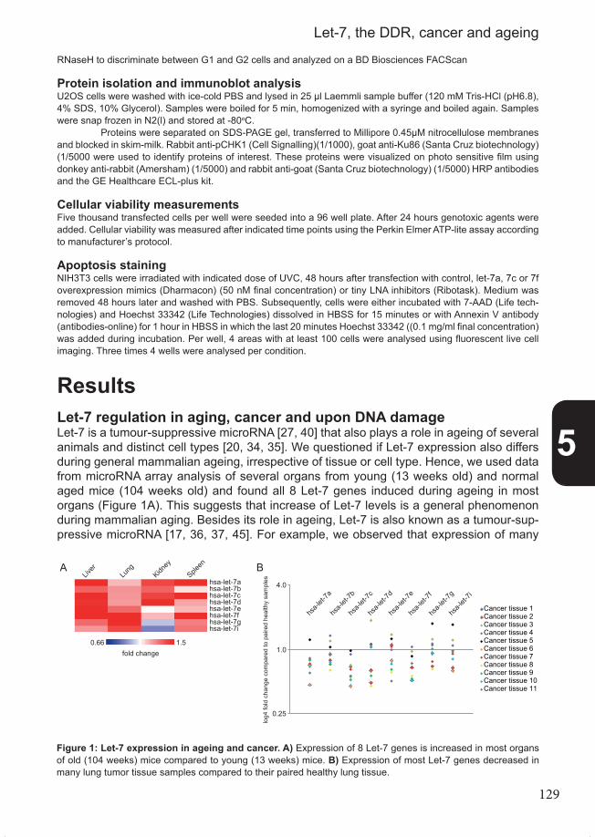
129
RNaseH to discriminate between G1 and G2 cells and analyzed on a BD Biosciences FACScan
Protein isolation and immunoblot analysisU2OS cells were washed with ice-cold PBS and lysed in 25 µl Laemmli sample buffer (120 mM Tris-HCl (pH6.8), 4% SDS, 10% Glycerol). Samples were boiled for 5 min, homogenized with a syringe and boiled again. Samples were snap frozen in N2(l) and stored at -80oC. Proteins were separated on SDS-PAGE gel, transferred to Millipore 0.45µM nitrocellulose membranes and blocked in skim-milk. Rabbit anti-pCHK1 (Cell Signalling)(1/1000), goat anti-Ku86 (Santa Cruz biotechnology)(1/5000 were used to identify proteins of interest. These proteins were visualized on photo sensitive film using donkey anti-rabbit (Amersham) (1/5000) and rabbit anti-goat (Santa Cruz biotechnology) (1/5000) HRP antibodies and the GE Healthcare ECL-plus kit.
Cellular viability measurementsFive thousand transfected cells per well were seeded into a 96 well plate. After 24 hours genotoxic agents were added. Cellular viability was measured after indicated time points using the Perkin Elmer ATP-lite assay according to manufacturer’s protocol.
Apoptosis stainingNIH3T3 cells were irradiated with indicated dose of UVC, 48 hours after transfection with control, let-7a, 7c or 7f overexpression mimics (Dharmacon) (50 nM final concentration) or tiny LNA inhibitors (Ribotask). Medium was removed 48 hours later and washed with PBS. Subsequently, cells were either incubated with 7-AAD (Life tech-nologies) and Hoechst 33342 (Life Technologies) dissolved in HBSS for 15 minutes or with Annexin V antibody (antibodies-online) for 1 hour in HBSS in which the last 20 minutes Hoechst 33342 ((0.1 mg/ml final concentration) was added during incubation. Per well, 4 areas with at least 100 cells were analysed using fluorescent live cell imaging. Three times 4 wells were analysed per condition.
ResultsLet-7 regulation in aging, cancer and upon DNA damageLet-7 is a tumour-suppressive microRNA [27, 40] that also plays a role in ageing of several animals and distinct cell types [20, 34, 35]. We questioned if Let-7 expression also differs during general mammalian ageing, irrespective of tissue or cell type. Hence, we used data from microRNA array analysis of several organs from young (13 weeks old) and normal aged mice (104 weeks old) and found all 8 Let-7 genes induced during ageing in most organs (Figure 1A). This suggests that increase of Let-7 levels is a general phenomenon during mammalian aging. Besides its role in ageing, Let-7 is also known as a tumour-sup-pressive microRNA [17, 36, 37, 45]. For example, we observed that expression of many Figure 1
Ahsa-let-7ahsa-let-7bhsa-let-7chsa-let-7dhsa-let-7ehsa-let-7fhsa-let-7ghsa-let-7i
1.50.66
Liver
Lung
Kidney
Spleen
fold change
B
Cancer tissue 1Cancer tissue 2Cancer tissue 3Cancer tissue 4Cancer tissue 5Cancer tissue 6Cancer tissue 7Cancer tissue 8Cancer tissue 9Cancer tissue 10Cancer tissue 11
hsa-l
et-7a
hsa-l
et-7i
hsa-l
et-7g
hsa-l
et-7f
hsa-l
et-7e
hsa-l
et-7d
hsa-l
et-7c
hsa-l
et-7b
4.0
1.0
0.25
log4
fold
cha
nge
com
pare
d to
pai
red
heal
thy
sam
ples
Figure 1: Let-7 expression in ageing and cancer. A) Expression of 8 Let-7 genes is increased in most organs of old (104 weeks) mice compared to young (13 weeks) mice. B) Expression of most Let-7 genes decreased in many lung tumor tissue samples compared to their paired healthy lung tissue.
5
Let-7, the DDR, cancer and ageing

130
Let-7 genes decreased in the majority of the lung cancer tissues that we analysed, as compared to the let-7 expression in healthy tissue from the same patient (Figure 1B). This underlines the role of Let-7 in tumorigenesis. Both cancer and ageing can be caused by DNA damage [1] and therefore we ques-tioned whether Let-7 expression responds to DNA damage. Since Let-7 is oppositely regu-lated in ageing organs versus cancer tissues, we investigated the Let-7 response in both wildtype and cancer cells, starting with NIH3T3 immortalized normal mouse fibroblasts. To induce DNA-damage, we used UV-radiation which directly damages the DNA with minimal side-effects on other bio-molecules compared to other DNA-damaging agents. Additional-ly, UV-induced DNA damage primarily consists of helix-distorting lesions, which impede efficient transcription and reduce cellular proliferation, impairments that are also common during natural ageing. Additionally, many of these helix-distorting lesions are poorly repai-red in untranscribed strands of the genome. Therefore they persist for considerable time in the remainder of the genome and by every round of replication they can lead to mutage-nesis, causing cancer. We analyzed previously generated microRNA array data of mouse NIH3T3 cells, 4 hours after treatment with 8J/m2 of UV [9]. As shown in figure 2A, all Let-7 family members were upregulated after UV treatment, mostly highly significant. This sug-gests that Let-7 is induced by UV-type DNA damage which is consistent with the idea that ageing-associated upregulation of Let-7 levels might be caused by DNA damage. To exclude species-specific or damage-specific effects, we also applied Ionizing Radiation (IR) to human C3RO fibroblasts. IR induces, besides numerous single strand breaks and oxidative damage, highly toxic DNA double strand breaks. As shown in figure 2B, multiple Let-7a genes are also induced in these fibroblasts 6h upon treatment with 8gy of IR, indicating that Let7 gene expression responds to different types of DNA damage in both mice and humans. We questioned if Let-7 regulation depends on ATR and ATM, the central DNA damage stress kinases that are directly activated by DNA damage and induce many downstream DDR-processes [46]. Therefore, we treated the cells with caffeine prior to irradiation, which inhibits activity of both kinases. Inhibition of ATM and ATR activity diminished IR-induced Let-7 regulation (Figure 2B), suggesting that ATM and ATR activity is required for DNA da-mage-induced Let-7 regulation. Besides normal primary fibroblasts, we also studied DNA damage-induced Let-7 regulation in cancer cells. Since Let-7 expression is decreased in many lung cancer tissue samples, we investigated A549, a lung cancer cell line. To induce DNA damage, we applied IR and compared Let-7 expression with its regulation in C3RO. In contrast to fibroblasts, expression of Let-7 was transiently reduced 6h after ionizing radiation (Figure 2C). Ad-ditionally, this decrease was not specific for A549, since Let-7 levels were also reduced in U2OS human sarcoma cells (Figure 2D), which was also shown by previous research [26]. Let-7 regulation was also dose dependent, as shown by treatment of A549 cells with DSB-inducing drug bleomycine (Figure 2E). Finally, we wanted to know if Let-7 regulation in A549 cells also depends on ATM activity, as in C3RO fibroblasts. When ATM activity was inhibited with KuDos ATM inhibitor, Let-7 expression was rescued (Figure 2F). These results strongly suggest that DNA damage directly induces regulation of Let-7 expression, although it depends on the status of the cells (normal vs cancer) which direction Let-7 is regulated.
Let-7 influences the DNA damage response and cellular viabilitySince Let-7 is regulated in several cells directly upon DNA damage, we questioned if Let-7 is required for the cellular response to DNA damage. We determined the clonogenic
5
Chapter 5

131
survival of U2OS cells after treatment with different types of genotoxic agents. We studied U2OS cells because these cells are often used for such studies. As Let-7 levels decreased in U2OS cells upon DNA damage (Figure 2D), we ectopically overexpressed Let-7 and applied increasing doses of UV-radiation, IR, cisplatin and doxorubicin. Each treatment in-duces different forms of DNA damage; doxorubicin is an IR-mimicking drug inducing DSBs,
Figure 2
A
0.00.51.01.52.02.53.03.5
hsa-l
et-7a
hsa-l
et-7b
hsa-l
et-7c
hsa-l
et-7d
hsa-l
et-7e
hsa-l
et-7f
hsa-l
et-7g
hsa-l
et-7i
Fold
cha
nge
0J/m8J/m
22**
* ** *
****
**
B
0.00.51.01.52.02.53.03.54.0
hsa-let 7a hsa-let 7c hsa-let 7f
Fold
cha
nge
0 Gy8 Gy0 Gy + caffeine8 Gy + caffeine
0.0 0.2 0.4 0.6 0.8 1.0 1.2 1.4
hsa-let-7a hsa-let-7c hsa-let-7f
Fold
cha
nge 0 Gy
8 Gy
C D
0.00.20.40.60.81.01.21.4
Fold
cha
nge
0 Gy8 Gy
hsa-let-7a hsa-let-7c hsa-let-7f
E
0.00.20.40.60.81.01.21.4
hsa-let-7a hsa-let-7c hsa-let-7f
Fold
cha
nge
0 µg/mL10 µg/mL50 µg/mL
0.0
0.2
0.4
0.6
0.8
1.0
1.2
1.4
hsa-let-7a hsa-let-7c hsa-let-7f
Fold
cha
nge
0 Gy8 Gy
0 Gy + Kudos 559338 Gy + Kudos 55933
F
Figure 2: Let-7 expression upon DNA damage. A) Expression of most of the Let-7 genes increased in NIH3T3 fibroblasts 4 hours after treatment with 8 J/m2. Fold change normalized against untreated sample. Error bars represent the standard deviation and asterisks indicate significance with either a P-value of 0.05 (*) or 0.01 (**) B) Increase of expression of indicated Let-7a genes relative to endogenous control tubulin was dependent on ATM/R activity in C3RO fibroblasts 6 hour after treatment with 8gy of ionizing radiation. Fold changes were calculated using ΔCt values and normalized against untreated sample. Error bars represent the SEM (N=3). C) Expression of indicated Let-7 genes decreased relative to endogenous control Tubulin in lung cancer cells A549 6 hours after treatment with 8gy of ionizing radiation. Fold changes were calculated using ΔCt values and normalized against untreated sample. Error bars represent the SEM (n=4). D) Expression of indicated Let-7 genes decreased relative to endogenous control Tubulin in osteosarcoma cells U2OS 8 hours after treatment with 8gy of ionizing radiation. Fold changes were calculated using ΔCt values and normalized against untreated sample. Error bars represent the SEM (n=4). E) Expression of indicated Let-7 genes relative to endogenous control Tubulin decreased do-se-dependently in A549 6 hours after Bleomycin treatment. Fold change was calculated using ΔCt values and normalized against untreated sample. Error bars represent the SEM (n=3) F) Decrease of expression of indicated Let-7 genes relative to endogenous control tubulin was dependent on ATM activity in A549 cells 6 hours after 8gy of ionizing radiation. Fold changes were calculated using ΔCt values and normalized against untreated sample. Error bars represent the SEM (n=3).
5
Let-7, the DDR, cancer and ageing

132
while cisplatin forms intra- and inter-strand crosslinks, disrupting the helix conformation. This hinders efficient replication and induces apoptosis, comparable to UV-induced DNA damage, which also impedes efficient transcription. These treatments showed that Let-7 sensitized U2OS for doxorubicin and IR, but induced resistance for UV and cisplatin (Figu-re 3). This suggests that Let-7 has different roles depending on which kind of DNA damage is induced. As Let-7 determined genotoxic sensitivity, we wondered which cellular responses are affected by Let-7 upon genotoxic insults. Hence, we studied the cell cycle, DNA dama-ge signalling, cellular viability and apoptosis in various cells after modulation of Let-7 levels and induction of DNA damage. First, we assessed if Let-7 impacts the cell cycle, since the phase of the cell cycle can influence how cells counter DNA damage. Additionally, Let-7 might modulate cell cycle arrest upon DNA damage. Therefore, we investigated the cell cycle distribution of control and Let-7 transfected U2OS cells upon mock and IR treatment. As figure 4A shows, Let-7 influenced the cell cycle distribution independent of DNA dama-ge by inducing an accumulation of cells in G2/M phase. These results suggest that Let-7 induces a G2/M cell cycle arrest, which was -as expected- also induced by IR alone (Figure 4B); the number of G2/M cells increased 2-fold in control transfected cells upon 8Gy of IR. In contrast, IR only increased the number of Let-7 transfected G2/M phase cells by ~1.3 fold (Figure 4B). This modest relative increase is explained by the high percentage of G2/M cells already present without IR treatment, which was already >60%. Let-7-induced G2/M cell cycle arrest can be caused by an increased number of cells that progressed through the G1 and S-phase with endogenous DNA damage. Figure 4A indeed shows a decrease of both G1 and -even more- S-phase cells. In S-phase, the DNA damage checkpoint response can be activated after detection of single strand DNA derived from DNA-repair intermediates and collapsed replication forks. These structures
Figure 3
A
1%
10%
100%
1000%
0 gy 1 gy 2 gy 3 gy 5 gy
Control miRNA mimichsa-let-7
C
1%
10%
100%
1000%
0 µM 0.1 µM 0.5 µM 1 µM 2 µM
Control miRNA mimichsa-let-7
D
0 µM 0.2 µM 1 µM 2 µM
40%
60%
80%
100%
120%
Control miRNA mimichsa-let-7
B
0.1%
1%
10%
100%
1000%
0J/m2 4J/m2 8J/m2 12J/m2 16J/m2
Control miRNA mimichsa-let-7
% s
urvi
val
% s
urvi
val
% s
urvi
val
% s
urvi
val
Figure 3: Let-7 influences the DDR. Let-7 modulated survival of U2OS cells after induction of DNA damage with A) doxorubicin, UV B), IR C) and cisplatin D) treatment. Increasing doses of DNA damage resulted in decreased (doxorubicin and IR) or increased (UV and cisplatin) colony formation of Let-7 transfected cells compared to control miR-mimic transfected cells. For each transfection, the number of colonies formed under untreated conditions was set at 100%. Error bars represent the SEM (n=3).
5
Chapter 5

133
facilitate activation of ATR via ATRIP and subsequent activation of the CHK1 protein kinase that induces cell cycle arrest [3]. Thus, we studied if Let-7 influenced phosphorylation of CHK1 in U2OS cells and observed that Let-7 expression decreased CHK1 phosphorylation in both mock- and IR-treated cells (Figure 4C). Decreased phosphorylation of CHK1 might explain the influence of Let-7 on the cell cycle (Figure 4A). These results on cell cycle dis-tribution and Chk1 phosphorylation show that Let-7 affects the cell cycle, which might partly depend on DNA damage. Since Let-7 determined genotoxic sensitivity in a genotoxin-specific manner (Fi-gure 3) and influenced cell cycle checkpoints (Figure 4), we wondered how Let-7 influen-ced cellular viability upon distinct genotoxic insults. To investigate this, we measured ATP levels after administration of cisplatin, mitomycin-C, another DNA-crosslinking agent, and DSB-inducing doxorubicin. All three genotoxic treatments showed higher ATP-levels in Let-7 transfected cells compared to control transfected cells (Figure 5A/B/C). This indicates that Let-7 helps to protect against the effects of both forms of genotoxic treatments, at least under the conditions tested. To examine how Let-7 might protect against these treatments we studied the effect of Let-7 on apoptosis in NIH3T3 cells, which clearly induce apoptosis after treatment with an UV-dose of 12 J/m2. The nuclei were stained with nuclear-stain Hoechst, while 7AAD or Annexin were used to respectively visualize early and late apop-totic cells. We observed that ectopic overexpression of Let-7 reduced the total number of cells (not shown), but also decreased the relative levels of both early (Figure 5D) and late apoptotic cells (Figure 5E), while the opposite occurred when Let-7 expression was silen-ced using tiny LNA-inhibitors (Figure 5F/G). This strongly suggests that Let-7 increases cellular viability upon DNA damage by inhibiting apoptosis.
0102030405060708090
100
G1 S G2/M
% o
f gat
ed c
ells
control microRNA mimichsa-let-7a
0102030405060708090
100
G1 S G2/M
% o
f gat
ed c
ells
Control microRNA mimichsa-let-7a
0 0 8 8 12 12
Contro
l
hsa-l
et-7
Contro
l
hsa-l
et-7
Contro
l
hsa-l
et-7
6hpChk1
Ku86
0.0
0.2
0.4
0.6
0.8
1.0
1.2
Fold
cha
nge
com
pare
d to
con
trol
Figure 4
A
B
C
Contro
l
hsa-l
et-7
Contro
l
hsa-l
et-7
Contro
l
hsa-l
et-7
0 0 8 8 12 12
- - -+ + + 2.5gy
gy
Figure 4: Let-7 modulates the cell cycle. A) Distribution of G1, S and G2/M-phase U2OS cells was measured 2 days after transfection with either Let-7 or a control miR-mimic. Values depict percentage of total number of cells analyzed per condition. B) Distribution of G1, S and G2/M-phase U2OS cells, 2 days after transfection with either Let-7 or control miR-mimic, were measured 8h after IR treatment and depicted as a percentage of the total number of analyzed cells per condition. Error bars represent standard deviation (n=2). C) Let-7 modulated CHK1 phosphorylation in U2OS cells 8h after 8 and 12 gy of IR. Ku86 is used as loading control.
5
Let-7, the DDR, cancer and ageing
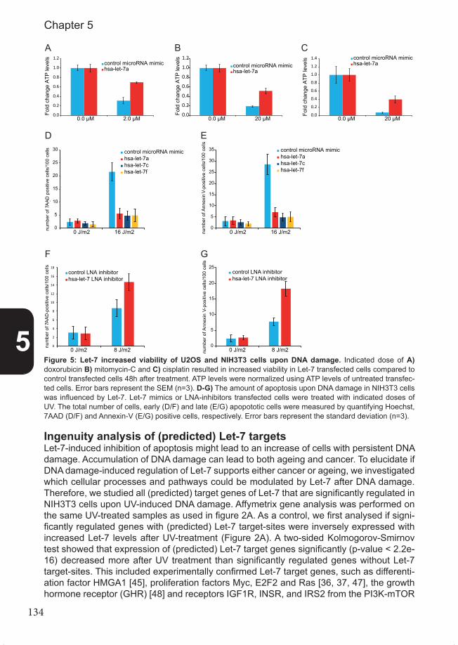
134
Ingenuity analysis of (predicted) Let-7 targetsLet-7-induced inhibition of apoptosis might lead to an increase of cells with persistent DNA damage. Accumulation of DNA damage can lead to both ageing and cancer. To elucidate if DNA damage-induced regulation of Let-7 supports either cancer or ageing, we investigated which cellular processes and pathways could be modulated by Let-7 after DNA damage. Therefore, we studied all (predicted) target genes of Let-7 that are significantly regulated in NIH3T3 cells upon UV-induced DNA damage. Affymetrix gene analysis was performed on the same UV-treated samples as used in figure 2A. As a control, we first analysed if signi-ficantly regulated genes with (predicted) Let-7 target-sites were inversely expressed with increased Let-7 levels after UV-treatment (Figure 2A). A two-sided Kolmogorov-Smirnov test showed that expression of (predicted) Let-7 target genes significantly (p-value < 2.2e-16) decreased more after UV treatment than significantly regulated genes without Let-7 target-sites. This included experimentally confirmed Let-7 target genes, such as differenti-ation factor HMGA1 [45], proliferation factors Myc, E2F2 and Ras [36, 37, 47], the growth hormone receptor (GHR) [48] and receptors IGF1R, INSR, and IRS2 from the PI3K-mTOR
0.0
0.2
0.4
0.6
0.8
1.0
1.2
0.0 µM 2.0 µM
Fold
cha
nge
ATP
leve
ls
control microRNA mimichsa-let-7a
0.0
0.2
0.4
0.6
0.8
1.0
1.2
0.0 µM 20 µM
Fold
cha
nge
ATP
leve
ls
control microRNA mimichsa-let-7a
0.0
0.2
0.4
0.6
0.8
1.0
1.2
1.4
0.0 µM 20 µM
Fold
cha
nge
ATP
leve
ls control microRNA mimichsa-let-7a
Figure 5
A B C
D
0
5
10
15
20
25
30
0 J/m2 16 J/m2num
ber o
f 7-
AAD
pos
itive
cel
ls/1
00 c
ells control microRNA mimic
hsa-let-7ahsa-let-7chsa-let-7f
0
5
10
15
20
25
30
35
0 J/m2 16 J/m2num
ber o
f Ann
exin
-V po
sitiv
e ce
lls/1
00 c
ells
control microRNA mimichsa-let-7ahsa-let-7chsa-let-7f
E
F G
0
2
4
6
8
10
12
14
16
18
0 J/m2 8 J/m2
num
ber o
f 7-
AAD
pos
itive
cel
ls/1
00 c
ells
control LNA inhibitorhsa-let-7 LNA inhibitor
0
5
10
15
20
25
0 J/m2 8 J/m2num
ber o
f Ann
exin
-V po
sitiv
e ce
lls/1
00 c
ells
control LNA inhibitorhsa-let-7 LNA inhibitor
Figure 5: Let-7 increased viability of U2OS and NIH3T3 cells upon DNA damage. Indicated dose of A) doxorubicin B) mitomycin-C and C) cisplatin resulted in increased viability in Let-7 transfected cells compared to control transfected cells 48h after treatment. ATP levels were normalized using ATP levels of untreated transfec-ted cells. Error bars represent the SEM (n=3). D-G) The amount of apoptosis upon DNA damage in NIH3T3 cells was influenced by Let-7. Let-7 mimics or LNA-inhibitors transfected cells were treated with indicated doses of UV. The total number of cells, early (D/F) and late (E/G) apopototic cells were measured by quantifying Hoechst, 7AAD (D/F) and Annexin-V (E/G) positive cells, respectively. Error bars represent the standard deviation (n=3).
5
Chapter 5

135
pathway [49]. These validated targets strongly indicate that Let-7 targets are indeed inver-sely regulated to Let-7 after UV-treatment. We wanted to know which biological processes and molecular pathways were as-sociated with differentially regulated (predicted) Let-7 target genes. Therefore we used Ingenuity Pathway Analysis (IPA), which identifies specific processes and molecular pa-thways that are enriched with UV-responsive and (predicted) Let-7 targets. IPA showed that, besides some specific disorders and generic processes such as post-translational modification and transcription, UV-responsive target genes were involved in processes such as proliferation, cancer and cell cycle control (Figure 6B). IPA also identifies speci-fic molecular pathways that are enriched within a certain set of genes of interest. Using UV-responsive and (predicted) Let-7 targets, various molecular pathways were identified by IPA that have a close link to both cancer and the DDR, such as p53-signalling. Other pathways are associated with control of cellular metabolism, growth and proliferation, such as peroxisome proliferator-activated nuclear receptors (PPAR), TGF-β, and PTEN-signal-ling (Figure 6C). Proteins of the PTEN signalling pathway overlap with downstream targets of IGF1R, INSR, and IRS2, which are components of the PI3K-mTOR pathway [49]. This pathway and PTEN-signalling, are strongly associated with both ageing and cancer, also in relationship to DNA damage [50-52]. This illustrates that Let-7 likely plays a role in both cancer and ageing in response to DNA damage.
Figure 5
A
Fold change
Cum
ulat
ive
dist
ribut
ion
UV-responsive (predicted) Let-7 target genes
UV-responsive non-Let-7 target genes
B
C
0123456789
-log(
p-va
lue)
0123456
-log(
p-va
lue)
0.000.05
0.100150.20
Ratio
0.25
Post-tra
nslatio
nal modi�ca
tion
Gene expression
Connective tis
sue diso
rders
Developmental diso
rder
Hereditary diso
rder
Metabolic dise
ase
Skeletal and m
uscular d
isord
ers
Cellular g
rowth
and prolife
ration
Cell cycle
Conective tis
sue development a
nd functio
n
Cellular a
ssembly and org
anizatio
n
Dermato
logical d
isease
s and co
nditions
Organism
al injury and abnorm
alities
Cellular fu
nction and m
aintenance
Cancer
PPARα/RXRα A
ctiva
tion
Molecu
lar m
echa
nism of
canc
er
PTEN sign
alling
Salvag
e path
ways o
f pyri
midine
Ribo
nucle
otide
s
Pancre
atic a
deno
carci
noma s
ignali
ng
cell c
ycle
regula
tion b
y BTG fa
mily pro
teins
Chronic
mye
loid l
euke
mia sig
nalin
g
Regula
tion o
f IL-2
expre
ssion
in ac
tivate
d and
anerg
ic T ly
mphoc
ytes
Estrog
en-m
ediat
ed S-ph
ase e
ntry
TGF-β sig
nalin
g
Pyrido
xal 5
’-pho
spha
te sa
lvage
pathw
ay
Antipro
liferat
ive ro
le of
TOB in T ce
ll sign
aling
Cholin
e bios
ynthe
sis III
Factor
s prom
oting
cardi
ogen
esis
in ve
rtebra
tes
p53 s
ignall
ing
1.0
0.8
0.6
0.4
0.2
0.0
0 2 4-2-4
Figure 6: IPA analysis links (predicted) Let-7 target genes to ageing and cancer pathways. A) Expression of predicted Let-7 target genes decreased more than average upon UV treatment. A cumulative distribution plot shows mRNA fold changes of genes with (blue) and without (red) predicted Let-7 MRE after 8J/m2 UV treatment of NIH3T3 primary fibroblasts. B) UV-responsive and predicted Let-7 target genes are enriched in biological processes of proliferation, cell cycle control and development. Top 15 out of 36 processes are shown that were identified by IPA analysis. C) UV-responsive and predicted Let-7 target genes are enriched in PTEN signalling, growth factor receptor signalling, PPAR signalling and TGF-β/SMAD signalling. Top 15 out of 49 processes are shown that were identified by IPA analysis.
5
Let-7, the DDR, cancer and ageing
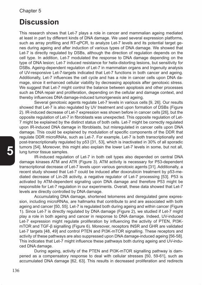
136
DiscussionThis research shows that Let-7 plays a role in cancer and mammalian ageing mediated at least in part by different kinds of DNA damage. We used several expression platforms, such as array profiling and RT-qPCR, to analyze Let-7 levels and its potential target ge-nes during ageing and after induction of various types of DNA damage. We showed that Let-7 is directly regulated by DSBs, although the direction of regulation depends on the cell type. In addition, Let-7 modulated the response to DNA damage depending on the type of DNA lesion; Let-7 induced resistance for helix-distorting lesions, but sensitivity for DSBs. Ageing-dependent regulation of Let-7 in mammalian organs and Ingenuity analysis of UV-responsive Let-7-targets indicated that Let-7 functions in both cancer and ageing. Additionally, Let-7 influences the cell cycle and has a role in cancer cells upon DNA da-mage, since it enhanced cellular viability by decreasing apoptosis after genotoxic stress. We suggest that Let-7 might control the balance between apoptosis and other processes such as DNA repair and proliferation, depending on the cellular and damage context, and thereby influences DNA damage-induced tumorigenesis and ageing. Several genotoxic agents regulate Let-7 levels in various cells [9, 26]. Our results showed that Let-7 is also regulated by UV treatment and upon formation of DSBs (Figure 2). IR-induced decrease of Let-7 expression was shown before in cancer cells [26], but the opposite regulation of Let-7 in fibroblasts was unexpected. This opposite regulation of Let-7 might be explained by the distinct status of both cells. Let-7 might be correctly regulated upon IR-induced DNA damage in fibroblasts, but misregulated in cancer cells upon DNA damage. This could be explained by modulation of specific components of the DDR that regulate DDR microRNAs, such as Let-7. For example, Let-7 is both transcriptionally and post-transcriptionally regulated by p53 [31, 53], which is inactivated in 30% of all sporadic tumors [54]. Moreover, this might also explain the lower Let-7 levels in some, but not all, lung tumor tissue samples. IR-induced regulation of Let-7 in both cell types also depended on central DNA damage kinases ATM and ATR (Figure 3). ATM activity is necessary for P53-dependent transcriptional decrease of Let-7 levels upon various genotoxic agents [31]. Additionally, a recent study showed that Let-7 could be induced after doxorubicin treatment by p53-me-diated decrease of Lin-28 activity, a negative regulator of Let-7 processing [53]. P53 is activated by ATM-dependent signaling upon DNA damage and therefore P53 might be responsible for Let-7 regulation in our experiments. Overall, these data showed that Let-7 levels are directly controlled by DNA damage. Accumulating DNA damage, shortened telomeres and deregulated gene expres-sion, including microRNAs, are hallmarks that contribute to and are associated with both ageing and cancer [50, 55]. Let-7 is regulated both during ageing and within cancer (Figure 1). Since Let-7 is directly regulated by DNA damage (Figure 2), we studied if Let-7 might play a role in both ageing and cancer in response to DNA damage. Indeed, UV-induced Let-7 expression might regulate proliferation by influencing the activity of PTEN, PI3K-mTOR and TGF-β signalling (Figure 6). Moreover, receptors INSR and GHR are validated Let-7 targets [48, 49] and control PTEN and PI3K-mTOR signalling. These receptors and activity of these pathways are also suppressed upon DNA damage-induced ageing [56-58]. This indicates that Let-7 might influence these pathways both during ageing and UV-indu-ced DNA damage. During ageing, activity of the PTEN and PI3K-mTOR signalling pathway is dam-pened as a compensatory response to deal with cellular stresses [50, 59-61], such as accumulated DNA damage [62, 63]. This results in decreased proliferation and redirects
5
Chapter 5

137
the cellular focus to cell maintenance. Abrogation of Let-7 targets INSR and GHR in KO mice results in growth defects and increases longevity [50]. Interestingly, increased Let-7 levels in Lin28 KO mice also results in small animals [64]. Additionally, these mice show a Let-7-mediated repression of PI3K-mTOR signalling. Further research should elucidate if Lin28 also influences ageing and if this is also mediated by DNA damage [53]. In line with this, increased Let-7 levels in fibroblasts with DNA damage (Figure 2) may also decrease proliferation by suppressing PI3K-mTOR signalling. Since DNA damage regulates Let-7 levels and Let-7 overexpression decreases proliferation [40], the number of S-phase cells (Figure 4) and potentially modulates PTEN and PI3K-mTOR signalling (Figure 6), DNA damage-induced Let-7 expression might therefore modulate cellular metabolism and main-tenance systems to cope with accumulating DNA damage. Previous research showed that Let-7 modulates cellular sensitivity upon DNA da-mage [26]. We showed that Let-7 both increased and decreased sensitivity depending on the type of DNA damage (Figure 3). This could be explained by the type of DNA lesion and the processes that Let-7 modulates. DSBs are highly lethal lesions and it was previously shown that some DSB repair protein levels are decreased upon Let-7 overexpression [40]. In addition, Let-7 also silenced DNA damage signaling (Figure 5B), which could result in the observed decrease of S-phase cells and accumulation of cells in G2/M (Figure 4). Consequently, elevated Let-7 levels, followed by IR treatment, may lead to an increased number of damaged cells that could explain the increased clonal survival sensitivity for DSB-inducing treatments (Figure 3). Therefore, cancer cells might decrease Let-7 expres-sion upon IR treatment (Figure 2) to enable correct cell cycle arrest and efficient DNA repair. Conversely, ageing cells with accumulated DNA damage (Figure 1) and fibroblasts with DSBs (Figure 2) might increase Let-7 expression to suppress DSB repair and dama-ge signaling, which are not necessary since Let-7 already suppresses proliferation [36]. Furthermore, this response allows more time for DNA repair of persistent helix-distorting lesions, such as induced by UV and cisplatin, which could explain the resistance that Let-7 induces for these treatments. However, using high doses, these treatments can also induce apoptosis. Let-7 in-hibits apoptosis [38, 65, 66] and we showed that it also decreases sensitivity for genotoxic–induced (Figure 5). Induction of Let-7 levels in fibroblasts (Figure 2) might also protect these cells against apoptosis induced by DSBs. This seems to contrast with the increased clonal survival sensitivity for DSB-inducing agents (Figure 3). However, apoptosis occurs within 24-48 hours, while clonal survival assesses longer-term proliferation capacity after genotoxic stress. To illustrate, decreased apoptosis induced by Let-7 may lead to an incre-ase of surviving, but damaged cells, which ultimately decreases their proliferation capacity. Apoptosis and DNA damage signaling might also be reduced during ageing-dependent increase of Let-7 (Figure 1) or after treatment of Let-7 overexpressing cells with helix-dis-torting agents (Figure 3). However, the resulting accumulating lesions are more easily by-passed during replication [67] than high levels of DSBs, causing fewer cells to accumulate in G2/M. This could subsequently result in the decreased sensitivity for helix-distorting les-ions, as shown in the clonal survival (Figure 3), while Let-7 increases sensitivity for DSBs. Clearly, Let-7 is directly responsive to DNA damage and influences the DDR as it may control the balance between cellular proliferation, DNA repair and apoptosis. This could be cell type specific as illustrated by the differential regulation of Let-7 upon DNA damage in primary fibroblasts and cancer cells. In addition, this balance could also de-pend on which kind of DNA damage is induced. Let-7 might sensitize cells for DSBs as it decreases DNA damage signalling and repair capacity [40], but might induce resistance for helix-distorting lesion by temporary decreasing proliferation and inhibiting apoptosis.
5
Let-7, the DDR, cancer and ageing

138
Thereby, the role of Let-7 in the DDR might influence both DNA damage-induced ageing and tumorigenesis. Therefore, this research could be of importance to better understand the complex function of microRNAs in both ageing and cancer.
5
Chapter 5

139
References 1. Hoeijmakers, J.H., DNA damage, aging, and cancer. N Engl J Med, 2009. 361(15): p. 1475-85.2. Harper, J.W. and S.J. Elledge, The DNA damage response: ten years after. Mol Cell, 2007. 28(5): p. 739-45.3. Bartek, J. and J. Lukas, DNA damage checkpoints: from initiation to recovery or adaptation. Curr Opin Cell Biol, 2007. 19(2): p. 238-45.4. Riley, T., et al., Transcriptional control of human p53-regulated genes. Nat Rev Mol Cell Biol, 2008. 9(5):p.402-12.5. Bartel, D.P., MicroRNAs: target recognition and regulatory functions. Cell, 2009. 136(2): p. 215-33.6. Guo, H., et al., Mammalian microRNAs predominantly act to decrease target mRNA levels. Nature, 2010.466(7308): p. 835-40.7. Kloosterman, W.P. and R.H. Plasterk, The diverse functions of microRNAs in animal development and disease. Dev Cell, 2006. 11(4): p. 441-50.8. Wouters, M.D., et al., MicroRNAs, the DNA damage response and cancer. Mutat Res, 2011.9. Pothof, J., et al., MicroRNA-mediated gene silencing modulates the UV-induced DNA-damage res- ponse.Embo J, 2009.10. Simone, N.L., et al., Ionizing radiation-induced oxidative stress alters miRNA expression. PLoS One, 2009.4(7): p. e6377.11. He, L., et al., microRNAs join the p53 network--another piece in the tumour-suppression puzzle. Nat Rev Cancer, 2007. 7(11): p. 819-22.12. Galluzzi, L., et al., miR-181a and miR-630 Regulate Cisplatin-Induced Cancer Cell Death. Cancer Res, 2010. 70(5): p. 1793-1803.13. Bu, Y., et al., Knockdown of Dicer in MCF-7 human breast carcinoma cells results in G1 arrest and increased sensitivity to cisplatin. Oncol Rep, 2009. 21(1): p. 13-7.14. Kumar, M.S., et al., Impaired microRNA processing enhances cellular transformation and tumorigene sis. Nat Genet, 2007. 39(5): p. 673-7.15. Karube, Y., et al., Reduced expression of Dicer associated with poor prognosis in lung cancer pa- tients. Cancer Sci, 2005. 96(2): p. 111-5.16. Medina, P.P., M. Nolde, and F.J. Slack, OncomiR addiction in an in vivo model of microRNA-21-indu- ced pre-B-cell lymphoma. Nature, 2010.17. Calin, G.A., et al., Human microRNA genes are frequently located at fragile sites and genomic regions involved in cancers. Proc Natl Acad Sci U S A, 2004. 101(9): p. 2999-3004.18. Boehm, M. and F. Slack, A developmental timing microRNA and its target regulate life span in C. ele- gans. Science, 2005. 310(5756): p. 1954-7.19. de Lencastre, A., et al., MicroRNAs both promote and antagonize longevity in C. elegans. Curr Biol, 2010.20(24): p. 2159-68.20. Ibanez-Ventoso, C., et al., Modulated microRNA expression during adult lifespan in Caenorhabditis elegans. Aging Cell, 2006. 5(3): p. 235-46.21. Kato, M., et al., Age-associated changes in expression of small, noncoding RNAs, including micro RNAs, in C. elegans. RNA, 2011. 17(10): p. 1804-20.22. Dimmeler, S. and P. Nicotera, MicroRNAs in age-related diseases. EMBO Mol Med, 2013. 5(2): p. 180-90.23. Smith-Vikos, T. and F.J. Slack, MicroRNAs and their roles in aging. J Cell Sci, 2012. 125(Pt 1): p. 7-17.24. He, X., L. He, and G.J. Hannon, The guardian’s little helper: microRNAs in the p53 tumor suppressor network. Cancer Res, 2007. 67(23): p. 11099-101.25. Ugalde, A.P., et al., Aging and chronic DNA damage response activate a regulatory pathway involving miR-29 and p53. Embo J, 2011. 30(11): p. 2219-32.26. Weidhaas, J.B., et al., MicroRNAs as potential agents to alter resistance to cytotoxic anticancer thera- py.Cancer Res, 2007. 67(23): p. 11111-6.27. Esquela-Kerscher, A., et al., The let-7 microRNA reduces tumor growth in mouse models of lung can- cer. Cell Cycle, 2008. 7(6): p. 759-64.28. Yu, F., et al., let-7 regulates self renewal and tumorigenicity of breast cancer cells. Cell, 2007. 131(6): p.1109-23.29. Kumar, M.S., et al., Suppression of non-small cell lung tumor development by the let-7 microRNA fa- mily. Proc Natl Acad Sci U S A, 2008. 105(10): p. 3903-8.30. Bussing, I., F.J. Slack, and H. Grosshans, let-7 microRNAs in development, stem cells and cancer. Trends Mol Med, 2008. 14(9): p. 400-9.31. Saleh, A.D., et al., Cellular stress induced alterations in microRNA let-7a and let-7b expression are
5
Let-7, the DDR, cancer and ageing

140
dependent on p53. PLoS One, 2011. 6(10): p. e24429.32. Chaudhry, M.A., Real-time PCR analysis of micro-RNA expression in ionizing radiation-treated cells. Cancer Biother Radiopharm, 2009. 24(1): p. 49-56.33. van Jaarsveld, M.T., et al., DNA damage responsive microRNAs misexpressed in human cancer modulate therapy sensitivity. Mol Oncol, 2013.34. Toledano, H., et al., The let-7-Imp axis regulates ageing of the Drosophila testis stem-cell niche. Natu- re, 2012. 485(7400): p. 605-10.35. Nishino, J., et al., Hmga2 promotes neural stem cell self-renewal in young but not old mice by redu- cing p16Ink4a and p19Arf Expression. Cell, 2008. 135(2): p. 227-39.36. Johnson, S.M., et al., RAS is regulated by the let-7 microRNA family. Cell, 2005. 120(5): p. 635-47.37. Sampson, V.B., et al., MicroRNA let-7a down-regulates MYC and reverts MYC-induced growth in Bur kitt lymphoma cells. Cancer Res, 2007. 67(20): p. 9762-70.38. Tsang, W.P. and T.T. Kwok, Let-7a microRNA suppresses therapeutics-induced cancer cell death by targeting caspase-3. Apoptosis, 2008. 13(10): p. 1215-22.39. Shimizu, S., et al., The let-7 family of microRNAs inhibits Bcl-xL expression and potentiates sorafe- nib-induced apoptosis in human hepatocellular carcinoma. J Hepatol, 2010. 52(5): p. 698-704.40. Johnson, C.D., et al., The let-7 microRNA represses cell proliferation pathways in human cells. Can- cer Res,2007. 67(16): p. 7713-22.41. Mondol, V. and A.E. Pasquinelli, Let’s make it happen: the role of let-7 microRNA in development. Curr Top Dev Biol, 2012. 99: p. 1-30.42. Hua, S., et al., Reduced miR-31 and let-7 maintain the balance between differentiation and quiescen- ce in lung cancer stem-like side population cells. Biomed Pharmacother, 2012. 66(2): p. 89-97.43. Pothof, J., et al., MicroRNA-mediated gene silencing modulates the UV-induced DNA-damage res- ponse. EMBO J, 2009. 28(14): p. 2090-9.44. Obad, S., et al., Silencing of microRNA families by seed-targeting tiny LNAs. Nat Genet, 2011. 43(4): p.371-8.45. Lee, Y.S. and A. Dutta, The tumor suppressor microRNA let-7 represses the HMGA2 oncogene. Ge- nes Dev, 2007. 21(9): p. 1025-30.46. Ciccia, A. and S.J. Elledge, The DNA damage response: making it safe to play with knives. Mol Cell, 2011.40(2): p. 179-204.47. Dong, Q., et al., MicroRNA let-7a inhibits proliferation of human prostate cancer cells in vitro and in vivo by targeting E2F2 and CCND2. PLoS One, 2010. 5(4): p. e10147.48. Lin, S., et al., Let-7b regulates the expression of the growth hormone receptor gene in deletion-type dwarf chickens. BMC Genomics, 2012. 13: p. 306.49. Zhu, H., et al., The Lin28/let-7 axis regulates glucose metabolism. Cell, 2011. 147(1): p. 81-94.50. Lopez-Otin, C., et al., The hallmarks of aging. Cell, 2013. 153(6): p. 1194-217.51. Hoesel, B. and J.A. Schmid, The complexity of NF-kappaB signaling in inflammation and cancer. Mol Cancer, 2013. 12: p. 86.52. Hanahan, D. and R.A. Weinberg, Hallmarks of cancer: the next generation. Cell, 2011. 144(5): p. 646- 74.53. Lee, J.Y., et al., Tumor suppressor p53 plays a key role in induction of both tristetraprolin and let-7 in human cancer cells. Nucleic Acids Res, 2013. 41(11): p. 5614-25.54. Vousden, K.H. and X. Lu, Live or let die: the cell’s response to p53. Nat Rev Cancer, 2002. 2(8): p. 594-604.55. Hanahan, D. and R.A. Weinberg, Hallmarks of cancer: the next generation. Cell, 2012. 144(5): p. 646- 74.56. van der Pluijm, I., et al., Impaired genome maintenance suppresses the growth hormone--insulin-like growth factor 1 axis in mice with Cockayne syndrome. PLoS Biol, 2007. 5(1): p. e2.57. Niedernhofer, L.J., et al., A new progeroid syndrome reveals that genotoxic stress suppresses the somatotroph axis. Nature, 2006. 444(7122): p. 1038-43.58. Feng, Z., et al., The regulation of AMPK beta1, TSC2, and PTEN expression by p53: stress, cell and tissue specificity, and the role of these gene products in modulating the IGF-1-AKT-mTOR pathways. Cancer Res, 2007. 67(7): p. 3043-53.59. Kenyon, C., The plasticity of aging: insights from long-lived mutants. Cell, 2005. 120(4): p. 449-60.60. Bartke, A., Minireview: role of the growth hormone/insulin-like growth factor system in mammalian aging. Endocrinology, 2005. 146(9): p. 3718-23.61. Steelman, L.S., et al., Roles of the Raf/MEK/ERK and PI3K/PTEN/Akt/mTOR pathways in controlling growth and sensitivity to therapy-implications for cancer and aging. Aging (Albany NY), 2011. 3(3): p.
5
Chapter 5

141
192-222.62. Schumacher, B., et al., Delayed and accelerated aging share common longevity assurance mechanis- ms. PLoS Genet, 2008. 4(8): p. e1000161.63. Garinis, G.A., et al., DNA damage and ageing: new-age ideas for an age-old problem. Nat Cell Biol, 2008. 10(11): p. 1241-7.64. Zhu, H., et al., The Lin28/let-7 axis regulates glucose metabolism. Cell, 2011. 147(1): p. 81-94.65. Wang, S., et al., Let-7/miR-98 regulate Fas and Fas-mediated apoptosis. Genes Immun, 2011. 12(2): p.149-54.66. Ding, Z., et al., MicroRNA hsa-let-7g targets lectin-like oxidized low-density lipoprotein receptor-1 ex- pression and inhibits apoptosis in human smooth muscle cells. Exp Biol Med (Maywood), 2012. 237(9):p.1093-100.67. Livneh, Z., O. Ziv, and S. Shachar, Multiple two-polymerase mechanisms in mammalian translesion DNA synthesis. Cell Cycle, 2010. 9(4): p. 729-35.
5
Let-7, the DDR, cancer and ageing

142

143
Chapter 6
General discussion ofmicroRNAs, the DNA damage response and cancer
Maikel D. Wouters1, Jan H. J. Hoeijmakers1, Joris Pothof1
1. Department of Genetics, Erasmus University Medical Center, Rotterdam, The Netherlan

144

145
DiscussionDNA damage plays a crucial role in ageing and cancer. The DNA damage response (DDR) protects the DNA molecule against DNA lesions and counteracts mutagenesis, thereby respectively delaying ageing and preventing tumorigenesis. The DDR enables cellular sur-vival upon DNA damage, for example by controlling cell cycle arrest and DNA repair. DNA damage can also lead to mutations, deletions and chromosomal rearrangements causing cancer. The DDR counteracts aberrant cellular proliferation upon excessive amounts of DNA damage, by inducing apoptosis or cellular senescence [1-3]. The DDR is tightly regu-lated to control all these processes in order to induce the correct response on DNA damage [3, 4]. For example, signaling kinases ATM and ATR (ATM/R) are the central players of the DDR as they phosphorylate many DDR targets that influence virtually every aspect of the DDR [5]. For example, ATM/R stabilizes p53 protein levels, which induces cell cycle arrest, while p53 also modulates transcription of many DDR genes, thereby inducing specific re-pair pathways, controlling apoptosis and cellular senescence [6]. Although regulation of the DDR is extensively studied at the transcriptional and post-translational level, less is known about post-transcriptional regulation of the DDR. MicroRNAs are important factors in post-transcriptional regulation of protein-coding genes and play important roles in many cellular processes, including apoptosis and cell cycle control [7, 8]. MicroRNAs are also regulated upon DNA damage and several DDR genes are regulated by microRNAs [9]. However, the total impact of microRNAs in the DDR is not clear yet. Hence, we systematically studied the role of microRNAs in the DDR. Both microRNAs and the DDR have a significant role in cancer. The DDR is a crucial barrier against tumorigenesis that should be overcome by a cell to progress into a more malignant state. Therefore, virtually all cancers contain defects in components of the DDR [10, 11]. Initially, this also sensitizes tumors for genotoxic treatments, until therapy resistance is acquired through modulating cellular processes such as the DDR [12-15]. Tu-mors often display misregulated microRNA expression. Expression of mature microRNAs is often suppressed by decreased activity of microRNA processing factors. Alternatively, specific microRNAs are misregulated in cancer [16-19], including microRNAs that are re-gulated by DNA damage [9]. We envisioned that these microRNAs might also modulate the DDR in cancer. Additionally, microRNAs are also known to modulate sensitivity for cancer treatments [7, 8]. Since tumors can be very sensitive to genotoxic therapy or become re-sistant to these treatments, it is conceivable that microRNAs with a role in the DDR could be important in genotoxic therapy sensitivity and resistance. Only several microRNAs are known to regulate DDR genes and a few of the-se microRNAs modulate sensitivity for genotoxic cancer therapy as described in Chapter 1. However, coherent, large scale research into the connection between microRNAs, the DDR, cancer and genotoxic therapy sensitivity has not yet been performed. This thesis describes our research to elucidate this connection by answering 4 main questions using array profiling and functional characterization of individual microRNAs: Which microRNAs are regulated upon DNA damage; how are microRNAs regulated after DNA damage, which DDR genes are regulated by DDR microRNAs and what is the effect of DDR microRNAs on cancer therapy sensitivity? The main conclusions of this thesis can be summarized as follows. Chapter 2 shows that at least one third of the microRNAs that are similarly regulated in different DNA-damaging conditions are misregulated in breast and lung tumors. Furthermore, a significant subset of these microRNAs modulates cancer therapy sensitivity. Chapter 3 demonstrates that expression of many Ionizing Radiation (IR)- and UV-responsive microR-
6
General discussion

146
NAs depends on several microRNA-processing RNA-binding proteins (RBP). These RBPs, and the microRNAs they regulate, are also likely misregulated in a subset of breast tumors.These DDR microRNAs may regulate various DDR genes. To illustrate, our previously pu-blished review predicts that microRNAs regulate 50% of all DDR genes [9]. For example, miR-24 regulates histone H2AX [20], an important protein with a role in the DNA damage checkpoint response. However, as described in chapter 4, we showed that adaptor protein MDC1 is also regulated by miR-24 and might have an important role upon DNA damage induced by IR. Finally, chapter 5 strongly suggests that Let-7 plays an important role in both cancer and aging through DDR-mediated regulation of proliferation pathways, the cell cycle and the apoptosis response.
Expression of microRNAs upon DNA damageThis thesis shows that many microRNAs are regulated upon diverse genotoxic insults. We profiled microRNA expression in different types of cells upon treatment with cisplatin, UV and IR. Although DNA damage-dependent microRNA expression was shown before for these separate treatments [21-23], a treatment-specific microRNA response or a gene-ral DNA damage microRNA response -independent of the applied DNA damaging agent- could not be extracted, since different microRNA array platforms, cell lines, DNA damages, dosages and time points were used, which may all influence the outcome. Hence, we used a standardized array-based [21] approach for these genotoxic insults to identify a general DNA damage microRNA response or a genotoxic agent, dose or cell type-specific res-ponse (Chapter 2 and 3). We studied the microRNA-DDR in several kinds of cell types to elucidate the ge-neral microRNA-DDR. However, most experimental cell types are derived from human tumors that exhibit defects in the DDR [10, 11] and have misregulated miRNA expression [16, 18, 24, 25]. Therefore, we used primary lung and breast epithelial cells to clarify the wild-type, general microRNA response upon DNA damage induced by cisplatin, causing DNA crosslinks, and IR which induces double strand breaks (DSBs). Although these DNA damages induced identical biological outcomes (repair/recovery versus apoptosis/senes-cence) in these cells, the general microRNA response was markedly different depending on the origin of the epithelial cell (Chapter 2). This observation has important implications for further studies on DDR microRNAs as it shows that a general microRNA DDR exists, but this response depends on the cell origin and therefore it is very important to correctly choose and study the cell type of interest. Additionally, we also profiled genotoxic treatment-specific microRNA expression. For example, we studied the specific UV-dependent microRNA DDR in human sarcoma U2OS cells and in wild-type, but immortalized, human and mouse fibroblasts [21](Chapter 3). Many UV-induced microRNAs were similarly regulated in these cells, showing that UV induces a common microRNA response, independent of cell type and species. This con-trasts with the different microRNA responses between primary breast and lung epithelial cells upon identical doses of either cisplatin and IR treatment (Chapter 2). Additionally, no IR-dependent microRNAs in primary epithelial cells could be compared with IR-induced microRNAs in U2OS and HeLa cells, because these cancer cells did not show a profound, early (4-6 hours) IR-response (Chapter 3). We did not study the IR-induced microRNA res-ponse in U2OS and HeLa at later time points, such as 12 hours after IR treatment, when primary epithelial cells did display several cisplatin and IR-specific microRNAs (Chapter 2). To summarize, although a general microRNA UV-DDR exists, IR and cisplatin have a spe-cific effect on microRNA expression, which depends on the origin of the cell and is slower induced than UV.
6
Chapter 6

147
The absence of an early IR response in U2OS and HeLa cells and the differences between the common UV-response and the various responses upon cisplatin and IR treat-ment could be explained as follows; every treatment induces distinct lesions and a strong DNA damage-induced response is slower initiated by IR and cisplatin-induced lesions. DSBs are very lethal lesions and mostly originate from IR-induced single strand breaks during replication or DNA repair [26]. Inter-strand crosslinks are the most lethal lesions that cisplatin can cause and also have to be encountered by the replication machinery to induce a strong response [27]. Since both damages and the subsequent response are dependent on the replication machinery to encounter the lesion, it requires more time before these genotoxic insults induce a microRNA response. In contrast, UV directly damages the DNA and inhibits transcription very efficiently [2], provoking an strong and early microRNA res-ponse (Chapter 3)[21]. Since transcription is an essential cellular process, this also could explain that this response is probably common for various cells (Chapter 3). In addition, the biological outcome after UV damage is quite similar for various cells [2], which is poten-tially supported by the similar microRNA responses. This suggests that UV damage is very suitable to study the microRNA DDR.
Mechanism of DNA damage-induced microRNA regulationIt has been postulated that the microRNA-mediated DDR is timed intermediately between post-translational and transcriptional responses [21], since many microRNAs are regulated relatively quickly, within hours after DNA damage [21, 28, 29]. We indeed observed that, 4 hours after irradiation, UV induced differential expression of a multitude of microRNAs (chapter 3), while IR and cisplatin induced microRNAs after 6 and 12 hours (chapter 2). It is, however, often unclear by which mechanism these microRNAs are regulated. Differential microRNA expression can be controlled at several levels; e.g. during transcription and post-transcriptionally. Intergenic microRNAs have their own promoters [30], while microRNAs located in introns are dependent on the transcription of protein-co-ding genes [31, 32]. Additionally, microRNAs are also profoundly processed post-tran-scriptionally, which is controlled by various RBPs. These RBPs recognize specific RNA sequences and thereby prevent or stimulate further processing of pri-microRNAs and pre-microRNAs into mature microRNAs [33, 34]. Although mature microRNAs are small molecules, pri-microRNAs can be large transcripts that require a significant amount of time to get transcribed [9]. Therefore, differential transcription generally does not occur 4-6 hours after a stimulus. Instead, post-transcriptional processing of microRNAs could be the main mechanism to quickly induce or inhibit microRNA expression. This explanation is further supported by the observation that primary or precursor microRNAs do not always correlate with their mature microRNA levels [29, 32, 35], which enables quick regulation upon a stimuli such as DNA damage. Several examples show that DDR proteins induce differential processing of mature microRNA levels [36, 37]. Additionally, RBPs were found to play a role in differential regu-lation of mature microRNA expression shortly upon DNA damage [29, 37]. For example, ATM-dependent phosphorylation of KHSRP induces differential microRNA processing a few hours after DNA damage [29]. Besides KHRSP, we studied other microRNA-proces-sing RBPs that were previously identified to be phosphorylated upon DNA damage. In-deed, we found that these proteins regulate mature microRNA levels upon IR-induced and UV-induced DNA damage, indicating that RBPs commonly regulate microRNA levels upon DNA damage (Chapter 3). Remarkably, these microRNA-processing RBPs seem to have a role in breast cancer since the microRNAs they regulate are differentially expressed in a subset of tumors compared to normal breast tissue. Additionally, these proteins might
6
General discussion

148
be specifically misregulated in luminal subtypes of breast cancer, indicating that microR-NA-processing RBPs not only have a role in cancer, but that the expression of the microR-NAs they control might even be used to support classification of cancer subtypes.
Role of microRNAs in the DDR, cancer and genotoxic cancer therapyDNA damage responsive microRNAs are often misregulated in cancer [9]. For example, UV-responsive microRNAs that were similarly regulated in several cell types (Chapter 3)[21] are also frequently misregulated microRNAs in cancer [38]. However, it is often unknown which role these DDR microRNAs have in tumorigenesis. Therefore, we sys-tematically identified multiple DDR microRNAs with a role in both cancer and genotoxic chemotherapy sensitivity by our large-scale microRNA profiling approach (Chapter 2). Mi-croRNAs similarly regulated upon different doses of cisplatin and IR-treatment were com-pared with misregulated microRNAs in cancer and found to extensively overlap. This over-lap demonstrates that besides transcription and post-translational dysregulation also the microRNA-mediated DDR is misexpressed in cancer. Strikingly, expression levels of these overlapping microRNAs in various cancer cells correlate with the chemotherapy sensitivity of these cells. Furthermore, overexpression of a subset of these microRNAs did modulate sensitivity of cancer cells for genotoxic therapy (Chapter 2). These observations underline the relevance of microRNAs in cancer and genotoxic cancer therapy. Some of the generally regulated DDR microRNAs that were misexpressed in can-cer, e.g. Let-7, miR-16 and miR-21, were already known to be regulated upon DNA dama-ge and to play a role in cancer and therapy sensitivity [28, 39-44]. For example, miR-21 is regulated upon UV, cisplatin and IR in several cells [21](chapter 2 and 3) and is an onco-genic microRNA that is frequently induced in various solid cancers [45], including our lung cancer dataset (Chapter 2). MiR-21 regulates several DDR genes [40, 46-48] and was previously found to modulate drug sensitivity via cell survival pathway induction [49-52]. Furthermore, we showed that DNA damage resistance is influenced by miR-21 through im-proved DNA repair or tolerance (Chapter 2), indicating an additional role for this oncogenic microRNA. Moreover, this example supports previous findings that individual microRNAs can modulate genotoxic cancer therapy sensitivity [28, 40-43, 53-55]. We also functionally studied the role of other microRNAs that modulated cellular sensitivity for various genotoxic insults and were previously found to be differentially regu-lated after DNA damage, e.g. Let-7 and miR-24. For example, expression of several Let-7 family-members was shown to decrease upon IR, etoposide and hydrogen peroxide treat-ment [21, 28, 56]. Additionally, we showed that Let-7a expression is silenced upon different doses of both cisplatin and IR in human primary epithelial mammary cells (Chapter 2). Decrease of Let-7 levels shortly upon IR treatment depends on p53 and ATM [56]. Addi-tionally, IR-dependent reduction of let-7 levels also depends on ATM (Chapter 5). These conclusions strongly suggest that Let-7 is directly regulated by DNA damage, facilitated by p53 and ATM. P53 activation depends on ATM-induced phosphorylation and p53 regulates mi-croRNA processing directly after DNA damage [36]. Therefore, ATM-induced activation of p53 might decrease Let-7 processing shortly upon DNA damage. In addition, we showed that UV-induced decrease of Let-7a levels also depends on the absence of microRNA-pro-cessing RBPs KHSRP and HNRNPUL1 (chapter 3). Further research should reveal if there is a connection between p53, KHSRP or HNRNPUL1. Nevertheless, our data shows that microRNA Let-7 is regulated post-transcriptionally and directly upon DNA damage. Let-7 is a tumor-suppressive DDR microRNA as it targets multiple oncogenic proliferation and differentiation factors [57, 58]. Additionally, Let-7 influences sensitivity for genotoxic
6
Chapter 6

149
cancer therapy [28, 41]. We identified Let-7a as a general responsive DDR microRNA in primary epithelial cells that is misregulated in breast cancer tissue (chapter 2 and 5). Although Let-7a did not sensitize cells for IR and several genotoxic chemotherapeutics in the breast cancer cells that we tested, Let-7 did modulate genotoxic sensitivity in human sarcoma U2OS cells. These observations confirm that microRNA let-7 has a role in the DDR, cancer and genotoxic therapy. Normally, let-7 expression is absent during early development [59]. However, Let-7 expression increases in the final stages of development [59], which induces cellular diffe-rentiation and senescence and decreases proliferation [57, 58], which may lead to aging. Therefore, besides a role in the DDR and cancer, it is not a surprise that Let-7 is also impli-cated aging [60-62]. Aging is also facilitated by accumulating DNA damage and is therefore strongly influenced by the DDR [1]. We found Let-7 levels to increase during aging in va-rious mouse tissues and regulation of Let-7 levels depended on DNA damage mediated by the DDR (chapter 5). Predicted and validated Let-7 targets within the ageing-associated PTEN and PI3K-mTOR pathways [63, 64] are significantly reduced upon DNA damage. Let-7-mediated decrease of these pathways could reduce cellular proliferation as part of a cellular survival response to counter DNA damage [65, 66] and thereby facilitating ageing (chapter 5). Furthermore, Let-7 levels might modulate cellular viability through controlling apoptosis and DNA repair. To summarize, we strongly suggest that besides tumorigenesis, Let-7 also influences aging by regulating the balance between apoptosis and cellular proli-feration upon DNA damage.
MiR-24 is another DDR microRNA that is studied in this thesis. Its expression is induced by UV and genotoxic agent H2O2 [67, 68] and this thesis shows that miR-24 is also induced in U2OS cells upon IR (chapter 4) and after UV treatment (chapter 3). We found that UV-de-pendent induction of miR-24 shortly after DNA damage is suppressed by microRNA-pro-cessing RBP KHSRP (chapter 3). However, miR-24 is profoundly induced multiple days after IR (chapter 4). High levels of miR-24 inhibit cellular proliferation and sensitizes various cells for DSBs (chapter 4)[20, 68-72], which indicates that regulation of MiR-24 levels is an important feature for the DDR. To illustrate, miR-24 influences IR-induced modulation of the cell cycle and it decreases NHEJ repair (chapter 4). Decreased proliferation, suppres-sion of DNA repair and subsequent increased sensitivity for genotoxic treatment mediated by miR-24 might play a role in cancer. MiR-24 has been characterized before as both a tumor-suppressor and oncogene [71, 73-75], which might be dependent on the cell type. We identified miR-24 expression to be reduced in lung cancer tissue (results not shown), indicating that miR-24 might be a tumor-suppressor in lung cancer. MiR-24-dependent effects on DNA repair and genotoxic sensitivity could not be mediated by reduction of miR-24-target H2AX [20] as we did not observe any miR-24-medi-ated silencing of H2AX levels after IR treatment. Instead, miR-24 regulates adaptor protein MDC1. Additionally, we discovered and characterized a smaller, alternative MDC1 isoform that was differentially regulated by miR-24 compared to the canonical MDC1 isoform. Alt-hough protein expression of the alternative MDC1 isoform was not confirmed and therefore its significance within the DDR is not clear yet, it shares many functional domains with the long, canonical MDC1 isoform. Surprisingly, the canonical isoform is targeted by miR-24 within a part of its ORF that the alternative isoform does not contain, explaining the differen-tial regulation of both isoforms (chapter 4). MiR-24-dependent differential regulation of the canonical MDC1 isoform would decrease DNA-repair, but might not compromise any of the other MDC1 functions in cells with high miR-24 expression levels when the small isoform is expressed. More importantly, differential regulation of isoforms by microRNAs could be
6
General discussion
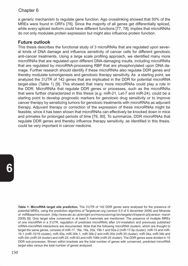
150
a generic mechanism to regulate gene function. Ago crosslinking showed that 50% of the MREs were found in ORFs [76]. Since the majority of all genes get differentially spliced, while every spliced isoform could have different functions [77, 78], implies that microRNAs do not only modulate protein expression but might also influence protein function.
Future outlookThis thesis describes the functional study of 3 microRNAs that are regulated upon sever-al kinds of DNA damage and influence sensitivity of cancer cells for different genotoxic anti-cancer treatments. Using a large scale profiling approach, we identified many more microRNAs that are regulated upon different DNA-damaging insults, including microRNAs that are regulated by microRNA-processing RBP that are phosphorylated upon DNA da-mage. Further research should identify if these microRNAs also regulate DDR genes and thereby modulate tumorigenesis and genotoxic therapy sensitivity. As a starting point, we analyzed the 3’UTR of 142 genes that are implicated in the DDR for potential microRNA target-sites (Table 1) [9]. This showed that many more microRNAs could play a role in the DDR. MicroRNAs that regulate DDR genes or processes, such as the microRNAs that were further characterized in this thesis (e.g. miR-21, Let-7 and miR-24), could be a starting point to develop prognostic markers for genotoxic drug sensitivity or to improve cancer therapy by sensitizing tumors for genotoxic treatments with microRNAs as adjuvant therapy. Adjuvant therapy or correction of the expression of these microRNAs might be feasible, since it has been shown that microRNAs can effectively be knocked down in mice and primates for prolonged periods of time [79, 80]. To summarize, DDR microRNAs that regulate DDR genes and thereby influence therapy sensitivity, as identified in this thesis, could be very important in cancer medicine.
Table 1: MicroRNA target site prediction. The 3’UTR of 142 DDR genes were analysed for the presence of potential MREs, using the prediction algoritms of Targetscan.org (version 5.0 of 5 december 2008) and Miranda of miRBase/microcosm (http://www.ebi.ac.uk/enright-srv/microcosm/cgi-bin/targets/v5/search.pl)(version march 2009) [9]. Only target sites conserved in at least 5 mammals are mentioned. The presence of multiple MREs of one microRNA in a 3’UTR, regulation of predicted microRNAs after UV-irradiation and previously published mRNA-microRNA interactions are documented. Note that the following microRNA clusters, which are thought to target the same genes, consists of miR-17, 18a, 19a, 20a, 19b-1 and 92a-2 (miR-17-5p cluster), miR-15 and miR-16-1 (miR-15/16 cluster), miR-30a, miR-30b-1, miR-30b-2 and miR-30d (miR-30 cluster), miR-34a, miR-34b and miR-34c (miR-34 cluster) and miR-25, miR-93 and miR-106b (miR-25 cluster). The DDR genes were divided in 15 DDR sub-processes. Shown within brackets are the total number of genes with conserved, predicted microRNA target sites versus the total number of genes analyzed.
6
Chapter 6

151
TABLE 1. Predicted microRNA target sites in DNA damage responsive genes
DNA damage responsive processes* DDR proteins
Predict
ed M
icroRNA ta
rget sit
e
Predict
ed by
Multiple
target
sites
in 3'
UTR
Regulat
ed af
ter UV 2
1
Publis
hed
Global Genome NER (2/5) RAD23B miR-106/302 TargetscanmiR-320 TargetscanmiR-199/199-5p Targetscan X XmiR-219 TargetscanmiR-590 TargetscanmiR-15/16 cluster Targetscan XmiR-196ab TargetscanmiR-136 TargetscanmiR-23ab Targetscan XmiR-181 TargetscanmiR-361 TargetscanmiR-17-5p cluster Targetscan X XmiR-328 TargetscanmiR-342 TargetscanmiR-30 cluster Targetscan X XmiR-182 TargetscanmiR-96/1271 TargetscanmiR-217 TargetscanmiR-362 Targetscan
RAD23A miR-361 Targetscan
Transcription-Coupled NER (9/11) CSA miR-491-3p MirbaseCOP9 miR-200bc/429 Targetscan XP300 miR-129 Targetscan X
miR-186 TargetscanmiR-150 TargetscanmiR-377 TargetscanmiR-22 Targetscan XmiR-132/212 TargetscanmiR-342/34 TargetscanmiR-145-5p MirbasemiR-148b MirbasemiR-301b MirbasemiR-507 MirbasemiR-363 MirbasemiR-434-3p Mirbase XmiR-200bc/429 Both X
CSB Let-7 Targetscan XRIT1 miR-125/351 Targetscan
miR-365 TargetscanmiR-27ab Targetscan XmiR-29abc Targetscan X
CUL4A miR-9 TargetscanmiR-137 TargetscanmiR-494 TargetscanmiR-103/107 Both XmiR-96/1271 Both X
TCEA1 miR-320/320abcd TargetscanmiR-340/340-5p Targetscan
TCEA2 miR-494 TargetscanHMGN1 miR-15/16 cluster Mirbase X
miR-34 cluster Mirbase XLet-7 Both X
6
General discussion

152
TABLE 1. Continued
DNA damage responsive processes* DDR proteins
Predict
ed M
icroRNA ta
rget sit
e
Predict
ed by
Multiple
target
sites
in 3'
UTR
Regulat
ed af
ter UV 2
1
Publis
hed
General Nucleotide Excision Repair (NER) (4/15) CCNH miR-23ab Targetscan XCDK7 miR-199/199-5p Targetscan XCAK miR-561 Both
miR-23ab Both XGTF2H1 miR-148 Targetscan
miR-10 Targetscanmir-30 cluster Mirbase XmiR-130/301/301 Mirbase X
Base Excision Repair (1/8) PARP miR-223 Both XmiR-7 Both X
Mismatch Repair (1/9) HMSH2 miR-210/590-5p TargetscanmiR-291b-3p Mirbase X
Non Homologeous End-Joining (3/8) NHEJ1 miR-505 TargetscanmiR-431 Targetscan
Artemis miR-140 cluster TargetscanmiR-383 Mirbase
Ku80 miR-340 TargetscanHomologeous Recombination (11/19) RPA miR-361 Targetscan X
RPA2 miR-130/301 Mirbase XMRE11 miR-384-3p Targetscan
miR-217 TargetscanmiR-153 TargetscanmiR-539 TargetscanmiR-9 Targetscan
RAD52 miR-758 BothRAD51A miR-758 Targetscan
miR-134 TargetscanRAD51B miR-145 TargetscanRAD50 miR-183 TargetscanBRCA1 miR-132/212 Targetscan
miR-205 TargetscanmiR-218 TargetscanmiR-125a TargetscanmiR-7 Targetscan X
RBBP8 miR-130/301 Targetscan XmiR-17-5p cluster Targetscan X X
BRIP1 miR-540 TargetscanPALB2 miR-576-3p Mirbase
Crosslink repair (3/8) FANCA miR-151-5p MirbasemiR-944 MirbasemiR-194 Mirbase
FANCF miR-590 TargetscanmiR-320 TargetscanmiR-30 cluster Targetscan X
FANCG miR-546 MirbasemiR-129-3p MirbasemiR-513-5p Mirbase
6
Chapter 6
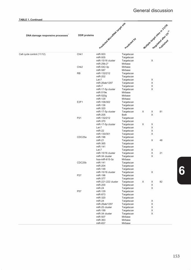
153
TABLE 1. Continued
DNA damage responsive processes* DDR proteins
Predict
ed M
icroRNA ta
rget sit
e
Predict
ed by
Multiple
target
sites
in 3'
UTR
Regulat
ed af
ter UV 2
1
Publis
hed
Cell cycle control (11/12) Chk1 miR-503 TargetscanmiR-505 TargetscanmiR-15/16 cluster Targetscan XmiR-29b-2* Mirbase
Chk2 miR-542-3p MirbasemiR-587 Mirbase
RB miR-132/212 TargetscanmiR-202 TargetscanLet-7 Targetscan XmiR-26ab/1297 Targetscan XmiR-7 Targetscan XmiR-17-5p cluster Targetscan XmiR-519e MirbasemiR-520g MirbasemiR-129 Mirbase
E2F1 miR-106/302 TargetscanmiR-136 TargetscanmiR-320 TargetscanmiR-17-5p cluster Targetscan X X 81miR-205 Both X
P21 miR-132/212 TargetscanmiR-370 TargetscanmiR-17-5p cluster Targetscan X XLet-7 Targetscan XmiR-22 Targetscan XmiR-130/301 Targetscan X
CDC25a miR-196 TargetscanmiR-21 Targetscan X 48miR-365 TargetscanmiR-141 TargetscanLet-7 Targetscan XmiR-15/16 cluster Targetscan X 21miR-34 cluster Targetscan Xhsa-miR-615-3p Mirbase
CDC25b miR-141 TargetscanmiR-204 TargetscanmiR-148 TargetscanmiR-15/16 cluster Targetscan X
P27 miR-196 TargetscanmiR-377 TargetscanmiR-221-222 cluster Targetscan X X 82miR-200 Targetscan XmiR-24 Targetscan X
P57 miR-129 TargetscanmiR-873 TargetscanmiR-320 TargetscanmiR-24 Targetscan XmiR-26ab/1297 Targetscan XmiR-25 cluster Targetscan XmiR-199 Targetscan XmiR-34 cluster Targetscan XmiR-507 MirbasemiR-363 MirbasemiR-657 Mirbase
6
General discussion

154
TABLE 1. Continued
DNA damage responsive processes* DDR proteins
Predict
ed M
icroRNA ta
rget sit
e
Predict
ed by
Multiple
target
sites
in 3'
UTR
Regulat
ed af
ter UV 2
1
Publis
hed
Cell cycle control (continued) Wee1 miR-106 TargetscanmiR-300 TargetscanmiR-155 TargetscanmiR-219 TargetscanmiR-503 TargetscanmiR-15/16 cluster Targetscan X XmiR-144 TargetscanmiR-221-222 cluster Targetscan Xhsa-miR-548d-5p MirbasemiR-155 MirbasemiR-301a MirbasemiR-140-5p MirbasemiR-15/16 cluster Mirbase X XmiR-606 MirbasemiR-17-5p cluster Mirbase X XmiR-627 Mirbase
PLK1 hsa-miR-491-3p MirbasemiR-568 MirbasemiR-100 MirbasemiR-604 MirbasemiR-767-3p Mirbase
Replication control (3/8) RECQ4 miR-544 Mirbase XRFC1 hsa-miR-345 Mirbase
hsa-miR-651 Mirbasehsa-miR-337-3p MirbasemiR-186 BothmiR-197 Both X
WRN miR-374/374ab TargetscanTranslesion Synthesis (1/2) Rev1 miR-183 Targetscan
miR-340 Targetscan X XmiR-96/1271 TargetscanmiR-486 Targetscan XmiR-182 TargetscanmiR-221-222 cluster Targetscan XmiR-139 TargetscanmiR-200bc/429 Targetscan XmiR-144 TargetscanmiR-101 TargetscanmiR-9 MirbasemiR-648 MirbasemiR-507 MirbasemiR-628-5p MirbasemiR-944 MirbasemiR-582-5p MirbasemiR-30 cluster Both XmiR-495/1192 Both XmiR-875 Both
DNA synthesis (7/10) Pol lambda Let-7 Targetscan XPCNA miR-548d-3p Mirbase X
miR-320 MirbasemiR-546 MirbasemiR-154 Both
LigaseII miR-27ab Mirbase XLigaseIII miR-512-5p Mirbase
miR-198 MirbasemiR-27ab Mirbase X
pol kappa miR-106/302 BothmiR-25 cluster Both X
Pol eta miR-218 TargetscanPol beta miR-188-5p Mirbase
miR-570 MirbasemiR-499-5p Mirbase X
6
Chapter 6
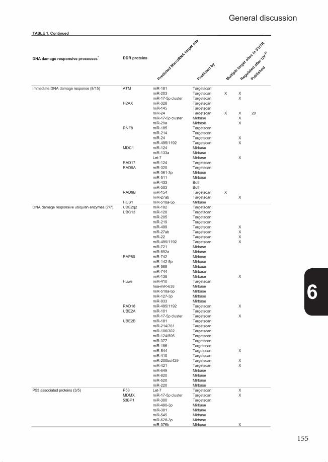
155
TABLE 1. Continued
DNA damage responsive processes* DDR proteins
Predict
ed M
icroRNA ta
rget sit
e
Predict
ed by
Multiple
target
sites
in 3'
UTR
Regulat
ed af
ter UV 2
1
Publis
hed
Immediate DNA damage response (8/15) ATM miR-181 TargetscanmiR-203 Targetscan X XmiR-17-5p cluster Targetscan X
H2AX miR-328 TargetscanmiR-145 TargetscanmiR-24 Targetscan X X 20miR-17-5p cluster Mirbase XmiR-29a Mirbase X
RNF8 miR-185 TargetscanmiR-214 TargetscanmiR-24 Targetscan XmiR-495/1192 Targetscan X
MDC1 miR-124 MirbasemiR-133a MirbaseLet-7 Mirbase X
RAD17 miR-124 TargetscanRAD9A miR-320 Targetscan
miR-361-3p MirbasemiR-511 MirbasemiR-433 BothmiR-503 Both
RAD9B miR-154 Targetscan XmiR-27ab Targetscan X
HUS1 miR-518a-5p MirbaseDNA damage responsive ubiquitin enzymes (7/7) UBE2q2 miR-182 Targetscan
UBC13 miR-128 TargetscanmiR-205 TargetscanmiR-219 TargetscanmiR-499 Targetscan XmiR-27ab Targetscan XmiR-22 Targetscan XmiR-495/1192 Targetscan XmiR-721 MirbasemiR-892a Mirbase
RAP80 miR-742 MirbasemiR-142-5p MirbasemiR-588 MirbasemiR-744 MirbasemiR-138 Mirbase X
Huwe miR-410 Targetscanhsa-miR-638 MirbasemiR-518a-5p MirbasemiR-127-3p MirbasemiR-933 Mirbase
RAD18 miR-495/1192 Targetscan XUBE2A miR-101 Targetscan
miR-17-5p cluster Targetscan XUBE2B miR-181 Targetscan
miR-214/761 TargetscanmiR-106/302 TargetscanmiR-124/506 TargetscanmiR-377 TargetscanmiR-186 TargetscanmiR-544 Targetscan XmiR-410 TargetscanmiR-200bc/429 Targetscan XmiR-421 Targetscan XmiR-649 MirbasemiR-820 MirbasemiR-520 MirbasemiR-220 Mirbase
P53 associated proteins (3/5) P53 Let-7 Targetscan XMDMX miR-17-5p cluster Targetscan X53BP1 miR-300 Targetscan
miR-490-3p MirbasemiR-381 MirbasemiR-545 MirbasemiR-628-3p MirbasemiR-376b Mirbase X
6
General discussion

156
References1. Hoeijmakers, J.H., DNA damage, aging, and cancer. N Engl J Med, 2009. 361(15): p. 1475-85.2. Hoeijmakers, J.H., Genome maintenance mechanisms for preventing cancer. Nature, 2001. 411(6835):p. 366-74.3. Harper, J.W. and S.J. Elledge, The DNA damage response: ten years after. Mol Cell, 2007. 28(5): p. 739-45.4. Bartek, J. and J. Lukas, DNA damage checkpoints: from initiation to recovery or adaptation. Curr Opin Cell Biol, 2007. 19(2): p. 238-45.5. Matsuoka, S., et al., ATM and ATR substrate analysis reveals extensive protein networks responsive to DNA damage. Science, 2007. 316(5828): p. 1160-6.6. Meulmeester, E., et al., ATM-mediated phosphorylations inhibit Mdmx/Mdm2 stabilization by HAUSP in favor of p53 activation. Cell Cycle, 2005. 4(9): p. 1166-70.7. Croce, C.M., Causes and consequences of microRNA dysregulation in cancer. Nat Rev Genet, 2009. 10(10): p. 704-14.8. Garofalo, M., G. Condorelli, and C.M. Croce, MicroRNAs in diseases and drug response. Curr Opin Pharmacol, 2008. 8(5): p. 661-7.9. Wouters, M.D., et al., MicroRNAs, the DNA damage response and cancer. Mutat Res, 2011. 717(1-2): p. 54-66.10. Gorgoulis, V.G., et al., Activation of the DNA damage checkpoint and genomic instability in human pre cancerous lesions. Nature, 2005. 434(7035): p. 907-13.11. Bartkova, J., et al., DNA damage response as a candidate anti-cancer barrier in early human tumori- genesis. Nature, 2005. 434(7035): p. 864-70.12. Bouwman, P. and J. Jonkers, The effects of deregulated DNA damage signalling on cancer chemothe- rapy response and resistance. Nat Rev Cancer, 2012. 12(9): p. 587-98.13. Lord, C.J. and A. Ashworth, The DNA damage response and cancer therapy. Nature, 2012. 481(7381):p. 287-94.14. Igney, F.H. and P.H. Krammer, Death and anti-death: tumour resistance to apoptosis. Nat Rev Cancer, 2002. 2(4): p. 277-88.15. Gordon, R.R. and P.S. Nelson, Cellular senescence and cancer chemotherapy resistance. Drug Re- sist Updat, 2012. 15(1-2): p. 123-31.16. Medina, P.P. and F.J. Slack, microRNAs and cancer: an overview. Cell Cycle, 2008. 7(16): p. 2485-92.17. Esquela-Kerscher, A. and F.J. Slack, Oncomirs - microRNAs with a role in cancer. Nat Rev Cancer, 2006. 6(4): p. 259-69.18. Calin, G.A., MicroRNAs and cancer: what we know and what we still have to learn. Genome Med, 2009.1(8): p. 78.19. Medina, P.P., M. Nolde, and F.J. Slack, OncomiR addiction in an in vivo model of microRNA-21-indu ced pre-B-cell lymphoma. Nature, 2010.20. Lal, A., et al., miR-24-mediated downregulation of H2AX suppresses DNA repair in terminally differen tiated blood cells. Nat Struct Mol Biol, 2009. 16(5): p. 492-8.21. Pothof, J., et al., MicroRNA-mediated gene silencing modulates the UV-induced DNA-damage res- ponse. EMBO J, 2009. 28(14): p. 2090-9.22. Simone, N.L., et al., Ionizing radiation-induced oxidative stress alters miRNA expression. PLoS One, 2009. 4(7): p. e6377.23. Galluzzi, L., et al., miR-181a and miR-630 Regulate Cisplatin-Induced Cancer Cell Death. Cancer Res,2010. 70(5): p. 1793-1803.24. Garzon, R., G.A. Calin, and C.M. Croce, MicroRNAs in Cancer. Annu Rev Med, 2009. 60: p. 167-79.25. Farazi, T.A., et al., miRNAs in human cancer. J Pathol, 2011. 223(2): p. 102-15.26. Jackson, S.P. and J. Bartek, The DNA-damage response in human biology and disease. Nature, 2009.461(7267): p. 1071-8.27. Dasari, S. and P. Bernard Tchounwou, Cisplatin in cancer therapy: Molecular mechanisms of action. Eur J Pharmacol, 2014. 740C: p. 364-378.28. Weidhaas, J.B., et al., MicroRNAs as potential agents to alter resistance to cytotoxic anticancer thera- py. Cancer Res, 2007. 67(23): p. 11111-6.29. Zhang, X., et al., The ATM Kinase Induces MicroRNA Biogenesis in the DNA Damage Response. Mol Cell, 2011. 41(4): p. 371-83.30. He, L., et al., A microRNA component of the p53 tumour suppressor network. Nature, 2007. 447(7148):p. 1130-4.31. Baer, C., R. Claus, and C. Plass, Genome-wide epigenetic regulation of miRNAs in cancer. Cancer
6
Chapter 6

157
Res, 2013. 73(2): p. 473-7.32. Jansson, M.D. and A.H. Lund, MicroRNA and cancer. Mol Oncol, 2012. 6(6): p. 590-610.33. Viswanathan, S.R., G.Q. Daley, and R.I. Gregory, Selective blockade of microRNA processing by Lin28. Science, 2008. 320(5872): p. 97-100.34. Trabucchi, M., et al., The RNA-binding protein KSRP promotes the biogenesis of a subset of microR NAs. Nature, 2009. 459(7249): p. 1010-4.35. Thomson, J.M., et al., Extensive post-transcriptional regulation of microRNAs and its implications for cancer. Genes Dev, 2006. 20(16): p. 2202-7.36. Suzuki, H.I., et al., Modulation of microRNA processing by p53. Nature, 2009. 460(7254): p. 529-33.37. Kawai, S. and A. Amano, BRCA1 regulates microRNA biogenesis via the DROSHA microprocessor complex. J Cell Biol, 2012. 197(2): p. 201-8.38. Calin, G.A. and C.M. Croce, MicroRNA signatures in human cancers. Nat Rev Cancer, 2006. 6(11): p. 857-66.39. Xia, L., et al., miR-15b and miR-16 modulate multidrug resistance by targeting BCL2 in human gastric cancer cells. Int J Cancer, 2008. 123(2): p. 372-9.40. Valeri, N., et al., MicroRNA-21 induces resistance to 5-fluorouracil by down-regulating human DNA MutS homolog 2 (hMSH2). Proc Natl Acad Sci U S A, 2010. 107(49): p. 21098-103.41. Blower, P.E., et al., MicroRNAs modulate the chemosensitivity of tumor cells. Mol Cancer Ther, 2008. 7(1): p.1-9.42. Shi, L., et al., MiR-21 protected human glioblastoma U87MG cells from chemotherapeutic drug temo- zolomide induced apoptosis by decreasing Bax/Bcl-2 ratio and caspase-3 activity. Brain Res, 2010.43. Li, Y., et al., MicroRNA-21 targets LRRFIP1 and contributes to VM-26 resistance in glioblastoma multi- forme. Brain Res, 2009. 1286: p. 13-8.44. Hwang, J.H., et al., Identification of microRNA-21 as a biomarker for chemoresistance and clinical out-come following adjuvant therapy in resectable pancreatic cancer. PLoS One, 2010. 5(5): p. e10630.45. Volinia, S., et al., A microRNA expression signature of human solid tumors defines cancer gene tar- gets. Proc Natl Acad Sci U S A, 2006. 103(7): p. 2257-61.46. Asangani, I.A., et al., MicroRNA-21 (miR-21) post-transcriptionally downregulates tumor suppressor Pdcd4 and stimulates invasion, intravasation and metastasis in colorectal cancer. Oncogene, 2008. 27(15): p. 2128-36.47. Liu, Z.L., et al., MicroRNA-21 (miR-21) expression promotes growth, metastasis, and chemo- or radio resistance in non-small cell lung cancer cells by targeting PTEN. Mol Cell Biochem, 2012.48. Wang, P., et al., microRNA-21 negatively regulates Cdc25A and cell cycle progression in colon cancer cells. Cancer Res, 2009. 69(20): p. 8157-65.49. Shi, L., et al., MiR-21 protected human glioblastoma U87MG cells from chemotherapeutic drug temo- zolomide induced apoptosis by decreasing Bax/Bcl-2 ratio and caspase-3 activity. Brain Res, 2010. 1352: p. 255-64.50. Meng, F., et al., Involvement of human micro-RNA in growth and response to chemotherapy in human cholangiocarcinoma cell lines. Gastroenterology, 2006. 130(7): p. 2113-29.51. Meng, F., et al., MicroRNA-21 regulates expression of the PTEN tumor suppressor gene in human hepatocellular cancer. Gastroenterology, 2007. 133(2): p. 647-58.52. Giovannetti, E., et al., MicroRNA-21 in pancreatic cancer: correlation with clinical outcome and phar macologic aspects underlying its role in the modulation of gemcitabine activity. Cancer Res, 2010. 70(11): p. 4528-38.53. Zhang, X., et al., Oncogenic Wip1 phosphatase is inhibited by miR-16 in the DNA damage signaling pathway. Cancer Res, 2010. 70(18): p. 7176-86.54. Fujita, Y., et al., Effects of miR-34a on cell growth and chemoresistance in prostate cancer PC3 cells. Biochem Biophys Res Commun, 2008. 377(1): p. 114-9.55. Raver-Shapira, N., et al., Transcriptional activation of miR-34a contributes to p53-mediated apoptosis. Mol Cell, 2007. 26(5): p. 731-43.56. Saleh, A.D., et al., Cellular stress induced alterations in microRNA let-7a and let-7b expression are dependent on p53. PLoS One, 2011. 6(10): p. e24429.57. Johnson, S.M., et al., RAS is regulated by the let-7 microRNA family. Cell, 2005. 120(5): p. 635-47.58. Sampson, V.B., et al., MicroRNA let-7a down-regulates MYC and reverts MYC-induced growth in Bur kitt lymphoma cells. Cancer Res, 2007. 67(20): p. 9762-70.59. Mondol, V. and A.E. Pasquinelli, Let’s make it happen: the role of let-7 microRNA in development. Curr Top Dev Biol, 2012. 99: p. 1-30.60. Ibanez-Ventoso, C., et al., Modulated microRNA expression during adult lifespan in Caenorhabditis elegans. Aging Cell, 2006. 5(3): p. 235-46.
6
General discussion

158
61. Toledano, H., et al., The let-7-Imp axis regulates ageing of the Drosophila testis stem-cell niche. Natu- re,2012. 485(7400): p. 605-10.62. Nishino, J., et al., Hmga2 promotes neural stem cell self-renewal in young but not old mice by redu cing p16Ink4a and p19Arf Expression. Cell, 2008. 135(2): p. 227-39.63. Zhu, H., et al., The Lin28/let-7 axis regulates glucose metabolism. Cell, 2011. 147(1): p. 81-94.64. Lin, S., et al., Let-7b regulates the expression of the growth hormone receptor gene in deletion-type dwarf chickens. BMC Genomics, 2012. 13: p. 306.65. Schumacher, B., et al., Delayed and accelerated aging share common longevity assurance mechanis- ms. PLoS Genet, 2008. 4(8): p. e1000161.66. Garinis, G.A., et al., DNA damage and ageing: new-age ideas for an age-old problem. Nat Cell Biol, 2008. 10(11): p. 1241-7.67. Pothof, J., et al., MicroRNA-mediated gene silencing modulates the UV-induced DNA-damage res- ponse. Embo J, 2009.68. Takagi, S., et al., MicroRNAs regulate human hepatocyte nuclear factor 4alpha, modulating the ex- pression of metabolic enzymes and cell cycle. J Biol Chem, 2010. 285(7): p. 4415-22.69. Lal, A., et al., miR-24 Inhibits cell proliferation by targeting E2F2, MYC, and other cell-cycle genes via binding to “seedless” 3’UTR microRNA recognition elements. Mol Cell, 2009. 35(5): p. 610-25.70. Amelio, I., et al., miR-24 triggers epidermal differentiation by controlling actin adhesion and cell migra tion. J Cell Biol, 2012. 199(2): p. 347-63.71. Mishra, P.J., et al., MiR-24 tumor suppressor activity is regulated independent of p53 and through a target site polymorphism. PLoS One, 2009. 4(12): p. e8445.72. Brunner, S., et al., Upregulation of miR-24 is associated with a decreased DNA damage response upon etoposide treatment in highly differentiated CD8(+) T cells sensitizing them to apoptotic cell death. Aging Cell, 2012. 11(4): p. 579-87.73. Guo, Y., et al., miR-24 functions as a tumor suppressor in Hep2 laryngeal carcinoma cells partly through down-regulation of the S100A8 protein. Oncol Rep, 2012. 27(4): p. 1097-103.74. Luzi, E., et al., The negative feedback-loop between the oncomir Mir-24-1 and menin modulates the Men1 tumorigenesis by mimicking the “Knudson’s second hit”. PLoS One, 2012. 7(6): p. e39767.75. Liu, X., et al., MicroRNA-24 targeting RNA-binding protein DND1 in tongue squamous cell carcinoma. FEBS Lett, 2010. 584(18): p. 4115-20.76. Hafner, M., et al., Transcriptome-wide identification of RNA-binding protein and microRNA target sites by PAR-CLIP. Cell, 2010. 141(1): p. 129-41.77. Kim, E., A. Magen, and G. Ast, Different levels of alternative splicing among eukaryotes. Nucleic Acids Res, 2007. 35(1): p. 125-31.78. Nilsen, T.W. and B.R. Graveley, Expansion of the eukaryotic proteome by alternative splicing. Nature, 2010. 463(7280): p. 457-63.79. Elmen, J., et al., LNA-mediated microRNA silencing in non-human primates. Nature, 2008. 452(7189): p. 896-9.80. Kota, J., et al., Therapeutic microRNA delivery suppresses tumorigenesis in a murine liver cancer mo del. Cell, 2009. 137(6): p. 1005-17.81. O’Donnell, K.A., et al., c-Myc-regulated microRNAs modulate E2F1 expression. Nature, 2005. 435(7043): p. 839-43.82. le Sage, C., et al., Regulation of the p27(Kip1) tumor suppressor by miR-221 and miR-222 promotes cancer cell proliferation. Embo J, 2007. 26(15): p. 3699-708.
6
Chapter 6

159

160

161
Appendix
SummaryNederlandse samenvatting
List of abbreviationsCurriculum Vitae
PhD portfolioAcknowledgements

162
Summary
DNA encodes all developmental instructions and heritable traits of an organism. It is of vital importance that DNA integrity is maintained to ensure proper gene function and to enable faithful replication of the genome. DNA is however continuously damaged by various endo-genous and exogenous agents, such as Reactive Oxygen Species (ROS) from cellular me-tabolism and ultraviolet light (UV) from the sun, respectively. Accumulating DNA damage decreases cellular viability and proliferation, thereby contributing to aging. DNA lesions can also lead to changes in the DNA-sequence such as mutations, deletions and chromosomal rearrangements, which eventually can lead to tumorigenesis. Therefore, it is of utmost importance that cells counteract the detrimental effects of DNA damage to prevent both cancer and premature aging. To this end, cells induce a potent cellular response upon DNA damage, designated the DNA damage response (DDR), which comprises DNA repair sys-tems and elaborate DNA damage checkpoint pathways. The DDR functions as a barrier for pre-malignant cells to progress into cancer cells. Most, if not all, cancer cells have therefore at least one defect in the DDR. This often sensitizes cancer cells to DNA damage, which is utilized, as mode of action, by irradiation and many cancer chemotherapeutics. However, the overt majority of tumors will acquire resistance against DNA damaging treatments, for example by modulating the DDR. Consequently, it is very important to study every aspect of the DDR to increase our knowledge of cancer biology and improve cancer medicine. The DDR controls numerous processes, such as the cell cycle, DNA repair and pro-grammed cell death, by regulating gene activity and expression. Much is known about DDR regulation at post-translational and transcriptional levels. However, less is known about post-transcriptional regulation, for example by microRNAs. MicroRNAs are endogenous, ~22-nucleotides-small, non-coding RNAs that negatively regulate gene expression levels by binding to the 3’untranslated-region (3’UTR) of target mRNAs, thereby inducing mRNA degradation or translation inhibition. It has been estimated that half of all protein-coding genes are controlled by microRNAs. MicroRNAs are essential for organismal development and diverse cellular function. They play an important role in cancer when misexpressed and several microRNAs are regulated after DNA damage. It is currently unclear to which extent and how microRNA expression is regulated by the DDR and their function therein. Studies linking the role of DDR microRNAs to tumo-rigenesiss and genotoxic drug sensitivity could lead to the identification of potential prog-nostic markers for genotoxic drug sensitivity and may even support novel microRNA-based therapies to sensitize tumors to genotoxic cancer therapy. To achieve these objectives, we addressed the following questions in this thesis. First, which microRNAs are regulated after DNA damage? Second, how is the expression of these DDR microRNAs regulated? Third, which DDR genes and processes are control-led by these microRNAs? And finally, what is the role of these microRNAs in cancer and therapy sensitivity? To answer these questions we used several approaches. In chapter 2 and 3, we used micro-arrays to profile microRNA expression after DNA damage exposure. In chapter 2, the normal microRNA response after DNA damage was delineated in human primary epithelial cells from breast and lung origin. Several DDR microRNAs were misexpressed in breast and lung cancer, which also modulated genotoxic cancer therapy sensitivity. This could improve our knowledge about the role of DDR microRNAs in cancer and genotoxic cancer therapy resistance. In chapter 3, we studied which factors control microRNA expression after Ioni-zing Radiation (IR) and UV-induced DNA damage in human sarcoma cells. We analyzed
A
Appendix

163
whether RNA binding proteins (RBPs) could control microRNA levels. First, we selected RBPs that are phosphorylated after DNA damage and function in microRNA processing. Then, we silenced those proteins by RNA interference and observed that the expression of several DDR microRNAs depends on these RBPs. Subsequent analysis suggests that some of these RBPs might not be functional in breast cancer. In chapter 4 and 5, we studied which DDR processes and genes are regulated by DNA damage responsive microRNAs. In chapter 4, we thoroughly investigated the role of miR-24, which is slowly induced by IR. We showed that miR-24 directly regulates MDC1, an adaptor protein in the DNA damage checkpoint response, not by targeting the 3’UTR but in the open reading frame. We discovered and characterized an alternative splice variant of MDC1 that is not under miR-24 control. Our results suggest that miR-24 alters the ratio between both splice variants, which implies that miR-24 in time alters checkpoint activity. In chapter 5, we studied the role of microRNA let-7 in the DDR and tried to esta-blish a link, mediated by let-7, between the DDR and cancer or aging. We studied Let-7 ex-pression in aged mice, human lung tumor samples, fibroblasts and cancer cells. We found that let-7 is directly regulated after DNA damage in general, but its function in cell survival depends on the type of damage. This research indicates that let-7 targets many genes and processes in response to DNA damage, suggesting a putative link between the DDR, these Let-7-mediated processes and cancer and aging. Many DDR genes are predicted targets of microRNAs (Table 1, Chapter 6). These microRNAs could also play a role in the DDR, like miR-24 and Let-7, and could be a star-ting point to develop prognostic markers to assess the sensitivity for genotoxic therapy or to support novel microRNA-based therapies. To summarize, DNA damage responsive mi-croRNAs control DDR genes and processes and could therefore influence therapy efficacy, as described in this thesis, which could be very important for cancer medicine.
A
Summary
164
Nederlandse samenvattingDNA bevat de erfelijke code voor de ontwikkeling en eigenschappen van elk organisme. Het DNA is georganiseerd in discrete instructies, genaamd genen. Het is van vitaal belang dat DNA integriteit behouden blijft, zodat genen functioneel blijven en het genoom correct gekopieerd kan worden, waardoor cellen kunnen delen en een organisme kan ontwikkelen, groeien, herstellen en uiteindelijk kan voortplanten. Het DNA wordt echter constant be-schadigd door verschillende stoffen en invloeden; van binnenuit, zoals zuurstof radicalen gevormd door het metabolisme van de cel, of van buitenaf door bijvoorbeeld het UV-licht van de zon. Opeenhoping van DNA schade vermindert cel vitaliteit en celdeling en bevor-dert veroudering. DNA schade kan ook leiden tot veranderingen in de DNA code, zoals mutaties, deleties of chromosomale veranderingen, welke kanker kunnen veroorzaken. Het is van cruciaal belang dat cellen de negatieve effecten van DNA schade tegengaan, zodat kanker en versnelde veroudering voorkomen kunnen worden. Om dit te bewerk-stelligen, activeren cellen een uitgebreide reactie op DNA schade, de zogenoemde DNA schade respons (DSR). De DSR vormt een barrière voor een pre-kwaadaardige cel om in een kankercel te ontwikkelen. Hierom zijn onderdelen van de DSR niet functioneel in kankercellen. Dit maakt deze kankercellen initieel gevoelig voor DNA schade, waarop het werkingsmechanisme van de meeste klassieke chemotherapeutica en bestraling is geba-seerd. Tumoren worden echter vaak resistent tegen DNA beschadigende behandelingen, onder andere door de DSR aan te passen. Daarom is het van groot belang elk onderdeel van de DSR goed te bestuderen, zodat meer kennis beschikbaar komt over het ontstaan en de behandeling van kanker. Nadat de DSR wordt geactiveerd door DNA schade, worden meerdere processen door de DSR gecontroleerd, zoals celdeling, DNA reparatie en, zodra het DNA van een cel teveel beschadigd is, geprogrammeerde celdood. De DSR reguleert deze processen door de activiteit en hoeveelheid van vele genproducten te beïnvloeden (genexpressie). Dit laatste kan op meerdere niveaus worden bewerkstelligd, zoals op transcriptioneel niveau te beïnvloeden hoeveel een gen wordt afgeschreven. Als een gen wordt afgeschreven ontstaat er een boodschapper RNA (in het Engels: messenger RNA (mRNA)). Dit mRNA molecuul wordt vertaald (translatie) in een eiwit dat de daadwerkelijke genfunctie uitvoert. Na translatie kan de hoeveelheid en de activiteit van een eiwit ook beïnvloed worden door de DSR, bijvoorbeeld d.m.v. de regulatie van eiwitafbraak. Er is veel kennis over hoe de DSR op transcriptioneel en op eiwit niveau genexpressie controleert en welke specifieke genproducten precies gereguleerd worden na DNA schade. Er is echter een niveau van genexpressie regulatie welke zich bevindt tussen het transcriptionele niveau en de regu-latie van eiwitten: Regulatie van de hoeveelheid en de translatie van mRNAs. Zeer kleine RNA moleculen, zogenaamde microRNAs, beïnvloeden op dit niveau de genexpressie. Over de rol van deze microRNAs in de DSR is weinig bekend. MicroRNAs zijn ongeveer 22-nucleotide-kleine RNAs die niet worden vertaald in een eiwit, maar de hoeveelheid eiwit van andere genen negatief beïnvloeden door te binden aan het uiteinde van een mRNA. Hierdoor wordt dit mRNA afgebroken en/of de translatie verhinderd. Vermoedelijk wordt de helft van alle eiwit-coderende genen geregu-leerd door microRNAs. MicroRNAs zijn belangrijk voor de ontwikkeling van organismen, ze spelen een belangrijke rol in kanker en de expressie van sommige microRNAs wordt ook gereguleerd na DNA schade. Het is echter onduidelijk hoeveel microRNAs gereguleerd worden na DNA schade en hoe de DSR precies microRNA niveaus beïnvloedt. Bovendien moeten nog veel DSR genen geïdentificeerd worden waarvan de expressie door microRNAs gereguleerd wordt. Het is cruciaal om de rol van microRNAs in de DSR te bestuderen, zodat we het ontstaan
A
Appendix

165
van kanker en de behandeling van kanker met DNA beschadigende medicijnen beter be-grijpen. Deze kennis kan ons begrip vergroten over gen regulatie na DNA schade en de daarop volgende veranderingen van verschillende cellulaire processen, zoals de cel cyclus en celdood. Dit onderzoek zou enerzijds microRNAs als potentiële prognostische markers kunnen identificeren, anderzijds zou dit de basis kunnen vormen om microRNA therapieën te ontwikkelen om klassieke therapie te ondersteunen door resistentie tegen te gaan. Om deze doelen te behalen, zijn de volgende vragen in deze dissertatie gesteld: Ten eerste, welke microRNAs worden gereguleerd na DNA schade? Ten tweede, hoe wor-den deze microRNAs gereguleerd na DNA schade? Ten derde, welke DSR genen en pro-cessen worden gereguleerd door deze microRNAs? En tenslotte, wat is de rol van deze microRNAs in kanker en therapie gevoeligheid? We hebben verschillende methoden gebruikt om deze vragen te beantwoorden. In hoofdstuk 2 en 3 hebben we expressie arrays gebruikt om microRNA hoeveelheden te meten na DNA schade. Hoofdstuk 2 beschrijft de normale microRNA respons na DNA schade in primaire epitheliale cellen van borst en long oorsprong. Wij hebben verschillende DSR microRNAs geïdentificeerd die foutief tot expressie komen in kanker en ook de reac-tie op DNA beschadigende therapie beïnvloeden. Dit kan onze kennis verbeteren over de rol van microRNAs in kanker en genotoxische therapie. In hoofdstuk 3 beschrijven we hoe microRNAs gereguleerd worden na ioniseren-de straling (IR) en UV-geïnduceerde DNA schade in humane sarcoma cellen. We onder-zochten of RNA-bindende eiwitten (RBE) de hoeveelheid van microRNAs kunnen regule-ren. Eerst selecteerden we RBE die geactiveerd worden na DNA schade en een rol hebben in microRNA maturatie. Regulatie van DSR microRNAs werd onderzocht na uitschakeling van deze RBEs. Dit liet zien dat regulatie van meerdere DSR microRNAs afhankelijk is van deze RBEs en dat deze eiwitten niet altijd functioneel zijn in borst kanker. Dit vergroot onze kennis over de rol van deze eiwitten in de DSR en in kanker. In hoofdstuk 4 en 5 hebben we onderzocht welke DSR processen en genen ge-reguleerd worden door DSR microRNAs. In hoofdstuk 4 hebben we de rol van miR-24 in de DSR bestudeerd, een microRNA dat gereguleerd wordt door IR. Ook laten we zien dat miR-24 MDC1, een DNA schade checkpoint eiwit, direct reguleert. Daarnaast vonden en karakteriseerden we een alternatieve isoform van MDC1 dat niet onder controle van miR-24 staat in vergelijking tot de normale MDC1 isoform. Onze resultaten wijzen erop dat miR-24 de ratio tussen beide isoformen zou kunnen veranderen, hetgeen mogelijk een andere activiteit van het DNA schade checkpoint zou bewerkstelligen. In hoofdstuk 5 hebben we de rol van microRNA let-7 in de DSR, kanker en verou-dering onderzocht. We bestudeerden let-7 expressie in verouderde muizen, humane long kanker samples, maar ook in fibroblasten en kankercellen behandeld met DNA schade. We vonden dat Let-7 direct gereguleerd wordt door meerdere soorten DNA schade, maar het type DNA schade is van belang voor de functie van let-7 in de context van cellulaire overle-ving. Dit onderzoek laat zien dat let-7 vele processen beïnvloed in de DSR en identificeert een potentiele connectie tussen de DSR en door let-7 geïnitieerde cellulaire processen, welke een rol spelen in beide kanker en veroudering. Voor vele DSR genen wordt voorspeld dat ze gereguleerd kunnen worden door microRNAs (Table 1, hoofdstuk 6). Deze microRNAs kunnen dus ook een rol spelen in de DSR, zoals miR-24 en let-7, en kunnen een startpunt zijn om prognostische markers te ont-wikkelen voor de gevoeligheid voor DNA beschadigende middelen of om kanker therapie te ondersteunen. Samenvattend, DSR microRNAs controleren DSR genen en processen en kunnen daardoor de effectiviteit van therapie beïnvloeden, zoals beschreven in deze dissertatie, hetgeen erg belangrijk kan zijn voor het genezen van kanker.
A
Nederlandse samenvatting

166
List of Abbreviations
3’UTR 3’untranslated region5’UTR 5’untranslated region53BP1 p53 binding protein 17AAD 7-Aminoactinomycin DAGO1-4 ArgonauteATM Ataxia Telangiectasia Mutated ATR Ataxia Telangiectasia and Rad3-related proteinBcl2 B-cell lymphoma 2BER Base Excision RepairBRCA1/2 Breast cancer susceptibility gene 1/2 BRDU BromodeoxyuridineBSA Bovine Serum AlbuminCDC25A Cell division cycle 25 homolog A CDK 6/4 Cyclin-dependent kinase 6/4CHK1/2 Checkpoint kinase 1/2Dapi 4’,6-diamidino-2-phenylindoleDDR DNA damage responseDhx9 DEAH box helicase 9DMEM Dulbecco’s modified Eagle’s mediumDNA Deoxyribonucleic acidDNA-PK DNA-dependent protein kinaseDre Danio rerioDSB Double strand breaksFACS Fluorescence-activated cell sortingFANCD2 Fanconi anemia group D2 protein FCS Feutal calf serum FITC Fluorescein isothiocyanateG1/G2-phase Gap1/2-cell cycle phaseGFP Green fluorescent proteinH2AX Histone2 AXHNRNPUL1 Heterogeneous nuclear ribonucleoprotein U-like 1 HR Homologous recombinationHRP Horseradish peroxidaseHsa Home sapiensIR Ionizing radiationIRIF Ionizing radiation induced fociKHSRP KH-type splicing regulatory proteinMDC1 Mediator of DNA damage checkpoint protein 1miRNA MicroRNAM-phase Mitosis cell cycle phaseMRE MicroRNA responsive elementMRN Mre11, Rad50 and Nbs1 complexmRNA Messenger RNAMSH2 MutS protein homolog 2ncRNAs Non-coding RNANER Nucleotide excision repairNHEJ Non-homologous end-joining
A
Appendix

167
ORF Open reading framePBS Phosphate buffered salinePCR Polymerase chain reactionPDCD4 Programmed cell death protein 4 PI Propidium iodidepiRNA Piwi-interacting RNApre-microRNA Precurser microRNApri-microRNA Primary microRNAPTEN Phosphatase and tensin homologPUM1/2 Pumilio RNA-binding family member 1/2PVDF Polyvinylidene difluoride RACE Rapid Amplification of cDNA EndsRBM38 RNA binding motif protein 38RBP RNA binding proteinRISC RNA-induced silencing complexRNA Ribonucleic acidRNAi RNA interferenceROS Reactive oxigen speciesRPA1 Replication protein A1rRNA ribosomal RNART-qPCR Real-time quatitative PCRSDS-PAAGE Sodium dodecyl sulfate polyacrylamide gel electrophoresissiRNA short interfering RNASnip1 Smad nuclear-interacting protein 1snoRNA Small nucleolar RNAssnRNA small nuclear RNAsS-phase Synthesis cell cycle phaseSSB single strand breaksTRBP TAR (HIV-1) RNA binding protein 2 tRNA transfer RNAU2OS osteosarcoma cellsUV ultravioletZcchc11 zinc finger, CCHC domain containing 11
A
List of Abbreviations

168
Curriculum Vitae
Name: M. D. Wouters (Maikel)Date of birth: 22 January 1983Place of birth: Capelle aan den IJssel, The Netherlands
Education
Period Education2000 – 2005: Technician school; Bio-Medical Laboratory Analysis, Hogeschool Rotterdam
2005 – 2008: Master in Molecular and Cellular Biology, Leiden University
2008 – 2014: PhD graduate, department of Genetics, Erasmus Medical Centre
Internships
Period Institute2002 – 2003: Department of Clinical Genetics, Erasmus Medical Centre
2004 – 2005: Department of Haematology, Erasmus Medical Centre
2006 – 2006: Department of Molecular Cell Biology, Institute Biology Leiden
2007 – 2008: Department of experimental Microbiology, Leiden University Medical Centre
Work experience
Period Institute2008 – 2013: PhD graduate, department of Genetics, Erasmus Medical Centre
2013 – 2014 Research assistant, Faculty of health and medicine, University of SurreyA
Appendix

169
Scientific publications
MicroRNAs, the DNA damage response and cancer Maikel D. Wouters, Dik van Gent, Jan H. J. Hoeijmakers, J. Pothof Mutation Research/Fundamental and Molecular Mechanisms of Mutagenesis 2011 Dec 1; 717(1-2):54-66.
DNA damage responsive microRNAs misexpressed in human cancer modulate therapy sensitivity Marijn T.M. van Jaarsveld, Maikel D. Wouters, Antonius W.M. Boersma, Marcel Smid, Wilfred F.J. van IJcken, Ron H.J. Mathijssen, Jan H.J. Hoeijmakers, John W.M. Martens, Steven van Laere, Erik A.C. Wiemer, Joris Pothof Molecular oncology, 2013 Dec 31
A
Curriculum Vitae

170
Phd portfolio
Courses
Molecular and cell biology 2008Genome maintenance and cancer 2008Radiation safety training (5b) 2009Safely working in the laboratory 2009Analysis of gene expression array data 2009Introduction in R programming 2009Photoshop and Illustrator course 2010English biomedical writing and communication 2011
Workshops
Annual MGC PhD workshop: Brugge 2009, poster presentation Köln 2010, oral presentation Maastricht 2011, oral presentation Dusseldorf 2012, poster presentation
Conferences and seminars
Journal club 2008-2012
MGC DNA repair 2-monthly group meetings 2008-2012 2011, oral presentation, Rotterdam, The Netherlands
Annual MGC symposium 2008-2012
CGC scientific meeting 2008, 2009, 2012
NWO-CW study group meeting on nucleic acids 2008, poster presentation Lunteren, The Netherlands,
NWO-CW study group meeting on Chemistry 2009, poster presentation related to biological & medical sciences Veldhoven, the Netherlands
NWO-CW study group meeting on protein research, 2010 nucleic acids, lipids & biomembranes, Veldhoven, the Netherlands
Responses to DNA damage: 2011, poster presentation from molecular mechanism to human disease Egmond aan zee, the Nether- lands
A
Appendix

171
A
PhD portfolio
International conferences
3rd EU-IP DNA repair workshop for young scientists 2009, oral presentation, Taormina, Sicily
Keystone meeting non-coding RNAs and cancer 2011, poster presentation Banff, Alberta, Canada
Teaching & coaching
Serena Bruens, 2009 Hoger laboratorium onderwijs, Hogeschool Rotterdam
Gisela Slaats 2010-2011 Molecular biology, Wageningen University,
Esther Jongste 2011 Hoger laboratorium onderwijs, Hogeschool Rotterdam

172
Acknowledgements
Finally, written last, but read first by all. Scientifically, the most unimportant part, but for many the only part that matters: The acknowledgements, the most personal piece of all. However, being very personal creates a problem, because I sometimes have the tendency to overshare personal thoughts, spicy details and politically incorrect statements. Anyhow, I decided not to censor myself, so hold on and lets start!
First, I would like to thank all the redheads in this world. Although a ‘red headed preferen-ce gene’ has not been discovered yet, I would like to specifically thank three red-headed women in particular. In order of appearance and importance…
My motherMa, je had het erg moeilijk met de gezondheid van jezelf en pa ten tijde van het begin van mijn PhD. Toch ben je altijd geïnteresseerd en ondersteunend geweest. Ik ben meer dan blij dat je je rust weer hebt terug gevonden en het leven weer vrolijk overziet. Mede met dank van Jan! Nu ik weer socialer kan zijn, zal ik dat dan ook zijn.
Linda, bedankt voor de fantastische tijd en de steun tijdens de tijd dat ik op het lab werkte en tijdens de moeilijke tijd met mn vader Je was er voor mij in het grootste gedeelte van m’n PhD, sorry dat ik er niet voor je kon zijn aan het begin van jouw PhD.
Petra, het was een geweldige, gezellige en leerzame ervaring om samen met jou PhD-bud-dy’s te zijn. Alleen een beetje onhandig dat ik er iets te enthousiast van werd.
I don’t know if a ‘science-gene exists’, but if it does, many people @ Erasmus have it for sure!
Joris, Super bedankt voor het begeleiden van mn praktische werk. Ik heb erg veel van je geleerd en vond je relaxte en benaderbare instelling erg fijn. De deur stond altijd open en je had altijd tijd voor een praatje en/of discussie over praktische zaken. Daardoor kon ik mij niet voorstellen dat je eigenlijk ook druk was. Het afronden van mn proefschrift was misschien ietwat frustrerend van beide kanten, maar dat krijg je van een Rotterdammer op een Rotterdamse universiteit; de roep om geen woorden, maar daden. Gelukkig, mede door Engeland heb ik herontdekt dat vrijheid en eigen verantwoordelijkheid een groot goed zijn, iets wat jij zeker erg hoog houdt.
Dear Jan, only in England I fully realized how an extraordinary research environment you created in which a high level of science is performed in a large degree of freedom with nor-mal, happy social-interactions. I admire your positive attitude and openness.
Beste promotiecommissie leden, veel dank voor het lezen, beoordelen en leveren van feedback op mijn proefschrift.
Erik, Marijn en Joey, ik heb met veel plezier met jullie samengewerkt. Marijn, jouw preci-sie en structuur zijn van een niveau die ik nooit zal benaderen. Zonder deze eigenschap-pen zou ik verloren zijn geweest in onze datasets. Erik, bedankt voor je nauwkeurige feedback op experimenten en manuscripten.
A
Appendix

173
A
Acknowledgements
Wim, dank voor het georganiseerde lab en je vragen, feedback en hulp, wetenschappelijk en voor mijn promotiestrubbelingen. Ik was altijd erg onder de indruk van je kennis en hoe gemakkelijk je dat paraat had. Ik heb erg genoten van de het BBQnoeien, thuis bij jou en Hannie. En zeer bedankt voor het nooit streng ingrijpen en de fantastische sfeer die daar-door mogelijk is op het lab. Ik zou nooit op een ander lab op de lab-benches hebben durven dansen, waarvoor dank!
I also don’t know if a ‘gezelligheids-gene’ exists, but the Vermeulen lab might be an unknown and very sloppy VMT-II lab in which that gene gets transduced in every lab mem-ber…
In general, I would like to thank everyone for the input, questions and feedback I got during my research and writing. And above all, thanks for creating a wonderful atmosphere, which will be very difficult, if not impossible, to ever find somewhere else…
First of all, Jurgen en Hannes, Jut en Jul, schaduw begeleiders, blijf vooral voor altijd samen in 1 kantoor! Overleg met jullie vrouwen of jullie ook in een communne kunnen gaan samenwonen, lekker kwartetten, en ga na jullie pensionering samen in een senioren woning wonen! In mijn gedachten kan het gewoon niet anders…Dank voor jullie hulp en kritische en constructieve wetenschappelijk input. Jullie zijn voor-beelden dat wetenschap en efficientie heus wel kan samengaan, zoals soms ook zou moe-ten. En dank voor jullie goed gevoel voor humor. Door jullie besef ik dat goede wetenschap vaak positief gecorreleerd is aan een goed gevoel voor humor. Dat betekent niet dat het intelligente humor hoeft te zijn, maar liefst, my favorite, poep, pies en seks grappen.
En dat brengt me automatisch bij Arjan, de koning van de platte opmerkingen! Altijd vrolijk, maar ook streng in de lab organisatie. En ik moet toegeven dat ik altijd stiekem erg genoten heb van je Haagse accent.
Kasper, gelukkig was ik niet alleen met onze microRNAs op het NER eiland! Dank voor het draaien van mijn arrays voor het RBP project. Ik kon mij altijd blijven verbazen over je droogklooterige en/of sarcastische opmerkingen en het was altijd erg fijn en handig om een persoonlijk IT helpdesk dichtbij te hebben.
Özge, my favourite Turkish (sorry Nesrin, she won, but you have the better ass!). I surpri-sed myself how emotional I got when reading your words about me in the acknowledge-ments of your thesis. At that moment I realized that I missed you most of the whole lab. And thank YOU for being more warm and sweet than I could ever have imagined possible in a human being. I am really happy that you choose to come back to Holland and if I get a job in Leiden we still will see each other easily!
Bert-Jaap, altijd erg gezellig, altijd in voor geouwehoer of een diepgaand gesprek. Als ik ie-mand had moeten kiezen die van een willekeurige plek in het Erasmus elke dag langs zou mogen komen, dan was jij dat zeker geweest. Ook omdat jij stiekem een onderzoeks-sub-ject voor mij was. Ik vond het contrast tussen je opgewektheid en je pessimistische wereld-beeld (ik weet eigenlijk niet hoe dat nu zit, ik hoop beter!) altijd erg interessant!
Loes, het was altijd heel erg fijn om je op de kast te kunnen jagen. Dan kon ik weer een dag voorruit. En ik vond het altijd erg leuk om stiekem je kont te checken. Hap, hap, hap,

174
A
Appendix
heb ik al gezegd dat het erg leuk was om je op de kast te kunnen jagen?
Serena, het was erg fijn om jou als eerste student begeleid te hebben. Voor mij de eerste aanwijzing dat ik het onderwijs een leuk alternatief vind, eventueel voor na de wetenschap. Ook al was je een slimme student en maakte je het mij daardoor niet zo moeilijk. Geheel terecht dat je nu een PhD op hetzelfde lab doet! Ook wil ik graag Gisela en Esther bedanken, even slim en hard werkend. Ik ben blij en dankbaar dat experimenten van al mijn studenten terug te vinden zijn in mijn proefschrift.
Barbara, my ***** buddy. Fun drinking nights with Cate and we had a lot of fun clubbing. I will never forget the secret, life-exploring experience we shared!
Christina, I really felt happy and honoured that Petra and me could introduce you in Rot-terdam. And hope to see you occasionally in Perron or Toffler!
It was a pity that I was already quite busy with writing my thesis, while a whole new bunch PhD students entered the lab. Still, I got a very promising impression and I think that every one of you will supplement the lab perfectly, in different ways.
Imke will replace the studentikoze attitude of Petra and me, you have to organisze the drinks now and should be the first to dance on the lab-benches!Franzi, you really seem to breath science!Yasemin, you are always open to listen and to talk with everyone!Mariangela, a Mediterranean contrast to the big mouths of the Dutch.PhD ladies, I wish you a lot of success in your PhD and in protecting your cookies and chocolate against Hannes
Last, but not least of group Vermeulen, I would like to thank, Karen who organized the lab to a new level, Maria, the biochemistry queen for her scientific input and Herve for the drinks we shared. And Jan de Wit for organizing the cell culture during the first half of my PhD.
De wetenschap en gezelligheid zou nooit mogelijk zijn geweest zonder de mannen de IT afdeling, met als extra dank voor ‘overloper’ Nils, de dames van de keuken en het secretariaat, waarvan in het bijzonder Jasperina voor haar administratieve hulp en luis-terende, Rotterdamse oor.
Ook wil ik iedereen van de andere kant van de 7e verdieping bedanken voor de hulp en gesprekken zodra ik mijn hoofd daar liet zien.
En dit geldt ook zeker voor de 6e verdieping!Dik en Koos, bedankt voor het delen van jullie kennis, hulp met experimenten en het schrijven van artikelen en alle andere feedback.Ricardo & Inger, thanks for helping me out with important experiments and producing nice results!Klaas, je was een aanvulling voor onze Ierse pub quiz avond! En erg tof dat je boven 1 van mn favoriete kroegen van Rotterdam woonde, de Regenboog!Anja, Nicole en Kishan. Jullie gezellige, Rotterdamse kant heb ik pas onlangs echt goed leren kennen op het promotiefeest van Klaas. Toen besefte ik eigenlijk dat ik jullie al eerder
A
Appendix

175
A
Acknowledgements
zo goed had willen leren kennen. Om het goed te maken, mogen jullie je zoveel mogelijk misdragen op mn promotiefeest! Omdat ik daar zelf zo van houd.
The nice thing of working at an university is the international environment! Therefore I would like to thank all the MGC workshops participants for the fun and science we shared!
Some international people I want to mention specifically, Nesrin, it was really fun running with you. And very nice when you runned in front of me so I had the better view! Did you finish writing yet? I will have my revenge on you now, the coming months! And hug sweet Fanny for me! But only when you are finished writing!
David, my dear David… I am still sure that I am straight, but I never came so close to lo-ving a guy as with you. I am still doubting about participating in your blowjob experiment, though… Diana, Me gustaría darte las gracias por tener siempre un espíritu positivo y por mostrarme en cuarto oscuro en el instituto JNI Aida, you are the most cheerful person I ever met!Mireille, ik kan mij alleen maar aansluiten bij de woorden die Ozge al schreef in haar dank-woord, je bent 1 van de dapperste personen die ik ken.
Nesrin, are you ready with writing yet?
I also would like to thank all the members of the ErasmusMC techno group for the fun nights we had, although I was never able to join the group when it was clubbing on its full strength. Specifically, I want to thank Cate, Jana and Kasia, my techno bitches, for the fun time we had at Awakenings, twice!
Nesrin, did you finish writing yet?
Naast wetenschap op het lab, heb ik ook veel plezier beleeft om een steentje bij te dragen aan wetenschapscommunicatie dmv science4you. Altijd leuk, leerzaam en gezellig met dank aan Marja en mijn mede-hulpjes, Thijs, Eline, Erik en Ruud.
Finaly, a part about my stay in England. Although it was not what I thought it would be to go abroad, much too boring and serious, but I don’t regret it, because otherwise I would never have met Ian, Daniela, Daan and Sarah, which were the few positive and fun people around. Thanks for all the help during my English adventure! And I would also like to thank Achmed, the manager of the Stoke pub, for creating a wonderful atmosphere in the only place I liked in the globally, universally, galactically boring town of Guildford!
Nesrin, did you finish writing yet?
En toen kwam ik dus halsoverkop terug in Nederland! En dit heeft mij doen beseffen wie mijn paranimfen moesten zijn! Karin, jij hebt mij het beste geholpen in het psychologische proces dat hier aan vooraf ging! Ook met jou komt het heus wel goed! En Renee, echt super bedankt dat ik bijna drie maanden in jouw huisje kon wonen, op je katten mocht pas-sen en de laatste hand kon leggen aan mijn proefschrift in deze fijne omgeving! Karin, het verstand denkt soms te veel, Renee het gevoel stuitert soms alle kanten op, maar samen als paranimfen symboliseren jullie de hoognodige balans tussen verstand en gevoel dat ik

176
door jullie terug aan het vinden ben!
Ook wil ik nog meer vrienden en familie bedanken! Annelies en Inge voor de psychologi-sche ondersteuning in Engeland. Inge via skype, als ik toch in de wetenschap blijf en per ongeluk een nobelprijs win dan draag ik deze op aan jou!En ik wil ook graag mn broer Kevin bedanken voor de mogelijkheid dat ik nu zn roommate ben! Wordt een toffe tijd!
Geen blad voor de mond! Daar wil ik graag mn vriendinnen Diane en Ellen Mae voor be-danken. Hoe goed wij samen kunnen lachen! We gaan snel weer samen eten! En Ellen, jij bewijst dat je naast leukste moeder ook makkelijk het leukste stap maatje kan zijn! Of meer… Over 2 jaar ;-)
Tenslotte wil ik het Apparaat bedanken voor de vriendschap! Karin, Natan, Sabrina, Mar-tine, Linda, Sijmen, Simon, Rowan, Maarten de super lay-out designer, Petra, Ties, Astrid, Renée, Imelda, Alwin, Tjeerd, Jos, Ines, Floris, Victor, Annette en Leendert. Ook al heb ik tegenwoordig misschien ook andere interesses en is 1 apparaat nooit ge-noeg, maar voor mij is alles nog steeds een apparaat en het apparaat is alles!
Peace and cheers,
Maikel, 14 november 2014, 11.42h te ROTTERDAM!!!!!!!!
PS: Nesrin, are you ready with writing yet?
A
Appendix
Copyright © 2022 FDOKUMEN