DNA damage-induced gene expression in Saccharomyces ...
19
REVIEW ARTICLE DNA damage-induced gene expression in Saccharomyces cerevisiae Yu Fu, Landon Pastushok & Wei Xiao Department of Microbiology and Immunology, University of Saskatchewan, Saskatoon, SK, Canada Correspondence: Wei Xiao, Department of Microbiology and Immunology, University of Saskatchewan, 107 Wiggins Road, Saskatoon, SK, Canada S7N 5E5. Tel.: 11 306 966 4308; fax: 11 306 966 4311; e-mail: [email protected] Received 15 February 2008; revised 27 May 2008; accepted 28 May 2008. First published online 8 July 2008. DOI:10.1111/j.1574-6976.2008.00126.x Editor: Martin Kupiec Keywords Saccharomyces cerevisiae ; DNA damage; transcriptional regulation; SOS response; cell-cycle checkpoint; signal transduction. Abstract After exposure to DNA-damaging agents, both prokaryotic and eukaryotic cells activate stress responses that result in specific alterations in patterns of gene expression. Bacteria such as Escherichia coli possess both lesion-specific responses as well as an SOS response to general DNA damage, and the molecular mechanisms of these responses are well studied. Mechanisms of DNA damage response in lower eukaryotes such as Saccharomyces cerevisiae are apparently different from those in bacteria. It becomes clear that many DNA damage-inducible genes are coregulated by the cell-cycle checkpoint, a signal transduction cascade that coordinates replication, repair, transcription and cell-cycle progression. On the other hand, among several well-characterized yeast DNA damage-inducible genes, their effectors and mechanisms of transcriptional regulation are rather different. This review attempts to summarize the current state of knowledge on the molecular mechanisms of DNA damage-induced transcriptional regulation in this model lower eukaryotic microorganism. Introduction DNA is the carrier of genetic information in most organisms. Any damage to the molecular structure of DNA has the potential to cause genomic instability, mutagenesis or even cell death. Unfortunately, DNA damage is unavoidable; DNA is continually exposed to insults resulting from exogenous and endogenous DNA-damaging agents as well as challenges posed by DNA replication. Therefore, it is not surprising that living organisms have developed numerous pathways to deal with DNA damage. In response to DNA damage, cells can alter many of their intracellular processes, including DNA repair, metabo- lism and cell-cycle progression, for cell survival and mainte- nance of genomic stability (Friedberg et al ., 2006). Such dynamic responses can be achieved through a variety of molecular mechanisms such as protein localization, protein degradation, posttranslational modification and genetic regula- tion. Generally speaking, altering gene expression is perhaps one of the most fundamental responses and it is reasonable to suggest that most, if not all, cellular processes are affected at the level of transcriptional regulation. In the field of DNA repair and mutagenesis, the notion that DNA damage causes an alteration in the expression profile of damage responsive genes has been an important area of research for many years. It is expected that the expression profile of a cell following DNA damage will provide us with critical information on how a cell protects itself from such stress. Transcriptional regulation in response to DNA damage has been studied extensively in model bacteria such as Escherichia coli. In contrast, such information is relatively scarce in eukaryotic organisms, from simple lower unicellular eukaryotes such as Saccharomyces cerevisiae to humans. In this review, we attempt to summarize recent findings related to gene regulation in response to DNA damage in lower eukaryotes, particularly the budding yeast S. cerevisiae, and to compare this response with the well-studied bacterial SOS response. Bacterial transcriptional responses to DNA damage The SOS response in E. coli When E. coli cells are subjected to DNA damage, about 48 unlinked genes are coordinately induced through a complex SOS regulatory network (Courcelle et al., 2001). The in- creased expression of these SOS regulon genes results in the elaboration of a set of physiological responses such as an enhanced capacity for recombination repair and excision FEMS Microbiol Rev 32 (2008) 908–926 c 2008 Federation of European Microbiological Societies Published by Blackwell Publishing Ltd. All rights reserved Downloaded from https://academic.oup.com/femsre/article/32/6/908/2683227 by guest on 04 July 2022
-
Upload
khangminh22 -
Category
Documents
-
view
1 -
download
0
Transcript of DNA damage-induced gene expression in Saccharomyces ...
R E V I E W A R T I C L E
DNAdamage-inducedgene expression inSaccharomyces cerevisiaeYu Fu, Landon Pastushok & Wei Xiao
Department of Microbiology and Immunology, University of Saskatchewan, Saskatoon, SK, Canada
Correspondence: Wei Xiao, Department of
Microbiology and Immunology, University of
Saskatchewan, 107 Wiggins Road,
Saskatoon, SK, Canada S7N 5E5. Tel.: 11 306
966 4308; fax: 11 306 966 4311;
e-mail: [email protected]
Received 15 February 2008; revised 27 May
2008; accepted 28 May 2008.
First published online 8 July 2008.
DOI:10.1111/j.1574-6976.2008.00126.x
Editor: Martin Kupiec
Keywords
Saccharomyces cerevisiae ; DNA damage;
transcriptional regulation; SOS response;
cell-cycle checkpoint; signal transduction.
Abstract
After exposure to DNA-damaging agents, both prokaryotic and eukaryotic cells
activate stress responses that result in specific alterations in patterns of gene
expression. Bacteria such as Escherichia coli possess both lesion-specific responses
as well as an SOS response to general DNA damage, and the molecular mechanisms
of these responses are well studied. Mechanisms of DNA damage response in lower
eukaryotes such as Saccharomyces cerevisiae are apparently different from those in
bacteria. It becomes clear that many DNA damage-inducible genes are coregulated
by the cell-cycle checkpoint, a signal transduction cascade that coordinates
replication, repair, transcription and cell-cycle progression. On the other hand,
among several well-characterized yeast DNA damage-inducible genes, their
effectors and mechanisms of transcriptional regulation are rather different. This
review attempts to summarize the current state of knowledge on the molecular
mechanisms of DNA damage-induced transcriptional regulation in this model
lower eukaryotic microorganism.
Introduction
DNA is the carrier of genetic information in most organisms.
Any damage to the molecular structure of DNA has the
potential to cause genomic instability, mutagenesis or even cell
death. Unfortunately, DNA damage is unavoidable; DNA is
continually exposed to insults resulting from exogenous and
endogenous DNA-damaging agents as well as challenges posed
by DNA replication. Therefore, it is not surprising that living
organisms have developed numerous pathways to deal with
DNA damage. In response to DNA damage, cells can alter many
of their intracellular processes, including DNA repair, metabo-
lism and cell-cycle progression, for cell survival and mainte-
nance of genomic stability (Friedberg et al., 2006). Such
dynamic responses can be achieved through a variety of
molecular mechanisms such as protein localization, protein
degradation, posttranslational modification and genetic regula-
tion. Generally speaking, altering gene expression is perhaps
one of the most fundamental responses and it is reasonable to
suggest that most, if not all, cellular processes are affected at the
level of transcriptional regulation. In the field of DNA repair
and mutagenesis, the notion that DNA damage causes an
alteration in the expression profile of damage responsive genes
has been an important area of research for many years. It is
expected that the expression profile of a cell following DNA
damage will provide us with critical information on how a cell
protects itself from such stress.
Transcriptional regulation in response to DNA damage has
been studied extensively in model bacteria such as Escherichia
coli. In contrast, such information is relatively scarce in
eukaryotic organisms, from simple lower unicellular eukaryotes
such as Saccharomyces cerevisiae to humans. In this review, we
attempt to summarize recent findings related to gene regulation
in response to DNA damage in lower eukaryotes, particularly
the budding yeast S. cerevisiae, and to compare this response
with the well-studied bacterial SOS response.
Bacterial transcriptional responses to DNAdamage
The SOS response in E. coli
When E. coli cells are subjected to DNA damage, about 48
unlinked genes are coordinately induced through a complex
SOS regulatory network (Courcelle et al., 2001). The in-
creased expression of these SOS regulon genes results in the
elaboration of a set of physiological responses such as an
enhanced capacity for recombination repair and excision
FEMS Microbiol Rev 32 (2008) 908–926c� 2008 Federation of European Microbiological SocietiesPublished by Blackwell Publishing Ltd. All rights reserved
Dow
nloaded from https://academ
ic.oup.com/fem
sre/article/32/6/908/2683227 by guest on 04 July 2022

repair, enhanced mutagenesis (due to error-prone transle-
sion DNA synthesis mediated by PolIV and PolV) and
inhibition of cell division. These responses have been
collectively termed the SOS response (Radman, 1975;
Witkin, 1976; Gottesman, 1981; Little & Mount, 1982).
Current model for transcriptional control of theSOS response
The SOS regulatory network is mainly controlled by two
proteins: RecA and LexA (Radman, 1975; Little & Mount,
1982; Walker, 1984). LexA is a transcriptional repressor that
binds to SOS boxes located near or inside the operator site of
the SOS-induced genes. SOS boxes are often palindromic
structures with a high degree of homology in nucleotide
sequence. An ideal symmetrical consensus sequence 50-
TACTGTATATATATACAGTA-30 was derived from the ana-
lysis of a pool of SOS boxes (Berg, 1988). The sequence
distinction in SOS boxes allows the LexA repressor to bind
to operators with different strengths (Lewis et al., 1994). The
LexA occupancy prevents accessibility to RNA polymerase
so as to inhibit the initiation of transcription. LexA is likely
to interact with DNA via its N-terminal domain, and the
C-terminus of LexA is required for its dimerization. The
dimerization of LexA is essential for its ability to repress
SOS-regulated genes in vivo. Meanwhile, LexA is able to
undergo a slow intramolecular self-cleavage termed auto-
digestion, whose rate significantly increases upon interac-
tion with RecA (Little, 1984, 1991, 1993).
The RecA protein of E. coli has at least three functions
in the SOS response. It not only plays a role in the
transcriptional regulation in response to DNA damage but
also directly participates in translesion synthesis and homo-
logous recombination. Following DNA damage, DNA
synthesis becomes discontinuous and single-strand DNA
(ssDNA) is produced by failed attempts to replicate da-
maged DNA. In the presence of ATP, RecA binds to the
ssDNA region and forms helical RecA-ssDNA nucleopro-
tein filaments. LexA then diffuses to deep grooves in the
RecA-ssDNA filaments and interacts with them in a man-
ner that results in autocatalytic cleavage of LexA at a scissile
peptide bond located between Ala84 and Gly85. Cleavage
of LexA inactivates its ability as a repressor, thus releasing
the SOS regulon genes from transcriptional repression. As
cells begin to recover from the inducing treatment by
various DNA repair and tolerance processes, the regions of
ssDNA disappear, and thus the inducing signal is dimin-
ished. Without the cleavage stimulated by RecA-ssDNA
filaments, the pool of LexA is boosted, which leads to
repression of the transcription of SOS regulon genes and a
return to the uninduced state (Walker, 1984; Friedberg
et al., 2006).
Fine tuning in the induction of the SOS response
The SOS regulatory system provides E. coli with a rapid
transcriptional response to the presence of DNA damage.
Furthermore, during SOS induction, the timing, the dura-
tion and the level of induction are diverse for different LexA-
regulated genes, suggesting a fine-tuning mechanism in the
SOS induction. The fine tuning is possibly determined by
at least four parameters: (1) the binding affinity of LexA
for the SOS box in the operator region; (2) the number of
SOS boxes in the operator region; (3) the location of the
SOS box relative to the promoter; and (4) the strength of
the promoter.
After exposing E. coli cells to DNA-damaging agents,
genes with operators that bind LexA relatively weakly are
the first to turn on fully. For example, uvrA, uvrB, ruvA,
ruvB, recN and sulA are induced within 5 min after 40 J m�2
UV radiation (Courcelle et al., 2001). uvrA and uvrB encode
proteins involved in nucleotide excision repair (NER); recN,
ruvA and ruvB encode proteins used for recombination
repair; while the protein product of the sulA gene can
temporarily arrest cell division to allow bacteria time to
complete the repair of damaged DNA. After the activation of
loosely controlled SOS genes, if the damage cannot be fully
repaired by NER and homologous recombination, genes
with operators that are tightly controlled by LexA will be
turned on. For example, the full induction of umuC and
umuD is not observed until 20 min after 40 J m�2 UV
irradiation (Courcelle et al., 2001). Similar to LexA, the
protein encoded by umuD also has a latent ability to auto-
digest, and the auto-digestion is strongly stimulated by the
interaction between the RecA/ssDNA nucleoprotein fila-
ment and UmuD (Nohmi et al., 1988). UmuC and a
posttranslationally processed form of UmuD (UmuD0) serve
as a mutagenic lesion-bypass DNA polymerase (PolV). This
last response allows the survival of E. coli after severe DNA
damage, but at the expense of introducing errors into the
genome.
In the SOS regulatory network, the transcript level of key
regulatory genes recA and lexA is also regulated by LexA,
thus forming a delicately controlled circuit (Brent &
Ptashne, 1980; Little et al., 1981; Brent, 1982). recA has one
SOS box positioned between the � 35 and � 10 regions of
the promoter, and a tandem pair of SOS boxes is located in
the lexA promoter region (Brent & Ptashne, 1981; Little
et al., 1981; Schnarr et al., 1991). LexA can bind to these SOS
boxes to prevent the initiation of transcription. Relative
binding affinity experiments reveal that LexA binds to the
recA operator more strongly than to the lexA operator and
many other operators of SOS regulon genes such as uvrA,
uvrB and uvrD (Brent & Ptashne, 1981; Peterson & Mount,
1987; Schnarr et al., 1991). Accordingly, it allows an inter-
mediate inducible state, bridging an uninduced state and a
FEMS Microbiol Rev 32 (2008) 908–926 c� 2008 Federation of European Microbiological SocietiesPublished by Blackwell Publishing Ltd. All rights reserved
909DNA damage-induced transcription in budding yeast
Dow
nloaded from https://academ
ic.oup.com/fem
sre/article/32/6/908/2683227 by guest on 04 July 2022
fully induced state (Little, 1983). A low amount of inducing
signal can thus lead to the activation of some of the SOS
functions, such as uvr1-dependent NER, without substan-
tial amplification of the RecA protein. When the inducing
signal continues to accumulate, recA will be induced, result-
ing in a full induction of SOS regulon genes. Meanwhile, due
to the repression of lexA by LexA itself, the SOS response
system is robust to prevent substantial induction caused by
very small amounts of inducing signal. Furthermore, the
strong repression of recA by LexA and the constant expres-
sion of LexA in SOS response ensure a fast return to the
uninduced state once the level of the inducing signal begins
to decrease (Walker, 1984).
In addition to the two central regulators RecA and LexA,
recent studies reveal that some other proteins are also
involved in the subtleties of SOS induction. All these
proteins appear to affect SOS regulation by modulating
RecA-ssDNA stability. RecX can block the assembly of the
RecA-ssDNA filament while not affecting the disassembly
through capping the assembly ends (Drees et al., 2004). As a
result, it strongly inhibits RecA-mediated DNA strand
exchange, ATPase and coprotease activities. The recX gene
is located 76-bp downstream of recA, and these two genes
belong to the same operon. Although recX is cotranscribed
with recA, recX transcription is downregulated with respect
to recA by an intrinsic transcription terminator that is
located between the recA and recX coding sequences. Despite
the presence of this terminator, a recA–recX message result-
ing from transcriptional read-through is detected at a level
of 5–10% of the recA message (Pages et al., 2003). RecX is
barely detected during vegetative growth, but robust expres-
sion of recX is observed after treating cells with DNA-
damaging agents (Stohl et al., 2003). The maximal recX
expression is observed at a later time than maximal expres-
sion of RecA after UV irradiation (Courcelle et al., 2001). All
these observations suggest that RecX is likely involved in the
subtle regulation that helps shut off the SOS response.
Early studies have reported that DinI can destabilize the
RecA-ssDNA filament when its concentration is 50–100-fold
above the natural RecA concentration, and it inhibits all
activities of RecA (Yasuda et al., 1998; Voloshin et al., 2001).
Therefore, DinI was initially thought to aid the return of
SOS-induced cells to a steady state. In contrast, recent
research has shown that DinI is also able to stabilize the
RecA-ssDNA filament to prevent its disassembly when
present at concentrations that are stoichiometric with or
somewhat greater than those of RecA (Lusetti et al., 2004).
Furthermore, DinI-mediated stabilization affects RecA-
mediated UmuD cleavage rather than RecA-mediated ATP
hydrolysis and LexA coprotease activities (Lusetti et al.,
2004), indicating that DinI could have a biological role in
fine-tuning the activity of UmuD in order to limit SOS
mutagenesis.
Role of DNA helicases and nucleases in SOSinduction
A critical step in the SOS response is the production of a
RecA-ssDNA filament, and this step requires DNA helicases
and nucleases. Either the RecFOR or the RecBCD pathway is
necessary for the SOS response after UV irradiation
(Ivancic-Bace et al., 2006). In E. coli, SOS induction im-
mediately after UV irradiation is dependent on the RecFOR
pathway. The RecFOR pathway includes the DNA helicase
RecQ, the nuclease RecJ and the RecFOR complex, which
facilitates RecA loading (Lusetti et al., 2006). It has been
suggested that the pathway may aid RecA binding to ssDNA
gaps. RecQ appears to be needed for fast degradation of the
LexA repressor (Hishida et al., 2004). This observation leads
to a model in which RecQ unwinds the template duplex in
front of a stalled fork on the leading strand, and then
switches over to the lagging strand to generate ssDNA on
the leading strand template, allowing formation of the RecA
filament in the 50–30 direction for SOS induction (Heyer,
2004; Hishida et al., 2004).
RecBCD, also known as Exonuclease V, contains helicase,
50–30 exonuclease, and RecA-loading activities. It can di-
rectly load RecA on the processed double-strand break
(DSB) ends (Singleton et al., 2004). SOS induction after
UV irradiation in recFOR mutants is not completely elimi-
nated but is delayed, and this induction is dependent on the
RecBCD enzyme (Thoms & Wackernagel, 1987; Hegde et al.,
1995; Whitby & Lloyd, 1995; Renzette et al., 2005). Accord-
ingly, it has been proposed that SOS induction requires
RecBCD when the DSB ends appear later because of NER
and replication fork collapse after UV irradiation (Ivancic-
Bace et al., 2006).
SOS-independent DNA damage induction inbacteria
Bacteria also possess damage-inducible systems that are
independent of the SOS response. For example, alkylating
or oxidative agents induce not only the SOS response but
also other more specific DNA damage responses.
Following exposure to a sublethal dose of an alkylating
agent (e.g. N-methyl-N0-nitro-N-nitrosoguanidine, MNNG),
bacteria such as E. coli manifest a pronounced resistance to
both lethal and mutagenic effects caused by a much higher
dose of the same or a similar alkylating agent (Jeggo et al.,
1977). The resistance is due to the induction of one or more
genes in response to low levels of the alkylating agents; the
protein products of these genes can directly remove the
alkylated adducts in DNA (Samson & Cairns, 1977). There-
fore, this regulatory pathway is called the adaptive response
to alkylation damage.
In E. coli, the Ada protein possesses dual functions and
plays a pivotal role in the adaptive response: it is a
FEMS Microbiol Rev 32 (2008) 908–926c� 2008 Federation of European Microbiological SocietiesPublished by Blackwell Publishing Ltd. All rights reserved
910 Y. Fu et al.
Dow
nloaded from https://academ
ic.oup.com/fem
sre/article/32/6/908/2683227 by guest on 04 July 2022

methyltransferase that removes alkyl groups from the
methylated base and a strong transcriptional activator of
several genes. The 39-kDa Ada protein is comprised of
two functional domains linked by a hinge region. The
C-terminal 19-kDa domain is capable of acquiring the
methyl group from a mutagenic O6-methylguanine or
O4-methylthymine to its Cys321 residue. The N-terminal
20-kDa domain specifically catalyzes the transfer of methyl
groups from methyl phosphotriester lesions to its Cys38
residue in a direct and irreversible way (Demple et al., 1985;
Nakabeppu & Sekiguchi, 1986; Teo et al., 1986; Lindahl
et al., 1988; Myers et al., 1993b; Takinowaki et al., 2006).
Methylation of the Cys38 triggers a conformational change
in the Ada protein, thereby dramatically (up to 1000-fold)
enhancing the sequence-specific DNA-binding affinity of
the Ada protein to the promoter regions of its own gene,
ada, and other alkylation resistance genes including alkA,
aidB and alkB (Teo et al., 1986; Sakumi & Sekiguchi, 1989;
Akimaru et al., 1990; Myers et al., 1993a; Sakashita et al.,
1993; Takinowaki et al., 2006). The transcriptional regula-
tory element (Ada box) in the ada gene to which methylated
Ada binds has been defined precisely, with the sequence 50-
AAAGCGCA-30. Methylated Ada also displays binding affi-
nity for the sequences 50-AAANNAAAGCGCA-30 and 50-
AAT(N)6GCAA-30 in the alkA and aidB promoter regions,
respectively (Teo et al., 1986; Nakamura et al., 1988; Landini
& Volkert, 1995).
Because the alkylation of Ada is irreversible, how is the
adaptive response switched off? Several mechanisms have
been proposed. One suggests that in the absence of alkyla-
tion adducts, the activated Ada is simply diluted by cell
divisions (Lindahl et al., 1988). Research has revealed that
physiologically relevant high concentrations of unmethy-
lated Ada are able to inhibit the activation of ada transcrip-
tion by methylated Ada, both in vitro and in vivo (Saget &
Walker, 1994). It is also possible that activated Ada is
proteolytically degraded. The proteolytic cleavage of acti-
vated Ada in the hinge region linking the N-terminal to
C-terminal domain of the protein might downregulate the
expression of ada. Consistent with this model, a methylated
20 kDa Ada can bind to the ada promoter; however, it does
not facilitate further binding of RNA polymerase to the
promoter nor does it promote ada transcription in vitro
(Akimaru et al., 1990).
The expression of genes responding to oxidative DNA
damage can be induced by the presence of reactive oxygen
species (ROS). Two regulatory responses induced by ROS
have been identified in E. coli – one controlled by soxRS and
the other by oxyR. In the soxRS regulatory system, SoxR
serves both as a sensor and as an activator. In the presence of
ROS, SoxR forms 2Fe–2S centers, which convert SoxR to an
active state. The activated SoxR induces transcription of
soxS, a positive regulator that stimulates transcription of
superoxide-responsive genes. Upon relief of oxidative stress,
SoxR is rapidly converted to its transcriptionally inactive
form, thus turning off the response (Wu & Weiss, 1992;
Hidalgo & Demple, 1994; Hidalgo et al., 1995; Ding et al.,
1996; Gaudu et al., 1997; Volkert & Landini, 2001). The oxyR
regulatory system is in response to hydrogen peroxide
(H2O2). H2O2 activates the transcriptional activity of OxyR
through formation of a disulfide bond between two of its
cysteine residues. Activated OxyR induces the transcription
of several oxidative stress genes (Zheng et al., 1998; Aslund
et al., 1999; Volkert & Landini, 2001).
Transcriptional responses to DNA damagein S. cerevisiae
From the above analyses, it appears that bacteria possess
transcriptional responses to specific types of DNA lesions as
well as general DNA damage, and that these regulatory
pathways are fairly well understood. In contrast, DNA
damage response in eukaryotes is apparently more complex
than in prokaryotes. Rather than direct cleavage of tran-
scription repressors, eukaryotes most likely use posttransla-
tional modifications, such as phosphorylation and
ubiquitination, and a sophisticated signal transduction
cascade to achieve gene regulation in response to DNA
damage. Here, we attempt to summarize the current knowl-
edge of mechanisms of DNA damage response in S. cerevi-
siae, a unicellular lower eukaryotic model organism.
Following a historic overview of identification and charac-
terization of genes whose transcript levels are altered follow-
ing treatment of cells with DNA-damaging agents, this
review focuses on regulatory responses that are either
common or unique to some well-studied genes.
Genome-wide DNA damage-induced expressionstudies in yeast
Following work in prokaryotes revealing that DNA damage
causes alterations in the transcriptional program of a cell, an
important initiative was to identify a comparable response
in the lower model eukaryote, S. cerevisiae. Although some
of these experiments were performed one gene at a time, a
precedent for systemic studies of transcription in bacteria
(Kenyon & Walker, 1980) and the emergence of high-
throughput technologies spurred the use of large-scale
screens for such studies in yeast. To originally identify some
of the genes that might be induced in response to DNA
damage in S. cerevisiae, two early studies utilizing different
genomic screens for DNA damage-induced genes are most
notable. The first was based on a transcriptional fusion
technique that is still widely used today for routine expres-
sion analysis. Using an experimental approach used pre-
viously in E. coli to screen for DNA damage-inducible genes
(Kenyon & Walker, 1980), Szostak and colleagues tested
FEMS Microbiol Rev 32 (2008) 908–926 c� 2008 Federation of European Microbiological SocietiesPublished by Blackwell Publishing Ltd. All rights reserved
911DNA damage-induced transcription in budding yeast
Dow
nloaded from https://academ
ic.oup.com/fem
sre/article/32/6/908/2683227 by guest on 04 July 2022

yeast genes as a random pool of lacZ fusions and screened
for coordinately regulated DNA damage-inducible genes
over various DNA damage treatments. From c. 8000 inde-
pendent clones, a total of six damage-inducible (DIN) genes
were identified, including one gene from a previous screen
(Ruby et al., 1983; Ruby & Szostak, 1985). Based on different
expression profiles of DIN genes to specific DNA-damaging
agents, the authors proposed at least two DIN classes. Such
grouping of inducible genes is analogous to the clustering of
large-scale data sets that is often necessary to manage
and interpret modern DNA microarray experiments. Mean-
while, McClanahan & McEntee (1984) used a differential
plaque hybridization strategy to identify genes whose tran-
script levels are altered after DNA damage treatment.
Radiolabeled cDNA probes were generated from poly(A)
mRNA harvested from control and treated cells, and
subsequently used for differential plaque hybridization.
By comparing the intensity of labeled probe bound between
the control and the treated samples, differential expression
of genes could be evaluated. Altogether, c. 9000
genomic clones were screened and four DNA damage-
responsive (DDR) genes were identified. The experimental
approach also enabled the identification of another set of
genes, with decreased expression in response to DNA
damage treatment. This latter finding testifies to the
complexity of the DNA damage response in eukaryotes,
because no such deactivation of genes was ever found in the
E. coli SOS response (Walker, 1984).
These two early screens were significant to the field of
transcriptional response to DNA damage in yeast. Not only
were they among the first to demonstrate altered levels of
transcripts in S. cerevisiae after DNA damage insult but also
they were carried out in a genome-wide manner and
foreshadowed some of the benefits and complications
associated with analogous DNA microarray experiments
that would follow. The powerful yet straightforward experi-
mental global approaches also paid dividends by consider-
ably expanding the number of genes known to be induced by
DNA damage at the time. Together, 10 new DNA damage-
inducible (DIN/DDR) and four DNA damage-repressive
genes were identified. These studies allowed estimation of
total DNA damage-responsive genes. Because the genomic
library of Ruby and Szostak likely contained only c. 500 yeast
genes representing about 8% of the genome, extrapolation
led to an estimation of roughly 80 DNA damage-inducible
genes in yeast. Significantly, the two independent screens
yielded nonoverlapping data sets, indicating that neither
screen was saturated and that the number of DNA damage-
inducible genes was probably underestimated. Thus, even in
the earliest stages of the studies of yeast DNA damage-
induced gene expression, it was obvious that S. cerevisiae
would have a much larger repertoire of DNA damage-
induced genes than E. coli.
With the exception of these studies, the identification
of DNA damage-inducible genes in the premicroarray era
was performed on a gene-by-gene basis, and the entire
complement of DNA damage-inducible genes grew very
slowly. A comprehensive list of these genes, including
relevant references, has been compiled and tabulated
(Friedberg et al., 1995) with references herein (McDonald
et al., 1997; Basrai et al., 1999; Bennett, 1999; Brusky
et al., 2000; Fry et al., 2003; Zaim et al., 2005; Fu & Xiao,
2006).
DNA microarray analyses in S. cerevisiae
Despite the above large-scale experimental approaches, a
plausible high-throughput and truly global approach was
not available until the emergence of genomic technologies.
In particular, DNA microarrays have provided the majority
of damage-induced gene expression data in budding yeast to
date. This wealth of expression data provided by DNA
microarrays has revealed a new realm for conceptualizing
regulatory networks in a global manner. In the following
paragraphs, we highlight only the first DNA microarray
studies of DNA damage-induced expression in yeast, and
note that there are numerous other significant studies that
cannot be feasibly covered in this review. Table 1 is provided
for an overview of genomic expression studies in S. cerevi-
siae focusing on DNA damage treatments or related gene
mutants. We refer the reader to the Yeast Functional
Genomics Database (http://yfgdb.princeton.edu/) for a na-
vigable compilation of these and related studies, 93 of which
are currently listed as genomic expression studies involving
stress.
The premiere microarray study on DNA damage-induced
expression in S. cerevisiae was published in 1999 (Jelinsky &
Samson, 1999). Representative sequences of 6218 yeast ORFs
were arrayed and cRNA probes were generated from untreated
cultures or cultures exposed to the DNA-alkylating agent
methyl methanesulfonate (MMS). As evidenced by the overlap
with the previous independent data mentioned above, the
results validated the use of DNA microarrays for damage-
induced expression analysis. Of the genes already known to be
induced by DNA-damaging agents by all other methods
combined, 86% (18 out of 21) were identified in the DNA
microarray experiment. The use of a single MMS dose and a
fourfold minimum cut-off likely explain the omission of the
three other previously identified inducible genes. Altogether, an
astonishing 325 yeast genes (c. 5% of entire yeast genes) were
found to be induced by MMS treatment, with a significant
portion (112) representing uncharacterized ORFs. In addition,
76 ORFs had reduced expression upon MMS exposure. The
reduced expression of some genes after DNA damage treatment
is reminiscent of the early DDR screen (McClanahan &
McEntee, 1984). Altogether, a single DNA microarray
FEMS Microbiol Rev 32 (2008) 908–926c� 2008 Federation of European Microbiological SocietiesPublished by Blackwell Publishing Ltd. All rights reserved
912 Y. Fu et al.
Dow
nloaded from https://academ
ic.oup.com/fem
sre/article/32/6/908/2683227 by guest on 04 July 2022

experiment augmented the complement of known DNA da-
mage-induced genes in S. cerevisiae by over 20-fold. Despite this
wealth of new data, however, meaningful conclusions regarding
transcriptional regulation were not possible with such an
abundance of raw, unorganized data.
A follow-up study (Jelinsky et al., 2000) provided a more
comprehensive analysis of transcriptional response to DNA
damage and demonstrated how large amounts of microarray
data can be manageably interpreted. This enabled the use of
a large dataset to address a specific hypothesis, namely that
MAG1 belongs to a novel regulatory network. The DNA
microarray data were computationally assembled into self-
organizing maps (SOMs) (Tamayo et al., 1999) that group
genes with similar expression profiles. Using SOMs, 18
different groups of genes were coregulated across various
experimental conditions. Of the genes in the SOM contain-
ing MAG1, it was noted that most contained an upstream
repressor sequence 2 element (URS2) previously identified
upstream of several genes involved in DNA repair and
metabolism (Xiao et al., 1993; Singh & Samson, 1995). It
was thus hypothesized and resolved that the genes within the
MAG1 SOM were regulated at the transcriptional level by
the same repressor element and constitute a regulatory
network. This study identified Rpn4 as a putative transcrip-
tional regulator of a large number of genes in response to
DNA damage. The fact that Rpn4 serves as a 26S protea-
some-associated protein as well as a transcriptional factor
that regulates at least 26 out of 32 proteasomal genes
(Mannhaupt et al., 1999) points to a possibility that
ubiquitin-mediated proteolysis may play a role in the
transcriptional response to DNA damage. Other studies
on the transcriptional response to DNA damage in yeast
using DNA microarray experiments have successfully ap-
plied similar methods of data management to test certain
hypotheses (see Table 1).
DNA damage checkpoint pathways andtranscriptional regulation
Both individual gene analyses and global transcriptional
studies in budding yeast indicate that many genes are
coordinately regulated in response to DNA damage.
Although transcriptional responses to specific damaging
agents result in distinct and signature profiles, the micro-
array data point to a general stress response pathway that
controls transcription. These observations are consistent
with previous reports based on individual gene analyses that
DNA damage and replication checkpoints are involved in a
transcriptional response to DNA damage.
The DNA damage checkpoint mutants were first isolated
by their failure to delay cell-cycle progression into mitosis
after irradiation with X-rays (Weinert & Hartwell, 1988;
Rowley et al., 1992; Weinert et al., 1994). These mutants
displayed increased radiation sensitivity, which could be
largely negated through reimposing an artificial arrest before
the M phase by an antitubulin agent. Hence, DNA damage
checkpoint was initially defined as a nonessential and
reversible response that slows down or arrests cell-cycle
progression in response to DNA damage, allowing time for
DNA repair (Hartwell & Weinert, 1989). At present, about
20 genes in budding yeast have been identified or antici-
pated to be involved in DNA damage checkpoints (Table 2)
(Elledge, 1996; Nyberg et al., 2002; Friedberg et al., 2006).
Table 1. Genomic DNA microarray studies of transcriptional response to
DNA-damaging agents and DNA repair-associated mutations in Sacchar-
omyces cerevisiae
References Cell type Mutant Treatment
Jelinsky & Samson (1999) Haploid MMS
Jelinsky et al. (2000) Haploid MMS, MNNG,
BCNU, t-BuOOH,
4-NQO, IR, MMC
Haploid rpn4 MMS
Gasch et al. (2000) Haploid H2O2
Gasch et al. (2001) Haploid MMS, IR, H2O2
Haploid dun1 MMS
Haploid mec1 MMS, IR
Haploid crt1 NA
De Sanctis et al. (2001) Haploid rad53,
rad6
IR
Oshiro et al. (2002) Haploid MMS, HU, UV,
Ostapenko & Solomon
(2003)
Haploid ctk1 HU
Green & Johnson (2004) Haploid tup1 NA
van Attikum et al. (2004) Haploid ino80,
arp8
MMS
Keller-Seitz et al. (2004) Diploid mec1 Aflatoxin B1
(N7-guanine
DNA adducts)
Mercier et al. (2005) All types IR
John et al. (2005) Haploid Cigarette smoke
extract
Benton et al. (2006) Haploid MMS, IR
Guo et al. (2006) Diploid Aflatoxin B1
Kelly et al. (2006) Haploid Azinomycin B
(interstrand
crosslinks and
alkylating agent)
Marques et al. (2006) Haploid H2O2
Shenton et al. (2006) Haploid H2O2
Workman et al. (2006) Haploid 30 TFs MMS
Kugou et al. (2007) Diploid mre11,
rad50
NA
Fu et al. (2008) Haploid rad6,
rad18
MMS
MMS, methyl methanesulfonate; MNNG, N-methyl-N0-nitro-N-nitroso-
guanidine; BCNU, 1,3-bis(2-chlorothyl)-1-nitrosourea; t-BuOOH, tert-
butyl hydroperoxide; 4-NQO, 4-nitroquinoline-n-oxide; IR, ionizing radia-
tions; MMC, mitomycin C; HU, hydroxyurea; UV, ultraviolet irradiation.
NA, not applicable.
FEMS Microbiol Rev 32 (2008) 908–926 c� 2008 Federation of European Microbiological SocietiesPublished by Blackwell Publishing Ltd. All rights reserved
913DNA damage-induced transcription in budding yeast
Dow
nloaded from https://academ
ic.oup.com/fem
sre/article/32/6/908/2683227 by guest on 04 July 2022

These genes are required for different phases of the cell cycle
and for different types of lesions. Because inactivation of
damage checkpoint genes affects the transcriptional re-
sponse to DNA damage as well as other cell fates, it has been
well accepted that checkpoint pathways play critical roles in
addition to cell-cycle arrest (Zhou & Elledge, 2000).
It is now clear that the checkpoint pathway comprises
a subroutine integrated into the larger DNA damage
response that regulates multifaceted responses (Zhou &
Elledge, 2000). Besides arresting cell-cycle progression, the
damage checkpoint pathway has been shown to promote
DNA repair (Mills et al., 1999), control telomere length
(Ritchie et al., 1999), activate transcription (Elledge, 1996)
and trigger apoptosis in metazoan cells (Roos & Kaina,
2006). In a broad sense, the damage checkpoint coordinately
regulates DNA damage responses including transcriptional
regulation.
Based on the data from previous research, a putative
model is proposed for the signal transduction pathways in
response to DNA damage, consisting of sensors, transducers
(including mediators) and effectors (Bachant & Elledge,
1998). The sensors are proteins that initially sense the
damaged DNA and initiate the signaling response. Transdu-
cers can be activated by the DNA damage signal passed
down from the sensor’s and then amplify and relay the signal
to the downstream effectors. The ultimate effectors are
defined as proteins that execute the cellular response. In the
case of transcriptional regulation, the effectors are likely to
be transcription factors that recognize cis-acting elements in
the promoter of DNA damage-inducible genes.
DNA damage sensors in S. cerevisiae
Because of the complexity of the source and type of DNA
damage, as well as the different cell-cycle stages at which
damage may occur, DNA damage sensors and their intrinsic
interactions have not been well established. Much of our
knowledge comes from studies with damage and replication
checkpoints.
The budding yeast proliferating cell nuclear antigen
(PCNA)-like 9-1-1 complex composed of Rad17, Ddc1 and
Mec3 is loaded on the DNA damage site with the assistance
of the clamp loader Rad24–Rfc complex, which leads to
activation of the main damage response kinase Mec1
(Kondo et al., 1999; Rouse & Jackson, 2002; Majka &
Burgers, 2003; Majka et al., 2006). Therefore, Rad24–Rfc
and the 9-1-1 complexes together have been speculated to be
sensors. Mec1 forms a stable complex with Lcd1/Ddc2, and
this complex itself has been anticipated to be a sensor due to
the ability of Ddc2 to bind to ends of single- and double-
stranded oligonucleotides in vitro and to recruit Mec1 to
double-strand break (DSB) sites in vivo in a manner
independent of the 9-1-1 complex (Kondo et al., 2001; Melo
et al., 2001; Rouse & Jackson, 2002; Niida & Nakanishi,
2006). Another sensor candidate is Rad9. Rad9 is required
for the activation of DNA damage checkpoint pathways in
budding yeast, is phosphorylated after DNA damage in a
Mec1- and Tel1-dependent manner and subsequently inter-
acts with the downstream kinase Rad53 (Naiki et al., 2004).
Rad9 displays the ability to associate with DSBs and controls
the DNA damage-specific induction of some repair,
Table 2. DNA damage sensors and signal transducers in Saccharomyces cerevisiae and their Schizosaccharomyces pombe and mammalian orthologs
Category Protein function S. cerevisiae S. pombe Mammals
Damage sensors RFC-like clamp loader Rad24 Rad17 RAD17
Clamp loader Rfc2-5 Rfc2-5 RFC2-5
PCNA-like clamp Ddc1 Rad9 RAD9
Rad17 Rad1 RAD1
Mec3 Hus1 HUS1
S-phase sensors Pole Pol2/Dun2 Cdc20 PoleDpb11 Cut5/Rad4 TopBP1
Drc1/Sld2 Drc1 Unknown
DNA helicase Sgs1 Rqh1 WRN, BLM, RTS
Sensor/transducer PI3K-like kinases Mec1 Rad3 ATR
ATR partner Lcd1/Ddc2 Rad26 ATRIP
Transducers PI3K-like kinases Tel1 Tel1 ATM
Damage mediator Rad9 Crb2 BRCA1, MDC1, 53BP1
S-phase mediator Mrc1 Mrc1 Claspin
Tof1 Swi1 Unknown
Csm3 Swi3 Unknown
Kinase Rad53 Cds1 CHK2
Kinase Chk1 Chk1 CHK1
Pds1 Cut2 PTTG
Effector kinase Dun1 Unknown Unknown
FEMS Microbiol Rev 32 (2008) 908–926c� 2008 Federation of European Microbiological SocietiesPublished by Blackwell Publishing Ltd. All rights reserved
914 Y. Fu et al.
Dow
nloaded from https://academ
ic.oup.com/fem
sre/article/32/6/908/2683227 by guest on 04 July 2022

replication and recombination genes (Aboussekhra et al.,
1996; Naiki et al., 2004). Furthermore, Rad9 is thought to
act in a pathway distinct from that of Rad24/9-1-1 for
damage checkpoint (Lydall & Weinert, 1995) and transcrip-
tional response (de la Torre-Ruiz et al., 1998), suggesting
that Rad9 may serve as an alternative DNA damage sensor.
The above candidate sensors primarily respond to DNA
damage caused by UV, ionizing radiations and endogenous
DSBs that induce G1/S and G2/M checkpoints. They also
respond to excessive telomere ssDNA in the cdc13-1 mutant
(Lydall & Weinert, 1995) and the accumulation of DSBs due
to a defective Cdc9 DNA ligase (Weinert & Hartwell, 1993),
both inducing the G2/M checkpoint.
The intra-S checkpoint is experimentally activated by
modest doses of alkylating agents, such as MMS, that cause
replication–blocking lesions. In addition, inhibiting replica-
tion by means other than DNA damage, such as treatment
with the ribonucleotide reductase (Rnr) inhibitor hydro-
xyurea, can activate similar cellular responses. Allele-specific
mutants in the catalytic subunit of DNA polymerase e (Pol2)
are defective in MMS- and hydroxyurea-induced RNR3
(Navas et al., 1995) and MAG1 (Zhu & Xiao, 1998) expres-
sion. Hence, Pol2 and its interacting partners Dpb11 and
Drc1 may serve as S-phase sensors. It is of great interest to
note that Dpb11 has been reported to interact with the Ddc1
subunit of 9-1-1 (Wang & Elledge, 2002), and that physcial
interactions between Pole and the human homologs of
Rad24/9-1-1 were also reported (Makiniemi et al., 2001;
Post et al., 2003), indicating that the damage signal may be
relayed between sensors. Recently, the helicase Sgs1, which is
a member of the E. coli RecQ helicase subfamily that includes
mammalian homologs of human BLM, WRN and RTS,
responsible for the Bloom, Werner and a subset of Roth-
mund–Thomson syndromes, respectively, was proposed as a
candidate sensor. Sgs1 possesses ATPase activity and prefer-
entially binds to branched DNA substrates (Bennett et al.,
1998, 1999). Sgs1 might be involved in the creation of the
signal for checkpoint activation, perhaps by resolving aber-
rantly paired double helices (Cobb et al., 2002). It functions
in the same epistasis group as Pole to activate Rad53 in the
presence of hydroxyurea, and this signaling pathway acts in
parallel to that of Rad17 and Rad24 (Navas et al., 1995; Frei
& Gasser, 2000). However, it remains unclear whether
putative S-phase-specific sensors like Pole and Sgs1 recog-
nize a subset of DNA damage or their activities lead to
common DNA damage intermediates such as ssDNA (like
RecA in the SOS response).
The above checkpoint proteins are placed in the sensor class
primarily based on genetic and biochemical properties or
inference, as little direct biochemical evidence yet exists to
support their sensor role. In principle, checkpoint sensors are
capable of directly recognizing DNA lesions or interacting with
pre-existing DNA damage repair proteins specific for indivi-
dual lesions. In this context, DNA repair proteins may serve as
damage sensors for both checkpoint and/or transcriptional
responses. A good example is the interaction between NER and
DNA damage checkpoint in response to UV treatment. A
genetic screen for mutants defective in the activation of
checkpoint following UV lesions but not other types of damage
resulted in the identification of RAD14, which is absolutely
required for the UV-induced damage checkpoint. Interestingly,
the checkpoint activation requires processing of UV lesions by
the NER complex instead of mere lesion recognition by Rad14,
and Rad14 physically interacts with Ddc1 (Giannattasio et al.,
2004). Similarly, the Mre11/Rad50/Nbs1 (MRN) DSB-binding
complex is required for the activation of ATM kinase in
mammalian cells (Lee & Paull, 2005), although a similar
function has not been reported for the corresponding Mre11/
Rad50/Xrs2 (MRX) complex in budding yeast. In both the
above cases, processing of UV-induced lesions by NER and
DSBs by MRN/MRX results in ssDNA regions that may serve
as an ultimate damage signal.
DNA damage transducers in S. cerevisiae
Considerable efforts have been made to understand signal
transducers in the damage and replication checkpoint path-
ways, and the same signal transduction cascade likely func-
tions in the transcriptional response to DNA damage. Most
of the transducers are protein kinases that, once activated by
the DNA damage signal passed down from the sensors,
amplify and relay the signal to the downstream effectors. In
S. cerevisiae, checkpoint protein kinases Mec1, Rad53 and
Dun1 are necessary for the transcriptional response of most,
if not all, genes to DNA damage, and they appear to be
central transducers in the regulation network (Zhou &
Elledge, 1993; Allen et al., 1994; Kiser & Weinert, 1996;
Gasch et al., 2001). The initiator of this signal transduction
kinase cascade in budding yeast appears to be Mec1 and its
regulatory subunit Lcd1/Ddc2, which is a serine/threonine
protein kinase belonging to the phosphatidylinositol-3-
kinase (PI3K) family. Mec1 is required for the activation of
Rad53 through phosphorylation of consensus PI3K sites
within Rad53 (Pellicioli & Foiani, 2005; Ma et al., 2006).
However, efficient and direct phosphorylation of Rad53
by Mec1 is only observed in the presence of Rad9 in vitro,
and the stimulatory activity of Rad9 requires both phos-
pho- and FHA-dependent interaction with Rad53, which
allows Rad53 to be recognized as a substrate for Mec1
(Sweeney et al., 2005). Hence, Rad9 is defined as a
checkpoint mediator (or adaptor) that serves to facilitate
and amplify signals. Rad9 functions primarily at G1/S and
G2/M, while Mrc1 and its mammalian homolog claspin
has been proposed to function as an S-phase or a replica-
tion checkpoint mediator. Mrc1 and Tof1 form a stable
replication-pausing complex at the stalled replication fork,
FEMS Microbiol Rev 32 (2008) 908–926 c� 2008 Federation of European Microbiological SocietiesPublished by Blackwell Publishing Ltd. All rights reserved
915DNA damage-induced transcription in budding yeast
Dow
nloaded from https://academ
ic.oup.com/fem
sre/article/32/6/908/2683227 by guest on 04 July 2022

which is required to anchor subsequent DNA repair events
(Katou et al., 2003).
Activation of Rad53 is an essential intermediary step in
yeast DNA damage responses that include delaying cell-cycle
progression, promoting repair processes, stabilizing stalled
replication forks and regulating transcription (Branzei &
Foiani, 2006). The kinase Dun1 is one of the identified
targets of Rad53, and the activation of Dun1 requires
phosphorylation by Rad53 (Chen et al., 2007). Dun1 was
originally identified as a DNA damage-uninducible (dun)
mutant defective in the transcriptional activation of genes
encoding Rnr in response to DNA damage (Zhou & Elledge,
1993), and was subsequently shown to be required for the
induction of other DNA damage-inducible genes (Basrai
et al., 1999; Zhu & Xiao, 2001; Fu & Xiao, 2006). Genomic
expression research showed that deletion of DUN1 affected
the expression of 4 1000 genes in response to MMS, and
the response in the dun1D mutant is largely the same as the
response seen in the mec1 mutant (Gasch et al., 2001),
suggesting that most of the Mec1-dependent effects on
genomic expression are mediated by the downstream Dun1
kinase. The mechanism used by Dun1 for DNA damage
induction appears to be rather diverse and requires further
investigation.
In response to DNA damage, the primary mechanism for
transcriptional regulation is likely to be at the stage of
transcription initiation. The downstream effectors are there-
fore expected to be transcription factors that directly influ-
ence transcription initiation. However, unlike damage
checkpoint transducers that appear to be aligned in a linear
cascade and control most if not all DNA damage-inducible
genes, the downstream effectors are clearly diverse and
dedicated to a particular gene or a group of genes (Fig. 1).
In the next section, selected examples are presented to
provide insights into the molecular mechanisms of DNA
damage-induced transcriptional response and their interac-
tion with the checkpoint cascade.
Mechanisms of transcriptional regulationof yeast genes in response to DNAdamage
RNR genes
Regulation of RNR genes is the best-known example of a
eukaryotic transcriptional response to DNA damage to date.
Rnr is an enzyme that converts nucleoside diphosphates
(NDPs) into deoxynucleoside diphosphates (dNDPs),
which represents the rate-limiting step in the production of
the four dNTPs for DNA synthesis and repair (Reichard,
1988; Elledge et al., 1993; Jordan & Reichard, 1998). Altered
levels or an imbalance of dNTP pools can lead to a higher
rate of spontaneous mutagenesis or cell death. Rnr is an
a2b2 tetramer, and four genes, RNR1-4, encode the subunits
of budding yeast Rnr: RNR1 and RNR3 encode a (large)
subunits, while RNR2 and RNR4 encode b (small) subunits.
Expression of all four genes is inducible at the transcrip-
tional level by a variety of DNA-damaging agents (Elledge
& Davis, 1987, 1989, 1990; Huang & Elledge, 1997), among
which RNR3 exhibits the highest level of induction, and
its transcriptional regulation has been examined most
extensively.
The transcriptional level of the RNR3 gene is very low
under normal conditions. However, when treating yeast cells
with DNA-damaging agents such as UV, MMS or 4-nitro-
quinoline-1-oxide (4-NQO), the transcript level of RNR3
can be increased 100–500-fold (Elledge & Davis, 1990). In
order to determine the mechanisms of induction, a series of
mutants have been isolated that cause constitutive expres-
sion of RNR3 (crt mutants) (Zhou & Elledge, 1992). These
negative regulators of RNR3 expression are divided into two
groups: indirect regulators that result in endogenous DNA
damage or a state of metabolic stress such as nucleotide
depletion that results in the upregulation of RNR3, and
Fig. 1. A working model of DNA damage-induced transcription in
budding yeast. In budding yeast, DNA damage-induced transcription is
controlled by signal transduction pathways including sensors, transdu-
cers and effectors. Mec1, Rad53 and Dun1 form the central kinase
cascade to regulate the DNA damage-induced transcription of most
genes examined. The phosphorylation of trans-acting factors (effectors)
may change their affinity for the corresponding cis-acting elements in
the promoter region of target genes, thus activating their transcription.
Only three sets of well-characterized genes are depicted. Solid ovals
indicate protein kinases. Dotted lines and question mark indicate that the
molecular mechanism of this step(s) remains unknown. Note that the
signal-sensor-transducer cascades are based on studies with cell-cycle
checkpoints and have not been extensively examined with transcriptional
regulation.
FEMS Microbiol Rev 32 (2008) 908–926c� 2008 Federation of European Microbiological SocietiesPublished by Blackwell Publishing Ltd. All rights reserved
916 Y. Fu et al.
Dow
nloaded from https://academ
ic.oup.com/fem
sre/article/32/6/908/2683227 by guest on 04 July 2022
direct regulators involved in the regulatory pathway, includ-
ing transcription factors. The CRT1, TUP1 (CRT4) and
SSN6 (CRT8) genes encode negative regulators that bind to
the RNR3 promoter. A second screen was carried out for the
mutations that disrupt the ability of DNA damage to induce
transcription of RNR3, and the corresponding genes were
designated DUN for DNA damage uninducible (Zhou &
Elledge, 1993). The nonessential serine/threonine protein
kinase Dun1 was isolated through this screen. Genetic
analysis of crt1, tup1 and ssn6 showed that these mutations
were epistatic to dun1, providing a strong genetic verifica-
tion that CRT1, TUP1 and SSN6 function downstream of
DUN1 (Huang et al., 1998). Combined with observations
that the Dun1 upstream kinases Mec1 and Rad53 are also
essential for the DNA damage-induced transcription of
RNR3 (Huang et al., 1998), the signal transduction pathway
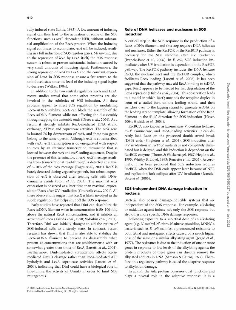
for RNR3 becomes conceivable (Fig. 2).
It is now clear that in response to DNA damage or
replication blocks, the Rad53 protein kinase is activated in
a Mec1-dependent manner, and activated Rad53 further
phosphorylates the protein kinase Dun1. The Mec1-Rad53-
Dun1 kinase cascade culminates in the phosphorylation of
Crt1. Crt1 is a DNA-binding protein that recognizes a 13-bp
consensus sequence termed the X box. Multiple X box-
related sequences of different strength can be found in the
promoter region of all four RNR genes. Crt1 is able to
recruit the Tup1–Ssn6 general repressor complex to suppress
the transcription of RNR genes. In the presence of DNA
damage or replication blocks, Crt1 is phosphorylated in a
Dun1-dependent manner and this phosphorylation
abolishes its ability to bind X boxes, leading to the transcrip-
tional induction of RNR genes (Huang et al., 1998).
Surprisingly, multiple X boxes were also identified in the
CRT1 promoter. Data from mutated X boxes showed that
they confer CRT1-dependent repression on CRT1 itself
(Huang et al., 1998). The expression level of CRT1 is very
low under normal growth conditions but is inducible by
DNA-damaging agents. Interestingly, the X boxes in the
CRT1 promoter consist of one with a weak affinity for Crt1
and one with a strong affinity. Thus, it is speculated that a
weak induction of CRT1 occurs immediately following DNA
damage, which provides a buffer against spurious transcrip-
tional activation of the pathway. Delaying full activation
may ensure a rapid restoration of the basal repressed state
when the DNA damage is repaired (Huang et al., 1998),
which may serve as an autonomous regulatory circuit (Fig.
2). This mechanism is reminiscent of the autonomous
regulation of the LexA repressor in the E. coli SOS response.
Crt1 mediates repression by recruiting the general re-
pressor Tup1–Ssn6 complex to RNR promoters via its
DNA damage
Mec1
Rad53
Dun1
P
Pol
Crt10
X-box CRT1
Crt1
RNR3Ccr4
CRT1 mRNA
Crt1
Crt1
X-boxCrt1
P
P
P
P
RNR3X-box
Rpd
3
Wtm
2
Hos2
Stalled replication
Fig. 2. Summary of the transcriptional regulation of RNR3. In response to DNA damage or stalled replication, activation of the Mec1-Rad53-Dun1
kinase cascade results in phosphorylation of the repressor protein Crt1, and phosphorylated Crt1 loses its binding affinity for the RNR3 promoter. With
the assistance of Wtm1, Rpd3 and Hos2, the transcription of RNR3 is highly activated. The transcription of CRT1 is negatively regulated by itself and
positively regulated by Crt10. Ccr4 influences Crt1 protein abundance by controlling the CRT1 mRNA stability.
FEMS Microbiol Rev 32 (2008) 908–926 c� 2008 Federation of European Microbiological SocietiesPublished by Blackwell Publishing Ltd. All rights reserved
917DNA damage-induced transcription in budding yeast
Dow
nloaded from https://academ
ic.oup.com/fem
sre/article/32/6/908/2683227 by guest on 04 July 2022
N-terminus (Huang et al., 1998; Li & Reese, 2000). Tup1-
Ssn6 recruitment establishes a nucleosomal array over the
promoter of RNR3 with a positioned nucleosome occupying
the TATA box to block access by the general transcriptional
machinery (Li & Reese, 2000, 2001; Sharma et al., 2003). The
derepression of RNR3 correlates with the disruption of the
nucleosome position. In response to DNA damage signals,
hyperphosphorylated Crt1 loses the ability to bind to the
RNR3 promoter, and the Tup1–Ssn6 complex is not re-
cruited to the promoter region. Consequently, the chromatin
structure is remodeled and thus increases the accessibility of
DNA to transcription factors. The chromatin remodeling at
the RNR3 promoter requires a number of general transcrip-
tional factors, such as TBP-associated factors (TAFIIS), and
RNA polymerase II. Furthermore, the remodeling is also
dependent on the SWI/SNF complex, which possesses a
DNA-stimulated ATPase activity and can destabilize histo-
ne–DNA interactions in an ATP-dependent manner (Sharma
et al., 2003). The preinitiation complex components TFIID
and RNA polymerase II aid in recruitment and retention of
the SWI/SNF complex to the RNR3 promoter.
Recently, it was reported that the expression of CRT1 is
also controlled by the novel regulator Crt10 (Fu & Xiao,
2006) (Fig. 2). CRT10 was identified through screening of
the S. cerevisiae deletion strains for hydroxyurea resistance.
Epistatic analysis indicates that CRT10 belongs to the CRT1
pathway. Deletion of CRT10 does not affect the transcript
level of TUP1 or SSN6 regardless of hydroxyurea treatment,
but significantly reduces the basal level as well as hydroxyur-
ea-induced expression of CRT1; thus, CRT10 appears to be a
positive regulator of CRT1 transcription (Fu & Xiao, 2006).
Furthermore, the dun1 mutation is epistatic to crt10 with
respect to both hydroxyurea sensitivity and RNR gene
expression. Interestingly, the expression of CRT10 itself is
induced by DNA-damaging agents and this induction
requires DUN1 (Fu & Xiao, 2006). The increased Crt10
activity may be required to bring Rnr activity back to a
normal level through increasing the expression of repressor
Crt1 once the DNA damage is repaired. Like CRT1, the
induction of CRT10 itself depends on DUN1, suggesting
that Crt10 functions downstream of Dun1 and forms
another component of the autoregulatory circuit to regulate
the expression of RNR genes (Fu & Xiao, 2006).
CCR4 encodes a component of the major cytoplasmic
deadenylase, which is involved in mRNA poly(A) tail short-
ening (Tucker et al., 2001). Cells defective in CCR4 display
particular sensitivity to the Rnr inhibiter hydroxyurea
(Woolstencroft et al., 2006). The ccr4 dun1 double mutants
exhibit synergistic sensitivity to hydroxyurea, and simulta-
neous overexpression of RNR2, RNR3 and RNR4 partially
rescues the hydroxyurea hypersensitivity of a ccr4 dun1
strain, implying that CCR4 and DUN1 function in different
pathways to regulate the activity of Rnr. Deletion of CRT1
suppresses hydroxyurea sensitivity of the ccr4 mutant, and
overexpression of CRT1 hypersensitizes ccr4 to hydroxyurea.
These observations lead to the conclusion that Ccr4 regu-
lates CRT1 mRNA poly(A) tail length and may thus subtly
influence the Crt1 protein abundance (Woolstencroft et al.,
2006) (Fig. 2).
More regulatory factors were found to be involved in the
transcriptional regulation of RNR genes. Wtm1 and Wtm2
have been reported to modulate the expression of RNR3
(Tringe et al., 2006). Moderate overexpression of both genes
or high-level expression of WTM2 alone upregulates RNR3-
lacZ in the absence of DNA damage. In response to hydro-
xyurea and g-rays, the expression level of RNR3 attenuated
45% in wtm2D mutants, but not in wtm1 mutants. Wtm2
was found to associate directly with the RNR3 promoter,
and the association correlates with its ability to increase
constitutive RNR3 expression. It remains unknown how
Wtm2 increases RNR3 transcription, although some obser-
vations hint that Wtm2 might enhance RNR3 transcription
by participating in chromatin remodeling (Tringe et al.,
2006). Furthermore, Wtm1 associates with and anchors the
Rnr2/Rnr4 complex in the nucleus in untreated cells. In
response to DNA damage or replication inhibition, the
interaction between Wtm1 and Rnr2/Rnr4 is disrupted and
this complex is released from the nucleus to the cytoplasm
(Lee & Elledge, 2006; Zhang et al., 2006). A recent report
shows that two histone deacetylases (HDAC), Rpd3 and
Hos2, are required for the transcriptional activation of
RNR3 in response to DNA damage (Sharma et al., 2007)
(Fig. 2). Although a direct association between Hos2 and the
RNR3 promoter has not been observed, Rpd3 was found to
specifically bind to X boxes in the RNR3 promoter region in
a Tup1- or a Crt1-independent manner. The HDAC activity
of Rpd3 and Hos2 is essential for the activation of RNR3.
The observation that the recruitment of RNA polymerase II
is dramatically reduced in the rpd3 hos2 mutant indicates
that Rpd3 and Hos2 activate the transcription by regulating
the assembly of the preinitiation complex or inducing
multiple rounds of RNA polymerase recruitment (Sharma
et al., 2007).
PHR1
PHR1 encodes a photolyase that specifically and exclusively
repairs pyrimidine dimers, which are the most abundant
lesions found in DNA following UV irradiation (Sancar,
1985). Interestingly, the transcription of PHR1 is induced
by various DNA-damaging agents such as MMS, MNNG,
UV irradiation, 4-NQO or g-ray (Robinson et al., 1986;
Sebastian et al., 1990) despite the fact that Phr1 only reverses
UV-induced pyrimidine dimers but not lesions induced by
other DNA-damaging agents. Three transcriptional regula-
tory elements have been defined within the PHR1 promoter
FEMS Microbiol Rev 32 (2008) 908–926c� 2008 Federation of European Microbiological SocietiesPublished by Blackwell Publishing Ltd. All rights reserved
918 Y. Fu et al.
Dow
nloaded from https://academ
ic.oup.com/fem
sre/article/32/6/908/2683227 by guest on 04 July 2022

region: an upstream activation sequence (UAS), an up-
stream repression sequence (URS) and an upstream essen-
tial sequence (UES) (Sancar et al., 1995). A 22-bp
interrupted palindrome comprises UASPHRI, and it is re-
sponsible for 80–90% of basal and induced expression. It
alone can activate transcription of a CYC1 minimal promo-
ter but does not confer damage responsiveness (Sancar et al.,
1995). URSPHR1 is defined to a 39-bp region that includes a
22-bp palindrome. Deletions or specific mutations within
URSPHR1 increase basal-level expression but decrease the
induction ratio. It functions as a strong URS and confers a
low-level damage inducibility when placed in the context of
a heterologous gene (Sancar et al., 1995). The UESPHR1 is
required for efficient derepression when URSPHR1 is present.
Deletion of URSPHR1 also eliminates the requirement for
UESPHR1 for transcriptional activation (Sancar et al., 1995).
Three proteins have been identified that regulate the
expression of PHR1 by binding to the upstream regulatory
elements. Ume6 is a bifunctional transcriptional regulator
involved in several metabolic pathways (Strich et al., 1994).
It is a positive regulator of PHR1 transcription and binds
specifically to the UASPHR1 (Sweet et al., 1997). Multiple
copies of Ume6 enhance the expression of PHR1; however,
deletion of UME6 reduces the expression of PHR1 during
vegetative growth, but only at a distinct cell-cycle phase
(Sweet et al., 1997; Sancar, 2000). Rph1 and Gis1, which are
35% identical to each other at the amino acid sequence level,
are two DNA damage-responsive repressors of PHR1 tran-
scription (Jang et al., 1999; Sancar, 2000). Both Rph1 and
Gis1 contain two putative zinc fingers that are 4 90%
identical overall and identical in the DNA-binding loop,
and they regulate the transcriptional response of PHR1
through binding to URSPHR1. Deletion of both RPH1 and
GIS1 is required to fully derepress PHR1 in the absence of
damage, suggesting that they are functionally redundant. In
vitro footprinting and binding competition studies indicate
that the sequence AG4 (C4T) within the URSPHR1 is the
binding site for Rph1 and Gis1 (Jang et al., 1999).
Induction of PHR1 is controlled by the DNA damage
signal transduction pathway. The serine and threonine
residues of Rph1 can be phosphorylated, and the phosphor-
ylation of Rph1 is increased in response to DNA damage.
The DNA damage-induced Rph1 phosphorylation requires
DNA damage checkpoint proteins Rad9, Rad17, Mec1 and
Rad53, indicating that the phosphorylation of Rph1 is under
the control of the Mec1-Rad53 DNA damage checkpoint
pathway (Kim et al., 2002). On the other hand, deletion of
DUN1, TEL1 or CHK1 does not affect the phosphorylation
of Rph1, indicating that PHR1 is regulated by a potentially
novel damage checkpoint that is distinct from the Mec1-
Rad53-Dun1 protein kinase cascade. Based on the results of
a coimmunoprecipitation assay, Rad53 does not appear to
interact physically with Rph1, indicating the existence of a
yet unknown kinase(s) in the Mec1-Rad53-Rph1 pathway
(Kim et al., 2002).
MAG1 and DDI1
MAG1 encodes a 3-methyladenine DNA glycosylase that
initiates the base excision repair pathway by removing lethal
lesions such as 3-methyladenine (Chen et al., 1989). MAG1
is not only induced by DNA-alkylating agents such as MMS
but also by UV, 4-NQO or hydroxyurea (Chen et al., 1990;
Chen & Samson, 1991; Xiao et al., 1993). By analyzing the
DNA sequence immediately upstream of MAG1, another
DNA damage-inducible gene, named DDI1 for DNA Da-
mage Inducible, was identified (Liu & Xiao, 1997). Similar
to MAG1, DDI1 is also induced by MMS, UV, 4-NQO and
hydroxyurea. Furthermore, both genes require a similar
dosage for maximum induction, and the induction profile
is similar (Liu & Xiao, 1997).
MAG1 and DDI1 lie in a head-to-head configuration and
are transcribed divergently. The expression of both MAG1
and DDI1 is controlled by common as well as distinct UAS
and URS elements, possibly through antagonistic mechan-
isms (Liu & Xiao, 1997). A UASMAG1 and a URSMAG1 have
been identified in the promoter region of MAG1 (Xiao et al.,
1993). Interestingly, fine mapping of the UASMAG1 sequence
reveals that it is located within the DDI1 protein coding
region, and the electrophoretic mobility shift assay using
labeled UASMAG1 as a probe detected sequence-specific
binding protein(s) (Liu & Xiao, 1997), although the identity
of the UASMAG1-binding protein has not been reported. The
expression of DDI1 is negatively regulated by a URSDDI1 in
its promoter region (Liu & Xiao, 1997). The intergenic
region between MAG1 and DDI1 also contains a cis-acting
element that coregulates the expression of both genes. In this
shared promoter region, UASDM, which contains two 8-bp
tandem repeat sequences 50-GGTGGCGA-30, is required for
the bidirectional expression of MAG1 and DDI1; deletion or
point mutations of this tandem repeat result in a reduced
basal level expression and significant reduction of damage-
induced expression (Liu & Xiao, 1997). Furthermore,
UASDM alone is able to confer a DNA damage response
when fused to a CYC1 promoter (Liu & Xiao, 1997),
indicating that this cis-acting element independently inter-
acts with a transcriptional factor(s). With a yeast one-hybrid
screen using UASDM as a bait, a transcriptional activator
called Pdr3 was isolated (Zhu & Xiao, 2004). Pdr3 binds to
UASDM in vivo and in vitro, and deletion of PDR3 reduced
both the basal level and the DNA damage-induced expres-
sion of MAG1 and DDI1. In addition, deletion of PDR3 does
not further affect MAG1 and DDI1 expression if UASDM is
deleted, indicating that UASDM is indeed the target for Pdr3
activation (Zhu & Xiao, 2004). Another transcriptional
activator, Rpn4, was shown to be required for MAG1
FEMS Microbiol Rev 32 (2008) 908–926 c� 2008 Federation of European Microbiological SocietiesPublished by Blackwell Publishing Ltd. All rights reserved
919DNA damage-induced transcription in budding yeast
Dow
nloaded from https://academ
ic.oup.com/fem
sre/article/32/6/908/2683227 by guest on 04 July 2022
(Jelinsky et al., 2000) and DDI1 (Zhu & Xiao, 2004)
expression; however, Rpn4 does not appear to bind UASDM.
Moreover, deletion of RPN4 does not alter MAG1 and DDI1
expression in the pdr3 mutant cells, suggesting that RPN4
acts upstream of PDR3 (Zhu & Xiao, 2004). Deletion of
PDR3 and RPN4 has no effect on the basal level or the DNA
damage-induced expression of PHR1, RNR2 or RNR3.
Meanwhile, Crt1 and Tup1/Ssn6, the repressors of the RNR
genes, and Rph1 and Gis1, the repressors of PHR1, are not
involved in the control of MAG1 expression (Zhu & Xiao,
2001). Hence, it becomes apparent that all three sets of well-
studied yeast damage-inducible genes (RNR, PHR1 and
MAG1-DDI1) have distinct regulators and that the regula-
tory mechanisms are also different from each other.
The expression of MAG1 and DDI1 is also controlled by
the DNA damage checkpoint. Mutation of POL2, MEC1 or
DUN1 reduces the DNA damage response of MAG1 (Zhu &
Xiao, 1998, 2001), suggesting that MAG1 is regulated by the
POL2-MEC1-RAD53-DUN1 checkpoint pathway. In con-
trast, DDI1 remains inducible in sad1-1 (rad53), dun1 or
mec1 mutants, but its induction is diminished in pds1 sad1-1
(rad53) or pds1 dun1 double mutants (Zhu & Xiao, 2001).
PDS1 encodes an anaphase inhibitor and functions down-
stream of CHK1; CHK1-PDS1 and RAD53-DUN1 may form
two parallel branches in the DNA damage checkpoints (Fig.
1) (Gardner et al., 1999; Schollaert et al., 2004). This
suggests that the CHK1-PDS1 and MEC1-RAD53-DUN1
checkpoint pathways may function redundantly in the
control of DDI1 expression (Zhu & Xiao, 2001).
A putative eukaryotic SOS response in buddingyeast?
Although cell-cycle checkpoints have been regarded as a
eukaryotic SOS response, their similarity to the bacterial
SOS response is limited. In budding yeast, the Rad51 protein
is a sequence homolog of E. coli RecA and possesses ssDNA-
binding and ATPase activities (Shinohara et al., 1992).
However, the only Rad51 enzymatic activity reported to date
is ATP- and ssDNA-dependent recombinase, and it is in-
volved in homologous recombination and homology-
mediated DNA repair and damage tolerance pathways (Sung,
1994; Paques & Haber, 1999; Gangavarapu et al., 2007).
Although RAD51 itself is DNA damage inducible (Shinohara
et al., 1992), deletion of RAD51 does not affect the expression
of the DNA damage-inducible genes examined (Y. Fu &
W. Xiao, unpublished results). Nevertheless, it remains
possible that there is a functional homolog of RecA in yeast
with respect to a transcriptional response but it shares no
sequence homology and enzymatic activity with RecA.
In a broad sense, E. coli RecA controls three important
cellular responses to DNA damage, namely homologous
recombination via RecBCD and RecFOR, TLS by working
with PolIV and PolV and the SOS response, which collec-
tively provides a survival mechanism when cells encounter
replication blocks and/or contain excessive ssDNA. The
above functions are reminiscent of the Rad6–Rad18 com-
plex in budding yeast. Rad6–Rad18 forms a stable complex
that is essential for the DNA postreplication repair (PRR)
pathway (Broomfield et al., 2001). The Rad6–Rad18 com-
plex possesses ssDNA-binding and ATPase activities (Bailly
et al., 1997) and assumes most if not all RecA functions to
coordinate broad cellular responses to DNA damage. Firstly,
Rad6–Rad18 as an E2–E3 ubiquitination complex mono-
ubiquitinates PCNA to promote Polz- and PolZ-mediated
TLS (Hoege et al., 2002; Stelter & Ulrich, 2003). Secondly,
this mono-Ub-PCNA is required for PCNA poly-ubiquiti-
nation via a Lys63 chain linkage for an error-free mode of
DNA damage tolerance (Hoege et al., 2002) reminiscent of
the RecFOR activity in E. coli (Chow & Courcelle, 2004).
Finally, although Rad6–Rad18 does not have homologous
recombinase activity like RecA, it may compete with the
Ubc9–Siz1 complex that sumorylates PCNA at the same
K164 residue; sumorylated PCNA recruits the DNA helicase
Srs2, which inhibits the recombinase activity of the yeast
RecA homolog Rad51 (Papouli et al., 2005; Pfander et al.,
2005). Hence, it would be of great interest to investigate
whether the Rad6–Rad18 complex also plays a role in an
SOS response and, if so, how it interacts with the damage
checkpoints.
Conclusions
In summary, transcriptional response to DNA damage is an
important process for cell survival of microorganisms under
such life-threatening stress. In bacteria such as E. coli,
the DNA damage-induced transcriptional regulation is
centrally controlled by an SOS response. All SOS-inducible
genes contain SOS boxes in their operator regions, and
their expression is repressed by the interaction between the
transcriptional repressor LexA and SOS boxes under
noninduced conditions. The DNA damage induction is
achieved through enhanced self proteolysis of LexA
assisted by ssDNA-activated RecA. In addition, bacteria also
possess regulatory mechanisms in response to specific DNA
damage.
DNA damage-induced transcriptional response in eukar-
yotic microorganisms is apparently more complicated and
less understood. Nevertheless, several conclusions can be
drawn from data analyses as presented in this review. Firstly,
the idea that eukaryotic microorganisms possess an SOS
response structurally conserved from bacteria has been
effectively ruled out. Secondly, in budding yeast, a large
number of yeast genes (more than 5%) are induced after
DNA damage. Almost all DNA damage-induced genes
individually investigated to date respond to a broad range
FEMS Microbiol Rev 32 (2008) 908–926c� 2008 Federation of European Microbiological SocietiesPublished by Blackwell Publishing Ltd. All rights reserved
920 Y. Fu et al.
Dow
nloaded from https://academ
ic.oup.com/fem
sre/article/32/6/908/2683227 by guest on 04 July 2022
of DNA-damaging agents regardless of whether the gene
function provides a survival value to the particular damage
(Birrell et al., 2002). This observation supports the hypoth-
esis that there may exist a coordinated signal transduction
pathway to regulate transcriptional response to DNA da-
mage. Thirdly, DNA damage checkpoint pathways variably
affect transcriptional response to DNA damage in budding
yeast and have been regarded as a functional analog of the
bacterial SOS response. Fourthly, unlike the SOS-inducible
genes in bacteria, few DNA damage-inducible genes in
budding yeast share a common promoter sequence, suggest-
ing that the downstream effectors are diversified. This is
consistent with reports that all three well-characterized sets
of damage-inducible genes in budding yeast are controlled
by different transcriptional regulators and furthermore,
their modes of regulation are different: while RNRs and
PHR1 are induced by derepression, MAG1 and DDI1 are
induced by activation. Fifthly, the search for a novel
mechanism(s) in eukaryotic microorganisms that may bet-
ter resume the RecA-mediated SOS response is underway
and may result in exciting insights into the eukaryotic SOS
response. Finally, although transcriptional response to DNA
damage is a convenient and useful indicator for the gene
function, transcriptional levels do not necessarily reflect
cellular protein levels, and this appears to be the case for
DNA damage response in budding yeast (Lee et al., 2007).
Hence, other posttranscriptional regulatory mechanisms
have to be taken into account when examining a particular
gene function and its product activity.
Acknowledgements
The authors wish to thank the laboratory members for
helpful discussion and Michelle Hanna for proofreading
the manuscript. This work is supported by the Natural
Sciences and Engineering Research Council of Canada
discovery grant OGP0138338 to W.X. Y.F. is a recipient of
the Arthur Smyth Memorial Scholarship from College of
Medicine, University of Saskatchewan.
References
Aboussekhra A, Vialard JE, Morrison DE, de la Torre-Ruiz MA,
Cernakova L, Fabre F & Lowndes NF (1996) A novel role for
the budding yeast RAD9 checkpoint gene in DNA damage-
dependent transcription. EMBO J 15: 3912–3922.
Akimaru H, Sakumi K, Yoshikai T, Anai M & Sekiguchi M (1990)
Positive and negative regulation of transcription by a cleavage
product of Ada protein. J Mol Biol 216: 261–273.
Allen JB, Zhou Z, Siede W, Friedberg EC & Elledge SJ (1994) The
SAD1/RAD53 protein kinase controls multiple checkpoints
and DNA damage-induced transcription in yeast. Genes Dev 8:
2401–2415.
Aslund F, Zheng M, Beckwith J & Storz G (1999) Regulation of
the OxyR transcription factor by hydrogen peroxide and the
cellular thiol-disulfide status. Proc Natl Acad Sci USA 96:
6161–6165.
Bachant JB & Elledge SJ (1998) Regulatory networks that control
DNA damage-inducible gene in Saccharomyces cerevisiae. DNA
Damage and Repair, Vol. 1: DNA Repair in Prokaryotes and
Lower Eukaryotes (Nickoloff JA & Hoekstra MF, eds), pp.
383–410. Humana Press, Totowa, NJ.
Bailly V, Lauder S, Prakash S & Prakash L (1997) Yeast DNA
repair proteins Rad6 and Rad18 form a heterodimer that has
ubiquitin conjugating, DNA binding, and ATP hydrolytic
activities. J Biol Chem 272: 23360–23365.
Basrai MA, Velculescu VE, Kinzler KW & Hieter P (1999)
NORF5/HUG1 is a component of the MEC1-mediated
checkpoint response to DNA damage and replication arrest in
Saccharomyces cerevisiae. Mol Cell Biol 19: 7041–7049.
Bennett RA (1999) The Saccharomyces cerevisiae ETH1 gene, an
inducible homolog of exonuclease III that provides resistance
to DNA-damaging agents and limits spontaneous
mutagenesis. Mol Cell Biol 19: 1800–1809.
Bennett RJ, Sharp JA & Wang JC (1998) Purification and
characterization of the Sgs1 DNA helicase activity of
Saccharomyces cerevisiae. J Biol Chem 273: 9644–9650.
Bennett RJ, Keck JL & Wang JC (1999) Binding specificity
determines polarity of DNA unwinding by the Sgs1 protein of
S. cerevisiae. J Mol Biol 289: 235–248.
Benton MG, Somasundaram S, Glasner JD & Palecek SP (2006)
Analyzing the dose-dependence of the Saccharomyces cerevisiae
global transcriptional response to methyl methanesulfonate
and ionizing radiation. BMC Genom 7: 305.
Berg OG (1988) Selection of DNA binding sites by regulatory
proteins: the LexA protein and the arginine repressor use
different strategies for functional specificity. Nucleic Acids Res
16: 5089–5105.
Birrell GW, Brown JA, Wu HI, Giaever G, Chu AM, Davis RW &
Brown JM (2002) Transcriptional response of Saccharomyces
cerevisiae to DNA-damaging agents does not identify the genes
that protect against these agents. Proc Natl Acad Sci USA 99:
8778–8783.
Branzei D & Foiani M (2006) The Rad53 signal transduction
pathway: replication fork stabilization, DNA repair, and
adaptation. Exp Cell Res 312: 2654–2659.
Brent R (1982) Regulation and autoregulation by lexA protein.
Biochimie 64: 565–569.
Brent R & Ptashne M (1980) The lexA gene product represses its
own promoter. Proc Natl Acad Sci USA 77: 1932–1936.
Brent R & Ptashne M (1981) Mechanism of action of the lexA
gene product. Proc Natl Acad Sci USA 78: 4204–4208.
Broomfield S, Hryciw T & Xiao W (2001) DNA postreplication
repair and mutagenesis in Saccharomyces cerevisiae. Mutat Res
486: 167–184.
Brusky J, Zhu Y & Xiao W (2000) UBC13, a DNA-damage-
inducible gene, is a member of the error-free postreplication
FEMS Microbiol Rev 32 (2008) 908–926 c� 2008 Federation of European Microbiological SocietiesPublished by Blackwell Publishing Ltd. All rights reserved
921DNA damage-induced transcription in budding yeast
Dow
nloaded from https://academ
ic.oup.com/fem
sre/article/32/6/908/2683227 by guest on 04 July 2022

repair pathway in Saccharomyces cerevisiae. Curr Genet 37:
168–174.
Chen J & Samson L (1991) Induction of S. cerevisiae MAG 3-
methyladenine DNA glycosylase transcript levels in response
to DNA damage. Nucleic Acids Res 19: 6427–6432.
Chen J, Derfler B, Maskati A & Samson L (1989) Cloning a
eukaryotic DNA glycosylase repair gene by the suppression of a
DNA repair defect in Escherichia coli. Proc Natl Acad Sci USA
86: 7961–7965.
Chen J, Derfler B & Samson L (1990) Saccharomyces cerevisiae
3-methyladenine DNA glycosylase has homology to the AlkA
glycosylase of E. coli and is induced in response to DNA
alkylation damage. EMBO J 9: 4569–4575.
Chen SH, Smolka MB & Zhou H (2007) Mechanism of Dun1
activation by Rad53 phosphorylation in Saccharomyces
cerevisiae. J Biol Chem 282: 986–995.
Chow KH & Courcelle J (2004) RecO acts with RecF and RecR to
protect and maintain replication forks blocked by UV-induced
DNA damage in Escherichia coli. J Biol Chem 279: 3492–3496.
Cobb JA, Bjergbaek L & Gasser SM (2002) RecQ helicases: at the
heart of genetic stability. FEBS Lett 529: 43–48.
Courcelle J, Khodursky A, Peter B, Brown PO & Hanawalt PC
(2001) Comparative gene expression profiles following UV
exposure in wild-type and SOS-deficient Escherichia coli.
Genetics 158: 41–64.
de la Torre-Ruiz MA, Green CM & Lowndes NF (1998) RAD9 and
RAD24 define two additive, interacting branches of the DNA
damage checkpoint pathway in budding yeast normally
required for Rad53 modification and activation. EMBO J 17:
2687–2698.
Demple B, Sedgwick B, Robins P, Totty N, Waterfield MD &
Lindahl T (1985) Active site and complete sequence of the
suicidal methyltransferase that counters alkylation
mutagenesis. Proc Natl Acad Sci USA 82: 2688–2692.
De Sanctis V, Bertozzi C, Costanzo G, Di Mauro E & Negri R
(2001) Cell cycle arrest determines the intensity of the global
transcriptional response of Saccharomyces cerevisiae to
ionizing radiation. Radiat Res 156: 379–387.
Ding H, Hidalgo E & Demple B (1996) The redox state of the
[2Fe–2S] clusters in SoxR protein regulates its activity as a
transcription factor. J Biol Chem 271: 33173–33175.
Drees JC, Lusetti SL, Chitteni-Pattu S, Inman RB & Cox MM
(2004) A RecA filament capping mechanism for RecX protein.
Mol Cell 15: 789–798.
Elledge SJ (1996) Cell cycle checkpoints: preventing an identity
crisis. Science 274: 1664–1672.
Elledge SJ & Davis RW (1987) Identification and isolation of the
gene encoding the small subunit of ribonucleotide reductase
from Saccharomyces cerevisiae: DNA damage-inducible gene
required for mitotic viability. Mol Cell Biol 7: 2783–2793.
Elledge SJ & Davis RW (1989) DNA damage induction of
ribonucleotide reductase. Mol Cell Biol 9: 4932–4940.
Elledge SJ & Davis RW (1990) Two genes differentially regulated
in the cell cycle and by DNA-damaging agents encode
alternative regulatory subunits of ribonucleotide reductase.
Genes Dev 4: 740–751.
Elledge SJ, Zhou Z, Allen JB & Navas TA (1993) DNA damage and
cell cycle regulation of ribonucleotide reductase. Bioessays 15:
333–339.
Frei C & Gasser SM (2000) The yeast Sgs1p helicase acts upstream
of Rad53p in the DNA replication checkpoint and colocalizes
with Rad53p in S-phase-specific foci. Genes Dev 14: 81–96.
Friedberg E, Walker G & Wolfram S (1995) DNA Repair and
Mutagenesis. ASM Press, Washington, DC.
Friedberg E, Walker G, Siede W, Wood R, Schultz R & Ellenberger
T (2006) DNA Repair and Mutagenesis. 2nd edn. ASM Press,
Washington, DC.
Fry RC, Sambandan TG & Rha C (2003) DNA damage and stress
transcripts in Saccharomyces cerevisiae mutant sgs1. Mech
Ageing Dev 124: 839–846.
Fu Y & Xiao W (2006) Identification and characterization of
CRT10 as a novel regulator of Saccharomyces cerevisiae
ribonucleotide reductase genes. Nucleic Acids Res 34:
1876–1883.
Fu Y, Zhu Y, Zhang K, Yeung M, Durocher D & Xiao W (2008)
Rad6-Rad18 mediates a eukaryotic SOS response by
ubiquitinating the 9-1-1 checkpoint clamp. Cell 133: 601–611.
Gangavarapu V, Prakash S & Prakash L (2007) Requirement of
RAD52 group genes for postreplication repair of UV-damaged
DNA in Saccharomyces cerevisiae. Mol Cell Biol 27: 7758–7764.
Gardner R, Putnam CW & Weinert T (1999) RAD53, DUN1 and
PDS1 define two parallel G2/M checkpoint pathways in
budding yeast. EMBO J 18: 3173–3185.
Gasch AP, Spellman PT, Kao CM et al. (2000) Genomic
expression programs in the response of yeast cells to
environmental changes. Mol Biol Cell 11: 4241–4257.
Gasch AP, Huang M, Metzner S, Botstein D, Elledge SJ & Brown
PO (2001) Genomic expression responses to DNA-damaging
agents and the regulatory role of the yeast ATR homolog
Mec1p. Mol Biol Cell 12: 2987–3003.
Gaudu P, Moon N & Weiss B (1997) Regulation of the soxRS
oxidative stress regulon. Reversible oxidation of the Fe–S
centers of SoxR in vivo. J Biol Chem 272: 5082–5086.
Giannattasio M, Lazzaro F, Longhese MP, Plevani P & Muzi-
Falconi M (2004) Physical and functional interactions between
nucleotide excision repair and DNA damage checkpoint.
EMBO J 23: 429–438.
Gottesman S (1981) Genetic control of the SOS system in E. coli.
Cell 23: 1–2.
Green SR & Johnson AD (2004) Promoter-dependent roles for
the Srb10 cyclin-dependent kinase and the Hda1 deacetylase in
Tup1-mediated repression in Saccharomyces cerevisiae. Mol
Biol Cell 15: 4191–4202.
Guo Y, Breeden LL, Fan W, Zhao LP, Eaton DL & Zarbl H (2006)
Analysis of cellular responses to aflatoxin B(1) in yeast
expressing human cytochrome P450 1A2 using cDNA
microarrays. Mutat Res 593: 121–142.
Hartwell LH & Weinert TA (1989) Checkpoints: controls that
ensure the order of cell cycle events. Science 246: 629–634.
FEMS Microbiol Rev 32 (2008) 908–926c� 2008 Federation of European Microbiological SocietiesPublished by Blackwell Publishing Ltd. All rights reserved
922 Y. Fu et al.
Dow
nloaded from https://academ
ic.oup.com/fem
sre/article/32/6/908/2683227 by guest on 04 July 2022
Hegde S, Sandler SJ, Clark AJ & Madiraju MV (1995) recO and
recR mutations delay induction of the SOS response in
Escherichia coli. Mol Gen Genet 246: 254–258.
Heyer WD (2004) Damage signaling: RecQ sends an SOS to you.
Curr Biol 14: R895–R897.
Hidalgo E & Demple B (1994) An iron–sulfur center essential for
transcriptional activation by the redox-sensing SoxR protein.
EMBO J 13: 138–146.
Hidalgo E, Bollinger JM Jr, Bradley TM, Walsh CT & Demple B
(1995) Binuclear [2Fe–2S] clusters in the Escherichia coli SoxR
protein and role of the metal centers in transcription. J Biol
Chem 270: 20908–20914.
Hishida T, Han YW, Shibata T, Kubota Y, Ishino Y, Iwasaki H &
Shinagawa H (2004) Role of the Escherichia coli RecQ DNA
helicase in SOS signaling and genome stabilization at stalled
replication forks. Genes Dev 18: 1886–1897.
Hoege C, Pfander B, Moldovan GL, Pyrowolakis G & Jentsch S
(2002) RAD6-dependent DNA repair is linked to modification
of PCNA by ubiquitin and SUMO. Nature 419: 135–141.
Huang M & Elledge SJ (1997) Identification of RNR4, encoding a
second essential small subunit of ribonucleotide reductase in
Saccharomyces cerevisiae. Mol Cell Biol 17: 6105–6113.
Huang M, Zhou Z & Elledge SJ (1998) The DNA replication and
damage checkpoint pathways induce transcription by
inhibition of the Crt1 repressor. Cell 94: 595–605.
Ivancic-Bace I, Vlasic I, Salaj-Smic E & Brcic-Kostic K (2006)
Genetic evidence for the requirement of RecA loading activity
in SOS induction after UV irradiation in Escherichia coli.
J Bacteriol 188: 5024–5032.
Jang YK, Wang L & Sancar GB (1999) RPH1 and GIS1 are
damage-responsive repressors of PHR1. Mol Cell Biol 19:
7630–7638.
Jeggo P, Defais TM, Samson L & Schendel P (1977) An adaptive
response of E. coli to low levels of alkylating agent: comparison
with previously characterised DNA repair pathways. Mol Gen
Genet 157: 1–9.
Jelinsky SA & Samson LD (1999) Global response of
Saccharomyces cerevisiae to an alkylating agent. Proc Natl Acad
Sci USA 96: 1486–1491.
Jelinsky SA, Estep P, Church GM & Samson LD (2000)
Regulatory networks revealed by transcriptional profiling of
damaged Saccharomyces cerevisiae cells: Rpn4 links base
excision repair with proteasomes. Mol Cell Biol 20: 8157–8167.
John L, Sharma G, Chaudhuri SP & Pillai B (2005) Cigarette
smoke extract induces changes in growth and gene expression
of Saccharomyces cerevisiae. Biochem Biophys Res Commun
338: 1578–1586.
Jordan A & Reichard P (1998) Ribonucleotide reductases. Annu
Rev Biochem 67: 71–98.
Katou Y, Kanoh Y, Bando M et al. (2003) S-phase checkpoint
proteins Tof1 and Mrc1 form a stable replication-pausing
complex. Nature 424: 1078–1083.
Keller-Seitz MU, Certa U, Sengstag C, Wurgler FE, Sun M &
Fasullo M (2004) Transcriptional response of yeast to aflatoxin
B1: recombinational repair involving RAD51 and RAD1. Mol
Biol Cell 15: 4321–4336.
Kelly GT, Liu C, Smith R III, Coleman RS & Watanabe CM (2006)
Cellular effects induced by the antitumor agent azinomycin B.
Chem Biol 13: 485–492.
Kenyon CJ & Walker GC (1980) DNA-damaging agents stimulate
gene expression at specific loci in Escherichia coli. Proc Natl
Acad Sci USA 77: 2819–2823.
Kim EM, Jang YK & Park SD (2002) Phosphorylation of Rph1, a
damage-responsive repressor of PHR1 in Saccharomyces
cerevisiae, is dependent upon Rad53 kinase. Nucleic Acids Res
30: 643–648.
Kiser GL & Weinert TA (1996) Distinct roles of yeast MEC and
RAD checkpoint genes in transcriptional induction after DNA
damage and implications for function. Mol Biol Cell 7:
703–718.
Kondo T, Matsumoto K & Sugimoto K (1999) Role of a complex
containing Rad17, Mec3, and Ddc1 in the yeast DNA damage
checkpoint pathway. Mol Cell Biol 19: 1136–1143.
Kondo T, Wakayama T, Naiki T, Matsumoto K & Sugimoto K
(2001) Recruitment of Mec1 and Ddc1 checkpoint proteins to
double-strand breaks through distinct mechanisms. Science
294: 867–870.
Kugou K, Sasanuma H, Matsumoto K, Shirahige K & Ohta K
(2007) Mre11 mediates gene regulation in yeast spore
development. Genes Genet Syst 82: 21–33.
Landini P & Volkert MR (1995) Transcriptional activation of the
Escherichia coli adaptive response gene aidB is mediated by
binding of methylated Ada protein. Evidence for a new
consensus sequence for Ada-binding sites. J Biol Chem 270:
8285–8289.
Lee JH & Paull TT (2005) ATM activation by DNA double-strand
breaks through the Mre11-Rad50-Nbs1 complex. Science 308:
551–554.
Lee MW, Kim BJ, Choi HK et al. (2007) Global protein expression
profiling of budding yeast in response to DNA damage. Yeast
24: 145–154.
Lee YD & Elledge SJ (2006) Control of ribonucleotide reductase
localization through an anchoring mechanism involving
Wtm1. Genes Dev 20: 334–344.
Lewis LK, Harlow GR, Gregg-Jolly LA & Mount DW (1994)
Identification of high affinity binding sites for LexA which
define new DNA damage-inducible genes in Escherichia coli.
J Mol Biol 241: 507–523.
Li B & Reese JC (2000) Derepression of DNA damage-regulated
genes requires yeast TAF(II)s. EMBO J 19: 4091–4100.
Li B & Reese JC (2001) Ssn6-Tup1 regulates RNR3 by positioning
nucleosomes and affecting the chromatin structure at the
upstream repression sequence. J Biol Chem 276: 33788–33797.
Lindahl T, Sedgwick B, Sekiguchi M & Nakabeppu Y (1988)
Regulation and expression of the adaptive response to
alkylating agents. Annu Rev Biochem 57: 133–157.
Little JW (1983) The SOS regulatory system: control of its state by
the level of RecA protease. J Mol Biol 167: 791–808.
FEMS Microbiol Rev 32 (2008) 908–926 c� 2008 Federation of European Microbiological SocietiesPublished by Blackwell Publishing Ltd. All rights reserved
923DNA damage-induced transcription in budding yeast
Dow
nloaded from https://academ
ic.oup.com/fem
sre/article/32/6/908/2683227 by guest on 04 July 2022

Little JW (1984) Autodigestion of lexA and phage l repressors.
Proc Natl Acad Sci USA 81: 1375–1379.
Little JW (1991) Mechanism of specific LexA cleavage:
autodigestion and the role of RecA coprotease. Biochimie 73:
411–421.
Little JW (1993) LexA cleavage and other self-processing
reactions. J Bacteriol 175: 4943–4950.
Little JW & Mount DW (1982) The SOS regulatory system of
Escherichia coli. Cell 29: 11–22.
Little JW, Mount DW & Yanisch-Perron CR (1981) Purified lexA
protein is a repressor of the recA and lexA genes. Proc Natl
Acad Sci USA 78: 4199–4203.
Liu Y & Xiao W (1997) Bidirectional regulation of two
DNA-damage-inducible genes, MAG1 and DDI1, from
Saccharomyces cerevisiae. Mol Microbiol 23: 777–789.
Lusetti SL, Voloshin ON, Inman RB, Camerini-Otero RD & Cox
MM (2004) The DinI protein stabilizes RecA protein
filaments. J Biol Chem 279: 30037–30046.
Lusetti SL, Hobbs MD, Stohl EA, Chitteni-Pattu S, Inman RB,
Seifert HS & Cox MM (2006) The RecF protein antagonizes
RecX function via direct interaction. Mol Cell 21: 41–50.
Lydall D & Weinert T (1995) Yeast checkpoint genes in DNA
damage processing: implications for repair and arrest. Science
270: 1488–1491.
Ma JL, Lee SJ, Duong JK & Stern DF (2006) Activation of the
checkpoint kinase Rad53 by the phosphatidyl inositol kinase-
like kinase Mec1. J Biol Chem 281: 3954–3963.
Majka J & Burgers PM (2003) Yeast Rad17/Mec3/Ddc1: a sliding
clamp for the DNA damage checkpoint. Proc Natl Acad Sci
USA 100: 2249–2254.
Majka J, Niedziela-Majka A & Burgers PM (2006) The checkpoint
clamp activates Mec1 kinase during initiation of the DNA
damage checkpoint. Mol Cell 24: 891–901.
Makiniemi M, Hillukkala T, Tuusa J et al. (2001) BRCT domain-
containing protein TopBP1 functions in DNA replication and
damage response. J Biol Chem 276: 30399–30406.
Mannhaupt G, Schnall R, Karpov V, Vetter I & Feldmann H
(1999) Rpn4p acts as a transcription factor by binding to
PACE, a nonamer box found upstream of 26S proteasomal and
other genes in yeast. FEBS Lett 450: 27–34.
Marques M, Mojzita D, Amorim MA, Almeida T, Hohmann S,
Moradas-Ferreira P & Costa V (2006) The Pep4p vacuolar
proteinase contributes to the turnover of oxidized proteins but
PEP4 overexpression is not sufficient to increase chronological
lifespan in Saccharomyces cerevisiae. Microbiology 152:
3595–3605.
McClanahan T & McEntee K (1984) Specific transcripts are
elevated in Saccharomyces cerevisiae in response to DNA
damage. Mol Cell Biol 4: 2356–2363.
McDonald JP, Levine AS & Woodgate R (1997) The
Saccharomyces cerevisiae RAD30 gene, a homologue of
Escherichia coli dinB and umuC, is DNA damage inducible and
functions in a novel error-free postreplication repair
mechanism. Genetics 147: 1557–1568.
Melo JA, Cohen J & Toczyski DP (2001) Two checkpoint
complexes are independently recruited to sites of DNA damage
in vivo. Genes Dev 15: 2809–2821.
Mercier G, Berthault N, Touleimat N, Kepes F, Fourel G, Gilson E
& Dutreix M (2005) A haploid-specific transcriptional
response to irradiation in Saccharomyces cerevisiae. Nucleic
Acids Res 33: 6635–6643.
Mills KD, Sinclair DA & Guarente L (1999) MEC1-dependent
redistribution of the Sir3 silencing protein from telomeres to
DNA double-strand breaks. Cell 97: 609–620.
Myers LC, Terranova MP, Ferentz AE, Wagner G & Verdine GL
(1993a) Repair of DNA methylphosphotriesters through a
metalloactivated cysteine nucleophile. Science 261: 1164–1167.
Myers LC, Verdine GL & Wagner G (1993b) Solution structure of
the DNA methyl phosphotriester repair domain of Escherichia
coli Ada. Biochemistry 32: 14089–14094.
Naiki T, Wakayama T, Nakada D, Matsumoto K & Sugimoto K
(2004) Association of Rad9 with double-strand breaks through
a Mec1-dependent mechanism. Mol Cell Biol 24: 3277–3285.
Nakabeppu Y & Sekiguchi M (1986) Regulatory mechanisms for
induction of synthesis of repair enzymes in response to
alkylating agents: ada protein acts as a transcriptional
regulator. Proc Natl Acad Sci USA 83: 6297–6301.
Nakamura T, Tokumoto Y, Sakumi K, Koike G, Nakabeppu Y &
Sekiguchi M (1988) Expression of the ada gene of Escherichia
coli in response to alkylating agents. Identification of
transcriptional regulatory elements. J Mol Biol 202: 483–494.
Navas TA, Zhou Z & Elledge SJ (1995) DNA polymerase epsilon
links the DNA replication machinery to the S phase
checkpoint. Cell 80: 29–39.
Niida H & Nakanishi M (2006) DNA damage checkpoints in
mammals. Mutagenesis 21: 3–9.
Nohmi T, Battista JR, Dodson LA & Walker GC (1988)
RecA-mediated cleavage activates UmuD for mutagenesis:
mechanistic relationship between transcriptional derepression
and posttranslational activation. Proc Natl Acad Sci USA 85:
1816–1820.
Nyberg KA, Michelson RJ, Putnam CW & Weinert TA (2002)
Toward maintaining the genome: DNA damage and
replication checkpoints. Annu Rev Genet 36: 617–656.
Oshiro G, Wodicka LM, Washburn MP, Yates JR III, Lockhart DJ
& Winzeler EA (2002) Parallel identification of new genes in
Saccharomyces cerevisiae. Genome Res 12: 1210–1220.
Ostapenko D & Solomon MJ (2003) Budding yeast CTDK-I is
required for DNA damage-induced transcription. Eukaryot
Cell 2: 274–283.
Pages V, Koffel-Schwartz N & Fuchs RP (2003) recX, a new SOS
gene that is co-transcribed with the recA gene in Escherichia
coli. DNA Repair (Amsterdam) 2: 273–284.
Papouli E, Chen S, Davies AA, Huttner D, Krejci L, Sung P &
Ulrich HD (2005) Crosstalk between SUMO and ubiquitin on
PCNA is mediated by recruitment of the helicase Srs2p. Mol
Cell 19: 123–133.
FEMS Microbiol Rev 32 (2008) 908–926c� 2008 Federation of European Microbiological SocietiesPublished by Blackwell Publishing Ltd. All rights reserved
924 Y. Fu et al.
Dow
nloaded from https://academ
ic.oup.com/fem
sre/article/32/6/908/2683227 by guest on 04 July 2022

Paques F & Haber JE (1999) Multiple pathways of recombination
induced by double-strand breaks in Saccharomyces cerevisiae.
Microbiol Mol Biol Rev 63: 349–404.
Pellicioli A & Foiani M (2005) Signal transduction: how rad53
kinase is activated. Curr Biol 15: R769–R771.
Peterson KR & Mount DW (1987) Differential repression of SOS
genes by unstable lexA41 (tsl-1) protein causes a ‘‘split-
phenotype’’ in Escherichia coli K-12. J Mol Biol 193: 27–40.
Pfander B, Moldovan GL, Sacher M, Hoege C & Jentsch S (2005)
SUMO-modified PCNA recruits Srs2 to prevent
recombination during S phase. Nature 436: 428–433.
Post SM, Tomkinson AE & Lee EY (2003) The human checkpoint
Rad protein Rad17 is chromatin-associated throughout the
cell cycle, localizes to DNA replication sites, and interacts with
DNA polymerase e. Nucleic Acids Res 31: 5568–5575.
Radman M (1975) SOS repair hypothesis: phenomenology of an
inducible DNA repair which is accompanied by mutagenesis.
Basic Life Sci 5A: 355–367.
Reichard P (1988) Interactions between deoxyribonucleotide and
DNA synthesis. Annu Rev Biochem 57: 349–374.
Renzette N, Gumlaw N, Nordman JT et al. (2005) Localization of
RecA in Escherichia coli K-12 using RecA-GFP. Mol Microbiol
57: 1074–1085.
Ritchie KB, Mallory JC & Petes TD (1999) Interactions of TLC1
(which encodes the RNA subunit of telomerase), TEL1, and
MEC1 in regulating telomere length in the yeast Saccharomyces
cerevisiae. Mol Cell Biol 19: 6065–6075.
Robinson GW, Nicolet CM, Kalainov D & Friedberg EC (1986) A
yeast excision-repair gene is inducible by DNA damaging
agents. Proc Natl Acad Sci USA 83: 1842–1846.
Roos WP & Kaina B (2006) DNA damage-induced cell death by
apoptosis. Trends Mol Med 12: 440–450.
Rouse J & Jackson SP (2002) Lcd1p recruits Mec1p to DNA
lesions in vitro and in vivo. Mol Cell 9: 857–869.
Rowley R, Hudson J & Young PG (1992) The wee1 protein kinase
is required for radiation-induced mitotic delay. Nature 356:
353–355.
Ruby SW & Szostak JW (1985) Specific Saccharomyces cerevisiae
genes are expressed in response to DNA-damaging agents. Mol
Cell Biol 5: 75–84.
Ruby SW, Szostak JW & Murray AW (1983) Cloning regulated
yeast genes from a pool of lacZ fusions. Methods Enzymol 101:
253–269.
Saget BM & Walker GC (1994) The Ada protein acts as both a
positive and a negative modulator of Escherichia coli’s response
to methylating agents. Proc Natl Acad Sci USA 91: 9730–9734.
Sakashita H, Sakuma T, Ohkubo T, Kainosho M, Sakumi K,
Sekiguchi M & Morikawa K (1993) Folding topology and DNA
binding of the N-terminal fragment of Ada protein. FEBS Lett
323: 252–256.
Sakumi K & Sekiguchi M (1989) Regulation of expression of the
ada gene controlling the adaptive response. Interactions with
the ada promoter of the Ada protein and RNA polymerase.
J Mol Biol 205: 373–385.
Samson L & Cairns J (1977) A new pathway for DNA repair in
Escherichia coli. Nature 267: 281–283.
Sancar GB (1985) Sequence of the Saccharomyces cerevisiae PHR1
gene and homology of the PHR1 photolyase to E. coli
photolyase. Nucleic Acids Res 13: 8231–8246.
Sancar GB (2000) Enzymatic photoreactivation: 50 years and
counting. Mutat Res 451: 25–37.
Sancar GB, Ferris R, Smith FW & Vandeberg B (1995) Promoter
elements of the PHR1 gene of Saccharomyces cerevisiae and
their roles in the response to DNA damage. Nucleic Acids Res
23: 4320–4328.
Schnarr M, Oertel-Buchheit P, Kazmaier M & Granger-Schnarr
M (1991) DNA binding properties of the LexA repressor.
Biochimie 73: 423–431.
Schollaert KL, Poisson JM, Searle JS, Schwanekamp JA,
Tomlinson CR & Sanchez Y (2004) A role for Saccharomyces
cerevisiae Chk1p in the response to replication blocks. Mol Biol
Cell 15: 4051–4063.
Sebastian J, Kraus B & Sancar GB (1990) Expression of the yeast
PHR1 gene is induced by DNA-damaging agents. Mol Cell Biol
10: 4630–4637.
Sharma VM, Li B & Reese JC (2003) SWI/SNF-dependent
chromatin remodeling of RNR3 requires TAF(II)s and the
general transcription machinery. Genes Dev 17: 502–515.
Sharma VM, Tomar RS, Dempsey AE & Reese JC (2007) Histone
deacetylases RPD3 and HOS2 regulate the transcriptional
activation of DNA damage-inducible genes. Mol Cell Biol 27:
3199–3210.
Shenton D, Smirnova JB, Selley JN et al. (2006) Global
translational responses to oxidative stress impact upon
multiple levels of protein synthesis. J Biol Chem 281:
29011–29021.
Shinohara A, Ogawa H & Ogawa T (1992) Rad51 protein
involved in repair and recombination in S. cerevisiae is a
RecA-like protein. Cell 69: 457–470.
Singh KK & Samson L (1995) Replication protein A binds to
regulatory elements in yeast DNA repair and DNA metabolism
genes. Proc Natl Acad Sci USA 92: 4907–4911.
Singleton MR, Dillingham MS, Gaudier M, Kowalczykowski SC
& Wigley DB (2004) Crystal structure of RecBCD enzyme
reveals a machine for processing DNA breaks. Nature 432:
187–193.
Stelter P & Ulrich HD (2003) Control of spontaneous and
damage-induced mutagenesis by SUMO and ubiquitin
conjugation. Nature 425: 188–191.
Stohl EA, Brockman JP, Burkle KL, Morimatsu K,
Kowalczykowski SC & Seifert HS (2003) Escherichia coli RecX
inhibits RecA recombinase and coprotease activities in vitro
and in vivo. J Biol Chem 278: 2278–2285.
Strich R, Surosky RT, Steber C, Dubois E, Messenguy F &
Esposito RE (1994) UME6 is a key regulator of nitrogen
repression and meiotic development. Genes Dev 8: 796–810.
Sung P (1994) Catalysis of ATP-dependent homologous DNA
pairing and strand exchange by yeast RAD51 protein. Science
265: 1241–1243.
FEMS Microbiol Rev 32 (2008) 908–926 c� 2008 Federation of European Microbiological SocietiesPublished by Blackwell Publishing Ltd. All rights reserved
925DNA damage-induced transcription in budding yeast
Dow
nloaded from https://academ
ic.oup.com/fem
sre/article/32/6/908/2683227 by guest on 04 July 2022

Sweeney FD, Yang F, Chi A, Shabanowitz J, Hunt DF & Durocher
D (2005) Saccharomyces cerevisiae Rad9 acts as a Mec1 adaptor
to allow Rad53 activation. Curr Biol 15: 1364–1375.
Sweet DH, Jang YK & Sancar GB (1997) Role of UME6 in
transcriptional regulation of a DNA repair gene in
Saccharomyces cerevisiae. Mol Cell Biol 17: 6223–6235.
Takinowaki H, Matsuda Y, Yoshida T, Kobayashi Y & Ohkubo T
(2006) The solution structure of the methylated form of the
N-terminal 16-kDa domain of Escherichia coli Ada protein.
Protein Sci 15: 487–497.
Tamayo P, Slonim D, Mesirov J et al. (1999) Interpreting patterns
of gene expression with self-organizing maps: methods and
application to hematopoietic differentiation. Proc Natl Acad
Sci USA 96: 2907–2912.
Teo I, Sedgwick B, Kilpatrick MW, McCarthy TV & Lindahl T
(1986) The intracellular signal for induction of resistance to
alkylating agents in E. coli. Cell 45: 315–324.
Thoms B & Wackernagel W (1987) Regulatory role of recF in the
SOS response of Escherichia coli: impaired induction of SOS
genes by UV irradiation and nalidixic acid in a recF mutant.
J Bacteriol 169: 1731–1736.
Tringe SG, Willis J, Liberatore KL & Ruby SW (2006) The WTM
genes in budding yeast amplify expression of the stress-
inducible gene RNR3. Genetics 174: 1215–1228.
Tucker M, Valencia-Sanchez MA, Staples RR, Chen J, Denis CL &
Parker R (2001) The transcription factor associated Ccr4 and
Caf1 proteins are components of the major cytoplasmic mRNA
deadenylase in Saccharomyces cerevisiae. Cell 104: 377–386.
van Attikum H, Fritsch O, Hohn B & Gasser SM (2004)
Recruitment of the INO80 complex by H2A phosphorylation
links ATP-dependent chromatin remodeling with DNA
double-strand break repair. Cell 119: 777–788.
Volkert MR & Landini P (2001) Transcriptional responses to
DNA damage. Curr Opin Microbiol 4: 178–185.
Voloshin ON, Ramirez BE, Bax A & Camerini-Otero RD (2001) A
model for the abrogation of the SOS response by an SOS
protein: a negatively charged helix in DinI mimics DNA in its
interaction with RecA. Genes Dev 15: 415–427.
Walker GC (1984) Mutagenesis and inducible responses to
deoxyribonucleic acid damage in Escherichia coli. Microbiol
Rev 48: 60–93.
Wang H & Elledge SJ (2002) Genetic and physical interactions
between DPB11 and DDC1 in the yeast DNA damage response
pathway. Genetics 160: 1295–1304.
Weinert TA & Hartwell LH (1988) The RAD9 gene controls the
cell cycle response to DNA damage in Saccharomyces cerevisiae.
Science 241: 317–322.
Weinert TA & Hartwell LH (1993) Cell cycle arrest of cdc mutants
and specificity of the RAD9 checkpoint. Genetics 134: 63–80.
Weinert TA, Kiser GL & Hartwell LH (1994) Mitotic checkpoint
genes in budding yeast and the dependence of mitosis on DNA
replication and repair. Genes Dev 8: 652–665.
Whitby MC & Lloyd RG (1995) Altered SOS induction associated
with mutations in recF, recO and recR. Mol Gen Genet 246:
174–179.
Witkin EM (1976) Ultraviolet mutagenesis and inducible
DNA repair in Escherichia coli. Bacteriol Rev 40:
869–907.
Woolstencroft RN, Beilharz TH, Cook MA, Preiss T, Durocher D
& Tyers M (2006) Ccr4 contributes to tolerance of replication
stress through control of CRT1 mRNA poly(A) tail length.
J Cell Sci 119: 5178–5192.
Workman CT, Mak HC, McCuine S et al. (2006) A systems
approach to mapping DNA damage response pathways.
Science 312: 1054–1059.
Wu J & Weiss B (1992) Two-stage induction of the soxRS
(superoxide response) regulon of Escherichia coli. J Bacteriol
174: 3915–3920.
Xiao W, Singh KK, Chen B & Samson L (1993) A common
element involved in transcriptional regulation of two DNA
alkylation repair genes (MAG and MGT1) of Saccharomyces
cerevisiae. Mol Cell Biol 13: 7213–7221.
Yasuda T, Morimatsu K, Horii T, Nagata T & Ohmori H (1998)
Inhibition of Escherichia coli RecA coprotease activities by
DinI. EMBO J 17: 3207–3216.
Zaim J, Speina E & Kierzek AM (2005) Identification of new
genes regulated by the Crt1 transcription factor, an effector of
the DNA damage checkpoint pathway in Saccharomyces
cerevisiae. J Biol Chem 280: 28–37.
Zhang Z, An X, Yang K et al. (2006) Nuclear localization of the
Saccharomyces cerevisiae ribonucleotide reductase small
subunit requires a karyopherin and a WD40 repeat protein.
Proc Natl Acad Sci USA 103: 1422–1427.
Zheng M, Aslund F & Storz G (1998) Activation of the OxyR
transcription factor by reversible disulfide bond formation.
Science 279: 1718–1721.
Zhou BB & Elledge SJ (2000) The DNA damage response: putting
checkpoints in perspective. Nature 408: 433–439.
Zhou Z & Elledge SJ (1992) Isolation of crt mutants constitutive
for transcription of the DNA damage inducible gene RNR3 in
Saccharomyces cerevisiae. Genetics 131: 851–866.
Zhou Z & Elledge SJ (1993) DUN1 encodes a protein kinase that
controls the DNA damage response in yeast. Cell 75:
1119–1127.
Zhu Y & Xiao W (1998) Differential regulation of two closely
clustered yeast genes, MAG1 and DDI1, by cell-cycle
checkpoints. Nucleic Acids Res 26: 5402–5408.
Zhu Y & Xiao W (2001) Two alternative cell cycle checkpoint
pathways differentially control DNA damage-dependent
induction of MAG1 and DDI1 expression in yeast. Mol Genet
Genomics 266: 436–444.
Zhu Y & Xiao W (2004) Pdr3 is required for DNA damage
induction of MAG1 and DDI1 via a bi-directional promoter
element. Nucleic Acids Res 32: 5066–5075.
FEMS Microbiol Rev 32 (2008) 908–926c� 2008 Federation of European Microbiological SocietiesPublished by Blackwell Publishing Ltd. All rights reserved
926 Y. Fu et al.
Dow
nloaded from https://academ
ic.oup.com/fem
sre/article/32/6/908/2683227 by guest on 04 July 2022