Assessment of methyl thiophanate–Cu (II) induced DNA damage in human lymphocytes
Upload
khangminh22Category
view
2download
0
IDENTIFICATION OF NOVEL DNA DAMAGE RESPONSE GENES USING
FUNCTIONAL GENOMICS
by
Michael Chang
A thesis submitted in conformity with the requirements
for the degree of Doctor of Philosophy
Graduate Department of Biochemistry
University of Toronto
© Copyright by Michael Chang (2005)

ii
Identification of novel DNA damage response genes using functional genomics
Doctor of Philosophy, 2005; Michael Chang; Department of Biochemistry, University of Toronto
ABSTRACT
The genetic information required for life is stored within molecules of DNA. This DNA is
under constant attack as a result of normal cellular metabolic processes, as well as exposure to
genotoxic agents. DNA damage left unrepaired can result in mutations that alter the genetic
information encoded within DNA. Cells have consequently evolved complex pathways to
combat damage to their DNA. Defects in the cellular response to DNA damage can result in
genomic instability, a hallmark of cancer cells. Identifying all the components required for this
response remains an important step in fully elucidating the molecular mechanisms involved. I
used functional genomic approaches to identify genes required for the DNA damage response in
Saccharomyces cerevisiae. I conducted a screen to identify genes required for resistance to a
DNA damaging agent, methyl methanesulfonate, and identified several poorly characterized
genes that are necessary for proper S phase progression in the presence of DNA damage.
Among the genes identified, ESC4/RTT107 has since been shown to be essential for the
resumption of DNA replication after DNA damage. Using genome-wide genetic interaction
screens to identify genes that are required for viability in the absence of MUS81 and MMS4, two
genes required for resistance to DNA damage, I helped identify ELG1, deletion of which causes
DNA replication defects, genomic instability, and an inability to properly recover from DNA
damage during S phase. I also used two-dimensional hierarchical clustering of synthetic genetic
interaction data determined by large-scale genetic network analysis to identify RMI1, which
encodes a new member of the highly conserved Sgs1-Top3 complex that is an important
suppressor of genomic instability.

iii
ACKNOWLEDGEMENTS
Many people contributed to the completion of this thesis. 90% of my gratitude should go to my
supervisor, Grant, without whom none of this would be possible. The other 10% goes to others
listed below, in no particular order.
Brenda Andrews and Craig Smibert: committee members
Co-authors: productive and successful scientific collaborations
Amy Tong and the Boone lab: help with SGA
Charlie Boone: transforming my unsuccessful pombe project into a successful cerevisiae thesis
Jiongwen Ou: best lab technician ever
Past and present members of the Brown lab: making the journey fun
Proceedings of the National Academy of Sciences, USA: authorizing the inclusion of published material in Ch. 3
EMBO Journal: authorizing the inclusion of published material in Ch. 4 and Ch. 5

iv
TABLE OF CONTENTS
ABSTRACT ................................................................................................................................................................... iiACKNOWLEDGEMENTS ......................................................................................................................................... iiiTABLE OF CONTENTS ............................................................................................................................................ ivLIST OF TABLES ....................................................................................................................................................... viLIST OF FIGURES .................................................................................................................................................... viiLIST OF ABBREVIATIONS .................................................................................................................................... viiiPUBLISHED MATERIAL NOT PRESENTED IN THIS THESIS ....................................................................... ix
1. GENERAL INTRODUCTION.................................................................................................................................... 1
1.1 DNA DAMAGE CHECKPOINTS....................................................................................................................... 11.2 RecQ DNA HELICASES .................................................................................................................................... 61.3 RecQ HELICASES AND TOPOISOMERASE III........................................................................................... 91.4 RecQ-Top3 AND DNA DAMAGE CHECKPOINT FUNCTIONS .............................................................. 101.5 OTHER PROTEINS IMPORTANT FOR PROCESSING STALLED FORKS ........................................ 121.6 ROLE OF HOMOLOGOUS RECOMBINATION IN RESTARTING STALLED FORKS ...................... 151.7 YEAST FUNCTIONAL GENOMICS............................................................................................................... 181.8 RATIONALE FOR THESIS PROJECT.......................................................................................................... 20
2. MATERIALS AND METHODS............................................................................................................................... 21
2.1 YEAST STRAINS AND MEDIA....................................................................................................................... 212.2 HIGH-THROUGHPUT MMS SCREEN ......................................................................................................... 242.3 MMS, HU, AND UV SENSITIVITY MEASUREMENTS ............................................................................. 242.4 CELL CYCLE SYNCHRONIZATION ............................................................................................................. 252.5 FLOW CYTOMETRY ........................................................................................................................................ 252.6 SYNTHETIC GENETIC ARRAY (SGA) ANALYSIS ................................................................................... 252.7 EPITOPE TAGGING, IMMUNOPRECIPITATION, IMMUNOBLOTTING, AND GEL FILTRATION....................................................................................................................................................................................... 262.8 PLASMID LOSS, FORWARD MUTATION RATE, AND Canr MUTATION SPECTRA ....................... 262.9 SGA MAPPING (SGAM) ANALYSIS ............................................................................................................. 272.10 FLUORESCENT MICROSCOPY ................................................................................................................. 282.11 RECOMBINATION AND GCR ASSAYS .................................................................................................... 282.12 PROTEIN HOMOLOGY SEARCHES ......................................................................................................... 28
3. A GENOME-WIDE SCREEN FOR METHYL METHANESULFONATE SENSITIVE MUTANTSREVEALS GENES REQUIRED FOR S PHASE PROGRESSION IN THE PRESENCE OF DNADAMAGE.......................................................................................................................................................................... 30
3.1 SUMMARY .......................................................................................................................................................... 313.2 INTRODUCTION ............................................................................................................................................... 313.3 RESULTS AND DISCUSSION........................................................................................................................ 33
4. Elg1 FORMS AN ALTERNATIVE RFC COMPLEX IMPORTANT FOR DNA REPLICATION ANDGENOME INTEGRITY................................................................................................................................................... 46
4.1 SUMMARY .......................................................................................................................................................... 474.2 INTRODUCTION ............................................................................................................................................... 474.3 RESULTS ............................................................................................................................................................ 494.4 DISCUSSION ..................................................................................................................................................... 67
5. RMI1/NCE4, A SUPPRESSOR OF GENOME INSTABILITY, ENCODES A MEMBER OF THE RecQHELICASE/TopoIII COMPLEX ................................................................................................................................... 72
5.1 SUMMARY .......................................................................................................................................................... 735.2 INTRODUCTION ............................................................................................................................................... 735.3 RESULTS ............................................................................................................................................................ 755.4 DISCUSSION ..................................................................................................................................................... 96

v
6. GENERAL DISCUSSION AND FUTURE DIRECTIONS ............................................................................... 101
6.1 SUMMARY ........................................................................................................................................................ 1016.2 FUNCTIONAL GENOMICS ........................................................................................................................... 1026.3 REPLICATION DEFECTS IN rmi1∆ ............................................................................................................ 1036.4 CHARACTERIZATION OF THE HUMAN Rmi1 HOMOLOGUE, hRMI1/BLAP75 ............................. 105
7. REFERENCES ........................................................................................................................................................ 108

vi
LIST OF TABLES
Table 1. Yeast strains used in this thesis. .................................................................................21Table 2. MMS sensitive deletion strains. .................................................................................34Table 3. Cross-sensitivity of MMS sensitive deletion mutants to HU, IR, and UV...................40Table 4. Transcriptional regulation of the 103 MMS response genes. ......................................42Table 5. elg1∆ genetic interactions. .........................................................................................62Table 6. rmi1∆ genetic interactions. ........................................................................................79

vii
LIST OF FIGURES
Figure 1. Checkpoint response pathways. ................................................................................. 2Figure 2. Role of HR in processing stalled replication forks. ...................................................16Figure 3. High-throughput MMS screen. .................................................................................33Figure 4. Confirmation of MMS sensitivity. ............................................................................36Figure 5. Effect of ploidy on MMS sensitivity.........................................................................38Figure 6. S phase progression analysis of selected MMS mutants. ...........................................43Figure 7. Genome-wide synthetic lethal screens with mus81∆ and mms4∆ identify the RFC
homologue Elg1. ..............................................................................................................51Figure 8. Alignment of S. cerevisiae RFC proteins. .................................................................53Figure 9. Elg1 forms complexes with Rfc2, 3, 4, and 5, but not with Rfc1, Rad24, or Ctf18. ...55Figure 10. Elg1 is required for the DNA damage response. .....................................................57Figure 11. Rad53 activation defects in elg1∆...........................................................................60Figure 12. Genome-wide synthetic genetic screens with elg1∆ identify homologous
recombination, fork re-start, and S phase checkpoint pathways.........................................62Figure 13. elg1∆ mutants display DNA replication defects. .....................................................65Figure 14. elg1∆ mutants are defective in recovery from MMS-induced replication fork stalling.
.........................................................................................................................................67Figure 15. Two-dimensional hierarchical clustering of synthetic genetic interactions determined
by SGA analysis. ..............................................................................................................76Figure 16. rmi1∆ mutants exhibit a growth defect that can be suppressed by mutation of SGS1.
.........................................................................................................................................78Figure 17. rmi1∆/rmi1∆ diploids are sporulation defective. .....................................................80Figure 18. Rmi1 physically associates with the Sgs1/Top3 complex........................................82Figure 19. The Rmi1-Sgs1 and Rmi1-Top3 interactions are not mediated by DNA. ................83Figure 20. Bulk DNA synthesis is not affected in rmi1∆ cells..................................................84Figure 21. rmi1∆ mutants exhibit Rad53 checkpoint activation during an unperturbed cell cycle.
.........................................................................................................................................86Figure 22. Spontaneous Rad52 focus formation in rmi1∆ cells. ...............................................88Figure 23. Deletion of RMI1 causes genomic instability. .........................................................89Figure 24. Rmi1 is required for the DNA damage response. ....................................................91Figure 25. Rmi1 homologues. .................................................................................................92Figure 26. ClustalW alignment of yeast Rmi1 homologues. ....................................................93Figure 27. Alignment of Rmi1 homologues.............................................................................95

viii
LIST OF ABBREVIATIONS
ATM: ataxia-telangiectasia mutatedATR: ataxia-telangiectasia relatedBIR: break-induced replicationBrdU: bromodeoxyuridineBS: Bloom syndromeCPT: camptothecinDNA: deoxyribonucleic acidDSB: double strand breakdsRNA: double-stranded RNAGCR: gross chromosomal rearrangementGFP: green fluorescent proteinHJ: Holliday junctionHR: homologous recombinationHU: hydroxyureaMMS: methyl methanesulfonateORF: open reading framePCNA: proliferating cell nuclear antigenIR: ionizing radiationrDNA: ribosomal DNARNA: ribonucleic acidRNAi: RNA interferenceRFB: replication fork barrierRFC: replication factor CRTS: Rothmund-Thomson syndromeSCE: sister chromatid exchangeSGA: synthetic genetic arraySGAM: synthetic genetic array mappingsiRNA: small interfering RNATAP: tandem affinity purificationtRNA: transfer RNAUV: ultraviolet radiationWS: Werner syndromeYFP: yellow fluorescent proteinYPD: yeast extract/peptone/dextrose

ix
PUBLISHED MATERIAL NOT PRESENTED IN THIS THESIS
Mayer, M. L., Pot, I., Chang, M., Xu, H., Aneliunas, V., Kwok, T., Newitt, R., Aebersold, R.,
Boone, C., Brown, G. W., and Hieter, P. (2004) Identification of protein complexes required for
sister chromatid cohesion. Mol. Biol. Cell 15: 1736-1745.
Tong, A.H.Y., Lesage, G., Bader, G., Ding, H., Xu, H., Xin, X., Young, J., Berriz, G.F., Brost,
R., Chang, M., Chen, Y., Cheng, X., Chua, G., Friesen, H., Goldberg, D.S, Haynes, J.,
Humphries, C., He, G., Hussein, S., Ke, L., Krogan, N., Li, Z., Levinson, J.N., Lu, H., Ménard,
P., Munyana, C., Parsons, A., Ryan, O., Tonikan, R., Roberts, T., Sdicu, A., Shapiro, J., Sheikh,
B., Suter, B., Wong, S.L., Zhang, L.V., Zhu, H., Burd, C.G., Munro, S., Sander, C., Rine, J.,
Greenblatt, J., Peter, M., Bretscher, A., Bell, G., Roth, F.P., Brown, G.W., Andrews, B., Bussey,
H., Boone, C. (2004) Genetic Interaction Networks: Large-scale Mapping of Synthetic Genetic
Interactions in Yeast. Science 303, 803-813.
Suter, B., Tong, A., Chang, M., Yu, L., Brown, G.W., Boone, C., and Rine, J. (2004) The
Origin Recognition Complex Links Replication, Sister Chromatid Cohesion, and Transcriptional
Silencing in Saccharomyces cerevisiae. Genetics 167: 579-591.

1
1. GENERAL INTRODUCTION
The genetic information required for all life is encoded in biological macromolecules of
deoxyribonucleic acid (DNA), which is constantly being damaged as a consequence of normal
cellular metabolism and by exposure to genotoxic agents. DNA damage left unrepaired results
in mutations that can alter the genetic information stored in the DNA. Cells have consequently
evolved complex mechanisms to combat mutation of their genetic material. Defects in the
cellular response to DNA damage can result in genomic instability, a hallmark of cancer cells.
When faced with DNA damage, cells respond by invoking DNA repair and DNA damage
checkpoint pathways, altering gene expression, or inducing programmed cell death (i.e.
apoptosis). Our lab is interested in understanding how cells cope with DNA damage during S
phase, a period in the cell cycle when DNA is particularly susceptible to mutagenic alterations.
My thesis work has largely focused on the identification of proteins required for the processing
of DNA replication forks stalled by the presence of DNA lesions or by deoxyribonucleotide
depletion. Proteins involved in the stabilization of stalled replication forks, and the recovery of
functional forks from those that have collapsed, are needed for restarting DNA replication when
replication is impeded, and are therefore important in ensuring cell viability and genomic
integrity.
1.1 DNA DAMAGE CHECKPOINTS
In eukaryotic cells, the integrity of the genome is protected by an elaborate set of surveillance
pathways designed to detect damage to the DNA, and to arrest cell cycle progression while the
damage is repaired. These pathways, termed "checkpoints", ensure that cells do not undergo
DNA replication or mitosis in the presence of DNA lesions, thereby preventing the chromosome
rearrangement, chromosome loss, and cell death that could result (Hartwell and Weinert, 1989).

2
Checkpoint pathways are also important to cope with stalled DNA replication forks, preventing
their collapse and ensuring that they are properly restarted. Four checkpoints that monitor
chromosome replication and integrity have been described in eukaryotic cells (reviewed in
(Elledge, 1996; Foiani et al., 2000; Melo and Toczyski, 2002; Rhind and Russell, 1998; Rhind
and Russell, 2000)). The three DNA damage checkpoints function during G1, S, and G2 phases
of the cell cycle. The fourth, the DNA replication or S/M checkpoint, delays mitosis and
suppresses initiation of DNA replication in response to replication arrest. The proteins that
comprise checkpoint pathways are highly conserved from yeast to humans (Figure 1).
Mutations in checkpoint genes are associated with cancer predisposition syndromes in humans
(Bertoni et al., 1999; Hartwell and Kastan, 1994; Hoekstra, 1997; Thacker, 1994), emphasizing
the relevance of these pathways to carcinogenesis. Checkpoint mutants also typically display
genomic instability, a hallmark of cancer cells.
Figure 1. Checkpoint response pathways in Saccharomyces cerevisiae, Schizosaccharomyces pombe, and Homo
sapiens. See text for details. (Melo and Toczyski, 2002)
Checkpoint proteins can be divided into three groups, sensors, mediators, and effectors.
Sensors are recruited to sites of DNA damage and are needed for the activation of effector
kinases. Mediator proteins are important to facilitate this activation. Phosphorylation of target

3
proteins by the effector kinases results in a number of cellular responses important for cell
viability and genomic integrity. The sensors comprise three groups of proteins: (i) the Mre11
complex and PI3-like protein kinases; (ii) the sliding clamp/clamp loader complexes; and (iii)
replication proteins such as Pol2, Dpb11, Cdc7-Dbf4, Drc1, and Rfc5. The PI3-like kinases
(ATM and ATR in humans, Tel1 and Mec1 in the budding yeast Saccharomyces cerevisiae)
play a central role in all checkpoint responses. The Mre11 complex (MRN: Mre11/Rad50/Nbs1
in humans; MRX: Mre11/Rad50/Xrs2 in S. cervisiae) is an important sensor of DNA double
strand breaks (DSBs) (Lisby et al., 2004). Activation of ATM/Tel1 (ATM in humans, Tel1 in S.
cerevisiae) in response to DSBs requires the Mre11 complex (Carson et al., 2003; Lee and
Paull, 2004; Lisby et al., 2004; Uziel et al., 2003). The ATR-like subfamily (ATR, Mec1)
seems to be the primary sensor that responds to S phase damage by UV light, the DNA
alkylating agent methyl methanesulfonate (MMS), and the replication inhibitors hydroxyurea
(HU) and aphidicolin (Abraham, 2001; Foiani et al., 2000; Rhind and Russell, 1998).
ATR/Mec1 forms a complex with the ATRIP/Ddc2 (ATRIP in humans, Ddc2 in S. cerevisiae)
protein (Cortez et al., 2001; Edwards et al., 1999; Paciotti et al., 2000) and this complex binds to
DNA strand breaks in vivo (Kondo et al., 2001; Melo et al., 2001; Zou and Elledge, 2003). The
checkpoint proteins Ddc1, Mec3, and Rad17 form a complex (known in humans and the fission
yeast Schizosaccharomyces pombe as the 9-1-1 complex) that is believed to be analogous to
PCNA (Kondo et al., 1999; St Onge et al., 1999; Venclovas and Thelen, 2000; Volkmer and
Karnitz, 1999). PCNA is a sliding clamp that encircles the DNA and tethers DNA polymerase
to the template, and is loaded onto DNA by replication factor C (RFC) (Waga and Stillman,
1998). By analogy to PCNA, the 9-1-1 complex is loaded onto DNA by an RFC-like complex
composed of Rad17 (Rad24 in S. cerevisiae) and 4 subunits of RFC (Green et al., 2000; Naiki et
al., 2000). Like ATR-ATRIP, the 9-1-1 complex binds to sites of DNA damage, specifically

4
DNA breaks, in vivo (Kondo et al., 2001; Melo et al., 2001). Although ATR-ATRIP and the 9-
1-1 complex are recruited to sites of DNA damage independently of each other (Melo et al.,
2001), recruitment of both complexes requires the binding of replication protein A (RPA) to
single-stranded DNA (Dart et al., 2004; Zou and Elledge, 2003; Zou et al., 2003). Thus it
appears that all DNA damage must be modified to expose tracts of single-stranded DNA in
order for the sensor proteins to recognize the damage and activate checkpoint pathways.
Two checkpoint effector kinases Chk1 and Chk2/Rad53 (Chk2 in humans, Rad53 in S.
cerevisiae) function downstream of the sensors. Although some shuffling of their roles appears
to have occurred in the course of evolution, there remains significant conservation of function.
In S. cerevisiae, Rad53 is the key downstream component of both the replication stress and
DNA damage signal transduction pathways (Allen et al., 1994; Sanchez et al., 1996; Sun et al.,
1996; Weinert et al., 1994). In mammalian cells, Chk1 responds in an ATR-dependent manner
to both DNA damage and HU-induced replication inhibition (Liu et al., 2000; Zhao and
Piwnica-Worms, 2001). The Rad53 homologue Chk2 appears more specific for IR damage,
acting downstream of ATM (Ahn et al., 2000; Blasina et al., 1999; Brown et al., 1999;
Chaturvedi et al., 1999; Matsuoka et al., 1998; Matsuoka et al., 2000; Melchionna et al., 2000).
Thus the roles of the effector kinases, particularly during S phase, are overlapping and complex.
Activation of Rad53 requires Mec1-Ddc2 and one of two classes of mediator proteins. The
prototypical mediator, Rad9, is required for Rad53 activation in response to DNA damage in the
G1 and G2 phases of the cell cycle (de la Torre-Ruiz et al., 1998; Weinert and Hartwell, 1988),
whereas Mrc1, Tof1, and Csm3 are critical during S phase where DNA damage causes stalling
of DNA replication (Alcasabas et al., 2001; Foss, 2001; Katou et al., 2003; Nedelcheva et al.,
2005; Osborn and Elledge, 2003; Tong et al., 2004; Xu et al., 2004).

5
Activation of checkpoint pathways causes cell cycle delay or arrest, presumably to allow
repair of lesions to occur (Hartwell and Weinert, 1989; Weinert and Hartwell, 1988). In
addition to cell cycle delay, checkpoints directly target DNA replication, recombination, and
repair proteins, and cause increased transcription of a number of genes (Bashkirov et al., 2000;
Brown and Kelly, 1999; Brush et al., 1996; D'Amours and Jackson, 2001; Gasch et al., 2001;
Grenon et al., 2001; Kihara et al., 2000; Marini et al., 1997; Snaith et al., 2000; Usui et al.,
2001; Weinreich and Stillman, 1999; Zhou and Elledge, 1993). Checkpoints also contribute to
the stability of replication forks. When replication forks are impeded by DNA lesions or
deoxyribonucleotide depletion, Mec1 and Rad53 prevent the collapse of stalled replication forks
and allow the resumption of DNA synthesis after stalling (Cobb et al., 2003; Sogo et al., 2002;
Tercero and Diffley, 2001; Tercero et al., 2003). Checkpoint defective mutants exhibit
premature dissociation of DNA polymerases from stalled forks (Cobb et al., 2003; Lucca et al.,
2004), and formation of abnormal DNA structures at replication forks, such as Holliday
junctions (HJs) and DNA double strand breaks (DSBs) (Cha and Kleckner, 2002; Lopes et al.,
2001; Sogo et al., 2002), which are detrimental to DNA replication, genomic integrity, and cell
survival. In mec1 and ATR mutants, these DSBs occur at specific chromosomal loci (Casper et
al., 2002; Cha and Kleckner, 2002) that are thought to cause stalling of replication forks. The
mediator proteins Mrc1, Tof1, and Csm3 interact directly with replication machinery at
replication forks (Katou et al., 2003; Nedelcheva et al., 2005; Osborn and Elledge, 2003), and
Mrc1 and Tof1 have been shown to be required for fork stability by facilitating activation of
Rad53 when fork progression is inhibited (Katou et al., 2003; Osborn and Elledge, 2003). It is
presently unclear how Mec1/ATR and Rad53/Chk2 act to stabilize stalled forks. Presumably
phosphorylation of target proteins, such as RPA (Brush and Kelly, 2000; Brush et al., 1996),
may be critical in this aspect. The identification of downstream targets of Mec1/ATR and

6
Rad53/Chk2 will be important in assessing the role of these checkpoint proteins at stalled
replication forks.
1.2 RecQ DNA HELICASES
Over the last decade, strong evidence has accumulated which suggests that RecQ DNA
helicases help process stalled replication forks to prevent genomic instability and cell death. S.
cerevisiae SGS1 is a member of the recQ DNA helicase family that unwinds DNA in a 3’-5’
direction (Bennett et al., 1998; Gray et al., 1997; Harmon and Kowalczykowski, 2001; Karow et
al., 1997; Puranam and Blackshear, 1994; Seki et al., 1994; Shen et al., 1998; Suzuki et al.,
1997; Umezu and Nakayama, 1993; Umezu et al., 1990). RecQ helicases all have a central
region of 350-400 residues that contains seven motifs found in many other DNA and RNA
helicases. Five human homologues of recQ (RECQL, BLM, WRN, RECQ4, and RECQ5) have
been identified to date. Loss of function mutations in BLM, WRN, and RECQ4 give rise to
Bloom syndrome (BS), Werner syndrome (WS), and Rothmund-Thomson syndrome (RTS),
respectively (Ellis et al., 1995; Kitao et al., 1999; Yu et al., 1996). Although the spectrum of
clinical features of each disease differs, they all result in a predisposition to cancer. Werner and
Rothmund-Thomson syndromes are also characterized by premature aging. A detailed
discussion of the clinical features of these diseases can be found elsewhere (German, 1995;
Hickson, 2003; Shen and Loeb, 2000b; Vennos and James, 1995).
The major defects of cells with mutated RecQ helicases, including BS, WS, and RTS
cells, are hyper-recombination and genomic instability. BS cells have elevated levels of sister-
chromatid exchanges (SCEs) (Chaganti et al., 1974) and gross chromosomal rearrangements
(GCRs) (German, 1993). Cells from WS patients display elevated levels of illegitimate
recombination and large chromosomal deletions (Mohaghegh and Hickson, 2001; Shen and

7
Loeb, 2000a; Shen and Loeb, 2000b). RTS cells also have an increased frequency of
chromosomal aberrations (Vennos and James, 1995). S. cerevisiae sgs1 mutants show elevated
levels of mitotic homologous recombination (HR), illegitimate recombination (Gangloff et al.,
1994; Watt et al., 1996; Yamagata et al., 1998), SCEs (Onoda et al., 2000), and GCRs (Myung
et al., 2001b; Myung and Kolodner, 2002). Cells lacking SGS1 are also mildly sensitive to
genotoxic agents such as methyl methanesulfonate (MMS), hydroxyurea (HU), and ultraviolet
(UV) radiation (Gangloff et al., 1994; Watt et al., 1996; Yamagata et al., 1998).
Several observations suggest that RecQ helicases function during S phase to process
abnormal replication intermediates resulting from stalled replication forks. In yeast and human
cells, levels of RecQ helicases peak in S phase (Dutertre et al., 2000; Frei and Gasser, 2000).
RecQ helicases co-localize with sites of DNA synthesis in yeast and Xenopus laevis (Chen et al.,
2001a; Frei and Gasser, 2000). Furthermore, the human homologues, BLM and WRN, are
required for normal S phase progression (Lonn et al., 1990; Poot et al., 1992). BS and WS cells
accumulate abnormal replication intermediates or retarded replication forks, resulting in a
prolonged S phase (Gianneli et al., 1977; Hanaoka et al., 1983; Hanaoka et al., 1985; Lonn et
al., 1990; Poot et al., 1992). In contrast, S phase is completed faster than wild type in sgs1∆
cells as a result of faster moving DNA replication forks (Versini et al., 2003). However,
completion of DNA replication is impeded at ribosomal DNA (rDNA), which contains a high
density of replication fork barriers (RFBs), protein-DNA complexes which prevent replication
forks from moving in the direction opposite to RNA polymerase I (Brewer and Fangman, 1988;
Kaliraman and Brill, 2002; Versini et al., 2003). It is possible that human DNA contains more
barriers to the progression of replication forks, which may account for the difference seen in
yeast and humans. RecQ helicases may be needed to stabilize or restart stalled replication forks.

8
Indeed, Sgs1 is required to stabilize DNA polymerases α and ε at sites of stalled replication
forks induced by HU treatment (Cobb et al., 2003).
Additional data support a role for RecQ helicases in processing stalled replication forks
by suppressing unwanted and detrimental recombination events at sites of stalled forks. The
Escherichia coli RecQ helicase, the founding member of the RecQ family, is part of the RecF
recombination pathway, responsible for replication recovery following DNA damage (Horii and
Clark, 1973; Kolodner et al., 1985). It has also been proposed that RecQ functions with the
RecJ exonuclease in a process that leads to the formation of a triple stranded DNA and blocks
formation of recombination intermediates until replication can restart (Courcelle and Hanawalt,
1999). In S. cerevisiae, sgs1 mutants accumulate recombination-dependent cruciforms or X-
structures, called Holliday junctions (HJs), at damaged replication forks (Liberi et al., 2005). As
discussed above, HJs can form as a result of the regression of collapsed replication forks, as
seen in rad53 mutants that have been exposed to HU (Sogo et al., 2002). If left unprocessed,
HJs can lead to arrest of DNA replication, genomic instability, and loss of cell viability.
Interestingly, the defects of S. pombe strains lacking rqh1+, the recQ homologue in fission yeast,
can be partially suppressed by expression of RusA (Doe et al., 2000), a bacterial resolvase of
HJs, suggesting that the presence of unresolved HJs is causing most, if not all, of the defects
seen in recQ mutants. RecQ, Sgs1, BLM, and WRN are all able to unwind DNA structures that
may be present at sites of stalled replication forks, including HJs (Bennett et al., 1999; Harmon
and Kowalczykowski, 1998; Mohaghegh et al., 2001; Shen et al., 1998). These data are
consistent with RecQ helicases functioning to stabilize stalled replication forks by reversing HJ
formation.
The human RecQ homologue BLM has been found to be a part of the BRCA-associated
genome surveillance complex, a complex that consists of proteins that have roles in the

9
recognition of aberrant DNA structures, in the repair of DNA damage, or in DNA damage
checkpoint activation (Wang et al., 2000). Given the DNA structure-specific helicase activity of
BLM, BLM may scan the genome for structural abnormalities (Oakley and Hickson, 2002),
likely including abnormalities that arise from stalling of replication forks.
1.3 RecQ HELICASES AND TOPOISOMERASE III
RecQ helicases function in concert with additional proteins, in particular topoisomerase III
(Top3). A subset of RecQ family members, including Sgs1, Rqh1, BLM, and RECQ5,
physically interact with Top3 (Ahmad and Stewart, 2005; Bennett et al., 2000; Fricke et al.,
2001; Johnson et al., 2000; Shimamoto et al., 2000; Wu et al., 2000). Moreover, the
functionality of an Sgs1 N-terminal truncation mutant that can no longer interact with Top3 can
be restored by replacing the truncated region with Top3, signifying the importance of the
interaction between Sgs1 and Top3 (Bennett and Wang, 2001). Top3 possesses only weak
DNA relaxation activity, suggesting that it is unlikely to participate in the maintenance of DNA
supercoiling homeostasis (Kim and Wang, 1992). E. coli RecQ stimulates Top3 to catenate and
decatenate covalently closed duplex DNA (Harmon et al., 1999). In addition, BLM stimulates
the DNA strand passage activity of Top3 (Oakley and Hickson, 2002). Moreover, BLM and
Top3 can work together to resolve a recombination intermediate containing a double Holliday
junction (Wu and Hickson, 2003). Like Sgs1, Top3 is required to stabilize DNA polymerase ε
at stalled replication forks, and both sgs1∆ and top3∆ mutants accumulate recombination-
dependent X-structures, further suggesting that Top3 functions with Sgs1 to prevent replication
fork collapse (Bjergbaek et al., 2004; Liberi et al., 2005). Two human topoisomerase III
homologues, TOP3α and TOP3β, have been identified (Hanai et al., 1996; Ng et al., 1999).
Deletion of murine TOP3α causes embryonic lethality (Li and Wang, 1998). Mice lacking

10
TOP3β develop to maturity, but have a reduced lifespan associated with multiple organ defects
(Kwan and Wang, 2001). S. cerevisiae strains lacking TOP3 exhibit a severe growth defect,
sensitivity to DNA damaging agents, and hyper-recombination (Gangloff et al., 1994; Wallis et
al., 1989). Most of the defects exhibited by top3 mutants can be suppressed by deletion of SGS1
(Chakraverty et al., 2001; Gangloff et al., 1994), a relationship that appears to be conserved in S.
pombe where mutations in the S. pombe rqh1+ can suppress the lethality of top3∆ mutants
(Maftahi et al., 1999). This suggests that Top3 is required to resolve a toxic DNA structure that
is generated by Sgs1.
1.4 RecQ-Top3 AND DNA DAMAGE CHECKPOINT FUNCTIONS
There is increasing evidence that RecQ helicases play a role in the checkpoint response during S
phase. S. cerevisiae cells lacking SGS1 are sensitive to the replication inhibitor HU, which
arrests cells in S phase (Frei and Gasser, 2000). A fraction of these HU-treated cells extend
microtubule spindles to mitotic length, a failure to completely arrest cells in S phase (Frei and
Gasser, 2000). sgs1∆ mutants also fail to slow the progress of S phase in response to MMS-
induced DNA damage (Frei and Gasser, 2000). Following exposure to HU or MMS, the
checkpoint kinase Rad53 is activated by phosphorylation and functions to stabilize stalled
replication forks, prevent the precocious firing of normally dormant replication origins, up-
regulate DNA damage response genes, and delay cell cycle progression to allow time for repair
of the damage (Paulovich and Hartwell, 1995; Pellicioli et al., 1999; Tercero and Diffley, 2001;
Tercero et al., 2003). In the absence of the RFC-like checkpoint complex member Rad24, Sgs1
is needed for complete activation of Rad53 upon exposure to HU (Frei and Gasser, 2000).
Consistent with a checkpoint role of Sgs1, Sgs1 co-localizes with Rad53 in S-phase-specific

11
foci, even in the absence of fork arrest (Frei and Gasser, 2000). These data support a role for
Sgs1 in activating a Rad53-dependent checkpoint response upon exposure to genotoxic agents.
RecQ helicases may also be a downstream target of checkpoint pathways. BLM is
phosphorylated by ATM in a cell-cycle-dependent manner, and in response to γ-irradiation,
although the functional significance of this phosphorylation remains unclear (Ababou et al.,
2000). Furthermore, BS cells are sensitive to HU and BLM is phosphorylated on two N-
terminal residues by ATR (Davies et al., 2004). BS cells ectopically expressing BLM protein
containing alanine substitutions of these two residues fail to recover from HU-induced
replication blockage, and arrest at a G2/M checkpoint (Davies et al., 2004).
Top3 also has a role in activating the Rad53-dependent checkpoint response upon
exposure to genotoxic agents. Like sgs1∆ mutants, S. cerevisiae strains lacking TOP3 are
sensitive to a variety of DNA damaging agents and are partially defective in slowing the rate of
S phase progression following exposure to DNA damaging agents (Chakraverty et al., 2001).
While sgs1∆ single mutants do not exhibit any detectable defects in Rad53 activation, top3∆
mutants fail to activate Rad53 fully after treatment with MMS (Chakraverty et al., 2001),
indicating that the efficiency of sensing the existence of DNA damage or signaling to the Rad53
checkpoint kinase is impaired. Like many other defects associated with top3 mutants, the defect
in activating Rad53 in the presence of MMS can be suppressed by deletion of SGS1
(Chakraverty et al., 2001). top3∆ mutants may have a compromised checkpoint due to impaired
progression into and through S phase (Bjergbaek et al., 2004). A rad24∆ top3∆ double mutant,
which does not exhibit these S phase defects or the slow growth exhibited by a top3∆ mutant, is
fully competent in activating Rad53 upon exposure to HU (Bjergbaek et al., 2004).
In addition to having a role in checkpoint activation, several studies in S. cerevisiae and
S. pombe have shown that top3 mutants accumulate DNA damage that results in checkpoint

12
activation (Chakraverty et al., 2001; Win et al., 2004). S. cerevisiae top3∆ mutants exhibit a
RAD24-dependent checkpoint delay in the G2 phase (Chakraverty et al., 2001). S. pombe cells
lacking top3+ arrest at G2/M in a Chk1-dependent manner (Win et al., 2004). These cells also
show phosphorylated Chk1 checkpoint kinase (Win et al., 2004), a marker for checkpoint
activation (Walworth et al., 1993). Thus, although top3 mutants are defective in activating the S
phase and S/M checkpoints, these mutants accumulate DNA damage that results in G2
checkpoint activation. E. coli RecQ has been shown to stimulate Top3 to catenate negatively
supercoiled plasmids (Harmon et al., 1999), suggesting a role in decatenating linked
chromosomes during the final stages of DNA replication. Such a role may explain the
checkpoint-mediated delay in G2 as failure to complete replication might activate the G2 DNA
damage checkpoint.
1.5 OTHER PROTEINS IMPORTANT FOR PROCESSING STALLED FORKS
Given the critical role of the RecQ-Top3 complex in preventing DNA replication fork collapse,
several genetic screens have been performed in S. cerevisiae in an attempt to identify genes in
parallel pathways to Sgs1-Top3-mediated fork stability (Mullen et al., 2001; Ooi et al., 2003;
Tong et al., 2001). Characterization of a number of these genetic interactions has revealed
additional, evolutionarily conserved proteins with putative roles in ensuring fork stability or fork
restart after replication arrest. These include two heterodimeric endonucleases, Mus81-Mms4
and Slx1-Slx4, and two DNA helicases, Srs2 (also known as Hpr5) and Rrm3. Several of these
genetic interactions are conserved in S. pombe, as deletion of the S. pombe recQ homologue
rqh1+ causes cell death or sickness in combination with deletions in mus81+, slx1+, slx4+, and
srs2+ (Boddy et al., 2000; Coulon et al., 2004; Doe et al., 2002; Wang et al., 2001a).

13
Mus81-Mms4. mus81 and mms4 mutants are sensitive to genotoxic agents, such as MMS, HU,
and camptothecin (CPT), which cause stalling of replication forks (Boddy et al., 2000; Interthal
and Heyer, 2000; Mullen et al., 2001; Parsons et al., 2004). Mus81-/- mice are viable and fertile,
but are hypersensitive to the DNA crosslinking agent mitomycin C, although not to γ-irradiation
(McPherson et al., 2004). Both homozygous Mus81-/- and heterozygous Mus81+/- mice exhibit
elevated levels of chromosomal aberrations and a predisposition to lymphomas and other
cancers (McPherson et al., 2004). Extensive biochemical studies of Mus81-Mms4 have been
performed with the S. cerevisiae, S. pombe, and human proteins. Mus81-Mms4 can resolve HJs
(Boddy et al., 2001; Chen et al., 2001b; Gaillard et al., 2003), although it has also been shown to
preferentially cleave 3’-flap or replication fork-like substrates (Bastin-Shanower et al., 2003;
Constantinou et al., 2002; Doe et al., 2002; Kaliraman et al., 2001). HJs accumulate in a DNA
polymerase α mutant, a mutant that likely causes elevated levels of fork stalling, that also lacks
Mus81, providing evidence that HJs may be resolved by Mus81-Mms4 in vivo (Gaillard et al.,
2003). Mus81-Mms4 may be required to resolve HJs formed from the collapsing of stalled
forks, rather than to stabilize stalled forks. Consistent with this view, S. pombe Mus81 is not
required to survive transient HU-induced stalled forks, but is required to survive fork collapse
induced by CPT (Kai et al., 2005). HU inhibits ribonucleotide reductase function, causing
replication fork stalling due to depletion of deoxyribonucleic acid pools (Reichard, 1988), while
CPT is a topoisomerase I inhibitor which causes accumulation of single-stranded nicks that can
cause replication fork collapse (Porter and Champoux, 1989a; Porter and Champoux, 1989b).
Slx1-Slx4. Slx1 and Slx4 form a second structure-specific endonuclease that cleaves branched
DNA structures (Fricke and Brill, 2003), although phenotypic data suggests that it functions in a
separate pathway than Mus81-Mms4. Though slx1 and slx4 are also sensitive to MMS and

14
CPT, (Deng et al., 2005; Fricke and Brill, 2003) they are less sensitive than mus81 and mms4
mutants, and are also not sensitive to chronic exposure to HU (Fricke and Brill, 2003; Mullen et
al., 2001). In addition, although the sgs1 mus81 and sgs1 mms4 synthetic lethality can be
suppressed by abolishing the homologous recombination (HR) pathway, the sgs1 slx1 and sgs1
slx4 synthetic lethality cannot (Bastin-Shanower et al., 2003; Fabre et al., 2002). This implies
that Mus81-Mms4 acts on recombination-dependent structures while Slx1-Slx4 does not. The
sgs1 slx4 synthetic lethality results from an inability to replicate the rDNA repeats on
chromosome XII properly (Kaliraman and Brill, 2002). Indeed, replication of the rDNA region,
unlike the replication of the rest of the genome, is retarded in sgs1∆ mutants (Versini et al.,
2003; Weitao et al., 2003). This effect is likely due to the presence of a replication fork barrier
(RFB) in each rDNA repeat (Kaliraman and Brill, 2002; Versini et al., 2003). Thus, it appears
that Slx1-Slx4 is required to process stalled forks at the RFB while Mus81-Mms4 is needed at
damage-induced collapsed forks.
Srs2 and Rrm3. Srs2 and Rrm3 are antirecombinogenic DNA helicases (Aguilera and Klein,
1988; Fabre et al., 2002; Keil and McWilliams, 1993). Both sgs1 srs2 and sgs1 rrm3 synthetic
growth defects can be suppressed by mutation of HR genes (McVey et al., 2001; Schmidt and
Kolodner, 2004; Torres et al., 2004b). Srs2 suppresses recombination, potentially at sites of
stalled forks, by removing Rad51 protein from single-stranded DNA, thus inhibiting the initial
step in HR (Krejci et al., 2003; Veaute et al., 2003). Rrm3 is a DNA helicase whose absence
causes replication forks to stall at over 1,000 discrete sites, including multiple sites in each of
the 150 rDNA repeats, tRNA genes, centromeres, telomeres, and the silent mating-type loci
(Ivessa et al., 2003; Ivessa et al., 2002; Ivessa et al., 2000). These sites are assembled into
nonnucleosomal protein-DNA complexes and disruption of these complexes alleviates the need

15
for Rrm3 to prevent fork stalling (Ivessa et al., 2003; Torres et al., 2004a). Thus, Rrm3 likely
acts to promote replication past protein-DNA complexes, although its exact mechanism of
action has yet to be elucidated.
1.6 ROLE OF HOMOLOGOUS RECOMBINATION IN RESTARTING STALLED
FORKS
The HR pathway, which is critical in repairing DSBs (Krogh and Symington, 2004), has also
been implicated in restarting stalled replication forks. Indeed, the Sgs1-Top3 and Mus81-Mms4
complexes both act on structures that are generated by the HR pathway involving Rad51,
Rad52, Rad54, Rad55, Rad57, and Rad59 (Bastin-Shanower et al., 2003; Kaliraman et al., 2001;
Oakley et al., 2002; Shor et al., 2002). In E. coli, it has been estimated that the generation of
DSBs at stalled replication forks occurs once every two to three rounds of replication (Michel et
al., 1997), while in S. cerevisiae, 22% of S phase cells contain Rad52 foci, which form as a
result of DSBs (Lisby et al., 2001). There are several ways in which a DSB could form at the
site of a stalled fork (Figure 2). If the replication fork encounters a single strand nick on the
template DNA, collapse of the fork and a DSB results. Regression of a stalled replication fork,
whereby the newly-synthesized DNA strands anneal to each other, would form a HJ with a
double-stranded DNA end (i.e. a DSB), that could be cleaved by endonucleases, also resulting
in a DSB (Seigneur et al., 1998). In either scenario, the DSB would result in a strand invasion
event from which replication can be restarted, a process termed break-induced replication, or
BIR (Kraus et al., 2001; Symington, 2002). A HJ produced by a regressed fork could also allow
limited synthesis using one of the nascent strands as template. Such a model would involve an
uncoupling between leading and lagging strand DNA synthesis, which has been demonstrated in
E. coli (Pages and Fuchs, 2003). The regressed fork could then reverse branch migrate to yield

16
a replication fork that has passed the stall-inducing lesion (Figure 2). This model may not
involve a DSB but the HR proteins would likely be needed for the annealing and branch
migration events.
Figure 2. Role of HR in processing stalled replication forks. See text for details. (Krogh and Symington, 2004)
Despite the appeal of such models, the role of HR at stalled forks is still controversial.
Although BIR has been shown to repair DSBs (Malkova et al., 1996), there is as yet no direct
evidence for BIR having a role in restart of stalled replication forks. Also, even though there is
evidence for regressed forks in E. coli (Courcelle et al., 2003; Grompone et al., 2004; Seigneur
et al., 1998), they have only been observed in S. cerevisiae in HU-arrested cells lacking the
checkpoint kinase Rad53 (Sogo et al., 2002). Furthermore, mutants that are defective in HR are
not sensitive to transient exposure to HU (Meister et al., 2005), despite being sensitive to

17
prolonged exposure (Chang et al., 2002). HU-stalled forks likely only collapse after prolonged
exposure to HU, a view supported by several observations. Induction of DSBs in S. cerevisiae
leads to a relocalization of the normally diffuse, nuclear Rad52 into punctate nuclear foci (Lisby
et al., 2001; Meister et al., 2003). In wild type cells, transient replication stress induced by
exposure to HU does not induce Rad52 relocalization (Lisby et al., 2004) suggesting that
collapsed forks and the resulting DSBs are not present. However, in S-phase checkpoint
mutants that accumulate collapsed forks upon transient exposure to HU (Lopes et al., 2001;
Sogo et al., 2002; Tercero and Diffley, 2001), Rad52 does form foci (Lisby et al., 2004) that
colocalize with the fork-associated protein PCNA (Meister et al., 2005). Levels of spontaneous
Rad52 foci are significantly reduced in wild type cells when exposed to HU, compared to
untreated cells (Lisby et al., 2004), suggesting that Rad52 may be actively excluded from stalled
forks in a checkpoint-dependent manner. The S-phase checkpoint may accomplish this by
modulating the phosphorylation state of Srs2 (Liberi et al., 2000), which can suppress
recombination at stalled forks (Fabre et al., 2002) by removing Rad51 from single-stranded
DNA (Krejci et al., 2003; Veaute et al., 2003). Interestingly, Rad52 foci do form upon
prolonged exposure to HU (Lisby et al., 2004), correlating with the sensitivity of HR mutants to
chronic HU exposure (Chang et al., 2002). An interesting possibility is that during prolonged
exposure to HU, replication fork collapse becomes increasingly likely, resulting in DSB
formation, relocalization of Rad52, and a requirement for HR for cell survival. Altogether,
these data suggest that HR may not be important for recovery of transient replication fork
stalling, but may become essential for viability when forks collapse.
Recent work by Lambert et al. in S. pombe has shown that recombination proteins
localize to sites of forks stalled at the fission yeast RFB RTS1, causing elevated levels of
recombination (Lambert et al., 2005). Recombination is important in this context for cell

18
viability, but also induces site-specific GCRs (Lambert et al., 2005). The authors propose that
recombination helps prevent cell death when forks stall, although at the expense of genomic
stability. This apparent discrepancy with data obtained using HU-induced stalled forks likely
illustrates a difference in response to different types of stalled forks. To generate an inducible
fork stall, Lambert et al. exploited the polar RFB near the S. pombe mat locus needed for mating
type switching (Dalgaard and Klar, 2001). Fork stalling at this RFB is a programmed event that
has evolved to permit mating type switching, and as such, is likely processed differently than
stalled forks induced by exposure to HU or DNA damaging agents. Unlike damage-induced
stalled forks, forks stalled at this RFB may be preferentially collapsed to yield a DSB needed for
mating type switching (Beach, 1983). This emphasizes the need to study a variety of stalled
forks to fully understand the complex mechanisms underlying this fundamental process.
1.7 YEAST FUNCTIONAL GENOMICS
Extensive genetic studies, especially in the budding yeast S. cerevisiae and fission yeast S.
pombe, have uncovered many components required for proper and efficient execution of DNA
damage response pathways. Many S. cerevisiae and S. pombe DNA damage response genes
have human homologues and mutations in a number of these genes have been implicated in
human cancers. For example, ataxia telangiectasia and Li Fraumeni syndrome have been linked
to mutations in ATM and CHK2, whose homologues in S. cerevisiae are TEL1 and RAD53,
respectively (Bell et al., 1999; Morrow et al., 1995; Savitsky et al., 1995). Therefore the
identification of DNA damage response genes in S. cerevisiae is likely to uncover novel genes
mutated in human cancers. Recent advances in genomic, proteomic, and bioinformatic
techniques have significantly increased the utility of model organisms, especially S. cerevisiae.
In particular, the construction of a complete collection of S. cerevisiae gene deletion mutants

19
(Giaever et al., 2002; Winzeler et al., 1999), along with libraries of conditional alleles of
essential genes (Kanemaki et al., 2003; Mnaimneh et al., 2004), has allowed for systematic
genetic analyses to determine gene function.
Of the ~6000 known or predicted genes in S. cerevisiae, about 75% are nonessential
(Giaever et al., 2002; Winzeler et al., 1999). This emphasizes the ability of yeast cells to
tolerate individual deletions of most genes, likely reflecting redundant pathways that have
evolved to buffer the phenotypic consequences of genetic variation (Hartman et al., 2001). This
high degree of genetic redundancy makes it difficult to determine the function of many genes,
but studying synthetic genetic interactions can circumvent this problem. A synthetic genetic
interaction occurs when a mutation in a gene suppresses, enhances, or modifies the phenotype of
a second mutation. In particular, if two mutations cause cell sickness or cell death, the synthetic
genetic interaction is termed synthetic sick or synthetic lethal, respectively. The creation of the
S. cerevisiae gene deletion mutants have enabled genome-wide, high-throughput synthetic
genetic interaction screens by using an approach termed synthetic genetic array (SGA) analysis
(Tong et al., 2001; Tong et al., 2004). Large-scale SGA analysis has allowed prediction of gene
function because genetic interactions often occur between functionally related genes, and
similar genetic interaction profiles tend to identify components of the same pathway (Tong et
al., 2004). To organize large-scale SGA data, two-dimensional hierarchical clustering is used.
The algorithm groups genes according to the similarity of their genetic interactions (Tong et al.,
2004). These and other functional genomic tools have greatly increased the speed at which
functional information can be obtained on uncharacterized genes, including genes with currently
unknown functions in the DNA damage response. In this thesis, I use these yeast functional
genomic tools to identify novel DNA damage response genes.

20
1.8 RATIONALE FOR THESIS PROJECT
Despite the fact that S. cerevisiae has been well studied over the span of several decades,
approximately 2,000 of its predicted ~6,000 open reading frames (ORFs) have not yet been
characterized experimentally (Hughes et al., 2004). Assigning functions to these
uncharacterized genes will undoubtedly uncover additional genes with important roles in DNA
damage response. Furthermore, many genes that have been characterized in other pathways
may have yet to be defined roles in responding to DNA damage. The goal of my project was to
use functional genomics to identify novel genes that function in the response to DNA damage,
particularly genes that are required for processing stalled replication forks. I have accomplished
this by utilizing an ordered array of S. cerevisiae mutants (a) to screen for genes required for
resistance to MMS, a known DNA replication fork stalling agent, and (b) to identify genetic
interactions with genes with known or putative roles in processing stalled replication forks, as
genetic interactions often occur between functionally related genes, and similar genetic
interaction profiles tend to identify components of the same pathway (Tong et al., 2004). Some
of these novel genes were subsequently characterized further through more hypothesis-driven
experiments.

21
2. MATERIALS AND METHODS
2.1 YEAST STRAINS AND MEDIA
Standard yeast media and growth conditions were used (Moreno et al., 1991; Sherman, 1991).
S. cerevisiae strains used in this study are derivatives of BY4741 (Brachmann et al., 1998) and
are listed in Table 1. Nonessential haploid deletion strains were made by the Saccharomyces
Gene Deletion Project (Winzeler et al., 1999), are isogenetic to BY4741, and can be obtained
from Open Biosystems (Huntsville, AL) or EUROSCARF (Frankfurt, Germany). To construct
MCY16, mec2-1 (also known as rad53-11) (Weinert et al., 1994) was amplified by PCR and co-
integrated with URA3 into Y3068 (Tong et al., 2001).
Table 1. Yeast strains used in this thesis.
Strain Names Genotype SourceBY4741 MATa his3Δ1 leu2Δ0 ura3Δ0 met15Δ0 (Brachmann et
al., 1998)MCY16 MATα mec2-1::URA3 can1Δ::MFA1pr-HIS3 his3Δ1 leu2Δ0
ura3Δ0(Chang et al.,2002)
Y3597 MATα mus81Δ::natR can1Δ::MFA1pr-HIS3 his3Δ1 leu2Δ0lys2Δ0 ura3Δ0
(Bellaoui et al.,2003)
Y3561 MATα mms4Δ::natR can1Δ::MFA1pr-HIS3 his3Δ1 leu2Δ0lys2Δ0 ura3Δ0
(Bellaoui et al.,2003)
Y4521 MATα elg1Δ::natR can1Δ::MFA1pr-HIS3 his3Δ1 leu2Δ0 lys2Δ0ura3Δ0
(Bellaoui et al.,2003)
MCY236 MATa mec2-1::URA3 his3Δ1 leu2Δ0 lys2Δ0 ura3Δ0 (Bellaoui et al.,2003)
KSC006 MATa ade1 his2 trp1 ura3 leu2 (Naiki et al.,2001)
KSC1372 MATa RFC1-FLAG::URA3 ade1 his2 trp1 ura3 leu2 (Naiki et al.,2001)
KSC1373 MATa RFC2-FLAG::TRP1 ade1 his2 trp1 ura3 leu2 (Naiki et al.,2001)
KSC1374 MATa RFC3-FLAG::URA3 ade1 his2 trp1 ura3 leu2 (Naiki et al.,2001)
KSC1375 MATa RFC4-FLAG::URA3 ade1 his2 trp1 ura3 leu2 (Naiki et al.,2001)
KSC1376 MATa RFC5-FLAG::TRP1 ade1 his2 trp1 ura3 leu2 (Naiki et al.,2001)
KSC1377 MATa RAD24-FLAG::URA3 ade1 his2 trp1 ura3 leu2 (Naiki et al.,2001)
YPH1483 MATa ura3-52 lys2-801 ade2-101 his3Δ200 trp1Δ63leu2Δ CTF18-13MYC::TRP1
(Mayer et al.,2001)
MBY44 MATa ELG1-13MYC::KanMX6 RFC1-FLAG::URA3 ade1 his2trp1 ura3 leu2
(Bellaoui et al.,2003)
MBY103 MATa ELG1-13MYC::KanMX6 RFC2-FLAG::TRP1 ade1 his2trp1 ura3 leu2
(Bellaoui et al.,2003)

22MBY104 MATa ELG1-13MYC::KanMX6 RFC3-FLAG::URA3 ade1 his2
trp1 ura3 leu2(Bellaoui et al.,2003)
MBY105 MATa ELG1-13MYC::KanMX6 RFC4-FLAG::URA3 ade1 his2trp1 ura3 leu2
(Bellaoui et al.,2003)
MBY106 MATa ELG1-13MYC::KanMX6 RFC5-FLAG::TRP1 ade1 his2trp1 ura3 leu2
(Bellaoui et al.,2003)
MBY107 MATa ELG1-3HA::KanMX6 RFC5-FLAG::TRP1 ade1 his2 trp1ura3 leu2
(Bellaoui et al.,2003)
MBY108 MATa ELG1-13MYC::KanMX6 RAD24-FLAG::URA3 ade1 his2trp1 ura3 leu2
(Bellaoui et al.,2003)
MBY110 MATa ELG1-3HA::KanMX6 ade1 his2 trp1 ura3 leu2 (Bellaoui et al.,2003)
MBY112 MATa ELG1-3HA::KanMX6 CTF18-13MYC::TRP1 ura3-52lys2-801 ade2-101 his3Δ200 trp1Δ63 leu2Δ
(Bellaoui et al.,2003)
MBY46 MATa rad24Δ::natR elg1Δ::kanR his3Δ1 leu2Δ0 ura3Δ0met15Δ0
(Bellaoui et al.,2003)
MBY66 MATa elg1Δ::natR ctf18Δ::kanR his3Δ1 leu2Δ0 ura3Δ0 met15Δ0 (Bellaoui et al.,2003)
MBY74 MATa rad24Δ::natR ctf18Δ::kanR his3Δ1 leu2Δ0 ura3Δ0met15Δ0
(Bellaoui et al.,2003)
MCY290 MATa elg1Δ::natR rad24Δ::kanR ctf18Δ::kanR his3Δ1 leu2Δ0ura3Δ0 met15Δ0
(Bellaoui et al.,2003)
MBY235 MATa his3Δ1 leu2Δ0 ura3Δ0 met15Δ0 [p426GAL1] (Bellaoui et al.,2003)
MBY236 MATa his3Δ1 leu2Δ0 ura3Δ0 met15Δ0 [p426GAL1-POL30] (Bellaoui et al.,2003)
MBY238 MATa elg1Δ::kanR his3Δ1 leu2Δ0 ura3Δ0 met15Δ0 [p426GAL1] (Bellaoui et al.,2003)
MBY239 MATa elg1Δ::kanR his3Δ1 leu2Δ0 ura3Δ0 met15Δ0 [p426GAL1-POL30]
(Bellaoui et al.,2003)
Y5646 MATα rmi1∆::natR lyp1∆ can1∆::MFA1pr-HIS3-MFα1pr-LEU2his3∆1 leu2∆0 ura3∆0 met15∆0 LYS2
(Bellaoui et al.,2003)
BY4742 MATα his3∆1 leu2∆0 ura3∆0 lys2∆0 (Brachmann etal., 1998)
MCY304 BY4742 with MATa rmi1∆::kanMX6 (Chang et al.,2005)
MCY352 BY4742 with SGS1-3HA-LEU2 (Chang et al.,2005)
MCY312 BY4741 with RMI1-TAP-HIS3 (Chang et al.,2005)
MCY353 BY4741 with MATα SGS1-3HA-LEU2 RMI1-TAP-HIS3 (Chang et al.,2005)
MCY348 BY4742 with TOP3-V5-VSV-kanMX6 I. StagljarMCY355 BY4741 with MET15 TOP3-V5-VSV-kanMX6 RMI1-TAP-HIS3 (Chang et al.,
2005)MCY356 BY4742 with LYS2 TOP3-V5-VSV-kanMX6 RMI1-TAP-HIS3
sgs1∆::kanMX6(Chang et al.,2005)
MCY365 BY4741 with MATα SGS1-3HA-LEU2 TOP3-TAP-HIS3rmi1∆::kanMX6
(Chang et al.,2005)
MCY367 BY4741 with MATα SGS1-3HA-LEU2 RMI1-TAP-HIS3top3∆::kanMX6
(Chang et al.,2005)
MCY372 BY4741 with sgs1::kanMX6 (pRS415) (Chang et al.,2005)
MCY373 BY4741 with sgs1::kanMX6 (pSM100-HA) (Chang et al.,2005)
MCY374 BY4741 with sgs1::kanMX6 (pSM100-hd-HA) (Chang et al.,2005)

23MCY375 MCY356 (pSM100-HA) (Chang et al.,
2005)MCY376 MCY356 (pSM100-hd-HA) (Chang et al.,
2005)MCY377 BY4742 with LYS2 TOP3-V5-VSV-kanMX6 rmi1∆::natMX6
sgs1∆::kanMX6 (pSM100-HA)(Chang et al.,2005)
MCY378 BY4742 with LYS2 TOP3-V5-VSV-kanMX6 rmi1∆::natMX6sgs1∆::kanMX6 (pSM100-hd-HA)
(Chang et al.,2005)
MCY379 BY4742 with LYS2 top3∆::natMX6 RMI1-TAP-HIS3sgs1∆::kanMX6 (pSM100-HA)
(Chang et al.,2005)
MCY380 BY4742 with LYS2 top3∆::natMX6 RMI1-TAP-HIS3sgs1∆::kanMX6 (pSM100-hd-HA)
(Chang et al.,2005)
MCY357 BY4741 (pWJ1344) (Chang et al.,2005)
MCY358 MCY304 (pWJ1344) (Chang et al.,2005)
MCY328 BY4741 with top3∆::kanMX6 (Chang et al.,2005)
MCY359 MCY328 (pWJ1344) (Chang et al.,2005)
MCY360 BY4741 with sgs1∆::kanMX6 (pWJ1344) (Chang et al.,2005)
RDY9 MATa mfa1::MFA1pr-HIS3 can1∆::natR leu2∆EcoRI::URA3-HOcs::leu2∆BstII leu2∆0 his3∆0 ura3∆0 met15∆0 lyp1∆
(Chang et al.,2005)
RDY10 MATa sgs1∆::kanMX6 mfa1::MFA1pr-HIS3 can1∆::natRleu2∆EcoRI::URA3-HOcs::leu2∆BstII leu2∆0 his3∆0 ura3∆0met15∆0 lyp1∆
(Chang et al.,2005)
RDY14 MATa rmi1∆::kanMX6 mfa1::MFA1pr-HIS3 can1∆::natRleu2∆EcoRI::URA3-HOcs::leu2∆BstII leu2∆0 his3∆0 ura3∆0met15∆0 lyp1∆
(Chang et al.,2005)
RDY15 MATa top3∆::kanMX6 mfa1::MFA1pr-HIS3 can1∆::natRleu2∆EcoRI::URA3-HOcs::leu2∆BstII leu2∆0 his3∆0 ura3∆0met15∆0 lyp1∆
(Chang et al.,2005)
CZY106 MATa mfa1::MFA1pr-HIS3 hxt13∆::URA3 his3∆1 ura3∆0 lyp1∆leu2∆0 met15∆0
(Chang et al.,2005)
CZY211 MATa sgs1∆::kanMX6 mfa1::MFA1pr-HIS3 hxt13∆::URA3his3∆1 ura3∆0 lyp1∆ leu2∆0 met15∆0
(Chang et al.,2005)
CZY232 MATa sgs1∆::natR mfa1::MFA1pr-HIS3 hxt13∆::URA3 his3∆1ura3∆0 lyp1∆ leu2∆0 met15∆0
(Chang et al.,2005)
CZY212 MATa top3∆::kanMX6 mfa1::MFA1pr-HIS3 hxt13∆::URA3his3∆1 ura3∆0 lyp1∆ leu2∆0 met15∆0
(Chang et al.,2005)
CZY213 MATa rmi1∆::kanMX6 mfa1::MFA1pr-HIS3 hxt13∆::URA3his3∆1 ura3∆0 lyp1∆ leu2∆0 met15∆0
(Chang et al.,2005)
MCY340 BY4741 with can1∆::MFA1-HIS3 rmi1∆::natMX6 (Chang et al.,2005)
MCY323 BY4741 with lyp1∆ rmi1∆::natMX6 sgs1∆::kanMX6 (Chang et al.,2005)
MCY335 BY4741 with sgs1∆::kanMX6 top3∆::natMX6 (Chang et al.,2005)
MCY345 BY4741 with sgs1∆::kanMX6 top3∆::kanMX6 rmi1∆::natMX6 (Chang et al.,2005)
MCY297 MATa/MATα his3∆1/his3∆1 leu2∆0/leu2∆0 ura3∆0/ura3∆0MET15/met15∆0 LYS2/lys2∆0
(Chang et al.,2005)
MCY370 MATa/MATα rmi1∆::kanMX6/rmi1∆::natMX6 his3∆1/his3∆1leu2∆0/leu2∆0 ura3∆0/ura3∆0 met15∆0/met15∆0 LYS2/lys2∆0LYP1/lyp1∆0
(Chang et al.,2005)
GBY635 SGS1-3HA-LEU2 TOP3-V5-VSV-kanMX6 RMI1-TAP-HIS3leu2∆0 his3∆1 ura3∆0
(Chang et al.,2005)

24
2.2 HIGH-THROUGHPUT MMS SCREEN
An ordered array of 4644 MATa viable haploid yeast gene deletion mutants, in duplicate at a
density of 768 colonies per plate, was replica pinned onto YPD and YPD+0.035% MMS. MMS
(Aldrich) plates contained 0.035% (v/v) MMS in YPD and were used within 24 hours of
preparation. The screen was performed 3 times using an automated pinning system, as
described (Tong et al., 2001). Plates were incubated at 30°C for 2 days before scoring.
2.3 MMS, HU, AND UV SENSITIVITY MEASUREMENTS
Cells were grown in YPD overnight at 30°C, diluted to a concentration of 1x10⁷ cells/mL, and
four additional ten-fold serial dilutions were made. 8 µL of each serial dilution was spotted
onto the indicated media and incubated at 30°C for 2 or 3 days. MMS (Aldrich) plates
contained 0.004%, 0.01%, or 0.035% (v/v) MMS in YPD and were used within 24h of
preparation. Hydroxyurea (HU) plates contained 10 mM, 50 mM HU, or 200 mM in YPD. For
the UV radiation sensitivity assay, cells were serially diluted, spotted onto YPD plates, exposed
to UV light at 100 J/m2, and incubated at 30°C. To determine viability after transient MMS
treatment, mid-log-phase cultures were incubated with 0.004% or 0.035% MMS, or 10 mM HU,
in YPD liquid at 30°C. Samples were collected at the indicated time points, diluted, plated on
YPD, and colonies were counted after incubation at 30°C for 3 days. The wild type control
strain used in MMS, HU, and UV sensitivity assays was BY4741 (Brachmann et al., 1998).

25
2.4 CELL CYCLE SYNCHRONIZATION
Cells were arrested in G1 by culturing in the presence of 2 µg/mL alpha mating factor for 2 h at
30°C in YPD pH3.9. Cells were released into the cell cycle by harvesting, washing, and
resuspending in YPD.
2.5 FLOW CYTOMETRY
Cells were harvested and fixed in 70% ethanol. Samples were then resuspended in 0.5 mL 0.1
mg/mL RNase A in 50 mM sodium citrate. After an overnight incubation at 37°C, 0.5 mL of 2
µM SYTOX Green in 50 mM sodium citrate was added. The samples were sonicated briefly
before analysis using a Becton-Dickinson FACScalibur.
2.6 SYNTHETIC GENETIC ARRAY (SGA) ANALYSIS
SGA analysis was carried out as described (Tong et al., 2001). The MATα SGA starting strains
containing mus81∆::natR (Y3597), mms4∆::natR (Y3561) and elg1∆::natR (Y4521) were used
to identify viable gene deletions that show synthetic genetic interactions with deletions in
mus81∆, mms4∆, and elg1∆, respectively. Genetic interactions were confirmed by tetrad
analysis on YPD (for mms4∆ and mus81∆) or on synthetic medium supplemented with sodium
glutamate as a nitrogen source (for elg1∆). Confirmed interactions and extent of fitness defect
are listed in Table 5.

26
2.7 EPITOPE TAGGING, IMMUNOPRECIPITATION, IMMUNOBLOTTING, AND GEL
FILTRATION
The construction of strains carrying 3HA- or 13MYC-tagged Elg1 was performed as described
(Longtine et al., 1998). Immunoprecipitation was performed essentially as described (Naiki et
al., 2001). Purified rabbit IgG Agarose (Sigma) was used to immunoprecipitate TAP-tagged
proteins, and immunoprecipitates were washed extensively with buffer containing 100 mM
NaCl. Proteins were resolved on 12% (for Chapter 4) or 7.5% (for Chapter 5) polyacylamide-
SDS gels, transferred to nitrocellulose membranes and subjected to immunoblot analysis with
anti-HA (16B12, Covance), anti-myc (9E10, Santa Cruz), anti-FLAG (M2, Sigma), anti-VSV
(P5D4; Roche), anti-tubulin (TAT-1) (Woods et al., 1989) or anti-TAP (PAP: Peroxidase-Anti-
Peroxidase Soluble Complex; Sigma) antibodies. Immunoblots were developed using
Supersignal ECL (Pierce). For detection of Rad53 and in situ autophosphorylation assays, cells
were fixed and extracts were prepared essentially as described (Pellicioli et al., 1999). Proteins
were separated on 8% or 4-12% polyacrylamide gels (Invitrogen), and immunoblots were
probed with anti-Rad53 (yC-19, Santa Cruz). Gel filtration of extracts of GBY635 was carried
out on a Superose 6 HR 5/20 column, essentially as described (Fricke et al., 2001).
2.8 PLASMID LOSS, FORWARD MUTATION RATE, AND Canr MUTATION
SPECTRA
Plasmid loss rate was measured using the plasmid YCp1 (Tye, 1999). Transformants were
streaked on YPD, single colonies were inoculated into YPD, and grown to saturation.
Probabilities of plasmid loss represent the averages of 10 independent experiments for wild-type
and elg1∆, and 7 independent experiments for ctf19∆, and were calculated as described (Boe
and Rasmussen, 1996). Mutation rates were determined by measuring the rate of forward

27
mutation to canavanine resistance as described previously (Huang et al., 2002). Fluctuation
tests were performed with 10 parallel cultures and median value from each was used to calculate
the spontaneous mutation rate by the method of the median (Lea and Coulson, 1949). Values
represent the average of three experiments. To examine the spectrum of Canr mutations, the
complete open reading frame of the CAN1 gene was amplified by PCR from independent Canr
colonies. Amplified DNA was analyzed by Hph1 restriction digestion and by sequencing.
2.9 SGA MAPPING (SGAM) ANALYSIS
SGAM analysis was carried out as described (Jorgensen et al., 2002; Tong et al., 2001; Tong et
al., 2004) to map the location of the extragenic suppressor in the rmi1∆::natR query strain
(Y5646). This essentially involved performing an SGA analysis using an rmi1∆ mutant strain
that contained the suppressor (supX) as the query. This analysis identified a group of linked but
functionally unrelated genes on chromosome XIII (Figure 16B). In the SGA technique, double
mutants are created by the normal shuffling of genes that occurs during meiosis, and selected
using the markers that are disrupting the query (natMX6) and array (kanMX6) strain genes. If
the SUPX genomic locus is unlinked to the array gene deletion, then 50% of the double mutants
will contain the supX suppressor allele, germinate, and give rise to a colony. However, if the
SUPX locus is tightly linked to the deleted array gene, double mutants that also contain the supX
suppressor will occur at a low frequency, because recombination is infrequent between tightly
linked genes. Since I hypothesized that rmi1∆ cells lacking supX would exhibit a growth defect,
the chromosomal location of supX was therefore identified by observing a lack of colony growth
for a set of double mutants whose array gene deletions were linked to the SUPX locus.

28
2.10 FLUORESCENT MICROSCOPY
Cells containing the plasmid pWJ1344, which expresses Rad52-YFP, were grown to logarithmic
phase at 23°C in SC medium lacking leucine. Microscopy was performed essentially as
described (Lisby et al., 2004; Lisby et al., 2003; Lisby et al., 2001).
2.11 RECOMBINATION AND GCR ASSAYS
Recombination assays were performed using a LEU2 direct repeat, as described (Smith and
Rothstein, 1999). Fluctuation tests of five colonies were repeated three times. GCR assays
were performed as described (Myung et al., 2001a). Fluctuation tests of three colonies were
repeated at least four times.
2.12 PROTEIN HOMOLOGY SEARCHES
A variety of publicly available and commercial databases were used to search for homologues to
the RMI1 gene and protein sequences, including NCBI (http://www.ncbi.nlm.nih.gov); SGD
(Saccharomyces Genome Database, (Christie et al., 2004)); Ensembl (http://www.ensembl.org,
(Hubbard et al., 2002)) genome assemblies; Celera (http://www.celera.com, (Kerlavage et al.,
2002)) human and mouse genome assemblies; DOE Joint Genome Institute fugu genome
assembly (http://www.jgi.doe.gov/fugu/index.html); tetraodon (Tetraodon nigroviridis) reads
and genome assembly at GENOSCOPE (http://www.genoscope.cns.fr/externe/tetraodon/); and
the sea squirt (Ciona savignyi) genome at the Center for Genome Research at Whitehead
institute (http://www-genome.wi.mit.edu/annotation/ciona/background.html) and at the DOE
Joint Genome Institute (http://www.jgi.doe.gov/programs/ciona.htm).
Programs used for homology searches were: BLAST (local generic and on the Paracel

29
Blaster system (Paracel, Inc.), and web implementations), Smith-Waterman algorithm for
identifying remote homologues (implemented at Paracel GeneMatcher2), and BLAT (web
implementation). GeneMatcher2 was also used for Hidden Markov Model searches.
Alignments were produced using ClustalW (Chenna et al., 2003) and ClustalX, and shaded
using BOXSHADE. The OB-fold nucleic acid binding domain family is Pfam accession
number PF01336.

30
3. A GENOME-WIDE SCREEN FOR METHYL METHANESULFONATE SENSITIVE
MUTANTS REVEALS GENES REQUIRED FOR S PHASE PROGRESSION IN THE
PRESENCE OF DNA DAMAGE
Published: Chang, M., Bellaoui, M., Boone, C., and Brown, G.W. (2002). A genome-wide
screen for methyl methanesulfonate-sensitive mutants reveals genes required for S phase
progression in the presence of DNA damage. Proc. Natl. Acad. Sci. USA 99, 16934-16939.
Data attribution: I performed the majority of the experimental work presented in this chapter.
Dr. Mohammed Bellaoui helped me perform some of the confirmations of MMS sensitivity by
spot dilution assays. Dr. Charles Boone provided me with use of his automated pinning robots.

31
3.1 SUMMARY
I performed a systematic screen of the set of ~5000 viable Saccharomyces cerevisiae haploid
gene deletion mutants and have identified 103 genes whose deletion causes sensitivity to the
DNA damaging agent methyl methanesulfonate (MMS). In total, 40 novel alkylation damage
response genes were identified. Comparison with the set of genes known to be transcriptionally
induced in response to MMS revealed surprisingly little overlap with those required for MMS
resistance, indicating that transcriptional regulation plays little, if any, role in the response to
MMS damage. Clustering of the MMS response genes on the basis of their cross-sensitivities to
hydroxyurea, ultraviolet radiation, and ionizing radiation revealed a DNA damage core of genes
required for responses to a broad range of DNA damaging agents. Of particular significance, I
identified a subset of genes that show a specific MMS response, displaying defects in S phase
progression only in the presence of MMS. These genes may promote replication fork stability
or processivity during encounters between replication forks and DNA damage.
3.2 INTRODUCTION
As described in Chapter 1, the budding yeast Saccharomyces cerevisiae has been an invaluable
tool for studying DNA damage response pathways. Many S. cerevisiae DNA damage response
genes have human homologues and mutations in a number of these genes have been implicated
in human diseases. Although several screens for S. cerevisiae DNA damage response genes
have been conducted over the past 30-40 years, additional genes are still being identified. The
set of viable S. cerevisiae deletion mutants (Winzeler et al., 1999) has allowed for genome-wide
studies to identify genes required for resistance to various cellular insults (Bennett et al., 2001;
Birrell et al., 2002; Birrell et al., 2001; Chan et al., 2000; Hanway et al., 2002). Here I report a

32
systematic analysis of the complete set of ~5000 viable gene deletion mutants to identify genes
that are required for resistance to the DNA damaging agent methyl methanesulfonate (MMS).
MMS is a monofunctional DNA alkylating agent and a known carcinogen (Beranek,
1990; Lawley, 1989), and primarily methylates DNA on N7-deoxyguanine and N3-deoxyadenine
(Pegg, 1984). Although the N7-methylguanine adduct may be non-toxic and non-mutagenic, N3-
methyladenine is a lethal lesion which inhibits DNA synthesis and needs to be actively repaired
(Beranek, 1990; Boiteux et al., 1984). The three pathways responsible for the removal of most
N3-methyladenine lesions are bypass repair (or postreplication repair), recombination repair, and
base excision repair (Xiao et al., 1996). All three pathways are required for wild-type resistance
to MMS induced DNA damage (Xiao et al., 1996). In addition, checkpoint proteins are required
to maintain cell viability in the presence of MMS (Tercero and Diffley, 2001; Weinert et al.,
1994).
Several studies have found that cells are most sensitive to MMS during progression
through S phase (Fung et al., 2002; Schwartz, 1989; Tercero and Diffley, 2001). Exposure to
MMS causes a checkpoint-independent reduction in the rate of replication fork progression,
likely due to a physical impediment of fork progression caused by alkylated DNA or some
intermediate in lesion processing (Tercero and Diffley, 2001). rad53 and mec1 checkpoint
mutants have high rates of replication fork termination suggesting that damage-induced fork
catastrophe is the cause of MMS sensitivity in checkpoint mutants (Tercero and Diffley, 2001).
Thus, in addition to identifying proteins involved in repair of MMS lesions and in regulating
cell cycle progression, a screen for MMS sensitive mutants may reveal novel proteins required
for DNA replication fork stability and processivity following alkylation damage.

33
3.3 RESULTS AND DISCUSSION
Figure 3. High-throughput MMS screen. The complete set of haploid yeast deletion mutants was arrayed in
duplicate onto 16 plates and pinned onto YPD media or YPD + 0.035% MMS (array plate 11 of 16 is shown).
Putative MMS sensitive mutants lead to the formation of smaller colonies when grown on MMS-containing media.
Screening for MMS sensitive deletion mutants
I performed a high-throughput MMS sensitivity screen by robotically pinning an ordered array
of ~4700 haploid yeast deletion mutants onto YPD or YPD plus 0.035% MMS (Figure 3).
From the 3 screens, 244 mutants were scored as MMS sensitive at least once, and 92 were
scored at least twice. Strains scored as sensitive at least 2 out of 3 times were verified by
spotting serial dilutions of the cells onto media containing 0.035% MMS (Figure 4). An
additional 38 mutants (19 that were scored as sensitive in 1 out of 3 screens, and 19 that were
not scored in any of the screens) were also chosen for verification because mutations in the
deleted genes were previously reported to be MMS sensitive. Thus, a total of 130 deletion
mutants were tested by spotting serial dilutions, of which 103 were confirmed to be MMS
sensitive (Table 2). In total, 40 genes with no previously known role in MMS response were
identified (Table 2, indicated in bold), including 15 uncharacterized genes. I also identified 48
genes that were not identified in genome-wide screens for sensitivity to UV or ionizing radiation

34
(Bennett et al., 2001; Birrell et al., 2001), illustrating the utility of performing screens with
different DNA damaging agents. 55 of the MMS resistance genes have readily identifiable
human homologues (Table 2, indicated with *), including 4 previously uncharacterized genes.
Twelve of the MMS resistance genes have an established link to a human disease (Rebhan et al.,
1997).
Table 2. MMS sensitive deletion strains.
Gene Hits MMSS Cellular Role1
AAT2* 3 +++ Amino-acid metabolismANC1*† 0 +++ Pol II transcriptionAOR1* 1 +++ UnknownAPN1 0 +++ DNA repairARO1 2 +++ Amino-acid metabolismARO7 2 +++ Amino-acid metabolismASF1* 3 +++ DNA synthesisBDF1* 1 +++ MeiosisBUD25 3 +++ Cell polarityBUR2 0 +++ Pol II transcriptionCAC2* 1 +++ DNA repairCDC40* 0 +++ MeiosisCDC50* 2 +++ Cell cycle controlCHL1*† 0 ++ MitosisCIK1 2 +++ MeiosisCSE2 3 + MitosisCTF18* 0 +++ Cell cycle controlCTF4/POB1* 2 +++ DNA synthesisCTF8 1 +++ Chromatin/chromosome structureDCC1 1 +++ Chromatin/chromosome structureDDC1 1 +++ Cell cycle controlDEG1 2 ++ Protein synthesisDOA1* 2 +++ Protein degradationDUN1* 1 +++ DNA repairERG3* 3 ++ Lipid, fatty-acid, sterol metabolismESC4 3 +++ Chromatin/chromosome structureGRR1* 3 +++ Amino-acid metabolismHOF1*† 3 ++ CytokinesisHPR1 0 +++ RecombinationHPR5/SRS2 2 +++ DNA repairHTL1 0 +++ UnknownISC1* 3 + Lipid, fatty-acid, sterol metabolismKIM3/MMS1 2 +++ Cell stressKRE22 1 +++ UnknownLSM1* 2 +++ RNA turnoverLSM6* 2 ++ RNA splicingLYS7* 2 +++ Amino-acid metabolismMAG1 2 +++ DNA repair

35MEC3 1 +++ Cell cycle controlMED1 2 ++ UnknownMET18/MMS19 0 +++ Amino-acid metabolismMMS2* 3 +++ DNA repairMMS22 3 +++ DNA repairMMS4/SLX2 2 +++ DNA repairMRE11*† 3 +++ DNA repairMUS81 3 +++ DNA repairNAT3* 3 +++ Protein modificationNCE4 2 +++ Cell wall maintenanceNPL6 2 + Nuclear-cytoplasmic transportNUP133 0 + Nuclear-cytoplasmic transportNUP84* 1 +++ Nuclear-cytoplasmic transportPOL32 2 +++ DNA synthesisRAD1*† 0 + DNA repairRAD5/REV2* 2 +++ DNA repairRAD6* 0 +++ DNA repairRAD9*† 3 ++ Cell cycle controlRAD17 1 +++ Cell cycle controlRAD18* 0 + DNA repairRAD24* 2 +++ Cell cycle controlRAD27* 3 +++ DNA synthesisRAD50*† 3 +++ DNA repairRAD51*† 3 +++ DNA repairRAD52* 3 +++ DNA repairRAD54* 3 +++ DNA repairRAD55* 3 +++ DNA repairRAD57*† 3 +++ DNA repairRAD59* 1 +++ DNA repairREM50 3 +++ DNA repairREV3* 0 +++ DNA repairRPB9* 2 ++ Pol II transcriptionRRN10 3 + Pol I transcriptionRTT101* 2 +++ Protein modificationSAE2 2 +++ MeiosisSEC66 2 +++ Cell cycle controlSGS1*† 3 +++ DNA repairSIT4* 2 + Cell cycle controlSLX4 2 +++ DNA repairSOD1*† 3 +++ Amino-acid metabolismSPT4* 1 + RecombinationSRB2 2 + Pol II transcriptionSRB5 2 ++ Pol II transcriptionSWI6* 2 +++ Cell cycle controlTOM37* 2 ++ Protein translocationTOP3* 3 +++ DNA repairUBC13* 3 +++ DNA repairUBP6* 2 +++ Protein modificationUME6 3 + MeiosisVID21 1 +++ UnknownVID31* 1 +++ UnknownVMA21 2 +++ Small molecule transport

36VPS36 2 ++ Amino-acid metabolismXRS2 3 +++ DNA repairYBL006C 1 ++ UnknownYBR099C 2 +++ UnknownYCK3* 2 +++ UnknownYEL045C 2 +++ UnknownYJL161W 1 + UnknownYLR218C* 3 + UnknownYLR235C 3 +++ UnknownYLR376C 2 +++ UnknownYMC2*† 2 +++ Small molecule transportYMR031W-A 2 +++ UnknownYOR275C* 3 +++ Cell stress+, mildly sensitive; ++, moderately sensitive; +++, extremely sensitive. Novel MMS resistance genes are indicatedin bold. *Genes with human homologues. †Genes involved in human disease. 1Cellular role as indicated in YPD(http://www.proteome.com/).
Figure 4. Confirmation of MMS sensitivity. Putative MMS sensitive strains were grown in YPD overnight at
30°C. Serial 10-fold dilutions were spotted onto YPD, YPD + 0.035% MMS, or YPD + 200 mM HU and
incubated at 30°C for 3 days. Strains in bold were scored as sensitive. A rad53 mutant was used as a positive
control.
I estimate that 63 MMS resistance genes have been reported in the literature
(Aboussekhra et al., 1989; Aguilera and Klein, 1990; Ajimura et al., 1993; Anderson, 1994;
Bennett et al., 2001; Chakraverty et al., 2001; Chen et al., 1989; Chua and Roeder, 1995;

37
Costanzo et al., 2001; Fricke et al., 2001; Gellon et al., 2001; Hanway et al., 2002; Hodges et
al., 1999; Hryciw et al., 2002; Huang et al., 2000; Leem et al., 1994; Longhese et al., 1997;
Mullen et al., 2001; Naiki et al., 2001; Nasim and Brychcy, 1979; Nitiss et al., 1996; Paulovich
et al., 1997; Petukhova et al., 1999; Piruat and Aguilera, 1996; Prakash and Prakash, 1977; Qian
et al., 1998; Rattray et al., 2001; Sommers et al., 1995; Tyler et al., 1999; Winston et al., 1984;
Wu and Wang, 1999; Xiao and Chow, 1998; Xiao et al., 2000), not including genes whose
deletion mutants were reported as weakly sensitive to MMS (Bennett et al., 2001), or genes for
which the haploid deletion mutant was not sensitive in my study. I successfully identified 78%
of the known MMS resistance genes in at least 1 out of 3 screens. If I focus on those genes that
were scored as sensitive in at least 2 out of 3 screens, I identified 72 mutants, 56% of known
MMS resistance genes, and had a false-positive rate of 21.7%. I estimate that approximately 26
MMS resistance genes were not identified in my screen [(72/0.56) - 103 = 26]. Some of these
26 mutants may already be included in the mutants that were scored as sensitive in only 1 out of
3 screens.
In the course of my studies I noted that 10 deletion mutants that were reported to confer
at least moderate MMS sensitivity in homozygous diploids (Bennett et al., 2001) were not
sensitive when tested as haploids. To ensure that this was not due to a general difference in
sensitivity between haploids and diploids I tested the MMS sensitivity of wild type diploids
(Figure 5A). There was no difference in MMS sensitivity between the wild-type haploid and
the corresponding diploid. Mohammed then tested the relevant homozygous diploid deletion
mutants for MMS sensitivity (Figure 5B and data not shown). The cnm67Δ/cnm67Δ,
dhh1Δ/dhh1Δ, yif2Δ/yif2Δ strains were found to be weakly sensitive to 0.035% MMS. The
bem1Δ/bem1Δ, nup12Δ/nup12Δ, hfi1/Δhfi1Δ, gos1Δ/gos1Δ, rvs161Δ/rvs161Δ, rvs167Δ/rvs167Δ
diploids were not sensitive to 0.035% MMS in Mohammed’s hands. We were unable to isolate

38
a clc1Δ/clc1Δ strain. Thus only three deletions that confer MMS sensitivity in a diploid did not
confer sensitivity in a haploid.
Figure 5. Effect of ploidy on MMS sensitivity. Strains were grown in YPD overnight at 30°C. Serial 10-fold
dilutions were spotted onto YPD or YPD + 0.035% MMS plates and incubated at 30°C for 3 days.
Cross-sensitivity of MMS sensitive mutants to UV, IR, and HU
I also tested the MMS sensitive mutants for their sensitivity to HU by spotting serial dilutions of
cultures onto YPD plates containing 200 mM HU (Figure 4). Two additional genome-wide
screens have recently been reported, one for deletion mutants that confer sensitivity to UV
radiation (Birrell et al., 2001) and one for deletion mutants that confer sensitivity to ionizing
radiation (Bennett et al., 2001). I combined my data for MMS and HU sensitivity with the
reported data for UV and IR sensitivity in order to cluster the MMS sensitive mutants based on
their cross-sensitivity to UV, IR, and HU (Table 3). Since both these genome-wide studies
missed known UV or IR sensitive mutants, I supplemented the data with literature reports (Ahne
et al., 1997; Bai and Symington, 1996; Bennett et al., 2001; Birrell et al., 2002; Bressan et al.,

39
1999; Eckardt-Schupp et al., 1987; Fasullo et al., 1999; Hryciw et al., 2002; Interthal and Heyer,
2000; Kozhin et al., 1995; Lauder et al., 1996; Lee et al., 2001; Moore, 1978; Rattray and
Symington, 1995; Wang and Elledge, 2002). I noted several interesting properties of these
mutants. First, although several lines of evidence indicate that both HU and MMS exert their
effects largely during S phase and cause stalling of replication forks, 13 of the 103 MMS
sensitive mutants displayed no significant sensitivity to HU. This indicates that MMS and HU
cause a different spectrum of DNA damage, and suggests that resistance to alkylation damage
requires some activities that are distinct from those involved in HU resistance. Despite this
clear difference, I also noted that a large cluster (37 genes out of 103) contained genes required
for both MMS and HU resistance, but not for UV or IR resistance, indicating that MMS and HU
are more alike in their action than they are to that of UV or IR. This cluster also contains most
of the genes with unknown function that were identified in the MMS screen. These genes may
have S phase specific roles in DNA damage response. Finally, I found just four genes whose
deletion conferred MMS sensitivity but not sensitivity to HU, UV, or IR. These genes may
function specifically to preserve viability following alkylation damage, and all have known roles
in DNA metabolism. MAG1 encodes a glycosylase that initiates base excision repair of N3-
methyladenine (Berdal et al., 1990; Chen et al., 1989; Chen et al., 1990). The CAC2 gene
product is a member of the chromatin assembly factor I (CAF-I) (Game and Kaufman, 1999;
Kaufman et al., 1997). REV3 encodes the catalytic subunit of DNA polymerase zeta and is
required for DNA damage-induced mutagenesis (Lawrence and Maher, 2001; Nelson et al.,
1996). SLX4 was identified in a screen for mutants that are synthetically lethal with deletion of
the DNA helicase gene SGS1 (Mullen et al., 2001).

40
Table 3. Cross-sensitivity of MMS sensitive deletion mutants to HU, IR, and UV.
HU IR UV n Gene/ORF
S 41
ASF1, BUR2, CDC40, CTF4,CTF8, DCC1, DUN1, GRR1,HOF1, HPR1, HTL1, KRE22,MEC3, MMS2, MMS4, MMS22,MRE11, MUS81, NPL6, NUP84,POL32, RAD5, RAD6, RAD17,RAD18, RAD24, RAD50, RAD51,RAD52, RAD54, RAD55, RAD57,RAD59, REM50, RPB9, SGS1,SRS2, VID31, XRS2, YBR099C,YLR235C
S
R 7 ANC1, ERG3, NAT3, SOD1,TOM37, UME6, YBL006C
S 5 DDC1, LSM1, MET18, MMS1,TOP3
S
RR 37
AAT2, AOR1, ARO1, ARO7,BDF1, BUD25, CDC50, CIK1,CSE2, CTF18, DEG1, DOA1,ESC4, ISC1, LSM6, LYS7, MED1,NCE4, RRN10, RTT101, SEC66,SIT4, SPT4, SRB2, SRB5, SWI6,UBP6, VMA21, VPS36, YCK3,YMC2, YEL045C, YJL161W,YLR218C, YLR376C, YMR031W-A, YOR275C
S 8 NUP133, CHL1, RAD1, RAD9,RAD27, SAE1, UBC13, VID21S
R 1 APN1S 0
R
R R 4 CAC2, MAG1, REV3, SLX4
As expected, I identified a large number of RAD genes in the MMS screen and all of
these are sensitive to at least one other DNA damaging agent. Clustering of the cross-sensitivity
data revealed a core subset of DNA damage response genes that are required for resistance to
HU, MMS, UV, and IR (Table 3, bold). This core includes genes involved in recombination
repair (MRE11, RAD50, RAD51, RAD52, RAD54, RAD55, RAD57, RAD59, XRS2) (Game,
2000), bypass repair (RAD5, RAD6, RAD18, MMS2) (Broomfield et al., 2001), DNA damage
checkpoint activation (RAD17, MEC3) (Lowndes and Murguia, 2000), and processing of repair
or recombination intermediates (MMS4, MUS81, SGS1, SRS2) (Boddy et al., 2001; Chen et al.,
2001b; Kaliraman et al., 2001; Klein, 2001; Lee et al., 1999). Other genes present in this core

41
have less defined roles in the repair of DNA damage. ASF1, which is regulated by the
checkpoint kinase Rad53, may function in chromatin assembly during DNA repair (Emili et al.,
2001). CTF4, CTF8 and DCC1 encode proteins that are required for sister chromatid cohesion
(Hanna et al., 2001; Mayer et al., 2001), defects in which might affect recombination repair
(Hartsuiker et al., 2001). Deletion of YBR099c and YLR235c remove the 3' ends of the MMS4
and TOP3 open reading frames, and so the sensitivity of these mutants likely reflects loss of
Mms4 or Top3 function.
Comparison of the MMS transcriptional profile with the set of genes required for MMS
resistance
Exposure to DNA damaging agents often results in differential gene expression (Fornace et al.,
1988; Herrlich et al., 1997; Kiser and Weinert, 1996; Zhan et al., 1993). Two groups have
independently examined the genomic expression response to MMS by DNA microarray analysis
(Gasch et al., 2001; Jelinsky and Samson, 1999). To determine the significance of the
transcriptional response to MMS, I compared the MMS transcriptional profiles with the set of
genes required for MMS resistance (Table 4). I found very little correlation between genes that
are transcriptionally induced by MMS and genes that are required for MMS resistance. Of the
616 genes whose transcription was induced greater than 1.5-fold by 0.02% MMS (Gasch et al.,
2001) there are only 8 whose deletion confers MMS sensitivity. Furthermore, none of these
genes were included in the 50 most highly induced genes. Similar results were seen when my
MMS sensitive data set was compared to those genes induced by 0.1% MMS (Jelinsky and
Samson, 1999), with only 7 genes of the 320 genes transcriptionally induced greater than 4-fold
conferring MMS sensitivity when deleted. Additionally, few MMS-induced genes are essential
(Gasch et al., 2001; Jelinsky and Samson, 1999; Winzeler et al., 1999), so the poor overlap

42
between the data sets is not due to the absence of essential genes on the deletion strain array. I
conclude that the transcriptional response plays little role in resistance to MMS induced cellular
damage. Many of the genes induced by MMS may not be specifically required for a cellular
response to MMS, but are coincidentally up-regulated because the cells have been stressed
(Gasch et al., 2001). The important responses to MMS-induced cellular damage are likely
effected immediately and require proteins that are already present in the cell. Consistent with
my results, recent data has indicated that transcriptional response plays little, if any, role in
sensitivity to IR, UV, cisplatin and hydrogen peroxide exposure (Birrell et al., 2002). Thus it
appears that the cellular response to a wide range of DNA damaging agents is largely
independent of transcriptional regulation.
Table 4. Transcriptional regulation of the 103 MMS response genes.
0.02% MMS, 60 minutes 0.1% MMS, 60 minutes
Induced ≥ 1.5-fold1 rank -fold induction2 Induced > 4-fold3 rank -fold induction4
RAD51 62 2.85 YJL161W 62 6.9DUN15 114 2.36 ARO1 64 6.8MAG1 205 2.0 UBC13 78 6.2
YJL161W 216 1.99 UBP6 107 5.6UBP6 248 1.91 TOM37 112 5.6
UBC13 361 1.74 MAG1 117 5.5SOD1 364 1.74 DUN1 244 4.3AOR1 490 1.59
1616 genes were induced ≥ 1.5-fold. 2Maximum induction was 16.7-fold. 3320 genes were induced ≥ 4-fold.4Maximum induction was 251-fold. 5Genes induced at both MMS concentrations are indicated in bold.
Cell cycle progression analysis of selected MMS sensitive mutants
Several studies have suggested that it is passage through S phase in the presence of MMS
damage that kills MMS sensitive mutants (Fung et al., 2002; Schwartz, 1989; Tercero and
Diffley, 2001). Hence, I examined the S phase progression of a subset of the MMS sensitive
mutants (Figure 6). I selected representative genes from pathways known to be involved in
repair of MMS damage, as well as several genes with poorly defined roles in MMS resistance.
Cells were arrested in G1 by alpha mating factor treatment and then released synchronously into

43
the cell cycle in either the presence or absence of MMS. Wild-type cells complete DNA
replication within 60 minutes after release from the block in the absence of MMS. Consistent
with previous reports, wild-type cells released in the presence of MMS show a slower
progression through S phase (Paulovich and Hartwell, 1995; Paulovich et al., 1997) due to a
slowing of replication fork progression and a checkpoint-dependent inhibition of late replication
origin firing (Tercero and Diffley, 2001), completing S phase by 120 minutes (Figure 6, top
panel).
Figure 6. S phase progression analysis of selected MMS mutants. Cells were arrested in G1 and released either in
the presence or absence of 0.035% MMS. Grey shaded histograms represent the cell cycle distribution of
asynchronous culture prior to cell cycle synchronization. Overlaid histograms (black lines) represent the cell cycle
distribution after release from G1 arrest ±0.035% MMS for the indicated times.

44
The mag1Δ mutant (defective in base excision repair) progresses normally in the
absence of MMS, but arrests with an intermediate DNA content in the presence of MMS. This
arrest was maintained for at least 20 h (data not shown), indicating that removal of N3-
methyladenine is essential for the completion of S phase, consistent with polymerases being
unable to synthesize through or bypass this lesion (Boiteux et al., 1984). The rad6Δ and rad52Δ
mutant strains (defective in bypass repair and homologous recombination repair, respectively)
displayed significantly slower progression through S phase in the presence of MMS compared
to the wild-type strain, suggesting that both bypass repair and recombination repair is required
for replication through MMS-induced lesions. The replication defects in rad6Δ and rad52Δ
were not as severe as in mag1Δ, perhaps indicating that Rad6 and Rad52 play a secondary role
in processing MMS induced lesions, downstream of Mag1. The rad50Δ strain (defective in
non-homologous end-joining and homologous recombination repair) progressed through S
phase more rapidly than the wild-type strain, consistent with a role for RAD50 in activating the
intra-S phase checkpoint response to MMS. A similar result was obtained with mre11Δ (data
not shown). RAD50 and MRE11 are required for Rad53 activation in response to double strand
DNA breaks (D'Amours and Jackson, 2001; Grenon et al., 2001), and my data suggest a similar
requirement following MMS damage. I found no significant role for the bypass polymerase
gene REV3 in S phase progression in the presence of MMS despite a role for RAD6. The RAD6
epistasis group consists of two major pathways, one involved in post-replication repair and one
in mutagenesis (Friedberg et al., 1995; Prakash et al., 1993). Rev3 is involved in mutagenesis
while Rad6 plays a critical role in both pathways (Friedberg et al., 1995; Prakash et al., 1993),
which may explain the differences seen between rad6Δ and rev3Δ in the S phase progression
assay.

45
I next tested several mutants identified in the MMS screen with less defined roles in
MMS response (Figure 6, bottom panel). Deletions in CAC2 or NCE4 do not affect the rate of
bulk DNA synthesis, suggesting that their roles in resistance to MMS induced damage may be
post-replicative. By contrast, both doa1Δ and esc4Δ are significantly defective in S phase
progression in the presence of MMS, displaying cells with incompletely replicated DNA 120
minutes and 150 minutes after release from the G1 block. DOA1 and ESC4 might be required
for replication fork stability or processivity when forks are stalled by DNA damage. Consistent
with this idea, DOA1 and ESC4 are both members of the cluster of genes required for resistance
to MMS and HU, drugs specifically affecting S phase progression, but not UV or IR (Table 3).
mus81Δ and slx4Δ are also defective in S phase progression, but to a lesser extent. MUS81
encodes a subunit of an endonuclease that is thought to act on stalled replication forks (Boddy et
al., 2001; Chen et al., 2001b; Kaliraman et al., 2001). Interestingly, the sensitivity of mec1 and
rad53 mutants to MMS is a result of replication forks terminating irreversibly at a high rate
(Tercero and Diffley, 2001). Furthermore, Mus81 physically interacts with Rad53 (Boddy et
al., 2000; Ho et al., 2002). MUS81 may be acting in the same pathway as the checkpoint genes
MEC1 and RAD53 in stabilizing or restarting stalled replication forks. Slx4, like Mus81, is
required for viability in the absence of the DNA helicase Sgs1 (Mullen et al., 2001). However,
mus81Δ mutants are sensitive to HU, UV, and IR while slx4Δ is not, suggesting that Slx4 may
be required to process DNA structures that arise specifically in the presence of alkylated DNA.
Thus, by utilizing a high-throughput genome wide screen for novel MMS resistance genes I
have identified a new class of genes that are required for S phase progression in the presence of
DNA damage.

46
4. Elg1 FORMS AN ALTERNATIVE RFC COMPLEX IMPORTANT FOR DNA
REPLICATION AND GENOME INTEGRITY
Published: Bellaoui, M., Chang, M., Ou, J., Xu, H., Boone, C., Brown, G.W. (2003). Elg1
forms an alternative RFC complex important for DNA replication and genome integrity.
EMBO J. 22, 4304-4313.
Data attribution: Dr. Mohammed Bellaoui performed the majority of the experiments in this
chapter, with contributions from Jiongwen Ou, Dr. Grant Brown, and me. I initiated the MUS81
and MMS4 SGA analyses (Figure 7A), performed some of the checkpoint assays (Figure 11A),
contributed to the confirmation of the ELG1 SGA analysis (Figure 12; Table 5), and performed
the plasmid loss assay (Figure 13B). Hong Xu in Dr. Charles Boone’s lab helped with the SGA
analyses.

47
4.1 SUMMARY
Genome-wide synthetic genetic interaction screens with mutants in the mus81 and mms4
replication fork processing genes identified a novel replication factor C (RFC) homologue,
Elg1, which forms an alternative RFC complex with Rfc2-5. This complex is distinct from the
DNA replication RFC, the DNA damage checkpoint RFC, and the sister chromatid cohesion
RFC. As expected from its genetic interactions elg1 mutants are sensitive to DNA damage.
Elg1 is redundant with Rad24 in the DNA damage response and contributes to activation of the
checkpoint kinase Rad53. We find that elg1 mutants display DNA replication defects and
genome instability, including increased recombination and mutation frequencies and
minichromosome maintenance defects. Mutants in elg1 show genetic interactions with
pathways required for processing of stalled replication forks, and are defective in recovery from
DNA damage during S phase. We propose that Elg1-RFC functions both in normal DNA
replication and in the DNA damage response.
4.2 INTRODUCTION
DNA replication is typically highly processive. Replication fork stalling or arrest can result
when replication forks encounter damage in the DNA, and at naturally occurring sequences such
as replication fork barriers and replication slow zones (Cha and Kleckner, 2002; Rothstein et al.,
2000). Several mechanisms by which replication forks can be restarted following arrest have
been described in bacteria (reviewed in (Michel, 2000; Michel et al., 2001)). Stalled forks are
susceptible to breakage and to replication fork reversal, both of which generate a double-strand
DNA end. A replication fork can then be re-established by homologous recombination followed
by Holliday junction resolution or by branch migration. By analogy, some of these same

48
processes are believed to occur in eukaryotes. In addition to these pathways, which are
principally involved in restarting what are presumably collapsed replication forks, recent work
has demonstrated that the S phase checkpoint pathway is responsible for stabilizing replication
forks and preventing fork collapse and formation of DNA structures that are substrates for
replication restart pathways (Lopes et al., 2001; Sogo et al., 2002; Tercero and Diffley, 2001).
Replication factor C (RFC) was first identified as a protein complex required for SV40
DNA replication in vitro (Tsurimoto and Stillman, 1989; Virshup and Kelly, 1989). RFC is a
five subunit complex that recognizes the primer-terminus and catalyzes the loading of the
sliding clamp PCNA (Hubscher, 1996; Mossi and Hubscher, 1998). PCNA acts as a
processivity factor for DNA polymerase δ on the leading and lagging strands. At least two
alternative forms of RFC have recently been identified in yeast and humans. In the first of these
the large subunit of RFC, Rfc1, is replaced by the Rad24 protein (Green et al., 2000). Rad24-
RFC loads a PCNA-like clamp consisting of Rad17, Ddc1, and Mec3 (Bermudez et al., 2003;
Majka and Burgers, 2003), is required for DNA damage checkpoint responses in G1 and G2,
and contributes to S phase checkpoint responses. In the second alternative RFC, Rfc1 is
replaced by Ctf18 (Hanna et al., 2001; Mayer et al., 2001; Naiki et al., 2001). Mutants in ctf18
have defects in sister chromatid cohesion (Hanna et al., 2001; Mayer et al., 2001), the process
by which newly replicated chromatids remain physically associated until entry into anaphase.
Ctf18-RFC also contains the Ctf8 and Dcc1 proteins, and mutations in ctf8 or dcc1 recapitulate
the cohesion defects observed in ctf18 mutants (Mayer et al., 2001). There is evidence that like
Rad24, Ctf18 also contributes to the S phase checkpoint response. ctf18 rad24 mutants have a
mild S-M checkpoint defect that is not evident in the single mutants, and have a Rad53
activation defect in S phase (Naiki et al., 2001).

49
Here I describe a functional genomics approach to identify previously uncharacterized
factors required for the DNA damage response, particularly those involved in replication fork
progression. To this end, Mohammed and I conducted genome-wide synthetic lethality screens
with deletion mutants in mus81 and mms4. Mus81 and Mms4 are subunits of an endonuclease
with a preference for branched DNA structures (Boddy et al., 2001; Chen et al., 2001b;
Kaliraman et al., 2001). In addition to this substrate preference, several lines of evidence
connect this enzyme to the processing of stalled DNA replication forks (Haber and Heyer, 2001;
Kaliraman et al., 2001; Mullen et al., 2001).
Mohammed and I identified a previously uncharacterized RFC1 homologue, ELG1.
Mohammed finds that Elg1 forms an RFC-like complex with the Rfc2-5 proteins, but not with
Rfc1 or its homologues Rad24 and Ctf18. ELG1 is functionally redundant with RAD24 in the
DNA damage response, yet does not share its primary role in checkpoint activation. Of
particular significance, elg1 mutants display defects in DNA replication both in the presence
and absence of DNA damage, suggesting that Elg1 functions directly in DNA replication. Cells
lacking elg1 require the intra-S phase checkpoint, homologous recombination proteins, and
pathways involved in replication restart following replication fork stalling for wild-type growth,
and are defective in recovery from DNA damage in S phase. We propose that Elg1-RFC
functions in lagging strand DNA synthesis to prevent replication fork stalling and to facilitate
re-start of stalled replication forks.
4.3 RESULTS
Genome-wide Synthetic Lethal Screens with mus81∆ and mms4∆ Identify ELG1
In order to identify novel genes that function to stabilize replication forks in vivo Mohammed
and I conducted genome-wide synthetic genetic interaction screens (Tong et al., 2001) with

50
strains carrying deletions of MUS81 or MMS4. Several lines of evidence suggest that Mus81
and Mms4 are involved in the processing of stalled replication forks (Haber and Heyer, 2001;
Kaliraman et al., 2001; Mullen et al., 2001). In these screens the query mutant was crossed with
the approximately 4600 strains that make up the complete set of S. cerevisiae viable haploid
deletion mutants. Double mutants that show reduced fitness compared to single mutants, as
evidenced by lack of growth or slow growth, were scored as positive. Each screen was
performed three times and interactions that were scored at least two times were confirmed by
tetrad analysis. The result of these screens is presented as a genetic interaction network in
Figure 7A, with tetrad analysis of the elg1∆ interactions shown in Figure 7B. Each line on the
network represents a synthetic lethal or synthetic sick (slow growth) interaction between the
linked genes. Consistent with models in which Mus81 and Mms4 function exclusively as a
heterodimer (Boddy et al., 2001; Kaliraman et al., 2001), the screen with mus81∆ identified the
same set of seven genes as the screen with mms4∆. We found strong genetic interactions
between mus81∆ and mms4∆ and deletions of either sgs1 or top3. These interactions have been
previously described and are believed to reflect the redundant roles of the Sgs1/Top3 and
Mus81/Mms4 pathways in the repair of stalled replication forks and/or the resolution of
recombination intermediates (Mullen et al., 2001). YLR235C overlaps the TOP3 gene and so
was likely identified due to its effect on Top3 function. In addition to these known interactions
we identified four novel interactions, with elg1∆, esc2∆, nce4∆, and vid22∆. We also found that
elg1∆ is synthetic lethal with mec2-1, a checkpoint-defective allele of RAD53 (Figure 7B),
which indicates that elg1∆ mutant cells require a Rad53-dependent checkpoint for viability.
Together, these data suggest that Elg1 is important for the integrity of DNA replication forks in
vivo.

51
Figure 7. Genome-wide synthetic lethal screens with mus81∆ and mms4∆ identify the RFC homologue Elg1. (A)
The results from synthetic genetic array analysis with mus81∆ and mms4∆ presented as a genetic interaction map.
Lines connecting genes represent synthetic lethality or synthetic slow growth. Red circles indicate novel genetic
interactions. (B) Tetrad confirmation of the elg1∆ crosses. Each column represents the four spores from a single
ascus. Double mutant colonies, as detected by selection for the dominant selectable marker linked to each gene, are
indicated by white arrowheads. (C) Schematic representation of the conserved sequence blocks in the S. cerevisiae
RFC-family genes. Elg1 contains six of the seven RFC boxes found in Rfc1.
ELG1 is a Member of the Replication Factor C Family
ELG1 (Enhanced Level of Genome instability) was first identified in a screen for increased Ty
transposon mobility, and elg1 mutants have been reported to confer an increase in direct repeat
recombination (Scholes et al., 2001). Using a LEU2 direct repeat assay (Smith and Rothstein,
1999), Grant confirmed that deletion of elg1 causes a 7-fold increase in recombination rate (data

52
not shown). Detailed examination of the hypothetical translation product of ELG1 revealed
extensive similarity to the Replication Factor C (RFC)-like protein family. Eight regions of
sequence similarity have been defined in RFC proteins (Cullmann et al., 1995). As indicated in
Figure 7C and in Figure 8, Elg1 contains all of the RFC boxes present in Rfc1 with the
exception of the ligase homology region, RFC box I. The Rfc-specific boxes II, IV, VI, and
VIII are present in Elg1, as is the ATP-binding motif contained in boxes III and V. RFC box
VII is present only in the small Rfc subunits, Rfc2-5, and is absent from Elg1. The RFC box VI
in Elg1 bears greater similarity to box VIa, found in the large Rfc subunits, than it does to box
VIb, found in the small subunits. Like the Rfc1 homologues Rad24 and Ctf18, Elg1 is
conserved throughout Eucaryota. Homologues of Elg1 are readily identifiable in Sch. pombe
(NP_595265), Drosophila melanogaster (AAF49530), mouse (BAC39389.1), and human
(CAC44537). Together, these data indicate that Elg1 is an Rfc1 homologue, and by analogy
with the Rfc1 homologues Rad24 and Ctf18 suggests that Elg1 forms an alternative RFC
complex with the small RFC subunits Rfc2-5.

53
Figure 8. Alignment of S. cerevisiae RFC proteins. Conserved RFC boxes II-VIII are shown below the alignment.
Sequences were aligned using DIALIGN (B. Morgenstern (1999). DIALIGN 2: improvement of the segment-to-
segment approach to multiple sequence alignment. Bioinformatics 15, 211 - 21).

54
Elg1 Forms a Novel RFC Complex
High-throughput protein-protein interaction screens have detected Elg1 in complexes with Rfc2,
Rfc4, and Rfc5 (Gavin et al., 2002; Ho et al., 2002). In order to test the possibility that Elg1
forms an RFC-like complex distinct from the canonical replication RFC Mohammed conducted
co-immunoprecipitation experiments with Elg1 and the five subunits of RFC. Elg1 protein was
not detectable in Rfc1 immunoprecipitates (Figure 9A, lane 4). In contrast, immunoprecipitates
of Rfc2, Rfc3, Rfc4, or Rfc5 all contained Elg1 (Figure 9A, lanes 6, 8, 10, and 12). Elg1 was
not immunoprecipitated in the absence of RFC, indicating that the immunoprecipitations were
specific (Figure 9A, lane 2). Therefore, Elg1 forms a complex with Rfc2-5 but not with Rfc1.
The simplest interpretation of these data is that Elg1 forms an alternative RFC complex in which
it substitutes for Rfc1 in binding to Rfc2-5.
Although genetic data suggest that Elg1 is functionally distinct from Ctf18 and Rad24 as
neither ctf18∆ nor rad24∆ shows as strong a genetic interaction with mus81∆, mms4∆, or rad53
(data not shown), it remained formally possible that Elg1 is a member of the Rad24-RFC or the
Ctf18-RFC complexes. To exclude these possibilities Mohammed first immunoprecipitated
Rad24 and assayed for the presence of Elg1 (Figure 9B). Elg1 was detected in Rfc5 complexes,
but not in Rad24 complexes. Mohammed next immunoprecipitated Elg1 and assayed for the
presence of Ctf18 (Figure 9C). Ctf18 was not present in Elg1 complexes, although again Rfc5
was present. We conclude that Elg1 forms a novel RFC complex with Rfc2-5.
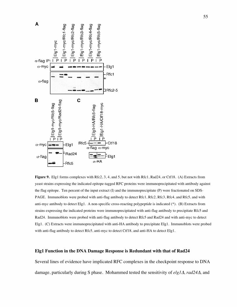
55
Figure 9. Elg1 forms complexes with Rfc2, 3, 4, and 5, but not with Rfc1, Rad24, or Ctf18. (A) Extracts from
yeast strains expressing the indicated epitope-tagged RFC proteins were immunoprecipitated with antibody against
the flag epitope. Ten percent of the input extract (I) and the immunoprecipitate (P) were fractionated on SDS-
PAGE. Immunoblots were probed with anti-flag antibody to detect Rfc1, Rfc2, Rfc3, Rfc4, and Rfc5, and with
anti-myc antibody to detect Elg1. A non-specific cross-reacting polypeptide is indicated (*). (B) Extracts from
strains expressing the indicated proteins were immunoprecipitated with anti-flag antibody to precipitate Rfc5 and
Rad24. Immunoblots were probed with anti-flag antibody to detect Rfc5 and Rad24 and with anti-myc to detect
Elg1. (C) Extracts were immunoprecipitated with anti-HA antibody to precipitate Elg1. Immunoblots were probed
with anti-flag antibody to detect Rfc5, anti-myc to detect Ctf18, and anti-HA to detect Elg1.
Elg1 Function in the DNA Damage Response is Redundant with that of Rad24
Several lines of evidence have implicated RFC complexes in the checkpoint response to DNA
damage, particularly during S phase. Mohammed tested the sensitivity of elg1∆, rad24∆, and

56
ctf18∆ mutants, as well as all pairwise double mutants, and the triple mutant, to the DNA
alkylating agent MMS, the replication inhibitor HU, and UV radiation (Figure 10A). In
addition, Mohammed measured mutant cell viability following exposure to 0.035% MMS in a 4
hour time course (Figure 10B). First, Mohammed found that the elg1∆ mutant was only mildly
sensitive to MMS, and was resistant to HU and UV. This contrasts with rad24∆, which was
significantly sensitive to all three agents, and with ctf18∆, which displayed sensitivity to MMS
and to HU, but was only modestly UV sensitive. These different sensitivities distinguish the
Rfc1 homologues, and indicate that Elg1 is not a central player in DNA damage repair.
Mohammed next examined the sensitivity of each possible double mutant combination
and the triple mutant. Both double mutants with rad24∆ were significantly more sensitive to all
agents than any of the single mutants, whereas the rad24∆ ctf18∆ elg1∆ triple mutant conferred
the greatest sensitivity to MMS. These findings indicate that the Rfc1 homologues are in
partially redundant pathways for DNA damage resistance.

57
Figure 10. Elg1 is required for the DNA damage response. (A) Ten-fold serial dilutions of cultures of the
indicated mutants were spotted on YPD, YPD containing 0.01% (v/v) MMS, 0.035% (v/v) MMS, 50 mM HU, or
on YPD that was subsequently exposed to 100 J/m2 UV. Plates were incubated at 30°C for 2-3 days. (B)
Logarithmically-growing cultures of the indicated mutants were incubated in YPD containing 0.035% (v/v) MMS
at 30°C. At the indicated times samples were withdrawn and plated on media lacking MMS to determine the
number of viable cells. Percent of viable cells relative to the number of viable cells at t=0 is shown. Plots represent
the average of three experiments and error bars span one standard deviation.
Elg1 Contributes to Rad53 Activation
Rad24 has a well-documented role in activation of the checkpoint kinase Rad53 (Naiki et al.,
2000; Pellicioli et al., 1999; Shimomura et al., 1998). Activation of Rad53 can be readily
assessed by immunoblot detection of a phosphorylation-dependent shift in Rad53 mobility
(Pellicioli et al., 1999). I assayed Rad53 activation in HU-arrested cells to assess S-M

58
checkpoint function in elg1∆ cells (Figure 11). ELG1 was not required for Rad53 activation
following HU arrest, as I observed wild-type levels of Rad53 phosphorylation in the elg1∆
mutant (Figure 11A). The ctf18∆ rad24∆ mutant displayed a clear defect in Rad53 activation,
as previously reported (Naiki et al., 2001) and consistent with the increased HU sensitivity of
this double mutant relative to the ctf18∆ and rad24∆ single mutants (Figure 10). The elg1∆
rad24∆ and elg1∆ ctf18∆ double mutants displayed slight defects in Rad53 activation, as
evidenced by the smaller fraction of Rad53 present in the most slowly-migrating form when
compared with wild-type (Figure 11A). As with ctf18∆ rad24∆, this might account for the
increased HU sensitivity of the double mutants compared to the single mutants (Figure 10). In
the elg1∆ rad24∆ ctf18∆ triple mutant phosphorylation of Rad53 was almost completely absent
(Figure 11A). I conclude that ELG1 contributes to the S-M checkpoint in the context of a rad24
mutation. The near absence of Rad53 phosphorylation in the triple mutant indicates that all of
the Rfc1 homologues can contribute to Rad53 activation in S phase.
Mohammed next treated cells with MMS to assess the role of Elg1 in the intra-S phase
checkpoint (Figure 11B). Again, elg1∆ cells displayed no defect in Rad53 activation.
However, when combined with rad24∆, both elg1∆ and ctf18∆ have significant Rad53
activation defects, consistent with roles in intra-S checkpoint response. It is worth noting,
however, that the elg1∆ ctf18∆ double mutant had little, if any, Rad53 activation defect,
indicating that Rad24 plays the more important role in the intra-S checkpoint.
Since we had hypothesized a role for Elg1 in replication fork integrity, Mohammed
assessed intra-S phase checkpoint function directly. Cells in asynchronous culture were treated
with MMS, and their accumulation in S phase was monitored by flow cytometry (Figure 11C).
Wild-type cells, with an intact intra-S phase checkpoint, accumulate in S phase by 2 hours, and
remain blocked with an intermediate DNA content for the 4 hour duration of the experiment.

59
This S phase accumulation is due to checkpoint-independent slowing of DNA replication fork
progression combined with a checkpoint-dependent inhibition of dormant and late origin firing
(Tercero and Diffley, 2001). In the rad53 mutant the checkpoint-dependent inhibition of origin
firing is abrogated and cells appeared to move through S phase more rapidly, accumulating with
a 2C DNA content by 3 hours. In the rad24∆ mutant, which is partially defective in the intra-S
checkpoint (Paulovich et al., 1997), cells moved through S phase more rapidly than wild-type
but not as rapidly as rad53 mutant cells. The elg1∆ resembled wild-type, and deleting elg1 did
not enhance the intra-S checkpoint defect in rad24∆. By contrast, the ctf18∆ rad24∆ mutant
was more defective in the intra-S checkpoint, accumulating with 2C DNA content with kinetics
similar to that observed with the rad53 mutant.

60
Figure 11. Rad53 activation defects in elg1∆. (A) S-M checkpoint. Logarithmically growing cultures were
arrested in G1 with α-factor and released into media containing 200 mM HU. At the indicated times (in hours)
samples were fixed and extracts fractionated on SDS-PAGE. Following transfer the immunoblot was probed with
anti-Rad53 antibody. Phosphorylation of Rad53 causes a shift in electrophoretic mobility (Rad53-P) and is a
marker for checkpoint activation. (B) Intra-S phase checkpoint. Logarithmically growing cultures were treated
with 0.035% (v/v) MMS. At the indicated times (in hours) samples were withdrawn and Rad53 activation was
analysed by immunoblotting. (C) Cell cycle progression in the presence of MMS was assessed by flow cytometry.
Logarithmically growing cultures were treated with 0.035% (v/v) MMS. At the indicated times samples were fixed
and the DNA contents of cells in each sample were analysed by flow cytometry. The positions of cells with 1C and
2C DNA contents are indicated on the histograms.

61
Cells Lacking Elg1 Require Homologous Recombination and Replication Fork Re-start
Pathways for Optimum Growth
To gain further insight into Elg1 function in vivo Mohammed and I performed a genome-wide
synthetic lethal screen with elg1∆. The synthetic genetic interactions confirmed by tetrad
analysis are presented in Figure 12 and in Table 5. Several functional clusters of genes are
readily apparent. First, elg1∆ interacts with members of the RAD52 epistasis group (rad51∆,
rad52∆, rad54∆, rad55∆, rad57∆), which are required for homologous recombination. The
genes rad50∆, mre11∆, and xrs2∆ were also identified. These interactions likely reflect the role
of these genes in homologous recombination rather than in non-homologous end-joining, as
elg1∆ is not synthetic lethal or sick with dnl4∆ or yku80 (data not shown). Of particular
significance, genes thought to be in redundant pathways for re-starting stalled replication forks
(Kaliraman et al., 2001; Mullen et al., 2001) have synthetic genetic interactions with elg1∆.
mus81∆, mms4∆, sgs1∆, top3∆, slx5∆, and slx8∆ all displayed a fitness defect in combination
with elg1∆. The last functional group of note comprises rad24∆, rad17∆, mec3∆, ddc1∆,
mrc1∆, and tof1∆. These genes are all linked to defects in S phase checkpoints (Alcasabas et
al., 2001; Foss, 2001; Osborn et al., 2002; Tanaka and Russell, 2001), and their identification in
the screen is consistent with lack of Elg1 causing DNA lesions or arrested replication forks
during S phase. Our comprehensive genetic analysis indicates that cells lacking Elg1 and
homologous recombination, replication fork re-start, or S phase checkpoint pathways have a
significant fitness defect, indicating that these pathways are required for optimal growth when
ELG1 is deleted. Finally, the functional classes of genes identified in the elg1 synthetic genetic
screen bear a striking similarity to those identified in screens with rad27∆ (Tong et al., 2001).
Therefore, the genetic data suggest that Elg1 performs a function that is similar to that of Rad27,

62
which plays an important role in Okazaki fragment maturation (Merrill and Holm, 1998;
Parenteau and Wellinger, 1999).
Figure 12. Genome-wide synthetic genetic screens with elg1∆ identify homologous recombination, fork re-start,
and S phase checkpoint pathways. The results of synthetic genetic array analysis with elg1∆ presented as a genetic
interaction map. Lines connecting genes represent synthetic lethality or synthetic slow growth. Colored circles
designate the cellular role of the interacting genes, as determined by perusal of relevant literature.
Table 5. elg1∆ genetic interactions.
Gene Growth defect1 Cellular roleRAD51 ++ Homologous recombinationRAD52 ++ Homologous recombinationRAD54 ++ Homologous recombinationRAD55 ++ Homologous recombinationRAD57 ++ Homologous recombinationRAD50 +++ NHEJ, HR, checkpointXRS2 +++ NHEJ, HR, checkpointMRE11 lethal NHEJ, HR, checkpointPOL32 +++ DNA repairRAD27 + DNA repair, DNA replicationHPR5/SRS2 + DNA repair, NHEJ, Checkpoint?, Fork re-start?MUS81 +++ Fork re-startMMS4 ++ Fork re-start

63
YBR099C +++ Fork re-startSGS1 ++ Fork re-startTOP3 + Fork re-startYLR235C + Fork re-startSLX5 + Fork re-startSLX8 + Fork re-startRAD24 + CheckpointRAD17 + CheckpointMEC3 + CheckpointDDC1 + CheckpointMRC1 ++ CheckpointTOF1 + CheckpointCTF4 +++ Sister chromatid cohesionCHL1 ++ Chromosome segregationBIM1 + Microtubule dynamicsKAR3 ++ Microtubule dynamicsMID1 + Small molecule transportYOR1 + Small molecule transportYBR094W + UnknownRTT109 + UnknownBRE1 + UnknownBRE5 + UnknownSWR1 + Unknown1Degree of synthetic fitness defect: ‘+++’=severe, ‘++’=moderate, ‘+’=mild.
Elg1 is Required for Replication Fidelity
Mutants defective in Okazaki fragment maturation, such as rfc1-1, pol30-52, and rad27∆ all
have increased forward mutation rates, but differ in the spectrum of mutants produced (Xie et
al., 1999; Xie et al., 2001). Whereas rfc1-1 and pol30-52 cause predominantly point mutations
and small insertions or deletions, rad27∆ causes large rearrangements. Grant assessed the
forward mutation rate to canavanine resistance of elg1∆, and found that it was almost 8-fold
higher than the wild-type strain (24.2 x 10⁻⁷ versus 3.24 x 10⁻⁷), indicating that Elg1 is important
for replication fidelity. To determine the spectrum of mutations caused by elg1∆ Grant
examined the CAN1 gene from 20 independent canr mutants from both wild-type and elg1∆ for
the presence of large insertion or deletion mutations. A difference in the size of a CAN1-derived
fragment was observed in 2 out of 20 canr wild-type strains, and in only 1 out of 20 canr elg1∆
strains (data not shown). DNA sequencing revealed that the mutations in the elg1∆ strains were

64
predominantly base substitution mutations, with some small (< 5 bp) deletions and insertions.
Therefore the elg1∆ mutation spectrum resembles that caused by defects in Rfc1 or PCNA.
elg1∆ Mutants are Defective in S Phase Progression
If elg1∆ cells are defective in fork re-start or have increased levels of fork stalling we would
expect to detect defects in S phase progression. Mohammed arrested cells in G1 phase and
released them synchronously into the cell cycle. Progression through S phase was measured by
flow cytometry. As shown in Figure 13A, the wild-type culture completed S phase by 60
minutes after release, as evidenced by the accumulation of cells with a 2C DNA content. In the
elg1∆ culture, however, a significant fraction of cells still had a 1C or intermediate (<2C) DNA
content at 60 minutes. Even at 80 minutes post-release some elg1∆ cells had not completed S
phase. Consistent with a role for Elg1 in S phase progression, I found that elg1∆ has a
probability of plasmid loss that is 7.6 times higher than wild-type in a minichromosome
maintenance assay (Figure 13B). This elevated plasmid loss was not suppressed significantly
by the presence of additional origins of replication and 2D gel analysis did not reveal any
initiation defects in elg1∆ (data not shown). Furthermore, Mohammed found that elg1∆ did not
have any detectable defect in sister chromatid cohesion (data not shown), suggesting that
plasmid loss in elg1∆ was not due to a segregation defect.
Since Elg1-RFC most likely functions as a clamp-loader, Mohammed tested whether
overexpression of PCNA could suppress the elg1∆ phenotype. As shown in Figure 13C
overexpression of PCNA rescued the MMS sensitivity of elg1∆, as evidenced by improved
growth on MMS plates when PCNA overexpression was induced by the presence of galactose.
Taken together these results suggest that Elg1 plays a direct role in DNA replication, most likely
during the elongation phase of DNA synthesis.

65
Figure 13. elg1∆ mutants display DNA replication defects. (A) Progression through S phase. Wild-type or elg1∆
cells were arrested in G1 (t=0) and released synchronously into the cell cycle. Samples were removed at the
indicated times and analysed by flow cytometry. The shaded histograms represent the cell cycle distribution of the
asynchronous cultures before the G1 arrest. Overlaid histograms represent the cell cycle distribution at the
indicated times after release from the G1 arrest. The positions of cells with 1C and 2C DNA contents are indicated.
(B) Plasmid loss in wild-type, elg1∆, and ctf19∆. The probability of plasmid loss per generation is plotted, and
error bars span one standard deviation. (C) Suppression of elg1∆ MMS sensitivity by PCNA overexpression.
Serial dilutions of wild type or elg1∆ cells carrying empty vector (v) or GAL1-POL30 plasmid (POL30) were
plated on synthetic media with 2% glucose (Glu; uninduced) or 2% galactose + 2% raffinose (Gal; induced), plus or
minus 0.01% MMS.

66
Abnormal Recovery from Replication Fork Stalling in elg1∆
Mohammed measured S phase progression in medium containing 0.035% MMS, a
concentration of MMS known to cause extensive fork stalling in wild-type cells (Tercero and
Diffley, 2001) (Figure 14A). In the presence of MMS S phase was prolonged in the wild-type
cells, which take 120 minutes to accumulate with 2C DNA content. The elg1∆ mutant was
clearly defective in S phase progression in the presence of MMS, with a significant fraction of
cells containing less than 2C DNA at 160 minutes post-release. Thus the introduction of fork
stalls causes further defects in DNA synthesis in elg1 mutants, consistent with Elg1 functioning
in preventing fork stalling or in re-starting stalled replication forks.
If Elg1 is required for replication fork integrity we expected to see prolonged activation
of Rad53 following MMS treatment during S phase. Wild-type and elg1∆ cells were arrested in
G1 and released into medium containing MMS for 1 hour to activate Rad53. The MMS was
then washed out and cells were allowed to recover. Rad53 activation during recovery from
MMS damage was assayed by immunoblot analysis (Figure 14B). In the wild-type strain Rad53
is dephosphorylated by 80 minutes following removal of MMS. By contrast, activated Rad53
persisted in the elg1∆ strain for at least 120 minutes, indicating that Elg1 is required for down-
regulation of the intra-S checkpoint response. Since the elg1∆ strain is at least 10-fold less
sensitive to MMS than mutants in MMS repair pathways (data not shown), these results suggest
that Elg1 is required for efficient fork re-start rather than direct repair of DNA lesions.

67
Figure 14. elg1∆ mutants are defective in recovery from MMS-induced replication fork stalling. (A) S phase
progression in the presence of MMS. Wild-type or elg1∆ cells were arrested in G1 (t=0) and released
synchronously into media containing 0.035% (v/v) MMS. Samples were removed at the indicated times and
analysed by flow cytometry. The shaded histograms represent the cell cycle distribution of the asynchronous
cultures before the G1 arrest. Overlaid histograms represent the cell cycle distribution at the indicated times after
release from the G1 arrest. (B) Checkpoint activation of Rad53 during recovery from MMS damage. Cells were
arrested in G1, released into MMS for 1 hour, and then transferred to medium lacking MMS (t=0). At the indicated
times samples were withdrawn and Rad53 activation was analysed by immunoblotting.
4.4 DISCUSSION
A Novel RFC-like Complex
Using a functional genomics approach we have identified Elg1 as a novel DNA replication
protein. Elg1 associates with Rfc2, Rfc3, Rfc4, and Rfc5, forming a fourth eukaryotic RFC-like
complex that is functionally distinct from the canonical RFC, and from Rad24-RFC and Ctf18-

68
RFC. Whether Elg1-RFC functions as a pentameric complex, like RFC and Rad24-RFC (Green
et al., 2000; Mossi and Hubscher, 1998), or requires accessory factors as found in Ctf18-RFC
(Mayer et al., 2001), awaits purification of Elg1-RFC. Eukaryotic cells have at least two sliding
clamps that are loaded by RFC-like enzymes, PCNA and the 9-1-1 complex. RFC loads PCNA
during leading and lagging strand DNA synthesis (reviewed in (Mossi and Hubscher, 1998)),
whereas Rad24-RFC loads the 9-1-1 clamp in vitro but cannot load PCNA (Bermudez et al.,
2003; Majka and Burgers, 2003). Ctf18-RFC binds to PCNA in vitro and in vivo (Ohta et al.,
2002), and so may function as a PCNA clamp loader under specialized circumstances. Our data
suggest a role for Elg1-RFC in DNA replication, and we therefore propose that it functions to
load or unload PCNA. In support of this Mohammed finds that overexpression of PCNA
suppresses the MMS sensitivity of elg1∆. Furthermore elg1∆ is synthetic sick when combined
with rad17∆, mec3∆, and ddc1∆, which encode the 9-1-1 clamp, but has no genetic interaction
with pol30-1, a mutant in the gene encoding PCNA (data not shown), suggesting that Elg1
functions in the same pathway as PCNA, but in a pathway that is parallel to that in which the 9-
1-1 clamp functions.
Elg1-RFC and Checkpoint Activation
A number of RFC-family proteins have been implicated in checkpoint activation. Rad24 is
required for the G1 and G2 DNA damage checkpoints (Siede et al., 1994; Weinert et al., 1994),
and is important (although not essential) for the intra-S damage checkpoint (Paulovich et al.,
1997; Pellicioli et al., 1999). A role for Rad24 in the S-M checkpoint is revealed in the context
of mutations in other RFC-family genes (Naiki et al., 2001; Shimomura et al., 1998). Similarly,
deletion of ctf18 has little effect on checkpoint activation unless rad24 is also deleted (Naiki et
al., 2001). Consistent with formation of Rad24-Rfc2-5 complexes (Green et al., 2000) mutants

69
in rfc2, rfc4, and rfc5 with checkpoint defects have also been described (Kim and Brill, 2001;
Noskov et al., 1998; Sugimoto et al., 1997; Sugimoto et al., 1996). Our data indicate that Elg1
does not play a primary role in checkpoint activation, as elg1∆ mutants show normal Rad53
activation in response to DNA damage in S phase and in response to replication fork arrest by
HU. Double mutants in rad24 and either elg1 or ctf18 were more defective in Rad53 activation
in S phase, and the triple mutant lacked detectable Rad53 activation. Thus it appears that Ctf18,
and to a lesser extent Elg1, can partially substitute for the primary role of Rad24 in Rad53
activation in S phase. Alternatively, Elg1 could contribute to Rad53 activation in an indirect
manner, for example through its effects on DNA replication.
When assessed directly, elg1∆ mutants display an intact intra-S phase checkpoint
response as these cells exhibit the slow progression through S phase in the presence of MMS
that is seen in wild-type cells. Mohammed did note, however, that elg1∆ rad24∆ double
mutants do not display an intra-S phase checkpoint defect that is greater than that seen with
rad24∆ alone. This was unexpected, as elg1∆ rad24∆ was more defective in Rad53 activation
than rad24∆. By contrast, in rad24∆ ctf18∆ S phase progression in the presence of MMS was
more rapid than wild-type or rad24∆, and resembled the progression seen in the completely
checkpoint-defective mec2-1 strain. Thus deletion of ctf18 in a rad24∆ strain exacerbates the
rad24∆ checkpoint defect, whereas deletion of elg1∆ does not. The implications of these data
are two-fold. First, the slow progression through S phase in elg1∆ is at least partially
checkpoint-independent, which is consistent with the elg1∆ mutant having a defect in DNA
replication fork integrity or re-start. Secondly, there is not a strict correlation between Rad53
activation and S phase progression in the presence of MMS in these mutants. This likely reflects
that the slow progression through S phase that is the hallmark of the intra-S phase checkpoint
(Paulovich and Hartwell, 1995; Paulovich et al., 1997) has both a checkpoint-dependent and a

70
checkpoint-independent component (Tercero and Diffley, 2001). Mohammed’s data suggest
that Elg1 functions in the checkpoint-independent component of the slow S phase progression,
again consistent with a role for Elg1 in preventing replication fork stalling or in re-starting
stalled forks.
Elg1-RFC and Genome Integrity
The phenotypes of elg1∆ indicate that Elg1 has an important role in maintaining genome
integrity. Elg1 suppresses recombination, protects against mutation, and is important for
recovery from DNA damage. Several aspects of the elg1∆ phenotype point to the presence of
double-strand DNA breaks, including the importance of homologous recombination for growth
of the elg1∆ mutant and the hyper-recombination phenotype. Other aspects of the phenotype,
while consistent with the presence of double-strand breaks, point to a more fundamental role for
Elg1 in DNA replication. These include the genetic requirement for the SGS1/TOP3 and
MUS81/MMS4 replication fork re-start pathways (Doe et al., 2002; Fabre et al., 2002;
Kaliraman et al., 2001) in the elg1∆ mutant cells, the slow S phase progression observed in both
the presence and absence of DNA damage, the increase in plasmid loss, the increase in mutation
rate, and the failure to recover efficiently from increased replication stalling induced by MMS.
We propose that deletion of ELG1 causes a decrease in replication fork processivity (an increase
in fork stalling or a defect in re-start of stalled forks) perhaps due to defective Okazaki fragment
maturation. Precedent for such a model comes from studies in both bacteria and yeast which
have indicated that mutants in Okazaki fragment processing cause DSBs, stimulate
recombination, confer DNA damage sensitivity, cause an increase in mutation frequency, and
render homologous recombination essential for viability (Michel et al., 2001; Rothstein et al.,
2000), phenotypes similar to those observed in elg1∆. Furthermore, defects in Okazaki

71
fragment synthesis can cause replication fork stalling (Flores et al., 2001), and there is a strong
correlation between replication stalling and genome instability (Aguilera et al., 2000; Michel,
2000; Michel et al., 2001; Rothstein et al., 2000).
Given the similarity of Elg1 to Rfc1 and its presence in RFC-like complexes, it is likely
that Elg1-RFC functions as a clamp loader or unloader. Biochemical experiments in vitro
indicate that a complex interplay between RFC, PCNA, and DNA polymerase δ governs
appropriate polymerase switching and Okazaki fragment sealing during lagging strand DNA
synthesis. Although it is clear that RFC is responsible for limiting the length of the nascent
strand synthesized by pol α, and for loading of PCNA to initiate the switch to synthesis by pol
δ (Ayyagari et al., 2003; Maga et al., 2001), it is not clear how PCNA is recycled on the lagging
strand. Eukaryotic cells in S phase have a great excess of Okazaki fragments over molecules of
PCNA (Mossi and Hubscher, 1998), and the stability of PCNA-DNA complexes suggests that
an unloading activity is required to recycle PCNA (Yao et al., 1996). A role for Elg1 in
unloading and recycling PCNA on the lagging strand fits well with the available data.
Additionally, this model predicts that PCNA should be limiting in elg1∆ mutants, and indeed
Mohammed found that PCNA overexpression suppresses the MMS sensitivity of elg1∆. Since
Elg1 is not essential its role in this process must be redundant, perhaps with RFC. Alternatively,
Elg1 could be important for loading of PCNA during re-start of stalled replication forks,
although in this model there must be a significant amount of fork stalling in an unperturbed S
phase to account for the increased plasmid loss and the slow S phase progression observed in
elg1∆. It will be of great interest to determine if the human homologue of Elg1 plays a similar
role in maintaining genome integrity.

72
5. RMI1/NCE4, A SUPPRESSOR OF GENOME INSTABILITY, ENCODES A
MEMBER OF THE RecQ HELICASE/TopoIII COMPLEX
Published: Chang, M, Bellaoui, M, Zhang, C., Desai, R., Morozov, P., Delgado-Cruzata, L.,
Rothstein, R., Freyer, G.A., Boone, C., Brown, G.W. (2005). RMI1/NCE4, a Suppressor of
Genome Instability, Encodes a Member of the RecQ Helicase/TopoIII Complex. EMBO J. 24:
2024-2033.
Data attribution: I performed the large majority of the experimental work presented in this
chapter. Dr. Mohammed Bellaoui performed the viability assays in HU and MMS. Dr.
Chaoying Zhang performed the gross chromosomal rearrangement assays. Ridhdhi Desai
performed the recombination assays. Dr. Pavel Morozov at the Columbia Genome Center
helped identify the human and mouse homologues of Rmi1. Lissette Delgado-Cruzata, a
student in Dr. Greg Freyer’s laboratory at Columbia University, performed the S. pombe
experiments. Dr. Rodney Rothstein, also at Columbia University, provided me with use of his
fluorescent microscope to do the Rad52-YFP localization experiments. Dr. Boone provided the
SGA technology and expertise. Dr. Grant Brown performed the gel filtration experiment and in
situ kinase assay.

73
5.1 SUMMARY
SGS1 encodes a DNA helicase whose homologues in human cells include the BLM, WRN and
RECQ4 genes, mutations in which lead to cancer-predisposition syndromes. Clustering of
synthetic genetic interactions identified by large-scale genetic network analysis revealed that the
genetic interaction profile of the gene RMI1 (RecQ Mediated Genome Instability, also known as
NCE4 and YPL024W) was highly similar to those of SGS1 and TOP3, suggesting a functional
relationship between Rmi1 and the Sgs1/Top3 complex. I show that Rmi1 physically interacts
with Sgs1 and Top3 and is a third member of this complex. Cells lacking RMI1 activate the
Rad53 checkpoint kinase, undergo a mitotic delay, and display increased relocalization of the
recombination repair protein Rad52, indicating the presence of spontaneous DNA damage.
Consistent with a role for RMI1 in maintaining genome integrity, rmi1∆ cells exhibit increased
recombination frequency and increased frequency of gross chromosomal rearrangements. In
addition, rmi1∆ strains fail to fully activate Rad53 upon exposure to DNA damaging agents
suggesting that Rmi1 is also an important part of the Rad53-dependent DNA damage response.
5.2 INTRODUCTION
As described in Chapter 1, Saccharomyces cerevisiae SGS1 is a member of the recQ family of
3’-5’ DNA helicases, which includes five human homologues (RECQL, BLM, WRN, RECQ4,
and RECQ5) (Watt et al., 1996; Watt et al., 1995). Loss of function mutations in BLM, WRN,
and RECQ4 give rise to Bloom syndrome (BS), Werner syndrome (WS), and Rothmund-
Thomson syndrome (RTS), respectively (Ellis et al., 1995; Kitao et al., 1999; Yu et al., 1996).
Though the spectrum of clinical features of each disease differs, they all result in a
predisposition to cancer. The major defects of cells with mutated recQ helicases are hyper-

74
recombination and genomic instability (Hickson, 2003). S. cerevisiae sgs1 mutants show
elevated levels of mitotic homologous recombination, illegitimate recombination (Gangloff et
al., 1994; Watt et al., 1996; Yamagata et al., 1998), sister chromatid exchanges (Onoda et al.,
2000), and gross chromosomal rearrangements (GCRs) (Myung et al., 2001b; Myung and
Kolodner, 2002). Cells lacking SGS1 are also moderately sensitive to genotoxic agents such as
methyl methanesulfonate (MMS) and hydroxyurea (HU) (Chang et al., 2002; Gangloff et al.,
1994; Watt et al., 1996; Yamagata et al., 1998).
A subset of RecQ family members physically interact with topoisomerase III (Top3)
homologues (Gangloff et al., 1994; Goodwin et al., 1999; Johnson et al., 2000; Wu et al., 2000).
E. coli RecQ stimulates Top3 to catenate and decatenate covalently closed duplex DNA
(Harmon et al., 1999) and BLM is able to stimulate the DNA strand passage activity of Top3α
(Oakley and Hickson, 2002). Furthermore, BLM and Top3α can work together to resolve a
recombination intermediate containing a double Holliday junction (Wu and Hickson, 2003). S.
cerevisiae strains lacking TOP3 exhibit a severe growth defect, sensitivity to DNA damaging
agents, and hyper-recombination (Chang et al., 2002; Gangloff et al., 1994; Wallis et al., 1989).
Most of the defects exhibited by top3 mutants can be suppressed by deletion of SGS1
(Chakraverty et al., 2001; Gangloff et al., 1994), a relationship that is conserved in
Schizosaccharomyces pombe where mutations in the recQ homologue rqh1+ can suppress the
lethality of top3∆ mutants (Maftahi et al., 1999). These data support models in which RecQ
helicase action produces a toxic DNA structure that is resolved by Top3 (Gangloff et al., 1994;
Ira et al., 2003; Wu and Hickson, 2003).
Two-dimensional hierarchical clustering of synthetic genetic interactions determined by
large-scale genetic network analysis in S. cerevisiae has proven useful for identifying genes
whose products function within the same pathway or complex (Tong et al., 2004). Such

75
clustering analysis revealed that the genetic interaction profile of the poorly characterized gene
RMI1 was highly similar to those of SGS1 and TOP3. I show that Rmi1 associates with Sgs1
and Top3 and that strains lacking RMI1 accumulate DNA damage in the absence of exogenous
genotoxic agents. My results indicate that the actions of Sgs1, Top3, and Rmi1 are required in
concert in order to maintain genome integrity.
5.3 RESULTS
Mutations in SGS1 can suppress the growth defects of an rmi1∆ mutant
Two-dimensional hierarchical clustering of large-scale synthetic genetic array (SGA) data
revealed that the set of genes that genetically interact with the uncharacterized gene RMI1 was
highly similar to that associated with SGS1, TOP3, and YLR235C (which overlaps the TOP3
open reading frame such that a deletion of this ORF likely results in a TOP3 hypomorph) (Tong
et al. 2004; Figure 15). Synthetic genetic interactions are usually orthogonal to protein-protein
interactions, but the products of genes with similar patterns of genetic interactions are often
found in the same cellular pathway or protein complex (Tong et al., 2004), suggesting that RMI1
might function in the SGS1/TOP3 pathway.

76
Figure 15. Two-dimensional hierarchical clustering of synthetic genetic interactions determined by SGA analysis
(Tong et al., 2004). (A) Synthetic genetic interactions are represented as red lines. Rows, 132 query genes;
columns, 1007 array genes. The cluster trees organize query (y-axis) and array genes (x-axis) that show similar
patterns of genetic interactions. (B) The relevant section (yellow outline in A) is expanded to allow visualization of
the RMI1/SGS1/TOP3 array gene cluster. Synthetic genetic interactions are represented as red squares.
Crossing the rmi1∆ strain from the Saccharomyces gene deletion collection with a wild
type strain revealed the presence of an extragenic suppressor in the rmi1∆ strain. Tetrads from
this cross were dissected to analyze the products of individual meioses (Figure 16A). The
resultant colonies were screened to identify those carrying the rmi1∆ mutation. I found that
roughly half (10 of 24) of the rmi1∆ isolates exhibited a slow growth phenotype, whereas the
other half (14 of 24) grew relatively normally, indicating that the original strain did indeed carry

77
a single extragenic suppressor mutation. To identify the suppressor (supX), I employed
synthetic genetic array mapping (SGAM) methodology, in which an rmi1∆ supX query strain
was crossed to an array of ~4600 viable gene deletion mutants (see Materials and Methods for
details). This method maps a group of genes that are tightly linked to the suppressor (Jorgensen
et al., 2002). Indeed, I identified a group of linked genes on chromosome XIII (Figure 16B),
indicating that the suppressor was in this region. The SGS1 gene was located in the middle of
this linkage group, and the sgs1∆ strain was not identified in the SGAM experiment, suggesting
that the suppressor might be a loss of function allele of SGS1. I crossed the rmi1∆ strain lacking
the suppressor with an sgs1∆ strain and found that the double mutants had a normal growth
phenotype (Figure 16C). The SGS1 allele was also sequenced from the rmi1∆ supX strain and
found that it carried a frame-shift mutation 691 nucleotides into the ORF and so encoded a
truncated protein lacking the helicase catalytic domain of Sgs1. Therefore deletion of RMI1
causes a slow growth phenotype that can be suppressed by deletion of SGS1. This is
reminiscent of the TOP3 gene, deletion of which causes slow growth that is suppressed by
mutation of SGS1 (Gangloff et al., 1994; Wallis et al., 1989).

78
Figure 16. rmi1∆ mutants exhibit a growth defect that can be suppressed by mutation of SGS1. (A) The
rmi1∆::kanMX6 strain was backcrossed to a WT strain (BY4741). The resulting diploids were sporulated and
tetrads were dissected on YPD. Each column represents the four spores from a single tetrad. The genotypes of the
resulting colonies are indicated with circles () for rmi1∆::kanMX6. (B) SGAM analysis using an rmi1∆::natMX6
query strain (which contains supX) revealed a set of colinear synthetic genetic interactions on chromosome XIII
(see Materials and Methods for details). A red bar indicates that deletion of the corresponding gene resulted in a
genetic interaction. Black bars represent essential genes, which are not a part of the gene deletion collection. Grey
bars indicate ORFs for which no deletion mutant was made as part of the Saccharomyces Gene Deletion Project
(Winzeler et al., 1999) and genes that are often found in control screens using a WT query strain, and therefore are
filtered from the results of SGA analyses. (C) An rmi1∆::natMX6 strain lacking supX was crossed to an
sgs1∆::kanMX6 strain. The resulting diploids were sporulated for tetrad analysis as in A. The genotypes of the
resulting colonies are indicated with boxes () for rmi1∆::natMX6 and circles () for sgs1∆::kanMX6.
rmi1∆ synthetic genetic interactions
Since the rmi1∆ mutant readily accumulates mutations in SGS1, I was unable to conduct an
RMI1 SGA analysis. Instead I adopted a candidate approach, analyzing genes with connections
to SGS1 and TOP3 function (Klein, 2001; Mullen et al., 2001; Tong et al., 2001; Tong et al.,

79
2004) (Table 6). I found that rmi1∆ is synthetic lethal when combined with mutations in genes
thought to play roles in restarting stalled replication forks: rrm3∆, mus81∆, mms4∆, slx1∆,
slx4∆, hex3∆, slx8∆, and hpr5∆. I also found that the slow growth phenotype of rmi1∆ was
suppressed by rad51∆, rad52∆, and rad54∆. This is consistent with models in which the
presence of the homologous recombination pathway facilitates creation of DNA processing
intermediates by Sgs1, which are toxic when Rmi1 is absent. Similar models have been
proposed to account for the suppression of top3∆ phenotypes by mutations in recombination
repair genes (Oakley et al., 2002; Shor et al., 2002). I also found that rmi1∆ did not display a
detectable genetic interaction with top3∆, consistent with RMI1 and TOP3 functioning in the
same pathway. Finally, I found that homozygous rmi1∆/rmi1∆ diploids are defective in
undergoing meiosis to produce four spore asci (Figure 17), indicating that like Sgs1 and Top3
(Gangloff et al., 1999; Watt et al., 1995), Rmi1 is essential for proper meiotic cell division.
Table 6. rmi1∆ genetic interactions.
Gene Interaction Proposed Functionsgs1∆ suppression RecQ helicasetop3∆ none Type I topoisomerase
rad53-11 lethality DNA damage checkpointmrc1∆ sickness S-phase DNA damage checkpointcsm3∆ sickness S-phase DNA damage checkpointtof1∆ sickness S-phase DNA damage checkpointrad9∆ none G2 DNA damage checkpointrad24∆ none G2 DNA damage checkpointrrm3∆ lethality DNA helicase; fork re-start
mus81∆ lethality nuclease subunit; fork re-startmms4∆ lethality nuclease subunit; fork re-startslx1∆ lethality nuclease subunit; fork re-startslx4∆ lethality nuclease subunit; fork re-starthex3∆ lethality fork re-startslx8∆ lethality fork re-starthpr5∆ lethality DNA helicase; fork re-startrad51∆ suppression homologous recombinationrad52∆ suppression homologous recombinationrad54∆ suppression homologous recombination

80
Figure 17. rmi1∆/rmi1∆ diploids are sporulation defective. Sporulation efficiency, as determined by the
percentage of cells that had formed four-spore asci, was scored following 5 days in sporulation medium at 30°C.
The value presented for each strain is the average of three trials.
Rmi1 physically interacts with Top3 and Sgs1
Genetic analysis placed RMI1 in the SGS1/TOP3 pathway and indicated in several ways that
rmi1∆ phenocopies top3∆. To gain insight into the mechanism underlying these genetic
observations, I tested whether Rmi1 physically associates with Sgs1 and Top3. Sgs1 and Top3
interact in vivo and in vitro (Bennett et al., 2000; Fricke et al., 2001; Gangloff et al., 1994)
however the apparent molecular mass of Sgs1/Top3 complexes in yeast extracts suggests that
the complexes are not heterodimeric and so may contain other proteins (Fricke et al., 2001). I
used strains containing SGS1, TOP3, and RMI1 epitope-tagged at their respective genomic loci
to perform co-immunoprecipitations. Rmi1 was found in complex with both Sgs1 and Top3
(Figure 18A and 18B). This complex was not disrupted in the presence of DNase I, indicating
that the interactions are not mediated by DNA (Figure 19). When Rmi1-TAP
immunoprecipitations were quantified by densitometry Grant found that 39% of Rmi1 was

81
depleted from the extract compared with 42% of Sgs1 (data not shown), indicating that a
significant fraction of Sgs1 is in complex with Rmi1. Grant next used gel filtration
chromatography to fractionate extract from the tagged strain. He found that Sgs1-HA, Top3-
VSV, and Rmi1-TAP co-elute in a high molecular weight complex (Figure 18C). Monomeric
Rmi1 was not detected. Together these data suggest that Rmi1 is in a heteromeric complex with
both Sgs1 and Top3 and functions as a subunit of the Sgs1/Top3 complex.

82
Figure 18. Rmi1 physically associates with the Sgs1/Top3
complex. (A-B) Extracts from yeast strains expressing the
indicated epitope-tagged proteins were
immunoprecipitated with IgG agarose. Ten
percent of the input extract (E) and the
immunoprecipitate (IP) were fractionated by SDS-
PAGE. Immunoblots were probed with anti-HA
antibody to detect Sgs1, with anti-VSV antibody to
detect Top3, or with peroxidase-anti-peroxidase to
detect Rmi1-TAP. (C) Extract from a yeast strain
expressing Sgs1-HA, Top3-VSV, and Rmi1-TAP
was fractionated on a Superose 6 gel filtration
column. Fractions were precipitated with TCA
and analysed by immunoblotting. The elution
positions of molecular weight standards are
indicated, as is the void volume of the column
(Vo). (D) Extracts from yeast strains expressing
Sgs1-HA and Top3-TAP or Sgs1-HA and Rmi1-
TAP in an rmi1∆ or top3∆ background,
respectively, were immunoprecipitated with IgG-
agarose to precipitate the TAP tagged protein
(lanes marked T) or with unconjugated agarose as
a control (lanes marked C). The precipitates were
immunoblotted and probed with anti-HA
antibodies to detect Sgs1-HA (top panel) or with
peroxidase-anti-peroxidase to detect the TAP
tagged proteins. (E) sgs1∆, sgs1∆ rmi1∆, and
sgs1∆ top3∆ strains were transformed with empty
vector (vector) or low copy plasmids expressing
HA-tagged Sgs1 (Sgs1) or helicase-dead Sgs1
(Sgs1-hd). TCA-fixed extracts were prepared and
fractionated by SGS-PAGE. Immunoblots were
probed with anti-HA antibody to detect Sgs1 or
Sgs1-hd, and with anti-tubulin antibodies as a
loading control.

83
Figure 19. The Rmi1-Sgs1 and Rmi1-Top3 interactions are not mediated by DNA. Extract from a yeast strain
expressing Sgs1-HA, Top3-VSV, and Rmi1-TAP was immunoprecipitated with IgG agarose following incubation
for 15 minutes in the presence (+) or absence (-) of 10U of DNase I and 10 mM MgCl2. Ten percent of the input
extract (E) and the entire immunoprecipitate (IP) were fractionated by SDS-PAGE. Immunoblots were probed with
anti-HA antibody to detect Sgs1 and with anti-VSV antibody to detect Top3.
Immunoprecipitates of Rmi1-TAP from extracts of an sgs1∆ strain contain Top3 (Figure
18B), indicating that the interaction of Rmi1 with Top3 does not require Sgs1. When
attempting reciprocal experiments I found that deletion of either TOP3 or RMI1 caused a
significant reduction in Sgs1 protein abundance (data not shown). Despite the reduced levels of
Sgs1, I detected Sgs1 in both Top3 immunoprecipitates from rmi1∆ cells and in Rmi1
immunoprecipitates from top3∆ cells (Figure 18D). Both wild type and catalytically inactive
helicase-dead mutant Sgs1 were poorly expressed in both rmi1∆ and top3∆ mutants (Figure
18E), indicating that the helicase activity of Sgs1 is not required for the observed reduction in
Sgs1 levels.

84
Figure 20. Bulk DNA synthesis is not affected in rmi1∆ cells. Wild-type or rmi1Δ cells were arrested in G1 (t =
0) and released synchronously into fresh YPD medium. Samples were removed at the indicated times and analyzed
by flow cytometry. The positions of cells with 1C and 2C DNA contents are indicated.
Cells lacking RMI1 display precocious checkpoint activation
RecQ helicases are thought to play a role in normal DNA replication. The human homologues
BLM and WRN are required for S phase progression (Lonn et al., 1990; Poot et al., 1992).
Completion of replication in the rDNA array is severely retarded in sgs1∆ mutants (Kaliraman
and Brill, 2002; Versini et al., 2003), and in vitro replication in Xenopus egg extracts in the
absence of Xblm results in DNA strand breaks (Li et al., 2004). I asked whether Rmi1 was also
required for S phase progression. Using cells released synchronously from a G1 arrest, I could
not detect a significant defect in bulk DNA synthesis, as assessed by flow cytometry (Figure
20). However, asynchronous rmi1∆ cultures exhibited an accumulation of budded cells with
one nucleus suggesting a delay in the late S/G2 phase of the cell cycle (Figure 21A). These
observations are similar to those made with top3∆ strains (Chakraverty et al., 2001; Gangloff et
al., 1994) and may indicate a checkpoint dependent mitotic delay. Grant and I assayed for
activation of the checkpoint kinase Rad53 in these cells, analysing both the phosphorylation

85
dependent mobility shift of Rad53 and Rad53 kinase activity (Figure 21B and 21C). I found
that rmi1∆ mutants displayed a modest mobility shift of Rad53 when released from a G1 arrest
in the absence of any DNA damaging agent (Figure 21B). This mobility shift is due to
phosphorylation and correlates with activation of Rad53 kinase activity (Pellicioli et al., 1999).
Grant measured Rad53 activation directly using an in situ kinase assay. Activation of Rad53 in
rmi1∆ was clearly evident in this assay, even in the sample from the asynchronous culture and
from the G1 arrested culture (Figure 21C). Activation of Rad53 was not evident in wild type
cells in either assay. The precocious Rad53 checkpoint activation is likely the cause of the
mitotic delay observed in rmi1∆, suggesting that DNA damage is arising in cells lacking Rmi1
during an unperturbed cell cycle. Consistent with this interpretation, I found that rmi1∆ is
synthetic lethal with rad53-11 (Figure 21D), a checkpoint defective allele of RAD53 (Weinert et
al., 1994), indicating that an intact checkpoint response is essential for the viability of cells
lacking Rmi1.

86
Figure 21. rmi1∆ mutants exhibit Rad53 checkpoint activation during an unperturbed cell cycle. (A)
Asynchronous cultures of wild type, rmi1∆, top3∆, sgs1∆, rmi1∆ sgs1∆, and top3∆ sgs1∆ were examined
microscopically to determine the % of cells with a bud. (B) Logarithmically growing cultures were arrested in G1
with alpha factor and released into fresh YPD media. At the indicated times, samples were fixed with TCA,
extracts were fractionated on SDS-PAGE, and immunoblotted to detect Rad53. The position of the activated
phosphorylated Rad53 is indicated. (C) Samples prepared as in B were fractionated on SDS-PAGE for in situ
kinase assay of Rad53 (upper panel). A parallel blot was probed with anti-tubulin antibody as a loading control
(lower panel). (D) An rmi1∆::kanMX6 strain was crossed to a rad53-11::URA3 strain. The resulting diploids were
sporulated and tetrads were dissected on YPD. The genotypes of the resulting colonies are indicated with boxes
() for rmi1∆::kanMX6 and with circles () for rad53-11::URA3. Inferred double mutants are indicated with a
box and circle.
I next investigated the requirement for other checkpoint proteins in rmi1∆ mutants. I
found that deletion of the G1 and G2 DNA damage checkpoint genes RAD24 or RAD9 had no

87
detectable effect on the rmi1∆ mutant. However, deletion of the S phase checkpoint genes
MRC1, TOF1, or CSM3 in the rmi1∆ mutant caused a synthetic sick phenotype (Table 6).
Therefore cells lacking RMI1 require the S phase checkpoint response for optimal growth,
suggesting that the DNA damage caused by deletion of RMI1 results from DNA replication
defects.
rmi1∆ mutants exhibit increased levels of Rad52 relocalization and genomic instability
RAD52 is essential for efficient homologous recombination. Rad52 relocalizes from a diffuse
nuclear localization to distinct subnuclear foci in response to DNA damage, particularly double
strand breaks (Lisby et al., 2004; Lisby et al., 2003; Lisby et al., 2001). Rad52 tagged with
yellow fluorescent protein (Lisby et al., 2004; Lisby et al., 2003) was visualized by fluorescence
microscopy in asynchronous mitotic haploid cells. As shown in Figure 22A, cells lacking RMI1
display subnuclear Rad52 foci whereas wild type cells show infrequent and transient foci.
Quantification of the data (Figure 22B) showed that rmi1∆, sgs1∆, and top3∆ all have elevated
levels of spontaneous Rad52 focus formation, indicating the presence of DNA damage requiring
homologous recombination for repair, likely double strand breaks. Elevated levels of Rad52
foci were observed both in S/G2/M (i.e. budded) cells and in G1 cells. Together with the data
indicating that Rad53 is activated in rmi1∆ mutants, these results suggest that DNA replication
in the absence of an intact Sgs1/Top3/Rmi1 pathway causes DNA lesions that result in genomic
instability (Ajima et al., 2002; Gangloff et al., 1994; Myung et al., 2001b), similar to the effect
observed in Xenopus egg extracts, in which replication in the absence of Xblm causes DNA
strand breaks (Li et al., 2004).

88
Figure 22. Spontaneous Rad52 focus formation in rmi1∆ cells. (A) Logarithmically growing cells expressing
Rad52-YFP were visualized by fluorescence microscopy. For each pair of images, the left panel is a DIC image
and the right panel is a fluorescence image showing Rad52-YFP. Representative cells are shown. (B) The
percentage of cells with Rad52 foci was determined for the indicated strains. G1 cells with Rad52 foci are
represented by the gold bars and S/G2/M cells with Rad52 foci are represented by the blue bars.
To assess the effect of the DNA damage that arises in rmi1∆ mutants, two assays for
genomic instability were applied. Both SGS1 and TOP3 are suppressors of homologous
recombination (Shor et al., 2002). Ridhdhi tested the effect of deletion of RMI1 on homologous
recombination using a LEU2 direct repeat assay (Smith and Rothstein, 1999). Consistent with
the observation that cells lacking RMI1 have high levels of Rad52 foci, Ridhdhi found that
rmi1∆ cells have an increased rate of recombination (Figure 23A), approximately 6-fold higher
than wild type. Chaoying measured the rate of gross chromosomal rearrangements (GCRs) in
rmi1∆, using an assay that detects large interstitial deletions, translocations, chromosome
fusions, and loss of a chromosome arm (Myung et al., 2001a). In this assay (Figure 23B), sgs1∆

89
and top3∆ showed increased GCR rates of approximately 30-fold over wild type, similar to
reported values (Myung et al., 2001b). In contrast, GCR rates in rmi1∆ cells were more than
150 times wild type levels. Thus, Rmi1 is a critical suppressor of gross chromosomal
rearrangements.
Figure 23. Deletion of RMI1 causes genomic instability. (A) Recombination rate was measured using a direct
repeat recombination assay. The average and standard deviation of three fluctuation tests are shown for each strain.
(B) Gross chromosomal rearrangement rate was measured. The average and standard deviation of four fluctuation
tests are shown for each strain.
RMI1 is required for the response to DNA damage
Both SGS1 and TOP3 are important for the response to DNA damage (Chakraverty et al., 2001;
Davey et al., 1998; Frei and Gasser, 2000; Stewart et al., 1997), therefore I tested whether
deletion of RMI1 caused sensitivity to DNA damaging agents (Figure 24A, B). The rmi1∆ cells,
like top3∆, displayed slow growth on YPD. The presence of the alkylating agent MMS (at

90
0.004%) or the replication inhibitor HU (at 10 mM) reduced colony formation by rmi1∆ by at
least an order of magnitude, indicating that the rmi1∆ mutant is sensitive to DNA damage and
replication stress. Wild type cells were unaffected by the levels of MMS and HU used.
Mohammed also tested whether rmi1∆ loses viability during transient exposure to the same
concentrations of MMS or HU. The rmi1∆ mutant rapidly lost viability during exposure to
MMS. During transient exposure to 10 mM HU the rmi1∆ mutant displayed little loss of
viability, although the growth of rmi1∆ was significantly impaired (Figure 24B). These results
are reminiscent of top3∆ which displays much greater sensitivity to transient MMS exposure
than it does to transient HU exposure (Chakraverty et al., 2001; Oakley et al., 2002). The DNA
damage sensitivity and loss of viability of rmi1∆ were suppressed by deletion of SGS1 with the
double mutant displaying growth similar to that of sgs1∆.
Top3 is important for full activation of Rad53 in response to DNA damage (Chakraverty
et al., 2001) while Sgs1 is necessary for Rad53 activation in the absence of Rad24 (Bjergbaek et
al., 2004; Frei and Gasser, 2000). I tested whether RMI1 was also important for Rad53
activation. Rad53 activation was measured after treatment with HU or MMS (Figure 24C).
Wild type cells showed a robust checkpoint response, resulting in phosphorylation-dependent
mobility shift of Rad53. By contrast, rmi1∆ mutants showed a defect in Rad53 activation in
response to both HU and MMS, as evidenced by incomplete phosphorylation of Rad53. This
defect can be suppressed by mutation of SGS1 (data not shown), similar to the suppression of
the Rad53 activation defect in a top3∆ mutant by deletion of SGS1 (Chakraverty et al., 2001).
Thus, in addition to causing DNA damage during S phase, deletion of RMI1 impedes full
checkpoint activation when cells are challenged with exogenous damaging agents, suggesting
that like Sgs1 and Top3, Rmi1 is upstream of Rad53 in the S phase checkpoint response.

91
Figure 24. Rmi1 is required for the DNA damage response. (A) Ten-fold serial dilutions of cultures of the
indicated mutants were spotted on YPD, YPD containing 0.004% (v/v) MMS, or YPD containing 10 mM HU. All
plates were incubated at 30°C for 2-3 days. (B) Logarithmically growing cultures of the indicated mutants were
incubated in YPD containing 0.004% (v/v) MMS or 10 mM HU at 30°C. At the indicated times, samples were
withdrawn and plated on YPD to determine the number of viable cells. The percentage of viable cells relative to
the number of viable cells at t = 0 is shown. Plots represent the average of three experiments, and error bars span 1
SD. (C) Logarithmically growing cultures were arrested in G1 with alpha factor and released into medium
containing either 0.035% (v/v) MMS or 150 mM HU. At the indicated times, samples were fixed and extracts
fractionated by SDS-PAGE. Following transfer, the immunoblot was probed with anti-Rad53 antibody.
Evolutionary conservation of Rmi1
Homologues of Sgs1 and Top3 are found throughout Eucaryota. Using local alignment
searches, Grant identified homologues of Rmi1 in six other yeast species. Sequence alignments

92
of yeast Rmi1 homologues indicated that these proteins share three blocks of high sequence
similarity (Figure 25A; Figure 26).
Figure 25. Rmi1 homologues. (A) Schematic diagrams of Rmi1 homologues from Saccharomyces cerevisiae (Sc),
Schizosaccharomyces pombe (Sp), Homo sapiens (Hs), and Mus musculus (Mm). Regions of high sequence
identity are indicated by the three shaded boxes. (B) S. pombe rmi1+ is a functional homologue of RMI1.
rmi1∆::G418R rqh1∆::ura4+ was crossed to rmi1+ rqh1+ and tetrads were dissected on YE5S. The genotypes of the
resulting colonies are indicated with boxes () for (inferred) rmi1∆::G418R and with circles () for rqh1∆::ura4+.
(C) Micrographs of rmi1∆::G418R rqh1∆::ura4+ microcolonies from C.
In fission yeast, the top3+ gene is essential for viability (Goodwin et al., 1999; Maftahi et
al., 1999), a phenotype that is suppressed by deletion of the fission yeast RecQ homologue
rqh1+ (Goodwin et al., 1999; Maftahi et al., 1999). Lissette asked whether the fission yeast
rmi1+ gene is, like top3+, essential for viability by replacing the rmi1+ ORF with a G418
resistance gene in a haploid strain carrying a deletion of the rqh1+ gene (rqh1∆::ura4+). This
strain was viable, indicating that rmi1+ is not essential in an rqh1∆ background. The
rmi1∆::G418R rqh1∆::ura4+ strain was crossed to a wild type strain and meiotic progeny were
examined following tetrad dissection (Figure 25B). All inferred rmi1∆ single mutants failed to
form colonies, indicating that rmi1+ is an essential gene. Examination of the resulting

93
microcolonies revealed that the rmi1∆ cells go through several divisions before arresting with an
elongated morphology (Figure 25C), a phenotype similar to that found with top3∆ mutants
(Maftahi et al., 1999). These results suggest that the fission yeast rmi1+ is the functional
homologue of budding yeast RMI1.
Figure 26. ClustalW alignment of yeast Rmi1 homologues. Saccharomyces cerevisiae Rmi1 (ScRmi1, Accession
number NP_015301) is aligned with Rmi1 homologues from Kluveromyces lactis (KlRmi1, XP_453604), Ashbya
gossypii (AgRmi1, AAS53829), Candida albicans (CaRmi1, EAK98148), Neurospora crassa (NcRmi1,
EAA29355), Magnaporthe grisea (MgRmi1, EAA51673), and Schizosaccharomyces pombe (SpRmi1,
CAA93226). Identical amino acids are shaded black and similar amino acids are shaded grey, at positions where
the identity or similarity is shared by at least four of the homologues. Regions of extensive sequence similarity are
designated I, II, and III.

94
Pavel extended Grant’s homology search to metazoan species and found that
homologues were not readily identified using local alignment searches such as BLAST. Pavel
used the three regions of sequence similarity from the yeast analysis to build a Hidden Markov
Model (HMM) for each region. The HMMs were then used to search the NCBI nonredundant
protein database, resulting in the identification of homologous proteins in humans and mice.
These putative Rmi1 homologues contain the three conserved regions that were evident in the
yeast homologues, and also contain a C-terminal extension (Figure 25A; Figure 27). The
human Rmi1 homologue is identical to the recently described BLAP75, a BLM-associated
protein that is important for genome integrity in human cells (Yin et al., 2005).

95
Figure 27. Alignment of Rmi1 homologues. Saccharomyces cerevisiae Rmi1 (ScRmi1, Accession number
NP_015301) is aligned with Rmi1 homologues from Candida albicans (CaRmi1, EAK98148),
Schizosaccharomyces pombe (SpRmi1, CAA93226), human (HsRmi1, NP_079221), and mouse (MmRmi1,
NP_0833180. Identical amino acids are boxed in black and similar amino acids are shaded grey, at positions where
the identity or similarity is shared by at least three of the homologues. Regions of extensive sequence similarity
from the yeast analysis are designated I, II, and III.

96
5.4 DISCUSSION
Rmi1 is a novel member of the Sgs1/Top3 complex
I have found that Rmi1 physically associates with both Sgs1 and Top3. Fractionation of cell
extracts by gel filtration chromatography and co-immunoprecipitation experiments indicated
that Rmi1 is in a high molecular weight heteromeric complex that contains both Sgs1 and Top3.
In the absence of Rmi1 the levels of Sgs1 decrease, an effect that is also observed in the absence
of the Sgs1 binding partner Top3. This suggests that interactions with both Rmi1 and Top3 are
important for Sgs1 stability. Finally, rmi1∆ shares many phenotypes with top3∆, including slow
growth and DNA damage sensitivities that are suppressed by deletion of SGS1, indicating that
Rmi1 is required for Top3 function in vivo (or vice versa). The simplest interpretation of these
data is that Rmi1 is a member of the functional Sgs1/Top3 complex. The exact stoichiometry
and architecture of the native cellular Sgs1/Top3/Rmi1 complex remains elusive as Sgs1 is
present in a very high molecular weight complex of some 1.3 MDa (Fricke et al., 2001),
suggesting that other proteins may also be present. Thus the interaction of Rmi1 with
Sgs1/Top3 may be direct or indirect.
The reduction of Sgs1 steady-state protein levels in an rmi1∆ or top3∆ background is
especially intriguing given that deleting SGS1 in these backgrounds improves cell viability.
Therefore it appears that even a very low level of Sgs1 is detrimental to cells lacking Rmi1 or
Top3. Although abolishing the helicase activity of Sgs1 improves viability of rmi1∆ (data not
shown) and top3∆ mutants (Mullen et al., 2001), levels of helicase-dead Sgs1 were still greatly
reduced in rmi1∆ and top3∆ compared to wild type (Figure 2E), indicating that the reduced
Sgs1 levels are unlikely to be a response to Sgs1 activity. The mechanism by which Sgs1 levels
are reduced is currently unknown, but the phenomenon appears to be evolutionarily conserved
in that deletion of top3+ in S. pombe results in a reduction in the level of a helicase-inactive

97
Rqh1 (Laursen et al., 2003). Although several models are consistent with my data, the simplest
explanation is that absence of either Rmi1 or Top3 from the Sgs1/Top3/Rmi1 complex
destabilizes Sgs1 but enough Sgs1 activity remains to cause reduced viability of rmi1∆ or top3∆
cells.
In vitro experiments using purified BLM and TOP3α have demonstrated that together,
these proteins can resolve a recombination intermediate containing a double Holliday junction
(Wu and Hickson, 2003). Deletion of RMI1 results in a phenotype very similar to that displayed
by top3∆ mutants, suggesting that Rmi1 may be important for the biochemical activity of Top3.
However, deletion of either TOP3 or RMI1 causes a reduction in Sgs1 levels and so is likely to
also compromise Sgs1 activity. Additionally, Rmi1 binds to both Top3 and Sgs1 further
indicating that Rmi1 may influence the activity of both complex members. It will be of
considerable interest to determine if and how the presence of Rmi1 affects the biochemical
properties of RecQ/Top3 complexes.
Accumulation of DNA damage in cells lacking RMI1
The mitotic cell cycle delay, precocious Rad53 activation, and synthetic genetic interactions
with genes required for DNA replication fork stability and the S phase checkpoint all point to
the accumulation of DNA lesions. The genetic suppression data suggest that these lesions are
generated from the processing of recombination intermediates by Sgs1. The exact nature of
these lesions has yet to be determined but the elevated levels of GCRs, Rad52 foci, and
recombination provide insight as to what these lesions may be. GCRs can take the form of non-
reciprocal translocations, interstitial deletions, chromosome fusions, and loss of a chromosome
arm followed by de novo telomere addition (Chen et al., 1998; Myung et al., 2001a). All of
these rearrangements require the creation of DSBs. Thus we know that at least a significant

98
fraction of the lesions generated in an rmi1∆ mutant are, or result in, DSBs. Consistent with this
hypothesis, Rad52 relocalizes into DSB repair foci in rmi1∆, presumably reflecting the observed
increase in recombination frequency. Recent work indicates that abnormal recombination
structures accumulate in sgs1∆ and top3∆ mutants when alkylation damage is present (Liberi et
al., 2005). Accumulation of these structures was not detected in the absence of DNA damage,
however. I found increased levels of Rad52 recombination repair foci in cells lacking Rmi1,
Sgs1, or Top3 in an otherwise unperturbed cell cycle, which argues that Sgs1/Top3/Rmi1
function is required to prevent DNA damage from occurring during normal cell cycle
progression. Interestingly, Ridhdhi found that the direct repeat recombination rate is higher in
an rmi1∆ mutant than in an sgs1∆ mutant, yet Rad52 foci form to the same extent in both. It has
been shown that multiple DSBs can localize to one Rad52 focus, thus the formation of Rad52
foci may not be directly proportional to the extent of DNA damage (Lisby et al., 2003). As
suggested by their slower growth rate and precocious checkpoint activation, it is likely that
rmi1∆ mutants accumulate more damage than sgs1∆ mutants, resulting in the higher rate of
recombination observed. Alternatively, the DNA lesions present in rmi1∆ cells may simply be
more recombinogenic than those present in sgs1∆ cells.
Defects in Rad53 checkpoint activation
Similar to top3∆ mutants (Chakraverty et al., 2001), cells lacking RMI1 are defective in fully
activating Rad53 in response to DNA damage induced by HU or MMS, a defect that can be
suppressed by the mutation of SGS1. The Sgs1/Top3/Rmi1 complex may be needed to process
DNA lesions in order to generate DNA structures that can be recognized by the DNA damage
checkpoint machinery, allowing for checkpoint activation. Sgs1 function in the absence of
Rmi1 or Top3 could generate a toxic DNA intermediate that is not efficiently recognized by the

99
checkpoint machinery. Alternatively, Rmi1 could be required in a more direct way to facilitate
Rad53 activation, perhaps by mediating localization of Rad53 to DNA lesions or stalled
replication forks. Either model is consistent with the weak Rad53 activation seen in the Rad53
protein blots of extracts from rmi1∆ mutants treated with MMS or HU (Figure 24C). Although
the failure of rmi1∆ mutants to support wild type checkpoint activation may seem at odds with
my data demonstrating precocious checkpoint activation in rmi1∆ in the absence of DNA
damaging agents, it is worth noting that Rad53 is in fact activated in rmi1∆ in response to MMS
or HU, but to lower levels than in wild type cells. Thus the spontaneous damage present in
rmi1∆ may cause more robust checkpoint activation if rmi1∆ mutants were not also
compromised in checkpoint activation. In this regard, it is interesting that I see evidence of
spontaneous DNA damage in G1 rmi1∆ cells (Figure 22B). In wild type cells, DNA damage
accrued during G1 does not induce Rad52 foci formation until cells progress into S phase (Lisby
et al., 2004). The presence of Rad52 foci in rmi1∆ G1 cells is likely due to progression through
mitosis despite the presence of DNA lesions. Although a single DSB is typically sufficient to
prevent passage through mitosis for several cell cycles (Lee et al., 1998), I would expect this
checkpoint mediated mitotic delay to be abrogated in mutants such as rmi1∆ that display
compromised checkpoint activation in response to DNA damage. Progression through mitosis
in the presence of DNA lesions could be a principal cause of the poor viability of rmi1∆
mutants.
Recent data suggests that top3∆ mutants appear to have a compromised checkpoint due
to impaired progression into and through S phase (Bjergbaek et al., 2004). A rad24∆ top3∆
double mutant, which does not exhibit these S phase defects or the slow growth exhibited by a
top3∆ mutant, is fully competent in activating Rad53 upon exposure to HU (Bjergbaek et al.,
2004). Flow cytometric analysis of rmi1∆ mutants failed to detect a significant delay in

100
progression into and through S phase, and I found that deletion of RAD24 does not suppress the
growth defect of an rmi1∆ mutant. Thus, the underlying mechanism by which the checkpoint is
compromised in rmi1∆ may differ from that in top3∆. Indeed, there are several aspects of the
rmi1∆ phenotype that are different from that of top3∆. rmi1∆ mutants grow slightly better than
top3∆ mutants and there is a large difference in their GCR rates. These phenotypic differences
are not surprising, given that loss of Top3 from the Rmi1/Sgs1/Top3 complex is likely to be
biochemically distinct from loss of Rmi1.
RMI1 function in higher eukaryotes
Grant and Pavel have identified homologues of budding yeast Rmi1 in several yeast species, as
well as mouse and human. The presence of three conserved regions in diverse species suggests
that these regions may constitute functional domains. Human Rmi1 has similarity to nucleic
acid binding OB-folds (Koonin et al., 2000) extending through conserved regions II and III,
raising the possibility that Rmi1 might bind DNA directly. Of particular interest, the putative
human Rmi1 homologue that Pavel identified by sequence similarity is identical to the recently
described BLAP75 (Yin et al., 2005). Thus, the role of Rmi1 in RecQ/Top3 function appears to
be conserved in all eukaryotes. Budding yeast Rmi1 is an important suppressor of DNA
damage and genomic instability, and is also required for a robust checkpoint response to DNA
damage and replication stress. It will be of great interest to determine if these functions are
conserved in hRmi1/BLAP75, and if hRMI1/BLAP75 polymorphisms are associated with human
cancers.

101
6. GENERAL DISCUSSION AND FUTURE DIRECTIONS
6.1 SUMMARY
DNA damage is a fairly common event and can lead to genomic instability if left unrepaired.
My thesis has focused primarily on how cells cope with DNA damage during S phase,
specifically replication fork stalling induced by DNA damage or replication stress. To fully
understand how cells respond when faced with stalled forks, it is important that all the required
components for this process are identified. To this end, I have taken functional genomic
approaches to identify genes required for coping with DNA damage during S phase. In my
screen for genes required for resistance to MMS-induced DNA damage, I identified several
poorly characterized genes, including ESC4 (also known as RTT107), that are necessary for
proper S phase progression in the presence of DNA damage (Chang et al., 2002). Rtt107
contains four BRCT domains, which are found in many proteins involved in the DNA damage
response or cell cycle regulation (Bork et al., 1997; Callebaut and Mornon, 1997). In agreement
with my findings, Rtt107 has since been further characterized as a target of the checkpoint
kinase Mec1 and is needed for the resumption of DNA replication after exposure to DNA
damage (Rouse, 2004). NCE4/RMI1 was also identified in my MMS screen (Chang et al.,
2002), a gene which our group and others have found to be an important suppressor of genomic
instability (Chang et al., 2005; Mullen et al., 2005). Using SGA to identify genes that are
required for viability in the absence of MUS81 and MMS4, I helped identify ELG1, deletion of
which causes replication defects, genomic instability, and an inability to properly recover from
DNA damage during S phase (Bellaoui et al., 2003). Lastly, I used two-dimensional
hierarchical clustering of synthetic genetic interaction data determined by large-scale genetic
network analysis in S. cerevisiae to identify RMI1, a novel component of the SGS1/TOP3
pathway (Chang et al., 2005). Subsequent characterization of Rmi1 showed that it is a third

102
member of the Sgs1-Top3 complex (Chang et al., 2005). In the absence of RMI1, cells
accumulate DNA damage and exhibit increased genomic instability (Chang et al., 2005). Thus,
I have successfully utilized several functional genomic approaches to identify new genes
important in the response to DNA damage and several of these genes are likely to have
conserved roles in the DNA damage response. For example, the BRCT domains contained in
Rtt107 are also found in many human DNA damage response proteins, and both Elg1 and Rmi1
have human homologues, which are candidate tumor suppressors. Taken together, my work
demonstrates the utility of yeast functional genomics in identifying novel human genes that may
function in the response to DNA damage and suppression of genomic instability.
6.2 FUNCTIONAL GENOMICS
My efforts have particularly emphasized the utility of the S. cerevisiae gene deletion
collection. Indeed, the gene deletion collection has become the backbone of many functional
genomic techniques. However, since haploid deletion mutants of essential genes are not viable,
essential genes are conspicuously absent from studies using this collection. Thus, the
approaches I have taken to identify novel DNA damage response genes can and should be
applied to collections of conditional mutants of essential genes (Kanemaki et al., 2003;
Mnaimneh et al., 2004). Screens for sensitivity to genotoxic agents can also be performed using
the heterozygous deletion mutant collection (Deutschbauer et al., 2005; Giaever et al., 2004).
Furthermore, it would be invaluable to have similar gene deletion collections in other
organisms. However, generating such collections has not been feasible due to the high degree
of difficulty in making gene deletions in most other organisms. The advent of RNA interference
(RNAi) technology to inhibit gene expression, although not as effective as gene deletion, has
circumvented this problem to some extent. Large-scale RNAi analysis has been employed in

103
Caenorhabditis elegans, Drosophila melanogaster, mouse cells, human cells, and Arabidopsis
(Armknecht et al., 2005; Berns et al., 2004; Boutros et al., 2004; Helliwell and Waterhouse,
2003; Kamath et al., 2003; Paddison et al., 2004; Waterhouse and Helliwell, 2003). Therefore
techniques such as SGA are, in principle, applicable to other model systems. Baugh et al. have
done a small-scale, systematic synthetic lethal analysis in C. elegans by using existing mutants
and RNAi to assemble a synthetic lethal matrix (Baugh et al., 2005). Living-cell microarrays
have also been recently developed for the screening of RNAi-inducing double-stranded RNAs
(dsRNAs) in Drosophila cells (Wheeler et al., 2004). In this technique, the dsRNA are printed
onto a standard glass microarray slide and used to transfect cells added on top of the microarray.
These microarrays can be used for cellular phenotyping and, by using multiple dsRNAs, genetic
interaction screening (Wheeler et al., 2004). Although these techniques are still in their infancy,
they hold tremendous promise in bringing the functional genomic approaches that have worked
so well in yeast to metazoan systems.
6.3 REPLICATION DEFECTS IN rmi1∆
Proteins required for the maintenance of genome integrity, including Sgs1 and Top3, often
function during DNA replication. Thus, elucidating the molecular function of Rmi1 would be
aided by examining the effect of the DNA damage generated in rmi1∆ mutants on DNA
replication. This can be most directly observed by analyzing DNA replication intermediates by
electron microscopy in an rmi1∆ mutant, and also in sgs1∆ and top3∆ mutants. This technique
has been successfully used to show that rad53 mutants exposed to HU exhibit extensive single-
stranded gaps, hemi-replicated intermediates, and accumulation of Holliday junctions through
fork reversal (Sogo et al., 2002). An alternative method to visualize replication intermediates is
by using neutral-neutral two dimensional (2D) gel electrophoresis (Brewer and Fangman, 1987).

104
In this technique, DNA replication intermediates are run in 2D where the first dimension
separates the DNA molecules proportional to their mass while the second dimension is run in
conditions such that the mobility of non-linear molecules is drastically influenced by their
shape. Considering rmi1∆ cells show evidence of DNA damage even in the absence of
exposure to genotoxic agents, abnormal replication intermediates might be observed even
without exposing rmi1∆ cells to HU or other genotoxic agents. However, using 2D gel
electrophoresis, both sgs1∆ and top3∆ mutants show elevated levels of recombination-
dependent X-structures only in the presence of MMS (Liberi et al., 2005). Although rmi1∆
mutants share many phenotypes with sgs1∆ and top3∆ mutants, rmi1∆ cells exhibit greatly
elevated levels of GCR compared to sgs1∆ and top3∆ cells (Chang et al., 2005), indicating that
loss of RMI1 likely results in DNA damage that is different, and more damaging to genomic
integrity, than loss of either SGS1 or TOP3. This damage may be observable by electron
microscopy or 2D gel electrophoretic analysis.
An alternative approach to assess the effect of the loss of RMI1 on DNA replication is to
use DNA combing, with which replication can be examined at the resolution of individual DNA
molecules. DNA combing is a combination of dynamic molecular combing and fluorescent
hybridization. In this technique, cells are released synchronously into S phase in the presence of
bromodeoxyuridine (BrdU), a thymidine analogue that can be incorporated into DNA and
visualized by a fluorochrome labeled antibody. Genomic DNA is stretched on a silanized cover
slip, resulting in parallel, aligned DNA molecules on the surface of the cover slip. Sites of
BrdU incorporation can be detected along individual DNA molecules with fluorescent
antibodies. Since combed DNA molecules are stretched uniformly, DNA combing can be used
to analyze many aspects of DNA replication including replication fork progression rate. Using
this approach, Versini et al. showed that replication forks move faster in sgs1∆ mutants than in

105
wild type cells (Versini et al., 2003). If true for top3∆ and rmi1∆ mutants as well, this may
imply that Sgs1-Top3-Rmi1 action at sites of stalled forks further delays replication fork
progression. Indeed, replication pause sites have been found throughout the yeast genome (Cha
and Kleckner, 2002; Deshpande and Newlon, 1996; Wang et al., 2001b). In a strain lacking
SGS1, TOP3, or RMI1, replication forks may proceed rapidly through these pause sites at the
expense of genomic integrity. If fork progression is not affected, or is slower, in rmi1∆ mutants
compared to wild type, this would clearly indicate that the damage accumulated in these cells is
different than that in sgs1∆ cells. The combination of the electron microscopy, 2D gel
electrophoretic analysis, and DNA combing should help elucidate the nature of the DNA
damage present in rmi1∆ cells, as compared to cells lacking SGS1 or TOP3.
6.4 CHARACTERIZATION OF THE HUMAN Rmi1 HOMOLOGUE, hRMI1/BLAP75
My work on Rmi1 has shown that it functions with Sgs1 and Top3 to suppress genomic
instability (Chang et al., 2005). Sequence homology searches identified a human homologue of
Rmi1 which is identical to the recently described BLAP75 (Yin et al., 2005). hRmi1/BLAP75
was found to be an integral part of BLM complexes, and loss of hRmi1/BLAP75 resulted in
reduced cell proliferation, destabilization of BLM complexes, and genomic instability as
evidenced by elevated levels of SCEs (Yin et al., 2005). It will be of considerable interest to
determine the effect of hRmi1/BLAP75 on BLM-associated activities.
In E. coli, RecQ can stimulate Top3 to catenate supercoiled DNA (Harmon et al., 1999).
BLM is able to stimulate the ability of hTop3α to relax supercoiled DNA, but in these studies,
no evidence of DNA catenation was detected (Wu et al., 2000). The bacterial and eukaryotic
RecQ-Top3 complexes may have evolved to perform different functions, perhaps involving a
different set of accessory proteins. Consistent with this, a bacterial homologue for Rmi1 has not

106
been identified (P. Morozov, personal communication). BLM can promote branch migration of
HJs suggesting that the function of hTop3α may be to resolve HJs (Karow et al., 2000).
However, hTop3α alone, or in combination with BLM, was unable to cleave a molecule
containing one HJ, suggesting that the torsional constraints on the four arms of an HJ that would
be present in vivo, but lacking in this in vitro substrate, are necessary for hTop3α action (Wu
and Hickson, 2003). Thus, Wu and Hickson tested the activity of BLM-hTop3α on a substrate
containing a double HJ, in which such torsional constraints are present, and found that together,
BLM and hTop3α can resolve this substrate (Wu and Hickson, 2003). Double HJ structures
exist in vivo and may be generated when both ends of a DSB invade a homologous sequence, as
proposed in models of homologous recombination (Schwacha and Kleckner, 1995; Szostak et
al., 1983). Since these experiments were performed in vitro using purified recombinant BLM
and hTop3α, hRmi1/BLAP75 was not present in the reactions. Thus, it will be important to
assess the role of hRmi1/BLAP75 on the activity of the BLM-hTop3α complex in these
experiments. hRmi1/BLAP75 contains a putative nucleic acid binding OB-fold (Chang et al.,
2005; Yin et al., 2005). Indeed, recent biochemical studies of yeast Rmi1 suggests that Rmi1
may be a structure-specific DNA binding protein with a preference for HJ structures (Mullen et
al., 2005). Therefore hRmi1 may function to target BLM-Top3α to appropriate substrates,
possibly by altering its substrate specificity. Rmi1 may also enhance or inhibit its activity,
perhaps by modulating the interaction of BLM with hTop3α, or of the BLM-Top3α complex
with other proteins. Our lab has purified hRmi1/BLAP75 to test these models.
The presence of a C-terminal extension in metazoan Rmi1, but lacking in yeast Rmi1, is
intriguing as it may suggest a divergence or gain of function in metazoan Rmi1. The C-terminal
extension could be the result of an ancestral gene fusing with RMI1 in the course of evolution.
Preliminary studies suggest that this may be the case, although all proteins identified which

107
exhibit homology to the C-terminal extension were either other Rmi1-like proteins, or were
encoded by hypothetical, computationally-predicted genes of unknown function (P. Morozov,
personal communication). Nevertheless, it will be of interest to perform mutational analysis on
hRmi1/BLAP75 to determine the significance of the C-terminal extension.
Caretaker genes are tumor suppressor genes that, when mutated, result in genomic
instability and predisposition to cancer (Kinzler and Vogelstein, 1997; Levitt and Hickson,
2002). This class of tumor suppressors includes a number of genes that are critical for genome
integrity, including the RecQ helicase genes BLM, WRN, and RTS. hRMI1/BLAP75 is an
attractive candidate for a novel human caretaker gene as deletion of yeast RMI1 (Chang et al.,
2005; Mullen et al., 2005), as well as depletion of hRmi1/BLAP75 by siRNA (Yin et al., 2005)
leads to significant genomic instability. hRMI1/BLAP75 is located on chromosome 9q22.1, a
region associated with frequent loss of heterozygosity in Chinese esophageal squamous cell
carcinomas (Lichun et al., 2004). Thus, it will be of great interest to determine if loss-of-
function mutations in hRMI1/BLAP75 are associated with this or other human cancers.
Furthermore, since yeast rmi1∆ mutants are readily suppressed by mutation of SGS1 (Chang et
al., 2005; Mullen et al., 2005), it is tempting to hypothesize that cases of Bloom, Werner, and
Rothmund-Thomson syndromes may be the result of an initial mutation in hRMI1/BLAP75. It
will be very interesting to determine if hRMI1/BLAP75 is mutated in BS, WS, and RTS cells as
it may provide insight into the molecular mechanisms behind the development of these diseases.

108
7. REFERENCES
Ababou, M., Dutertre, S., Lecluse, Y., Onclercq, R., Chatton, B., and Amor-Gueret, M. (2000). ATM-
dependent phosphorylation and accumulation of endogenous BLM protein in response to ionizing
radiation. Oncogene 19, 5955-5963.
Aboussekhra, A., Chanet, R., Zgaga, Z., Cassier-Chauvat, C., Heude, M., and Fabre, F. (1989). RADH, a
gene of Saccharomyces cerevisiae encoding a putative DNA helicase involved in DNA repair.
Characteristics of radH mutants and sequence of the gene. Nucleic Acids Res 17, 7211-7219.
Abraham, R. T. (2001). Cell cycle checkpoint signalling through the ATM and ATR kinases. Genes Dev
15, 2177-2196.
Aguilera, A., Chavez, S., and Malagon, F. (2000). Mitotic recombination in yeast: elements controlling
its incidence. Yeast 16, 731-754.
Aguilera, A., and Klein, H. L. (1988). Genetic control of intrachromosomal recombination in
Saccharomyces cerevisiae. I. Isolation and genetic characterization of hyper-recombination mutations.
Genetics 119, 779-790.
Aguilera, A., and Klein, H. L. (1990). HPR1, a novel yeast gene that prevents intrachromosomal excision
recombination, shows carboxy-terminal homology to the Saccharomyces cerevisiae TOP1 gene. Mol
Cell Biol 10, 1439-1451.
Ahmad, F., and Stewart, E. (2005). The N-terminal region of the Schizosaccharomyces pombe RecQ
helicase, Rqh1p, physically interacts with Topoisomerase III and is required for Rqh1p function. Mol
Genet Genomics 273, 102-114.
Ahn, J. Y., Schwarz, J. K., Piwnica-Worms, H., and Canman, C. E. (2000). Threonine 68
phosphorylation by ataxia telangiectasia mutated is required for efficient activation of Chk2 in response
to ionizing radiation [In Process Citation]. Cancer Res 60, 5934-5936.
Ahne, F., Jha, B., and Eckardt-Schupp, F. (1997). The RAD5 gene product is involved in the avoidance
of non-homologous end-joining of DNA double strand breaks in the yeast Saccharomyces cerevisiae.
Nucleic Acids Res 25, 743-749.
Ajima, J., Umezu, K., and Maki, H. (2002). Elevated incidence of loss of heterozygosity (LOH) in an
sgs1 mutant of Saccharomyces cerevisiae: roles of yeast RecQ helicase in suppression of aneuploidy,

109
interchromosomal rearrangement, and the simultaneous incidence of both events during mitotic growth.
Mutat Res 504, 157-172.
Ajimura, M., Leem, S. H., and Ogawa, H. (1993). Identification of new genes required for meiotic
recombination in Saccharomyces cerevisiae. Genetics 133, 51-66.
Alcasabas, A. A., Osborn, A. J., Bachant, J., Hu, F., Werler, P. J., Bousset, K., Furuya, K., Diffley, J. F.,
Carr, A. M., and Elledge, S. J. (2001). Mrc1 transduces signals of DNA replication stress to activate
Rad53. Nat Cell Biol 3, 958-965.
Allen, J. B., Zhou, Z., Siede, W., Friedberg, E. C., and Elledge, S. J. (1994). The SAD1/RAD53 protein
kinase controls multiple checkpoints and DNA damage-induced transcription in yeast. Genes Dev 8,
2401-2415.
Anderson, C. W. (1994). Protein kinases and the response to DNA damage. Semin Cell Biol 5, 427-436.
Armknecht, S., Boutros, M., Kiger, A., Nybakken, K., Mathey-Prevot, B., and Perrimon, N. (2005).
High-throughput RNA interference screens in Drosophila tissue culture cells. Methods Enzymol 392, 55-
73.
Ayyagari, R., Gomes, X. V., Gordenin, D. A., and Burgers, P. M. (2003). Okazaki fragment maturation
in yeast. I. Distribution of functions between FEN1 AND DNA2. J Biol Chem 278, 1618-1625.
Bai, Y., and Symington, L. S. (1996). A Rad52 homolog is required for RAD51-independent mitotic
recombination in Saccharomyces cerevisiae. Genes Dev 10, 2025-2037.
Bashkirov, V. I., King, J. S., Bashkirova, E. V., Schmuckli-Maurer, J., and Heyer, W. D. (2000). DNA
repair protein Rad55 is a terminal substrate of the DNA damage checkpoints. Mol Cell Biol 20, 4393-
4404.
Bastin-Shanower, S. A., Fricke, W. M., Mullen, J. R., and Brill, S. J. (2003). The mechanism of Mus81-
Mms4 cleavage site selection distinguishes it from the homologous endonuclease Rad1-Rad10. Mol Cell
Biol 23, 3487-3496.
Baugh, L. R., Wen, J. C., Hill, A. A., Slonim, D. K., Brown, E. L., and Hunter, C. P. (2005). Synthetic
lethal analysis of Caenorhabditis elegans posterior embryonic patterning genes identifies conserved
genetic interactions. Genome Biol 6, R45.
Beach, D. H. (1983). Cell type switching by DNA transposition in fission yeast. Nature 305, 682-688.

110
Bell, D. W., Varley, J. M., Szydlo, T. E., Kang, D. H., Wahrer, D. C., Shannon, K. E., Lubratovich, M.,
Verselis, S. J., Isselbacher, K. J., Fraumeni, J. F., et al. (1999). Heterozygous germ line hCHK2
mutations in Li-Fraumeni syndrome [see comments]. Science 286, 2528-2531.
Bellaoui, M., Chang, M., Ou, J., Xu, H., Boone, C., and Brown, G. W. (2003). Elg1 forms an alternative
RFC complex important for DNA replication and genome integrity. EMBO J 22, 4304-4313.
Bennett, C. B., Lewis, L. K., Karthikeyan, G., Lobachev, K. S., Jin, Y. H., Sterling, J. F., Snipe, J. R.,
and Resnick, M. A. (2001). Genes required for ionizing radiation resistance in yeast. Nat Genet 29, 426-
434.
Bennett, R. J., Keck, J. L., and Wang, J. C. (1999). Binding specificity determines polarity of DNA
unwinding by the Sgs1 protein of S. cerevisiae. J Mol Biol 289, 235-248.
Bennett, R. J., Noirot-Gros, M. F., and Wang, J. C. (2000). Interaction between yeast sgs1 helicase and
DNA topoisomerase III. J Biol Chem 275, 26898-26905.
Bennett, R. J., Sharp, J. A., and Wang, J. C. (1998). Purification and characterization of the Sgs1 DNA
helicase activity of Saccharomyces cerevisiae. J Biol Chem 273, 9644-9650.
Bennett, R. J., and Wang, J. C. (2001). Association of yeast DNA topoisomerase III and Sgs1 DNA
helicase: studies of fusion proteins. Proc Natl Acad Sci USA 98, 11108-11113.
Beranek, D. T. (1990). Distribution of methyl and ethyl adducts following alkylation with
monofunctional alkylating agents. Mutat Res 231, 11-30.
Berdal, K. G., Bjoras, M., Bjelland, S., and Seeberg, E. (1990). Cloning and expression in Escherichia
coli of a gene for an alkylbase DNA glycosylase from Saccharomyces cerevisiae; a homologue to the
bacterial alkA gene. EMBO J 9, 4563-4568.
Bermudez, V. P., Lindsey-Boltz, L. A., Cesare, A. J., Maniwa, Y., Griffith, J. D., Hurwitz, J., and
Sancar, A. (2003). Loading of the human 9-1-1 checkpoint complex onto DNA by the checkpoint clamp
loader hRad17-replication factor C complex in vitro. Proc Natl Acad Sci USA 100, 1633-1638.
Berns, K., Hijmans, E. M., Mullenders, J., Brummelkamp, T. R., Velds, A., Heimerikx, M., Kerkhoven,
R. M., Madiredjo, M., Nijkamp, W., Weigelt, B., et al. (2004). A large-scale RNAi screen in human cells
identifies new components of the p53 pathway. Nature 428, 431-437.

111
Bertoni, F., Codegoni, A. M., Furlan, D., Tibiletti, M. G., Capella, C., and Broggini, M. (1999). CHK1
frameshift mutations in genetically unstable colorectal and endometrial cancers. Genes Chrom Cancer
26, 176-180.
Birrell, G. W., Brown, J. A., Wu, H. I., Giaever, G., Chu, A. M., Davis, R. W., and Brown, J. M. (2002).
Transcriptional response of Saccharomyces cerevisiae to DNA-damaging agents does not identify the
genes that protect against these agents. Proc Natl Acad Sci USA 99, 8778-8783.
Birrell, G. W., Giaever, G., Chu, A. M., Davis, R. W., and Brown, J. M. (2001). A genome-wide screen
in Saccharomyces cerevisiae for genes affecting UV radiation sensitivity. Proc Natl Acad Sci USA 98,
12608-12613.
Bjergbaek, L., Cobb, J. A., Tsai-Pflugfelder, M., and Gasser, S. M. (2004). Mechanistically distinct roles
for Sgs1p in checkpoint activation and replication fork maintenance. EMBO J 26, 405-417.
Blasina, A., de Weyer, I. V., Laus, M. C., Luyten, W. H., Parker, A. E., and McGowan, C. H. (1999). A
human homologue of the checkpoint kinase Cds1 directly inhibits Cdc25 phosphatase. Curr Biol 9, 1-10.
Boddy, M. N., Gaillard, P. H., McDonald, W. H., Shanahan, P., Yates, J. R., 3rd, and Russell, P. (2001).
Mus81-Eme1 are essential components of a Holliday junction resolvase. Cell 107, 537-548.
Boddy, M. N., Lopez-Girona, A., Shanahan, P., Interthal, H., Heyer, W. D., and Russell, P. (2000).
Damage tolerance protein mus81 associates with the FHA1 domain of checkpoint kinase cds1. Mol Cell
Biol 20, 8758-8766.
Boe, L., and Rasmussen, K. V. (1996). Suggestions as to quantitative measurements of plasmid loss.
Plasmid 36, 153-159.
Boiteux, S., Huisman, O., and Laval, J. (1984). 3-Methyladenine residues in DNA induce the SOS
function sfiA in Escherichia coli. EMBO J 3, 2569-2573.
Bork, P., Hofmann, K., Bucher, P., Neuwald, A. F., Altschul, S. F., and Koonin, E. V. (1997). A
superfamily of conserved domains in DNA damage-responsive cell cycle checkpoint proteins. Faseb J
11, 68-76.
Boutros, M., Kiger, A. A., Armknecht, S., Kerr, K., Hild, M., Koch, B., Haas, S. A., Consortium, H. F.,
Paro, R., and Perrimon, N. (2004). Genome-wide RNAi analysis of growth and viability in Drosophila
cells. Science 303, 832-835.

112
Brachmann, C. B., Davies, A., Cost, G. J., Caputo, E., Li, J., Hieter, P., and Boeke, J. D. (1998).
Designer deletion strains derived from Saccharomyces cerevisiae S288C: a useful set of strains and
plasmids for PCR-mediated gene disruption and other applications. Yeast 14, 115-132.
Bressan, D. A., Baxter, B. K., and Petrini, J. H. (1999). The Mre11-Rad50-Xrs2 protein complex
facilitates homologous recombination-based double-strand break repair in Saccharomyces cerevisiae.
Mol Cell Biol 19, 7681-7687.
Brewer, B. J., and Fangman, W. L. (1987). The localization of replication origins on ARS plasmids in S.
cerevisiae. Cell 51, 463-471.
Brewer, B. J., and Fangman, W. L. (1988). A replication fork barrier at the 3' end of yeast ribosomal
RNA genes. Cell 55, 637-643.
Broomfield, S., Hryciw, T., and Xiao, W. (2001). DNA postreplication repair and mutagenesis in
Saccharomyces cerevisiae. Mutat Res 486, 167-184.
Brown, A. L., Lee, C. H., Schwarz, J. K., Mitiku, N., Piwnica-Worms, H., and Chung, J. H. (1999). A
human Cds1-related kinase that functions downstream of ATM protein in the cellular response to DNA
damage. Proc Natl Acad Sci USA 96, 3745-3750.
Brown, G. W., and Kelly, T. J. (1999). Cell cycle regulation of Dfp1, an activator of the Hsk1 protein
kinase. Proc Natl Acad Sci USA 96, 8443-8448.
Brush, G. S., and Kelly, T. J. (2000). Phosphorylation of the replication protein A large subunit in the
Saccharomyces cerevisiae checkpoint response. Nucleic Acids Res 28, 3725-3732.
Brush, G. S., Morrow, D. M., Hieter, P., and Kelly, T. J. (1996). The ATM homologue MEC1 is required
for phosphorylation of replication protein A in yeast. Proc Natl Acad Sci USA 93, 15075-15080.
Callebaut, I., and Mornon, J. P. (1997). From BRCA1 to RAP1: a widespread BRCT module closely
associated with DNA repair. FEBS Lett 400, 25-30.
Carson, C. T., Schwartz, R. A., Stracker, T. H., Lilley, C. E., Lee, D. V., and Weitzman, M. D. (2003).
The Mre11 complex is required for ATM activation and the G2/M checkpoint. EMBO J 22, 6610-6620.
Casper, A. M., Nghiem, P., Arlt, M. F., and Glover, T. W. (2002). ATR regulates fragile site stability.
Cell 111, 779-789.

113
Cha, R. S., and Kleckner, N. (2002). ATR homolog Mec1 promotes fork progression, thus averting
breaks in replication slow zones. Science 297, 602-606.
Chaganti, R. S., Schonberg, S., and German, J. (1974). A manyfold increase in sister chromatid
exchanges in Bloom's syndrome lymphocytes. Proc Natl Acad Sci USA 71, 4508-4512.
Chakraverty, R. K., Kearsey, J. M., Oakley, T. J., Grenon, M., de La Torre Ruiz, M. A., Lowndes, N. F.,
and Hickson, I. D. (2001). Topoisomerase III acts upstream of Rad53p in the S-phase DNA damage
checkpoint. Mol Cell Biol 21, 7150-7162.
Chan, T. F., Carvalho, J., Riles, L., and Zheng, X. F. (2000). A chemical genomics approach toward
understanding the global functions of the target of rapamycin protein (TOR). Proc Natl Acad Sci USA
97, 13227-13232.
Chang, M., Bellaoui, M., Boone, C., and Brown, G. W. (2002). A genome-wide screen for methyl
methanesulfonate-sensitive mutants reveals genes required for S phase progression in the presence of
DNA damage. Proc Natl Acad Sci USA 99, 16934-16939.
Chang, M., Bellaoui, M., Zhang, C., Desai, R., Morozov, P., Delgado-Cruzata, L., Rothstein, R., Freyer,
G. A., Boone, C., and Brown, G. W. (2005). RMI1/NCE4, a suppressor of genome instability, encodes a
member of the RecQ helicase/Topo III complex. EMBO J 24, 2024-2033.
Chaturvedi, P., Eng, W. K., Zhu, Y., Mattern, M. R., Mishra, R., Hurle, M. R., Zhang, X., Annan, R. S.,
Lu, Q., Faucette, L. F., et al. (1999). Mammalian Chk2 is a downstream effector of the ATM-dependent
DNA damage checkpoint pathway. Oncogene 18, 4047-4054.
Chen, C., Umezu, K., and Kolodner, R. D. (1998). Chromosomal rearrangements occur in S. cerevisiae
rfa1 mutator mutants due to mutagenic lesions processed by double-strand-break repair. Mol Cell 2, 9-
22.
Chen, C. Y., Graham, J., and Yan, H. (2001a). Evidence for a replication function of FFA-1, the
Xenopus orthologue of Werner syndrome protein. J Cell Biol 152, 985-996.
Chen, J., Derfler, B., Maskati, A., and Samson, L. (1989). Cloning a eukaryotic DNA glycosylase repair
gene by the suppression of a DNA repair defect in Escherichia coli. Proc Natl Acad Sci USA 86, 7961-
7965.

114
Chen, J., Derfler, B., and Samson, L. (1990). Saccharomyces cerevisiae 3-methyladenine DNA
glycosylase has homology to the AlkA glycosylase of E. coli and is induced in response to DNA
alkylation damage. EMBO J 9, 4569-4575.
Chen, X. B., Melchionna, R., Denis, C. M., Gaillard, P. H., Blasina, A., Van de Weyer, I., Boddy, M. N.,
Russell, P., Vialard, J., and McGowan, C. H. (2001b). Human Mus81-associated endonuclease cleaves
Holliday junctions in vitro. Mol Cell 8, 1117-1127.
Chenna, R., Sugawara, H., Koike, T., Lopez, R., Gibson, T. J., Higgins, D. G., and Thompson, J. D.
(2003). Multiple sequence alignment with the Clustal series of programs. Nucleic Acids Res 31, 3497-
3500.
Christie, K. R., Weng, S., Balakrishnan, R., Costanzo, M. C., Dolinski, K., Dwight, S. S., Engel, S. R.,
Feierbach, B., Fisk, D. G., Hirschman, J. E., et al. (2004). Saccharomyces Genome Database (SGD)
provides tools to identify and analyze sequences from Saccharomyces cerevisiae and related sequences
from other organisms. Nucleic Acids Res 32 Database issue, D311-314.
Chua, P., and Roeder, G. S. (1995). Bdf1, a yeast chromosomal protein required for sporulation. Mol
Cell Biol 15, 3685-3696.
Cobb, J. A., Bjergbaek, L., Shimada, K., Frei, C., and Gasser, S. M. (2003). DNA polymerase
stabilization at stalled replication forks requires Mec1 and the RecQ helicase Sgs1. EMBO J 22, 4325-
4336.
Constantinou, A., Chen, X. B., McGowan, C. H., and West, S. C. (2002). Holliday junction resolution in
human cells: two junction endonucleases with distinct substrate specificities. EMBO J 21, 5577-5585.
Cortez, D., Guntuku, S., Qin, J., and Elledge, S. J. (2001). ATR and ATRIP: partners in checkpoint
signalling. Science 294, 1713-1716.
Costanzo, M. C., Crawford, M. E., Hirschman, J. E., Kranz, J. E., Olsen, P., Robertson, L. S., Skrzypek,
M. S., Braun, B. R., Hopkins, K. L., Kondu, P., et al. (2001). YPD, PombePD and WormPD: model
organism volumes of the BioKnowledge library, an integrated resource for protein information. Nucleic
Acids Res 29, 75-79.
Coulon, S., Gaillard, P. H., Chahwan, C., McDonald, W. H., Yates, J. R., 3rd, and Russell, P. (2004).
Slx1-Slx4 are subunits of a structure-specific endonuclease that maintains ribosomal DNA in fission
yeast. Mol Biol Cell 15, 71-80.

115
Courcelle, J., Donaldson, J. R., Chow, K. H., and Courcelle, C. T. (2003). DNA damage-induced
replication fork regression and processing in Escherichia coli. Science 299, 1064-1067.
Courcelle, J., and Hanawalt, P. C. (1999). RecQ and RecJ process blocked replication forks prior to the
resumption of replication in UV-irradiated Escherichia coli. Mol Gen Genet 262, 543-551.
Cullmann, G., Fien, K., Kobayashi, R., and Stillman, B. (1995). Characterization of the five replication
factor C genes of Saccharomyces cerevisiae. Mol Cell Biol 15, 4661-4671.
D'Amours, D., and Jackson, S. P. (2001). The yeast Xrs2 complex functions in S phase checkpoint
regulation. Genes Dev 15, 2238-2249.
Dalgaard, J. Z., and Klar, A. J. (2001). A DNA replication-arrest site RTS1 regulates imprinting by
determining the direction of replication at mat1 in S. pombe. Genes Dev 15, 2060-2068.
Dart, D. A., Adams, K. E., Akerman, I., and Lakin, N. D. (2004). Recruitment of the cell cycle
checkpoint kinase ATR to chromatin during S-phase. J Biol Chem 279, 16433-16440.
Davey, S., Han, C. S., Ramer, S. A., Klassen, J. C., Jacobson, A., Eisenberger, A., Hopkins, K. M.,
Lieberman, H. B., and Freyer, G. A. (1998). Fission yeast rad12+ regulates cell cycle checkpoint control
and is homologous to the Bloom's syndrome disease gene. Mol Cell Biol 18, 2721-2728.
Davies, S. L., North, P. S., Dart, A., Lakin, N. D., and Hickson, I. D. (2004). Phosphorylation of the
Bloom's syndrome helicase and its role in recovery from S-phase arrest. Mol Cell Biol 24, 1279-1291.
de la Torre-Ruiz, M. A., Green, C. M., and Lowndes, N. F. (1998). RAD9 and RAD24 define two
additive, interacting branches of the DNA damage checkpoint pathway in budding yeast normally
required for Rad53 modification and activation. EMBO J 17, 2687-2698.
Deng, C., Brown, J. A., You, D., and Brown, J. M. (2005). Multiple Endonucleases Function to Repair
Covalent Topoisomerase I Complexes in Saccharomyces cerevisiae. Genetics 170, 591-600.
Deshpande, A. M., and Newlon, C. S. (1996). DNA replication fork pause sites dependent on
transcription. Science 272, 1030-1033.
Deutschbauer, A. M., Jaramillo, D. F., Proctor, M., Kumm, J., Hillenmeyer, M. E., Davis, R. W., Nislow,
C., and Giaever, G. (2005). Mechanisms of haploinsufficiency revealed by genome-wide profiling in
yeast. Genetics 169, 1915-1925.

116
Doe, C. L., Ahn, J. S., Dixon, J., and Whitby, M. C. (2002). Mus81-Eme1 and Rqh1 involvement in
processing stalled and collapsed replication forks. J Biol Chem 277, 32753-32759.
Doe, C. L., Dixon, J., Osman, F., and Whitby, M. C. (2000). Partial suppression of the fission yeast
rqh1(-) phenotype by expression of a bacterial Holliday junction resolvase. EMBO J 19, 2751-2762.
Dutertre, S., Ababou, M., Onclercq, R., Delic, J., Chatton, B., Jaulin, C., and Amor-Gueret, M. (2000).
Cell cycle regulation of the endogenous wild type Bloom's syndrome DNA helicase. Oncogene 19, 2731-
2738.
Eckardt-Schupp, F., Siede, W., and Game, J. C. (1987). The RAD24 (= Rs1) gene product of
Saccharomyces cerevisiae participates in two different pathways of DNA repair. Genetics 115, 83-90.
Edwards, R. J., Bentley, N. J., and Carr, A. M. (1999). A Rad3-Rad26 complex responds to DNA
damage independently of other checkpoint proteins. Nat Cell Biol 1, 393-398.
Elledge, S. J. (1996). Cell cycle checkpoints: preventing an identity crisis. Science 274, 1664-1672.
Ellis, N. A., Groden, J., Ye, T. Z., Straughen, J., Lennon, D. J., Ciocci, S., Proytcheva, M., and German,
J. (1995). The Bloom's syndrome gene product is homologous to RecQ helicases. Cell 83, 655-666.
Emili, A., Schieltz, D. M., Yates, J. R., 3rd, and Hartwell, L. H. (2001). Dynamic interaction of DNA
damage checkpoint protein Rad53 with chromatin assembly factor Asf1. Mol Cell 7, 13-20.
Fabre, F., Chan, A., Heyer, W. D., and Gangloff, S. (2002). Alternate pathways involving Sgs1/Top3,
Mus81/ Mms4, and Srs2 prevent formation of toxic recombination intermediates from single-stranded
gaps created by DNA replication. Proc Natl Acad Sci USA 99, 16887-16892.
Fasullo, M., Koudelik, J., AhChing, P., Giallanza, P., and Cera, C. (1999). Radiosensitive and mitotic
recombination phenotypes of the Saccharomyces cerevisiae dun1 mutant defective in DNA damage-
inducible gene expression. Genetics 152, 909-919.
Flores, M. J., Bierne, H., Ehrlich, S. D., and Michel, B. (2001). Impairment of lagging strand synthesis
triggers the formation of a RuvABC substrate at replication forks. EMBO J 20, 619-629.
Foiani, M., Pellicioli, A., Lopes, M., Lucca, C., Ferrari, M., Liberi, G., Muzi Falconi, M., and Plevani, P.
(2000). DNA damage checkpoints and DNA replication controls in Saccharomyces cerevisiae. Mutat Res
451, 187-196.

117
Fornace, A. J., Jr., Alamo, I., Jr., and Hollander, M. C. (1988). DNA damage-inducible transcripts in
mammalian cells. Proc Natl Acad Sci USA 85, 8800-8804.
Foss, E. J. (2001). Tof1p regulates DNA damage responses during S phase in Saccharomyces cerevisiae.
Genetics 157, 567-577.
Frei, C., and Gasser, S. M. (2000). The yeast Sgs1p helicase acts upstream of Rad53p in the DNA
replication checkpoint and colocalizes with Rad53p in S-phase-specific foci. Genes Dev 14, 81-96.
Fricke, W. M., and Brill, S. J. (2003). Slx1-Slx4 is a second structure-specific endonuclease functionally
redundant with Sgs1-Top3. Genes Dev 17, 1768-1778.
Fricke, W. M., Kaliraman, V., and Brill, S. J. (2001). Mapping the DNA topoisomerase III binding
domain of the Sgs1 DNA helicase. J Biol Chem 276, 8848-8855.
Friedberg, E. C., Walker, G. C., and Siede, W. (1995). DNA Repair and Mutagenesis (Washington, D.C.,
American Society for Microbiology).
Fung, A. D., Ou, J., Bueler, S., and Brown, G. W. (2002). A conserved domain of Schizosaccharomyces
pombe dfp1(+) is uniquely required for chromosome stability following alkylation damage during S
phase. Mol Cell Biol 22, 4477-4490.
Gaillard, P. H., Noguchi, E., Shanahan, P., and Russell, P. (2003). The endogenous Mus81-Eme1
complex resolves Holliday junctions by a nick and counternick mechanism. Mol Cell 12, 747-759.
Game, J. C. (2000). The Saccharomyces repair genes at the end of the century. Mutat Res 451, 277-293.
Game, J. C., and Kaufman, P. D. (1999). Role of Saccharomyces cerevisiae chromatin assembly factor-I
in repair of ultraviolet radiation damage in vivo. Genetics 151, 485-497.
Gangloff, S., de Massy, B., Arthur, L., Rothstein, R., and Fabre, F. (1999). The essential role of yeast
topoisomerase III in meiosis depends on recombination. EMBO J 18, 1701-1711.
Gangloff, S., McDonald, J. P., Bendixen, C., Arthur, L., and Rothstein, R. (1994). The yeast type I
topoisomerase Top3 interacts with Sgs1, a DNA helicase homolog: a potential eukaryotic reverse gyrase.
Mol Cell Biol 14, 8391-8398.
Gasch, A. P., Huang, M., Metzner, S., Botstein, D., Elledge, S. J., and Brown, P. O. (2001). Genomic
expression responses to DNA-damaging agents and the regulatory role of the yeast ATR homolog
Mec1p. Mol Biol Cell 12, 2987-3003.

118
Gavin, A. C., Bosche, M., Krause, R., Grandi, P., Marzioch, M., Bauer, A., Schultz, J., Rick, J. M.,
Michon, A. M., Cruciat, C. M., et al. (2002). Functional organization of the yeast proteome by
systematic analysis of protein complexes. Nature 415, 141-147.
Gellon, L., Barbey, R., Auffret van der Kemp, P., Thomas, D., and Boiteux, S. (2001). Synergism
between base excision repair, mediated by the DNA glycosylases Ntg1 and Ntg2, and nucleotide
excision repair in the removal of oxidatively damaged DNA bases in Saccharomyces cerevisiae. Mol
Genet Genomics 265, 1087-1096.
German, J. (1993). Bloom syndrome: a mendelian prototype of somatic mutational disease. Medicine
(Baltimore) 72, 393-406.
German, J. (1995). Bloom's syndrome. Dermatol Clin 13, 7-18.
Giaever, G., Chu, A. M., Ni, L., Connelly, C., Riles, L., Veronneau, S., Dow, S., Lucau-Danila, A.,
Anderson, K., Andre, B., et al. (2002). Functional profiling of the Saccharomyces cerevisiae genome.
Nature 418, 387-391.
Giaever, G., Flaherty, P., Kumm, J., Proctor, M., Nislow, C., Jaramillo, D. F., Chu, A. M., Jordan, M. I.,
Arkin, A. P., and Davis, R. W. (2004). Chemogenomic profiling: identifying the functional interactions
of small molecules in yeast. Proc Natl Acad Sci USA 101, 793-798.
Gianneli, F., Benson, P. F., Pawsey, S. A., and Polani, P. E. (1977). Ultraviolet light sensitivity and
delayed DNA-chain maturation in Bloom's syndrome fibroblasts. Nature 265, 466-469.
Goodwin, A., Wang, S. W., Toda, T., Norbury, C., and Hickson, I. D. (1999). Topoisomerase III is
essential for accurate nuclear division in Schizosaccharomyces pombe. Nucleic Acids Res 27, 4050-
4058.
Gray, M. D., Shen, J. C., Kamath-Loeb, A. S., Blank, A., Sopher, B. L., Martin, G. M., Oshima, J., and
Loeb, L. A. (1997). The Werner syndrome protein is a DNA helicase. Nat Genet 17, 100-103.
Green, C. M., Erdjument-Bromage, H., Tempst, P., and Lowndes, N. F. (2000). A novel Rad24
checkpoint protein complex closely related to replication factor C. Curr Biol 10, 39-42.
Grenon, M., Gilbert, C., and Lowndes, N. F. (2001). Checkpoint activation in response to double-strand
breaks requires the Mre11/Rad50/Xrs2 complex. Nat Cell Biol 3, 844-847.

119
Grompone, G., Ehrlich, D., and Michel, B. (2004). Cells defective for replication restart undergo
replication fork reversal. EMBO Rep 5, 607-612.
Haber, J. E., and Heyer, W. D. (2001). The fuss about Mus81. Cell 107, 551-554.
Hanai, R., Caron, P. R., and Wang, J. C. (1996). Human TOP3: a single-copy gene encoding DNA
topoisomerase III. Proc Natl Acad Sci USA 93, 3653-3657.
Hanaoka, F., Takeuchi, F., Matsumura, T., Goto, M., Miyamoto, T., and Yamada, M. (1983). Decrease
in the average size of replicons in a Werner syndrome cell line by Simian virus 40 infection. Exp Cell
Res 144, 463-467.
Hanaoka, F., Yamada, M., Takeuchi, F., Goto, M., Miyamoto, T., and Hori, T. (1985). Autoradiographic
studies of DNA replication in Werner's syndrome cells. Adv Exp Med Biol 190, 439-457.
Hanna, J. S., Kroll, E. S., Lundblad, V., and Spencer, F. A. (2001). Saccharomyces cerevisiae CTF18
and CTF4 are required for sister chromatid cohesion. Mol Cell Biol 21, 3144-3158.
Hanway, D., Chin, J. K., Xia, G., Oshiro, G., Winzeler, E. A., and Romesberg, F. E. (2002). Previously
uncharacterized genes in the UV- and MMS-induced DNA damage response in yeast. Proc Natl Acad Sci
USA 29, 29.
Harmon, F. G., DiGate, R. J., and Kowalczykowski, S. C. (1999). RecQ helicase and topoisomerase III
comprise a novel DNA strand passage function: a conserved mechanism for control of DNA
recombination. Mol Cell 3, 611-620.
Harmon, F. G., and Kowalczykowski, S. C. (1998). RecQ helicase, in concert with RecA and SSB
proteins, initiates and disrupts DNA recombination. Genes Dev 12, 1134-1144.
Harmon, F. G., and Kowalczykowski, S. C. (2001). Biochemical characterization of the DNA helicase
activity of the escherichia coli RecQ helicase. J Biol Chem 276, 232-243.
Hartman, J. L. t., Garvik, B., and Hartwell, L. (2001). Principles for the buffering of genetic variation.
Science 291, 1001-1004.
Hartsuiker, E., Vaessen, E., Carr, A. M., and Kohli, J. (2001). Fission yeast Rad50 stimulates sister
chromatid recombination and links cohesion with repair. EMBO J 20, 6660-6671.
Hartwell, L. H., and Kastan, M. B. (1994). Cell cycle control and cancer. Science 266, 1821-1828.

120
Hartwell, L. H., and Weinert, T. A. (1989). Checkpoints: controls that ensure the order of cell cycle
events. Science 246, 629-634.
Helliwell, C., and Waterhouse, P. (2003). Constructs and methods for high-throughput gene silencing in
plants. Methods 30, 289-295.
Herrlich, P., Blattner, C., Knebel, A., Bender, K., and Rahmsdorf, H. J. (1997). Nuclear and non-nuclear
targets of genotoxic agents in the induction of gene expression. Shared principles in yeast, rodents, man
and plants. Biol Chem 378, 1217-1229.
Hickson, I. D. (2003). RecQ helicases: caretakers of the genome. Nat Rev Cancer 3, 169-178.
Ho, Y., Gruhler, A., Heilbut, A., Bader, G. D., Moore, L., Adams, S. L., Millar, A., Taylor, P., Bennett,
K., Boutilier, K., et al. (2002). Systematic identification of protein complexes in Saccharomyces
cerevisiae by mass spectrometry. Nature 415, 180-183.
Hodges, P. E., McKee, A. H., Davis, B. P., Payne, W. E., and Garrels, J. I. (1999). The Yeast Proteome
Database (YPD): a model for the organization and presentation of genome-wide functional data. Nucleic
Acids Res 27, 69-73.
Hoekstra, M. F. (1997). Responses to DNA damage and regulation of cell cycle checkpoints by the ATM
protein kinase family. Current Opinion In Genetics And Development 7, 170-175.
Horii, Z., and Clark, A. J. (1973). Genetic analysis of the recF pathway to genetic recombination in
Escherichia coli K12: isolation and characterization of mutants. J Mol Biol 80, 327-344.
Hryciw, T., Tang, M., Fontanie, T., and Xiao, W. (2002). MMS1 protects against replication-dependent
DNA damage in Saccharomyces cerevisiae. Mol Genet Genomics 266, 848-857.
Huang, M. E., de Calignon, A., Nicolas, A., and Galibert, F. (2000). POL32, a subunit of the
Saccharomyces cerevisiae DNA polymerase delta, defines a link between DNA replication and the
mutagenic bypass repair pathway. Curr Genet 38, 178-187.
Huang, M. E., Rio, A. G., Galibert, M. D., and Galibert, F. (2002). Pol32, a subunit of Saccharomyces
cerevisiae DNA polymerase delta, suppresses genomic deletions and is involved in the mutagenic bypass
pathway. Genetics 160, 1409-1422.
Hubbard, T., Barker, D., Birney, E., Cameron, G., Chen, Y., Clark, L., Cox, T., Cuff, J., Curwen, V.,
Down, T., et al. (2002). The Ensembl genome database project. Nucleic Acids Res 30, 38-41.

121
Hubscher, U., Maga, G., and Podust, V. (1996). DNA replication accessory proteins. In DNA
Replication in Eukaryotic Cells, M. DePamphilis, ed. (Cold Spring Harbor, N.Y., Cold Spring Harbor
Laboratory Press), pp. 525-543.
Hughes, T. R., Robinson, M. D., Mitsakakis, N., and Johnston, M. (2004). The promise of functional
genomics: completing the encyclopedia of a cell. Curr Opin Microbiol 7, 546-554.
Interthal, H., and Heyer, W. D. (2000). MUS81 encodes a novel helix-hairpin-helix protein involved in
the response to UV- and methylation-induced DNA damage in Saccharomyces cerevisiae. Mol Gen
Genet 263, 812-827.
Ira, G., Malkova, A., Liberi, G., Foiani, M., and Haber, J. E. (2003). Srs2 and Sgs1-Top3 suppress
crossovers during double-strand break repair in yeast. Cell 115, 401-411.
Ivessa, A. S., Lenzmeier, B. A., Bessler, J. B., Goudsouzian, L. K., Schnakenberg, S. L., and Zakian, V.
A. (2003). The Saccharomyces cerevisiae helicase Rrm3p facilitates replication past nonhistone protein-
DNA complexes. Mol Cell 12, 1525-1536.
Ivessa, A. S., Zhou, J. Q., Schulz, V. P., Monson, E. K., and Zakian, V. A. (2002). Saccharomyces
Rrm3p, a 5' to 3' DNA helicase that promotes replication fork progression through telomeric and
subtelomeric DNA. Genes Dev 16, 1383-1396.
Ivessa, A. S., Zhou, J. Q., and Zakian, V. A. (2000). The Saccharomyces Pif1p DNA helicase and the
highly related Rrm3p have opposite effects on replication fork progression in ribosomal DNA. Cell 100,
479-489.
Jelinsky, S. A., and Samson, L. D. (1999). Global response of Saccharomyces cerevisiae to an alkylating
agent. Proc Natl Acad Sci USA 96, 1486-1491.
Johnson, F. B., Lombard, D. B., Neff, N. F., Mastrangelo, M. A., Dewolf, W., Ellis, N. A., Marciniak, R.
A., Yin, Y., Jaenisch, R., and Guarente, L. (2000). Association of the Bloom syndrome protein with
topoisomerase IIIalpha in somatic and meiotic cells. Cancer Res 60, 1162-1167.
Jorgensen, P., Nelson, B., Robinson, M. D., Chen, Y., Andrews, B., Tyers, M., and Boone, C. (2002).
High-resolution genetic mapping with ordered arrays of Saccharomyces cerevisiae deletion mutants.
Genetics 162, 1091-1099.
Kai, M., Boddy, M. N., Russell, P., and Wang, T. S. (2005). Replication checkpoint kinase Cds1
regulates Mus81 to preserve genome integrity during replication stress. Genes Dev 19, 919-932.

122
Kaliraman, V., and Brill, S. J. (2002). Role of SGS1 and SLX4 in maintaining rDNA structure in
Saccharomyces cerevisiae. Curr Genet 41, 389-400.
Kaliraman, V., Mullen, J. R., Fricke, W. M., Bastin-Shanower, S. A., and Brill, S. J. (2001). Functional
overlap between Sgs1-Top3 and the Mms4-Mus81 endonuclease. Genes Dev 15, 2730-2740.
Kamath, R. S., Fraser, A. G., Dong, Y., Poulin, G., Durbin, R., Gotta, M., Kanapin, A., Le Bot, N.,
Moreno, S., Sohrmann, M., et al. (2003). Systematic functional analysis of the Caenorhabditis elegans
genome using RNAi. Nature 421, 231-237.
Kanemaki, M., Sanchez-Diaz, A., Gambus, A., and Labib, K. (2003). Functional proteomic identification
of DNA replication proteins by induced proteolysis in vivo. Nature 423, 720-724.
Karow, J. K., Chakraverty, R. K., and Hickson, I. D. (1997). The Bloom's syndrome gene product is a 3'-
5' DNA helicase. J Biol Chem 272, 30611-30614.
Karow, J. K., Constantinou, A., Li, J. L., West, S. C., and Hickson, I. D. (2000). The Bloom's syndrome
gene product promotes branch migration of holliday junctions. Proc Natl Acad Sci USA 97, 6504-6508.
Katou, Y., Kanoh, Y., Bando, M., Noguchi, H., Tanaka, H., Ashikari, T., Sugimoto, K., and Shirahige,
K. (2003). S-phase checkpoint proteins Tof1 and Mrc1 form a stable replication-pausing complex.
Nature 424, 1078-1083.
Kaufman, P. D., Kobayashi, R., and Stillman, B. (1997). Ultraviolet radiation sensitivity and reduction of
telomeric silencing in Saccharomyces cerevisiae cells lacking chromatin assembly factor-I. Genes Dev
11, 345-357.
Keil, R. L., and McWilliams, A. D. (1993). A gene with specific and global effects on recombination of
sequences from tandemly repeated genes in Saccharomyces cerevisiae. Genetics 135, 711-718.
Kerlavage, A., Bonazzi, V., di Tommaso, M., Lawrence, C., Li, P., Mayberry, F., Mural, R., Nodell, M.,
Yandell, M., Zhang, J., and Thomas, P. (2002). The Celera Discovery System. Nucleic Acids Res 30,
129-136.
Kihara, M., Nakai, W., Asano, S., Suzuki, A., Kitada, K., Kawasaki, Y., Johnston, L. H., and Sugino, A.
(2000). Characterization of the yeast Cdc7/Dbf4 complex purified from insect cells: its protein kinase
activity is regulated by Rad53p. J Biol Chem 275, 35051-35062.

123
Kim, H. S., and Brill, S. J. (2001). Rfc4 interacts with Rpa1 and is required for both DNA replication and
DNA damage checkpoints in Saccharomyces cerevisiae. Mol Cell Biol 21, 3725-3737.
Kim, R. A., and Wang, J. C. (1992). Identification of the yeast TOP3 gene product as a single strand-
specific DNA topoisomerase. J Biol Chem 267, 17178-17185.
Kinzler, K. W., and Vogelstein, B. (1997). Cancer-susceptibility genes. Gatekeepers and caretakers.
Nature 386, 761, 763.
Kiser, G. L., and Weinert, T. A. (1996). Distinct roles of yeast MEC and RAD checkpoint genes in
transcriptional induction after DNA damage and implications for function. Mol Biol Cell 7, 703-718.
Kitao, S., Lindor, N. M., Shiratori, M., Furuichi, Y., and Shimamoto, A. (1999). Rothmund-thomson
syndrome responsible gene, RECQL4: genomic structure and products. Genomics 61, 268-276.
Klein, H. L. (2001). Mutations in recombinational repair and in checkpoint control genes suppress the
lethal combination of srs2Delta with other DNA repair genes in Saccharomyces cerevisiae. Genetics 157,
557-565.
Kolodner, R., Fishel, R. A., and Howard, M. (1985). Genetic recombination of bacterial plasmid DNA:
effect of RecF pathway mutations on plasmid recombination in Escherichia coli. J Bacteriol 163, 1060-
1066.
Kondo, T., Matsumoto, K., and Sugimoto, K. (1999). Role of a complex containing Rad17, Mec3, and
Ddc1 in the yeast DNA damage checkpoint pathway. Mol Cell Biol 19, 1136-1143.
Kondo, T., Wakayama, T., Naike, T., Matsumoto, K., and Sugimoto, K. (2001). Recruitment of Mec1
and Ddc1 checkpoint proteins to double-strand breaks through distinct mechanisms. Science 294, 867-
870.
Koonin, E. V., Wolf, Y. I., and Aravind, L. (2000). Protein fold recognition using sequence profiles and
its application in structural genomics. Adv Protein Chem 54, 245-275.
Kozhin, S. A., Chepurnaya, O. V., and Korolev, V. G. (1995). The RAD58 (XRS4) gene: map position
on the right arm of chromosome XIII. Yeast 11, 1211-1213.
Kraus, E., Leung, W. Y., and Haber, J. E. (2001). Break-induced replication: a review and an example in
budding yeast. Proc Natl Acad Sci USA 98, 8255-8262.

124
Krejci, L., Van Komen, S., Li, Y., Villemain, J., Reddy, M. S., Klein, H., Ellenberger, T., and Sung, P.
(2003). DNA helicase Srs2 disrupts the Rad51 presynaptic filament. Nature 423, 305-309.
Krogh, B. O., and Symington, L. S. (2004). Recombination proteins in yeast. Annu Rev Genet 38, 233-
271.
Kwan, K. Y., and Wang, J. C. (2001). Mice lacking DNA topoisomerase IIIbeta develop to maturity but
show a reduced mean lifespan. Proc Natl Acad Sci USA 98, 5717-5721.
Lambert, S., Watson, A., Sheedy, D. M., Martin, B., and Carr, A. M. (2005). Gross chromosomal
rearrangements and elevated recombination at an inducible site-specific replication fork barrier. Cell 121,
689-702.
Lauder, S., Bankmann, M., Guzder, S. N., Sung, P., Prakash, L., and Prakash, S. (1996). Dual
requirement for the yeast MMS19 gene in DNA repair and RNA polymerase II transcription. Mol Cell
Biol 16, 6783-6793.
Laursen, L. V., Ampatzidou, E., Andersen, A. H., and Murray, J. M. (2003). Role for the fission yeast
RecQ helicase in DNA repair in G2. Mol Cell Biol 23, 3692-3705.
Lawley, P. D. (1989). Mutagens as carcinogens: development of current concepts. Mutat Res 213, 3-25.
Lawrence, C. W., and Maher, V. M. (2001). Eukaryotic mutagenesis and translesion replication
dependent on DNA polymerase zeta and Rev1 protein. Biochem Soc Trans 29, 187-191.
Lea, D. E., and Coulson, C. A. (1949). The Distribution of the Numbers of Mutants in Bacterial
Populations. Journal of Genetics 49, 264-285.
Lee, J. H., Choi, I. Y., Kil, I. S., Kim, S. Y., Yang, E. S., and Park, J. W. (2001). Protective role of
superoxide dismutases against ionizing radiation in yeast. Biochim Biophys Acta 1526, 191-198.
Lee, J. H., and Paull, T. T. (2004). Direct activation of the ATM protein kinase by the
Mre11/Rad50/Nbs1 complex. Science 304, 93-96.
Lee, S. E., Moore, J. K., Holmes, A., Umezu, K., Kolodner, R. D., and Haber, J. E. (1998).
Saccharomyces Ku70, mre11/rad50 and RPA proteins regulate adaptation to G2/M arrest after DNA
damage. Cell 94, 399-409.
Lee, S. K., Johnson, R. E., Yu, S. L., Prakash, L., and Prakash, S. (1999). Requirement of yeast SGS1
and SRS2 genes for replication and transcription. Science 286, 2339-2342.

125
Leem, S. H., Ropp, P. A., and Sugino, A. (1994). The yeast Saccharomyces cerevisiae DNA polymerase
IV: possible involvement in double strand break DNA repair. Nucleic Acids Res 22, 3011-3017.
Levitt, N. C., and Hickson, I. D. (2002). Caretaker tumour suppressor genes that defend genome
integrity. Trends Mol Med 8, 179-186.
Li, W., Kim, S. M., Lee, J., and Dunphy, W. G. (2004). Absence of BLM leads to accumulation of
chromosomal DNA breaks during both unperturbed and disrupted S phases. J Cell Biol 165, 801-812.
Li, W., and Wang, J. C. (1998). Mammalian DNA topoisomerase IIIalpha is essential in early
embryogenesis. Proc Natl Acad Sci USA 95, 1010-1013.
Liberi, G., Chiolo, I., Pellicioli, A., Lopes, M., Plevani, P., Muzi-Falconi, M., and Foiani, M. (2000).
Srs2 DNA helicase is involved in checkpoint response and its regulation requires a functional Mec1-
dependent pathway and Cdk1 activity. EMBO J 19, 5027-5038.
Liberi, G., Maffioletti, G., Lucca, C., Chiolo, I., Baryshnikova, A., Cotta-Ramusino, C., Lopes, M.,
Pellicioli, A., Haber, J. E., and Foiani, M. (2005). Rad51-dependent DNA structures accumulate at
damaged replication forks in sgs1 mutants defective in the yeast ortholog of BLM RecQ helicase. Genes
Dev 19, 339-350.
Lichun, Y., Ching Tang, C. M., Wai Lau, K., and Lung, M. L. (2004). Frequent loss of heterozygosity on
chromosome 9 in Chinese esophageal squamous cell carcinomas. Cancer Lett 203, 71-77.
Lisby, M., Barlow, J. H., Burgess, R. C., and Rothstein, R. (2004). Choreography of the DNA damage
response: spatiotemporal relationships among checkpoint and repair proteins. Cell 118, 699-713.
Lisby, M., Mortensen, U. H., and Rothstein, R. (2003). Colocalization of multiple DNA double-strand
breaks at a single Rad52 repair centre. Nat Cell Biol 5, 572-577.
Lisby, M., Rothstein, R., and Mortensen, U. H. (2001). Rad52 forms DNA repair and recombination
centers during S phase. Proc Natl Acad Sci USA 98, 8276-8282.
Liu, Q., Guntuku, S., Cui, X. S., Matsuoka, S., Cortez, D., Tamai, K., Luo, G., Carattini-Rivera, S.,
DeMayo, F., Bradley, A., et al. (2000). Chk1 is an essential kinase that is regulated by Atr and required
for the G(2)/M DNA damage checkpoint. Genes Dev 14, 1448-1459.

126
Longhese, M. P., Paciotti, V., Fraschini, R., Zaccarini, R., Plevani, P., and Lucchini, G. (1997). The
novel DNA damage checkpoint protein ddc1p is phosphorylated periodically during the cell cycle and in
response to DNA damage in budding yeast. EMBO J 16, 5216-5226.
Longtine, M. S., McKenzie, A., 3rd, Demarini, D. J., Shah, N. G., Wach, A., Brachat, A., Philippsen, P.,
and Pringle, J. R. (1998). Additional modules for versatile and economical PCR-based gene deletion and
modification in Saccharomyces cerevisiae. Yeast 14, 953-961.
Lonn, U., Lonn, S., Nylen, U., Winblad, G., and German, J. (1990). An abnormal profile of DNA
replication intermediates in Bloom's syndrome. Cancer Res 50, 3141-3145.
Lopes, M., Cotta-Ramusino, C., Pellicioli, A., Liberi, G., Plevani, P., Muzi-Falconi, M., Newlon, C. S.,
and Foiani, M. (2001). The DNA replication checkpoint response stabilizes stalled replication forks.
Nature 412, 557-561.
Lowndes, N. F., and Murguia, J. R. (2000). Sensing and responding to DNA damage. Curr Opin Genet
Dev 10, 17-25.
Lucca, C., Vanoli, F., Cotta-Ramusino, C., Pellicioli, A., Liberi, G., Haber, J., and Foiani, M. (2004).
Checkpoint-mediated control of replisome-fork association and signalling in response to replication
pausing. Oncogene 23, 1206-1213.
Maftahi, M., Han, C. S., Langston, L. D., Hope, J. C., Zigouras, N., and Freyer, G. A. (1999). The
top3(+) gene is essential in Schizosaccharomyces pombe and the lethality associated with its loss is
caused by Rad12 helicase activity. Nucleic Acids Res 27, 4715-4724.
Maga, G., Villani, G., Tillement, V., Stucki, M., Locatelli, G. A., Frouin, I., Spadari, S., and Hubscher,
U. (2001). Okazaki fragment processing: modulation of the strand displacement activity of DNA
polymerase delta by the concerted action of replication protein A, proliferating cell nuclear antigen, and
flap endonuclease-1. Proc Natl Acad Sci USA 98, 14298-14303.
Majka, J., and Burgers, P. M. (2003). Yeast Rad17/Mec3/Ddc1: A sliding clamp for the DNA damage
checkpoint. Proc Natl Acad Sci USA 100, 2249-2254.
Malkova, A., Ivanov, E. L., and Haber, J. E. (1996). Double-strand break repair in the absence of RAD51
in yeast: a possible role for break-induced DNA replication. Proc Natl Acad Sci USA 93, 7131-7136.
Marini, F., Pellicioli, A., Paciotti, V., Lucchini, G., Plevani, P., Stern, D. F., and Foiani, M. (1997). A
role for DNA primase in coupling DNA replication to DNA damage response. EMBO J 16, 639-650.

127
Matsuoka, S., Huang, M., and Elledge, S. J. (1998). Linkage of ATM to cell cycle regulation by the
Chk2 protein kinase. Science 282, 1893-1897.
Matsuoka, S., Rotman, G., Ogawa, A., Shiloh, Y., Tamai, K., and Elledge, S. J. (2000). Ataxia
telangiectasia-mutated phosphorylates Chk2 in vivo and in vitro. Proc Natl Acad Sci USA 97, 10389-
10394.
Mayer, M. L., Gygi, S. P., Aebersold, R., and Hieter, P. (2001). Identification of RFC(Ctf18p, Ctf8p,
Dcc1p): an alternative RFC complex required for sister chromatid cohesion in S. cerevisiae. Mol Cell 7,
959-970.
McPherson, J. P., Lemmers, B., Chahwan, R., Pamidi, A., Migon, E., Matysiak-Zablocki, E., Moynahan,
M. E., Essers, J., Hanada, K., Poonepalli, A., et al. (2004). Involvement of mammalian Mus81 in genome
integrity and tumor suppression. Science 304, 1822-1826.
McVey, M., Kaeberlein, M., Tissenbaum, H. A., and Guarente, L. (2001). The short life span of
Saccharomyces cerevisiae sgs1 and srs2 mutants is a composite of normal aging processes and mitotic
arrest due to defective recombination. Genetics 157, 1531-1542.
Meister, P., Poidevin, M., Francesconi, S., Tratner, I., Zarzov, P., and Baldacci, G. (2003). Nuclear
factories for signalling and repairing DNA double strand breaks in living fission yeast. Nucleic Acids
Res 31, 5064-5073.
Meister, P., Taddei, A., Vernis, L., Poidevin, M., Gasser, S. M., and Baldacci, G. (2005). Temporal
separation of replication and recombination requires the intra-S checkpoint. J Cell Biol 168, 537-544.
Melchionna, R., Chen, X. B., Blasina, A., and McGowan, C. H. (2000). Threonine 68 is required for
radiation-induced phosphorylation and activation of Cds1. Nat Cell Biol 2, 762-765.
Melo, J., and Toczyski, T. (2002). A unified view of the DNA-damage checkpoint. Curr Op Cell Biol 14,
237-245.
Melo, J. A., Cohen, J., and Toczyski, D. P. (2001). Two checkpoint complexes are independently
recruited to sites of DNA damage in vivo. Genes Dev 15, 2809-2821.
Merrill, B. J., and Holm, C. (1998). The RAD52 recombinational repair pathway is essential in pol30
(PCNA) mutants that accumulate small single-stranded DNA fragments during DNA synthesis. Genetics
148, 611-624.

128
Michel, B. (2000). Replication fork arrest and DNA recombination. Trends Biochem Sci 25, 173-178.
Michel, B., Ehrlich, S. D., and Uzest, M. (1997). DNA double-strand breaks caused by replication arrest.
EMBO J 16, 430-438.
Michel, B., Flores, M. J., Viguera, E., Grompone, G., Seigneur, M., and Bidnenko, V. (2001). Rescue of
arrested replication forks by homologous recombination. Proc Natl Acad Sci USA 98, 8181-8188.
Mnaimneh, S., Davierwala, A. P., Haynes, J., Moffat, J., Peng, W. T., Zhang, W., Yang, X., Pootoolal,
J., Chua, G., Lopez, A., et al. (2004). Exploration of essential gene functions via titratable promoter
alleles. Cell 118, 31-44.
Mohaghegh, P., and Hickson, I. D. (2001). DNA helicase deficiencies associated with cancer
predisposition and premature ageing disorders. Hum Mol Genet 10, 741-746.
Mohaghegh, P., Karow, J. K., Brosh Jr, R. M., Jr., Bohr, V. A., and Hickson, I. D. (2001). The Bloom's
and Werner's syndrome proteins are DNA structure-specific helicases. Nucleic Acids Res 29, 2843-2849.
Moore, C. W. (1978). Bleomycin-induced mutation and recombination in Saccharomyces cerevisiae.
Mutat Res 58, 41-49.
Moreno, S., Klar, A., and Nurse, P. (1991). Molecular genetic analysis of fission yeast
Schizosaccharomyces pombe. Methods Enzymol 194, 795-823.
Morrow, D. M., Tagle, D. A., Shiloh, Y., Collins, F. S., and Hieter, P. (1995). TEL1, an S. cerevisiae
homolog of the human gene mutated in ataxia telangiectasia, is functionally related to the yeast
checkpoint gene MEC1. Cell 82, 831-840.
Mossi, R., and Hubscher, U. (1998). Clamping down on clamps and clamp loaders--the eukaryotic
replication factor C. Eur J Biochem 254, 209-216.
Mullen, J. R., Kaliraman, V., Ibrahim, S. S., and Brill, S. J. (2001). Requirement for three novel protein
complexes in the absence of the Sgs1 DNA helicase in Saccharomyces cerevisiae. Genetics 157, 103-
118.
Mullen, J. R., Nallaseth, F. S., Lan, Y. Q., Slagle, C. E., and Brill, S. J. (2005). Yeast rmi1/nce4 controls
genome stability as a subunit of the sgs1-top3 complex. Mol Cell Biol 25, 4476-4487.
Myung, K., Chen, C., and Kolodner, R. D. (2001a). Multiple pathways cooperate in the suppression of
genome instability in Saccharomyces cerevisiae. Nature 411, 1073-1076.
129
Myung, K., Datta, A., Chen, C., and Kolodner, R. D. (2001b). SGS1, the Saccharomyces cerevisiae
homologue of BLM and WRN, suppresses genome instability and homeologous recombination. Nat
Genet 27, 113-116.
Myung, K., and Kolodner, R. D. (2002). Suppression of genome instability by redundant S-phase
checkpoint pathways in Saccharomyces cerevisiae. Proc Natl Acad Sci USA 99, 4500-4507.
Naiki, T., Kondo, T., Nakada, D., Matsumoto, K., and Sugimoto, K. (2001). Chl12 (Ctf18) forms a novel
replication factor C-related complex and functions redundantly with Rad24 in the DNA replication
checkpoint pathway. Mol Cell Biol 21, 5838-5845.
Naiki, T., Shimomura, T., Kondo, T., Matsumoto, K., and Sugimoto, K. (2000). Rfc5, in cooperation
with rad24, controls DNA damage checkpoints throughout the cell cycle in Saccharomyces cerevisiae.
Mol Cell Biol 20, 5888-5896.
Nasim, A., and Brychcy, T. (1979). Cross sensitivity of mutator strains to physical and chemical
mutagens. Can J Genet Cytol 21, 129-137.
Nedelcheva, M. N., Roguev, A., Dolapchiev, L. B., Shevchenko, A., Taskov, H. B., Stewart, A. F., and
Stoynov, S. S. (2005). Uncoupling of unwinding from DNA synthesis implies regulation of MCM
helicase by Tof1/Mrc1/Csm3 checkpoint complex. J Mol Biol 347, 509-521.
Nelson, J. R., Lawrence, C. W., and Hinkle, D. C. (1996). Thymine-thymine dimer bypass by yeast DNA
polymerase zeta. Science 272, 1646-1649.
Ng, S. W., Liu, Y., Hasselblatt, K. T., Mok, S. C., and Berkowitz, R. S. (1999). A new human
topoisomerase III that interacts with SGS1 protein. Nucleic Acids Res 27, 993-1000.
Nitiss, J. L., Rose, A., Sykes, K. C., Harris, J., and Zhou, J. (1996). Using yeast to understand drugs that
target topoisomerases. Ann N Y Acad Sci 803, 32-43.
Noskov, V. N., Araki, H., and Sugino, A. (1998). The RFC2 gene, encoding the third-largest subunit of
the replication factor C complex, is required for an S-phase checkpoint in Saccharomyces cerevisiae.
Mol Cell Biol 18, 4914-4923.
Oakley, T. J., Goodwin, A., Chakraverty, R. K., and Hickson, I. D. (2002). Inactivation of homologous
recombination suppresses defects in topoisomerase III-deficient mutants. DNA Repair (Amst) 1, 463-
482.

130
Oakley, T. J., and Hickson, I. D. (2002). Defending genome integrity during S-phase: putative roles for
RecQ helicases and topoisomerase III. DNA Repair (Amst) 1, 175-207.
Ohta, S., Shiomi, Y., Sugimoto, K., Obuse, C., and Tsurimoto, T. (2002). A proteomics approach to
identify proliferating cell nuclear antigen (PCNA)-binding proteins in human cell lysates. Identification
of the human CHL12/RFCs2-5 complex as a novel PCNA-binding protein. J Biol Chem 277, 40362-
40367.
Onoda, F., Seki, M., Miyajima, A., and Enomoto, T. (2000). Elevation of sister chromatid exchange in
Saccharomyces cerevisiae sgs1 disruptants and the relevance of the disruptants as a system to evaluate
mutations in Bloom's syndrome gene. Mutat Res 459, 203-209.
Ooi, S. L., Shoemaker, D. D., and Boeke, J. D. (2003). DNA helicase gene interaction network defined
using synthetic lethality analyzed by microarray. Nat Genet 35, 277-286.
Osborn, A. J., and Elledge, S. J. (2003). Mrc1 is a replication fork component whose phosphorylation in
response to DNA replication stress activates Rad53. Genes Dev 17, 1755-1767.
Osborn, A. J., Elledge, S. J., and Zou, L. (2002). Checking on the fork: the DNA-replication stress-
response pathway. Trends Cell Biol 12, 509-516.
Paciotti, V., Clerici, M., Lucchini, G., and Longhese, M. P. (2000). The checkpoint protein Ddc2,
functionally related to S. pombe Rad26, interacts with Mec1 and is regulated by Mec1-dependent
phosphorylation in budding yeast. Genes Dev 14, 2046-2059.
Paddison, P. J., Silva, J. M., Conklin, D. S., Schlabach, M., Li, M., Aruleba, S., Balija, V.,
O'Shaughnessy, A., Gnoj, L., Scobie, K., et al. (2004). A resource for large-scale RNA-interference-
based screens in mammals. Nature 428, 427-431.
Pages, V., and Fuchs, R. P. (2003). Uncoupling of leading- and lagging-strand DNA replication during
lesion bypass in vivo. Science 300, 1300-1303.
Parenteau, J., and Wellinger, R. J. (1999). Accumulation of single-stranded DNA and destabilization of
telomeric repeats in yeast mutant strains carrying a deletion of RAD27. Mol Cell Biol 19, 4143-4152.
Parsons, A. B., Brost, R. L., Ding, H., Li, Z., Zhang, C., Sheikh, B., Brown, G. W., Kane, P. M., Hughes,
T. R., and Boone, C. (2004). Integration of chemical-genetic and genetic interaction data links bioactive
compounds to cellular target pathways. Nat Biotechnol 22, 62-69.

131
Paulovich, A. G., and Hartwell, L. H. (1995). A checkpoint regulates the rate of progression through S
phase in S. cerevisiae in response to DNA damage. Cell 82, 841-847.
Paulovich, A. G., Margulies, R. U., Garvik, B. M., and Hartwell, L. H. (1997). RAD9, RAD17, and
RAD24 are required for S phase regulation in Saccharomyces cerevisiae in response to DNA damage.
Genetics 145, 45-62.
Pegg, A. E. (1984). Methylation of the O6 position of guanine in DNA is the most likely initiating event
in carcinogenesis by methylating agents. Cancer Invest 2, 223-231.
Pellicioli, A., Lucca, C., Liberi, G., Marini, F., Lopes, M., Plevani, P., Romano, A., Di Fiore, P. P., and
Foiani, M. (1999). Activation of Rad53 kinase in response to DNA damage and its effect in modulating
phosphorylation of the lagging strand DNA polymerase. EMBO J 18, 6561-6572.
Petukhova, G., Van Komen, S., Vergano, S., Klein, H., and Sung, P. (1999). Yeast Rad54 promotes
Rad51-dependent homologous DNA pairing via ATP hydrolysis-driven change in DNA double helix
conformation. J Biol Chem 274, 29453-29462.
Piruat, J. I., and Aguilera, A. (1996). Mutations in the yeast SRB2 general transcription factor suppress
hpr1- induced recombination and show defects in DNA repair. Genetics 143, 1533-1542.
Poot, M., Hoehn, H., Runger, T. M., and Martin, G. M. (1992). Impaired S-phase transit of Werner
syndrome cells expressed in lymphoblastoid cell lines. Exp Cell Res 202, 267-273.
Porter, S. E., and Champoux, J. J. (1989a). The basis for camptothecin enhancement of DNA breakage
by eukaryotic topoisomerase I. Nucleic Acids Res 17, 8521-8532.
Porter, S. E., and Champoux, J. J. (1989b). Mapping in vivo topoisomerase I sites on simian virus 40
DNA: asymmetric distribution of sites on replicating molecules. Mol Cell Biol 9, 541-550.
Prakash, L., and Prakash, S. (1977). Isolation and characterization of MMS-sensitive mutants of
Saccharomyces cerevisiae. Genetics 86, 33-55.
Prakash, S., Sung, P., and Prakash, L. (1993). DNA repair genes and proteins of Saccharomyces
cerevisiae. Annu Rev Genet 27, 33-70.
Puranam, K. L., and Blackshear, P. J. (1994). Cloning and characterization of RECQL, a potential human
homologue of the Escherichia coli DNA helicase RecQ. J Biol Chem 269, 29838-29845.

132
Qian, Z., Huang, H., Hong, J. Y., Burck, C. L., Johnston, S. D., Berman, J., Carol, A., and Liebman, S.
W. (1998). Yeast Ty1 retrotransposition is stimulated by a synergistic interaction between mutations in
chromatin assembly factor I and histone regulatory proteins. Mol Cell Biol 18, 4783-4792.
Rattray, A. J., McGill, C. B., Shafer, B. K., and Strathern, J. N. (2001). Fidelity of mitotic double-strand-
break repair in Saccharomyces cerevisiae: a role for SAE2/COM1. Genetics 158, 109-122.
Rattray, A. J., and Symington, L. S. (1995). Multiple pathways for homologous recombination in
Saccharomyces cerevisiae. Genetics 139, 45-56.
Rebhan, M., Chalifa-Caspi, V., Prilusky, J., and Lancet, D. (1997). GeneCards: integrating information
about genes, proteins and diseases. Trends Genet 13, 163.
Reichard, P. (1988). Interactions between deoxyribonucleotide and DNA synthesis. Annu Rev Biochem
57, 349-374.
Rhind, N., and Russell, P. (1998). Mitotic DNA damage and replication checkpoints in yeast. Curr Opin
Cell Biol 10, 749-758.
Rhind, N., and Russell, P. (2000). Chk1 and Cds1: linchpins of the DNA damage and replication
checkpoint pathways. J Cell Sci 113, 3889-3896.
Rothstein, R., Michel, B., and Gangloff, S. (2000). Replication fork pausing and recombination or
"gimme a break". Genes Dev 14, 1-10.
Rouse, J. (2004). Esc4p, a new target of Mec1p (ATR), promotes resumption of DNA synthesis after
DNA damage. EMBO J 23, 1188-1197.
Sanchez, Y., Desany, B. A., Jones, W. J., Liu, Q., Wang, B., and Elledge, S. J. (1996). Regulation of
RAD53 by the ATM-like kinases MEC1 and TEL1 in yeast cell cycle checkpoint pathways [see
comments]. Science 271, 357-360.
Savitsky, K., Bar-Shira, A., Gilad, S., Rotman, G., Ziv, Y., Vanagaite, L., Tagle, D. A., Smith, S., Uziel,
T., Sfez, S., and al, e. (1995). A single ataxia telangiectasia gene with a product similar to PI-3 kinase
[see comments]. Science 268, 1749-1753.
Schmidt, K. H., and Kolodner, R. D. (2004). Requirement of Rrm3 helicase for repair of spontaneous
DNA lesions in cells lacking Srs2 or Sgs1 helicase. Mol Cell Biol 24, 3213-3226.

133
Scholes, D. T., Banerjee, M., Bowen, B., and Curcio, M. J. (2001). Multiple regulators of Ty1
transposition in Saccharomyces cerevisiae have conserved roles in genome maintenance. Genetics 159,
1449-1465.
Schwacha, A., and Kleckner, N. (1995). Identification of double Holliday junctions as intermediates in
meiotic recombination. Cell 83, 783-791.
Schwartz, J. L. (1989). Monofunctional alkylating agent-induced S-phase-dependent DNA damage.
Mutat Res 216, 111-118.
Seigneur, M., Bidnenko, V., Ehrlich, S. D., and Michel, B. (1998). RuvAB acts at arrested replication
forks. Cell 95, 419-430.
Seki, M., Miyazawa, H., Tada, S., Yanagisawa, J., Yamaoka, T., Hoshino, S., Ozawa, K., Eki, T.,
Nogami, M., Okumura, K., and et al. (1994). Molecular cloning of cDNA encoding human DNA
helicase Q1 which has homology to Escherichia coli Rec Q helicase and localization of the gene at
chromosome 12p12. Nucleic Acids Res 22, 4566-4573.
Shen, J. C., Gray, M. D., Oshima, J., and Loeb, L. A. (1998). Characterization of Werner syndrome
protein DNA helicase activity: directionality, substrate dependence and stimulation by replication protein
A. Nucleic Acids Res 26, 2879-2885.
Shen, J. C., and Loeb, L. A. (2000a). Werner syndrome exonuclease catalyzes structure-dependent
degradation of DNA. Nucleic Acids Res 28, 3260-3268.
Shen, J. C., and Loeb, L. A. (2000b). The Werner syndrome gene: the molecular basis of RecQ helicase-
deficiency diseases. Trends Genet 16, 213-220.
Sherman, F. (1991). Getting started with yeast. Methods Enzymol 194, 3-21.
Shimamoto, A., Nishikawa, K., Kitao, S., and Furuichi, Y. (2000). Human RecQ5beta, a large isomer of
RecQ5 DNA helicase, localizes in the nucleoplasm and interacts with topoisomerases 3alpha and 3beta.
Nucleic Acids Res 28, 1647-1655.
Shimomura, T., Ando, S., Matsumoto, K., and Sugimoto, K. (1998). Functional and physical interaction
between Rad24 and Rfc5 in the yeast checkpoint pathways. Mol Cell Biol 18, 5485-5491.

134
Shor, E., Gangloff, S., Wagner, M., Weinstein, J., Price, G., and Rothstein, R. (2002). Mutations in
homologous recombination genes rescue top3 slow growth in Saccharomyces cerevisiae. Genetics 162,
647-662.
Siede, W., Friedberg, A. S., Dianova, I., and Friedberg, E. C. (1994). Characterization of G1 checkpoint
control in the yeast Saccharomyces cerevisiae following exposure to DNA-damaging agents. Genetics
138, 271-281.
Smith, J., and Rothstein, R. (1999). An allele of RFA1 suppresses RAD52-dependent double-strand
break repair in Saccharomyces cerevisiae. Genetics 151, 447-458.
Snaith, H. A., Brown, G. W., and Forsburg, S. L. (2000). Schizosaccharomyces pombe Hsk1p is a
potential cds1p target required for genome integrity. Mol Cell Biol 20, 7922-7932.
Sogo, J. M., Lopes, M., and Foiani, M. (2002). Fork reversal and ssDNA accumulation at stalled
replication forks owing to checkpoint defects. Science 297, 599-602.
Sommers, C. H., Miller, E. J., Dujon, B., Prakash, S., and Prakash, L. (1995). Conditional lethality of
null mutations in RTH1 that encodes the yeast counterpart of a mammalian 5'- to 3'-exonuclease required
for lagging strand DNA synthesis in reconstituted systems. J Biol Chem 270, 4193-4196.
St Onge, R. P., Udell, C. M., Casselman, R., and Davey, S. (1999). The human G2 checkpoint control
protein hRAD9 is a nuclear phosphoprotein that forms complexes with hRAD1 and hHUS1. Mol Biol
Cell 10, 1985-1995.
Stewart, E., Chapman, C. R., Al-Khodairy, F., Carr, A. M., and Enoch, T. (1997). rqh1+, a fission yeast
gene related to the Bloom's and Werner's syndrome genes, is required for reversible S phase arrest.
EMBO J 16, 2682-2692.
Sugimoto, K., Ando, S., Shimomura, T., and Matsumoto, K. (1997). Rfc5, a replication factor C
component, is required for regulation of Rad53 protein kinase in the yeast checkpoint pathway. Mol Cell
Biol 17, 5905-5914.
Sugimoto, K., Shimomura, T., Hashimoto, K., Araki, H., Sugino, A., and Matsumoto, K. (1996). Rfc5, a
small subunit of replication factor C complex, couples DNA replication and mitosis in budding yeast.
Proc Natl Acad Sci USA 93, 7048-7052.

135
Sun, Z., Fay, D. S., Marini, F., Foiani, M., and Stern, D. F. (1996). Spk1/Rad53 is regulated by Mec1-
dependent protein phosphorylation in DNA replication and damage checkpoint pathways. Genes Dev 10,
395-406.
Suzuki, N., Shimamoto, A., Imamura, O., Kuromitsu, J., Kitao, S., Goto, M., and Furuichi, Y. (1997).
DNA helicase activity in Werner's syndrome gene product synthesized in a baculovirus system. Nucleic
Acids Res 25, 2973-2978.
Symington, L. S. (2002). Role of RAD52 epistasis group genes in homologous recombination and
double-strand break repair. Microbiol Mol Biol Rev 66, 630-670.
Szostak, J. W., Orr-Weaver, T. L., Rothstein, R. J., and Stahl, F. W. (1983). The double-strand-break
repair model for recombination. Cell 33, 25-35.
Tanaka, K., and Russell, P. (2001). Mrc1 channels the DNA replication arrest signal to checkpoint kinase
Cds1. Nat Cell Biol 3, 966-972.
Tercero, J. A., and Diffley, J. F. (2001). Regulation of DNA replication fork progression through
damaged DNA by the Mec1/Rad53 checkpoint. Nature 412, 553-557.
Tercero, J. A., Longhese, M. P., and Diffley, J. F. (2003). A central role for DNA replication forks in
checkpoint activation and response. Mol Cell 11, 1323-1336.
Thacker, J. (1994). Cellular radiosensitivity in ataxia-telangiectasia. International Journal Of Radiation
Biology 66, S87-96.
Tong, A. H., Evangelista, M., Parsons, A. B., Xu, H., Bader, G. D., Page, N., Robinson, M.,
Raghibizadeh, S., Hogue, C. W., Bussey, H., et al. (2001). Systematic genetic analysis with ordered
arrays of yeast deletion mutants. Science 294, 2364-2368.
Tong, A. H., Lesage, G., Bader, G. D., Ding, H., Xu, H., Xin, X., Young, J., Berriz, G. F., Brost, R. L.,
Chang, M., et al. (2004). Global mapping of the yeast genetic interaction network. Science 303, 808-813.
Torres, J. Z., Bessler, J. B., and Zakian, V. A. (2004a). Local chromatin structure at the ribosomal DNA
causes replication fork pausing and genome instability in the absence of the S. cerevisiae DNA helicase
Rrm3p. Genes Dev 18, 498-503.

136
Torres, J. Z., Schnakenberg, S. L., and Zakian, V. A. (2004b). Saccharomyces cerevisiae Rrm3p DNA
helicase promotes genome integrity by preventing replication fork stalling: viability of rrm3 cells
requires the intra-S-phase checkpoint and fork restart activities. Mol Cell Biol 24, 3198-3212.
Tsurimoto, T., and Stillman, B. (1989). Purification of a cellular replication factor, RF-C, that is required
for coordinated synthesis of leading and lagging strands during simian virus 40 DNA replication in vitro.
Mol Cell Biol 9, 609-619.
Tye, B. K. (1999). Minichromosome maintenance as a genetic assay for defects in DNA replication.
Methods 18, 329-334.
Tyler, J. K., Adams, C. R., Chen, S. R., Kobayashi, R., Kamakaka, R. T., and Kadonaga, J. T. (1999).
The RCAF complex mediates chromatin assembly during DNA replication and repair. Nature 402, 555-
560.
Umezu, K., and Nakayama, H. (1993). RecQ DNA helicase of Escherichia coli. Characterization of the
helix-unwinding activity with emphasis on the effect of single-stranded DNA-binding protein. J Mol Biol
230, 1145-1150.
Umezu, K., Nakayama, K., and Nakayama, H. (1990). Escherichia coli RecQ protein is a DNA helicase.
Proc Natl Acad Sci USA 87, 5363-5367.
Usui, T., Ogawa, H., and Petrini, J. H. (2001). A DNA damage response pathway controlled by Tel1 and
the Mre11 complex. Mol Cell 7, 1255-1266.
Uziel, T., Lerenthal, Y., Moyal, L., Andegeko, Y., Mittelman, L., and Shiloh, Y. (2003). Requirement of
the MRN complex for ATM activation by DNA damage. EMBO J 22, 5612-5621.
Veaute, X., Jeusset, J., Soustelle, C., Kowalczykowski, S. C., Le Cam, E., and Fabre, F. (2003). The Srs2
helicase prevents recombination by disrupting Rad51 nucleoprotein filaments. Nature 423, 309-312.
Venclovas, C., and Thelen, M. P. (2000). Structure-based predictions of Rad1, Rad9, Hus1 and Rad17
participation in sliding clamp and clamp-loading complexes. Nucleic Acids Res 28, 2481-2493.
Vennos, E. M., and James, W. D. (1995). Rothmund-Thomson syndrome. Dermatol Clin 13, 143-150.
Versini, G., Comet, I., Wu, M., Hoopes, L., Schwob, E., and Pasero, P. (2003). The yeast Sgs1 helicase
is differentially required for genomic and ribosomal DNA replication. EMBO J 22, 1939-1949.

137
Virshup, D. M., and Kelly, T. J. (1989). Purification of replication protein C, a cellular protein involved
in the initial stages of simian virus 40 DNA replication in vitro. Proc Natl Acad Sci USA 86, 3584-3588.
Volkmer, E., and Karnitz, L. M. (1999). Human homologs of Schizosaccharomyces pombe rad1, hus1,
and rad9 form a DNA damage-responsive protein complex. J Biol Chem 274, 567-570.
Waga, S., and Stillman, B. (1998). The DNA replication fork in eukaryotic cells. Annu Rev Biochem 67,
721-751.
Wallis, J. W., Chrebet, G., Brodsky, G., Rolfe, M., and Rothstein, R. (1989). A hyper-recombination
mutation in S. cerevisiae identifies a novel eukaryotic topoisomerase. Cell 58, 409-419.
Walworth, N., Davey, S., and Beach, D. (1993). Fission yeast chk1 protein kinase links the rad
checkpoint pathway to cdc2. Nature 363, 368-371.
Wang, H., and Elledge, S. J. (2002). Genetic and physical interactions between DPB11 and DDC1 in the
yeast DNA damage response pathway. Genetics 160, 1295-1304.
Wang, S. W., Goodwin, A., Hickson, I. D., and Norbury, C. J. (2001a). Involvement of
Schizosaccharomyces pombe Srs2 in cellular responses to DNA damage. Nucleic Acids Res 29, 2963-
2972.
Wang, Y., Cortez, D., Yazdi, P., Neff, N., Elledge, S. J., and Qin, J. (2000). BASC, a super complex of
BRCA1-associated proteins involved in the recognition and repair of aberrant DNA structures. Genes
Dev 14, 927-939.
Wang, Y., Vujcic, M., and Kowalski, D. (2001b). DNA replication forks pause at silent origins near the
HML locus in budding yeast. Mol Cell Biol 21, 4938-4948.
Waterhouse, P. M., and Helliwell, C. A. (2003). Exploring plant genomes by RNA-induced gene
silencing. Nat Rev Genet 4, 29-38.
Watt, P. M., Hickson, I. D., Borts, R. H., and Louis, E. J. (1996). SGS1, a homologue of the Bloom's and
Werner's syndrome genes, is required for maintenance of genome stability in Saccharomyces cerevisiae.
Genetics 144, 935-945.
Watt, P. M., Louis, E. J., Borts, R. H., and Hickson, I. D. (1995). Sgs1: a eukaryotic homolog of E. coli
RecQ that interacts with topoisomerase II in vivo and is required for faithful chromosome segregation.
Cell 81, 253-260.

138
Weinert, T. A., and Hartwell, L. H. (1988). The RAD9 gene controls the cell cycle response to DNA
damage in Saccharomyces cerevisiae. Science 241, 317-322.
Weinert, T. A., Kiser, G. L., and Hartwell, L. H. (1994). Mitotic checkpoint genes in budding yeast and
the dependence of mitosis on DNA replication and repair. Genes Dev 8, 652-665.
Weinreich, M., and Stillman, B. (1999). Cdc7p-Dbf4p binds to chromatin during S phase and is
regulated by both the APC and the RAD53 checkpoint pathway. EMBO J 18, 5334-5346.
Weitao, T., Budd, M., and Campbell, J. L. (2003). Evidence that yeast SGS1, DNA2, SRS2, and FOB1
interact to maintain rDNA stability. Mutat Res 532, 157-172.
Wheeler, D. B., Bailey, S. N., Guertin, D. A., Carpenter, A. E., Higgins, C. O., and Sabatini, D. M.
(2004). RNAi living-cell microarrays for loss-of-function screens in Drosophila melanogaster cells. Nat
Methods 1, 127-132.
Win, T. Z., Goodwin, A., Hickson, I. D., Norbury, C. J., and Wang, S. W. (2004). Requirement for
Schizosaccharomyces pombe Top3 in the maintenance of chromosome integrity. J Cell Sci 117, 4769-
4778.
Winston, F., Chaleff, D. T., Valent, B., and Fink, G. R. (1984). Mutations affecting Ty-mediated
expression of the HIS4 gene of Saccharomyces cerevisiae. Genetics 107, 179-197.
Winzeler, E. A., Shoemaker, D. D., Astromoff, A., Liang, H., Anderson, K., Andre, B., Bangham, R.,
Benito, R., Boeke, J. D., Bussey, H., et al. (1999). Functional characterization of the S. cerevisiae
genome by gene deletion and parallel analysis. Science 285, 901-906.
Woods, A., Sherwin, T., Sasse, R., MacRae, T. H., Baines, A. J., and Gull, K. (1989). Definition of
individual components within the cytoskeleton of Trypanosoma brucei by a library of monoclonal
antibodies. J Cell Sci 93, 491-500.
Wu, L., Davies, S. L., North, P. S., Goulaouic, H., Riou, J. F., Turley, H., Gatter, K. C., and Hickson, I.
D. (2000). The Bloom's syndrome gene product interacts with topoisomerase III. J Biol Chem 275, 9636-
9644.
Wu, L., and Hickson, I. D. (2003). The Bloom's syndrome helicase suppresses crossing over during
homologous recombination. Nature 426, 870-874.
139
Wu, X., and Wang, Z. (1999). Relationships between yeast Rad27 and Apn1 in response to
apurinic/apyrimidinic (AP) sites in DNA. Nucleic Acids Res 27, 956-962.
Xiao, W., and Chow, B. L. (1998). Synergism between yeast nucleotide and base excision repair
pathways in the protection against DNA methylation damage. Curr Genet 33, 92-99.
Xiao, W., Chow, B. L., Broomfield, S., and Hanna, M. (2000). The Saccharomyces cerevisiae RAD6
group is composed of an error-prone and two error-free postreplication repair pathways. Genetics 155,
1633-1641.
Xiao, W., Chow, B. L., and Rathgeber, L. (1996). The repair of DNA methylation damage in
Saccharomyces cerevisiae. Curr Genet 30, 461-468.
Xie, Y., Counter, C., and Alani, E. (1999). Characterization of the repeat-tract instability and mutator
phenotypes conferred by a Tn3 insertion in RFC1, the large subunit of the yeast clamp loader. Genetics
151, 499-509.
Xie, Y., Liu, Y., Argueso, J. L., Henricksen, L. A., Kao, H. I., Bambara, R. A., and Alani, E. (2001).
Identification of rad27 mutations that confer differential defects in mutation avoidance, repeat tract
instability, and flap cleavage. Mol Cell Biol 21, 4889-4899.
Xu, H., Boone, C., and Klein, H. L. (2004). Mrc1 is required for sister chromatid cohesion to aid in
recombination repair of spontaneous damage. Mol Cell Biol 24, 7082-7090.
Yamagata, K., Kato, J., Shimamoto, A., Goto, M., Furuichi, Y., and Ikeda, H. (1998). Bloom's and
Werner's syndrome genes suppress hyperrecombination in yeast sgs1 mutant: implication for genomic
instability in human diseases. Proc Natl Acad Sci USA 95, 8733-8738.
Yao, N., Turner, J., Kelman, Z., Stukenberg, P. T., Dean, F., Shechter, D., Pan, Z. Q., Hurwitz, J., and
O'Donnell, M. (1996). Clamp loading, unloading and intrinsic stability of the PCNA, beta and gp45
sliding clamps of human, E. coli and T4 replicases. Genes Cells 1, 101-113.
Yin, J., Sobeck, A., Xu, C., Meetei, A. R., Hoatlin, M., Li, L., and Wang, W. (2005). BLAP75, an
essential component of Bloom's syndrome protein complexes that maintain genome integrity. EMBO J
24, 1465-1476.
Yu, C. E., Oshima, J., Fu, Y. H., Wijsman, E. M., Hisama, F., Alisch, R., Matthews, S., Nakura, J., Miki,
T., Ouais, S., et al. (1996). Positional cloning of the Werner's syndrome gene. Science 272, 258-262.

140
Zhan, Q., Carrier, F., and Fornace, A. J., Jr. (1993). Induction of cellular p53 activity by DNA-damaging
agents and growth arrest. Mol Cell Biol 13, 4242-4250.
Zhao, H., and Piwnica-Worms, H. (2001). ATR-mediated checkpoint pathways regulate phosphorylation
and activation of human Chk1. Mol Cell Biol 21, 4129-4139.
Zhou, Z., and Elledge, S. J. (1993). DUN1 encodes a protein kinase that controls the DNA damage
response in yeast. Cell 75, 1119-1127.
Zou, L., and Elledge, S. J. (2003). Sensing DNA damage through ATRIP recognition of RPA-ssDNA
complexes. Science 300, 1542-1548.
Zou, L., Liu, D., and Elledge, S. J. (2003). Replication protein A-mediated recruitment and activation of
Rad17 complexes. Proc Natl Acad Sci USA 100, 13827-13832.
Copyright © 2022 FDOKUMEN