Expression of a novel non-coding mitochondrial RNA in human proliferating cells
Oxidative DNA Damage Bypass in Arabidopsis thalianaRequires DNA Polymerase l and Proliferating CellNuclear Antigen 2 W
Alessandra Amoroso,a,1 Lorenzo Concia,b,1 Caterina Maggio,b Cecile Raynaud,c Catherine Bergounioux,c
Emmanuele Crespan,a Rino Cella,b and Giovanni Magaa,2
a Institute of Molecular Genetics, National Research Council, 27100 Pavia, Italyb Department of Genetics and Microbiology, University of Pavia, 27100 Pavia, Italyc Institut de Biotechnologie des Plantes, Unite Mixte de Recherche, Centre National de la Recherche Scientifique 8618,
Plateau du Moulon, Universite Paris-Sud, 91405 Orsay, France
The oxidized base 7,8-oxoguanine (8-oxo-G) is the most common DNA lesion generated by reactive oxygen species. This
lesion is highly mutagenic due to the frequent misincorporation of A opposite 8-oxo-G during DNA replication. In
mammalian cells, the DNA polymerase (pol) family X enzyme DNA pol l catalyzes the correct incorporation of C opposite
8-oxo-G, together with the auxiliary factor proliferating cell nuclear antigen (PCNA). Here, we show that Arabidopsis thali-
ana DNA pol l, the only member of the X family in plants, is as efficient in performing error-free translesion synthesis past
8-oxo-G as its mammalian homolog. Arabidopsis, in contrast with animal cells, possesses two genes for PCNA. Using in
vitro and in vivo approaches, we observed that PCNA2, but not PCNA1, physically interacts with DNA pol l, enhancing its
fidelity and efficiency in translesion synthesis. The levels of DNA pol l in transgenic plantlets characterized by over-
expression or silencing of Arabidopsis POLL correlate with the ability of cell extracts to perform error-free translesion
synthesis. The important role of DNA pol l is corroborated by the observation that the promoter of POLL is activated by UV
and that both overexpressing and silenced plants show altered growth phenotypes.
INTRODUCTION
The DNA of all living organisms is subjected to damage by
physical and chemical environmental agents (UV and ionizing
radiations, chemical mutagens, etc.) and by free radicals or
alkylating agents endogenously generated by metabolism (Britt,
1999). DNA is also damaged because of errors during its repli-
cation. The DNA lesions produced by these damaging agents
may result in base change, base loss, base mismatch, base
deletion or insertion, linked pyrimidines, strand breaks, and intra-
and interstrand cross-links (Bray and West, 2005). These DNA
lesions can be both genotoxic and cytotoxic. Plants are partic-
ularly affected by the UV-B radiation of sunlight, which pene-
trates cells and damages their genome by inducing DNA–protein
and DNA–DNA cross-links, thymidine dimers, and oxidative
damage through the generation of reactive oxygen species
(ROS) (Collins, 1999). ROS are produced not only through the
action of exogenous agents, but also during normal cell metab-
olism. When ROS react with DNA, themost frequently generated
lesion (103 to 104 per cell/per day in human cells) is 7,8-dihydro-
8-oxoguanine (8-oxo-G), which is potentially mutagenic (Kamiya,
2003, 2004). In fact, the presence of 8-oxo-G in the replicating
strand can lead to frequent misincorporation of A opposite the
lesion by the replicative DNA polymerases (DNA pols) a, d, and «,
resulting in an error-prone synthesis (Maga et al., 2009).
Removal of A:8-oxo-G mismatches arising from the activity of
replicative DNA pols requires a two-step mechanism. First, the
mismatch is recognized by the glycosylase MutY, which re-
moves the incorrectly paired A, leaving a 1-nucleotide gap on the
DNA with the 8-oxo-G as the template base. At this point, a DNA
pol is required that incorporates dCTP opposite the lesion to
reconstitute a C:8-oxo-G base pair; this is subsequently recog-
nized by a second glycosylase, Ogg1, which removes the oxi-
dized base. Thus, the presence of a specialized translesion DNA
pol able to efficiently incorporate C opposite 8-oxo-G is of
paramount importance in the mechanism of tolerance toward
oxidative DNA damage. In human cells, we have recently shown
that, after removal of the erroneously incorporated A opposite
8-oxo-G by the glycosylase MutYH, the subsequent error-free
bypass of the lesion requires the specializedDNApol l, alongwith
the auxiliary proteins proliferating cell nuclear antigen (PCNA)
and Replication Protein A (RP-A) (Maga et al., 2007, 2008), to
catalyze the correct incorporation of C opposite 8-oxo-G during
the resynthesis step, reconstituting a C:8-oxo-G base pair that
could subsequently be repaired by the base excision repair
mechanism (Macpherson et al., 2005).
In plants, the general knowledge about DNA pol l structure
and functions is still limited. Analysis of the Arabidopsis thaliana
1 These authors contributed equally to this work.2 Address correspondence to [email protected] author responsible for distribution of materials integral to thefindings presented in this article in accordance with the policy describedin the Instructions for Authors (www.plantcell.org) is: Giovanni Maga([email protected]).WOnline version contains Web-only data.www.plantcell.org/cgi/doi/10.1105/tpc.110.081455
The Plant Cell, Vol. 23: 806–822, February 2011, www.plantcell.org ã 2011 American Society of Plant Biologists

genome shows that this enzyme, encoded by the gene
At1g10520, is the only member of the X polymerase family.
Likewise, a single gene member of the X-family encoding DNA
pol l (POLL) was identified in the rice (Oryza sativa) genome. The
Os DNA pol l protein consists of 552 amino acid residues with a
molecular mass of 60.9 kD and shares high sequence identity
with ArabidopsisDNA pol l. Both Os DNA pol l and At DNA pol l
show a strongly conserved PIP box (PCNA binding domain;
Warbrick, 1998). At DNA pol l possesses the amino acidic
stretch QKLGLKYF, common to the DNA pol l of two other
dicots, Populus trichocarpa and Vitis vinifera. Conversely, Os
DNA pol l has a PIP box (QRIGLKFF) found to be conserved in
the monocot Sorghum bicolor. Both amino acid sequences are
different from the animal DNA pol l PIP-box (i.e., QAIGLKHY in
mammals), thus reflecting a different phylogenetic origin.
A second PCNA-interacting box (VPVLELF) has been identi-
fied in human (Homo sapiens) DNA pol l (Maga et al., 2004). At
DNA pol l also contains a similar domain (VRTISLF). The Os DNA
pol l contains a BRCT domain in the N-terminal region and a pol
X domain in the C-terminal domain, appearing similar to mam-
malian DNA pol l (Uchiyama et al., 2004). The comparison
between the Os DNA pol l amino acid sequence and those from
other known eukaryotic DNA polymerases, belonging to the X
family, revealed that it shares 29.6% identity with Hs DNA pol l
and 60.5% with At DNA pol l. Sequence conservation between
Os DNA pol l and Hs DNA pol l was higher (37%) in the
C-terminal domain. Moreover, the N-terminal BCRT domain,
which proved to be an important mediator of the protein–protein
interactions involved in the DNA repair mechanisms, showed a
lower degree of identity with Hs DNA pol l (Uchiyama et al.,
2004). From a biochemical and functional point of view, it was
found that the Os DNA pol l catalysis is metal dependent, and
Mn2+ appeared to be an activator more efficient than Mg2+. Os
DNA pol l catalyzes DNA synthesis with high fidelity, preferen-
tially using poly(dA)/oligo(dT) as a template, and these observa-
tions were similar to those reported for Hs DNA pol l.
As previously mentioned, Hs DNA pol l exerts a deoxyribo-
nucleotide-terminal transferase activity and directly interacts
with human PCNA, whichwas shown to increase its processivity.
PCNA is an essential component of the DNA replication and
repair machinery of all eukaryotic organisms (Umar et al., 1996;
Gary et al., 1999; Maga and Hubscher, 2003). The primary
sequence of PCNA is highly conserved in all eukaryotes, and
PCNA homologs have been described in many plants (Krishna
et al., 1994a, 1994b). PCNA genes have been duplicated in
Arabidopsis: PCNA1 (At1g07370) is on chromosome 1 in a re-
gion duplicated from a chromosome-2 segment encompassing
PCNA2 (At2g29570) (Blanc et al., 2003). Both Arabidopsis PCNA
proteins (Shultz et al., 2007) have a nuclear location.
RP-A is a heterotrimeric protein conserved in all eukaryotes. It
is the major single-stranded DNA (ssDNA) binding protein and
stabilizes ssDNA during DNA replication, repair, and transcrip-
tion (Iftode et al., 1999; Fanning et al., 2006). In plants, RP-A
participates also in the repair and replication of plastid DNA
(Ishibashi et al., 2006). In Arabidopsis, the largest subunits of
RP-A (RP-A1) has the ability to bind ssDNA, while the two small
subunits (RP-A2 and RP-A3) stabilize the complex and the
interactions with the replicative machinery (Zou et al., 2006).
Plants are the only known eukaryotes with multiple copies of the
RP-A genes. Arabidopsis possesses five putative genes for RP-
A1 and two genes each for RP-A2 andRP-A3 (Shultz et al., 2007).
Given the conservation in plants of all the essential components
of the 8-oxo-G tolerance pathway identified in human cells, we
investigated their functional relationships in carrying out the
bypass of this highly mutagenic lesion. In addition, we examined
the effects of human PCNA and RP-A on the activity of At DNA
pol l synthesis opposite the 8-oxo-G lesion. Our results show
that a highly efficient mechanism for error-free bypass of the
8-oxo-G lesion operates also in plant cells. We identified impor-
tant differences in the functional interaction among DNA pol
l and the two PCNA proteins present inArabidopsis cells, as well
as a high degree of functional conservation between the plant
and human machineries. Moreover, the use of transgenic plants
characterized by an altered expression of DNA pol l allowed us
to establish a correlation between this enzyme and translesion
synthesis (TLS).
RESULTS
Purification and Characterization of Recombinant
Arabidopsis DNA Pol l
To characterize the biochemical features of DNA pol l, the
corresponding coding sequence (CDS) was expressed in Esch-
erichia coli, and the recombinant His-tagged protein was purified
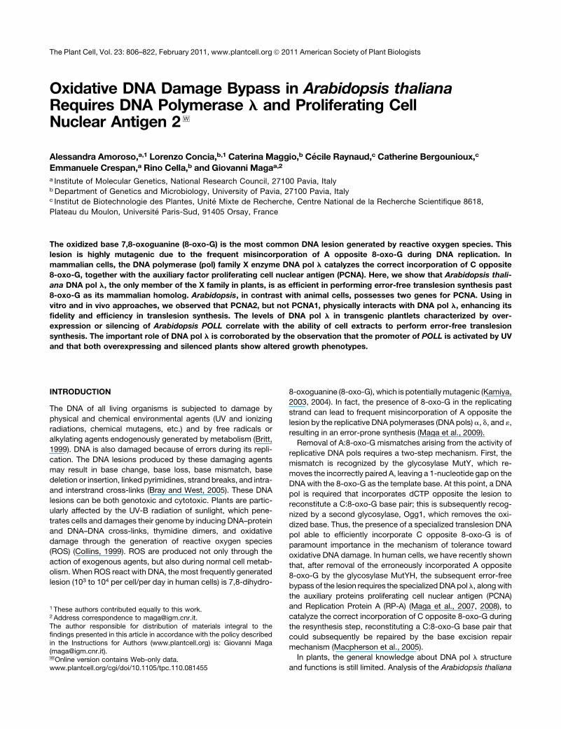
by fast protein liquid chromatography (FPLC). Figure 1A shows
the elution profile of polymerase activity from the last chroma-
tographic step (Mono Q). To distinguish DNA pol l activity
from that of any interfering bacterial DNA pol possibly present
in the eluted fractions, the assay was performed in parallel in
the presence of Ic (N-9-fluorenylmethoxycarbonyl-aminoalkyl-
triphosphate), a selective inhibitor of eukaryotic DNA pol l
(Crespan et al., 2005). The single peak of activity detected in
fractions 8 to 14 was inhibited by Ic, thus confirming that the
enzymatic activity measured was actually that of DNA pol l. A
small residual pol activity in fractions 18 to 21, which was not
sensitive to Ic, was due to E. coli DNA pol I contamination and
was discarded. SDS-PAGE analysis of the same fractions
showed the presence of a single polypeptide of the expected
molecular mass, indicating a degree of homogeneity of the
recombinant protein higher than 95% (Figure 1). Protein identity
was further confirmed by protein gel blot analysis using anti-Hs
DNA pol l polyclonal antibodies (Figure 1C). With respect to
enzymological parameters, DNApoll, as anticipated, required the
presence of bivalent metal ions for the catalysis, with a preference
forMn2+overMg2+ (Figure 1D) anddisplayedoptimal activity at pH
8.0 (Figure 1E). Moreover, the catalytic efficiency was very sen-
sitive to salt concentration, displaying almost complete inhibition
at NaCl concentration higher that 50 mM (Figure 1F).
Expression and Purification of Recombinant Arabidopsis
PCNA1 and PCNA2
Contrary to animal cells, which possess only one PCNA gene,
Arabidopsis has two PCNA genes. To obtain recombinant PCNA1
8-Oxo-G Bypass by Arabidopsis DNA Pol l 807
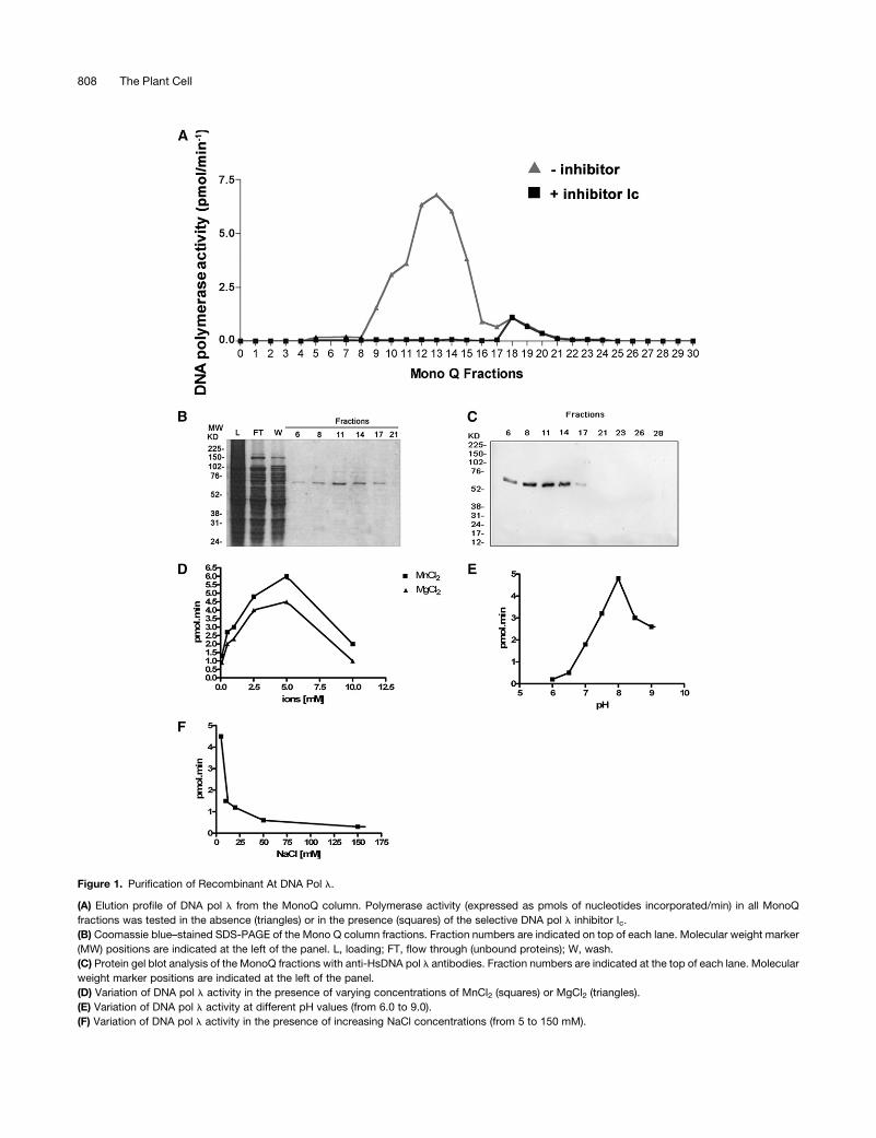
Figure 1. Purification of Recombinant At DNA Pol l.
(A) Elution profile of DNA pol l from the MonoQ column. Polymerase activity (expressed as pmols of nucleotides incorporated/min) in all MonoQ
fractions was tested in the absence (triangles) or in the presence (squares) of the selective DNA pol l inhibitor Ic.
(B) Coomassie blue–stained SDS-PAGE of the Mono Q column fractions. Fraction numbers are indicated on top of each lane. Molecular weight marker
(MW) positions are indicated at the left of the panel. L, loading; FT, flow through (unbound proteins); W, wash.
(C) Protein gel blot analysis of the MonoQ fractions with anti-HsDNA pol l antibodies. Fraction numbers are indicated at the top of each lane. Molecular
weight marker positions are indicated at the left of the panel.
(D) Variation of DNA pol l activity in the presence of varying concentrations of MnCl2 (squares) or MgCl2 (triangles).
(E) Variation of DNA pol l activity at different pH values (from 6.0 to 9.0).
(F) Variation of DNA pol l activity in the presence of increasing NaCl concentrations (from 5 to 150 mM).
808 The Plant Cell
and PCNA2, the corresponding CDSs were cloned and ex-
pressed as His-tagged proteins in E. coli cells. The recombinant
proteins were purified by FPLC and the eluted fractions analyzed
by SDS-PAGE and protein blots, using an antibody against
human PCNA that cross-reacts with both plant proteins. In the
case of PCNA1, a single polypetide of the expected molecular
mass could be detected, with a peak in fractions 10 to 24 (Figures
2A and 2B). PCNA2 was similarly produced, as shown in Figure
2C (fractions 12 to 18). Protein gel blot analysis with anti-Hs
PCNA antibodies again confirmed its identity (Figure 2D). Thus,
both PCNA1 and PCNA2 were successfully expressed and
purified as His-tagged proteins.
DNA Pol l Recognizes 8-Oxo-G on a Template as a Normal
Guanine and Preferentially Incorporates dCTP over dATP
Opposite This Lesion
The mutagenic potential of 8-oxo-G when present on the tem-
plate strand is due to its ability to direct the preferential incor-
poration of dATPby someDNApols opposite the lesion.Wehave
shown that Hs DNA pol l, on the contrary, preferentially incor-
porates the correct dCTP opposite 8-oxo-G (Maga et al., 2007).
Thus, the next step was to test the efficiency of nucleotide
incorporation by DNA pol l opposite an 8-oxo-G lesion present
into a 1-nucleotide gap substrate. As shown in Figure 3A, both
dCTP (lanes 2 to 5) and dATP (lanes 6 to 9) were incorporated
opposite the lesion. At high dCTP concentrations, a limited (1
nucleotide) strand displacement synthesis could be observed
(lanes 2 to 4) as revealed by the incorporation of a second dCTP
residue opposite to the template G immediately downstream of
the lesion. This feature was observed also for the human enzyme
(Singhal and Wilson, 1993). When the same experiment was
repeated using a control substrate containing an unmodified
guanine, only dCTP was incorporated (Figure 3B, lanes 2 to 6),
confirming that the incorporation of dATPwas strictly dependent
on the presence of an 8-oxo-G and was not due to an intrinsic
infidelity of DNA pol l. Quantification of the products allowed us
to determine the kinetic parameters for these reactions. As
shown in Figure 3C, the catalytic efficiency (Vmax/Km) for dATP
incorporation opposite 8-oxo-Gwas 3-fold higher than for dATP,
thus confirming a preferential dCTP incorporation opposite the
lesion. Remarkably, the Vmax/Km value for dCTP incorporation
opposite 8-oxo-G (8.8 pmol 3 mM21 3 h21 3 mg21) was nearly
identical to the one displayed opposite a normal G (7.4 pmol 3mM21 3 h21 3 mg21) (Figure 3D), suggesting that DNA pol l
recognized an 8-oxo-G lesion on the template as a normal
guanine.
ArabidopsisPCNA2butNotPCNA1 Increases theSelectivity
for dCTP Incorporation Opposite 8-Oxo-G by DNA Pol l
Hs PCNA was shown to increase the processivity of Hs DNA
pol l and its fidelity during 8-oxo-G bypass. We produced both
PCNA recombinant proteins from Arabidopsis and sought to
verify whether they had the same effect on At DNA pol l activity.
To investigate whether PCNA2 contributed also to translesion
Figure 2. Purification of Recombinant Arabidopsis PCNA1 and PCNA2.
(A) Coomassie blue staining after SDS-PAGE of the fractions containing PCNA1. Fraction numbers are indicated on top of each lane. Molecular weight
marker (MW) positions are indicated at the left of the panel. L, loading; FT, flow through (unbound proteins); W, wash.
(B) Protein gel blot analysis of the PCNA1-containing fractions with anti-PCNA antibody. Fraction numbers are indicated on top of each lane. Molecular
weight marker (MW) positions are indicated at the left of the panel.
(C) As in (A), but with the fractions containing PCNA2.
(D) As in (B), but with the fractions containing PCNA2.
8-Oxo-G Bypass by Arabidopsis DNA Pol l 809

synthesis, increasing concentrations of dATP or dCTP were
incubated with a 1-nucleotide gapped substrate containing the
8-oxo-G lesion in the presence or in the absence of PCNA2.
Quantification of the data showed that the incorporation effi-
ciency, defined as the Vmax/Km ratio, for dATP incorporation
opposite to the 8-oxo-G damage was decreased 4-fold, while
the incorporation of dCTP was not affected (Figure 3E). To
better investigate the effects of PCNA2 on 8-oxo-G bypass,
similar experiments were performed at dATP or dCTP concen-
trations of 1mM (Figure 4A), 10mM (Figure 4B), or 100mM (Figure
4C). Quantification of the data allowed us to determine dose–
response curves for At PCNA2 inhibition at different nucleotide
Figure 3. PCNA2 Favors Error-Free Translesion Synthesis by DNA Pol l Opposite the 8-Oxo-G Lesion.
(A) Incorporation was monitored with varying concentrations of dCTP (lanes 2 to 5) or dATP (lanes 6 to 9). Lane 1, control in the absence of nucleotides.
The structure of the substrate is shown on top of the panel. The position of the band corresponding to the incorporation opposite the 8-oxo-G lesion (+1)
is indicated at the left of the panel.
(B) Incorporation of dATP and dCTP opposite to a normal G by DNA pol l was monitored with increasing concentration of dCTP (lanes 2 to 6) or dATP
(lanes 7 to 11). Lane 1, control in the absence of nucleotides. The structure of the substrate is shown on top of the panel. The position of the band
corresponding to the incorporation opposite the undamaged base (+1) is indicated on the left of the panel.
(C) Quantification of dCTP (triangles) and dATP (circles) incorporation opposite 8-oxo-G. The Km and Vmax values are reported for both nucleotides.
(D) Quantification of dCTP incorporation (triangles) opposite normal G. The corresponding Km and Vmax values are indicated. Values are the mean of
three independent experiments. Error bars are 6 SD.
(E) Variation of the initial velocity (v), induced by DNA pol l on the 1-nucleotide gapped 8-oxo-G template for dCTP incorporation and in the presence
(white circles) or in the absence (black circles) of At PCNA2 or for dATP incorporation in the presence (white squares) or in the absence (black circles) of
At PCNA2 as a function of nucleotide substrate concentration. Values are themean of three biological replicates. The corresponding Km and Vmax values
are indicated. Error bars are 6 SD.
810 The Plant Cell
concentrations. As shown in Figure 4D, while the effect on dCTP
incorporation was modest (10 to 25% inhibition), a strong inhi-
bition of dATP incorporationwas observed, which increasedwith
the nucleotide concentration, showing a 75% reduction of in-
corporation at 100mMdATP. This was reflected by a decrease of
the apparent equilibrium dissociation constant (Kd) for PCNA2 as
a function of increasing dATP concentrations (Figure 4E). Thus,
PCNA2 acted as an a-competitive inhibitor of dATP incorpora-
tion by DNA pol l, with respect to the nucleotide substrate.
These data suggest that PCNA2 binds to the ternary complex of
DNA pol l and its substrates nucleic acid and nucleotides.
Similar assays were performed with PCNA1. Dose–response
curves showed a clear inhibition of both dATP and dCTP incor-
poration opposite 8-oxo-G (Figure 4F), the extent of which,
however, was not influenced by the nucleotide concentration,
as shown by the similar values obtained for the Kd of PCNA1
(Figure 4G).
Arabidopsis PCNA2, but Not PCNA1, Physically Interacts
with DNA Pol l
The results described above indicated a different effect of
PCNA1 PCNA2 on the bypass activity of DNA pol l. In particular,
they suggested a physical interaction between PCNA2 and the
translesion polymerase. Since both PCNA and DNA pol l had a
His-tag, pull-down experiments could not be performed. Coim-
munoprecipitation experiments were attempted, but were not
conclusive, due to the high tendency of DNA pol l to unspecifi-
cally bind to Protein A or Protein G Sepharose (data not shown).
Thus, a protein dot blot experiment was devised, where both
PCNA proteins were separately spotted onto a nitrocellulose
membrane and overlaid with DNA pol l. After extensive washing
of the membrane, the presence of DNA pol l bound to PCNA
spotted on the filter was revealed by anti-Hs DNA pol l polyclo-
nal antibodies. As shown in Figure 5A, DNA pol l could bind only
to PCNA2 (membrane A). As a control, an identical membrane
was incubated with buffer without DNA pol l (membrane B) and
revealed by anti-Hs PCNA antibodies to visualize the presence of
the two proteins. The interaction appeared to be specific, since
no signal was detected in positions corresponding to equivalent
amounts of BSA, spotted as negative controls (membrane A).
Similarly, no cross-reaction between anti-Hs DNA pol l anti-
bodies and PCNA1was noticed, as indicated by the absence of a
signal corresponding to the spotted area, whereas antibodies
recognized the recombinant At DNA pol l spotted on the
membrane as a positive control (membrane A). These results
confirm that the functional interaction between DNA pol l and
PCNA2 during translesion synthesis, which allows bypassing of
the 8-oxo-G lesion with high fidelity, is due to their direct physical
interaction. Moreover, this physical interaction was selective,
since no binding was observed with PCNA1.
HumanPCNAandRP-ACanSubstitute for thePlantProteins
to Favor the Error-Free Bypass Synthesis by At DNA Pol l
Opposite 8-Oxo-G
Hs RP-A and Hs PCNA have previously been shown in our
laboratory to be able to increase the efficiency of translesion
synthesis by Hs DNA pol l in animal cells. Due to the high level of
conservation between At DNA pol l, At PCNA2, and their
respective humanorthologs, we analyzed the effect of the human
proteins Hs RP-A and Hs PCNA on the ability of At DNA pol l to
bypass the 8-oxo-G damage. As shown in Figure 5B, there was a
significant inhibitory effect of Hs RP-A and Hs PCNA proteins on
At DNA pol l efficiency for dATP incorporation opposite 8-oxo-G
damage, which was higher than in the case of dCTP. Indeed,
dATP incorporation in the presence of the highest tested
amounts of Hs PCNA and Hs RP-A (2.5 mM) was almost unde-
tectable, whereas under the same conditions, total dCTP incor-
poration was reduced by 50%.
Human RP-A Cooperates with At PCNA2 to Increase the
Fidelity of 8-Oxo-G Bypass by At DNA Pol l
Since Hs RP-A and Hs PCNA could synergistically enhance the
fidelity of 8-oxo-G bypass by At DNA pol l, we tested whether At
PCNA2 displayed the same effect when combinedwith Hs RP-A.
As shown in Figure 5C, addition of At PCNA2 alone or in
combination with Hs RP-A did not affect dCTP incorporation
opposite 8-oxo-G (lanes 2 to 7). On the contrary, At PCNA2,
which alone was effective in reducing dATP incorporation (lane
9), in combination with Hs RP-A resulted in a strongly enhanced
inhibition of dATP insertion (lanes 10 to 13). Overall, these data
demonstrate that the functional interaction of RP-A with At DNA
pol l and PCNA in 8-oxo-G bypass is highly conserved, as it
occurs also between human and plant proteins. Such a high level
of evolutionary conservation highlights the importance of this
pathway in preventing the incorrect dATP incorporation opposite
the 8-oxo-G damage, thus reducing its mutagenic potential.
Both PCNA1 and PCNA2 Are Localized in the Nucleus of
Plant Cells, but Only PCNA2 Interacts with DNA Pol l in Vivo
Since in vitro experiments indicated a functional cooperation
between DNA pol l and PCNA2, we confirmed the physical
interaction between these two proteins in vivo. Transient ex-
pression assays using BY-2 tobacco (Nicotiana tabacum) pro-
toplasts with constructs encoding At PCNA1, At PCNA2, and At
DNA pol l N-terminally fused to green fluorescent protein (GFP)
and yellow fluorescent protein (YFP), under the control of cau-
liflower mosaic virus 35S promoter (Guilley et al., 1982), showed
an in vivo nuclear localization of all three proteins (see Supple-
mental Figure 1 online). Additional assays also showed that
nuclear localization was not affected by the N- or C-terminal
position of the tag (data not shown). As shown in Figures 6A and
6E, both PCNA1 andPCNA2 localize in the nucleus, but not in the
nucleolus. On the contrary, DNA pol l accumulates in the whole
nucleus even though GFP labeling of the nucleolus was weak
(Figures 6B and 6F). The in vivo colocalization of DNA pol l with
PCNA1 and PCNA2 was tested by cotransfecting tobacco BY-2
protoplasts with two different constructs encoding the fluores-
cent fusion proteins GFP-At DNA pol l and AtPCNA1-RFP (for
red fluorescent protein), or GFP-At DNA pol l and At PCNA2-
RFP. Digital merging of red and green single-channel fluores-
cent images confirmed the nuclear colocalization (Figures 6C
and 6G).
8-Oxo-G Bypass by Arabidopsis DNA Pol l 811

Figure 4. The Inhibition of dATP Incorporation by PCNA2, but Not by PCNA1, Is Dependent on the Nucleotide Concentration.
(A) DNA pol l (50ng) was incubated in the presence of the 1-nucleotide gapped 8-oxo-G template and with 1 mM of either dATP (lanes 1 to 6) or dCTP
(lanes 7 to 12) in the absence (lanes 1, 2, 7, and 8) or in the presence (lanes 3 to 6 and 9 to 12) of increasing amounts of PCNA2 (15, 30, 60, and 90 ng).
Lane 13, control reaction in the absence of DNA pol l.
(B) As in (A), but in the presence of 10 mM dATP or dCTP.
(C) As in (A), but in the presence of 100 mM dATP or dCTP.
(D) Effects of increasing amounts of PCNA2 on the relative incorporation of different concentrations (1, 10, and 100 mM) of dATP and dCTP opposite to
8-oxo-G by DNA pol l. Values are the mean of three independent biological replicates. Error bars represent 6 SD values.
(E) Variations of the apparent dissociation constant of PCNA2 (Kd) for DNA pol l as a function of increasing concentrations of dATP (light-gray bars) or
dCTP (dark-gray bars) on a 1-nucleotide gapped 8-oxo-G template. Values are themean of three independent biological replicates. Error bars represent
6 SD values.
(F) Effects of increasing amounts of PCNA1 on the relative incorporation of different concentrations (1, 10, and 100 mM) of dATP and dCTP opposite
8-oxo-G by DNA pol l. Values are the mean of three independent biological replicates. Error bars represent 6 SD values.
(G) Variations of the apparent dissociation constant of PCNA1 (Kd) for DNA pol l as a function of increasing concentrations of dATP (light-gray bars) or
dCTP (dark-gray bars) on a 1-nucleotide gapped 8-oxo-G template. Values are themean of three independent biological replicates. Error bars represent
6 SD values.
To prove the physical interaction between DNA pol l and
PCNA1 or PCNA2, we performed a bimolecular fluorescence
complementation assay (Figure 7). BY-2 protoplasts were co-
transfected with a construct encoding a fusion protein between
At DNA pol l and YFP C-terminal region (amino acids 156 to 238)
(At POLL-YFPC) and with those encoding either At PCNA1 or At
PCNA2 fused to YFP N-terminal region (amino acids 1 to 155)
(At PCNA1-YFPN or At PCNA2-YFPN). Both YFP moieties were
C-terminally fused. Fluorescence was observed only with PCNA2
as a result of molecular complementation of YFPN and YFPC due
to in vivo physical interaction between DNA pol l and PCNA2
(Figure 7C), while no fluorescence was observed with PCNA1
(Figure 7A). The construct At PCNA1-YFPNwas previously proven
to encode a functional fusion protein able to interact with ATXR5,6
proteins (Raynaud et al., 2006).
Arabidopsis DNA Pol l Is Responsible for Error-Free
Bypass of 8-Oxo-G Lesions in Planta
POLL overexpressing plants (POLOE) were produced by Agro-
bacterium tumefaciens–mediated transformation of Columbia
(Col-0) flowers using the vector pGBIN1-At POLL (see Supple-
mental Figure 2 online) where the CDS encoding DNA pol l is
under the control of the constitutive cauliflower mosaic virus 35S
promoter. Three-week-old kanamycin-resistant T1 lines were
analyzed by quantitative RT-PCR (qRT-PCR) with respect to the
level of POLL transcripts. Out of more than 20 overexpressors, a
line characterized by a 70-fold increased content of POLL
transcripts was chosen to evaluate its behavior in a translesion
assay (Figure 8B). The protein level of overexpressing lines was
assessed by protein gel blot analysis (see Supplemental Figure 3
online). The relative increase in protein level with respect to wild-
type plants was 26 0.5-fold, which was lower than the increase
of the relevant transcript level. This suggests that DNA pol l is
subjected to a tight posttranslational regulation that controls
protein accumulation, as already demonstrated in the case of
mammalian cells (Wimmer et al., 2008).
The only available Arabidopsis line with an insertion in the
POLL gene (At1g10520) was the SALK_075391C line, in which
the T-DNA (4.5 kb) is located in the intron between the 9th and
10th exons, corresponding in the splicedmRNA to an exon-exon
junction at 1167 bp starting from ATG, within the codon for S389
of the translated peptide (Figure 8A). Since the active site of DNA
pol l spans from amino acid 346 to 411, it was anticipated that
the insertion could prevent the production of a functional protein.
Following further selection on kanamycin-containing medium,
the analysis by PCR using genomic DNA (see Supplemental
Figure 3 online) showed that SALK_075391C knockdown line
was actually homozygous for the T-DNA insertion. Moreover,
qRT-PCR (Figure 8B) and protein gel blot analysis (Figure 8C)
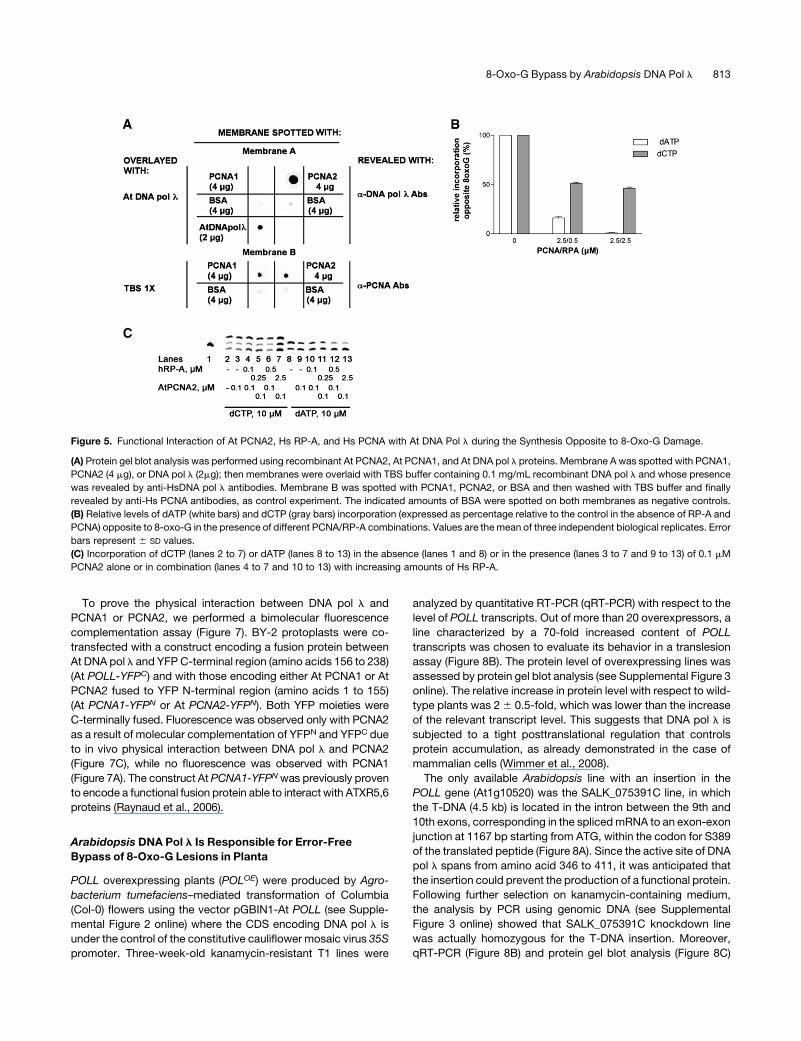
Figure 5. Functional Interaction of At PCNA2, Hs RP-A, and Hs PCNA with At DNA Pol l during the Synthesis Opposite to 8-Oxo-G Damage.
(A) Protein gel blot analysis was performed using recombinant At PCNA2, At PCNA1, and At DNA pol l proteins. Membrane A was spotted with PCNA1,
PCNA2 (4 mg), or DNA pol l (2mg); then membranes were overlaid with TBS buffer containing 0.1 mg/mL recombinant DNA pol l and whose presence
was revealed by anti-HsDNA pol l antibodies. Membrane B was spotted with PCNA1, PCNA2, or BSA and then washed with TBS buffer and finally
revealed by anti-Hs PCNA antibodies, as control experiment. The indicated amounts of BSA were spotted on both membranes as negative controls.
(B) Relative levels of dATP (white bars) and dCTP (gray bars) incorporation (expressed as percentage relative to the control in the absence of RP-A and
PCNA) opposite to 8-oxo-G in the presence of different PCNA/RP-A combinations. Values are themean of three independent biological replicates. Error
bars represent 6 SD values.
(C) Incorporation of dCTP (lanes 2 to 7) or dATP (lanes 8 to 13) in the absence (lanes 1 and 8) or in the presence (lanes 3 to 7 and 9 to 13) of 0.1 mM
PCNA2 alone or in combination (lanes 4 to 7 and 10 to 13) with increasing amounts of Hs RP-A.
8-Oxo-G Bypass by Arabidopsis DNA Pol l 813
failed to detect POLL transcripts or protein, thus proving the
actual knocking down of the POLL gene. Both types of plants,
POLL overexpressors and knockdown, are characterized by an
altered phenotype. Overexpressors show a retarded growth,
while the knockdown features early flowering (see Supplemental
Figure 4 online). The reasons of these phenotypes are currently
under investigation; however, taking into account the modest
increase in protein levels observed in POLLOE plants (see Sup-
plemental Figure 3 online), these alterations reflect the impor-
tance of the maintenance of a balanced level of DNA pol l in the
living plant organism.
To further investigate the role of DNA pol l in 8-oxo-G lesion
bypass, we prepared cell extracts from 10-d-old seedlings of
overexpressing POLL (POLLOE) and knockdown (POLLKD) lines.
The extracts were tested in in vitro translesion assays in com-
parison with extracts coming from Col-0 plants (POLLwt) (see
Figure 8D for a representative experiment). The error-free (dCTP)
incorporation opposite the 8-oxo-G lesion (Figure 8E) was in-
creased;2-fold in the POLLOE extracts, while it was reduced in
the POLLKD extracts with respect to POLLwt. The level of error-
prone (dATP) bypass was basically unaffected. These data
strongly suggest that DNA pol l is an essential component of
the machinery responsible for error-free 8-oxo-G bypass in
plants.
Arabidopsis POLL Expression Is Higher in Young Tissues
and Is Induced by UV Irradiation
On the basis of annotated coding sequences of Arabidopsis
available at The Arabidopsis Information Resource website
(www.Arabidopsis.org), the length of the intergenic region up-
stream of the coding sequence of POLL (At1g10520) is only 177
bp long because of the proximity of the upstream gene
(At1g10510). The analysis of the putative promoter by PlantPAN
(Plant Promoter Analysis Navigator; http://PlantPAN.mbc.nctu.
edu.tw), which takes into account 197 Arabidopsis transcription
factors from PLACE, TRANSFAC, AGRIS, and JASPAR data-
bases (Chang et al., 2008), revealed the presence of T-box and
SORLIP5 sites, which are overrepresented in promoters of light-
activated genes (Chan et al., 2001; Hudson and Quail, 2003),
along with two distinct Myb4 binding sites. The Myb family of
transcription factors is known to mediate biotic and abiotic
stresses in plants (Chen et al., 2002). In particular, the Myb4
subgroup was found to repress the transcription of UV response
genes, since the related mutants were found to be more tolerant
to UV. Moreover, it was observed that MYB4 transcription is
downregulated after UV-B irradiation (Jin et al., 2000). Thus, the
presence of two Myb4 binding sites in the putative POLL pro-
moter implicates UV as a possible regulator of POLL expression.
To prove this hypothesis, we compared the relative level of 10-d-
old At POLL transcripts between control and UV-irradiated
seedlings and 30-d-old leaves by qRT-PCR. Three independent
biological replicates were performed for each group of samples.
The amount of POLL transcripts found in untreated seedling and
leaves was ;9.5 and 1.8% of the geometric average of two
independent housekeeping genes, respectively (Figure 8F). The
fact that in seedlings the level of POLL transcripts was higher
than in mature leaves is consistent with the observation that the
major DNA repair pathway in rice mature leaves is due to
photoreactivation performed by cyclobutane pyrimidine dimers
photolyase and (6-4) photolyase (Kimura et al., 2004). On the
other hand, the higher expression level in seedlings may reflect a
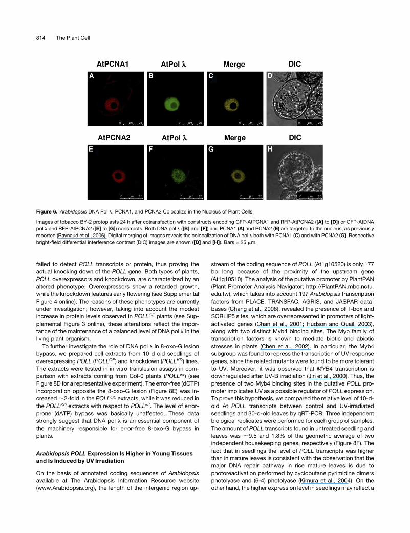
Figure 6. Arabidopsis DNA Pol l, PCNA1, and PCNA2 Colocalize in the Nucleus of Plant Cells.
Images of tobacco BY-2 protoplasts 24 h after cotransfection with constructs encoding GFP-AtPCNA1 and RFP-AtPCNA2 ([A] to [D]) or GFP-AtDNA
pol l and RFP-AtPCNA2 ([E] to [G]) constructs. Both DNA pol l ([B] and [F]) and PCNA1 (A) and PCNA2 (E) are targeted to the nucleus, as previously
reported (Raynaud et al., 2006). Digital merging of images reveals the colocalization of DNA pol l both with PCNA1 (C) and with PCNA2 (G). Respective
bright-field differential interference contrast (DIC) images are shown ([D] and [H]). Bars = 25 mm.
814 The Plant Cell
higher rate of cell proliferation and DNA replication due the
presence of meristematic tissues. In fact, the horthologous rice
POLLwas shown to be expressed at a higher level in proliferating
cells (Uchiyama et al., 2004).
Following UV irradiation, a remarkable increase of POLL
expression was observed both in 10-d-old seedlings and in
leaves. In particular, irradiated seedlings showed an increase of
over 3.7-fold from ;9.5 to >35.5% with respect to the expres-
sion of housekeeping genes (Figure 8F). In leaves, the expression
level raised from 1.8 to ;11.3% (6-fold) following irradiation
(Figure 8F). To confirm the increased POLL expression caused
by UV, we produced transgenic plants containing a chimeric
reporter uidA gene driven by the POLL promoter. Results of
histochemical staining showed that b-glucuronidase activity was
detectable only in irradiated plants, thus indicating a very low
basal expression of POLL under normal conditions (see Supple-
mental Figure 5 online). These data clearly show that POLL
transcription is induced by UV.
DISCUSSION
DNA pol l is the only member of DNA family X present in higher
plants. Arabidopsis DNA pol l shows 30% similarity with its
human homolog. On the other hand, while human cells possess
only one gene for each of the three subunits (p70, p32, and p14)
of the ssDNAbinding protein RP-A and one for the auxiliary factor
PCNA, Arabidopsis contains two genes for PCNA, five genes for
the p70, and two each for p32 and p14 subunits of RP-A (Kimura
and Sakaguchi, 2006). Whether this fact simply constitutes an
example of extreme functional redundancy or reflects a diversi-
fication of functions occurring in plant cells with respect to animal
cells is not known. Our recent results have defined the role of
human PCNA and RP-A in increasing the fidelity of synthesis of
human DNA pol l in bypassing the 8-oxo-G lesion (Maga et al.,
2008). To investigatewhether themechanismoperating in animal
cells was also involved in increasing the tolerance to oxidative
DNA damage in plants, we produced recombinant Arabidopsis
DNA pol l, PCNA1, and PCNA2. These proteins were then tested
in vitro in specific translesion synthesis assays in the presence of
an 8-oxo-G lesion.
The results presented here, while confirming that DNA pol l is
likely playing a major role in the error-free bypass of 8-oxo-G
lesions in plants, highlight some important differences with re-
spect to animal cells. Themost intriguing one is the diversification
of functions of the two plant PCNA proteins. Our analysis showed
that both PCNA1 and PCNA2 are expressed and localized in the
nucleus, together with DNA pol l. However, only PCNA2 phys-
ically interacts with DNA pol l both in vitro and in vivo. This
differential behavior is also reflected at the enzymatic level.
PCNA2 enhances the fidelity of DNA pol l by reducing its ability
to incorrectly incorporate dATP opposite 8-oxo-G, whereas
PCNA1 shows a general inhibition of translesion synthesis by
DNA pol l. Interestingly, the effect of PCNA2 is dependent on
nucleotide concentration and exerts its maximal efficiency in
preventing error-pronebypassof the 8-oxo-G lesionbyDNApoll
precisely when it is most needed, that is, in the presence of high
concentrations of dATP, that should give the highest probability
for misincorporation. At PCNA1 and At PCNA2 are nearly iden-
tical (99% similarity) and show a very high similarity (85%) to Hs
PCNA. In particular, all the conservedboxes thatwere found to be
involved in the interaction between Hs DNA pol l and Hs PCNA
(SHV motif, amino acids 43 to 45; QLGI motif amino acids 125 to
128; LAPKmotif, amino acids 251 to 254) are conserved also in At
PCNA1 and AtPCNA2. However, PCNA1 and PCNA2 show two
and three extra amino acids, respectively, at their C terminus
compared with the human protein.
The recent resolution of the crystal structure of At PCNA1 and
At PCNA2 in complex with a peptide derived from the human p21
protein, a known interactor of Hs PCNA, further confirmed the
functional equivalence of both plant proteins with the Hs PCNA
(Strzalka et al., 2009). PCNA1 and PCNA2 have been also
proposed to be able to form heterotrimers, an observation that
is particularly relevant in the light of the different functional
effects of these two proteins revealed by our study.
Thus, it is possible to speculate that PCNA2 might act as a
specialized DNA repair or translesion auxiliary factor, promoting
the switch from highly error-prone replicative and/or repair plant
DNA pols to the more faithful DNA pol l. On the other hand,
PCNA1might be required for other functions (for example during
DNA replication or certain types of DNA repair) not requiring the
action of DNA pol l. Heterotrimers composed of mixed PCNA1
and PCNA2 monomers might also display different properties
with respect to the corresponding homotrimers, possibly adding
a further level of regulation to plant PCNA functions. Further
mutagenesis and in vivo expression studies will help to clarify
these issues.
DNA pol h, a Y-family TLS polymerase ofArabidopsis, is able to
rescue a yeast UV-sensitive phenotype only when cotransformed
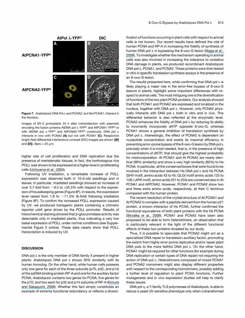
Figure 7. Arabidopsis DNA Pol l and PCNA2, but Not PCNA1, Interact in
the Nucleus.
Images of BY-2 protoplasts 24 h after cotransfection with plasmids
encoding the fusion proteins AtDNA pol l-YFPC and AtPCNA1-YFPN or
with AtDNA pol l-YFPC and AtPCNA2-YFPN constructs. DNA pol l
interacts in vivo with PCNA2 (A) but not with PCNA1 (C). Respective
bright-field differential interference contrast (DIC) images are shown ([B]
and [D]). Bars = 25 mm.
8-Oxo-G Bypass by Arabidopsis DNA Pol l 815

Figure 8. Arabidopsis DNA Pol l Is Responsible for Error-Free Bypass of 8-Oxo-G Lesion in Plant Cells.
(A) Schematic drawing of the protein domains encoded by DNA pol l gene. Functional domains are reported. BRCT, BRCA-1 C-terminal domain;
POLXc, DNA polymerase family X catalytic domain. The Ser-389 residue corresponding to T-DNA insertion is indicated by a black arrow. The portion of
catalytic domain (amino acids 379 to 461) corresponding to amplicon produced by qRT-PCR with primers POLL FW and POLL RV is indicated by the
dotted line. Primers position is reported as white triangles.
(B) Relative expression level of POLL in 10-d-old seedlings of different lines. qRT-PCR results show that POLL transcript levels in wild-type (POLWT),
knockdown (POLKD), and overexpressing (POLOE) lines. Transcript levels are expressed as percentage of the average expression level of two
housekeeping genes (H.K.): EF1a translation elongation factor (At5g60390) and glyceraldehyde-3-phosphate dehydrogenase C2 (At1g13440). POLL
transcripts are;14.7% of H.K in the overexpressing line and only 0.2% in Col-0. No expression was detected in knockdown line. Error bars represent6
SD of two technical replicates
(C) Protein gel blot analysis of the expression of DNA pol l in wild-type (lanes 5 to 8) and KD (lanes 1 to 4) cell extracts (CE). Top panel: protein blot with
anti-Hs DNA pol l antibodies. Bottom panel: Ponceau staining of the same membrane. The migration position of DNA pol l is indicated with an arrow.
(D) Reactions were performed with cell extracts under the conditions specified in Methods. POLLOE (lanes 1 to 4), POLLwt (lanes 5 to 8), or POLLKD
(lanes 9 to 12) crude extracts (10 mg total proteins) were incubated in the presence of 100 mMdATP (lanes 1, 3, 5, 7, 9, and 11) or 100 mMdCTP (lanes 2,
4, 6, 8, 10, and 12) with the 39/72 8-oxo-G DNA substrate or with the 39/72 control DNA substrate. Lanes 13 (with 8-oxo-G substrate) and 14 (with
control substrate) are control reactions in the absence of extracts.
(E) Relative activity, expressed as percentage of elongated primer ends, for dATP (light white bars) and dCTP (light-gray bars) in the 39/72 8-oxo-G DNA
substrate and for dATP (dark-gray bars) and dCTP (black bars) in the 39/72 8-oxo-G DNA control substrate. Values are the mean of two independent
biological replicates. Error bars are 6 SD.
(F) qRT-PCR on 10-d-old seedlings and on mature leaves (30 d old) show a higher transcript level in young tissues, 9.5% (64.1%) of H.K. versus 1.8%
(60.5%). After 30 min of UV irradiation (0.3 mW/cm2, corresponding to 2.9 J/m2/s, at 243-nm wavelength), transcript level was ;3.7-fold higher in
seedlings and >6-fold higher in mature leaves, 35.5% (67%) versus 11.3% (62.7%). Error bars represent 6 SD values among three independent
biological replicates.
816 The Plant Cell

with the PCNA2 gene but not with PCNA1 (Anderson et al., 2008;
Kunz, 2008), further emphasizing the specific role of PCNA2
protein as an auxiliary factor in translesion synthesis. PCNA1 and
PCNA2 differ by eight amino acidic residues, but only Asn-201,
present in PCNA2, was proven to be critical for the interaction
with DNA pol h in yeast. In fact, when Asn was substituted with
Lys (Lys-201 as is the case of PCNA1), PCNA2 failed to com-
plement the RAD18 mutant.
Since monoubiquitylation of PCNA Lys-164, close to residue
201 in the three-dimensional structure, is required for the func-
tionality of DNApolh (van der Kempet al., 2009), it was proposed
that the amino acidic substitution might have some differential
effect on PCNA1 and PCNA2 monoubiquitylation, thus influenc-
ing their functional interaction with DNA pol h (Kunz, 2008).
Recent data point to a role of ubiquitylation of PCNA in TLS
(Chen et al., 2010; Das-Bradoo et al., 2010) suggesting a major
role for posttranslational modulation of PCNA function rather
than transcriptional regulation. It is tempting to speculate that
PCNA is always present in the nuclear environment, and its
interaction with translesion DNA pols is prevented unless DNA
damage induces a rapid activation through ubiquitylation of
PCNA, thus allowing interaction with TLS pols, the fidelity of
which is lower compared with that of replicative ones.
However, PCNA monoubiquitylation in plants is still to be
confirmed, and Arabidopsis has no clear homolog of RAD18, the
enzyme that can perform PCNA monoubiquitylation in humans
and yeast (Potuschak et al., 1998; Kraft et al., 2005). Neverthe-
less, it is not possible to exclude that monoubiquitylation of
PCNA2 might further enhance DNA pol l fidelity.
We previously demonstrated that, in animal cells, DNA pol l is
the specialized enzyme required for error-free bypass of 8-oxo-G
lesions, and it is recruited to the damage site thanks to the
coordinated action of two auxiliary proteins: PCNA and RP-A.
Here, we show that At DNA pol l, the plant homolog of Hs DNA
pol l, fulfills a similar function in coordination with At PCNA2. In
addition, we show that the human proteins RP-A and PCNA can
cooperate with a plant DNA pol l. Such a high degree of
evolutionary conservation is a characteristic of essential bio-
chemical processes, resembling the interaction of replicative
DNA pols with the auxiliary factors PCNA and Replication Factor
C, which is conserved from yeast to man. Thus, our results
further support an essential role of the DNA pol l–dependent
pathway for oxidative DNA damage tolerance in all eukaryotic
organisms.
Sequence annotation of the genomes of Arabidopsis and rice
has shown the presence in plants of conserved genes encoding
components of repair pathways, including those for specialized
DNA pols necessary for TLS (Kimura and Sakaguchi, 2006).
Some of these have been characterized. For instance, it was
shown that a cDNA encoding DNA pol h of Arabidopsis is able to
complement a UV-sensitive yeast strain deficient in this poly-
merase (Santiago et al., 2006). Increased UV sensitivity was also
reported for a Rev1mutant (Takahashi et al., 2005). In the case of
ArabidopsisDNApol k, deletion studies ofPOLK showed that the
ability to bypass 8-oxo-G increases following the removal of the
C-terminal region (not involved in catalytic activity), thus indicat-
ing a possible regulatory role of this region (Garcıa-Ortiz et al.,
2007).
Also, Arabidopsis DNA pol z, which belongs to the B family,
appears to be involved in TLS since deletion of its Rev3 subunits
is responsible for a drastic increase in UV sensitivity (Sakamoto
et al., 2003).
A reverse-genetic analysis was used to evaluate the role of
genes encoding At DNA pol h (At POLH) and At DNA pol z (At
POLZ) in the tolerance of roots to UV radiation, and it was shown
that these pols contribute to the integrity of stem cells of the root
meristem (Curtis and Hays, 2007). To evaluate the role played
by At DNA pol l in translesion synthesis in vivo, we analyzed
transgenic plants characterized by either overexpression or
silencing of POLL. Results of TLS assays using cell extracts of
these plants showed that dCTP incorporation due to error-free
bypass of 8-oxo-G lesion was increased ;2-fold in POLLOE
extracts, while it was reduced in the POLLKD cell extracts with
respect to control.
Additional evidence pointing to a major role of At POLL in TLS
comes from expression studies following UV irradiation of young
plants. It is well known that UV, besides inducing the formation of
cyclobutane pyrimidine dimers and photoproducts, contributes
to ROS production. Thus, the observation that UV irradiation
causes an increase of POLL transcripts both in seedlings and
mature leaves is consistent with its role in the response to
oxidative DNA damage.
METHODS
Chemicals
Deoxynucleotideswere purchased fromGene Spin. All the other reagents
were of analytical grade andpurchased fromFluka orMerck. The 39-, 32-,
and 72-oligonucleotides, either unlabeled or 59-labeled, were purchased
from MWG.
DNA Substrates
All oligonucleotides were purified from polyacrylamide denaturing gels.
The sequences were as follows: 72-mer template, 39-ATGTTGGTTC-
TCGTATGCTGCCGGTCACGGCTTAAGTGTGGCGGCCGCGGGTTG-
GAGGGCTTATAGATTATG- 59; 39-mer primer, 59-TACAACCAAGAGCA-
TACGACGGCCAGTGCCGAATTCACA-39; 32-mer primer, 59-CGCCGG-
CGCCCAACCTCCCGAATATCTAATAC-39. The72-mer, either undamaged
or containing the 8-oxo-G lesion (8-oxo-dG-CE Phosphoramide from
Glen Research), and the corresponding primer were chemically synthe-
sized and purified on a 12% (w/v) polyacrylamide, 7 M urea, and 10%
formamide gel. After elution and ethanol precipitation, their concentra-
tions were determined spectrophotometrically. The bold letter corre-
sponds to the position of the 8-oxo-G lesion. The 39-mer primer was
59-labeled with Blue FAM Fluorescence group by the Eurofin Company.
Each labeled primer was mixed to the complementary template oligonu-
cleotide at 1:1:1 (M/M) ratio in the presence of 25mMTrisHCl, pH 8.0, and
50 mM KCl, heated at 758C for 10 min, and then slowly cooled down at
room temperature.
Antibodies and Proteins
Monoclonal antibody against PCNA (clone PC10, mouse) was purchased
from Santa Cruz Biotechnology. Antibodies against human DNA pol l
(polyclonal rabbit) were a gift from U. Hubscher, University of Zurich-
Irchel. Recombinant human PCNA and RPA were expressed and purified
in the laboratory of U. Hubscher as described (Maga et al., 2008).
8-Oxo-G Bypass by Arabidopsis DNA Pol l 817
Buffers
The following buffers were used: lysis buffer, 0.1 M NaPO4, pH 8, 0.01 M
TrisHCl, pH 8, 0.01% Nonidet NP-40; equilibrium Ni-NTA buffer, 50 mM
TrisHCl, pH 8, 50 mM NaCl, 5% (v/v) glycerol, and 5 mM imidazole-HCl;
elution Ni-NTA buffer, 50 mM TrisHCl, pH 8, 50 mM NaCl, 5% (v/v)
glycerol, and 500 mM imidazole-HCl; Equilibrium Mono Q buffer, 20 mM
TrisHCl, pH 8, 8 mM NaCl, 5% (v/v) glycerol, and 1 mM EDTA, pH 8;
Elution Mono Q buffer, 20 mM TrisHCl, pH 8, 1 M NaCl, 5% (v/v) glycerol,
and 1 mM EDTA, pH 8; Dialysis Mono Q buffer, 50 mM TrisHCl, pH 7.5,
and 20% (v/v) glycerol; TDB buffer, 40 mM Tris-HCl, pH 7.5, 1 mM DTT,
and 0.2 mg/mL BSA.
Cloning, Expression, and Purification of His-At DNA Pol l
Arabidopsis thaliana MM1 suspension cells were maintained as de-
scribed (Menges and Murray, 2002). Total RNA was extracted from
actively proliferating cells 2 d after transfer to fresh medium using an
RNeasy Mini Kit (Qiagen) following the manufacturer’s instructions. One
microgram of total RNA was reverse transcribed with the ImProm-II
reverse transcription system (Promega).
The coding sequences of AtDNA pol l were amplified using the
HotStart HiFidelity Polymerase Kit with the following primers: attB1-
PolL, 59-GGGGACAAGTTTGTACAAAAAAGCAGGCTTAATGGCGGCA-
AAGCGAGGGAGAAA-39; attB2-PolL, 59-GGGGACCACTTTGTACAAGA-
AAGCTGGGTA[TTA]TTAGAGATTCCTCTCGTGTGG-39. The recognition
sequences for BP Recombinase II (Invitrogen) are underlined. For
C-terminal fusion proteins, the stop codon (brackets) was omitted.
The PCR product was purified and cloned by site-specific recombina-
tion, performed with Gateway BP Clonase II enzyme mix (Invitrogen), fol-
lowing the manufacturer’s instructions, into the entry vector pDONR221
(Invitrogen) and sequenced (Macrogen).
Then, the CDS of At DNA pol l was subcloned with a Gateway LR
recombinase into the expression vector pEXP1-DEST (Invitrogen), car-
rying a His-tag in frame with the CDS at the N-terminal end.
For expression and purification of His-tagged At DNA pol l, pEXP1-
DEST-pol l was used to transform Escherichia coli DH5a bacteria grown
in 1.5 liters of Luria-Bertani medium containing ampicillin (50 mg/mL) to
reach an OD600 of 0.4 at 378C. At DNA pol l expression was induced for 4
h at 378C by adding isopropyl b-thiogalactoside to a 1 mM final concen-
tration.
Cells were centrifuged at 3000g for 1 h, and bacterial pellets were
resuspended in a 10mL lysis buffer for 1 h on icewith lysozyme (1mg/mL).
Next, they were briefly sonicated to destroy cell membranes and reduce
viscosity and subjected to a mechanical lysis by a glass Dounce homog-
enizer followed by incubation for 10 min in ice. The insoluble material was
removed by centrifugation for 90 min at 100,000g and the supernatants
loaded onto 1 mL FPLC-Ni-NTA His Bind Resin column (Pharmacia)
equilibrated with equilibriumNi-NTA buffer. The columnwaswashed with
8 mL equilibrium buffer, and His-At DNA pol l was then eluted with an
8-mL gradient from 5 to 500mM imidazole in elution buffer. The presence
and the purity of protein were determined by SDS-PAGE, protein blotting,
Coomassie Brilliant Blue staining, and in vitro polymerase activity assay.
To eliminate imidazole, fractions were pooled and dialyzed for 4 h at 48C
against the EquilibriumMono Q buffer. The pool was loaded onto a Mono
Q column (Pharmacia) equilibrated in the same buffer. The column was
elutedwith a linear gradient from8mM to 1MNaCl. Fractionswere tested
by protein blotting, Coomassie Brilliant Blue staining, and with an in vitro
polymerase assay. For SDS-PAGE and protein blotting, aliquots of the
fractions that contained proteins of interest were electrophoresed in a
12% SDS-polyacrylamide gel and transferred by standard procedures to
a nitrocellulose filter. The membrane was blocked for 3 h with milk-TBS
and then incubated for 3 h with the primary anti-pol l antibodies. After
washing with TBS containing 0.1% Tween 20, the membrane was
incubated with anti-rabbit (Pierce) IgG conjugated to peroxidase. Immu-
nodetection was performed using a light-enhanced chemiluminescence
(ECL) detection system according to Amersham’s instructions.
Cloning, Expression, and Purification of His-At PCNA1 and 2
The CDSs of Arabidopsis PCNA1 and PCNA2 were amplified as previ-
ously described in the case of DNA pol l using the the following
primers: attB1-PCNA1, 59-GGGGACAAGTTTGTACAAAAAAGCAGGCT-
TAATGTTGGAGCTACGTCTT-39; attB2-PCNA1 NS, 59-GGGGAC-
CACTTTGTACAAGAAAGCTGGGTA[TTA]GGGATTAGTGTCTTC-39; attB1-
PCNA2, 59-GGGGACAAGTTTGTACAAAAAAGCAGGCTTAATGTTG-
GAGCTTCGTTTA-39; attB2-PCNA2 NS, 59-GGGGACCACTTTGTACAA-
GAAAGCTGGGTA[TTA]TTCTGGTTTGGTGTC-39.
The recognition sequences for BP Recombinase II (Invitrogen) are
underlined; for C-terminal fusion proteins, the stop codons (in brackets)
were omitted. The PCR products were purified, cloned by site-specific
Gateway recombination into the entry vector pDONR221 (Invitrogen), and
sequenced (Macrogen). The CDSs of At PCNA1 and At PCNA2 were then
subcloned into the expression vector pEXP1-DEST as previously de-
scribed.
The plasmids pEXP1-DEST-PCNA1 and pEXP-DEST-PCNA2 were
used to transform E. coli DH5a (VWR). Protein expression was induced
as in the case of DNA pol l. The two proteins were purified by a Ni-NTA
His Bind Resin column (Pharmacia) and aMono Q column (Pharmacia) as
described for DNA pol l purification. Fractions were tested by protein gel
blotting and Coomassie Brilliant Blue staining.
In Vitro Polymerase Assays
The reaction mixtures contained the enzymatic fraction of DNA pol l,
PCNA1, or PCNA2 from chromatography profile in a 10-mL final volume
containing Buffer SCA (50 mM Tris-HCl, pH 7.5, 1 mM DTT, 0.20 mg/mL
BSA, and 2% glycerol), 0.01 mM [3H-TTP] (1490 cpm/pmol), deoxynu-
cleotide triphosphate (1.5 Ci/mmol), 10 mM MgCl2, and 0.5 mg poly(dA)/
oligo(dT) 5:1 in the presence of different concentrations of purified
enzyme and with fixed doses of Ic, a specific DNA pol l polymerase
inhibitor, previously developed in our laboratory (Crespan et al., 2005). All
the reaction mixtures were incubated for 20 min at 378C, and subse-
quently 10 mL from each sample was spotted onto glass GF/C fiber filters
(Printed Filtermat A; WALLAC). Filters were washed three times with 5%
TCA and once with ethanol for 5 min and then dried; finally, EcoLume
Scintillation cocktail (ICN, Research Products Division) was added to
detect the acid-precipitable radioactivity by the Perkin-Elmer Trilux
MicroBeta 1450 counter.
The effect ofmetal ions on theDNApol l activity was assayed under the
conditions described above in the presence of increasing concentrations
of MnCl2 or MgCl2. The pH influence on the DNA pol l activity was
assayed under the conditions described above in the presence of 25 mM
Tris-HCl at different pH values.
In Vitro Translesion Synthesis Assays
For denaturing gel analysis of theDNAproducts, the reactionmixtures (10
mL) contained 25 nM 59-(6-carboxyfluorescein)- labeled dsDNA substrate
(8-oxo-G damaged and control), 50 nM At DNA pol l, 1 mM Mg2+, and
different concentrations of deoxynucleotide triphosphate and purified Hs
PCNA, Hs RPA, At PCNA1, and At PCNA2 as indicated in the figure
legends. Reaction mixtures were incubated for 25 min at 378C and then
stopped by addition of standard denaturing gel loading buffer (95%
formamide, 10 mM EDTA, xylene cyanol, and bromophenol blue), heated
at 958C for 5 min and loaded on a 7 M urea 12% polyacrylamide gel.
The reaction products were analyzed using the Molecular Dynamics
818 The Plant Cell
PhosphorImager (Typhoon Trio GE Healthcare) and quantified by Image
Quant and GraphPad Prism programs.
Steady State Kinetic Analysis
Reactions were performed as described above. Quantification was done
by scanning densitometry with a PhosphorImager (Typhoon Trio; GE
Healthcare). The initial velocity of the reaction was calculated (Image
Quant and GraphPad Prism 3.0) from the values of integrated gel band
intensities as follows:
I�T =IT2 1
where T is the target site, the template position of interest; I*T is the sumof
the integrated intensities at positions T, T +1, . . ., T + n.
All of the intensity values were normalized to the total intensity of the
corresponding lane to correct for differences in gel loading. The apparent
Km and Vmax values were calculated by plotting the initial velocities in
dependence of the nucleotide [deoxynucleotide triphosphate] or primer
[39-OH] substrate concentrations and fitting the data according to
Michaelis- Menten equation as follows:
v ¼ Vmax=ð1þ Km=½S�Þwhere [E]0 was the input enzyme concentration and [S] was the vari-
able substrate. Substrate incorporation efficiencies were defined as
the Vmax/Km ratio. Reactions were performed as describe above.
Protein Dot Blotting
DNA pol l or both the recombinant PCNA were spotted on two identical
nitrocellulosemembranes (Hybond; Amersham), as indicated in the figure
legends, and the membranes were blocked by incubation with skimmed
milk. Then, membrane A was overlaid with DNA pol l and revealed by
anti-Hs DNA pol l antibody. BSA protein was also spotted as a control.
Membrane B, loaded with both the PCNA1 and PCNA2, was treated as
described above but was not incubated with DNA pol l and revealed
using specific anti-HsPCNA antibody. After washing with TBS containing
Tween 20, the membranes were incubated with the appropriate IgG
conjugated to peroxidase. Immunodetection was performed using a
light-enhanced chemiluminescence detection system (ECL; GE Health-
care) according to themanufacturer’s instructions. The images were then
analyzed by the QuantyOne software.
Transgenes Constructs
The pGHC-XR, pGHC-GX, and pGHC-XG plasmids were obtained
by subcloning into the pFF19 plasmid (Timmermans et al., 1990) the
Gateway fluorescent cassette excised from binary vectors pK7RWG2,
pK7FWG2, and pK7WGF2 (Karimi, 2005), respectively.
The sequences encoding DNA pol l, PCNA1, and PCNA2 cloned in the
pDONR221 vector were subcloned into the suitable vectors with a
Gateway LR recombinase, as previously described. pDONR221-AtPC-
NA1 and pDONR221-AtPCNA2 were recombined with pGHC-RX and
pGHC-GX, and pDONR221-AtDNA pol l with pGHC-GX and pGHC-XG.
For bimolecular fluorescence complementation assays, we used pUC-
SPYNE and pUC-SPYCE (Walter, 2004), carrying a cassette containing
each a single half of YFP coding sequence, adjacent to the MCS; pUC-
SPYNE carries the N-terminal half of YFP (YFPN, amino acids 1 to 155)
and pUC-SPYCE the C-terminal half of YFP (YFPC, amino acids 156 to
239). Cloning of CDSs of interest into the MCS resulted in a C-terminal
fusion protein with YFPN (pUC-SPYNE) or YFPC (pUC-SPYCE).
DNA pol l coding sequence was amplified with the following primers:
59-GAGTCTAGAATGGCGGCAAAGCGAGGGAG-39 and 59-CTCGGT-
ACCGAGATTCCTCTCGTGTGGCTC-39. The restriction sites are under-
lined. The PCR product was purified, digested with XbaI andKpnI, cloned
into the pUC-SPYCE polylinker, and sequenced. PCNA1 and PCNA2
coding sequences cloned in pUC-SPYNE were kindly provided by C.
Raynaud (Institut de Biotechnologie des Plantes).
Protoplast Isolation and Transformation
Nicotiana tabacum BY-2 suspension cells were grown in the dark at 238C
in MS medium supplemented with 0.5 mg L21 2,4-D (Duchefa), 0.05 mg
L21 kinetin (Duchefa), and 30 g L21 sucrose. Actively proliferating cells
2 d after transfer to fresh medium were centrifuged and the pellet resus-
pended in 50 mL of MGM (MS medium supplemented with 170 mM
glucose and 170 mM mannitol) containing 1% (w/v) Cellulase Onozuka
R10 (Duchefa) and 0.1% (w/v) Pectolyase Y-23 (Duchefa).
Protoplasts were filtered through a 100-mm nylon mesh, washed with
50 mL of MGM medium, resuspended in 4 mL of MSS medium (MS
medium supplemented with 28 M sucrose), counted, and diluted to
106/mL.
For each transfection, 40 mg of plasmid was mixed with 150 mL of
protoplasts and 450 mL of polyethylene glycol solution [25% (w/v)
polyethylene glycol 6000, 0.45 M mannitol, and 0.1 M Ca(NO3)2, pH 9].
After 30 min of incubation in the dark, 3 mL of Ca(NO3)2 0.275 M was
added, and then protoplasts were washed with MGM medium and
incubated in the dark for 24 h before observation.
Light and Confocal Microscopy
Transfected protoplasts were observed with a Leica SP2 inverted con-
focal microscopy station. To observe GFP, excitation was 488 nm, and
the spectral detector was set between 500 and 540 nm. To observe YFP,
excitation was at 514 nm, and the spectral detector was set between 520
and 550 nm. To observe RFP, excitation was at 543 nm, and the spectral
detector was set between 580 and 640 nm.
Images were analyzed with ImageJ (http://rsbweb.nih.gov/ij/). Colocal-
ization was revealed using the colocalization threshold plugin (http://www.
macbiophotonics.ca/imagej/colour_analysis.htm#6.3%20Colocalisation%
20Threshold).
Plant Transformation, Growth, and UV Irradiation
All Arabidopsis transgenic lines used in this study were generated in the
Col ecotype (Col-0) by the floral dip method (Clough and Bent, 1998)
using the Agrobacterium tumefaciens GV3101/pMP90 strain. Progeny
seeds were selected on half-strength MS medium without sucrose and
containing kanamycin (50 mg/L). Kanamycin-resistant selection was
performed as reported by Harrison et al. (2006).
Plants were then grown for 3 weeks in a growth chamber in short-day
conditions (10 h light and 14 h dark), transferred to soil, and grown to
maturity in long-day conditions (16 h light and 8 h dark) at 70% relative
humidity and 23 6 38C. Insertion mutant information was obtained from
the SIGnAL website at http://signal.salk.edu.
UV irradiation was performed for 30min at 0.3mW/cm2, corresponding
to 2.9 J/m2/s, at 243 nm of wavelength. For determination of At POLL
transcription level in untreated and UV-irradiated tissues with qRT-PCR,
biological triplicates were performed.
RNA Extraction and Retrotranscription
RNA was extracted from 500 mg of fresh tissue. Samples frozen with
liquid nitrogen were grinded and then processed using the Aurum RNA
Fatty and Fibrous kit (Bio-Rad). Nine microliters of the extracted RNA
were retrotranscribed using the ImProm-II reverse transcription system
(Promega) with oligo(dT) as primer, following the manufacturer’s instruc-
tions.
8-Oxo-G Bypass by Arabidopsis DNA Pol l 819
Real-Time qPCR
Real-time qPCRwas performedwith GoTaq qPCRMaster Mix (Promega)
following the manufacturer’s instruction in a Rotorgene 2000 thermal-
cycler (Corbett) using two-step cycling condition (958C for 2min, followed
by 40 cycles of 958C for 20 s, 608C for 20 s, and 728C for 20 s).
Three independent biological replicateswere performed for each group
of samples. Two reactions were set up in parallel, using 0.5 mL of cDNA
diluted 1:4 and 0.5 mL of each primer (0.5mMfinal concentration) in 20 mL
of final volume. For each pair of primers, an NTC (no template control)
reaction was set up, in double.
Raw data exported from the Rotorgene 2000 were analyzed using
LinRegPCR 11.0 computer software. Primer efficiencies were calculated
using an assumption-free method (Ruijter et al., 2009) that uses a linear
regression of the fluorescence curve to estimate the efficiency for each
run, with the following procedure. After individual baseline correction,
reactions were grouped by gene, and a common window of linearity
(three to five cycles) was determined to calculatemean efficiency for each
gene through linear regression. Then, the fluorescence value at one cycle
before the upper limit of window of linearity was taken as common
threshold (Nt) for each gene and used, together with the relevant effi-
ciency (Eg) and the fractional number of cycles needed to reach the
threshold (Ct), to calculate the starting concentration (No) of each
individual reaction using the formula No = Nt/(Eg^Ct) (Ruijter et al.,
2009), where No is expressed in arbitrary fluorescence units. POLL
transcription level was normalized on the geometric average (Hellemans
et al., 2007) of two housekeeping genes, the EF1a translation elongation
factor (At5g60390) and glyceraldehyde-3-phosphate dehydrogenase C2
(At1g13440), whose expression levels were previously reported to be
constant in different tissues (Czechowski et al., 2005). Primers used were
as follows: EF1a FW, 59-TGAGCACGCTCTTCTTGCTTTCA-39; EF1a
RV, 59-GGTGGTGGCATCCATCTTGTTACA-39; G3P FW, 59-TTGGTGA-
CAACAGGTCAAGCA-39; 3P RV, 59-AAACTTGTCGCTCAATGCAATC-39;
POLL FW, 59-TATCGTTGTCACCCATCCTG-39; POLL RV, 59-CTGTC-
CAGGCTATGAGTCCA-39.
POLL FW and POLL RV anneal along the CDS at 1137 to 1156 bp and
1365 to 1384 bp from ATG, respectively, thus producing an amplicon of
247 bp that spans the insertion point of T-DNA, located at 1158 bp
starting from ATG.
Accession Numbers
Sequence data from this article can be found in the GenBank/EMBL
database or the Arabidopsis Genome Initiative database under the
following accession numbers: At1g10520 (POLL), At1g07370 (PCNA1),
At2g29570 (PCNA2), At5g60390 (EF1a), At1g13440 (glyceraldehyde-3-
phosphate dehydrogenase C2), and At4g38620 (Myb4). The SALK ID
number for the T-DNA line used in this article is SALK_075391C.
Supplemental Data
The following materials are available in the online version of this article.
Supplemental Figure 1. Images of BY-2 Protoplasts Transfected
with Plasmids Encoding the Fusion Proteins GFP-At DNA pol l, GFP-
At PCNA1, or GFP-At PCNA2.
Supplemental Figure 2. Map of pGBIN1-At POLL Binary Vector Plas-
mid Used for Agrobacterium-Assisted Production of ATDNAPOLLOE
Plants.
Supplemental Figure 3. Genotyping of Overexpressing and Knock-
down Lines.
Supplemental Figure 4. Phenotypes of Col-0, At POLLOE, and At
pollKD 30-d-Old Plants Grown in Short-Day Conditions.
Supplemental Figure 5. GUS Histochemical Staining of Arabidopsis
Plantlets Following UV Irradiation.
ACKNOWLEDGMENTS
This work was partially supported by Associazione Italiana per la Ricerca
sul Cancro Grant IG4538 2008-2010 to G.M. E.C. is the recipient of a
Fondazione Italiana per la Ricerca sul Cancro fellowship and Fondo
Agevolazioni alla Ricerca.
Received November 17, 2010; revised January 13, 2011; accepted
January 23, 2011; published February 15, 2011.
REFERENCES
Anderson, H.J., et al. (2008). Arabidopsis thaliana Y-family DNA poly-
merase eta catalyses translesion synthesis and interacts functionally
with PCNA2. Plant J. 55: 895–908.
Blanc, G., Hokamp, K., and Wolfe, K.H. (2003). A recent polyploidy
superimposed on older large-scale duplications in the Arabidopsis
genome. Genome Res. 13: 137–144.
Bray, C.M., and West, C.E. (2005). DNA repair mechanisms in plants:
Crucial sensors and effectors for the maintenance of genome integ-
rity. New Phytol. 168: 511–528.
Britt, A.B. (1999). Molecular genetics of DNA repair in higher plants.
Trends Plant Sci. 4: 20–25.
Chan, C.S., Guo, L., and Shih, M.C. (2001). Promoter analysis of the
nuclear gene encoding the chloroplast glyceraldehyde-3-phosphate
dehydrogenase B subunit of Arabidopsis thaliana. Plant Mol. Biol. 46:
131–141.
Chang, W.C., Lee, T.Y., Huang, H.D., Huang, H.Y., and Pan, R.L.
(2008). PlantPAN: Plant promoter analysis navigator, for identifying
combinatorial cis-regulatory elements with distance constraint in plant
gene groups. BMC Genomics 9: 561.
Chen, J., Ai, Y., Wang, J., Haracska, L., and Zhuang, Z. (2010).
Chemically ubiquitylated PCNA as a probe for eukaryotic translesion
DNA synthesis. Nat. Chem. Biol. 6: 270–272.
Chen, W., et al. (2002). Expression profile matrix of Arabidopsis
transcription factor genes suggests their putative functions in re-
sponse to environmental stresses. Plant Cell 14: 559–574.
Clough, S.J., and Bent, A.F. (1998). Floral dip: A simplified method
for Agrobacterium-mediated transformation of Arabidopsis thaliana.
Plant J. 16: 735–743.
Collins, A.R. (1999). Oxidative DNA damage, antioxidants, and cancer.
Bioessays 21: 238–246.
Crespan, E., Zanoli, S., Khandazhinskaya, A., Shevelev, I., Jasko, M.,
Alexandrova, L., Kukhanova, M., Blanca, G., Villani, G., Hubscher,
U., Spadari, S., and Maga, G. (2005). Incorporation of non-nucleoside
triphosphate analogues opposite to an abasic site by human DNA
polymerases beta and lambda. Nucleic Acids Res. 33: 4117–4127.
Curtis, M.J., and Hays, J.B. (2007). Tolerance of dividing cells to
replication stress in UVB-irradiated Arabidopsis roots: Requirements
for DNA translesion polymerases h and z. DNA Repair (Amst.) 6:
1341–1358.
Czechowski, T., Stitt, M., Altmann, T., Udvardi, M.K., and Scheible,
W.R. (2005). Genome-wide identification and testing of superior
reference genes for transcript normalization in Arabidopsis. Plant
Physiol. 139: 5–17.
Das-Bradoo, S., Nguyen, H.D., and Bielinsky, A.K. (2010). Damage-
specific modification of PCNA. Cell Cycle 9: 3674–3679.
820 The Plant Cell
Fanning, E., Klimovich, V., and Nager, A.R. (2006). A dynamic model
for replication protein A (RPA) function in DNA processing pathways.
Nucleic Acids Res. 34: 4126–4137.
Garcıa-Ortiz, M.V., Roldan-Arjona, T., and Ariza, R.R. (2007). The
noncatalytic C-terminus of AtPOLK Y-family DNA polymerase affects
synthesis fidelity, mismatch extension and translesion replication.
FEBS J. 274: 3340–3350.
Gary, R., Kim, K., Cornelius, H.L., Park, M.S., and Matsumoto, Y.
(1999). Proliferating cell nuclear antigen facilitates excision in long-
patch base excision repair. J. Biol. Chem. 274: 4354–4363.
Guilley, H., Dudley, R.K., Jonard, G., Balazs, E., and Richards, K.E.
(1982). Transcription of Cauliflower mosaic virus DNA: Detection of
promoter sequences, and characterization of transcripts. Cell 30:
763–773.
Harrison, S.J., Mott, E.K., Parsley, K., Aspinall, S., Gray, J.C., and
Cottage, A. (2006). A rapid and robust method of identifying trans-
formed Arabidopsis thaliana seedlings following floral dip transforma-
tion. Plant Methods 2: 1–7.
Hellemans, J., Mortier, G., De Paepe, A., Speleman, F., and
Vandesompele, J. (2007). qBase relative quantification framework
and software for management and automated analysis of real-time
quantitative PCR data. Genome Biol. 8: R19.
Hudson, M.E., and Quail, P.H. (2003). Identification of promoter motifs
involved in the network of phytochrome A-regulated gene expression
by combined analysis of genomic sequence and microarray data.
Plant Physiol. 133: 1605–1616.
Iftode, C., Daniely, Y., and Borowiec, J.A. (1999). Replication protein A
(RPA): The eukaryotic SSB. Crit. Rev. Biochem. Mol. Biol. 34: 141–180.
Ishibashi, K., Suzuki, K., Ando, Y., Takakura, C., and Inoue, H.
(2006). Nonhomologous chromosomal integration of foreign DNA is
completely dependent on MUS-53 (human Lig4 homolog) in Neuros-
pora. Proc. Natl. Acad. Sci. USA 103: 14871–14876.
Jin, H., Cominelli, E., Bailey, P., Parr, A., Mehrtens, F., Jones, J.,
Tonelli, C., Weisshaar, B., and Martin, C. (2000). Transcriptional
repression by AtMYB4 controls production of UV-protecting sun-
screens in Arabidopsis. EMBO J. 19: 6150–6161.
Kamiya, H. (2003). Mutagenic potentials of damaged nucleic acids
produced by reactive oxygen/nitrogen species: Approaches using
synthetic oligonucleotides and nucleotides: Survey and summary.
Nucleic Acids Res. 31: 517–531.
Kamiya, H. (2004). Mutagenicities of 8-hydroxyguanine and 2-hydroxy-
adenine produced by reactive oxygen species. Biol. Pharm. Bull. 27:
475–479.
Karimi, M., De Meyer, B., and Hilson, P. (2005). Modular cloning in
plant cells. Trends Plant Sci. 10: 103–105.
Kimura, S., and Sakaguchi, K. (2006). DNA repair in plants. Chem. Rev.
106: 753–766.
Kimura, S., Tahira, Y., Ishibashi, T., Mori, Y., Mori, T., Hashimoto, J.,
and Sakaguchi, K. (2004). DNA repair in higher plants; photoreacti-
vation is the major DNA repair pathway in non-proliferating cells while
excision repair (nucleotide excision repair and base excision repair) is
active in proliferating cells. Nucleic Acids Res. 32: 2760–2767.
Kraft, E., Stone, S.L., Ma, L., Su, N., Gao, Y., Lau, O.S., Deng, X.W.,
and Callis, J. (2005). Genome analysis and functional characterization
of the E2 and RING-type E3 ligase ubiquitination enzymes of Arabi-
dopsis. Plant Physiol. 139: 1597–1611.
Krishna, T.S., Fenyo, D., Kong, X.P., Gary, S., Chait, B.T., Burgers,
P., and Kuriyan, J. (1994b). Crystallization of proliferating cell nuclear
antigen (PCNA) from Saccharomyces cerevisiae. J. Mol. Biol. 241:
265–268.
Krishna, T.S., Kong, X.P., Gary, S., Burgers, P.M., and Kuriyan, J.
(1994a). Crystal structure of the eukaryotic DNA polymerase pro-
cessivity factor PCNA. Cell 79: 1233–1243.
Kunz, B.A. (2008). A role for PCNA2 in translesion synthesis by
Arabidopsis thaliana DNA polymerase eta. Plant Signal. Behav. 3:
897–898.
Macpherson, P., Barone, F., Maga, G., Mazzei, F., Karran, P., and
Bignami, M. (2005). 8-Oxoguanine incorporation into DNA repeats in
vitro and mismatch recognition by MutSalpha. Nucleic Acids Res. 33:
5094–5105.
Maga, G., Blanca, G., Shevelev, I., Frouin, I., Ramadan, K., Spadari,
S., Villani, G., and Hubscher, U. (2004). The human DNA polymerase
lambda interacts with PCNA through a domain important for DNA
primer binding and the interaction is inhibited by p21/WAF1/CIP1.
FASEB J. 18: 1743–1745.
Maga, G., Crespan, E., Wimmer, U., van Loon, B., Amoroso, A.,
Mondello, C., Belgiovine, C., Ferrari, E., Locatelli, G., Villani, G.,
and Hubscher, U. (2008). Replication protein A and proliferating cell
nuclear antigen coordinate DNA polymerase selection in 8-oxo-gua-
nine repair. Proc. Natl. Acad. Sci. USA 105: 20689–20694.
Maga, G., and Hubscher, U. (2003). Proliferating cell nuclear antigen
(PCNA): A dancer with many partners. J. Cell Sci. 116: 3051–3060.
Maga, G., van Loon, B., Crespan, E., Villani, G., and Hubscher, U.
(2009). The block of DNA polymerase delta strand displacement
activity by an abasic site can be rescued by the concerted action of
DNA polymerase beta and Flap endonuclease 1. J. Biol. Chem. 284:
14267–14275.
Maga, G., Villani, G., Crespan, E., Wimmer, U., Ferrari, E., Bertocci,
B., and Hubscher, U. (2007). 8-Oxo-guanine bypass by human
DNA polymerases in the presence of auxiliary proteins. Nature 447:
606–608.
Menges, M., and Murray, J.A.H. (2002). Synchronous Arabidopsis
suspension cultures for analysis of cell-cycle gene activity. Plant J. 30:
203–212.
Potuschak, T., Stary, S., Schlogelhofer, P., Becker, F., Nejinskaia,
V., and Bachmair, A. (1998). PRT1 of Arabidopsis thaliana encodes a
component of the plant N-end rule pathway. Proc. Natl. Acad. Sci.
USA 95: 7904–7908.
Raynaud, C., Sozzani, R., Glab, N., Domenichini, S., Perennes, C.,
Cella, R., Kondorosi, E., and Bergounioux, C. (2006). Two cell-cycle
regulated SET-domain proteins interact with proliferating cell nuclear
antigen (PCNA) in Arabidopsis. Plant J. 47: 395–407.
Ruijter, J.M., Ramakers, C., Hoogaars, W.M., Karlen, Y., Bakker, O.,
van den Hoff, M.J., and Moorman, A.F. (2009). Amplification effi-
ciency: Linking baseline and bias in the analysis of quantitative PCR
data. Nucleic Acids Res. 37: e45.
Sakamoto, A., Lan, V.T., Hase, Y., Shikazono, N., Matsunaga, T., and
Tanaka, A. (2003). Disruption of the AtREV3 gene causes hypersen-
sitivity to ultraviolet B light and gamma-rays in Arabidopsis: Implica-
tion of the presence of a translesion synthesis mechanism in plants.
Plant Cell 15: 2042–2057.
Santiago, M.J., Alejandre-Duran, E., and Ruiz-Rubio, M. (2006).
Analysis of UV-induced mutation spectra in Escherichia coli by DNA
polymerase eta from Arabidopsis thaliana. Mutat. Res. 601: 51–60.
Shultz, R.W., Tatineni, V.M., Hanley-Bowdoin, L., and Thompson,
W.F. (2007). Genome-wide analysis of the core DNA replication
machinery in the higher plants Arabidopsis and rice. Plant Physiol.
144: 1697–1714.
Singhal, R.K., and Wilson, S.H. (1993). Short gap-filling synthesis by
DNA polymerase beta is processive. J. Biol. Chem. 268: 15906–
15911.
Strzalka, W., Oyama, T., Tori, K., and Morikawa, K. (2009). Crystal
structures of the Arabidopsis thaliana proliferating cell nuclear antigen
1 and 2 proteins complexed with the human p21 C-terminal segment.
Protein Sci. 18: 1072–1080.
Takahashi, S., Sakamoto, A., Sato, S., Kato, T., Tabata, S., and
8-Oxo-G Bypass by Arabidopsis DNA Pol l 821

Tanaka, A. (2005). Roles of Arabidopsis AtREV1 and AtREV7 in
translesion synthesis. Plant Physiol. 138: 870–881.
Timmermans, M.C., Maliga, P., Vieira, J., and Messing, J. (1990). The
pFF plasmids: Cassettes utilising CaMV sequences for expression of
foreign genes in plants. J. Biotechnol. 14: 333–344.
Uchiyama, Y., Kimura, S., Yamamoto, T., Ishibashi, T., and Sakaguchi,
K. (2004). Plant DNA polymerase lambda, a DNA repair enzyme that
functions in plant meristematic and meiotic tissues. Eur. J. Biochem.
271: 2799–2807.
Umar, A., Buermeyer, A.B., Simon, J.A., Thomas, D.C., Clark, A.B.,
Liskay, R.M., and Kunkel, T.A. (1996). Requirement for PCNA in
DNA mismatch repair at a step preceding DNA resynthesis. Cell 87:
65–73.
van der Kemp, P.A., de Padula, M., Burguiere-Slezak, G., Ulrich,
H.D., and Boiteux, S. (2009). PCNA monoubiquitylation and DNA
polymerase eta ubiquitin-binding domain are required to prevent
8-oxoguanine-induced mutagenesis in Saccharomyces cerevisiae.
Nucleic Acids Res. 37: 2549–2559.
Walter, M., Chaban, C., Schutze, K., Batistic, O., Weckermann, K.,
Nake, C., Blazevic, D., Grefen, C., Schumacher, K., Oecking, C.,
Harter, K., and Kudla, J. (2004). Visualization of protein interactions
in living plant cells using bimolecular fluorescence complementation.
Plant J. 40: 428–438.
Warbrick, E. (1998). PCNA binding through a conserved motif. Bio-
essays 20: 195–199.
Wimmer, U., Ferrari, E., Hunziker, P., and Hubscher, U. (2008).
Control of DNA polymerase lambda stability by phosphorylation and
ubiquitination during the cell cycle. EMBO Rep. 9: 1027–1033.
Zou, J., Zhang, S., Zhang, W., Li, G., Chen, Z., Zhai, W., Zhao, X., Pan,
X., Xie, Q., and Zhu, L. (2006). The rice HIGH-TILLERING DWARF1
encoding an ortholog of Arabidopsis MAX3 is required for negative
regulation of the outgrowth of axillary buds. Plant J. 48: 687–698.
822 The Plant Cell

Supplemental Data. Amoroso et al. (2011). Plant Cell 10.1105/tpc.110.081455
1
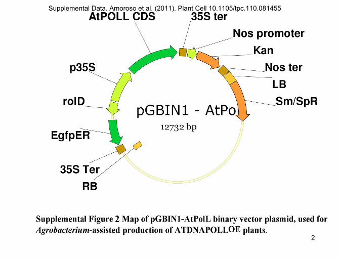
Supplemental Data. Amoroso et al. (2011). Plant Cell 10.1105/tpc.110.081455
2
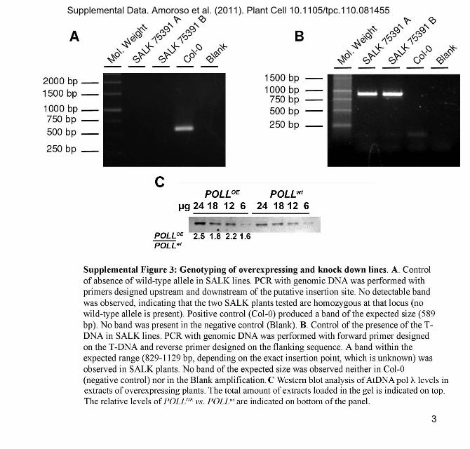
Supplemental Data. Amoroso et al. (2011). Plant Cell 10.1105/tpc.110.081455
3

Supplemental Data. Amoroso et al. (2011). Plant Cell 10.1105/tpc.110.081455
4

Supplemental Data. Amoroso et al. (2011). Plant Cell 10.1105/tpc.110.081455
5

DOI 10.1105/tpc.110.081455; originally published online February 15, 2011; 2011;23;806-822Plant Cell
Emmanuele Crespan, Rino Cella and Giovanni MagaAlessandra Amoroso, Lorenzo Concia, Caterina Maggio, Cécile Raynaud, Catherine Bergounioux,
Proliferating Cell Nuclear Antigen 2 andλ Requires DNA Polymerase Arabidopsis thalianaOxidative DNA Damage Bypass in
This information is current as of April 4, 2013
Supplemental Data http://www.plantcell.org/content/suppl/2011/02/04/tpc.110.081455.DC1.html
References http://www.plantcell.org/content/23/2/806.full.html#ref-list-1
This article cites 57 articles, 23 of which can be accessed free at:
Permissions https://www.copyright.com/ccc/openurl.do?sid=pd_hw1532298X&issn=1532298X&WT.mc_id=pd_hw1532298X
eTOCs http://www.plantcell.org/cgi/alerts/ctmain
Sign up for eTOCs at:
CiteTrack Alerts http://www.plantcell.org/cgi/alerts/ctmain
Sign up for CiteTrack Alerts at:
Subscription Information http://www.aspb.org/publications/subscriptions.cfm
is available at:Plant Physiology and The Plant CellSubscription Information for
ADVANCING THE SCIENCE OF PLANT BIOLOGY © American Society of Plant Biologists
Copyright © 2022 FDOKUMEN