Light and melatonin schedule neuronal differentiation in the habenular nuclei
Downloaded from www.microbiologyresearch.org by
IP: 54.242.247.86
On: Wed, 21 Sep 2016 16:41:19
The distribution pattern of proliferating cell nuclearantigen in the nuclei of Leishmania donovani
Devanand Kumar,1 Neha Minocha,1 Kalpana Rajanala2 and Swati Saha1
Correspondence
Swati Saha
1Department of Microbiology, University of Delhi South Campus, Benito Juarez Road, New Delhi110021, India
2National Institute of Immunology, Aruna Asaf Ali Marg, New Delhi 110067, India
Received 31 July 2009
Accepted 27 August 2009
DNA replication in eukaryotes is a highly conserved process marked by the licensing of multiple
origins, with pre-replication complex assembly in G1 phase, followed by the onset of replication at
these origins in S phase. The two strands replicate by different mechanisms, and DNA synthesis is
brought about by the activity of the replicative DNA polymerases Pol d and Pol e. Proliferating cell
nuclear antigen (PCNA) augments the processivity of these polymerases by serving as a DNA
sliding clamp protein. This study reports the cloning of PCNA from the protozoan Leishmania
donovani, which is the causative agent of the systemic disease visceral leishmaniasis. PCNA was
demonstrated to be robustly expressed in actively proliferating L. donovani promastigotes. We
found that the protein was present primarily in the nucleus throughout the cell cycle, and it was
found in both proliferating procyclic and metacyclic promastigotes. However, levels of expression
of PCNA varied through cell cycle progression, with maximum expression evident in G1 and S
phases. The subnuclear pattern of expression of PCNA differed in different stages of the cell
cycle; it formed distinct subnuclear foci in S phase, while it was distributed in a more diffuse
pattern in G2/M phase and post-mitotic phase cells. These subnuclear foci are the sites of active
DNA replication, suggesting that replication factories exist in Leishmania, as they do in higher
eukaryotes, thus opening avenues for investigating other Leishmania proteins that are involved in
DNA replication as part of these replication factories.
DNA replication in eukaryotes is the culmination of eventsoccurring in two distinct stages. In the first stage, originsget licensed to replicate by the establishment of multi-protein complexes called pre-replication complexes (pre-RCs) at origins in G1 phase, and, in the second stage, theselicensed origins get activated, and fire in S phase (Bell &Dutta, 2002; Diffley, 2004). DNA synthesis of the twostrands occurs by two different mechanisms, wherein onestrand is synthesized continuously, and the other issynthesized discontinuously. Several enzymes and proteinfactors are responsible for these processes, and DNApolymerases d and e play a major role in DNA synthesis.The DNA polymerase processivity factor proliferating cellnuclear antigen (PCNA) considerably augments theprocessivity of these enzymes (Moldovan et al., 2007;Bravo et al., 1987; Prelich et al., 1987). Replication proteinsand their auxiliary factors are organized in complexes
called replisomes. Several replicons and their replisomesare ordered into clusters called replication factories or foci.Replication foci can be indirectly or directly labelled byshort pulses with fluorescently labelled nucleotides, andthey are visible by fluorescence microscopy (Lengronneet al., 2001; Pasero et al., 1997; Gilbert, 2001; Kitamuraet al., 2006). Each replication focus is believed to harbourseveral replication forks (Berezney et al., 2000; Cook,2001).
Replication foci are rich in the enzymes and accessoryproteins that are involved in DNA replication. Theseenzymes and proteins include DNA polymerases, PCNA,replication factor C, replication protein A and DNA ligases,and cell cycle regulators such as cyclin A (Leonhardt et al.,2000; Cardoso et al., 1993). PCNA was the first protein tobe identified at these replication foci, and it has beenwidely used as a marker for these foci in higher eukaryotes,as it is conserved, and is absolutely essential for DNAreplication. Though first identified as the DNA polymerased processivity factor, PCNA forms a trimeric ring aroundDNA, anchoring not only DNA polymerases, but alsointeracting with other replication-associated factors, suchas Cdt1 and MCM10 (Das-Bradoo et al., 2006; Arias &Walter, 2006).
Abbreviations: DAPI, 49,6-diamidino-2-phenylindole; ORC, origin recog-nition complex; PCNA, proliferating cell nuclear antigen; pre-RC, pre-replication complex.
The GenBank/EMBL/DDBJ accession number for the LdPCNAsequence reported in this paper is GQ249893.
A CLUSTALW analysis of LdPCNA and PCNAs from various trypanoso-matids is available with the online version of this paper.
Microbiology (2009), 155, 3748–3757 DOI 10.1099/mic.0.033217-0
3748 033217 G 2009 SGM Printed in Great Britain

Downloaded from www.microbiologyresearch.org by
IP: 54.242.247.86
On: Wed, 21 Sep 2016 16:41:19
While PCNA has been extensively investigated in highereukaryotes, among protozoans it has been characterized inPlasmodium falciparum (Patterson et al., 2002; Kilbey et al.,1993; Horrocks et al., 1996; Li et al., 2002) and Toxoplasmagondii (Guerini et al., 2000, 2005) only. In both of theseorganisms, there are two distinct PCNAs. In T. gondii, onePCNA orthologue remains nuclear throughout the cellcycle, while the second orthologue is nuclear in S phase,but is uniformly distributed throughout the cell in earlyG1-phase and mitotic cells (Guerini et al., 2005). In P.falciparum, the two PCNAs are expressed during the sexualand asexual stages, though only PfPCNA1 has all theconserved motifs of PCNA. PfPCNA1 has been shown tointeract with (origin recognition complex) ORC5 andORC1 of P. falciparum, and it co-localizes with both ORC1and ORC5 in immunofluorescence studies (Gupta et al.,2008, 2009). PfORC1 carries a PIP domain (the domainresponsible for docking PCNA-interacting proteins withPCNA), and genetic complementation analysis using ayeast ORC1 mutant strain has revealed that while wild-typePfORC1 can complement mutant ScORC1, PIP mutants ofPfORC1 cannot. This finding points to the importance ofthe PfORC1–PCNA interaction in modulating cell viability,probably through DNA replication.
Not much has been reported about the pre-replication andreplication machinery of the genomes of trypanosomatids.The expression of the pre-RC protein ORC1 fromLeishmania major has been analysed using GFP-ORC1(Kumar et al., 2008), and the protein has been found to benuclear throughout the cell cycle. The pre-RC protein Cdc45of Trypanosoma brucei has been demonstrated to interactwith BRCA1 of T. brucei (Oyola et al., 2009). Here, we haveexamined PCNA in the trypanosomatid Leishmania dono-vani (LdPCNA), and characterized its expression in L.donovani promastigotes. LdPCNA was found to be wellexpressed in proliferating promastigotes. An analysis of thesubcellular localization of the protein in relation to thedifferent stages of the cell cycle revealed that, while LdPCNAwas nuclear throughout the cell cycle, maximum expressionwas seen in G1 and S phases. Furthermore, PCNA formeddistinct subnuclear foci in S phase. BrdU-labelling experi-ments indicated that these subnuclear foci were sites ofactive DNA replication. Our results point towards theexistence of replication factories in Leishmania, and indicatethat LdPCNA is a promising tool for investigating otherLeishmania proteins that are involved in DNA replication aspart of these replication factories.
METHODS
Leishmania culture. L. donovani 1S promastigotes were cultured in
M199 medium supplemented with 10 % fetal bovine serum, 2 mM
L-glutamine, 100 mM adenine and 5 mg haemin ml21.
Genomic DNA isolation. About 16109 promastigotes were lysed in
10 mM Tris/HCl (pH 8.0), 10 mM EDTA, 150 mM NaCl and 1 %
SDS. After treatment with 100 mg proteinase K ml21 (Roche), and
phenol/chloroform extraction, the genomic DNA was precipitatedwith ethanol.
Cloning of PCNA. The primers PCNA-F (59-CACCGGATCCGAA-TTCATGCTCGAGGCTCAG-39) and PCNA-R (59-TACTGCAGT-CCCTCCGCATCGTCCACCTTG-39) were used to amplify the PCNAgene from L. donovani genomic DNA, using the high-performanceproofreading enzyme Phu DNA polymerase (Finnzymes). The PCRproduct was cloned into the pENTR/D-TOPO vector (Invitrogen),and subcloned into the EcoRI and PstI sites of the expression vectorpASK-IBA43plus (IBA BioTAGnology) for overexpression of recom-binant LdPCNA.
Overexpression and purification of PCNA. BL21 Codon Plus cells(Stratagene) harbouring plasmid pASK-PCNA were grown to mid-exponential phase, and expression of PCNA was induced with 200 nganhydrotetracycline ml21. Cells were further incubated at 16 uC for16218 h. To assess protein solubility, harvested cells were resus-pended in PBS, lysed on ice by sonication, and the lysate was clarifiedby centrifugation (10 000 g). After solubilization of the pellet in 36SDS-sample loading buffer, the lysate and pellet were analysed forPCNA expression by SDS-PAGE. PCNA was purified by affinitychromatography using Strep-Tactin II resin (IBA BioTAGnology),according to the instructions of the manufacturer.
Glutaraldehdye cross-linking. An 8–10 mg quantity of purifiedPCNA was incubated for 5 min in 10 mM Tris/HCl (pH 8.0)containing 0.1 % glutaraldehyde. Reactions were stopped by theaddition of 36 SDS-sample loading buffer, and the product wasresolved by SDS-PAGE on 8 % gels.
Raising antibodies to PCNA, and their characterization.Polyclonal antibodies were raised in mouse and rabbit. Antibodieswere tested at dilutions of 1 : 10 000, 1 : 5000 and 1 : 2500 by Westernblotting, as described by Sambrook et al. (1989).
Preparation of Leishmania extracts. L. donovani whole-cellextracts were prepared using the M-PER kit (Pierce Biotechnology),and nuclear and cytosolic extracts were prepared using the NE-PERkit (Pierce Biotechnology).
Immunofluorescence analysis. For examining the subcellularlocalization of PCNA, harvested promastigotes were washed withPBS and fixed with 2 % paraformaldehyde for 20 min, and cellspreads were made on poly-lysine-coated coverslips. The cells werepermeabilized with 0.1 % Triton X-100 in PBS for 5 min, and thenwashed, and subjected to blocking with 10 % chicken serum(Invitrogen). After washing, the cells were incubated with anti-PCNA antibody (1 : 100 dilution) for 1–2 h at room temperature, andthen washed, and incubated with Texas-red-labelled anti-mousesecondary antibody (Jackson ImmunoResearch Laboratories). Afterwashing, the coverslips were mounted in anti-fade solution contain-ing 49,6-diamidino-2-phenylindole (DAPI) (Vectashield; VectorLaboratories). Cells were viewed and images were acquired using a6100 objective, utilizing a motorized epifluorescence microscope(Upright Axioimager M1; Carl Zeiss MicroImaging) equipped with ahigh-resolution camera (AxioCam MRm Rev. 2; Carl ZeissMicroImaging). Images were analysed by AxioVision Software Rel.4.4 (Carl Zeiss MicroImaging).
To quantify PCNA expression, fluorescence intensity of nuclearPCNA was quantified using ImageJ software (Wayne Rasband,National Institutes of Health, Bethesda, MD, USA).
For examining replication foci, exponentially growing promastigoteswere pulsed with 40 mM BrdU for 20 min. Cells were then harvested,washed with PBS, and fixed in 2 % paraformaldehyde. Cell spreadswere permeabilized and blocked, and then incubated with both anti-
LdPCNA expression in Leishmania donovani
http://mic.sgmjournals.org 3749

Downloaded from www.microbiologyresearch.org by
IP: 54.242.247.86
On: Wed, 21 Sep 2016 16:41:19
BrdU antibody (GE Healthcare) and rabbit anti-PCNA antibody
(1 : 400 dilution). After washing, Texas-red-labelled anti-mouse
(Jackson ImmunoResearch Laboratories) and Alexa-Fluor-488-
labelled anti-rabbit (Invitrogen) secondary antibodies were applied.
After washing, the coverslips were mounted, and imaging of nuclei
was done using an inverted motorized confocal microscope (LSM
510; Carl Zeiss MicroImaging) with a 6100 objective by collecting
images at high speed in different focal planes (Z stacks) throughout
the entire nucleus.
For examining the distribution pattern of PCNA after exposure to
UV, exponentially growing cells were pulsed with BrdU for 10 min,
irradiated using a hand-held UV lamp (254 nm), and further
incubated for 10 min. Cells were harvested for immunofluorescence
analysis by using BrdU and PCNA antibodies, as described above.
RESULTS AND DISCUSSION
Cloning and purification of PCNA
While two PCNA orthologues have been found in each ofthe other protozoan species that have been studied (Li et al.,2002; Guerini et al., 2005; Patterson et al., 2002), theannotation of the L. major whole genome sequencerevealed the presence of a single PCNA orthologue (Ivenset al., 2005). Based on the published L. major sequence, wedesigned end primers to amplify PCNA from L. donovanigenomic DNA. The ~0.8 kb amplicon obtained (Fig. 1a)was cloned, and sequenced from both ends usingoverlapping primers. Clones of amplicons obtained fromtwo independent PCRs were sequenced. An analysis of thederived amino acid sequence of LdPCNA showed that theprotein was 293 aa in length, and had the conserved motifsthat typify PCNAs of other eukaryotic organisms (Fig. 1b).A comparative pairwise analysis of the amino acid sequenceof LdPCNA with PCNAs from other eukaryotes (Fig. 1c)revealed that LdPCNA shared 55–65 % homology and 30–40 % identity with those PCNAs. When comparingLdPCNA with PCNAs from other trypanosomatids (basedon the published genome sequences) (Supplementary Fig.S1), we found that while the protein shared between 95 and100 % identity with PCNAs from other Leishmania species,it shared about 75 % identity with PCNAs of T. brucei andTrypanosoma cruzi. The C-terminal insertion apparent inFig. 1(c) is conserved in all the trypanosomatids.
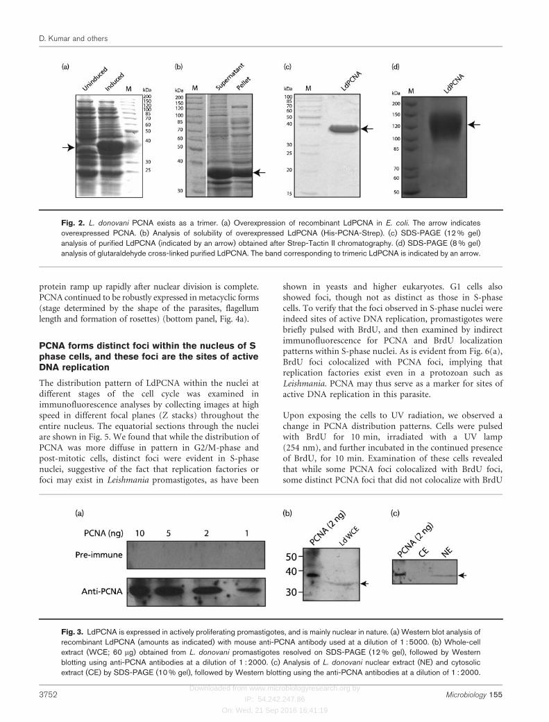
In order to examine PCNA expression in L. donovanipromastigotes, LdPCNA was overexpressed in Escherichiacoli, the recombinant protein was purified, and antibodieswere raised against it. The gene was subcloned into the E.coli expression vector pASK-IBA43plus, and the proteinwas overexpressed in E. coli BL21 Codon Plus cells (Fig.2a). The overexpressed protein was about 60 % soluble(Fig. 2b), and it was purified by affinity chromatographyusing a Strep-Tactin II matrix. The pure protein (Fig. 2c)was found to exist as a trimer by using glutaraldehydecross-linking (Fig. 2d). This result is in keeping with thepresence of the conserved residues responsible for trimerintersubunit interactions in PCNA (Fig. 1b). All eukaryoticPCNAs reported to date form a ring-shaped homotrimeric
complex that encircles DNA, and form a sliding clamp(Krishna et al., 1994; Gulbis et al., 1996; Bowman et al.,2004).
PCNA is well-expressed in actively proliferatingLeishmania promastigotes
Polyclonal antibodies against LdPCNA were raised in mice,and subsequently in rabbits. To assess the specificity andefficacy of the antibodies raised, Western blotting analysiswas carried out using recombinant LdPCNA. While pre-immune serum did not react with recombinant LdPCNA,anti-PCNA antiserum detected as little as 1 ng LdPCNA ata dilution of 1 : 5000 (Fig. 3a). The expression of PCNA inL. donovani promastigotes was analysed in whole-cellextracts of actively proliferating promastigotes using theseantibodies. A single band of the expected size was detected(recombinant LdPCNA is 2 kDa larger because of the tagsat both ends), indicating that PCNA was well expressedand that the antibodies were of high specificity (Fig. 3b). Innuclear and cytosolic extracts made from asynchronouscultures of these promastigotes, PCNA was detected in thenuclear extracts only (Fig. 3c), indicating that the protein isprimarily nuclear in nature.
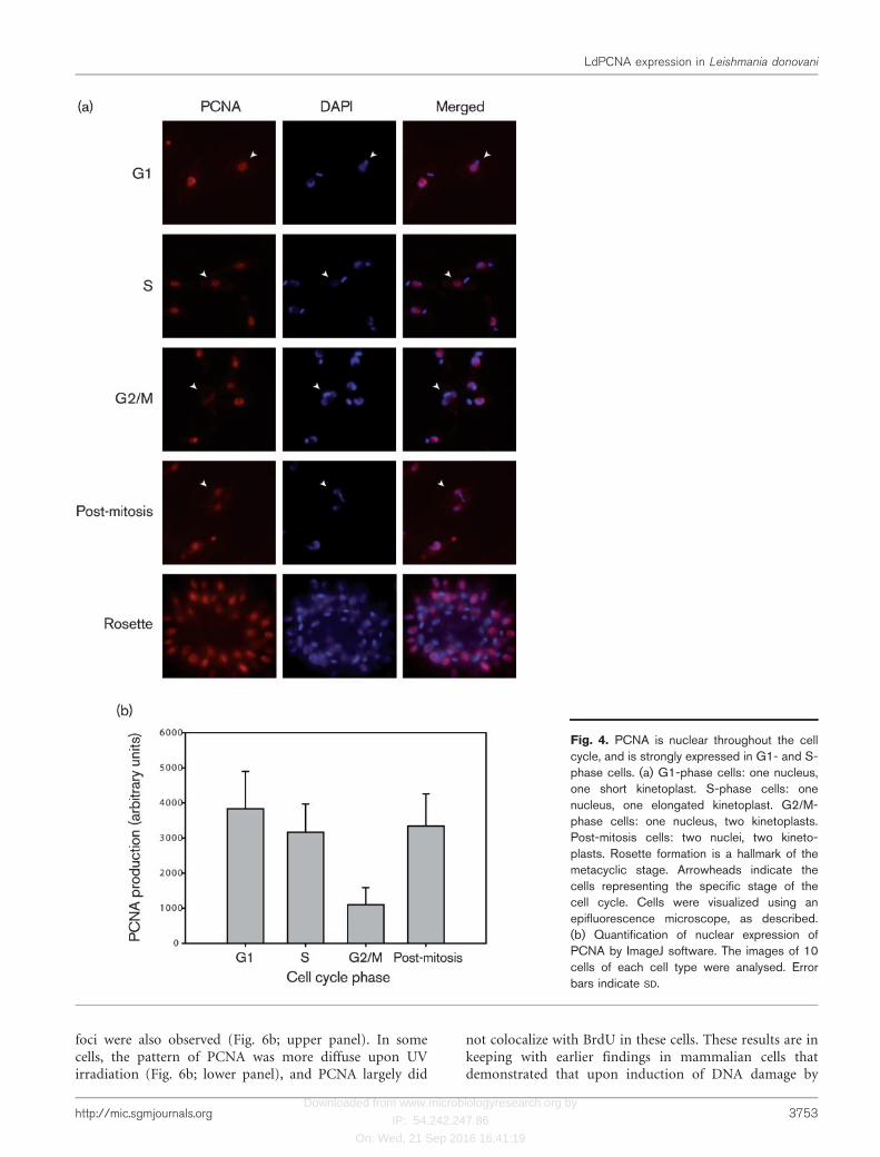
PCNA is nuclear throughout the cell cycle, but ismainly expressed in G1 and S phases
The subcellular localization of LdPCNA was examined tosee if the specific import of PCNA into the nucleus in onlyG1 and S phases could be a means of regulation of DNAreplication in Leishmania promastigotes. Immuno-fluorescence analysis of cells harvested from asynchronousprocyclic cultures was carried out using the anti-PCNAantibodies we raised. In trypanosomatids, due to thestaggered timing of replication of kinetoplast DNA ascompared with the genome, the kinetoplast can be used asa marker for cell cycle stage of an individual cell (Siegel etal., 2008). While cells in G1 phase contain one nucleus andone short roundish kinetoplast, S-phase cells contain onenucleus and one elongated kinetoplast. Cells in G2/Mphase contain one nucleus and two kinetoplasts, askinetoplast replication is complete by this stage of the cellcycle, and the newly synthesized kinetoplasts are alreadyseparate. Post-mitotic cells have two completely separatednuclei and two kinetoplasts. Indirect immunofluorescencestudies with anti-PCNA antibodies revealed that thisprotein was nuclear at all stages of the cell cycle (Fig. 4a).We were unable to detect PCNA in the cytoplasm in anyphase of cell cycle. However, in over 400 cells examined,expression was always robust in G1- and S-phase cells, butvery weak in mitotic cells (Fig. 4a). Quantification offluorescence intensity of nuclear PCNA at different stagesof the cell cycle revealed that expression in G2/M phase wasabout 2.5- to 3-fold lower than in G1 or S phase (Fig. 4b).Post-mitotic cells showed almost as much expression ofPCNA as G1- and S-phase cells, suggesting that whilePCNA probably degrades during G2/M phase, levels of the
D. Kumar and others
3750 Microbiology 155

Downloaded from www.microbiologyresearch.org by
IP: 54.242.247.86
On: Wed, 21 Sep 2016 16:41:19
Fig. 1. Cloning of PCNA from L. donovani. (a) Amplification of PCNA from L. donovani genomic DNA. The ~0.8 kb PCNA
gene product is indicated by an arrow. M, DNA marker. (b) Schematic representation of the conserved domains in LdPCNA. (c)CLUSTALW (www.ebi.ac.uk/Tools/clustalw2) analysis of LdPCNA and PCNAs from other eukaryotes viewed using the Jalviewmultiple alignment editor (Waterhouse et al., 2009). H. sapiens, Homo sapiens; X. laevis, Xenopus laevis; D. melanogaster,Drosophila melanogaster; S. pombe, Schizosaccharomyces pombe; S. cerevisiae, Saccharomyces cerevisiae.
LdPCNA expression in Leishmania donovani
http://mic.sgmjournals.org 3751
Downloaded from www.microbiologyresearch.org by
IP: 54.242.247.86
On: Wed, 21 Sep 2016 16:41:19
protein ramp up rapidly after nuclear division is complete.PCNA continued to be robustly expressed in metacyclic forms(stage determined by the shape of the parasites, flagellumlength and formation of rosettes) (bottom panel, Fig. 4a).
PCNA forms distinct foci within the nucleus of Sphase cells, and these foci are the sites of activeDNA replication
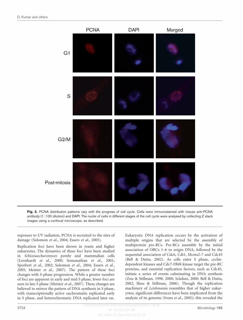
The distribution pattern of LdPCNA within the nuclei atdifferent stages of the cell cycle was examined inimmunofluorescence analyses by collecting images at highspeed in different focal planes (Z stacks) throughout theentire nucleus. The equatorial sections through the nucleiare shown in Fig. 5. We found that while the distribution ofPCNA was more diffuse in pattern in G2/M-phase andpost-mitotic cells, distinct foci were evident in S-phasenuclei, suggestive of the fact that replication factories orfoci may exist in Leishmania promastigotes, as have been
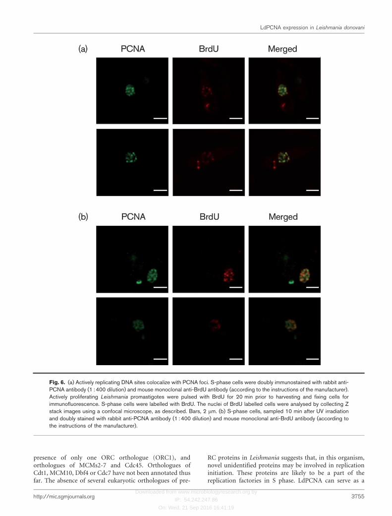
shown in yeasts and higher eukaryotes. G1 cells alsoshowed foci, though not as distinct as those in S-phasecells. To verify that the foci observed in S-phase nuclei wereindeed sites of active DNA replication, promastigotes werebriefly pulsed with BrdU, and then examined by indirectimmunofluorescence for PCNA and BrdU localizationpatterns within S-phase nuclei. As is evident from Fig. 6(a),BrdU foci colocalized with PCNA foci, implying thatreplication factories exist even in a protozoan such asLeishmania. PCNA may thus serve as a marker for sites ofactive DNA replication in this parasite.
Upon exposing the cells to UV radiation, we observed achange in PCNA distribution patterns. Cells were pulsedwith BrdU for 10 min, irradiated with a UV lamp(254 nm), and further incubated in the continued presenceof BrdU, for 10 min. Examination of these cells revealedthat while some PCNA foci colocalized with BrdU foci,some distinct PCNA foci that did not colocalize with BrdU
Fig. 2. L. donovani PCNA exists as a trimer. (a) Overexpression of recombinant LdPCNA in E. coli. The arrow indicatesoverexpressed PCNA. (b) Analysis of solubility of overexpressed LdPCNA (His-PCNA-Strep). (c) SDS-PAGE (12 % gel)analysis of purified LdPCNA (indicated by an arrow) obtained after Strep-Tactin II chromatography. (d) SDS-PAGE (8 % gel)analysis of glutaraldehyde cross-linked purified LdPCNA. The band corresponding to trimeric LdPCNA is indicated by an arrow.
Fig. 3. LdPCNA is expressed in actively proliferating promastigotes, and is mainly nuclear in nature. (a) Western blot analysis ofrecombinant LdPCNA (amounts as indicated) with mouse anti-PCNA antibody used at a dilution of 1 : 5000. (b) Whole-cellextract (WCE; 60 mg) obtained from L. donovani promastigotes resolved on SDS-PAGE (12 % gel), followed by Westernblotting using anti-PCNA antibodies at a dilution of 1 : 2000. (c) Analysis of L. donovani nuclear extract (NE) and cytosolicextract (CE) by SDS-PAGE (10 % gel), followed by Western blotting using the anti-PCNA antibodies at a dilution of 1 : 2000.
D. Kumar and others
3752 Microbiology 155
Downloaded from www.microbiologyresearch.org by
IP: 54.242.247.86
On: Wed, 21 Sep 2016 16:41:19
foci were also observed (Fig. 6b; upper panel). In somecells, the pattern of PCNA was more diffuse upon UVirradiation (Fig. 6b; lower panel), and PCNA largely did
not colocalize with BrdU in these cells. These results are inkeeping with earlier findings in mammalian cells thatdemonstrated that upon induction of DNA damage by
Fig. 4. PCNA is nuclear throughout the cellcycle, and is strongly expressed in G1- and S-phase cells. (a) G1-phase cells: one nucleus,one short kinetoplast. S-phase cells: onenucleus, one elongated kinetoplast. G2/M-phase cells: one nucleus, two kinetoplasts.Post-mitosis cells: two nuclei, two kineto-plasts. Rosette formation is a hallmark of themetacyclic stage. Arrowheads indicate thecells representing the specific stage of thecell cycle. Cells were visualized using anepifluorescence microscope, as described.(b) Quantification of nuclear expression ofPCNA by ImageJ software. The images of 10cells of each cell type were analysed. Errorbars indicate SD.
LdPCNA expression in Leishmania donovani
http://mic.sgmjournals.org 3753
Downloaded from www.microbiologyresearch.org by
IP: 54.242.247.86
On: Wed, 21 Sep 2016 16:41:19
exposure to UV radiation, PCNA is recruited to the sites ofdamage (Solomon et al., 2004; Essers et al., 2005).
Replication foci have been shown in yeasts and highereukaryotes. The dynamics of these foci have been studiedin Schizosaccharomyces pombe and mammalian cells(Leonhardt et al., 2000; Somanathan et al., 2001;Sporbert et al., 2002; Solomon et al., 2004; Essers et al.,2005; Meister et al., 2007). The pattern of these focichanges with S-phase progression. While a greater numberof foci are apparent in early and mid S phase, fewer foci areseen in late S phase (Meister et al., 2007). These changes arebelieved to mirror the pattern of DNA synthesis in S phase,with transcriptionally active euchromatin replicated earlyin S phase, and heterochromatic DNA replicated later on.
Eukaryotic DNA replication occurs by the activation ofmultiple origins that are selected by the assembly ofmultiprotein pre-RCs. Pre-RCs assemble by the initialassociation of ORCs 1–6 to origin DNA, followed by thesequential association of Cdc6, Cdt1, Mcms2-7 and Cdc45(Bell & Dutta, 2002). As cells enter S phase, cyclin-dependent kinases and Cdc7-Dbf4 kinase target the pre-RCproteins, and essential replication factors, such as Cdc45,initiate a series of events culminating in DNA synthesis(Zou & Stillman, 1998, 2000; Sclafani, 2000; Bell & Dutta,2002; Sheu & Stillman, 2006). Though the replicationmachinery of Leishmania resembles that of higher eukar-yotes, significant differences have been implicated from theanalysis of its genome (Ivens et al., 2005); this revealed the
Fig. 5. PCNA distribution patterns vary with the progress of cell cycle. Cells were immunostained with mouse anti-PCNAantibody (1 : 100 dilution) and DAPI. The nuclei of cells in different stages of the cell cycle were analysed by collecting Z stackimages using a confocal microscope, as described.
D. Kumar and others
3754 Microbiology 155
Downloaded from www.microbiologyresearch.org by
IP: 54.242.247.86
On: Wed, 21 Sep 2016 16:41:19
presence of only one ORC orthologue (ORC1), andorthologues of MCMs2-7 and Cdc45. Orthologues ofCdt1, MCM10, Dbf4 or Cdc7 have not been annotated thusfar. The absence of several eukaryotic orthologues of pre-
RC proteins in Leishmania suggests that, in this organism,novel unidentified proteins may be involved in replicationinitiation. These proteins are likely to be a part of thereplication factories in S phase. LdPCNA can serve as a
Fig. 6. (a) Actively replicating DNA sites colocalize with PCNA foci. S-phase cells were doubly immunostained with rabbit anti-PCNA antibody (1 : 400 dilution) and mouse monoclonal anti-BrdU antibody (according to the instructions of the manufacturer).Actively proliferating Leishmania promastigotes were pulsed with BrdU for 20 min prior to harvesting and fixing cells forimmunofluorescence. S-phase cells were labelled with BrdU. The nuclei of BrdU labelled cells were analysed by collecting Zstack images using a confocal microscope, as described. Bars, 2 mm. (b) S-phase cells, sampled 10 min after UV irradiationand doubly stained with rabbit anti-PCNA antibody (1 : 400 dilution) and mouse monoclonal anti-BrdU antibody (according tothe instructions of the manufacturer).
LdPCNA expression in Leishmania donovani
http://mic.sgmjournals.org 3755
Downloaded from www.microbiologyresearch.org by
IP: 54.242.247.86
On: Wed, 21 Sep 2016 16:41:19
marker for these factories, and can be utilized to seek outother Leishmania replication proteins, including any novelproteins that may be involved in the process.
ACKNOWLEDGEMENTS
We thank Dr Vinay Nandicoori and Dr Sagar Sengupta for the use of
the fluorescence microscope, and the laboratory of Dr Deepak Pental
for DNA sequencing. This work was supported by a grant from the
Council of Scientific and Industrial Research, India, and the
Department of Biotechnology, India. D. K. is a Senior Research
Fellow supported from this grant. N. M. is a University Grants
Commission Senior Research Fellow.
REFERENCES
Arias, E. E. & Walter, J. C. (2006). PCNA functions as a molecular
platform to trigger Cdt1 destruction and prevent re-replication. Nat
Cell Biol 8, 84–90.
Bell, S. P. & Dutta, A. (2002). DNA replication in eukaryotic cells.
Annu Rev Biochem 71, 333–374.
Berezney, R., Dubey, D. D. & Huberman, J. A. (2000). Heterogeneity
of eukaryotic replicons, replicon clusters, and replication foci.
Chromosoma 108, 471–484.
Bowman, G. D., O’Donnell, M. & Kuriyan, J. (2004). Structural analysis
of a eukaryotic sliding DNA clamp–clamp loader complex. Nature
429, 724–730.
Bravo, R., Frank, R., Blundell, P. A. & Macdonald-Bravo, H. (1987).Cyclin/PCNA is the auxiliary protein of DNA polymerase-d. Nature
326, 515–517.
Cardoso, M. C., Leonhardt, H. & Nadal-Ginard, B. (1993). Reversal of
terminal differentiation and control of DNA replication: cyclin A and
Cdk2 specifically localize at subnuclear sites of DNA replication. Cell
74, 979–992.
Cook, P. R. (2001). Principles of Nuclear Structures and Function. New
York: Wiley-Liss.
Das-Bradoo, S., Ricke, R. M. & Bielinsky, A. K. (2006). Interaction
between PCNA and diubiquitinated Mcm10 is essential for cell
growth in budding yeast. Mol Cell Biol 26, 4806–4817.
Diffley, J. F. (2004). Regulation of early events in chromosome
replication. Curr Biol 14, R778–R786.
Essers, J., Theil, A. F., Baldeyron, C., van Cappellen, W. A.,Houtsmuller, A. B., Kanaar, R. & Vermeulen, W. (2005). Nuclear
dynamics of PCNA in DNA replication and repair. Mol Cell Biol 25,
9350–9359.
Gilbert, D. M. (2001). Making sense of eukaryotic DNA replication
origins. Science 294, 96–100.
Guerini, M. N., Que, X., Reed, S. L. & White, M. W. (2000). Two genes
encoding unique proliferating-cell-nuclear-antigens are expressed in
Toxoplasma gondii. Mol Biochem Parasitol 109, 121–131.
Guerini, M. N., Behnke, M. S. & White, M. W. (2005). Biochemical and
genetic analysis of the distinct proliferating cell nuclear antigens of
Toxoplasma gondii. Mol Biochem Parasitol 142, 56–65.
Gulbis, J. M., Kelman, Z., Hurwitz, J., O’Donnell, M. & Kuriyan, J.(1996). Structure of the C-terminal region of p21(WAF1/CIP1)
complexed with human PCNA. Cell 87, 297–306.
Gupta, A., Mehra, P. & Dhar, S. K. (2008). Plasmodium falciparum
origin recognition complex subunit 5: functional characterization and
role in DNA replication foci formation. Mol Microbiol 69, 646–665.
Gupta, A., Mehra, P., Deshmukh, A., Dar, A., Mitra, P., Roy, N. & Dhar,S. K. (2009). Functional dissection of the catalytic carboxyl-terminaldomain of human malaria parasite Plasmodium falciparum origin
recognition complex subunit 1 (PfORC1). Eukaryot Cell 8, 1341–
1351.
Horrocks, P., Jackson, M., Cheesman, S., White, J. H. & Kilbey, B. J.(1996). Stage specific expression of proliferating cell nuclear antigenand DNA polymerase d from Plasmodium falciparum. Mol Biochem
Parasitol 79, 177–182.
Ivens, A. C., Peacock, C. S., Worthey, E. A., Murphy, L., Aggarwal, G.,Berriman, M., Sisk, E., Rajandream, M. A., Adlem, E. & other authors(2005). The genome of the kinetoplastid parasite, Leishmania major.
Science 309, 436–442.
Kilbey, B. J., Fraser, I., McAleese, S., Goman, M. & Ridley, R. G.(1993). Molecular characterisation and stage-specific expression of
proliferating cell nuclear antigen (PCNA) from the malarial parasite,Plasmodium falciparum. Nucleic Acids Res 21, 239–243.
Kitamura, E., Blow, J. J. & Tanaka, T. U. (2006). Live-cell imagingreveals replication of individual replicons in eukaryotic replication
factories. Cell 125, 1297–1308.
Krishna, T. S., Kong, X. P., Gary, S., Burgers, P. M. & Kuriyan, J.(1994). Crystal structure of the eukaryotic DNA polymerase
processivity factor PCNA. Cell 79, 1233–1243.
Kumar, D., Mukherji, A. & Saha, S. (2008). Expression and subcellular
localization of ORC1 in Leishmania major. Biochem Biophys Res
Commun 375, 74–79.
Lengronne, A., Pasero, P., Bensimon, A. & Schwob, E. (2001).Monitoring S phase progression globally and locally using BrdU
incorporation in TK(+) yeast strains. Nucleic Acids Res 29, 1433–1442.
Leonhardt, H., Rahn, H. P., Weinzierl, P., Sporbert, A., Cremer, T.,Zink, D. & Cardoso, M. C. (2000). Dynamics of DNA replication
factories in living cells. J Cell Biol 149, 271–280.
Li, J. L., Warren, A. V. & Cox, L. S. (2002). Identification of a secondproliferating cell nuclear antigen in the human malarial pathogen
Plasmodium falciparum. Int J Parasitol 32, 1683–1692.
Meister, P., Taddei, A., Ponti, A., Baldacci, G. & Gasser, S. M. (2007).Replication foci dynamics: replication patterns are modulated
by S-phase checkpoint kinases in fission yeast. EMBO J 26, 1315–1326.
Moldovan, G. L., Pfander, B. & Jentsch, S. (2007). PCNA, the maestro
of the replication fork. Cell 129, 665–679.
Oyola, S. O., Bringaud, F. & Melville, S. E. (2009). A kinetoplastid
BRCA2 interacts with DNA replication protein CDC45. Int J Parasitol39, 59–69.
Pasero, P., Braguglia, D. & Gasser, S. M. (1997). ORC-dependent
and origin-specific initiation of DNA replication at defined foci inisolated yeast nuclei. Genes Dev 11, 1504–1518.
Patterson, S., Whittle, C., Robert, C. & Chakrabarti, D. (2002).Molecular characterization and expression of an alternate prolif-
erating cell nuclear antigen homologue, PfPCNA2, in Plasmodium
falciparum. Biochem Biophys Res Commun 298, 371–376.
Prelich, G., Tan, C. K., Kostura, M., Mathews, M. B., So, A. G.,Downey, K. M. & Stillman, B. (1987). Functional identity of
proliferating cell nuclear antigen and a DNA polymerase-d auxiliaryprotein. Nature 326, 517–520.
Sambrook, J., Fristch, E. F. & Maniatis, T. (1989). Molecular Cloning:a Laboratory Manual, 2nd edn. Cold Spring Harbor, NY: Cold Spring
Harbor Laboratory.
Sclafani, R. A. (2000). Cdc7p-Dbf4p becomes famous in the cell cycle.J Cell Sci 113, 2111–2117.
D. Kumar and others
3756 Microbiology 155

Downloaded from www.microbiologyresearch.org by
IP: 54.242.247.86
On: Wed, 21 Sep 2016 16:41:19
Sheu, Y. J. & Stillman, B. (2006). Cdc7-Dbf4 phosphorylates MCMproteins via a docking site-mediated mechanism to promote S phaseprogression. Mol Cell 24, 101–113.
Siegel, T. N., Hekstra, D. R. & Cross, G. A. (2008). Analysis of theTrypanosoma brucei cell cycle by quantitative DAPI imaging. MolBiochem Parasitol 160, 171–174.
Solomon, D. A., Cardoso, M. C. & Knudsen, E. S. (2004). Dynamictargeting of the replication machinery to sites of DNA damage. J CellBiol 166, 455–463.
Somanathan, S., Suchyna, T. M., Siegel, A. J. & Berezney, R. (2001).Targeting of PCNA to sites of DNA replication in the mammalian cellnucleus. J Cell Biochem 81, 56–67.
Sporbert, A., Gahl, A., Ankerhold, R., Leonhardt, H. & Cardoso, M. C.(2002). DNA polymerase clamp shows little turnover at established
replication sites but sequential de novo assembly at adjacent origin
clusters. Mol Cell 10, 1355–1365.
Waterhouse, A. M., Procter, J. B., Martin, D. M. A., Clamp, M. & Barton,G. J. (2009). Jalview Version 2 – a multiple sequence alignment editor
and analysis workbench. Bioinformatics 25, 1189–1191.
Zou, L. & Stillman, B. (1998). Formation of a preinitiation complex by
S-phase cyclin CDK-dependent loading of Cdc45p onto chromatin.
Science 280, 593–596.
Zou, L. & Stillman, B. (2000). Assembly of a complex containing
Cdc45p, replication protein A, and Mcm2p at replication origins
controlled by S-phase cyclin-dependent kinases and Cdc7p-Dbf4p
kinase. Mol Cell Biol 20, 3086–3096.
Edited by: L. Knoll
LdPCNA expression in Leishmania donovani
http://mic.sgmjournals.org 3757
Copyright © 2022 FDOKUMEN