Preferential DNA damage prevention by the E. coli AidB gene: A new mechanism for the protection of...
20
Preferential DNA damage prevention by the E. coli AidB gene: a new mechanism for protection of specific genes Valentina Rippa 1,2 , Angela Duilio 2 , Pamela di Pasquale 2 , Angela Amoresano 2 , Paolo Landini 3 , and Michael R. Volkert 1,* 1 Department of Microbiology and Physiological Systems, University of Massachusetts Medical School, Worcester, MA, 01655, USA 2 Department of Organic Chemistry and Biochemistry, University Federico II of Naples, Naples, Italy 3 Department of Biomolecular Sciences and Biotechnology, University of Milan, Milan, Italy Abstract aidB is one of four genes of E. coli that is induced by alkylating agents and regulated by Ada protein. Three genes (ada, alkA, and alkB) encode DNA repair proteins that remove or repair alkylated bases. However, the role of AidB remains unclear despite extensive efforts to determine its function in cells exposed to alkylating agents. The E. coli AidB protein was identified as a component of the protein complex that assembles at strong promoters. We demonstrate that AidB protein preferentially binds to UP elements, AT rich transcription enhancer sequences found upstream of many highly expressed genes, several DNA repair genes, and housekeeping genes. AidB allows efficient transcription from promoters containing an UP element upon exposure to a DNA methylating agent and protects downstream genes from DNA damage. The DNA binding domain is required to target AidB to specific genes preferentially protecting them from alkylation damage. However, deletion of AidB’s DNA binding domain does not prevent its antimutagenic activity, instead this deletion appears to allow AidB to function as a cytoplasmic alkylation resistance protein. Our studies identify the role of AidB in alkylating agent exposed cells and suggest a new cellular strategy in which a subset of the genome is preferentially protected from damage by alkylating agents. Keywords AidB protein; alkylating agents; DNA protection; UP element Introduction The E. coli aidB gene is one of four genes of the adaptive response to alkylation damage and is regulated by Ada protein (For review see: [1,2]). Ada protein is a methyltransferase that functions as a transcriptional activator after transfer of a methyl group from DNA to a © 2011 Elsevier B.V. All rights reserved. * Corresponding Author: [email protected], 1 Department of Microbiology and Physiological Systems, University of Massachusetts Medical School, Rm. S6-117, Worcester, MA, 01655, USA, Phone: 508-856-2314; Fax 508-856-5920. Publisher's Disclaimer: This is a PDF file of an unedited manuscript that has been accepted for publication. As a service to our customers we are providing this early version of the manuscript. The manuscript will undergo copyediting, typesetting, and review of the resulting proof before it is published in its final citable form. Please note that during the production process errors may be discovered which could affect the content, and all legal disclaimers that apply to the journal pertain. NIH Public Access Author Manuscript DNA Repair (Amst). Author manuscript; available in PMC 2012 September 5. Published in final edited form as: DNA Repair (Amst). 2011 September 5; 10(9): 934–941. doi:10.1016/j.dnarep.2011.06.001. NIH-PA Author Manuscript NIH-PA Author Manuscript NIH-PA Author Manuscript
-
Upload
independent -
Category
Documents
-
view
2 -
download
0
Transcript of Preferential DNA damage prevention by the E. coli AidB gene: A new mechanism for the protection of...

Preferential DNA damage prevention by the E. coli AidB gene: anew mechanism for protection of specific genes
Valentina Rippa1,2, Angela Duilio2, Pamela di Pasquale2, Angela Amoresano2, PaoloLandini3, and Michael R. Volkert1,*
1Department of Microbiology and Physiological Systems, University of Massachusetts MedicalSchool, Worcester, MA, 01655, USA2Department of Organic Chemistry and Biochemistry, University Federico II of Naples, Naples,Italy3Department of Biomolecular Sciences and Biotechnology, University of Milan, Milan, Italy
AbstractaidB is one of four genes of E. coli that is induced by alkylating agents and regulated by Adaprotein. Three genes (ada, alkA, and alkB) encode DNA repair proteins that remove or repairalkylated bases. However, the role of AidB remains unclear despite extensive efforts to determineits function in cells exposed to alkylating agents. The E. coli AidB protein was identified as acomponent of the protein complex that assembles at strong promoters. We demonstrate that AidBprotein preferentially binds to UP elements, AT rich transcription enhancer sequences foundupstream of many highly expressed genes, several DNA repair genes, and housekeeping genes.AidB allows efficient transcription from promoters containing an UP element upon exposure to aDNA methylating agent and protects downstream genes from DNA damage. The DNA bindingdomain is required to target AidB to specific genes preferentially protecting them from alkylationdamage. However, deletion of AidB’s DNA binding domain does not prevent its antimutagenicactivity, instead this deletion appears to allow AidB to function as a cytoplasmic alkylationresistance protein. Our studies identify the role of AidB in alkylating agent exposed cells andsuggest a new cellular strategy in which a subset of the genome is preferentially protected fromdamage by alkylating agents.
KeywordsAidB protein; alkylating agents; DNA protection; UP element
IntroductionThe E. coli aidB gene is one of four genes of the adaptive response to alkylation damage andis regulated by Ada protein (For review see: [1,2]). Ada protein is a methyltransferase thatfunctions as a transcriptional activator after transfer of a methyl group from DNA to a
© 2011 Elsevier B.V. All rights reserved.*Corresponding Author: [email protected], 1Department of Microbiology and Physiological Systems, University ofMassachusetts Medical School, Rm. S6-117, Worcester, MA, 01655, USA, Phone: 508-856-2314; Fax 508-856-5920.Publisher's Disclaimer: This is a PDF file of an unedited manuscript that has been accepted for publication. As a service to ourcustomers we are providing this early version of the manuscript. The manuscript will undergo copyediting, typesetting, and review ofthe resulting proof before it is published in its final citable form. Please note that during the production process errors may bediscovered which could affect the content, and all legal disclaimers that apply to the journal pertain.
NIH Public AccessAuthor ManuscriptDNA Repair (Amst). Author manuscript; available in PMC 2012 September 5.
Published in final edited form as:DNA Repair (Amst). 2011 September 5; 10(9): 934–941. doi:10.1016/j.dnarep.2011.06.001.
NIH
-PA Author Manuscript
NIH
-PA Author Manuscript
NIH
-PA Author Manuscript

cysteine residue in its amino terminal domain. The alkylation of Ada is stable and activatesit to function as a transcriptional activator that induces expression of the ada-alkB operon,the alkA and aidB genes. Ada, AlkA and AlkB are enzymes that repair different alkyl lesionsin DNA. Ada removes alkyl groups from O6alkylguanine, O4 alkylthymine by transferringthem to a cysteine residue in its C-terminal domain [3]. Its amino terminal domain is also amethyltransferase that repairs one stereoisomer of alkylated phosphates by transferring themto a cysteine residue in its N-terminal domain [4,5]. AlkA is a glycosylase that removes 6different types of alkylated bases from DNA [6] and AlkB is an a-ketoglutarate-Fe(II)-dependent DNA dioxygenase that repairs 1-alkyladenine and 3-alkylcytosine lesions byoxidizing the alkyl groups to unstable derivatives that spontaneously decay restoring thebases to their original state [7,8].
The role for AidB in alkylated cells has remained an unsolved problem. AidB has similarityto the acyl-CoA dehydrogenase family of metabolic enzymes and has weak isovaleryl CoA-dehydrogenase activity [9,10]. AidB was also shown to be a flavoprotein that bindsnonspecifically to double stranded DNA. This observation led to the suggestion that it mightbe a repair enzyme [9]. The recent crystal structure of AidB revealed that its flavin bindingsite lies within an interior channel, while its DNA binding site is accessible only from theexterior of the protein and is spatially distant from its flavin binding region. Based on theseobservations, it was suggested that AidB might instead bind and protect DNA byinactivating alkylators before they are able to react with DNA.
In this study we demonstrate that AidB has sequence specific DNA binding activity thattargets AidB to UP element-containing genes. We propose the gene specific targeting ofAidB protein to be a new cellular strategy that results in preferential protection fromalkylation damage and counteracts transcription inhibition by alkylating agents at a subset ofthe genome, i.e., at genes controlled by promoters with UP elements.
Materials and MethodsBacterial strains and plasmids
The bacterial strains and plasmids used in this work are listed in Table 1.
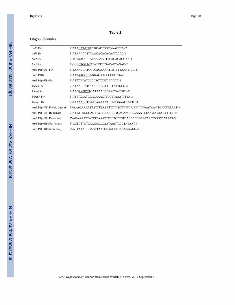
Cloning of the aidB geneThe E. coli aidB gene was amplified from the bacterial chromosome by PCR using theprimers listed in Table 2. The amplification product was digested with NdeI and HindIII(underlined in Table 2) and cloned into the pET22b (+) vector (Novagen) creating theplasmid pET22b-aidB. The resulting expression vector contains a 6X histidine tag to allowprotein purification by Ni2+ affinity chromatography. Plasmid construction was verified byautomated DNA sequencing. The recombinant AidB protein was produced and purified asdescribed previously [11].
Electrophoretic mobility shift assay (EMSA)EMSA experiments were performed using rrnB P1wt as the biotin-labeled DNA probe.Sense and antisense oligonucleotides (Table 2) were annealed by incubation at 95°C for 5min and successive gradual cooling to room temperature. Purified recombinant AidB wasincubated with 20 ng of biotinylated DNA rrnB P1wt for 20 min at room temperature in 20µl of buffer Z (25 mM HEPES pH 7.6, 50 mM KCl, 12.5 mM MgCl2, 1 mM DTT, 20%glycerol, 0.1% triton). Protein-DNA complexes were separated on 5% nativepolyacrylamide gel (29:1 cross-linking ratio) in 0.5x TBE (45 mM Tris pH 8.0, 45 mM boricacid, 1 mM EDTA) at 200 V (20 V/cm) at room temperature. Afterwards, electrophoretictransfer to a nylon membrane was carried out in 0.5× TBE at 380 mA for 45 min, and the
Rippa et al. Page 2
DNA Repair (Amst). Author manuscript; available in PMC 2012 September 5.
NIH
-PA Author Manuscript
NIH
-PA Author Manuscript
NIH
-PA Author Manuscript

transferred DNA was cross-linked to the membrane with UV light. After incubation inblocking buffer for 1 h at room temperature, the membrane was incubated with streptavidin-HRP conjugate (Sigma) for 30 min at room temperature. The membrane was washed andvisualized with SuperSignal chemiluminescence reagent (Pierce).
Competition experiments were performed using increasing quantities (100×–500×) of eitherunlabelled rrnB P1wt, which contains its UP element used as a specific competitor or rrnBP1ΔUP is used as a non-specific competitor.
Construction of fusion plasmids for transcription assaysThe lacZ gene was amplified from genomic DNA of E. coli by PCR using the primers listedin Table 2. The amplification product was digested with HindIII and XhoI (underlined inTable 2) and cloned into the pET22b (+) vector (Novagen) generating the plasmid pET22b-lacZ. The rrnB P1 promoter with (rrnB P1wt) and without its UP element (rrnB P1ΔUP),PleuA and PompF were amplified by PCR, digested with SphI and HindIII and inserted intopET22b-lacZ linearized with the same restriction enzymes. The resulting plasmids,designated as listed in Table 1, were verified by automated DNA sequencing.
In vivo transcription assaysMG1655 and MV5924 E. coli strains were individually transformed with pET22b-lacZ,pET22b-P rrnB P1wt-lacZ, pET22b-P rrnB P1ΔUP-lacZ, pET22b-PleuA-lacZ and pET22b-PompF-lacZ plasmids. These bacterial cultures grown overnight in LB medium at 30°C,were diluted 1:100 in fresh medium. At an A600 nm of 0.4, the cultures were divided in fouraliquots: one was not supplemented and the other three aliquots were supplemented withMNNG (5µg/ml), ENNG (5µg/ml), MMS 0.04%, respectively. Cellular pellets werecollected during the exponential growth phase. β-galactosidase activity from the promoters-lacZ fusions was determined by measuring ONPG-hydrolysis, as described by Miller [12]and was compared to the activity obtained using a promoterless lacZ gene.
Isolation of plasmid DNA and damage assayThe MG1655 and MV5924 E. coli strains bearing pET22b-lacZ were grown overnight in LBmedium at 30°C; these bacterial cultures were then diluted 1:100 in fresh medium. At anA600 nm of 0.4, the cultures were divided in four aliquots: one was not supplemented and theother three aliquots were supplemented with MNNG (5µg/ml), ENNG (5µg/ml), MMS0.04%, respectively. After addition of alkylating agent, the bacterial cells were allowed togrow for 3 h; the plasmid DNA was isolated and served as a probe for the estimation ofalkylated bases. The plasmids were divided into 2 aliquots, one of which was treated withthe E. coli AlkA (a kind gift from Patrick J. O’Brien) and AP Endo (NEB); the other aliquotdid not receive further treatment (control). Treatment with AlkA was performed in 70 mMMOPS, pH 7.5, 1 mM EDTA, 1mM DTT, 5% glycerol for 30 min at 37°C, followed bytreatment with AP Endo for 1 h at 37°C. Then the samples were subjected to electrophoresisin 0.8% agarose gel for ~1 h at 80 V using 40 mM Tris, pH 7.8, 1 mM EDTA buffer.
Determination of DNA damage in the lacZ geneMG1655 and MV5924 E. coli strains were individually transformed with pET22b-lacZ,pET22b-P rrnB P1wt-lacZ, pET22b-P rrnB P1ΔUP-lacZ. These bacterial cultures grownovernight in LB medium at 30°C, were diluted 1:100 in fresh medium. At an A600 nm of 0.4,the cultures were divided in two aliquots, and one was supplemented with 0.04% MMS toactivate the adaptive response. The bacterial cells were allowed to grow for 3h. Then, theplasmids under study were isolated from these bacterial cells and were digested with HindIIIand XhoI to release the lacZ fragment. To estimate the presence of alkyl lesions, the DNA
Rippa et al. Page 3
DNA Repair (Amst). Author manuscript; available in PMC 2012 September 5.
NIH
-PA Author Manuscript
NIH
-PA Author Manuscript
NIH
-PA Author Manuscript

fragments were treated or not with the AlkA and AP Endo proteins. The samples were thensubjected to electrophoresis on alkaline agarose gels in 30 mM NaOH, 1 mM EDTA, pH 8buffer, at 60 V for 3 h at 25°C. The gel was neutralized by soaking in a solution containing1.5 M NaCl and 1 M Tris-HCl, pH 7.6 for 1 h. Finally, the gel was stained in TE buffer (10mM Tris-HCl, 1 mM EDTA, pH 7.4) containing SYBR® Gold for 30 min at 25°C and thesamples were then analysed for single-strand DNA breaks.
Cell survival and mutagenesisCell survival was tested by growing cells to a density of 1–3 × 10⁸ cells/ml, treating withMNNG for 30 min, then diluting cells in phosphate buffered saline containing 4% Na2S2O3to inactivate residual MNNG [13], then plating cells on LB plates [13]. Mutation frequencieswere determined using DSEM plates [13]. Cultures grown to approximately 3 × 10⁸ cellsper ml then spread on alkylating agent containing plates and incubated for 3 days at 37°Cand Arg+ mutant colonies counted. Since alkylating agents are relatively unstable, platescontaining mutagens were made by first adding alkylators at volumes needed to attain thespecified final concentration, then adding 25 ml cooled (50°C) DSEM medium. The plateswere cooled for 20 min, then dried by incubation at 37°C for 20 min with covers removedand immediately inoculated. All mutagenesis measurements were made at sub-lethal dosesof alkylators using strains deficient in most alkylation specific DNA repair mechanisms ada-alkBΔ25:: CmR alkA1 tag-1 aidBΔ35::TetR) carrying either the vector pTrc99A, orpTrc99A derivatives that express aidB alleles.
ResultsTo identify proteins that bind the upstream regions of strong promoters, we investigated theprotein complex that assembles at the upstream elements of the rrnB P1 promoter (seeSupplemental Data and Supplemental FigureS1) by comparing proteins that bind to asequence containing the −35 region and the UP element, an A/T rich enhancer sequence thatconstitutes the upstream element of many genes [14–16],, but not to a similar sequencelacking the UP element. The presence of the E. coli AidB protein among these proteins wasunexpected, and suggested a possible regulatory role for AidB in transcription.
AidB preferentially binds DNA containing UP elementsFigure 1 shows that AidB protein binds to DNA containing the rrnB P1wt promoter retardingthe fragment in an electrophoretic mobility shift experiment (EMSA). When rrnB P1wtDNA is used as competitor, there is a rapid loss of binding to the labeled DNA. However,when the rrnB P1ΔUP promoter lacking the UP element is used as competitor, no inhibitionof binding to the labeled rrnB P1wt sequence is seen even when it is added at a 500-foldexcess. This indicates that AidB protein preferentially binds to rrnB P1 promoter only whenthe UP element is present. Similar results were also seen when random DNA containing thesame base pair composition was used as competitor (see supplemental data). Thepreferential binding is not restricted to the rrnB P1wt promoter, since binding of AidB to itsown promoter also requires the presence of the UP element [11].
Functional analysis of AidB during transcriptionIn order to determine whether the presence of AidB at the rrnB P1 promoter might be ofbiological significance, we tested its effect on transcription from this promoter by in vivotranscription assays. In addition, we tested other promoters that differ with respect topresence or absence of an UP element, namely: 1) the rrnB p1 promoter with its UP element(rrnBWT), 2) the rrnB promoter deleted of its UP element (rrnBΔUP), 3) PleuA, which lacksan UP element and 4) PompF, which has an UP element. All promoters were individuallyfused to a promoterless lacZ gene contained in the reporter plasmid pET22b-lacZ. Both
Rippa et al. Page 4
DNA Repair (Amst). Author manuscript; available in PMC 2012 September 5.
NIH
-PA Author Manuscript
NIH
-PA Author Manuscript
NIH
-PA Author Manuscript

MG1655 (wild type) and MV5924 (ΔaidB) E. coli strains were then transformed with thefusion plasmids and grown in LB medium, either in the absence or in the presence ofalkylating agents (MMS, MNNG, ENNG). After 2 hours incubation in the presence orabsence of the alkylating agent, β-galactosidase activity was measured during theexponential growth phase. As shown in Fig. 2A, wild type and aidB mutant strains notexposed to alkylators showed identical levels of β-galactosidase activity, indicating that thepresence of AidB has no effect on transcription in untreated cells experiencing normalgrowth.
When MG1655 cells are treated with MMS, MNNG, or ENNG, transcription is reduced byroughly 2-fold for all promoters tested. In contrast, the aidB mutant showed a much moresevere reduction in transcription, especially at the rrnBWT and PompF, the two UP elementcontaining promoters (Fig. 2). This suggests that the interaction of AidB protein with thisclass of promoters is of functional significance and that AidB prevents transcription blockby alkylation stress. The smaller effect of the aidB deletion on the two promoters lacking UPelements is consistent with preferential binding of AidB to this region (Fig. 1). Takentogether, these data strongly suggest that AidB is required for high levels of transcriptionduring alkylation stress and that it has a more pronounced effect on transcription frompromoters containing an upstream UP element sequence. While it is formally possible thatAidB is a transcriptional regulator of UP element containing genes when alkylating agentsare present, a more likely explanation, based on its role as part of an alkylation inducibleDNA repair response, is that AidB prevents or repairs DNA damage in specific regions ofthe genome, preferentially preserving the coding capacity of genes transcribed from UPelement containing promoters.
AidB reduces the level of alkylation damage in DNATo test directly if AidB might be able to prevent or repair alkylation damage to DNA thepET22b-lacZ plasmid was isolated from wild type and aidB mutant cells grown either in theabsence or in the presence of alkylators (MMS, MNNG, ENNG) and served as a probe forthe estimation of alkylated bases in DNA. The plasmids were divided into 2 aliquots, onewas treated with E. coli AlkA (a gift from Patrick J. O’Brien) and AP Endonuclease IV (APEndo) (New England Biolabs); the other aliquot did not receive further treatment and servedas a control. The AlkA glycosylase recognizes and removes a wide variety of alkylatedbases converting them to abasic sites [6] and AP Endo is an apurinic/apyrimidinic (AP)endonuclease that converts the abasic sites to nicks [17,18]. The combined action of thesetwo enzymes on a damage1d plasmid results in the conversion of the covalently closedcircular (supercoiled) DNA to open circular and, if lesions are closely spaced, linear forms.AlkA treated and untreated plasmids were then subjected to electrophoresis on agarose gelsand tested for conversion of the supercoiled form to open circular and linear forms. Asshown in Fig. 3A, alkyl lesions were not detected in plasmids isolated from bacteria grownin LB medium without the addition of alkylating agents, indicating there is no detectableendogenous damage or non-specific cleavage by these enzymes in vitro. When plasmidDNA isolated from wild type cells exposed to alkylating agents was analyzed (Fig. 3B–D),treatment with AlkA and AP Endo did not result in nicking (Lane 2) indicating a lack ofDNA damage, but when DNA isolated from the alkylating agent treated aidB mutant wasanalyzed, the supercoiled fraction was completely absent after AlkA/AP Endo treatment andthere was an increase in both open circular and linear forms (Lanes 4). These results indicatethat the presence of AidB reduces the level of alkylation damage in plasmid DNA.Moreover, AidB protects DNA from all three alkylating agents tested, although they differin the nature of DNA lesions they produce. Indeed, MNNG methylates and ENNG ethylatesDNA more effectively at O6-G than MMS. In contrast MMS methylates double stranded
Rippa et al. Page 5
DNA Repair (Amst). Author manuscript; available in PMC 2012 September 5.
NIH
-PA Author Manuscript
NIH
-PA Author Manuscript
NIH
-PA Author Manuscript

DNA primarily at N7-G and N3-A sites and in single stranded DNA regions it alsomethylates N1-A and N3-C more efficiently than MNNG [19].
AidB preferentially protects DNA regions downstream of an UP elementSince AidB appears to protect DNA from alkylating agents very effectively (Figure 3), thelack of an alkylation sensitivity phenotype of aidB mutants remains a puzzle (see Figure 5and [13,20]). The observation that AidB protein allows more effective transcription of geneswith UP element promoters in the presence of an alkylating agent (Fig. 2) and has a higheraffinity for UP element containing promoters [11], suggests that AidB may not protect theentire genome equally and may show a preference for UP element containing regions ofDNA. To test this possibility, we analyzed the effect of AidB on alkylation damage in vivoin the lacZ fragment. In this experiment, we used three plasmids, each carrying the lacZgene fused to three different upstream sequences: the rrnBWT promoter, the rrnBΔUPpromoter, and a third plasmid carrying a promoterless lacZ gene. We investigated whetherthe presence of AidB might affect the content of alkyl lesions within the lacZ sequences.Wild type and aidB mutant cells containing these plasmids were first treated with MMS.After isolation, the plasmids were digested with restriction enzymes to release the lacZfragment. The lacZ fragment was then purified by agarose gel electrophoresis and dividedinto two aliquots. One aliquot was treated with the AlkA/AP Endo to nick the DNA at thelesion sites. The samples were then subjected to electrophoresis on alkaline agarose gel todenature DNA and to separate nicked from full-length ssDNA fragments. Since onlyundamaged strands will run as full-length molecules, the fraction of strands containinglesions can be estimated by comparing the AlkA/AP Endo treated samples with controls nottreated with AlkA/AP Endo. Figure 4 shows that the aidB mutant cells are not able to protectthe lacZ gene regardless of the upstream sequence present and all DNA samples are equallysensitive to AlkA/AP Endo treatment (Lanes 8, 10,12). In Wild type cells, essentially allDNA from the rrnBWT bearing plasmid exposed to AlkA/AP Endo remains as full length(Fig. 4, compare Lanes 3 and 4). When lacZ is fused to the rrnBΔUP promoter, the sampletreated with AlkA/AP Endo (Lane 6) shows a clear decrease in the amount of full-lengthfragments compared with the control DNA not treated with AlkA/AP Endo (Lane 5).Treatment of lacZ from the promoterless plasmid with AlkA/AP Endo resulted in an almostcomplete loss of full-length DNA fragments indicating a higher level of damage (CompareLanes 1 and 2), thus confirming that the presence of AidB is required for the protectionagainst alkyl damage. It also suggests that transcription itself cannot be solely responsiblefor damage prevention, since transcription at the onset of damage is identical in wild typeand the aidB mutant (Fig. 2). Additionally, there is no detectable difference in damage levelswhen DNA samples isolated from the aidB mutant are compared with one another despitethe markedly higher level of transcription of the lacZ gene transcribed from the rrnB P1wtelement versus the promoterless lacZ. By contrast, plasmids from MMS treated wild typecells show a clear difference in their levels of protection from alkylation damage. Figure 4shows that lacZ fused to the UP element containing the rrnB P1wt promoter is well protectedfrom MMS exposure when compared, either to the samples from the aidB mutant, or lacZfused to the rrnB promoter that lacks the UP element, or has no promoter.
These results demonstrate that AidB preferentially protects the DNA of genes transcribedfrom UP element-containing promoters to a greater extent than DNA fragments bearingpromoters lacking an UP element.
AidB does not confer cellular resistance to alkylation damageThe aidB mutant strain MV5924was tested for its sensitivity to MNNG damage. Figure 5shows that even a complete deletion of aidB results in little or no sensitivity to MNNG whencompared with its isogenic wild type. The aidB mutation also does not affect MMS
Rippa et al. Page 6
DNA Repair (Amst). Author manuscript; available in PMC 2012 September 5.
NIH
-PA Author Manuscript
NIH
-PA Author Manuscript
NIH
-PA Author Manuscript

sensitivity (Figure S3, supplementary material). These results are consistent with previousobservations that mutants carrying insertions in the aidB gene show no increase insensitivity to alkylating agents [13]. A lack of alkylation sensitivity of the aidB deletionmutant is inconsistent with a general DNA damage prevention mechanism, since the abilityto prevent damage throughout the genome should result in increased resistance. However,protection of only some DNA regions would prevent damage to only those genes that aretargets for AidB protein and is unlikely to have a major effect on overall survival.
Based on the 3-dimensional structure of AidB protein, it has been suggested that AidB isunlikely to function as a DNA repair protein. Instead it may bind DNA and enzymaticallyinactivate alkylators as they approach the DNA. This notion is based on the observation thatthe dehydrogenase active site of AidB is spatially distant from its DNA binding face andaccessible only from the exterior of the protein [23]. Since a DNA repair protein predictsthat the DNA binding domain will be required for activity, we constructed an AidB mutantthat lacks the entire DNA binding domain (aidBΔ440–541). This mutant has previously beenshown to have IVD activity identical to the wild type, but no detectable DNA bindingactivity [11]. Since wild type AidB protein functions as an antimutator when cells are grownin the presence of MNNG, we tested if the DNA binding deficient AidB mutant proteinretains the antimutator activity. Figure 6 shows that the aidBΔ440–541 mutant allele is asactive as the wild type allele in the antimutator assay, indicating that DNA binding is notrequired for the alkylation resistance function of AidB and suggesting DNA binding insteadserves to target AidB to specific genes.
DiscussionThe biological role of AidB has long been uncertain. Our data demonstrate that AidBprevents DNA damage by alkylating agents and counteracts the block to transcription thatresults upon exposure to alkylating agents, especially in genes that are transcribed frompromoters containing UP elements. These effects are seen after treatment with MMS,MNNG and ENNG, three alkylating agents that produce different DNA lesions or damagespectra [24]. The result that ENNG damage is also prevented is especially interesting sinceENNG lesions are repaired not only the by adaptive response repair system, but also bynucleotide excision repair in E. coli [25].
The result that AidB can prevent DNA damage seems inconsistent with the result that loss ofAidB function by a complete deletion has little or no effect on MMS or MNNG sensitivityof the mutant strain (Figure 5 and S3). However, effects of an aidB mutation on DNAdamage and mutagenesis were seen at sublethal doses of alkylating agents (Figures 3–6).Since Ada dependent aidB induction is relatively weak compared to that of other adaptiveresponse genes [13,20], it is possible that AidB protein levels are too low to provideadequate protection against lethal doses of alkylating agents. The primary function of AidBmay be to protect DNA from the low levels of alkylators that are produced as by-products ofstationary phase metabolism [26–29], a possibility that is consistent with the observationthat aidB is induced and expressed at elevated levels in stationary phase [10,30,31].
The result that the AidB protein specifically binds to DNA sequences that include the UPelement [11] (see also Fig. 1), suggests that the lack of increased sensitivity to high levels ofalkylating agents in the aidB mutant (Fig. 5) may also be due to the fact that AidB onlyprotects a subset of the genome, leaving other genes, including essential ones, exposed toDNA damage. The aidB mutant phenotype is consistent with targeted repair or damageprevention and is analogous to the effect seen in strains that lack the ability to carry outtranscription-coupled repair (TCR) of UV damage, the only other gene specific repair ordamage prevention system currently known. A TCR deficient mfd mutant shows only a
Rippa et al. Page 7
DNA Repair (Amst). Author manuscript; available in PMC 2012 September 5.
NIH
-PA Author Manuscript
NIH
-PA Author Manuscript
NIH
-PA Author Manuscript
modest decrease in cellular resistance to UV, but a dramatic reduction in the rate at whichrepair of active genes occurs [32–34]. Thus, the AidB prevention mechanism appears to be acellular strategy to preferentially protect a subset of genes. In this case the genes includeones important for basic metabolic processes and key DNA repair genes. AidB is targetedtowards genes whose promoters have upstream UP elements. This includes genes such asmost of the ribosomal RNA genes and many tRNA genes as well as several key DNA repairgenes required for recovery from alkylation damage such as recA, polA, sulA, recN the ada-alkB operon, and aidB itself [14,35,36].
The presence of a functional aidB gene protects UP element genes from alkylation damageand results in more efficient transcription in the presence of alkylating agents. lacZ fused tothe two UP element containing rrn and ompF promoters are transcribed 10- and 6- fold moreefficiently in the presence of an alkylating agent than lacZ fused to an rrn promoter whoseUP element has been deleted, or the leuA promoter, which has no UP element. Although it ispossible that AidB has regulatory effects on these genes, a lower level of template damageshould clearly contribute to the transcription efficiency.
Promoters lacking an UP element, and thus not efficiently bound by AidB protein still showa slightly higher level of transcription in wild type versus aidB mutants upon alkylation (2.2and 2-fold enhancement for rrnBΔUP and Pleu, Fig. 2). It is unclear if this aidB-dependentenhancement of transcription in the presence of an alkylating agent represents some directprotection by aidB, or is an indirect effect of the elevated levels of ribosomes, tRNAs andpossibly other components of the translational machinery that are transcribed at a higherlevels in the aidB+ strain under these conditions. The observation that the protection of lacZfused to the rrn promoter lacking an UP element and the observation that plasmid DNAshows better protection in wild type than in an aidB mutant strain (Fig. 4), suggests thatthere may be some general protection resulting from the presence of aidB, especially when itis highly expressed, or induced for a long period of time as in these experiments. Underthese conditions AidB may initially protect the genes preferentially targeted, followed byother parts of the genome if AidB protein accumulates to sufficiently high levels. Theprecise mechanism of action of AidB remains to be determined, though it is possible that itprovides protection of DNA adjacent to its preferred binding site, either by simplyinactivating alkylating agents and reducing the local concentration, or by polymerizing intomultimers that extend from the initial binding site. In the latter case, it is likely to protectboth by shielding the DNA and by inactivating alkylators.
However, the MNNG resistance resulting from expression of the DNA binding deficientaidB mutant protein, AidB(Δ440–541) indicates that the mutant lacking DNA bindingactivity still functions to prevent alkylation mutagenesis. This observation makes it unlikelythat AidB functions by simply binding and coating the DNA, thus preventing access byalkylators. The ability of the DNA binding defective AidB protein to prevent mutagenesissuggests that AidB is not a DNA repair protein, since DNA repair would be inhibited bylack of DNA binding activity. Instead, AidB is more likely to function to prevent damage bydetoxifying alkylating agents, which could reduce DNA alkylation even in the absence ofDNA binding activity by reducing the intracellular concentration of active alkylators. A rolefor AidB in alkylating agent detoxification is also consistent with earlier work on AidB andanalysis of the structural features of the protein [10,23]. Determination of the precisemechanism by which AidB may inactivate alkylating agents requires further work toexamine the chemistry of the hypothetical process.
It is unclear how widespread preferential damage prevention mechanisms such as AidB are,if other prokaryotes and eukaryotes have similar damage prevention proteins, or if thestrategy of preferential DNA protection extends to mechanisms that prevent damage by
Rippa et al. Page 8
DNA Repair (Amst). Author manuscript; available in PMC 2012 September 5.
NIH
-PA Author Manuscript
NIH
-PA Author Manuscript
NIH
-PA Author Manuscript
other agents. In E. coli the dps gene is highly expressed in stationary phase and preventsoxidative DNA damage. Unlike AidB, however, this protein is produced at very high levelsand appears to function as a genome wide protective protein. It is unclear if it may also havea preference for specific sequences when it is expressed at lower levels [37].
HighlightsIn this manuscript we demonstrate that aidB binds preferentially to UP elementcontaining genes and preferentially protects them from alkylation damage. Additionally,the reduction in transcription normally seen in the presence of alkylators is much greaterin the aidB mutant than in wild type, indicating that transcription capacity is preservedwhen aidB is functional. We further demonstrate that aidB overexpression reducesmutagenesis and that this does not require the DNA binding domain, suggesting that itdoes not repair DNA, but prevents damage from occurring.
Supplementary MaterialRefer to Web version on PubMed Central for supplementary material.
AcknowledgmentsWe thank Patrick J. O’Brien (University of Michigan) for AlkA protein. This work was funded in part by NIHGrant CA100122 to MRV, Italian MIUR PRIN 2008 and FIRB 2007 Italian Human ProteoNet grants to AD.
REFERENCES1. Volkert MR, Landini P. Transcriptional responses to DNA damage. Curr. Opin. Microbiol. 2001;
4:178–185. [PubMed: 11282474]2. Landini P, Volkert MR. Regulatory responses of the adaptive response to alkylation damage: a
simple regulon with complex regulatory features. J. Bacteriol. 2000; 182:6543–6549. [PubMed:11073893]
3. McCarthy TV, Karran P, Lindahl T. Inducible repair of O-alkylated DNA pyrimidines inEscherichia coli. EMBO J. 1984; 3:545–550. [PubMed: 6370685]
4. Teo I, Sedgwick B, Kilpatrick MW, McCarthy TV, Lindahl T. The intracellular signal for inductionof resistance to alkylating agents in E. coli. Cell. 1986; 45:315–324. [PubMed: 3009022]
5. Nakabeppu Y, Sekiguchi M. Regulatory mechanisms for induction of synthesis of repair enzymes inresponse to alkylating agents: Ada protein acts as a transcriptional regulator. Proc. Natl. Acad. Sci.USA. 1986; 83:6297–6301. [PubMed: 3529081]
6. O'Brien PJ, Ellenberger T. The Escherichia coli 3-methyladenine DNA glycosylase AlkA has aremarkably versatile active site. J Biol Chem. 2004; 279:26876–26884. [PubMed: 15126496]
7. Trewick SC, Henshaw TF, Hausinger RP, Lindahl T, Sedgwick B. Oxidative demethylation byEscherichia coli AlkB directly reverts DNA base damage. Nature. 2002; 419:174–178. [PubMed:12226667]
8. Falnes PO, Johansen RF, Seeberg E. AlkB-mediated oxidative demethylation reverses DNA damagein Escherichia coli. Nature. 2002; 419:178–182. [PubMed: 12226668]
9. Rohankhedkar MS, Mulrooney SB, Wedemeyer WJ, Hausinger RP. The AidB component of theEscherichia coli adaptive response to alkylating agents is a flavin-containing, DNA-binding protein.J Bacteriol. 2006; 188:223–230. [PubMed: 16352838]
10. Landini P, Hajec LI, Volkert MR. Structure and transcriptional regulation of the Escherichia coliadaptive response gene aidB. J. Bacteriol. 1994; 176:6583–6589. [PubMed: 7961409]
11. Rippa V, Amoresano A, Esposito C, Landini P, Volkert M, Duilio A. Specific DNA binding andregulation of its own expression by the AidB protein in Escherichia coli. J Bacteriol. 2010;192:6136–6142. [PubMed: 20889740]
Rippa et al. Page 9
DNA Repair (Amst). Author manuscript; available in PMC 2012 September 5.
NIH
-PA Author Manuscript
NIH
-PA Author Manuscript
NIH
-PA Author Manuscript
12. Miller, JH. Experiments in Molecular Genetics. Cold Spring Harbor Laboratories, Cold SpringHarbor; 1972.
13. Volkert MR, Nguyen DC. Induction of specific Escherichia coli genes by sublethal treatments withalkylating agents. Proc. Natl. Acad. Sci. USA. 1984; 81:4110–4114. [PubMed: 6330740]
14. Gourse RL, Ross W, Gaal T. UPs and downs in bacterial transcription initiation: the role of thealpha subunit of RNA polymerase in promoter recognition. Mol Microbiol. 2000; 37:687–695.[PubMed: 10972792]
15. Estrem ST, Gaal T, Ross W, Gourse RL. Identification of an UP element consensus sequence forbacterial promoters. Proc. Natl. Acad. Sci. USA. 1998; 95:9761–9766. [PubMed: 9707549]
16. Ross W, Gosink KK, Salomon J, Igarashi K, Zou C, Ishihama A, Severinov K, Gourse RL. A thirdrecognition element in bacterial promoters: DNA binding by the a subunit of RNA polymerase.Science. 1993; 262:1407–1413. [PubMed: 8248780]
17. Ljungquist S, Lindahl T. Relation between Escherichia coli endonucleases specific for apurinicsites in DNA and exonuclease III. Nucleic Acids Res. 1977; 4:2871–2879. [PubMed: 333397]
18. Ljungquist S. A new endonuclease from Escherichia coli acting at apurinic sites in DNA. J BiolChem. 1977; 252:2808–2814. [PubMed: 16002]
19. Singer B. The chemical effects of nucleic acid alkylation and their relation to mutagenesis andcarcinogenesis. Prog Nucleic Acid Res Mol Biol. 1975; 15:219–284. [PubMed: 237307]
20. Volkert MR, Nguyen DC, Beard KC. Escherichia coli gene induction by alkylation treatment.Genetics. 1986; 112:11–26. [PubMed: 3080354]
21. Volkert MR. Adaptive response of Escherichia coli to alkylation damage. Environ. Molec.Mutagen. 1988; 11:241–255. [PubMed: 3278898]
22. Lindahl T, Sedgwick B, Sekiguchi M, Nakabeppu Y. Regulation and expression of the adaptiveresponse to alkylating agents. Annu. Rev. Biochem. 1988; 57:133–157. [PubMed: 3052269]
23. Bowles T, Metz AH, O'Quin J, Wawrzak Z, Eichman BF. Structure and DNA binding of alkylationresponse protein AidB. Proc Natl Acad Sci U S A. 2008; 105:15299–15304. [PubMed: 18829440]
24. Singer, B.; Grunberger, D. Molecular Biology of Mutagens and Carcinogens. New York: PlenumPress; 1983.
25. Bonanno K, Wyrzykowski J, Chong W, Matijasevic Z, Volkert MR. Alkylation resistance of E.coli cells expressing different isoforms of human alkyladenine DNA glycosylase (hAAG). DNARep. 2002; 1:507–517.
26. Rebeck GW, Samson L. Increased spontaneous mutation and alkylation sensitivity of Escherichiacoli strains lacking the ogt O6-methylguanine DNA repair methyltransferase. J. Bacteriol. 1991;173:2068–2076. [PubMed: 2002008]
27. Sedgwick B. Nitrosated peptides and polyamines as endogenous mutagens in O6-alkylguanine-DNA alkyltransferase deficient cells. Carcinogenesis. 1997; 18:1561–1567. [PubMed: 9276631]
28. Taverna P, Sedgwick B. Generation of edogenous methylating agents by nitrosation in Escherichiacoli. J. Bacteriol. 1996; 178:5105–5111. [PubMed: 8752326]
29. Sedgwick B. Oxidation of methylhydrazines to mutagenic methylating derivatives and inducers ofthe adaptive response of Escherichia coli to alkylation damage. Cancer Res. 1992; 52:2693–2697.
30. Landini P, Hajec LI, Nguyen LH, Burgess RR, Volkert MR. The leucine-responsive protein (Lrp)acts as a specific repressor for ss-dependent transcription of the Escherichia coli aidB gene. Molec.Microbiol. 1996; 20:947–955. [PubMed: 8809748]
31. Volkert MR, Hajec LI, Matijasevic Z, Fang F, Prince R. Induction of the Escherichia coli aidBgene under oxygen limiting conditions requires a functional rpoS (katF) gene. J. Bacteriol. 1994;176:7638–7645. [PubMed: 8002588]
32. Selby CP, Sancar A. Mechanisms of transcription-repair coupling and mutation frequency decline.Microbiol Rev. 1994; 58:317–329. [PubMed: 7968917]
33. Selby CP, Sancar A. Molecular Mechanism of Transcription-Repair Coupling. Science. 1993;260:53–58. [PubMed: 8465200]
34. Selby CP, Witkin E, Sancar A. Escherichia coli mfd mutant deficient in "mutation frequencydecline" lacks strand-specific repair: in vitro complementation with purified coupling factor. Proc.Natl. Acad. Sci. USA. 1991; 88 11574-11478.
Rippa et al. Page 10
DNA Repair (Amst). Author manuscript; available in PMC 2012 September 5.
NIH
-PA Author Manuscript
NIH
-PA Author Manuscript
NIH
-PA Author Manuscript

35. Ross W, Aiyar SE, Salomon J, Gourse RL. Escherichia coli promoters with UP elements ofdifferent strengths: modular structure of bacterial promoters. J Bacteriol. 1998; 180:5375–5383.[PubMed: 9765569]
36. Landini P, Volkert MR. RNA polymerase a subunit binding site in positively controlled promoters:a new model for RNA polymerase/promoter interaction and transcriptional activation in the E. coliada and aidB genes. EMBO J. 1995; 14:4329–4335. [PubMed: 7556074]
37. Martinez A, Kolter R. Protection of DNA during oxidative stress by the nonspecific DNA-bindingprotein Dps. J. Bacteriol. 1997; 179:5188–5194. [PubMed: 9260963]
38. Jensen KF. The Escherichia coli K-12 "wild types" W3110 and MG1655 have an rph frameshiftmutation that leads to pyrimidine starvation due to low pyrE expression levels. J Bacteriol. 1993;175:3401–3407. [PubMed: 8501045]
39. Murphy KC, Campellone KG. Lambda Red-mediated recombinogenic engineering ofenterohemorrhagic and enteropathogenic E. coli. BMC Mol Biol. 2003; 4:11. [PubMed:14672541]
40. Volkert MR. Altered induction of the adaptive response to alkylation damage in Escherichia colirecF mutants. J. Bacteriol. 1989; 171:99–103. [PubMed: 2536670]
Rippa et al. Page 11
DNA Repair (Amst). Author manuscript; available in PMC 2012 September 5.
NIH
-PA Author Manuscript
NIH
-PA Author Manuscript
NIH
-PA Author Manuscript
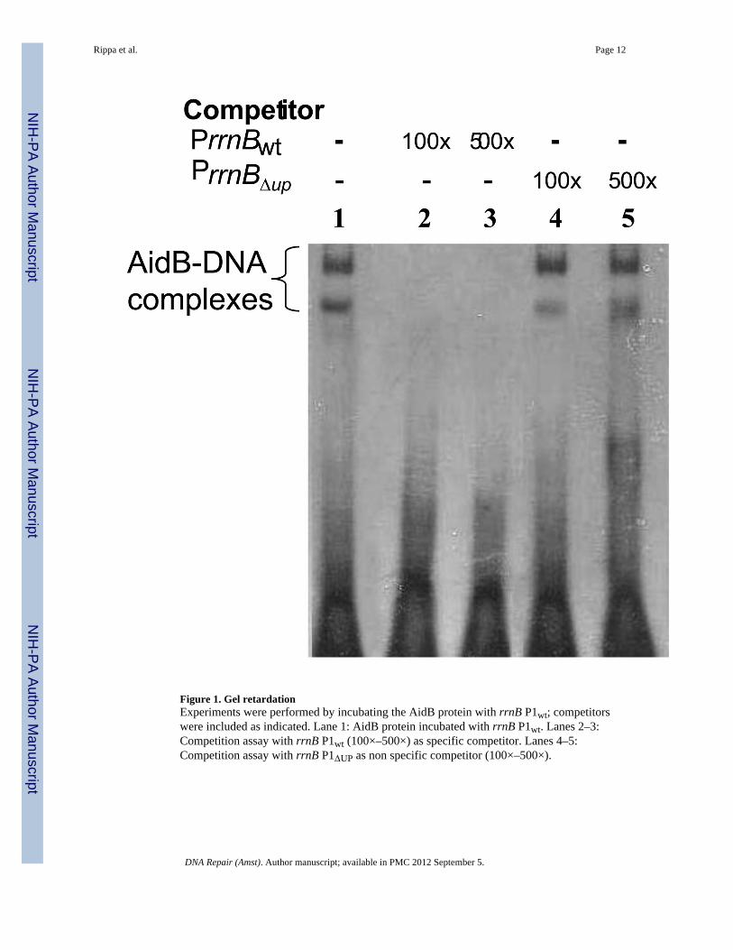
Figure 1. Gel retardationExperiments were performed by incubating the AidB protein with rrnB P1wt; competitorswere included as indicated. Lane 1: AidB protein incubated with rrnB P1wt. Lanes 2–3:Competition assay with rrnB P1wt (100×–500×) as specific competitor. Lanes 4–5:Competition assay with rrnB P1ΔUP as non specific competitor (100×–500×).
Rippa et al. Page 12
DNA Repair (Amst). Author manuscript; available in PMC 2012 September 5.
NIH
-PA Author Manuscript
NIH
-PA Author Manuscript
NIH
-PA Author Manuscript

Figure 2. In vivo transcription of lacZ fused to different promotersThe pET22b-P rrnB P1wt-lacZ, pET22b-P rrnB P1ΔUP-lacZ, pET22b-PleuA-lacZ andpET22b-PompF-lacZ plasmids were individually introduced into MG1655 (wild type) and
Rippa et al. Page 13
DNA Repair (Amst). Author manuscript; available in PMC 2012 September 5.
NIH
-PA Author Manuscript
NIH
-PA Author Manuscript
NIH
-PA Author Manuscript
MV5924 (ΔaidB) E. coli strains and the specific activity of β-galactosidase was determinedin the absence (A) and in the presence of MNNG (5µg/ml) (B), ENNG (5µg/ml) (C), MMS0.04% (D. The activities of promoters are reported in Miller units; the activity obtainedusing a promoterless lacZ gene was subtracted. Numbers above bars refer to the ratio of theβ-galactosidase activity of the promoter measured in the wild type cells to the activity of thatsame promoter in the aidB mutant strain. Means and standard deviations have beencalculated from four independent assays.
Rippa et al. Page 14
DNA Repair (Amst). Author manuscript; available in PMC 2012 September 5.
NIH
-PA Author Manuscript
NIH
-PA Author Manuscript
NIH
-PA Author Manuscript

Figure 3. Plasmid damage assayThe pET22b-lacZ DNA was isolated from wild type (Lanes 1–2) and ΔaidB (Lanes 3–4) E.coli strains grown in the absence (A) or in the presence of MNNG (5µg/ml) (B), MMS0.04% (C), ENNG (5µg/ml) (D), digested (Lanes 2, 4) or not (Lanes 1, 3) with AlkA and APEndo and subjected to agarose gel electrophoresis. Lane 5, 1 Kb DNA marker (NEB). OC:open circular; L: linear; SC: supercoiled.
Rippa et al. Page 15
DNA Repair (Amst). Author manuscript; available in PMC 2012 September 5.
NIH
-PA Author Manuscript
NIH
-PA Author Manuscript
NIH
-PA Author Manuscript
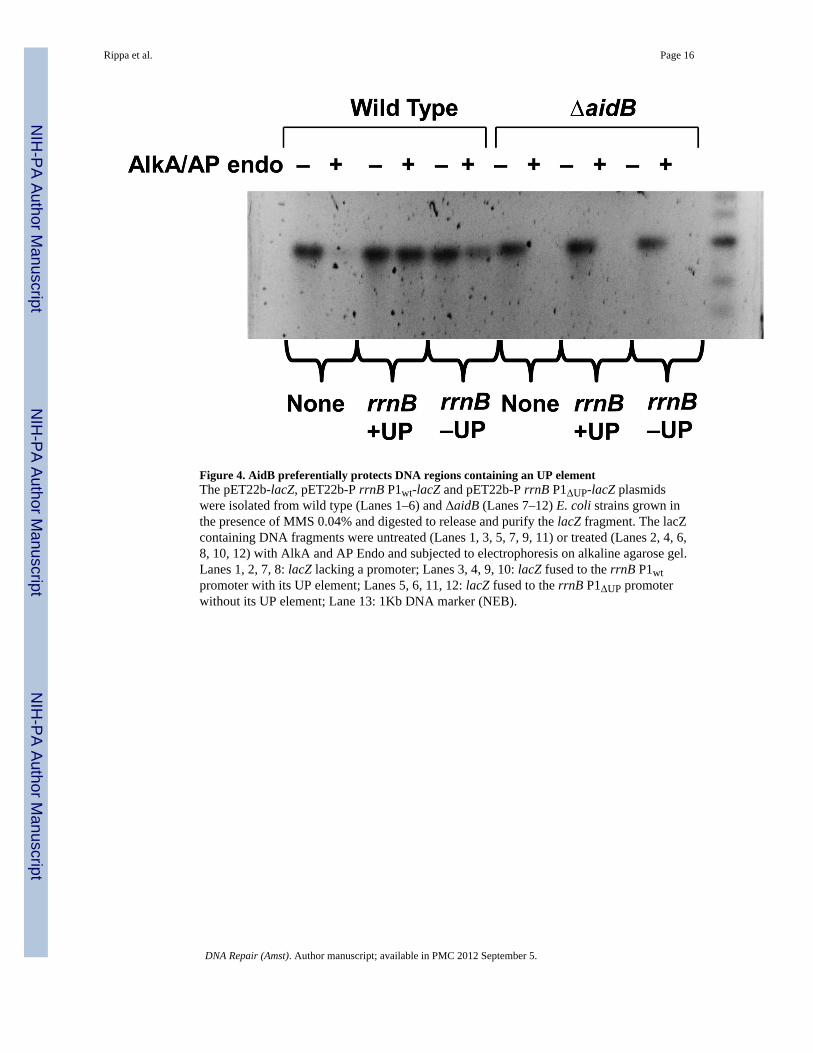
Figure 4. AidB preferentially protects DNA regions containing an UP elementThe pET22b-lacZ, pET22b-P rrnB P1wt-lacZ and pET22b-P rrnB P1ΔUP-lacZ plasmidswere isolated from wild type (Lanes 1–6) and ΔaidB (Lanes 7–12) E. coli strains grown inthe presence of MMS 0.04% and digested to release and purify the lacZ fragment. The lacZcontaining DNA fragments were untreated (Lanes 1, 3, 5, 7, 9, 11) or treated (Lanes 2, 4, 6,8, 10, 12) with AlkA and AP Endo and subjected to electrophoresis on alkaline agarose gel.Lanes 1, 2, 7, 8: lacZ lacking a promoter; Lanes 3, 4, 9, 10: lacZ fused to the rrnB P1wtpromoter with its UP element; Lanes 5, 6, 11, 12: lacZ fused to the rrnB P1ΔUP promoterwithout its UP element; Lane 13: 1Kb DNA marker (NEB).
Rippa et al. Page 16
DNA Repair (Amst). Author manuscript; available in PMC 2012 September 5.
NIH
-PA Author Manuscript
NIH
-PA Author Manuscript
NIH
-PA Author Manuscript

Figure 5. Alkylating agent sensitivity of strains deficient in adaptive response genesCells were grown to a density of approximately 1–3 × 10⁸ cells/ml and treated with MNNGas indicated. Wild type (MG1655), aidBΔ35::TetR (MV5924). Each data point represents 3(15, 90 µg/ml) or 6 repetitions (0, 30 and 60 µg/ml). Standard errors of the mean are shownwhere visible beyond the data point.
Rippa et al. Page 17
DNA Repair (Amst). Author manuscript; available in PMC 2012 September 5.
NIH
-PA Author Manuscript
NIH
-PA Author Manuscript
NIH
-PA Author Manuscript

Figure 6. Antimutagenic activity of wild type and AidB(Δ440–541)Cells were grown to a density of approximately 3 × 10⁸ cells per ml and undiluted cultureswere spread on DSEM plates ± alkylating agents and mutation frequencies determined. Allmutagenesis experiments were conducted at sublethal doses of alkylating agents usingstrains deficient in most alkylation specific DNA repair mechanisms: aidB+ (MV6782, ada-alkBΔ25:: CmR alkA1 tag-1 aidBΔ35::TetR/pTrc99A-AidB+); aidBΔ440–541 (MV6790,ada-alkBΔ25::CmR alkA1 tag-1 aidBΔ35::TetR/pTrc99A-AidBΔ440–451); vector control(MV6780, ada-alkBΔ25::CmR alkA1 tag-1 aidBΔ35::TetR/pTrc99A).
Rippa et al. Page 18
DNA Repair (Amst). Author manuscript; available in PMC 2012 September 5.
NIH
-PA Author Manuscript
NIH
-PA Author Manuscript
NIH
-PA Author Manuscript

NIH
-PA Author Manuscript
NIH
-PA Author Manuscript
NIH
-PA Author Manuscript
Rippa et al. Page 19
Table 1
Bacterial strains and plasmids
Strains/plasmids DescriptionReference
orsource
Strains
MG1655 wild-type; F− λ− ilvG rfb50 rph1 [38]
MV5924 aidBΔ::TetR derivative of MG1655 in which the aidB gene is replaced by a tetracycline resistancecassette using the methods of Murphy and Campellone [39]
[11]
MV6774 ada-alkBΔ25::CmR alkA1 tag-1 aidBΔ35::TetR derivative of MV1161 [13] This study
MV6780 ada-alkBΔ25::CmR alkA1 tag-1 aidBΔ35::TetR/pTrc99A) This study
MV6782 ada-alkBΔ25::CmR alkA1 tag-1 aidBΔ35::TetR/pMV435 (pTrc99A-AidB+) This study
MV6790 ada-alkBΔ25::CmR alkA1 tag-1 aidBΔ35::TetR/pMV1526 (pTrc99A-AidBΔ440– 451) This study
Plasmids
pET22b(+) carries an N-terminal pelB signal sequence for potential periplasmic localization, plus an optionalC-terminal His-tag sequence
This study
pET22b-aidB pET22bΔ(NdeI-HindIII)Ω(aidB gene) This study
pET22b-lacZ pET22bΔ(HindIII-XhoI)Ω(lacZ gene) This study
pET22b-PrrnB(+UP)-lacZ pET22b-lacZΔ(SphI-HindIII)ΩPrrnB(+UP) This study
pET22b-PrrnB(−UP)-lacZ pET22b-lacZΔ(SphI-HindIII)ΩPrrnB(−UP) This study
pET22b-PleuA-lacZ pET22b-lacZΔ(SphI-HindIII)ΩPleuA This study
pET22b-PompF-lacZ pET22b-lacZΔ(SphI-HindIII)ΩPompF This study
pTrc99A E. coli expression vector [10]
pMV435 pTrc99A-AidB+ [10]
pMV1526 pTrc99A-AidBΔ440–451 This study
DNA Repair (Amst). Author manuscript; available in PMC 2012 September 5.
NIH
-PA Author Manuscript
NIH
-PA Author Manuscript
NIH
-PA Author Manuscript
Rippa et al. Page 20
Table 2
Oligonucleotides
aidB Fw 5'-ATACATATGGTGCACTGGCAAACTCA-3'
aidB Rv 5'-ATAAAGCTTTAACACACACACTCCCC-3'
lacZ Fw 5'-TGTAAGCTTATAACAATTTCACACAGGAA-3'
lacZ Rv 5'-CGGCTCGAGTTATTTTTGACACCAGAC-3'
rrnB P1(+UP) Fw 5'-TAAAGCATGCTCAGAAAATTATTTTAAATTTC-3'
rrnB P1Rv 5'-ATTAAGCTTAGGAGAACCCCGCTGA-3'
rrnB P1(−UP) Fw 5'-ATTTGCATGCCCTCTTGTCAGGCC-3'
PleuA Fw 5'-ATAAGCATGCGGGACGTTTTTATTGCG-3'
PleuA Rv 5'-AAGAAGCTTGATAAAGCGAACGATGTG-3'
PompF Fw 5'-ATTTGCATGCACAAAGTTCCTTAAATTTTA-3'
PompF Rv 5'-TAAAAGCTTAATAAAAATTTACGGAACTATTG-3'
rrnB P1(+UP) Fw bio (emsa) 5'-bio-AGAAAATTATTTTAAATTTCCTCTTGTCAGGCCGGAATAAC TC CCTATAAT-3'
rrnB P1(+UP) Rv (emsa) 5'-ATTATAGGGAGTTATTCCGGCCTGACAAGAGGAAATTTAA AATAA TTTTCT-3'
rrnB P1(+UP) Fw (emsa) 5’-AGAAAATTATTTTAAATTTCCTCTTGTCAGGCCGGAATAAC TCCCT ATAAT-3’
rrnB P1(−UP) Fw (emsa) 5’-CCTCTTGTCAGGCCGGAATAACTCCCTATAAT-3’
rrnB P1(−UP) Rv (emsa) 5’-ATTATAGGGAGTTATTCCGGCCTGACAAGAGG-3’
DNA Repair (Amst). Author manuscript; available in PMC 2012 September 5.