
Predictions of melatonin suppression during the early ... - Nature
Upload
independentCategory
view
0download
0
© The Author 2013. Published by Oxford University Press. All rights reserved. For Permissions, please email: [email protected]
Carcinogenesis vol.34 no.5 pp.1051–1061, 2013 doi:10.1093/carcin/bgt025Advance Access publication January 25, 2013
Blockage of melatonin receptors impairs p53-mediated prevention of DNA damage accumulation
Raffaela!Santoro1, Federica!Mori1, Marina!Marani1, Giuseppe!Grasso2, Anna!Maria Cambria2, Giovanni!Blandino3, Paola!Muti4 and Sabrina!Strano1,*1Molecular Chemoprevention Group, Molecular Medicine Area, Regina Elena National Cancer Institute, Rome 00144, Italy, 2Department of Oncology, Division of Pathology, S.!Vincenzo Hospital, Taormina, Italy, 3Translational Oncogenomics Unit-ROC, Molecular Medicine Area, Regina Elena National Cancer Institute, Rome 00144, Italy and 4Department of Oncology, McMaster University, Hamilton, Ontario L8V 5C2, Canada
*To whom correspondence should be addressed. Tel:!+39 0652662974; Fax: +39 0652662880; Email: [email protected]
Melatonin has been known to be a chemopreventive agent since its levels inversely correlate with the risk of developing cancer. We have recently shown that melatonin induces p38-dependent phos-phorylation of both p53 and histone H2AX. This is associated with a p53-mediated increase in repair of both endogenous and chemo-therapy-induced DNA damage. In addition, the inhibition of p38 activities impairs melatonin’s capability to induce a p53-depend-ent DNA damage response and thus its ability to maintain genome integrity. Since melatonin-induced p53 phosphorylation requires an intact p38 phosphorylation cascade and p38 can be activated by G proteins, we supposed that melatonin’s activities could be mediated by its G-protein-coupled membrane receptors, MT1 and MT2. Here, we show that the activation of the p53-dependent DNA damage response by melatonin is indeed mediated by MT1 and MT2. As a result, the absence of either receptor impairs mel-atonin’s ability to reduce both cell proliferation and clonogenic potential of cancer cells. In addition, this causes an impairment of the p53-dependent DNA damage response. By providing molecu-lar insight, our "ndings might have translational impact, suggest-ing the involvement of melatonin receptors in tumorigenesis.
Introduction
Melatonin (N-acetyl-5-methoxytryptamine) is a mammal pineal hor-mone mainly secreted by the pineal gland at the hypothalamic level. Melatonin synthesis occurs mainly at night and is tuned by enzymes inhibited by the sunlight (1). Increasing observational evidence associ-ates melatonin concentration to cancer occurrence (2–9). In particular, two epidemiological studies have demonstrated an inverse correlation between overnight urinary levels of melatonin and the incidence of breast cancer (8,9). The "rst study was conducted within the ORDET cohort and tested the concentration of a melatonin metabolite, 6-sulfa-toxymelatonin, in 178 postmenopausal women with incident breast cancer and 710 matched controls (8), whereas the second one refers to a case–control study conducted within the NHIS cohort on 357 postmenopausal women with incident breast cancer and 533 matched controls (9). These and other similar studies suggest that melatonin might be able to induce tumour suppressor pathways in vivo (10,11). Molecular studies have shown that melatonin is able to induce both the p53 tumour suppressor and its target gene p21 (10,12–14). In response to a plethora of stressful conditions, especially those caus-ing DNA damage, p53 undergoes post-translational modi"cations, becomes more stable and its transcriptional activities are enhanced (15–23). Particularly, upon DNA damage, p53 is phosphorylated by a subset of kinases. Ataxia telangiectasia mutant and ataxia telangi-ectasia related phosphorylate p53 upon DNA double strand breaks, whereas p38 can phosphorylate p53 in response to single strand
breaks or in the absence of ataxia telangiectasia mutant (15,22,23). In addition, in the presence of radical oxygen species, both p53 and p38 become phosphorylated (24–29).
We have recently published that melatonin triggers a p38- and PML-dependent phosphorylation cascade, resulting in p53 phospho-rylation in Ser-15 (14).
Having a high lipid-water solubility, melatonin is able to enter the cell by diffusing across the cellular membrane and act as a radical scavenger. In the same way, it can cross the nuclear membrane and bind its nuclear receptor RZR/ROR!. This is then recruited onto RZR regulatory elements present in the promoters of several genes, such as p21 and 5-lipoxygenase, which are thereby transcriptionally regulated (10). In addition, melatonin regulates signalling pathways by binding to its membrane receptors, MTNR1A and MTNR1B, also known as MT1 and MT2, which exist in the homodimeric and heterodimeric states (30–33). Both MT1 and MT2 are G-protein-coupled membrane receptors which, upon ligand binding, activate G proteins and inhibit cyclic adenosine 3",5"-monophosphate formation (10,34). It has been shown that both MT1 and MT2 receptors, upon melatonin binding, can also activate MAP kinases, among which c-Jun, in Chlorocebus aethiops Cos-7 cells (30). Since we found that melatonin treatment leads to p38 accumulation (14), we investigated whether this was receptor mediated. In the present manuscript, we show that melatonin triggers p53 phosphorylation through the activation of its receptors, MT1 and MT2. In fact, we show that both chemical inhibition and selective gene silencing of the receptors impair melatonin’s ability to trigger p38 phosphorylation and accumulation, thereby inhibiting p53 phosphorylation. This pairs with a decrease in melatonin’s abil-ity to reduce cell proliferation and prevent DNA damage. Together with recent translational studies showing reduced levels of melatonin receptors in cancer samples as compared with their matched controls (35,36), these "ndings suggest that the involvement of melatonin receptors in triggering anticancer pathways.
Materials and methods
Cell culture and transfectionHCT116 and MCF-7 cells were cultured in Dulbecco’s modi"ed Eagle’s medium supplemented with 10% fetal calf serum, 2 mM #-glutamine, 100 U/ml penicillin and 100!#g/ml streptomycin (Gibco, Life Technologies, Carlsbad, CA). Cell lines were grown at 37°C, 5% CO2. Transfections were performed with Lipofectamine 2000 (Life Technologies) according to the manufacturer’s recommendations, using the following small interfering RNA (siRNA): siGFP 5"-AAGUUCAGCGUGUCCGGGGAG(dTdT)-3"; siMTNR1A 5"-GGAAUACAGGAGAAUUAUA(dTdT)-3"; siMTNR1B 5"-CUAGCUACUUACUGGCUUA(dTdT)-3" (Euro"ns MWG, Ebersberg, Germany). Culture medium was replaced 24 h post-transfection and cells were allowed to grow for additional 72 h before any treatment.
ReagentsMelatonin (M5250) and Luzindole (L2407) were purchased from Sigma–Aldrich (St Louis, MO).
Growth curves and colony-forming!assaysFor growth curves, 7 $ 103 cells/well were seeded in 6-well dishes. Cells were harvested at the indicated times by trypsin detachment and counted automatically using a Guava EasyCyte 8HT %ow cytometer (Millipore, Billerica, MA). Cell concentration (cells/ml) is shown on the y-axis. With regard to colony-forming assays, 5 $ 102 cells were seeded in 35-mm dishes and grown for 15!days. Cells were stained with crystal violet and colonies were counted. To maintain melatonin levels, the medium was changed every 48 h in both experiments.
Western!blotsTotal protein extracts were prepared by lysing cells in 8 M urea (Bio-Rad, Hercules, CA). Receptors were extracted with 8 M urea/10 mM [(3-cholamidopropyl)-dimethyl-ammonio]1-propanesulfonate (Bio-Rad). All Abbreviation: siRNA, small interfering RNA.
1051
at McM
aster University Library on M
ay 6, 2013http://carcin.oxfordjournals.org/
Dow
nloaded from
R.Santoro et!al.
protein extracts were quanti"ed by Bradford assay and equal amounts were loaded onto sodium dodecyl sulfate–polyacrylamide gel electrophoresis (either pre-cast 4–20% Novex Tris-Glycine gels from Life Technologies or home-made gels), transferred to polyvinylidene %uoride membranes (Immobilon-P, Millipore) and underwent immunoblot with the indicated anti-bodies. Antibodies to phospho-p53 Ser-15 (9284, Cell Signaling, Danvers, MA), $-actin (A2288, AC-74, Sigma), p21 (2947, Cell Signaling), nucleo-lin (ab13541, 4E2, Abcam, Cambridge, UK), MTNR1A (ab87639, Abcam), MTNR1B (ab128469, Abcam), %H2AX (9718, 20E3, Cell Signaling), p38 (9212, Cell Signaling), phospho-p38 Thr180/Tyr182 (9211, Cell Signaling) and p53 (sc126, DO1, Santa Cruz Biotechnology) were diluted in 5% bovine serum albumin in Tris-buffered saline/0.1% Tween-20. Secondary anti-mouse and anti-rabbit antibodies were purchased from Bio-Rad. Images were acquired using a VersaDoc MP instrument (Bio-Rad).
Comet!assaysFollowing transfection with the indicated siRNA, cells were treated with melatonin for 2 h and then irradiated at a dose of 0.05 J/cm2 using a Bio-Sun irradiation apparatus (Vilbert Lourmat, Marne-la-Vallée, France) as described previously (37) and allowed to repair DNA for 4 h. After treatment, cells were detached with trypsin and embedded in 1% low melting agarose (Sigma) in phosphate-buffered saline and spread onto microscopy slides coated previously with 1% agarose (Bio-Rad). Cells were lysed in the lysis solution (2.5 M NaCl, 100 mM ethylenediaminetetraacetic acid, 10 mM Tris base, 8 g/l NaOH, 1% Triton X-100, 10% dimethyl sulfoxide) for 1 h at room temperature and then run in running solution (300 mM NaOH, 1 mM ethylenediaminetetraacetic acid, pH 13.0) for 30 min at 25 V and 250 mA. DNA was equilibrated with 0.4 M Tris (pH 8.0) and slides were dried with methanol. DNA was stained with propidium iodide (Sigma) and pictures were taken using $60 magni"-cation on an Axiovert 200M microscope and Axiovision acquisition program (Zeiss). At least 300 cells were scored for each slide.
Mice xenograftsFour-week-old CD1 nude mice (n!=!7/each group, Charles River) were sub-cutaneously injected with either HCT116 p53 wt or HCT116 p53&/& cell lines (2 $ 106 cells/mouse) treated with 1!#M melatonin or vehicle (ethanol) for 72 h, changing the medium every 24 h. Tumour volume was evaluated twice a week. Animals were killed and xenograft excised after 2 weeks from the injection. Tumour volume (mm3) was calculated as follows: 0.5!$ D1 $ D2, where D1 and D2 are the larger and smaller diameters measured by caliper. All tumori-genicity assays were carried out according to the guidelines set by the Internal Ethical Committee.
Results
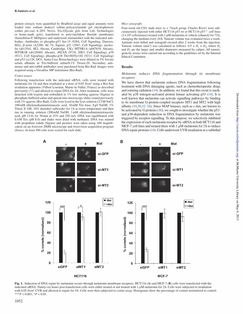
Melatonin reduces DNA fragmentation through its membrane receptorsWe have shown that melatonin reduces DNA fragmentation following treatment with DNA damaging agents, such as chemotherapeutic drugs and ionizing radiation (14). In addition, we found that this event is medi-ated by p38 mitogen-activated protein kinase activating p53 (14). It is well known that melatonin can activate signalling pathways by binding to its membrane G-protein-coupled receptors MT1 and MT2 with high af"nity (10,30,32–34). Since MAP kinases, such as c-Jun, are known to be activated by G proteins (30), we sought to investigate whether the p53- and p38-dependent reduction in DNA fragmentation by melatonin was triggered by receptor signalling. To this purpose, we selectively inhibited the expression of each melatonin receptor by siRNA in both HCT116 and MCF-7 cell lines and treated them with 1!&M melatonin for 2 h to induce DNA repair proteins (14). Cells underwent UVB irradiation at a sublethal
Fig.!1. Induction of DNA repair by melatonin occurs through melatonin membrane receptors. HCT116 (A) and MCF-7 (B) cells were transfected with the indicated siRNA. Ninety-six hours post-transfection cells were either treated or not treated with 1!&M melatonin for 2 h. Cells were subjected to irradiation with 0.05 J/cm2 UVB and allowed to repair for 4 h. Cells were then subjected to comet assay. Histograms show the percentage of comets normalized to control. ***P!<!0.001; *P!<!0.05.
1052
at McM
aster University Library on M
ay 6, 2013http://carcin.oxfordjournals.org/
Dow
nloaded from

Melatonin receptors trigger p53-dependent DNA!repair
dose and were allowed to repair damaged DNA. As shown previously with chemotherapeutic drugs and gamma-irradiation (14), melatonin was able to induce repair of fragmented DNA following UVB irradia-tion as well, both in HCT116 (Figure!1A) and in MCF-7 (Figure!1B). However, in the absence of either MT1 or MT2, the number of cells showing DNA fragmentation was not affected by melatonin treatment in either of the cell lines (Figure!1A and B). In addition, comet assays were performed in both HCT116 (Supplementary Figure S1A, available at Carcinogenesis Online) and MCF-7 (Supplementary Figure S1B, avail-able at Carcinogenesis Online) by either pre-treating or not pre-treating the cells with 1 nM luzindole, a melatonin antagonist binding to and inac-tivating both MT1 and MT2 receptors, which is known to inhibit both receptors, though it has a 25-fold higher af"nity for MT2 (38). Luzindole impaired melatonin’s ability to reduce DNA fragmentation following UVB irradiation (HCT116 cells in Supplementary Figure S1A, avail-able at Carcinogenesis Online and MCF-7 cells in Supplementary Figure S1B, available at Carcinogenesis Online). Both the approaches indicated that receptor function is necessary for melatonin’s ability to induce repair of fragmented DNA following UVB irradiation.
Receptor-mediated reduction of DNA damage is p53 dependentWe have shown previously that melatonin’s ability to reduce DNA fragmentation was impaired in cells lacking p53 (14). In order to understand whether receptor-mediated DNA repair induced by
melatonin was triggered mainly by p53, we performed comet assays (as described for Figure!1) in syngenic HCT116 cells, either wild-type (wt) or null for p53. As for other types of DNA damage (14), melatonin was not able to induce repair of fragmented DNA caused by UVB irradiation in the absence of p53 (Figure! 2B). Moreover, although removal of either MT1 or MT2 by siRNA impaired melatonin’s abil-ity to reduce DNA fragmentation in HCT116 wild-type cells, it did not have any further effect in HCT116 p53 null cells (Figure!2A and B) demonstrating that removal of either receptor impairs the activa-tion of p53 by melatonin. Of note, HCT116 p53 null cells have been reported to express two p53 isoforms (39–41). Thus, our results are consistent with the need of full-length p53 for DNA repair.
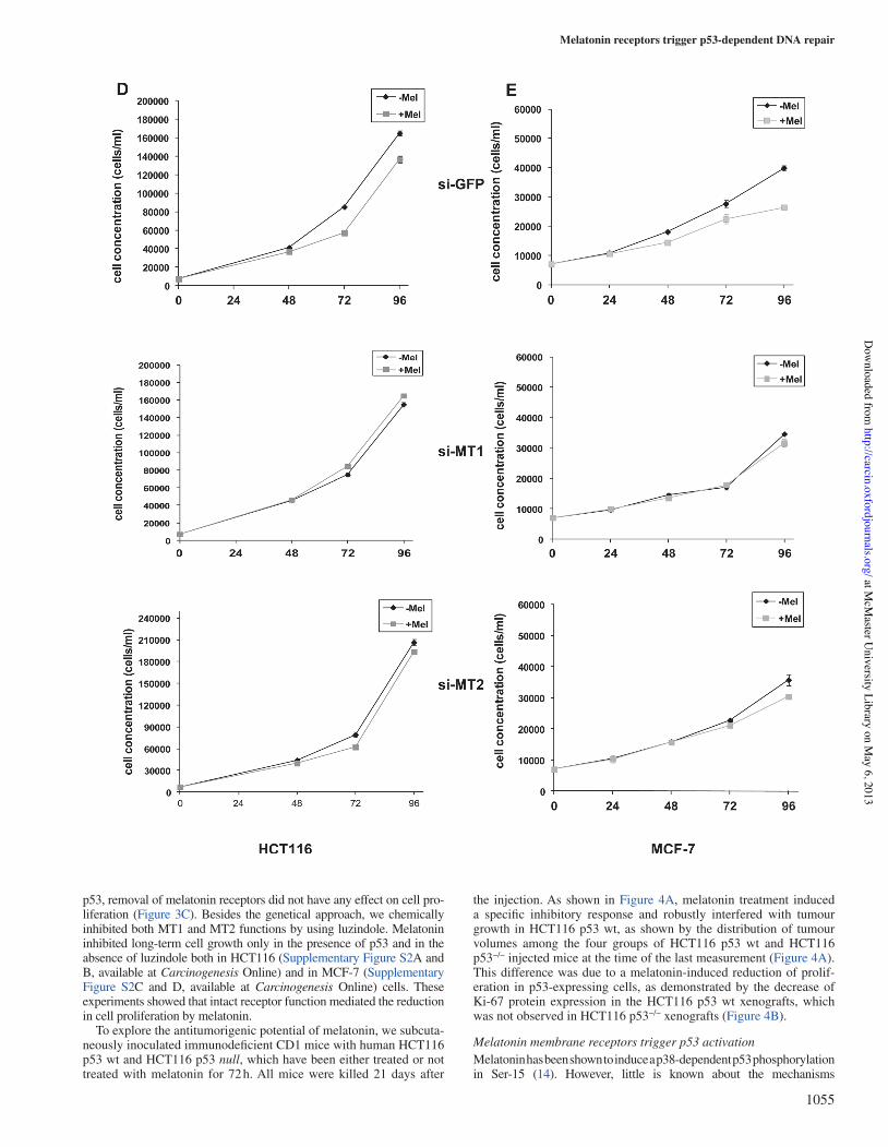
Blockage of melatonin receptors impairs melatonin’s ability to reduce cell proliferationMelatonin has proven to be able to inhibit proliferation of cancer cells (12–14). Here, we investigated whether activation of receptors was involved in this process. In order to do this, we silenced MT1 or MT2 expression in MCF-7 (Figure! 3A), HCT116 p53 wt (Figure! 3B) and HCT116 p53 null (Figure!3C) cells. Colony-forming assays showed that depletion of either receptors by siRNA impaired melatonin’s ability to inhibit long-term proliferation (Figure!3A and B). The same effects were observed in short-term proliferation, as assessed by growth curves in both HCT116 (Figure!3D) and MCF-7 (Figure!3E). In the absence of
Fig.!2. Melatonin membrane receptors trigger p53-dependent DNA repair. HCT116 wt (A) and HCT116 p53&/& (B) cells were transfected with the indicated siRNA and either treated or not with 1!&M melatonin for 2 h. Cells were subjected to irradiation with 0.05 J/cm2 UVB and allowed to repair for 4 h. Cells were then subjected to comet assay. Histograms show the percentage of comets normalized to control. ***P!<!0.001.
1053
at McM
aster University Library on M
ay 6, 2013http://carcin.oxfordjournals.org/
Dow
nloaded from

R.Santoro et!al.
Fig.!3. Melatonin inhibition of cell proliferation is receptor mediated. MCF7 (A), HCT116 wt (B) and HCT116 p53&/& (C) cells were transfected as in Figure!1, seeded and allowed to form colonies for 15!days. Culture medium (without or with 1!&M melatonin) was replaced every second day to keep melatonin levels constant. Colonies were stained and counted. Histograms show the percentage of cells relative to untreated cells. ***P!<!0.001; **P!<!0.01. HCT116 (D) and MCF-7 (E) cells were seeded and allowed to grow for the indicated time. Culture medium (without or with 1!&M melatonin) was replaced every second day to keep melatonin levels constant. Cell number was counted with the Guava EasyCyte 8HT %ow cytometer. Cell concentration is shown in the graphs.
1054
at McM
aster University Library on M
ay 6, 2013http://carcin.oxfordjournals.org/
Dow
nloaded from
Melatonin receptors trigger p53-dependent DNA!repair
p53, removal of melatonin receptors did not have any effect on cell pro-liferation (Figure!3C). Besides the genetical approach, we chemically inhibited both MT1 and MT2 functions by using luzindole. Melatonin inhibited long-term cell growth only in the presence of p53 and in the absence of luzindole both in HCT116 (Supplementary Figure S2A and B, available at Carcinogenesis Online) and in MCF-7 (Supplementary Figure S2C and D, available at Carcinogenesis Online) cells. These experiments showed that intact receptor function mediated the reduction in cell proliferation by melatonin.
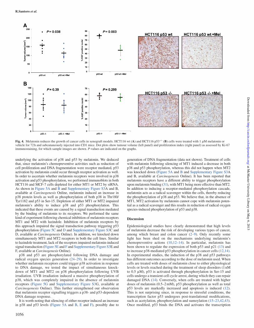
To explore the antitumorigenic potential of melatonin, we subcuta-neously inoculated immunode"cient CD1 mice with human HCT116 p53 wt and HCT116 p53 null, which have been either treated or not treated with melatonin for 72 h. All mice were killed 21!days after
the injection. As shown in Figure!4A, melatonin treatment induced a speci"c inhibitory response and robustly interfered with tumour growth in HCT116 p53 wt, as shown by the distribution of tumour volumes among the four groups of HCT116 p53 wt and HCT116 p53&/& injected mice at the time of the last measurement (Figure!4A). This difference was due to a melatonin-induced reduction of prolif-eration in p53-expressing cells, as demonstrated by the decrease of Ki-67 protein expression in the HCT116 p53 wt xenografts, which was not observed in HCT116 p53&/& xenografts (Figure!4B).
Melatonin membrane receptors trigger p53 activationMelatonin has been shown to induce a p38-dependent p53 phosphorylation in Ser-15 (14). However, little is known about the mechanisms
1055
at McM
aster University Library on M
ay 6, 2013http://carcin.oxfordjournals.org/
Dow
nloaded from
R.Santoro et!al.
underlying the activation of p38 and p53 by melatonin. We deduced that, since melatonin’s chemopreventive activities such as reduction of cell proliferation and DNA fragmentation were receptor mediated, p53 activation by melatonin could occur through receptor activation as well. In order to ascertain whether melatonin receptors were involved in p38 activation and p53 phosphorylation, we performed immunoblots in both HCT116 and MCF-7 cells depleted for either MT1 or MT2 by siRNA. As shown in Figure!5A and B and Supplementary Figure S3A and B, available at Carcinogenesis Online, melatonin induced an increase in p38 protein levels as well as phosphorylation of both p38 in Thr180/Tyr1182 and p53 in Ser-15. Depletion of either MT1 or MT2 impaired melatonin’s ability to induce p38 and p53 phosphorylation. This indicated that these events are caused by a signal transduction mediated by the binding of melatonin to its receptors. We performed the same kind of experiment following chemical inhibition of melatonin receptors MT1 and MT2 with luzindole. Inhibition of melatonin receptors by this approach impaired the signal transduction pathway triggering p53 phosphorylation (Figure!5C and D and Supplementary Figure S3C and D, available at Carcinogenesis Online). In addition, we knocked down simultaneously MT1 and MT2 receptors in both the cell lines. Similar to luzindole treatment, lack of the receptors impaired melatonin-induced signal transduction (Figure!5E and F and Supplementary Figure S3E and F, available at Carcinogenesis Online).
p38 and p53 are phosphorylated following DNA damage and radical oxygen species generation (24–29). In order to investigate whether melatonin receptors could trigger p38 activation in response to DNA damage, we tested the impact of simultaneous knock-down of MT1 and MT2 on p38 phosphorylation following UVB irradiation. UVB irradiation induced a massive phosphorylation of p38, which was completely impaired in the absence of melatonin receptors (Figure! 5G and Supplementary Figure S3G, available at Carcinogenesis Online). This further strengthened our observation that melatonin receptor signalling triggers a p38- and p53-dependent DNA damage response.
It is worth noting that silencing of either receptor induced an increase in p38 and p53 levels (Figure! 5A and B, E and F), possibly due to
generation of DNA fragmentation (data not shown). Treatment of cells with melatonin following silencing of MT1 induced a decrease in both p38 and p53 phosphorylation, whereas this did not happen when MT2 was knocked down (Figure!5A and B and Supplementary Figure S3A and B, available at Carcinogenesis Online). It has been reported that melatonin receptors have a different ability to trigger phosphorylation upon melatonin binding (31), with MT1 being more effective than MT2. In addition to inducing a receptor-mediated phosphorylation cascade, melatonin acts as a radical scavenger within the cells, thereby reducing the phosphorylation of p38 and p53. We believe that, in the absence of MT1, MT2 activation by melatonin cannot cope with melatonin poten-tial as a radical scavenger and this results in reduction of radical oxygen species-induced phosphorylation of p53 and p38.
Discussion
Epidemiological studies have clearly demonstrated that high levels of melatonin decrease the risk of developing various types of cancer, among which breast and colon cancer (2–9). Only recently some light has been shed on the mechanisms underlying melatonin’s chemopreventive actions (10,12–14). In particular, melatonin has been shown to regulate the expression of both p53 and p21 (13) and to increase p38-mediated p53 phosphorylation in cancer cells (12,14). In experimental studies, the induction of the p38 and p53 pathways has different outcomes according to the dose of melatonin used. When cells are treated with doses of melatonin close to either physiological levels or levels reached during the treatment of sleep disorders (1 nM to 0.5!&M), p53 is activated through phosphorylation in Ser-15 and cells undergo a transient cell cycle arrest, during which they can repair damaged DNA (14). Conversely, when cells are treated with higher doses of melatonin (0.5–2 mM), p53 phosphorylation as well as total p53 levels are markedly increased and apoptosis is induced (12). This is not surprising since, in response to stressful conditions, the transcription factor p53 undergoes post-translational modi"cations, such as acetylation, phosphorylation and sumoylation (15–23,42,43). Once modi"ed, p53 binds the DNA and activates the transcription
Fig.!4. Melatonin reduces the growth of cancer cells in xenograft models. HCT116 wt (A) and HCT116 p53&/& (B) cells were treated with 1!&M melatonin or vehicle for 72 h and subcutaneously injected into CD1 mice. Dot plots show tumour volume (left panel) and proliferation index (right panel) as assessed by Ki-67 immunostaining, for which sample images are shown. P-values are indicated on the graphs.
1056
at McM
aster University Library on M
ay 6, 2013http://carcin.oxfordjournals.org/
Dow
nloaded from

Melatonin receptors trigger p53-dependent DNA!repair
of a plethora of target genes, which can cause a variety of cellular responses, ranging from cell cycle arrest to apoptosis (17,19,43,44). In particular, when cells undergo sublethal DNA damage, p53 is phosphorylated by DNA damage activated kinases, such as ataxia telangiectasia mutant, ataxia telangiectasia related and p38 mitogen-activated protein kinase, and activates genes inducing cell cycle arrest, such as p21 (15,17,21–23). When this occurs, a DNA repair response is induced, in which phosphorylated p53 plays a central role (19,21,44). We have demonstrated previously that melatonin is able to induce
a p38-dependent phosphorylation cascade, which determines the phosphorylation of both p53 in Ser-15 and histone H2AX in Ser-139 (14). Here, we show that melatonin triggers a p38- and p53-mediated DNA damage response through activating receptors (Figures 1 and 2). In fact, by both selective silencing of melatonin receptors MT1 and MT2 and their chemical inhibition with melatonin’s antagonist luzindole, melatonin’s ability to reduce DNA fragmentation in response to ionizing radiation was impaired. Along the same line, both removal and inactivation of melatonin receptors weakened
Fig.!5. Blockage of melatonin membrane receptors impairs melatonin-induced activation of p53. HCT116 (A) and MCF-7 (B) cells were transfected with the indicated siRNA. Ninety-six hours post-transfection cells were treated with 1!&M melatonin for the indicated time. Cell extracts were subjected to immunoblot with the indicated antibodies. HCT116 (C) and MCF-7 (D) cells were treated with 1 nM luzindole for 15 min, then with melatonin for the indicated time. Cell extracts were subjected to immunoblot with the indicated antibodies. HCT116 (E) and MCF-7 (F) cells were transfected with the indicated siRNA. Ninety-six hours post-transfection cells were treated with 1!&M melatonin for the indicated time. Cell extracts were subjected to immunoblot with the indicated antibodies. (G) HCT116 cells were transfected with the indicated siRNA. Ninety-six hours post-transfection cells were irradiated with 0.05 J/cm2 UVB. Cell extracts were prepared at the indicated time post-irradiation and were subjected to immunoblot with the indicated antibodies. Western blot signal quanti"cation for each panel is shown in Supplementary Figure S3, available at Carcinogenesis Online.
1057
at McM
aster University Library on M
ay 6, 2013http://carcin.oxfordjournals.org/
Dow
nloaded from

R.Santoro et!al.
Fig.!6. Expression of melatonin receptors is reduced in colon cancer. Box plots showing reduced MT1 (left) and MT2 (right) expression in tumour versus normal samples in six publicly available casuistries from Oncomine. (A) Kaiser casuistry [0. No value (5); 1.!Cecum adenocarcinoma (17); 2.!Colon adenocarcinoma (41); 3.!Colon mucinous adenocarcinoma (13); 4.!Colon signet ring cell adenocarcinoma (2); 5.!Colon small cell carcinoma (2); 6.!Rectal adenocarcinoma (8); 7.!Rectal mucinous adenocarcinoma (4); 8.!Rectal signet ring cell adenocarcinoma (1); 9.!Rectosigmoid adenocarcinoma (10); 10. Rectosigmoid mucinous adenocarcinoma (2); Kaiser colon. Genome Biol 2007/07/05, 105 samples, MTNR1A Information: mRNA, 19 574 measured genes. Reporter information: Human Genome U133 Plus 2.0 Array]; (B) Skrypczak casuistry [0. No value (24); 1.!Colorectal adenocarcinoma (45); 2.!Colorectal carcinoma (36); Skrzypczak colorectal. PLoS One 2010/10/01, 105 samples, MTNR1A Information: mRNA, 19 574 measured genes. Reporter Information: Human Genome U133 Plus 2.0 Array]; (C) Hong casuistry [0. No value (12); 1.!Colorectal carcinoma (70); Hong colorectal. Clin Exp Metastasis 2010/02/01, 82 samples, MTNR1A Information: mRNA, 19 574 measured genes. Reporter Information: Human Genome U133 Plus 2.0 Array]. Numbers between brackets indicate the number of samples; (D) Farmer casuistry [1. Apocrine breast carcinoma (6); 2.!Basal-like subtype of invasive breast carcinoma (16); 3.!Luminal-like subtype of invasive breast carcinoma (27); Farmer breast. Oncogene 2005/07/07, 49 samples, MTNR1A Information: mRNA, 12 624 measured genes. Reporter Information: Human Genome U133A Array]; (E) Desmedt casuistry [1. Breast carcinoma (8); 2.!Invasive ductal and invasive lobular breast carcinoma (11); 3.!Invasive lobular breast carcinoma (13); 4.!Invasive tubular and lobular carcinoma (4); 5.!Invasive ductal breast carcinoma (158); 6.!Medullary breast carcinoma (2); 7.!Mucinous breast carcinoma (2); Desmedt breast. Clin Cancer Res 2007/06/01, 198 samples, MTNR1A Information: mRNA, 12 624 measured genes. Reporter Information: Human Genome U133A Array]; (F) Ginestier casuistry [1. Invasive ductal breast carcinoma (45); 2.!Invasive lobular breast carcinoma (1); 3.!Medullary breast carcinoma (5); 4.!Invasive mixed breast carcinoma (2); 5.!Tubular breast carcinoma (1); 6.!Invasive breast carcinoma (1); Ginestier breast. Clin Cancer Res 2006/08/01, 55 samples, MTNR1B Information: mRNA, 19 574 measured genes. Reporter Information: Human Genome U133 Plus 2.0 Array].
1058
at McM
aster University Library on M
ay 6, 2013http://carcin.oxfordjournals.org/
Dow
nloaded from

Melatonin receptors trigger p53-dependent DNA!repair
melatonin’s activity towards reduction of cell proliferation (Figure!3). Melatonin’s anticancer activities can be attributed to p53 activation by p38 (14). Furthermore, intact receptor signalling was necessary for melatonin-induced DNA damage response. Hence, we proposed that blockage of melatonin receptor signalling by either gene silencing or chemical inhibition would result in lack of phosphorylation of p53 following melatonin treatment. Indeed, our experiments showed that melatonin could not induce p53 phosphorylation in Ser-15 in the absence of intact receptor signalling, which paired to a lack in the increase of p38 protein levels (Figure!5). Moreover, we showed that removal of either receptor impaired melatonin’s anticancer activities. Based on these results, we could speculate that melatonin triggers its chemopreventive activities by binding to and activating MT1/MT2 heterodimers. This is con"rmed by previous studies showing that melatonin-activated signal transduction is stronger when melatonin binds to MT1/MT2 heterodimers as compared with MT1/MT1 and MT2/MT2 homodimers (31). In light of these results, we suggested
that melatonin receptors could play an important role in tumorigenesis. In order to ascertain whether they are actually involved in cancer pathogenesis, we interrogated tumour data sets with Oncomine (www.oncomine.org). We used three independent casuistries of colon cancers and three of breast cancers and analysed the transcript levels for MT1 and MT2 in cancer versus normal samples (45–50). The data sets were produced with Affymetrix Human Genome U133 Plus 2.0 Array and the analyses were performed by using Oncomine (www.oncomine.org). For colon cancers, the "rst (Kaiser, Figure!6A) and the second (Skrzypczak, Figure!6B) casuistries are comprised of 105 samples each; the third one (Hong, Figure! 6C) is composed of 82 samples. For breast cancers, the "rst casuistry (Farmer, Figure!6D) is comprised of 49 samples, the second (Desmedt, Figure!6E) of 198 samples and the third one (Ginestier, Figure!6E) of 55 samples. In all the casuistries, both MT1 and MT2 mRNA expression levels were downregulated in cancer versus normal samples, as shown by box plots, regardless of tumour!type.
1059
at McM
aster University Library on M
ay 6, 2013http://carcin.oxfordjournals.org/
Dow
nloaded from

R.Santoro et!al.
Moreover, we show that melatonin treatment signi"cantly reduced the tumour volume as well as the Ki-67 proliferation index of p53-expressing cells in vivo (Figure! 4), con"rming the key role of melatonin in the control of cancer cells proliferation.
It has to be noted that published data evidenced an inverse correlation between MT1 expression and tumour development (51–53), whereas oth-ers found a positive correlation (54–56). Importantly, some authors showed an inverse correlation between melatonin serum levels and MT1 expres-sion (57), shading the possibility that enhanced expression of melatonin receptors in cancer could be due to lower levels of circulating melatonin.
Several trials have been conducted with melatonin as an adjuvant for chemotherapy in cancer patients (58–60) and systematic reviews on these data have been published (60,61). The main aim of these studies was to understand whether melatonin could have an active role in suppressing tumour growth, whereas little attention was paid to the side effects of chemotherapy. A!systematic review considered and analysed also the information related to the physiological response to chemotherapeutic treatments (61). Meta-analysis showed that the use of melatonin as an adjuvant for chemotherapy signi"cantly improved relapse and survival rate as compared with chemotherapy alone. Moreover, melatonin treatment ameliorated chemotherapy side effects, such as immune response and thrombocytopenia.
Our data provide in vitro and in vivo evidences that melatonin anti-cancer activities, including DNA damage response and inhibition of cell proliferation, are mediated by the activation of the MT1 and MT2 receptors and analyses on sample data sets for breast and colon cancer show that both MT1 and MT2 mRNA levels are reduced in cancer as compared with normal colon samples. This represents important infor-mation for both the design and the analysis of trials involving mela-tonin either as an adjuvant for chemotherapy or as a chemopreventive agent. In fact, when analysing the effects of melatonin on cell growth and relapse-free survival, MT1 and MT2 expression in tumours under study might acquire great importance. It is expected that patients with tumours expressing low levels of melatonin receptors would not respond to melatonin treatment in terms of decreasing tumour progression and increasing relapse-free survival. However, the same patients could bene-"t from melatonin’s ability to reduce bystander effects, increase immune response and ameliorate side effects due to chemotherapy administra-tion (thrombocytopenia, anaemia and asthenia). This is con"rmed by the fact that meta-analysis showed that 1!year survival rate was signi"cantly increased by melatonin co-administration with chemotherapy (61).
As for the prospective chemopreventive studies, it could be of interest the analysis of possibly arising tumours in subjects treated with mela-tonin. In fact, melatonin could demonstrate its effectiveness in prevent-ing the formation of a wide range of tumours, while tumours arising despite the treatment could be due to mutations, hypermethylation or deletion of either MT1 or MT2. In light of these implications, our work might acquire strong relevance for clinical trials involving melatonin both as a chemopreventive agent and as an adjuvant for chemotherapy.
Supplementary material
Supplementary Figures 1–4 can be found at http://carcin.oxfordjournals.org/
Funding
Associazione Italiana Ricerca sul Cancro and Fondo per gli Investimenti della Ricerca di Base (RBAP11LP2W), Ministry for Education (stipend to R.S.); Fondo per gli Investimenti della Ricerca di Base (RBAP10XKNC), Ministry for Education (stipend to F.M.); Italian Health Of"ce; Fondazione Veronesi.
Acknowledgements
We thank Tania Merlino for kindly revising the English and Maria Ferraiuolo for technical help.
Con"ict of Interest Statement: None declared.
References
1. Pandi-Perumal,S.R. et!al. (2008) Physiological effects of melatonin: role of melatonin receptors and signal transduction pathways. Prog. Neurobiol., 85, 335–353.
2. Alpert,M. et!al. (2009) Nighttime use of special spectacles or light bulbs that block blue light may reduce the risk of cancer. Med. Hypotheses, 73, 324–325.
3. Flynn-Evans,E.E. et!al. (2009) Total visual blindness is protective against breast cancer. Cancer Causes Control, 20, 1753–1756.
4. Kliukiene,J. et!al. (2001) Risk of breast cancer among Norwegian women with visual impairment. Br. J.!Cancer, 84, 397–399.
5. Kloog,I. et!al. (2008) Light at night co-distributes with incident breast but not lung cancer in the female population of Israel. Chronobiol. Int., 25, 65–81.
6. Lipton,J. et! al. (2009) Melatonin de"ciency and disrupted circadian rhythms in pediatric survivors of craniopharyngioma. Neurology, 73, 323–325.
7. Schernhammer,E.S. et! al. (2010) Urinary 6-sulphatoxymelatonin levels and risk of breast cancer in premenopausal women: the ORDET cohort. Cancer Epidemiol. Biomarkers Prev., 19, 729–737.
8. Schernhammer,E.S. et!al. (2008) Urinary 6-sulfatoxymelatonin levels and risk of breast cancer in postmenopausal women. J. Natl Cancer Inst., 100, 898–905.
9. Schernhammer,E.S. et!al. (2009) Urinary melatonin levels and postmen-opausal breast cancer risk in the Nurses’ Health Study cohort. Cancer Epidemiol. Biomarkers Prev., 18, 74–79.
10. Jung,B. et!al. (2006) Melatonin in cancer management: progress and prom-ise. Cancer Res., 66, 9789–9793.
11. Blask,D.E. et! al. (2011) Circadian regulation of molecular, dietary, and metabolic signaling mechanisms of human breast cancer growth by the nocturnal melatonin signal and the consequences of its disruption by light at night. J. Pineal Res., 51, 259–269.
12. Kim,C.H. et!al. (2010) Melatonin induces apoptotic cell death via p53 in LNCaP cells. Korean J.!Physiol. Pharmacol., 14, 365–369.
13. Mediavilla,M.D. et! al. (1999) Melatonin increases p53 and p21WAF1 expression in MCF-7 human breast cancer cells in vitro. Life Sci., 65, 415–420.
14. Santoro,R. et! al. (2012) Melatonin triggers p53Ser phosphorylation and prevents DNA damage accumulation. Oncogene, 31, 2931–2942.
15. Banin,S. et! al. (1998) Enhanced phosphorylation of p53 by ATM in response to DNA damage. Science, 281, 1674–1677.
16. Carbone,R. et! al. (2002) PML NBs associate with the hMre11 complex and p53 at sites of irradiation induced DNA damage. Oncogene, 21, 1633–1640.
17. Gottlieb,T.M. et!al. (2002) Cross-talk between Akt, p53 and Mdm2: possi-ble implications for the regulation of apoptosis. Oncogene, 21, 1299–1303.
18. Haupt,Y. et! al. (1997) Mdm2 promotes the rapid degradation of p53. Nature, 387, 296–299.
19. Helton,E.S. et!al. (2007) p53 modulation of the DNA damage response. J. Cell. Biochem., 100, 883–896.
20. Kubbutat,M.H. et!al. (1997) Regulation of p53 stability by Mdm2. Nature, 387, 299–303.
21. Lakin,N.D. et!al. (1999) Regulation of p53 in response to DNA damage. Oncogene, 18, 7644–7655.
22. She,Q.B. et!al. (2000) ERKs and p38 kinase phosphorylate p53 protein at serine 15 in response to UV radiation. J. Biol. Chem., 275, 20444–20449.
23. Shiloh,Y. (2003) ATM and related protein kinases: safeguarding genome integrity. Nat. Rev. Cancer, 3, 155–168.
24. Keshari,R.S. et!al. (2012) Reactive oxygen species-induced activation of ERK and p38 MAPK mediates PMA-induced NETs release from human neutrophils. J. Cell. Biochem., 114, 532–40.
25. Robinson,K.A. et!al. (1999) Redox-sensitive protein phosphatase activity regulates the phosphorylation state of p38 protein kinase in primary astro-cyte culture. J. Neurosci. Res., 55, 724–732.
26. Yue,Y. et! al. (2002) The relative order of mK(ATP) channels, free radi-cals and p38 MAPK in preconditioning’s protective pathway in rat heart. Cardiovasc. Res., 55, 681–689.
27. Chen,T. et!al. (2009) Selenocystine induces caspase-independent apoptosis in MCF-7 human breast carcinoma cells with involvement of p53 phospho-rylation and reactive oxygen species generation. Int. J.!Biochem. Cell Biol., 41, 666–676.
28. Li,G.X. et!al. (2007) Differential involvement of reactive oxygen species in apoptosis induced by two classes of selenium compounds in human pros-tate cancer cells. Int. J.!Cancer, 120, 2034–2043.
29. Xie,S. et!al. (2001) Reactive oxygen species-induced phosphorylation of p53 on serine 20 is mediated in part by polo-like kinase-3. J. Biol. Chem., 276, 36194–36199.
1060
at McM
aster University Library on M
ay 6, 2013http://carcin.oxfordjournals.org/
Dow
nloaded from

Melatonin receptors trigger p53-dependent DNA!repair
30. Chan,A.S. et!al. (2002) Melatonin mt1 and MT2 receptors stimulate c-Jun N-terminal kinase via pertussis toxin-sensitive and -insensitive G proteins. Cell. Signal., 14, 249–257.
31. Maurice,P. et!al. (2010) Molecular organization and dynamics of the mela-tonin MT1 receptor/RGS20/G(i) protein complex reveal asymmetry of receptor dimers for RGS and G(i) coupling. EMBO J., 29, 3646–3659.
32. Xi,S.C. et!al. (2000) Potential involvement of mt1 receptor and attenuated sex steroid-induced calcium in%ux in the direct anti-proliferative action of melatonin on androgen-responsive LNCaP human prostate cancer cells. J. Pineal Res., 29, 172–183.
33. Dubocovich,M.L. (2007) Melatonin receptors: role on sleep and circadian rhythm regulation. Sleep Med., 8 (suppl. 3), 34–42.
34. Jockers,R. et! al. (2008) Melatonin receptors, heterodimerization, sig-nal transduction and binding sites: what’s new? Br. J.! Pharmacol., 154, 1182–1195.
35. Leon,J. et!al. (2011) Gender-related invasion differences associated with mRNA expression levels of melatonin membrane receptors in colorectal cancer. Mol. Carcinog., 51, 608–18.
36. Nemeth,C. et!al. (2011) Decreased expression of the melatonin receptor 1 in human colorectal adenocarcinomas. J. Biol. Regul. Homeost. Agents, 25, 531–542.
37. Kovacs,D. et!al. (2012) The eumelanin intermediate 5,6-dihydroxyindole-2-carboxylic acid is a messenger in the cross-talk among epidermal cells. J. Invest. Dermatol., 132, 1196–1205.
38. Dubocovich,M.L. et!al. (1998) Selective MT2 melatonin receptor antago-nists block melatonin-mediated phase advances of circadian rhythms. FASEB J., 12, 1211–1220.
39. Aoubala,M. et!al. (2011) p53 directly transactivates '133p53!, regulat-ing cell fate outcome in response to DNA damage. Cell Death Differ., 18, 248–258.
40. Courtois,S. et!al. (2002) DeltaN-p53, a natural isoform of p53 lacking the "rst transactivation domain, counteracts growth suppression by wild-type p53. Oncogene, 21, 6722–6728.
41. Murray-Zmijewski,F. et! al. (2006) p53/p63/p73 isoforms: an orchestra of isoforms to harmonise cell differentiation and response to stress. Cell Death Differ., 13, 962–972.
42. Bernardi,R. et! al. (2004) PML regulates p53 stability by sequestering Mdm2 to the nucleolus. Nat. Cell Biol., 6, 665–672.
43. Oren,M. (2003) Decision making by p53: life, death and cancer. Cell Death Differ., 10, 431–442.
44. Lukas,J. et!al. (2009) DNA repair: new tales of an old tail. Nature, 458, 581–583.
45. Hong,Y. et!al. (2010) A ‘metastasis-prone’ signature for early-stage mis-match-repair pro"cient sporadic colorectal cancer patients and its implica-tions for possible therapeutics. Clin. Exp. Metastasis, 27, 83–90.
46. Kaiser,S. et! al. (2007) Transcriptional recapitulation and subversion of embryonic colon development by mouse colon tumor models and human colon cancer. Genome Biol., 8, R131.
47. Skrzypczak,M. et!al. (2010) Modeling oncogenic signaling in colon tumors by multidirectional analyses of microarray data directed for maximization of analytical reliability. PLoS ONE, 5, e13091.
48. Desmedt,C. et!al. (2007) Strong time dependence of the 76-gene prognostic signature for node-negative breast cancer patients in the TRANSBIG mul-ticenter independent validation series. Clin. Cancer Res., 13, 3207–3214.
49. Farmer,P. et!al. (2005) Identi"cation of molecular apocrine breast tumours by microarray analysis. Oncogene, 24, 4660–4671.
50. Ginestier,C. et! al. (2006) Prognosis and gene expression pro"ling of 20q13-ampli"ed breast cancers. Clin. Cancer Res., 12, 4533–4544.
51. Hill,S.M. et! al. (2011) Declining melatonin levels and MT1 receptor expression in aging rats is associated with enhanced mammary tumor growth and decreased sensitivity to melatonin. Breast Cancer Res. Treat., 127, 91–98.
52. Nemeth,C. et!al. (2012) Decreased expression of the melatonin receptor 1 in human colorectal adenocarcinomas. J. Biol. Regul. Homeost. Agents, 25, 531–542.
53. Nakamura,E. et!al. (2008) Frequent silencing of a putative tumor suppres-sor gene melatonin receptor 1 A!(MTNR1A) in oral squamous-cell carci-noma. Cancer Sci., 99, 1390–1400.
54. Danielczyk,K. et!al. (2009) The expression of MT1 melatonin receptor and Ki-67 antigen in melanoma malignum. Anticancer Res., 29, 3887–3895.
55. Dillon,D.C. et!al. (2002) Differential expression of high-af"nity melatonin receptors (MT1) in normal and malignant human breast tissue. Am. J.!Clin. Pathol., 118, 451–458.
56. Lai,L. et!al. (2009) Alteration of the MT1 melatonin receptor gene and its expression in primary human breast tumors and breast cancer cell lines. Breast Cancer Res. Treat., 118, 293–305.
57. Barrett,P. et!al. (1996) Regulation of the Mel 1a melatonin receptor mRNA and protein levels in the ovine pars tuberalis: evidence for a cyclic adeno-sine 3",5"-monophosphate-independent Mel 1a receptor coupling and an autoregulatory mechanism of expression. Mol. Endocrinol., 10, 892–902.
58. Lissoni,P. et!al. (1991) Clinical results with the pineal hormone melatonin in advanced cancer resistant to standard antitumor therapies. Oncology, 48, 448–450.
59. Lissoni,P. et!al. (1999) Decreased toxicity and increased ef"cacy of can-cer chemotherapy using the pineal hormone melatonin in metastatic solid tumour patients with poor clinical status. Eur. J.!Cancer, 35, 1688–1692.
60. Mills,E. et!al. (2005) Melatonin in the treatment of cancer: a systematic review of randomized controlled trials and meta-analysis. J. Pineal Res., 39, 360–366.
61. Wang,Y.M. et!al. (2012) The ef"cacy and safety of melatonin in concurrent chemotherapy or radiotherapy for solid tumors: a meta-analysis of rand-omized controlled trials. Cancer Chemother. Pharmacol., 69, 1213–20.
Received July 13, 2012; revised December 20, 2012; accepted January 22, 2013
1061
at McM
aster University Library on M
ay 6, 2013http://carcin.oxfordjournals.org/
Dow
nloaded from
Copyright © 2022 FDOKUMEN