Restraint Stress Impairs Oocyte Developmental Potential in Mice
Upload
independentCategory
view
3download
0
Biochemical and Biophysical Research Communications 388 (2009) 161–166
Contents lists available at ScienceDirect
Biochemical and Biophysical Research Communications
journal homepage: www.elsevier .com/locate /ybbrc
Oxytocin deficiency impairs maternal skeletal remodeling
Xuan Liu a,b, Kengo Shimono c, Ling-Ling Zhu a, Jianhua Li a, Yuanzhen Peng a, Aliza Imam a,Jameel Iqbal a, Surinder Moonga a, Graziana Colaianni d, Cai Su a, Zuhong Lu b, Masahiro Iwamoto c,Maurizio Pacifici c, Alberta Zallone d, Li Sun a, Mone Zaidi a,*
a The Mount Sinai Bone Program, Mount Sinai School of Medicine, NY, USAb State Key Laboratory of Bioelectronics, Southeast University, Nanjing, Chinac Department of Orthopedics, Thomas Jefferson University, Philadelphia, USAd Department of Histology, University of Bari, Italy
a r t i c l e i n f o a b s t r a c t
Article history:Received 23 July 2009Available online 3 August 2009
Keywords:Pituitary hormonesOsteoclastOsteoblastBone density
0006-291X/$ - see front matter � 2009 Elsevier Inc. Adoi:10.1016/j.bbrc.2009.07.148
* Corresponding author. Address: Mount Sinai SchooLevy Place, New York, NY 10029, USA. Fax: +1 212 42
E-mail address: [email protected] (M. Zaidi).
We have reported that the posterior pituitary hormone, oxytocin (OT), known for its effects in inducingparturition, lactation and social bonding, is also a skeletal hormone. Here, we demonstrate that OT plays akey role in enabling maternal skeletal mobilization during pregnancy by enhancing the formation of boneresorbing osteoclasts. Osteoclast formation ex vivo is thus diminished in pregnant mothers with geneticOT-deficiency. OT�/� pups at day E20 also show a defect in trabecular bone. lCT measurements revealnormal bone volume, but increased trabecular numbers, suggesting that trabeculae in OT�/� pups arehypomineralized. We suggest that OT facilitates intergenerational transfer of calcium ions from a preg-nant mother to the pups.
� 2009 Elsevier Inc. All rights reserved.
Introduction
Originating as a highly conserved nanopeptide for electrolytehomeostasis in primitive vertebrates over 400 million years ago[1], OT has acquired a primary role in mammalian lactation [2].While the peptide also facilitates parturition, it is not indispen-sible for this function [2]. OT null mice deliver normally, butare unable to nurse their pups absent a milk ejection reflex.This defect is fully reversible upon peripheral OT injection,attesting to a hormonal rather than a central mechanism [2].The central effects of OT, mediated by hypothalamic neurons,include the regulation of social behavior, such as sexual andmaternal behavior, affiliation and social memory, as well as,in males, penile erection and ejaculation [3–6]. The central ac-tions of OT can be elicited by intracerebroventricular injection,and the global deletion of OT in mice causes social amnesiaand aggressiveness [7]. Thus, OT acts by two distinct mecha-nisms: a hormonal action obligatory to milk let down when cir-culating levels are high [8], and a central neurogenic pathwaythat regulates behavior.
The rise of OT during late pregnancy and lactation coincideswith rapid fetal skeletogenesis and post-natal bone modeling,both requiring an elaborate supply of calcium ions for mineraliza-
ll rights reserved.
l of Medicine, One Gustave L.6 8312.
tion. In fact, about 30 of a total of 1000 grams maternal calcium ismobilized during late pregnancy [9]. This calcium arises from ele-vated maternal bone resorption relative to declining bone forma-tion during the last trimester [10]. Following childbirth, a further80 g of calcium is secreted into milk during lactation; this cal-cium, in contrast, comes from an absolute increase in boneresorption [9]. Pregnant and lactating women thus suffer themost rapid bone loss of their lifetime, up to 10% over 6 months,mainly at trabecular sites [11]. The recovery of bone formation,through a yet uncharacterized mechanism, lends to a near-resto-ration of bone mass to virgin levels at weaning [10]. If bone for-mation is insufficient, the end result is ‘‘pregnancy- and lactation-associated osteoporosis”.
We hypothesized that a pituitary hormone that controls par-turition and lactation might also regulate intergenerational cal-cium transfer, and hence, maternal bone mass. In an excitingset of observations [12], we showed that mice lacking OT orits receptor (Oxtr) display severe low-turnover osteoporosisdue to defective bone formation. That this defect is recapitu-lated in haploinsufficient OT+/� mice suggests that the skeletaleffects of OT are dominant, unlike the lactation defect, seenonly in OT null mice [2,12]. The injection of OT into wild typemice expectedly increases bone mass by inducing a state ofhigh bone turnover, in which an increase in bone formationpredominates [12].
Our evidence suggesting that OT is an anabolic bone hormone,with additional effects on resorption [12], led us to hypothesizethat OT mediates the high remodeling of pregnancy, at least in

162 X. Liu et al. / Biochemical and Biophysical Research Communications 388 (2009) 161–166
part. We were also prompted to speculate that fetal skeletal devel-opment was OT-sensitive. Here, we report that maternal OT-defi-ciency results in attenuated osteoclast and osteoblast formationex vivo, and the resulting OT-deficiency in the fetus impairs trabec-ular development, and likely, trabecular mineralization.
Fig. 1. Oxytocin (OT) deficiency attenuates the increases in osteoclast formation ex vivomultinucleated osteoclasts in bone marrow cell cultures from 18-day pregnant femalesimages (B). This increase corresponds to an elevation in plasma C-telopeptide (CTX) levewas mimicked by exposure to OT in culture (10�8 M, D and E), as well as by intraperitonewas attenuated in pregnant mice deficient in OT, namely in the heterozygote (+/�) andcontrol mice in each group, and wild type littermates in panels H and I; Student’s t-test
Materials and methods
Ex vivo assays. To study osteoclast formation, bone marrow cellsfrom mice were cultured for 1 day with M-CSF (30 ng/ml). Non-adherent cells were then collected, purified by Ficoll-PackPlus
during late pregnancy. Mean tartrate-resistant acid phosphatase- (TRAP-) positiveplotted as percent of control, non pregnant females (A) or shown as representativels, which approached significance (p = 0.06) (C). The enhanced osteoclast formational (IP) OT injection, 25 lg/mouse, thrice a week, for 5 weeks (F and G). This increasehomozygote (�/�) groups (H and I). Statistics: mean + SEM; comparisons against
, **p < 0.01, n = 3–5 mice per group, 8 wells per experiment.

X. Liu et al. / Biochemical and Biophysical Research Communications 388 (2009) 161–166 163
(Amersham Pharmacia Biotech Inc., Arlington Height, IL), and incu-bated with RANK-L (50 ng/ml) and M-CSF (30 ng/ml) for 4–6 days.This was followed by staining for tartarate-resistant acid phospha-tase (TRAP) using a kit (Sigma) per manufacturer. The number ofTRAP-positive cells was counted. To validate results from theosteoclast formation assay, parallel measurements of C-telopep-tide, a bone resorption marker, were made by ELISA using a kit(RAT LAPS, Nordic Bioscience Diagnostics, Hovedgade, Denmark).
To study osteoblast formation, adherent stromal cells (after re-moval of the hematopoetic stem cell fraction from bone marrow)were cultured in the presence of ascorbic acid-2-phosphate(1 mM) to initiate differentiation. At around 10 days, multi-cellularfibroblastoid colonies (or CFU-fs) appeared; we stained these colo-nies for alkaline phosphatase using a kit (Sigma) per manufacturer,and counted alkaline phosphatase-positive CFU-fs. In separate cul-tures, the colonies were allowed to mature to form mineralizing,colony-forming units-osteoblastoid (CFU-ob) that were von Kossastained and counted at 21 days.
All results were expressed as mean ± SEM and displayed as per-cent of control (or wild type) cultures. Comparisons were made be-tween groups using the Student’s t-test.
Gross skeletal phenotyping. Pups were obtained at day E20 fromOT+/� mothers and stained with alizarin red and alcian blue tovisualize gross abnormalities in bone and cartilage, respectively.The carcasses were cleaned and fixed overnight in ethanol (95%v/v) and then stained with alcian blue (Sigma A-3157; in aceticacid 20% v/v, and ethanol 80% v/v). Thereafter, the skeletons weretransferred back into ethanol (95% v/v) for 3 h and incubated inKOH (2% w/v), and then in a solution of alizarin red S (SigmaA5533) in KOH (1% w/v) and glycerol (20% v/v). The skeletons werefinally stored n a 1:1 mixture of glycerol and ethanol (95% v/v) forvisualization of gross abnormalities.
lCT measurements of trabecular structure. A total 16 femurs wereisolated from four OT�/� and OT+/+ pups. The bones were subjectedto lCT analysis using a CT40 scanner (SCANCO USA Inc., Southeast-ern, PA) at 45 kV and 177 lA. Bone structure and quality were ana-lyzed by the proprietary software (SCANCO USA Inc). The thresholdvalue for all analyses was set at 177. Indices analyzed includedbone volume/total volume (BV/TV), density of connectivity of tra-becular bone (Conn.D), trabecular number per mm3 (DT-Tb.N), sep-aration of trabecular bone or marrow thickness (DT-Tb.Sp), andtrabecular bone thickness (DT-Tb.Th). Average values of the pairof femurs were first calculated, and statistical comparisons madeusing the Student’s t-test.
Results and discussion
Fig. 1A and B show enhanced TRAP-positive osteoclast forma-tion in bone marrow cultures, as means and representative images,respectively, from mice pregnant for 18 days. The increase inosteoclastogenesis corresponds with a near-doubling of the plasmalevels of C-telopeptide, a marker of bone resorption, shown inFig. 1C. That osteoclast formation and serum C-telopeptide levelscorrelate validates the use of ex vivo bone marrow cell culturesfor our experiments. This is important as plasma C-telopeptide lev-els arise from bone resorption and provide a secondary functionalend-point, while osteoclastogenesis assays measure the formationof the cells themselves. In addition, we have found previously thatOT has dual actions on the osteoclast [12]: it stimulates osteo-clastogenesis, but inhibits bone resorption by mature osteoclasts.Thus, we consider it valuable to use changes in ex vivo osteoclasto-genesis as valid surrogates for measuring OT responsiveness.
The pregnancy-induced increases in osteoclastogenesis weremimicked by the addition of OT (10�8 M) to bone marrow cell cul-tures (Fig. 1D and E), as well as by intraperitoneal injections of OT
(25 lg/mouse) thrice weekly for 5 weeks (Fig. 1F and G). This isconsistent with similar increases noted earlier by us in responseto two OT injections given 12 h apart [12], indicating a very rapidcellular response seen well before changes in serum remodelingmarkers ensue.
Overall, these findings not only confirmed an osteoclastogenicaction of OT in vitro and in vivo, but also questioned whether theenhanced osteoclast formation was due to elevated OT levels dur-ing late pregnancy [8]. We therefore measured osteoclast forma-tion ex vivo in pregnant mice genetically devoid of OT (JacksonLaboratory, Bar Harbor, ME). Of note is that homozygote OT�/�
mice have impaired lactation and poor social bonding, but are fullyfertile and without a defect in fecundity [2]. In contrast, haploin-sufficient, heterozygote OT+/� mice are known to lactate and deli-ver normally, and have no discernable basal reproductivephenotype [2]. However, Fig. 1H and I show profound decreasesin osteoclast formation in both genotypes. In particular, pregnantOT�/� mice showed a �80% reduction in osteoclastogenesis com-pared with wild type pregnant littermates.
Impressively, that these decrements in osteoclastogenesis wereevident even after 5 days of incubation of bone marrow cells withRANK-L and M-CSF suggested that the chronic absence of OT hadinduced a defect that did not reverse in culture. Together the re-sults demonstrate that the osteoclastogenesis triggered in preg-nant mice is, at least in part, dependent upon an intact OT axis. Itis therefore likely that the increased serum levels of OT duringpregnancy [8] drive the initial osteoclastogenesis that tends tomobilize maternal calcium towards fetal skeletogenesis. This re-sponse, we have shown, is attenuated in mice deficient in OT.
Considering that OT stimulates osteoblastogenesis as an ana-bolic hormone [12], we next examined whether osteoblast forma-tion ex vivo was enhanced in pregnant wild type mice, and whetherthis response was attenuated in OT-deficiency. We studied the for-mation of both early, alkaline phosphatase-positive CFU-f, and late,mineralizing von Kossa-positive CFU-ob colonies. We found, as ex-pected, that pregnancy strongly stimulated CFU-f formation(Fig. 2A and B). This was mimicked by intraperitoneal OT injection,thrice weekly, as before, indicating that OT might, at least in part,mediate this increase (Fig. 2C and D).
However, complete OT-deficiency in OT�/� mice only modestlydecreased CFU-f formation with respect to wild type pregnant con-trols, whereas haploinsufficiency had no effect in decreasing CFU-f(Fig. 2E and F). In contrast, mineralizing CFU-ob formation was sig-nificantly decreased in OT+/�mice, but surprisingly so, not as muchin the homozygotes (Fig. 2G and H). Together, the findings show amodest effect of OT depletion on osteoblast formation during preg-nancy. This is not unexpected as recovery of bone formation occursmainly during lactation to approach virgin levels at weaning [9].
The uncoupling of bone remodeling with net skeletal loss dur-ing lactation has been attributed to hypoestrogenemia [13]. How-ever, menopausal estrogen deficiency causes irreversible bone loss[14], while the bone loss of pregnancy and lactation is mostlyreversible in both rodents and humans [15]. Elegant studies byWysolmerski and colleagues suggest that PTHrP, a hormone indis-pensible for mammary gland development, is also pro-resorptiveduring lactation [13,16]. The evidence is solid: when PTHrP is de-leted conditionally in mammary cells, not only do serum PTHrPlevels drop as expected, lactation-induced hyper-resorption isattenuated, thus reducing bone loss [16].
It is hard to imagine, though, that a single or even two mecha-nisms are solely responsible for a process as fundamental to pro-creation and skeletal morphogenesis as maternal hyper-resorption. We now implicate OT, which is also a mammarygland-specific peptide [2], in the regulation of intergenerationalcalcium transfer by stimulating maternal osteoclastogenesis dur-
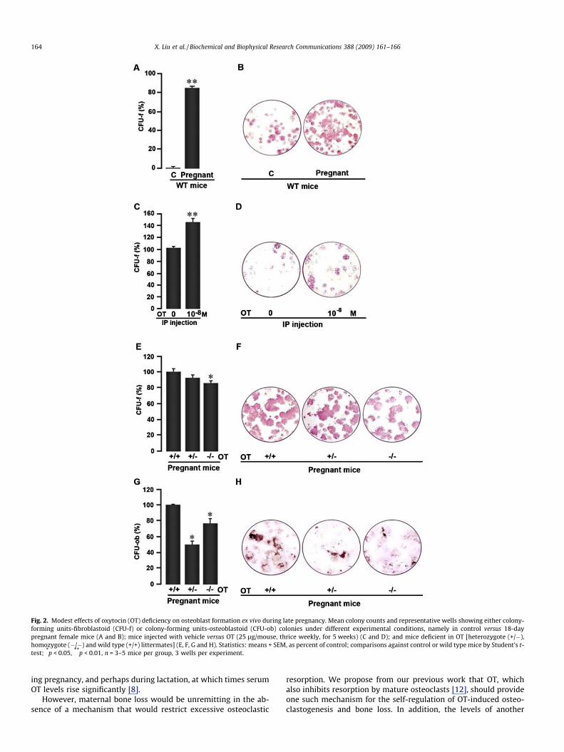
Fig. 2. Modest effects of oxytocin (OT) deficiency on osteoblast formation ex vivo during late pregnancy. Mean colony counts and representative wells showing either colony-forming units-fibroblastoid (CFU-f) or colony-forming units-osteoblastoid (CFU-ob) colonies under different experimental conditions, namely in control versus 18-daypregnant female mice (A and B); mice injected with vehicle versus OT (25 lg/mouse, thrice weekly, for 5 weeks) (C and D); and mice deficient in OT [heterozygote (+/�),homozygote (�/�) and wild type (+/+) littermates] (E, F, G and H). Statistics: means + SEM, as percent of control; comparisons against control or wild type mice by Student’s t-test; *p < 0.05, **p < 0.01, n = 3–5 mice per group, 3 wells per experiment.
164 X. Liu et al. / Biochemical and Biophysical Research Communications 388 (2009) 161–166
ing pregnancy, and perhaps during lactation, at which times serumOT levels rise significantly [8].
However, maternal bone loss would be unremitting in the ab-sence of a mechanism that would restrict excessive osteoclastic
resorption. We propose from our previous work that OT, whichalso inhibits resorption by mature osteoclasts [12], should provideone such mechanism for the self-regulation of OT-induced osteo-clastogenesis and bone loss. In addition, the levels of another

X. Liu et al. / Biochemical and Biophysical Research Communications 388 (2009) 161–166 165
osteoclast-inhibitory hormone, calcitonin, rise during pregnancyand lactation [17,18]. A high circulating calcitonin likely also pre-vents the excessive loss of calcium. However, while we provide ge-netic evidence for OT as a hormone that stimulatesosteoclastogenesis during pregnancy, and likely during lactation,mechanisms restricting excessive resorption have yet to beexplored.
In view of a plausible role for OT in maternal skeletal remodeling,we next chose to examine the skeletons of pups at E20. Fig. 3A showsOT+/�, OT�/� and wild type pups derived from heterozygote mothers.These pups had apparently normal skeletons, specifically displaying
Fig. 3. Oxytocin (OT) deficiency impairs skeletogenesis in utero. (A) No overt defectsoxytocin-deficient (heterozygote or homozygote) pups derived at E20 compared with thsignificant decreases and increases, respectively, in trabecular spacing (DT-Tb.Sp) andtrabecular thickness, as determined in epiphyseal trabecular bone (B). Representative imaMeans + SD shown for each parameter; comparisons between OT�/� pups and wild litte
no runting or bone/cartilage defects. Measurements of mainly tra-becular bone at the epiphysis showed no difference in BV/TV(Fig. 3B and C). However, trabecular number was significantly in-creased, without any change in trabecular thickness (Fig. 3B). Thisresulted, as would be expected, in a significant reduction in trabec-ular spacing (Fig. 3B). The increase in trabecular number is reminis-cent of reduced resorption in OT-deficiency; however, theexpectation from this finding in isolation would be an osteopetroticphenotype manifest by an increased BV/TV. Nonetheless, BV/TV wasunchanged, even in the face of greater trabecular numbers. This, tous, implies that each trabeculum is less mineralized, or indeed, total
or runting in alizarin red (bone) or alcian blue (cartilage) stained skeletons fromose of wild type littermates from the same heterozygote mother. Micro-CT revealedtrabecular number (DT-Tb.N), but without differences in bone volume (BV/TV) orges of epiphyseal and diaphyseal sections are shown in panels C and D, respectively.rmates (OT+/+) by Student’s t-test; *p < 0.05, n = 4 mice per group.

Fig. 4. Hypothetical schema for the control of osteoclast formation and resorptivefunction during pregnancy and lactation. The increased production of oxytocin (OT)from the posterior pituitary stimulates osteoclastogenesis by acting on theosteoclast precursor. During lactation, this is further accelerated by a transientdecline in serum estrogen (E2) and parathyroid hormone-related protein (PTHrP)secretion from mammary epithelial cells: all of these hormonal influences appear toplay a role in maternal skeletal mobilization. However, to prevent excessive skeletalloss, OT inhibits bone resorption by mature osteoclasts, as does the hormonecalcitonin (CT), produced in excess from the parafollicular C-cells of the thyroid.
166 X. Liu et al. / Biochemical and Biophysical Research Communications 388 (2009) 161–166
mineral per unit volume is lower in OT�/�pups than in wild type con-trols. While we have not measured mineral per se, we can concludesafely that OT is essential for optimal skeletal maturation in the fe-tus, and that the absence of OT leads to increased numbers of trabec-ulae that appear hypomineralized. Finally, lCT measurements at thediaphysis showed no obvious differences between OT�/� pups andwild type littermates (Fig. 3D).
Fig. 4 schematizes our current understanding of the hormonalregulation of bone resorption during pregnancy and/or lactation.It is known that hypoestrogenemia stimulates osteoclastogenesisduring lactation [13]. The breast-derived hormone, PTHrP doesthe same [13,16], as does OT, during late pregnancy, and likely dur-ing lactation. Our previous studies also implicate OT in stimulatingbone formation [12], particularly when the maternal skeleton re-quires restoration to virgin levels at weaning. Calcitonin appearsto aid this process by inhibiting resorption through its direct osteo-clastic action, as does OT, as a means of self-regulation.
We are currently unclear which hormonal action predominates,or indeed, the precise sequence in which these and other, yetunidentified hormones and cytokines, orchestrate to regulate aprocess as fundamental as intergenerational calcium transfer.While we have just touched the surface, our study highlights theimportance of further investigations using genetically-modifiedmice to probe into how the fetal skeleton is mineralized at the ex-pense of the mother, and in particular, how the mother recoversfrom this heavy burden of calcium loss.
Acknowledgments
M.Z. and S.L. acknowledge the National Institutes of Health forsupport (AG 23176, DK70526 and DK80459). X.L. is a graduate stu-dent sponsored by State Key Laboratory of Bioelectronics, South-east University, Nanjing, China. J.I. acknowledges the support ofthe American Federation for Aging Research.
References
[1] R. Acher, J. Chauvet, M.T. Chauvet, G. Michel, Y. Rouille, Molecular evolution ofneurohypophysial hormones in relation to osmoregulation: the two fishoptions, Fish Physiol. Biochem. 17 (1997) 1–6.
[2] K. Nishimori, L.J. Young, Q. Guo, Z. Wang, T.R. Insel, M.M. Matzuk, Oxytocin isrequired for nursing but is not essential for parturition or reproductivebehavior, Proc. Natl. Acad. Sci. USA 93 (1996) 11699–11704.
[3] W.S. Young 3rd, E. Shepard, A.C. DeVries, A. Zimmer, M.E. LaMarca, E.I. Ginns, J.Amico, R.J. Nelson, L. Hennighausen, K.U. Wagner, Targeted reduction ofoxytocin expression provides insights into its physiological roles, Adv. Exp.Med. Biol. 449 (1998) 231–240.
[4] T.R. Insel, C.R. Harbaugh, Lesions the hypothalamic paraventricular nucleusdisrupts the initiation of maternal behavior, Physiol. Behav. 45 (1989) 1033–1041.
[5] R.C. Mantella, R.R. Vollmer, X. Li, J.A. Amico, Female oxytocin-deficient micedisplay enhanced anxiety-related behavior, Endocrinology 144 (2003) 2291–2296.
[6] A. Argiolas, M. Collu, G.L. Gessa, M.R. Melis, G. Serra, The oxytocin antagonistd(CH2)5Tyr(Me)-Orn8-vasotocin inhibits male copulatory behavior in rats,Eur. J. Pharmacol. 149 (1988) 389–392.
[7] J.N. Ferguson, L.J. Young, E.F. Hearn, M.M. Matzuk, T.R. Insel, J.T. Winslow,Social amnesia in mice lacking the oxytocin gene, Nat. Genet. 25 (2000) 284–288.
[8] M.Y. Dawood, O. Ylikorkala, D. Trivedi, F. Fuchs, Oxytocin in maternalcirculation and amniotic fluid during pregnancy, J. Clin. Endocrinol. Metab.49 (1979) 429–434.
[9] J.J. Wysolmerski, The evolutionary origins of maternal calcium and bonemetabolism during lactation, J. Mammary Gland Biol. Neoplasia 7 (2002) 267–276.
[10] M. Sowers, D. Eyre, D.W. Hollis, J.F. Randolph, B. Shapiro, M.L. Jannausch, M.Crutchfield, Biochemical markers of bone turnover in lactating and non-lactating postpartum women, J. Clin. Endocrinol. Metab. 80 (1995) 2210–2216.
[11] L.D. Ritchi, E.B. Fung, B.P. Halloran, J.R. Turnland, M.D. VanLoan, C.E. Cann, J.C.King, A longitudinal study of calcium homeostasis during human pregnancyand lactation and after resumption of menses, Am. J. Clin. Nutr. 76 (1998) 693–701.
[12] R. Tamma, G. Colaianni, L.-L. Zhu, A. DiBenedetto, G. Greco, G. Montemurro, N.Patano, M. Strippoli, R. Vergari, L. Mancini, S. Colucci, M. Grano, R. Faccio, X.Liu, J.H. Li, S. Usmani, M. Bachar, I. Bab, K. Nishimori, L. Young, C. Buettener, J.Iqbal, L. Sun, M. Zaidi, A. Zallone, Oxytocin is an anabolic bone hormone, Proc.Natl. Acad. Sci. USA 106 (2009) 7149–7154.
[13] J.N. VaHouten, J.J. Wysolmerski, Low estrogen and high parathyroid hormone-related peptide levels contribute to accelerated bone resorption and bone lossn lactating mice, Endocrinology 144 (2003) 5521–5529.
[14] M. Zaidi, Skeletal remodeling in health and disease, Nat. Med. 13 (2007) 791–801.
[15] C.S. Kovacs, G.H. Fulehan, Calcium and bone disorders during pregnancy andlactation, Endocrinol. Metab. Clin. North Am. 35 (2006) 21–51.
[16] J.N. VanHouten, P. Dann, A.F. Stewart, C.J. Watson, M. Pollak, A.C. Karapalis, J.J.Wysolmerski, Mammary-specific deletion of parathyroid hormone-relatedprotein preserves bone mass during lactation, J. Clin. Invest. 112 (2003) 1429–1436.
[17] L.H. Briemer, I. MacIntyre, M. Zaidi, Peptides from the calcitonin genes:molecular genetics, structure and function, Biochem. J. 255 (1988) 377–390.
[18] J.P. Woodrow, C.J. Sharpe, N.J. Fudge, A.O. Hoff, R.F. Gagel, C.S. Kovacs,Calcitonin plays a critical role in regulating skeletal mineral metabolismduring lactation, Endocrinology 147 (2006) 4010–4021.
Copyright © 2022 FDOKUMEN