Oxytocin and socioemotional aging: Current knowledge and future trends
55
IN HUMAN NEUROSCIENCE Oxytocin and Socioemotional Aging-Current Knowledge and Future Trends Natalie C. Ebner, Gabriela M. Maura, Kai Macdonald, Lars Westberg and Håkan Fischer Journal Name: Frontiers in Human Neuroscience ISSN: 1662-5161 Article type: Original Research Article Received on: 28 May 2013 Accepted on: 01 Aug 2013 Provisional PDF published on: 01 Aug 2013 Frontiers website link: www.frontiersin.org Citation: Ebner NC, Maura GM, Macdonald K, Westberg L and Fischer H(2013) Oxytocin and Socioemotional Aging─Current Knowledge and Future Trends. Front. Hum. Neurosci. 7:487. doi:10.3389/fnhum.2013.00487 Article URL: http://www.frontiersin.org/Journal/Abstract.aspx?s=537& name=human%20neuroscience&ART_DOI=10.3389 /fnhum.2013.00487 (If clicking on the link doesn't work, try copying and pasting it into your browser.) Copyright statement: © 2013 Ebner, Maura, Macdonald, Westberg and Fischer. This is an open-access article distributed under the terms of the Creative Commons Attribution License (CC BY). The use, distribution or reproduction in other forums is permitted, provided the original author(s) or licensor are credited and that the original publication in this journal is cited, in accordance with accepted academic practice. No use, distribution or reproduction is permitted which does not comply with these terms. This Provisional PDF corresponds to the article as it appeared upon acceptance, after rigorous peer-review. Fully formatted PDF and full text (HTML) versions will be made available soon.
Transcript of Oxytocin and socioemotional aging: Current knowledge and future trends

IN HUMAN NEUROSCIENCE
Oxytocin and Socioemotional Aging-Current Knowledge and Future Trends Natalie C. Ebner, Gabriela M. Maura, Kai Macdonald, Lars Westberg and Håkan Fischer
Journal Name: Frontiers in Human Neuroscience
ISSN: 1662-5161
Article type: Original Research Article
Received on: 28 May 2013
Accepted on: 01 Aug 2013
Provisional PDF published on: 01 Aug 2013
Frontiers website link: www.frontiersin.org
Citation: Ebner NC, Maura GM, Macdonald K, Westberg L and FischerH(2013) Oxytocin and Socioemotional Aging─Current Knowledgeand Future Trends. Front. Hum. Neurosci. 7:487.doi:10.3389/fnhum.2013.00487
Article URL: http://www.frontiersin.org/Journal/Abstract.aspx?s=537&name=human%20neuroscience&ART_DOI=10.3389/fnhum.2013.00487
(If clicking on the link doesn't work, try copying and pasting it into your browser.)
Copyright statement: © 2013 Ebner, Maura, Macdonald, Westberg and Fischer. This is anopen-access article distributed under the terms of the CreativeCommons Attribution License (CC BY). The use, distribution orreproduction in other forums is permitted, provided the originalauthor(s) or licensor are credited and that the originalpublication in this journal is cited, in accordance with acceptedacademic practice. No use, distribution or reproduction ispermitted which does not comply with these terms.
This Provisional PDF corresponds to the article as it appeared upon acceptance, after rigorous
peer-review. Fully formatted PDF and full text (HTML) versions will be made available soon.

Running head: OXYTOCIN AND SOCIOEMOTIONAL AGING
Oxytocin and Socioemotional Aging─Current Knowledge and Future Trends
Natalie C. Ebner*,1 Gabriela M. Maura,
1 Kai MacDonald,
2 Lars Westberg,
3 & Håkan Fischer
4,5
1Department of Psychology, University of Florida, Gainesville, FL, USA,
2Department of Psychiatry,
University of California, San Diego, CA, USA, 3Department of Pharmacology, University of Gothenburg,
Gothenburg, Sweden, 4Department of Psychology, Stockholm University, Stockholm, Sweden,
5Aging
Research Center, Karolinska Institute, Stockholm, Sweden
Correspondence: Dr. Natalie C. Ebner
University of Florida
Department of Psychology
P.O. Box 112250
Gainesville, FL 32611, USA
Phone: (1) 203 691 0371
Email: [email protected]
Abstract: 236 characters
Main Text: 54 manuscript pages; 1 table, 1 box, 4 figures

OXYTOCIN AND SOCIOEMOTIONAL AGING
2
Abstract
The oxytocin (OT) system is involved in various aspects of social cognition and prosocial behavior.
Specifically, OT has been examined in the context of social memory, emotion recognition, cooperation,
trust, empathy, and bonding, and─though evidence is somewhat mixed─intranasal OT appears to benefit
aspects of socioemotional functioning. However, most of the extant data on aging and OT is from animal
research and human OT research has focused largely on young adults. As such, though we know that
various socioemotional capacities change with age, we know little about whether age-related changes in
the OT system may underlie age-related differences in socioemotional functioning. In this review, we take
a genetic-neuro-behavioral approach and evaluate current evidence on age-related changes in the OT
system as well as the putative effects of these alterations on age-related socioemotional functioning.
Looking forward, we identify informational gaps and propose an Age-Related Genetic, Neurobiological,
Sociobehavioral Model of Oxytocin (AGeNeS-OT model) which may structure and inform investigations
into aging-related genetic, neural, and sociocognitive processes related to OT. As an exemplar of the use
of the model, we report exploratory data suggesting differences in socioemotional processing associated
with genetic variation in the oxytocin receptor gene (OXTR) in samples of young and older adults.
Information gained from this arena has translational potential in depression, social stress, and anxiety─all
of which have high relevance in aging─and may contribute to reducing social isolation and improving
well-being of individuals across the lifespan.
Keywords: oxytocin, aging, socioemotional functioning, amygdala, anterior cingulate

OXYTOCIN AND SOCIOEMOTIONAL AGING
3
Oxytocin and Socioemotional Aging─Current Knowledge and Future Trends
Social and emotional processes and their associated genetic and neurobiological mechanisms in aging
are still incompletely understood (Nielsen & Mather, 2011). In this paper we propose to combine
neuroendocrine, genetic, and sociobehavioral approaches to examine the role of the oxytocin (OT) system
in the context of socioemotional aging. Aspects of the OT system warranting investigation include: 1)
changes in endogenous and dynamic OT levels; 2) changes in systems which directly impact OT function
(i.e., gonadal hormones); 3) genetic variation in aspects of the OT system, including the gene for oxytocin
(OXT), its receptor (OXTR), and the related CD38 system; 4) changes in OT-rich neural regions; 5) the
effect of exogenous OT. There is increasing evidence that OT plays a significant role in many of the
socioemotional capacities that undergo age-related changes. However, to date, very little is known about
the role of OT in human aging (Huffmeijer, van IJzendoorn, & Bakermans-Kranenburg, 2012). Thus, it
will be crucial for future research to clarify links between age-related changes in the abovementioned
aspects of the OT system and changes in neural processing and subsequent alterations in experience as
well as behavior in socioemotional domains in older compared to young adults.
To foreshadow, this focused review conceptually integrates two lines of research. First, we summarize
evidence for age-associated changes in socioemotional capacities (Isaacowitz et al., 2007; Scheibe &
Carstensen, 2010; Ruffman, Henry, Livingstone, & Phillips, 2008). Second, we review evidence for the
involvement of OT in socioemotional functioning (Bartz, Zaki, Bolger, & Ochsner, 2011; Meyer-
Lindenberg, Domes, Kirsch, & Heinrichs, 2011; Van IJzendoorn & Bakermans-Kranenburg, 2012).
Synthesizing these two lines of work, we present an Age-Related Genetic, Neurobiological,
Sociobehavioral Model of Oxytocin (AGeNeS-OT model) which may stimulate questions and organize
investigations into the role of OT in socioemotional aging. As an example of the use of the AGeNeS–OT
model, we report preliminary data suggesting neural and behavioral differences in socioemotional

OXYTOCIN AND SOCIOEMOTIONAL AGING
4
processing associated with genetic variations in OXTR in samples of young and older adults. We conclude
by suggesting future directions for research implied by the model. Ultimately, these investigations will
increase our understanding of the role of OT in aging and will have the potential for generating new
interventions to improve health and well-being.
Socioemotional Functioning and Aging
From life’s beginning, humans are confronted with critical, survival-enhancing socioemotional stimuli
related to self and others. To maintain successful social interactions and avoid the negative consequences
of social isolation (Baumeister & Leary, 1995; Norman et al., 2011), we learn to quickly and accurately
process, respond to, and remember social cues (Adolphs, 2003; Baron-Cohen, Tager-Flusberg, & Cohen,
2000; Grady & Keightley, 2002). Socioemotional functioning may become particularly relevant in old age
when─due to the experience of increasing physical ailment, dependency, and age-related social
losses─the experience of social isolation often increases with negative effects on physical and mental
health (Cornwell & Waite, 2009).
The extant literature suggests a mixed picture of age-related changes in socioemotional capabilities:
Some capacities (e.g., emotion regulation, emotional problem solving) improve with age, whereas other
skills (e.g., recognition of emotions in others) decline (cf. Scheibe & Carstensen, 2010). In particular,
across various studies, older compared to young adults show increased emotion regulation capacity
(Blanchard-Fields, Mienaltowski, & Seay, 2007; Carstensen, 2006; Riediger, Schmiedek, Wagner, &
Lindenberger, 2009; Scheibe & Blanchard-Fields, 2009; Voelkle, Ebner, Lindenberger, & Riediger, 2013)
and greater confidence in this ability (Gross & Levenson, 1997; Kessler & Staudinger, 2009; Lawton,
Kleban, Rajagopal, & Dean, 1992). The majority of older adults are well-adjusted emotionally and report
relatively high levels of affective well-being and emotional stability as documented in cross-sectional
(Carstensen, Pasupathi, Mayr, & Nesselroade, 2000) as well as longitudinal (Carstensen et al., 2011)

OXYTOCIN AND SOCIOEMOTIONAL AGING
5
studies (see also Charles, Reynolds, & Gatz, 2001; Teachman, 2006). In addition, older compared to
young adults are at least equally (and often more) effective in their ability to regulate their emotional
experiences, autonomic arousal, and outward display of negative emotions in language and faces when
instructed to do so (Kunzmann, Kupperbusch, & Levenson, 2005; Magai, Consedine, Krivoshekova,
Kudadjie-Gyamfi, & McPherson, 2006; Phillips, Henry, Hosie, & Milne, 2008), and show improved
socioemotional problem solving capacity (Blanchard-Fields et al., 2007).
At the same time, older adults often show increased difficulties in accurate recognition of social and
emotional cues (for reviews see Isaacowitz et al., 2007; Ruffman et al., 2008; see also Ebner & Johnson,
2010; see Figure 1A). Recent functional magnetic resonance imaging (fMRI) data suggests that these
difficulties are associated with greater activity in dorsomedial prefrontal cortex (dmPFC) in older
compared to young adults during facial emotion reading (Ebner, Johnson, & Fischer, 2012; Keightley,
Chiew, Winocur, & Grady, 2007; Williams et al., 2006; see Figure 1C). This association comports with
previous evidence that dmPFC is involved in complex processing and cognitive and emotional control
(Amodio & Frith, 2006). Another age-related change in socioemotional functioning is that older
compared to young adults demonstrate more interpersonal trust (Castle et al., 2012; List, 2004). This
change may be due to the difficulty older adults often have in “reading” the emotions of others, as
suggested by recent findings that older compared to young adults are less proficient at detecting lies,
mediated by deficits in emotion recognition (Ruffman, Murray, Halberstadt, & Vater, 2012). With respect
to changes in memory, there is evidence that the majority of older adults experience declines in
remembering critical socioemotional cues, including names (Crook et al., 1993; Verhaeghen & Salthouse,
1997) and faces (Bartlett, Leslie, Tubs, & Fulton, 1989; Ebner & Johnson, 2009; Grady et al., 1995; see
Figure 1B). Finally, in terms of social motivation, there is robust evidence that older adults are more
OXYTOCIN AND SOCIOEMOTIONAL AGING
6
avoidance-oriented and less approach-oriented than young adults (Ebner, Freund, & Baltes, 2006; Freund,
2006; Nikitin, Schoch, & Freund, in press).
Importantly, the mechanisms underlying these age-related changes in socioemotional functioning are
not well understood yet. One potential explanation is differences in visual processing (Ebner, He, &
Johnson, 2011; Isaacowitz, Wadlinger, Goren, & Wilson, 2006), perhaps as a function of age-related
changes in motivation (Carstensen, 2006; Mather & Carstensen, 2005; Samanez-Larkin & Carstensen,
2011). In particular, there is evidence that older compared to young adults spend more time looking at
positive than negative information (Isaacowitz et al., 2006) and, when processing faces, spend less time
viewing the eye region and more time viewing the mouth (Firestone, Turk-Browne, & Ryan, 2007). This
age-differential visual processing pattern may be important given that the eye vs. mouth regions of a face
carry different socioemotional information (Calder, Young, Keane, & Dean, 2000; Ebner et al., 2011). A
complementary, mechanistic explanation for age-related changes in socioemotional function may be
changes in brain structure or function in regions associated with socioemotional processing such as
amygdala, PFC, insula, or fusiform gyrus (Cacioppo, Norris, Decety, Monteleone, & Nusbaum, 2009;
Ebner et al., 2012; Grady, 2008; Keightley et al., 2007; see Ruffman et al., 2008; Samanez-Larkin &
Carstensen, 2011; St. Jaques, Winecoff, & Cabeza, 2013, for overviews). For instance, there is well-
documented, age-related structural decline in regions such as the lateral PFC (lPFC), insula, and striatum
(Raz, 2005; Raz et al., 2005). Regarding functional changes, one common finding is an age-related
decrease in amygdala activation during the perception of emotional stimuli (especially negative stimuli)
accompanied by an age-related increase in activity in a number of lPFC and mPFC regions (Fischer,
Nyberg, & Bäckman, 2010; Fischer, Sandblom, Gavazzeni, Fransson, Wright, & Bäckman, 2005;
Gunning-Dixon et al., 2003; Iidaka et al., 2002; Tessitore et al., 2005; but see Ebner, Johnson,

OXYTOCIN AND SOCIOEMOTIONAL AGING
7
Rieckmann, Durbin, Johnson, & Fischer, 2013; Mather & Carstensen, 2005; Wright, Wedig, Williams,
Rauch, & Albert, 2006).
Crucially, however, extant literature suggests that age-related differences in socioemotional
processing cannot be explained solely by age-related visuoperceptual and/or neurocognitive changes
(Samanez-Larkin & Carstensen, 2011). In addition, it may be that changes in socioemotional function are
also linked with age-related alterations in neuroendocrine function. In particular, the neuropeptide OT
appears as a particularly promising candidate, given increasing evidence of its role in socioemotional
domains (Bartz et al., 2011; Donaldson & Young, 2008; Insel & Fernald, 2004; Meyer-Lindenberg et al.,
2011; Norman et al., 2011). However, to date, we know very little about age-related changes in the OT
system, particularly in the context of socioemotional aging (Huffmeijer et al., 2012).
Oxytocin and Socioemotional Functioning
OT is a nine amino acid peptide, with peripheral and central functions (Gimpl & Fahrenholz, 2001). It
is synthesized in magnocellular neurosecretory cells of paraventricular nuclei (PVN) and supraoptic
nuclei (SON) of the hypothalamus and released through the posterior pituitary gland into the periphery
(Insel, 2010). OT is also released into the brain by magnocellular dendrites (Leng & Ludwig, 2006) and
by OT-releasing neurons projecting to specific brain regions such as the amygdala, hippocampus, and
striatum (Kimura, Tanizawa, Mori, Brownstein, & Okayama, 1992; Knobloch et al., 2012; Landgraf &
Neumann, 2004). Human and animal studies combined suggest that the function of the OT system is
reflected at a variety of physiological and anatomical levels, including: 1) peripheral hormone levels (i.e.,
plasma and saliva); 2) central hormone levels (i.e., in cerebrospinal fluid (CSF)); 3) histological levels
(i.e., presence and size of OT cells); 4) receptor levels (in OT receptor binding in defined brain regions);
5) genetic levels, or the level of “neuropeptidergic individuality” (MacDonald, 2012; i.e., polymorphisms

OXYTOCIN AND SOCIOEMOTIONAL AGING
8
related to OXT or OXTR, or genes related to OT release (i.e., CD38; Sauer, Montag, Reuter, & Kirsch,
2013; Sauer, Montag, Worner, Kirsch, & Reuter, 2012).
In particular, accumulating evidence suggests that OT may serve as a key effector in socioemotional
functioning such as emotion recognition, memory for faces, interpersonal trust, and bonding as briefly
summarized next (see Bartz et al., 2011; Meyer-Lindenberg et al., 2011; Norman et al., 2011; Zink &
Meyer-Lindenberg, 2012, for comprehensive overviews).
After the discovery that certain neuropeptides could be delivered intranasally to the human brain
(Born, Lange, Kern, McGregor, Bickel, & Fehm, 2002), a number of experimental studies using
intranasal OT revealed intriguing effects on diverse aspects of socioemotional functioning. For example,
research in healthy adults suggests that OT impairs performance in verbal memory tasks (Ferrier, Kennet,
& Devlin, 1980; Heinrichs, Meinlschmidt, Wippich, Ehlert, & Hellhammer, 2004; but see Feifel,
MacDonald, Cobb, & Minassian, 2012), while enhancing recognition of social (i.e., faces) but not non-
social stimuli (Rimmele, Hediger, Heinrichs, & Klaver, 2009; see also Heinrichs et al., 2004), especially
for neutral and angry compared to happy faces (Savaskan, Ehrhardt, Shulz, Walter, & Schächinger, 2008).
Furthermore, intranasal administration of OT increases overall gaze time toward faces (Andari, Duhamel,
Zalla, Herbrecht, Leboyer, & Sirigu, 2010; Averbeck, 2010; Gamer, Zurowski, & Büchel, 2010;
Guastella, Mitchell, & Dadds, 2008) and increases emotion recognition, specifically of happy and fearful
faces (and under certain conditions angry faces; see Shahrestani, Kemp, & Guastella, in press, for a recent
review).
In addition, recent studies have shown that intranasal OT increases facial trustworthiness and
attractiveness ratings (Theodoridou, Rowe, Penton-Voak, & Rogers, 2009) as well as interpersonal trust
and the willingness to take social risks (Baumgartner, Heinrichs, Vonlanthen, Fischbacher, & Fehr, 2008;
Kosfeld, Heinrichs, Zak, Fischbacher, & Fehr, 2005; Phan, Sripada, Angstadt, & McCabe, 2010). These

OXYTOCIN AND SOCIOEMOTIONAL AGING
9
effects of OT on trust seem to be particularly pronounced in positive social interactions (Mikolajczak,
Pinon, Lane, de Timary, & Luminet, 2010; Zak, Kurzban, & Matzner, 2005) and with respect to in-group
versus out-group members (Van IJzendoorn & Bakermans-Kranenburg, 2012). Moreover, these effects
seem moderated by interindividual differences (Rockliff, Karl, McEwan, Gilbert, Matos, & Gilbert, 2011;
but see Guastella, Alvares, Hickie, Chan, Chen, & Banati, 2013), including genetic polymorphisms
associated with OT function (Riedl & Javor, 2011; see also MacDonald, 2012; Rodrigues, Saslow, Garcia,
John, & Keltner, 2009, for reviews).
Besides these effects on facial processing and trust, intranasal OT has been shown to influence social
approach behavior, attachment, bonding, and social rejection with associated health benefits (Ditzen,
Schaer, Gabriel, Bodenmann, Ehlert, & Heinrichs, 2009; Fekete et al., 2013; Gouin et al., 2010; Scheele
et al., 2012; Schneiderman, Zagoory-Sharon, Leckman, & Feldman, 2012). For example, intranasal OT
increased positive relative to negative behaviors during a laboratory couple conflict and reduced post-
conflict cortisol levels (Ditzen et al., 2009). This potential stress reducing-effect of OT has been further
documented by evidence that participants with increased plasma OT healed faster and had a greater
number of positive interactions with partners during a 24-hour hospital stay (Gouin et al., 2010; see also
Kéri & Kiss, 2011; Kiss, Levy-Gigi, & Kéri, 2011; see Taylor, Gonzaga, Klein, Hu, Greendale, &
Seeman, 2006, for a discussion of OT’s role during relaxation vs. stress; see also Feldman, Gordon, &
Zagoory-Sharon, 2011).
An ever-expanding body of neuroimaging data suggests that OT’s effects on socioemotional
functioning are due to its attenuation of the neural circuitry for anxiety and aversion and its activation of
social reward neural networks (cf. Yoshida et al., 2009; Zink & Meyer-Lindenberg, 2012). In particular, a
number of studies have provided evidence that the amygdala might be a key structure for the mediation of
the social-cognitive effects of OT (Domes, Heinrichs, Gläscher, Büchel, Braus & Herpertz, 2007; Kirsch

OXYTOCIN AND SOCIOEMOTIONAL AGING
10
et al., 2005; Labuschagne, Phan, Wood, Angstadt, Chua, & Heinrichs, 2010; Petrovic, Kalisch, Singer, &
Dolan, 2008; Riem et al., 2011; Singer et al., 2008; Zink & Meyer-Lindenberg, 2012; cf. Huffmeijer et
al., 2012; but see Domes et al., 2010). For example, OT attenuates amygdala response to fear-inducing
stimuli (Kirsch et al., 2005). Baumgartner et al. (2008; see also Kosfeld et al., 2005; Mikolajczak et al.,
2010) provide evidence that OT reduced betrayal aversion to breaches of trust via a reduction in bilateral
amygdala activation and midbrain regions and greater ventral striatum and orbitofrontal cortex (OFC)
activity. Furthermore, there are suggestions of specific modulatory influences of OT on subregions within
the amygdala during processing of socioemotional information (Gamer et al., 2010; see also Huber,
Veinante, & Stoop, 2005; Knobloch et al., 2012; Viviani et al., 2011). These central effects, importantly,
occur in interaction with a network of other neurochemicals including estrogen, dopamine, and serotonin
(Riedl & Javor, 2011).
Thus there are suggestions in the literature that OT increases approach-related behaviors, while
decreasing withdrawal-related behaviors (Kemp & Guastella, 2010). At the same time, however, there is
evidence suggesting that OT may play a somewhat more complex role in social behavior than simply
directing approach vs. avoidance behavior and/or attentional biases to positive and negative information,
respectively. Rather, OT may increase social engagement, salience of social agents, and social value of
processed information, largely independent of valence (Bartz et al., 2011; Shamay-Tsoory, 2010;
Shamay-Tsoory, Fischer, Dvash, Harari, Perach-Bloom, Levkovitz, 2009; Tops, 2009). In line with this
suggestion, brain regions such as the ventral tegmentum, PFC, nucleus accumbens, and insula associated
with the social-reward neural network have shown sensitive to OT (Balleine, Delgado, & Hikosaka, 2007;
Groppe, et al., 2013; Riem et al., 2011; Scheele et al., 2013; Wittfoth-Schardt et al., 2012).
The central effects of OT are mediated by its G-protein-coupled receptor, located on a variety of
tissues including the brain, heart, kidney, and uterus (Gimpl & Fahrenholz, 2001; Loup, Tribollet,

OXYTOCIN AND SOCIOEMOTIONAL AGING
11
Dubois-Dauphin, & Dreifuss, 1991). Polymorphisms of the gene encoding the OT receptor, OXTR, have
been shown to contribute to individual differences in various social phenotypes (cf. Ebstein, Knafo,
Mankuta, Chew, & Lai, 2012; Gimpl & Fahrenholz, 2001; Kumsta, Chen, Pape, & Heinrichs, 2013;
Meyer-Lindenberg et al., 2011; Westberg & Walum, 2013; Zink & Meyer-Lindenberg, 2012). For
example, OXTR single nucleotide polymorphisms (SNPs) have been associated with lower positive affect
(Lucht et al., 2009), lower levels of responsiveness of mothers to their toddlers (Bakermans-Kranenburg
& van IJzendoorn, 2008), lower empathy scores and increased stress reactivity (Rodrigues et al., 2009),
nonverbal displays of prosociality (Kogan, Saslow, Impett, Oveis, Keltner, & Saturn, 2011), and pair-
bonding (Walum et al., 2012). OXTR SNPs have also been studied in relation with autism spectrum
disorder (ASD; see Ebstein et al., 2012, for a review), with evidence that they contribute to risk for some
phenotypes observed in ASD (Egawa, Watanabe, Endo, Tamura, Masuzawa, & Someya, 2012; but see
Tansey et al., 2010).
Taken together, this review highlights the importance of simultaneously considering behavioral,
neural, and genetic perspectives when examining OT’s role in socioemotional functioning, as will be
discussed in more detail below (see Figure 2). In addition, it raises five important caveats and
informational gaps. First, some of the effects associated with OT are inconsistent and come from small,
homogeneous samples, creating a need for replication of key findings in larger, more representative
samples.
Second, many of OT’s effects seem to vary by individual difference variables such as the level of
social proficiency (Bartz et al., 2011; but Guastella et al., 2013). Third, there is increasing evidence
suggesting that the effects of OT are dependent on context (Domes, Heinrichs, Michel, Berger, &
Herpertz, 2007) and influenced by early life experiences (see MacDonald, 2012, for a review). For
example, women (Heim, Young, Newport, Mletzko, Miller, & Nemeroff, 2008) and men (Meinlschmidt

OXYTOCIN AND SOCIOEMOTIONAL AGING
12
& Heim, 2007) who were abused or neglected as children showed altered OT system sensitivity as adults
(e.g., decreased CSF level of OT; see also Fries, Ziegler, Kurian, Jacoris, & Pollack, 2005; Winslow,
Noble, Lyons, Sterk, & Insel, 2003; but see Anderson, 2006; cf. MacDonald, 2012, for a review).
Fourth, due to both theoretical safety concerns using OT in women as well as the complexity
introduced by OT’s sex-specific effects, a large majority of studies conducted so far refer to men
exclusively, even though there are growing indications that some of OT’s effects may differ by sex
(Domes et al., 2010; Guastella, Carson, Dadds, Mitchell, & Cox, 2009; Marsh, Yu, Pine, & Blair, 2010;
Savaskan et al., 2008; cf. MacDonald, 2012). This sex-specific pattern raises the possibility that the
effects of OT on social cognition may be differentially regulated by gonadal steroids (estrogen and
testosterone) or other sex-specific biological factors (Choleris, Clipperton-Allen, Phan, & Kavaliers,
2009; Gabor, Phan, Clipperton-Allen, Kavaliers, & Choleris, 2012; see also van Anders, Goldey, & Kuo,
2011; see also Weisman & Feldman, 2013).
A fifth shortcoming in the current human literature on oxytocin—critical in the present context—is
that current studies have almost exclusively been conducted with young adults. Given the
abovementioned evidence of age-group differences in socioemotional functioning (Samanez-Larkin &
Carstensen, 2011; Scheibe & Carstensen, 2010), a comprehensive examination of aging-related aspects of
the OT system (including genetic, neurobiological, and behavioral aspects) is warranted (Huffmeijer et
al., 2012).
Oxytocin and Aging
Despite a significant need for research addressing the growing older segment of the population,
research on OT and aging is scarce and inconclusive. To date, the few studies that have addressed age-
related differences in the OT system almost exclusively refer to non-human species with limited
applicability to humans (Quinn, 2005). Also, studies conducted to date are characterized by large

OXYTOCIN AND SOCIOEMOTIONAL AGING
13
methodological differences in terms of species examined, OT parameters measured, brain regions
targeted, etc., which makes a direct comparison difficult and a meta-analytic approach not feasible. Most
importantly, a theoretical framework for generating hypotheses regarding age-related differences in the
OT system (including changes in endogenous OT physiology, function, and differential response to
exogenous OT) is entirely lacking (cf. Huffmeijer et al., 2012).
Table 1 provides a summary of the current studies on OT and aging. Whereas some studies suggest no
noticeable effects of aging on the OT system (Arletti, Benelli, Poggioli, Luppi, Menozzi, & Bertolini,
1995; Fliers, De Vries, & Swaab, 1985; Wierda et al., 1991; Zbuzek, Fuchs, Zbuzek, & Wu, 1988), other
studies report age-related change (Arsenijevic, Dreifuss, Vallet, Marguerat, & Tribollet, 1995; Fliers &
Swaab, 1983; Melis, Mauri, & Argiolas, 1995; Melis, Stancampiano, Fratta, & Argiolas, 1992; Parker,
Hoffman, Hyde, Cummings, & Maestripieri, 2010). Notably, some of the studies reporting comparability
of the OT system across older and young subjects refer to peripheral OT levels (Fliers & Swaab, 1983;
Melis et al., 1992; Zbuzek et al., 1988), whereas several of the studies documenting age-related change
relate to central OT levels (Fliers & Swaab, 1983; Melis et al., 1992; Arsenijevic et al., 1995; Parker,
Hoffman, Hyde, Cummings, & Maestripieri, 2010). Thus, it is possible that aging may change OT
transmission in the CNS but not in the neurohypophyseal (peripheral) system (Melis, Spano, Succu, &
Argiolas, 1999). A summary of the evidence reported in Table 1 would be that existent evidence does not
allow yet a firm conclusion of the existence or direction of age-related changes in the OT system, leaving
the question open to empirical examination.
To our knowledge, only one very recent study explicitly examined the effects of intranasal OT in a
group of older men (mean age of 80 years) focusing on OT’s effects on social engagement and physical
health (Barraza, Grewal, Ropacki, Perez, Gonzalez, & Zak, 2013). Results from this double-blind,
placebo-controlled 10-day clinical trial suggested improvement in dispositional gratitude in older adults in
OXYTOCIN AND SOCIOEMOTIONAL AGING
14
the OT compared to the placebo group. In addition, the OT group had a slower decline in physical
functioning and decreased self-reported fatigue than the placebo group. No changes in mood,
cardiovascular states, or social activity and engagement patterns were observed across the study interval.
Importantly, this study did not include a comparison group of young adults and did not extensively
explore OT’s effects on other aspects of socioemotional functioning. Thus, it is critical to follow up on
these first promising findings regarding OT and aging and to conduct systematic examinations of age-
differences in baseline levels of OT. In addition, a comprehensive evaluation of both single-dose as well
as longer-term administration of intranasal OT and its effect on socioemotional functioning in young and
older men and women is warranted. Finally, these studies should take into account genetic variations
related to OT.
Oxytocin and Socioemotional Aging: Age-Related Genetic, Neurobiological, Sociobehavioral Model of
Oxytocin
Based on the following rationale, we propose an OT X Age interaction (see Figure 3C) as the guiding
working hypothesis for future research on the role of OT in socioemotional aging: As mentioned above,
there is early evidence that the beneficial effects of OT in socioemotional domains (see Figure 3A) vary
by individual factors (Bartz et al., 2011). Notably, “preexisting social impairment” seems to play a role, in
that more socially impaired individuals benefit more from OT than less socially impaired individuals
(Bartz et al., 2010; Guastella et al., 2010; but see Bakermans-Kranenburg & van IJzendoorn, 2013). Also
there may be a “ceiling effect”, a point beyond which OT cannot further improve social abilities (Bartz et
al., 2011). As laid out above, older adults experience deficits in various socioemotional capacities
(Scheibe & Carstensen, 2010; see Figure 3A), rendering them more socially impaired than young adults in
some regards. Therefore, it may well be that OT is particularly beneficial in older compared to young
adults (see Figure 3C).

OXYTOCIN AND SOCIOEMOTIONAL AGING
15
However, an alternative hypothesis exists: As reported above, even though some aspects of
socioemotional functioning (i.e., emotion recognition and memory for emotional information) decline
with age, other aspects increase or remain stable. That is, given broad evidence for a positivity effect and
for healthy socioemotional functioning in old age (Carstensen, 2006; Carstensen et al., 2011), as well as
some evidence for increased trustworthiness in old age (Castle et al., 2012), on average, older adults can
be described as highly positive, trustworthy, and prosocial. These characteristics may be adaptive in some
contexts (e.g., social interactions within close relationships) but maladaptive in others (e.g., putting aging
adults at greater susceptibility to fraud). This reasoning, combined with the current lack of proof that
aging is associated with declines in the OT system and mixed evidence regarding OT’s effect on cognitive
performance (Feifel et al., 2012; Heinrichs et al., 2004), suggest the possibility that under certain
circumstances OT may have harmful effects in older adults. Given that OT is currently being investigated
in clinical populations such as schizophrenia (cf. MacDonald & Feifel, 2012, 2013), comprising samples
of people who are late middle-aged, a thorough investigation of age-related aspects of the OT
system─including beneficial or detrimental effects on outcome measures in socioemotional as well as
cognitive domains─will be crucial.
As summarized above, the OT system is represented at genetic, neural, and behavioral levels (Meyer-
Lindenberg et al., 2011). Furthermore, each of these levels and their functional interactions are influenced
by the aging process. We therefore propose for future research in the domain of OT and socioemotional
aging to adopt an Age-Related Genetic, Neurobiological, Sociobehavioral Model of Oxytocin (AGeNeS-
OT model; Figure 2). In particular, this model suggests that a comprehensive examination of the central
OT system should consider interactions between OT-related genes (OXT, OXTR, CD38; Meyer-
Lindenberg et al., 2011; Sauer et al., 2013), the brain (e.g., amygdala, frontal cortex, brainstem, ventral
tegmental area; Balleine et al., 2007; Baumgartner et al., 2008; Gamer et al., 2010; Kirsch et al., 2005;

OXYTOCIN AND SOCIOEMOTIONAL AGING
16
Pedersen, Caldwell, Walker, Ayers, & Mason, 1994), and behavior (e.g., social memory, emotion
identification, approach/avoidance biases; Domes et al., 2010; Lischke, Berger, Prehn, Heinrichs,
Herpertz, & Domes, 2011; Rimmele et al., 2009), by combining genetics, functional and structural brain
imaging, and sociobehavioral measures. Crucially, the model proposes that interactions between
neuroendocrine and sociobehavioral factors need to be considered from a developmental perspective,
taking age variations into account. Along these lines, the model offers a theoretical framework to address
vital research questions: (1) Are OT-related genotypes associated with composition and quality of social
networks in the elderly? How do brain structures involved in social processing such as mPFC and OFC,
temporoparietal junction, or amygdala mediate these relationships? (2) Is older adults’ increased social
avoidance compared to approach motivation represented in neural processing differences in brain
networks involving PFC and amygdala? To what extent do these associations interact with OT-related
genotypes? (3) Are detrimental effects that early abuse has on morbidity and mortality in the elderly
moderated by OT-related genotypes or OT levels? How is this relationship structurally and functionally
represented in the brain? (4) Are effects of social relationships on cognitive functioning in the elderly
mediated by the OT system (either OT-levels or OT-related genotypes)? Do structural changes in brain
regions such as the hippocampus underlie this relationship?
In the attempt to provide a concrete empirical application of the AGeNeS-OT Model, we here present a
preliminary report of an experiment in which we examined associations between OXTR polymorphisms,
brain activity and behavioral response during reading of facial emotions in young and older adults. This
exploratory, secondary data analysis was based on our group’s previous finding of increased activation in
ventromedial PFC (vmPFC) during emotion identification of happy compared to angry faces and
increased dorsomedial PFC (dmPFC) activity to angry compared to happy faces (Ebner et al., 2012; see
also Keightley et al., 2007) in both young and older adults. In the present set of analyses, we examined the

OXYTOCIN AND SOCIOEMOTIONAL AGING
17
extent to which these processing differences in mPFC would be further qualified when considering OXTR
polymorphisms in both of the age groups. In particular, we examined (1) the extent to which OXTR
polymorphisms were associated with differences in young and older adults’ brain activity in bilateral
mPFC (Ebner et al., 2012; Haxby, Hoffman, & Gobbini, 2000, 2002; Pessoa & Adolphs, 2010) during a
facial emotion reading task; and (2) the extent to which OXTR polymorphisms were associated with
young and older adults’ ability to read facial emotions.
Young (n = 25, 12 females, M = 25.1 years [SD = 3.6, range = 20-31] and older (n = 29, 17 females,
M = 68.3 years [SD = 2.8, range = 65-74]) healthy participants underwent fMRI on a 3T Siemens
Magnetom TrioTim scanner, while identifying happy, neutral, and angry facial emotions (see Ebner et al.,
2012, for details on participants, study design, and image acquisition). Participants were subsequently
genotyped by KBioscience (http://www.kbioscience.co.uk) using KASPar methodology for 14 OXTR
single nucleotide polymorphisms (SNPs in order from the 3’ to the 5’ end: rs7632287, rs6770632,
rs1042778, rs237887, rs2268493, rs2254298, rs53576, rs237897, rs4686302, rs4564970, rs2301261,
rs2268498, rs2270465, rs75775), previously shown to be associated with social behavior (Apicella,
Cesarini, Johannesson, Dawes, Lichtenstein, & Wallace, 2010; Ebstein et al. 2012; Meyer-Lindenberg et
al., 2011; Walum et al., 2012; Westberg & Walum, 2013).
Data from this event-related fMRI study was analyzed using Statistical Parametric Mapping (SPM5;
Wellcome Department of Imaging Neuroscience) and pre-processing and data analysis was conducted as
reported in Ebner et al. (2012). The following T-contrasts were specified across young and older adults,
based on our previous findings (Ebner et al., 2012): (1) Happy Faces > Angry Faces, (2) Angry Faces >
Happy Faces. We focused on select regions of interest (ROIs: bilateral medial frontal gyrus and anterior
cingulate gyrus) in which we had previously seen processing differences for happy vs. angry faces, at a
threshold of p < .05, FDR corrected. For each region of activation identified by these two contrasts, peak

OXYTOCIN AND SOCIOEMOTIONAL AGING
18
voxel beta values were extracted for each participant to produce a single value for each condition of
interest. These values are depicted in the bar graphs of Figure 4. In the fashion of follow-up F- and t-tests
(p < .05), for each of the14 OXTR SNPs that were genotyped, we examined differences in brain activation
between polymorphisms across the total sample as well as separately for young and older adults. The
most consistent associations found in these analyses were in relation to OXTR rs237887 (cf. Israel, Knafo,
Ebstein, Lerer, Shalev, & Uzefovsky, 2009; Lerer, Levi, Salomon, Darvasi, Yirmiya, & Ebstein, 2008;
Liu et al., 2010; Lori et al., 2012; but see Apicella et al., 2010).
OXTR rs237887 AA carriers (n = 10 young participants; n = 10 older participants) and GA/GG
carriers (n = 15 young participants; n = 19 older participants) were comparable in terms of chronological
age, level of education, cognitive status (e.g., Mini Mental State Examination; Folstein, Folstein, &
McHugh, 1975; 2-Back Digits Task; Kirchner, 1958; Verbal Fluency Task; Lezak, 1995), and affective
variables (Geriatric Depression Scale; Brink, Yesavage, Lum, Heersema, Adey, & Rose, 1982; Gottfries,
1997; State-Trait Anxiety Inventory; Spielberger, Gorsuch, & Lushene, 1970).
For the contrast Happy Faces > Angry Faces, we found greater BOLD response to happy compared to
angry faces in bilateral anterior cingulate cortex (ACC; MNI: x = 3, y = 45, z = 0 and x = -3, y = 51, z =
0) and bilateral mPFC (MNI: x = 3, y = 60, z = -3 and x = -3, y = 57, z = -3). Figure 4A shows brain
activity in left ACC (MNI: x = -3, y = 51, z = 0) for this contrast. To then examine associations between
OXTR rs237887 polymorphisms and brain activity during facial emotion identification of happy vs. angry
faces in young and older adults, we conducted follow-up univariate ANOVA collapsed across young and
older participants on extracted beta values at the peak voxel of activation. Left ACC activity was greater
for AA carriers than GA/GG carriers (F(1,51) = 6.51, p = .014, ηp2
= .11; see Figure 4B). More
interestingly, however, this effect was more pronounced in older than young adults, as tested in univariate
ANOVAs conducted separately within young and older participants (Young participants: F(1,23) = 2.38,

OXYTOCIN AND SOCIOEMOTIONAL AGING
19
p = .136, ηp2
= .09; Older participants: F(1,26) = 3.09, p = .035, ηp2
= .16; see Figure 4C). A comparable
pattern of results was found for right ACC (F(1,51) = 6.34, p = .015, ηp2
= .11). In addition, the results for
left (F(1,51) = 3.24, p = .078, ηp2
= .06) and right (F(1,51) = 1.29, p = .261, ηp2
= .03) mPFC pointed in the
same direction but were not significant.
ACC is a brain region that has been shown to be associated with affective processing (Amodio &
Frith, 2006; Bush, Luu, & Posner, 2000; Ebner et al., 2012), suggesting that AA compared to GA/GG
carriers may process happy compared to angry faces more affectively. This interpretation was further
supported by the finding that AA-genotype carriers of OXTR rs237887 (M = 1111 ms, SD = 171 ms) were
faster at labeling happy expressions than individuals carrying a G-allele (M = 1212 ms, SD = 173 ms;
F(1,50) = 4.26, p = .044, ηp2
= .08), with comparable effects in young and older participants. No
comparable effect was found for accuracy in emotion expression identification. However, interestingly,
greater recruitment of right ACC in individuals carrying a G-allele was positively correlated (r = .35; p =
.049) with accuracy in reading happy faces but uncorrelated in AA-genotype carriers (r = .05; p = .838).
This suggests that GA/GG carriers, as the group who needed more time on the task (see Figure 5A),
benefitted from recruiting ACC during the facial emotion reading task. This positive brain-behavior
correlation in GA/GG carriers was comparable in young and older participants (Fisher’s z = -0.42; p =
.337).
For the contrast Angry Faces > Happy Faces, we found greater BOLD response to angry compared to
happy faces in left mPFC (MNI: x = -6, y = 15, z = 51). In a follow-up univariate ANOVA collapsed
across young and older participants on extracted beta values at the peak voxel of activation, activity in left
mPFC did not vary by OXTR rs237887 polymorphism (p > .05).
To our knowledge this is the first study that considers young and older participants in a genetic-neuro-
behavioral examination of facial emotion processing, as suggested in the AGeNeS-OT model. Though this

OXYTOCIN AND SOCIOEMOTIONAL AGING
20
secondary data analysis was largely exploratory and replication in a larger independent sample of young
and older adults is warranted, our study provides some first indication of a role of OXTR rs237887 in
reading positive compared to negative facial expressions, with some variation as a function of the age of
the participant. Intriguingly, OXTR rs237887 has previously been associated with susceptibility for ASD
(Liu et al., 2010), prosocial behavior (Israel et al., 2009, but see Apicella et al., 2010), and face
recognition (Lori et al., 2012). We found improved processing of happy compared to angry faces for AA
carriers compared to GA/GG carriers, as reflected in their faster response time in reading happy faces and
their increased recruitment of ACC during emotion reading of happy compared to angry faces. Examining
young and older participants separately, this increased activation of ACC in AA compared to GA/GG
carriers was more pronounced in older than young participants. This is very interesting given broad
evidence of preferential processing of positive over negative information in older compared to young
adults (Mather & Carstensen, 2005). In addition, our findings suggest that GA/GG carriers’ ability to
correctly identify happy faces improved when recruiting ACC during the task.
Future Trends in Research on Oxytocin and Socioemotional Aging
Taken together, this research review indicates that a targeted investigation of age-related changes in
the OT system—especially one that considers genetic, neural, and behavioral processe—has the potential
to substantively increase our understanding of socioemotional change in aging. We believe that our
AGeNeS-OT model will be a fruitful conceptual basis in that it raises a set of vital research questions
necessary to refine our understanding of OT-related dynamics in aging in socioemotional contexts (see
Box 1). In addition, future research along those lines has great potential to inform both pharmacological
and psychosocial interventions targeting social and emotional dysfunction in the elderly. In particular,
there is an increasing body of research suggesting a significant role of OT in the context of various
disorders characterized by socioemotional dysfunction such as social-bonding deficits or related to social

OXYTOCIN AND SOCIOEMOTIONAL AGING
21
anxiety and stress (Heinrichs, Baumgartner, Kirschbaum, & Elhert, 2003; Taylor et al., 2006; Zetzche,
Frasch, Jirikowski, Murck, & Steiger, 1996; see MacDonald & Feifel, 2012, for an overview), deficits
with great relevance in an aging context. Thus, future research towards implementation of
pharmacological neuropeptide treatments with the potential to decrease emotional and social stress,
anxiety, and depression (Arletti & Bertolini, 1987; Carter & Altemus, 1997) will be important. These
interventions may consequently promote positive social interaction and willingness to engage in more
frequently rewarding social risks (Heinrichs et al., 2003; Kosfeld et al., 2005), improving health and life
quality up until late in life.

OXYTOCIN AND SOCIOEMOTIONAL AGING
22
References
Adolphs, R. (2003). Cognitive neuroscience of human social behaviour. Nature Reviews
Neuroscience, 4(3), 165-178. doi:10.1038/nrn1056
Amodio, D. M., & Frith, C. D. (2006). Meeting of the minds: The medial frontal cortex and social
cognition. Nature Reviews Neuroscience, 7, 268–277. doi:10.1038/nrn1884
Andari, E., Duhamel, J., Zalla, T., Herbrecht, E., Leboyer, M., & Sirigu, A. (2010). Promoting social
behavior with oxytocin in high-functioning autism spectrum disorders. Proceedings of the National
Academy of Sciences of the United States of America, 107(9), 4389-4394.
doi:10.1073/pnas.0910249107
Anderson, G. M. (2006). Report of altered urinary oxytocin and AVP excretion in neglected orphans
should be reconsidered. Journal of Autism and Developmental Disorders, 36(6), 829-830.
doi:10.1007/s10803-006-0153-7
Apicella, C. L., Cesarini, D., Johannesson, M., Dawes, C. T., Lichtenstein, P., & Wallace, B. (2010). No
association between oxytocin receptor (OXTR) gene polymorphisms and experimentally elicited
social preferences. PloS One, 5(6), e11153. doi:10.1371/journal.pone.0011153
Arletti, R., Benelli, A., Poggioli, R., Luppi, P., Menozzi, B., & Bertolini, A. (1995). Aged rats are still
responsive to the antidepressant and memory-improving effects of oxytocin. Neuropeptides, 29(3),
177-182. doi:10.1016/0143-4179(95)90021-7
Arletti, R., & Bertolini, A. (1987). Oxytocin acts as an antidepressant in two animal models of depression.
Life Sciences, 41(14), 1725-1730. doi:10.1016/0024-3205(87)90600-X
Arsenijevic, Y., Dreifuss, J. J., Vallet, P., Marguerat, A., & Tribollet, E. (1995). Reduced binding of
oxytocin in the rat brain during aging. Brain Research, 698(1–2), 275-279. doi:10.1016/0006-
8993(95)01020-V

OXYTOCIN AND SOCIOEMOTIONAL AGING
23
Averbeck, B. B. (2010). Oxytocin and the salience of social cues. Proceedings of the National Academy
of Sciences of the United States of America, 107(20), 9033-9034. doi:10.1073/pnas.1004892107
Bakermans-Kranenburg, M. J., & van IJzendoorn, M. H. (2008). Oxytocin receptor (OXTR) and
serotonin transporter (5-HTT) genes associated with observed parenting. Social Cognitive & Affective
Neuroscience, 3(2), 128-134. doi:10.1093/scan/nsn004
Bakermans-Kranenburg, M.J., & van IJzendoorn, M. H. (2013). Sniffing around oxytocin: Review and
meta-analyses of trials in healthy and clinical groups with implications for pharmacotherapy.
Translational Psychiatry, 3, e258. doi:10.1038/tp.2013.34
Balleine, B. W., Delgado, M. R., & Hikosaka, O. (2007). The role of the dorsal striatum in reward and
decision-making. The Journal of Neuroscience, 27(31), 8161-8165. doi:10.1523/JNEUROSCI.1554-
07.2007
Baron-Cohen, S. E., Tager-Flusberg, H. E., & Cohen, D. J. (Eds.). (2000). Understanding other minds:
Perspectives from developmental cognitive neuroscience. New York, NY: Oxford University Press.
Barraza, J., Grewal, N., Ropacki, S., Perez, P., Gonzalez, A., & Zak, P. (2013). Effects of a 10-day
oxytocin trial in older adults on health and well-being. Experimental & Clinical
Psychopharmacology, 21(2), 85-92. doi:10.1037/a0031581
Bartlett, J. C., Leslie, J. E., Tubbs, A., & Fulton, A. (1989). Aging and memory for pictures of faces.
Psychology and Aging, 4, 276-283. doi:10.1037/0882-7974.4.3.276
Bartz, J. A., Zaki, J., Bolger, N., Hollander, E., Ludwig, N. N., Kolevzon, A., & Ochsner, K. N. (2010).
Oxytocin selectively improves empathic accuracy. Psychological Science, 21(10), 1426-1428.
doi:10.1177/0956797610383439
Bartz, J. A., Zaki, J., Bolger, N., & Ochsner, K. N. (2011). Social effects of oxytocin in humans: Context
and person matter. Trends in Cognitive Sciences, 15(7), 301-309. doi:10.1016/j.tics.2011.05.002

OXYTOCIN AND SOCIOEMOTIONAL AGING
24
Baumeister, R. F., & Leary, M. R. (1995). The need to belong: Desire for interpersonal attachments as a
fundamental human motivation. Psychological Bulletin, 117(3), 497. doi:10.1037/0033-
2909.117.3.497
Baumgartner, T., Heinrichs, M., Vonlanthen, A., Fischbacher, U., & Fehr, E. (2008). Oxytocin shapes the
neural circuitry of trust and trust adaptation in humans. Neuron, 58(4), 639-650.
doi:10.1016/j.neuron.2008.04.009
Blanchard-Fields, F., Mienaltowski, A., & Seay, R. B. (2007). Age differences in everyday problem-
solving effectiveness: Older adults select more effective strategies for interpersonal problems. The
Journals of Gerontology Series B: Psychological Sciences and Social Sciences, 62(1), P61-P64.
doi:10.1093/geronb/62.1.P61
Born, J., Lange, T., Kern, W., McGregor, G. P., Bickel, U., & Fehm, H. L. (2002). Sniffing
neuropeptides: A transnasal approach to the human brain. Nature Neuroscience, 5(6), 514-516.
doi:10.1038/nn0602-849
Brink, T. L., Yesavage, J. A., Lum, O., Heersema, P. H., Adey, M., & Rose, T. L. (1982). Screening tests
for geriatric depression. Clinical Gerontologist, 1(1), 37-43. doi:10.1300/J018v01n01_06
Bush, G., Luu, P., Posner, M. (2000). Cognitive and emotional influences in anterior cingulate cortex.
Trends in Cognitive Sciences, 4(6), 215-222. doi:10.1016/S1364-6613(00)01483-2
Cacioppo, J. T., Norris, C. J., Decety, J., Monteleone, G., & Nusbaum, H. (2009). In the eye of the
beholder: Individual differences in perceived social isolation predict regional brain activation to
social stimuli. Journal of Cognitive Neuroscience, 21(1), 83-92. doi:10.1162/jocn.2009.21007
Calder, A. J., Young, A. W., Keane, J., & Dean, M. (2000). Configural information in facial expression
perception. Journal of Experimental Psychology: Human Perception and Performance, 26(2), 527-
551. doi:10.1037/0096-1523.26.2.527

OXYTOCIN AND SOCIOEMOTIONAL AGING
25
Carstensen, L. L., Pasupathi, M., Mayr, U., & Nesselroade, J. R. (2000). Emotional experience in
everyday life across the adult life span. Journal of Personality and Social Psychology, 79(4), 644.
doi:10.1037/0022-3514.79.4.644
Carstensen, L. L. (2006). The influence of a sense of time on human development. Science, 312(5782),
1913-1915. doi:10.1126/science.1127488
Carstensen, L. L., Turan, B., Scheibe, S., Ram, N., Ersner-Hershfield, H., Samanez-Larkin, G. R., . . .
Nesselroade, J. R. (2011). Emotional experience improves with age: Evidence based on over 10 years
of experience sampling. Psychology & Aging, 26(1), 21-33. doi:10.1037/a0021285
Carter, C. S., & Altemus, M. (1997). Integrative functions of lactational hormones in social behavior and
stress management. Annals of the New York Academy of Sciences, 807(1), 164-174.
doi:10.1111/j.1749-6632.1997.tb51918.x
Castle, E., Eisenberger, N. I., Seeman, T. E., Moons, W. G., Boggero, I. A., Grinblatt, M. S., & Taylor, S.
E. (2012). Neural and behavioral bases of age differences in perceptions of trust. Proceedings of the
National Academy of Sciences, 109(51), 20848-20852. doi:10.1073/pnas.1218518109
Charles, S. T., Reynolds, C. A., & Gatz, M. (2001). Age-related differences and change in positive and
negative affect over 23 years. Journal of Personality and Social Psychology, 80(1), 136-151.
doi:10.1037/0022-3514.80.1.136
Choleris, E., Clipperton-Allen, A. E., Phan, A., & Kavaliers, M. (2009). Neuroendocrinology of social
information processing in rats and mice. Frontiers in Neuroendocrinology, 30(4), 442-459.
doi:10.1016/j.yfrne.2009.05.003
Cornwell, E. Y., & Waite, L. J. (2009). Social disconnectedness, perceived isolation, and health among
older adults. Journal of Health and Social Behavior, 50(1), 31-48.
doi:10.1177/002214650905000103

OXYTOCIN AND SOCIOEMOTIONAL AGING
26
Crook, T. H., Lebowitz, B. D., Pirozzolo, F. J., Zappalà, G., Cavarzeran, F., Measso, G., & Massari, D. C.
(1993). Recalling names after introduction: Changes across the adult life span in two cultures.
Developmental Neuropsychology, 9(2), 103-113. doi:10.1080/87565649109540547
Ditzen, B., Schaer, M., Gabriel, B., Bodenmann, G., Ehlert, U., & Heinrichs, M. (2009). Intranasal
oxytocin increases positive communication and reduces cortisol levels during couple conflict.
Biological Psychiatry, 65(9), 728-731. doi:10.1016/j.biopsych.2008.10.011
Domes, G., Heinrichs, M., Gläscher, J., Büchel, C., Braus, D. F., & Herpertz, S. C. (2007). Oxytocin
attenuates amygdala responses to emotional faces regardless of valence. Biological Psychiatry,
62(10), 1187-1190. doi:10.1016/j.biopsych.2007.03.025
Domes, G., Heinrichs, M., Michel, A., Berger, C., & Herpertz, S. C. (2007). Oxytocin improves “mind-
reading” in humans. Biological Psychiatry, 61(6), 731-733. doi:10.1016/j.biopsych.2006.07.015
Domes, G., Lischke, A., Berger, C., Grossmann, A., Hauenstein, K., Heinrichs, M., & Herpertz, S. C.
(2010). Effects of intranasal oxytocin on emotional face processing in
women. Psychoneuroendocrinology, 35(1), 83-93. doi:10.1016/j.psyneuen.2009.06.016
Donaldson, Z. R., & Young, L. J. (2008). Oxytocin, vasopressin, and the neurogenetics of sociality.
Science, 322(5903), 900-904. doi:10.1126/science.1158668
Ebner, N. C., Freund, A. M., & Baltes, P. B. (2006). Developmental changes in personal goal orientation
from young to late adulthood: From striving for gains to maintenance and prevention of
losses. Psychology and Aging, 21(4), 664-678. doi:10.1037/0882-7974.21.4.664
Ebner, N. C., He, Y., & Johnson, M. K. (2011). Age and emotion affect how we look at a face: Visual
scan patterns differ for own-age versus other-age emotional faces. Cognition & Emotion, 25(6), 983-
997. doi:10.1080/02699931.2010.540817

OXYTOCIN AND SOCIOEMOTIONAL AGING
27
Ebner, N. C., & Johnson, M. K. (2009). Young and older emotional faces: Are there age group differences
in expression identification and memory? Emotion, 9(3), 329–339. doi:10.1037/a0015179
Ebner, N. C., & Johnson, M. K. (2010). Age-group differences in interference from young and older
emotional faces. Cognition & Emotion, 24, 1095–1116. doi:10.1080/02699930903128395
Ebner, N. C., Johnson, M. K., & Fischer, H. (2012). Neural mechanisms of reading facial emotions in
young and older adults. Frontiers in Psychology, 3, 223. doi:10.3389/fpsyg.2012.00223
Ebner, N. C., Johnson, M. R., Rieckmann, A., Durbin, K. A., Johnson, M. K., & Fischer, H. (2013).
Processing own-age vs. other-age faces: Neuro-behavioral correlates and effects of emotion.
Neuroimage, 78(0), 363-371. doi:10.1016/j.neuroimage.2013.04.029
Ebstein, R. P., Knafo, A., Mankuta, D., Chew, S. H., & Lai, P. S. (2012). The contributions of oxytocin
and vasopressin pathway genes to human behavior. Hormones and Behavior, 61(3), 359-379.
doi:10.1016/j.yhbeh.2011.12.014
Egawa, J., Watanabe, Y., Endo, T., Tamura, R., Masuzawa, N., & Someya, T. (2012). Association
between OXTR and clinical phenotypes of autism spectrum disorders. Psychiatry Research, 208(1),
99-100. doi:10.1016/j.psychres.2012.11.007
Feifel, D., MacDonald, K., Cobb, P., & Minassian, A. (2012). Adjunctive intranasal oxytocin improves
verbal memory in people with schizophrenia. Schizophrenia Research, 139(1–3), 207-210.
doi:10.1016/j.schres.2012.05.018
Fekete, E. M., Seay, J., Antoni, M. H., Mendez, A. J., Fletcher, M. A., Szeto, A., & Scheiderman, N.
(2013). Oxytocin, social support, and sleep quality in low-income minority women living with HIV.
Behavioral Sleep Medicine, 11, 1-15. doi: 10.1080/15402002.2013.791297

OXYTOCIN AND SOCIOEMOTIONAL AGING
28
Feldman, R., Gordon, I., & Zagoory‐Sharon, O. (2011). Maternal and paternal plasma, salivary, and
urinary oxytocin and parent–infant synchrony: Considering stress and affiliation components of
human bonding. Developmental Science, 14(4), 752-761. doi:10.1111/j.1467-7687.2010.01021.x
Ferrier, B. M., Kennet, D. J., & Devlin, M. C. (1980). Influence of oxytocin on human memory processes.
Life Sciences, 27(24), 2311-2317. doi:10.1016/0024-3205(80)90499-3
Firestone, A. A., Turk-Browne, N. B., & Ryan, J. D. (2007). Age-related deficits in face recognition are
related to underlying changes in scanning behavior. Aging, Neuropsychology, and Cognition: A
Journal on Normal and Dysfunctional Development, 14(6), 594-607.
doi:10.1080/13825580600899717
Fischer, H., Sandblom, J., Gavazzeni, J., Fransson, P., Wright, C. I., & Bäckman, L. (2005). Age-
differential patterns of brain activation during perception of angry faces. Neuroscience Letters,
386(2), 99–104. doi:10.1016/j.neulet.2005.06.002
Fischer, H., Nyberg, L., & Bäckman, L. (2010). Age-related differences in brain regions supporting
successful encoding of emotional faces. Cortex, 46(4), 490-497. doi:10.1016/j.cortex.2009.05.011
Fliers, E., De Vries, G. J., & Swaab, D. F. (1985). Changes with aging in the vasopressin and oxytocin
innervation of the rat brain. Brain Research, 348(1), 1-8. doi:10.1016/0006-8993(85)90351-8
Fliers, E., & Swaab, D. F. (1983). Activation of vasopressinergic and oxytocinergic neurons during aging
in the wistar rat. Peptides, 4(2), 165-170. doi:10.1016/0196-9781(83)90108-0
Folstein, M. F., Folstein, S. E., & McHugh, P. R. (1975). Mini-mental state: A practical method for
grading the cognitive state of patients for the clinician. New York, NY: Pergamon Press.
Freund, A. M. (2006). Age-differential motivational consequences of optimization versus compensation
focus in younger and older adults. Psychology and Aging, 21(2), 240-252. doi:10.1037/0882-
7974.21.2.240

OXYTOCIN AND SOCIOEMOTIONAL AGING
29
Fries, A. B. W., Ziegler, T. E., Kurian, J. R., Jacoris, S., & Pollak, S. D. (2005). Early experience in
humans is associated with changes in neuropeptides critical for regulating social behavior.
Proceedings of the National Academy of Sciences of the United States of America, 102(47), 17237-
17240. doi:10.1073/pnas.0504767102
Gabor, C. S., Phan, A., Clipperton-Allen, A., Kavaliers, M., & Choleris, E. (2012). Interplay of oxytocin,
vasopressin, and sex hormones in the regulation of social recognition. Behavioral Neuroscience,
126(1), 97-109. doi:10.1037/a0026464
Gamer, M., Zurowski, B., & Büchel, C. (2010). Different amygdala subregions mediate valence-related
and attentional effects of oxytocin in humans. Proceedings of the National Academy of Sciences,
107(20), 9400-9405. doi:10.1073/pnas.1000985107
Gimpl, G., & Fahrenholz, F. (2001). The oxytocin receptor system: Structure, function, and regulation.
Physiological Reviews, 81(2), 629-683.
Gottfries, C. G. (1997). Recognition and management of depression in the elderly. International Clinical
Psychopharmacology, 12, S31-S36. doi:10.1097/00004850-199712007-00006
Gouin, J., Carter, C. S., Pournajafi-Nazarloo, H., Glaser, R., Malarkey, W. B., Loving, T. J., . . . Kiecolt-
Glaser, J. K. (2010). Marital behavior, oxytocin, vasopressin, and wound healing.
Psychoneuroendocrinology, 35(7), 1082-1090. doi:10.1016/j.psyneuen.2010.01.009
Grady, C. L. (2008), Cognitive neuroscience of aging. Annals of the New York Academy of Sciences,
1124, 127–144. doi:10.1196/annals.1440.009
Grady, C. L., & Keightley, M. L. (2002). Studies of altered social cognition in neuropsychiatric disorders
using functional neuroimaging. Canadian Journal of Psychiatry, 47(4), 327-336.

OXYTOCIN AND SOCIOEMOTIONAL AGING
30
Grady, C. L., McIntosh, A. R., Horwitz, B., Maisog, J. M., Ungerleider, L.G., Mentis, M.J., Pietrini, P.,
Schapiro, M. B., & Haxby, J. V. (1995). Age-related reductions in human recognition memory due to
impaired encoding. Science, 269(5221), 218–221. doi:10.1126/science.7618082
Groppe, S. E., Gossen, A., Rademacher, L., Hahn, A., Westphal, L., Gründer, G., & Spreckelmeyer, K. N.
(2013). Oxytocin influences processing of socially relevant cues in the ventral tegmental area of the
human brain. Biological Psychiatry, 74(3), 172-179. doi:10.1016/j.biopsych.2012.12.023
Gross, J. J., & Levenson, R. W. (1997). Hiding feelings: The acute effects of inhibiting negative and
positive emotion. Journal of abnormal psychology, 106(1), 95-103. doi:10.1037/0021-843X.106.1.95
Guastella, A., Alvares, G., Hickie, I., Chan, H., Chen, T., & Banati, R. (2013). Unitary hormonal models,
peripheral markers, and evaluation of response to drug: A response to Weisman and Feldman.
Psychoneuroendocrinology, 38(5), 627-628. doi:10.1016/j.psyneuen.2013.03.005
Guastella, A. J., Carson, D. S., Dadds, M. R., Mitchell, P. B., & Cox, R. E. (2009). Does oxytocin
influence the early detection of angry and happy faces? Psychoneuroendocrinology, 34(2), 220-225.
doi:10.1016/j.psyneuen.2008.09.001
Guastella, A. J., Einfeld, S. L., Gray, K. M., Rinehart, N. J., Tonge, B. J., Lambert, T. J., & Hickie, I. B.
(2010). Intranasal oxytocin improves emotion recognition for youth with autism spectrum disorders.
Biological Psychiatry, 67(7), 692-694. doi:10.1016/j.biopsych.2009.09.020
Guastella, A. J., Mitchell, P. B., & Dadds, M. R. (2008). Oxytocin increases gaze to the eye region of
human faces. Biological Psychiatry, 63(1), 3-5. doi:10.1016/j.biopsych.2007.06.026
Gunning-Dixon, F., Gur, R. C., Perkins, A. C., Schroeder, L., Turner, T., Turetsky, B. I., . . . Gur, R. E.
(2003). Aged-related differences in brain activation during emotional face processing. Neurobiology
of Aging, 24(2), 285-295. doi:10.1016/S0197-4580(02)00099-4

OXYTOCIN AND SOCIOEMOTIONAL AGING
31
Haxby, J. V., Hoffman, E. A., & Gobbini, M. I. (2000). The distributed human neural system for face
perception. Trends in Cognitive Sciences, 4(6), 223-233. doi:10.1016/S1364-6613(00)01482-0
Haxby, J. V., Hoffman, E. A., & Gobbini, M. I. (2002). Human neural systems for face recognition and
social communication. Biological Psychiatry, 51(1), 59-67. doi:10.1016/S0006-3223(01)01330-0
Heim, C., Young, L. J., Newport, D. J., Mletzko, T., Miller, A. H., & Nemeroff, C. B. (2008). Lower CSF
oxytocin concentrations in women with a history of childhood abuse. Molecular Psychiatry, 14(10),
954-958. doi:10.1038/mp.2008.112
Heinrichs, M., Baumgartner, T., Kirschbaum, C., & Ehlert, U. (2003). Social support and oxytocin
interact to suppress cortisol and subjective responses to psychosocial stress. Biological Psychiatry,
54(12), 1389-1398. doi:10.1016/S0006-3223(03)00465-7
Heinrichs, M., Meinlschmidt, G., Wippich, W., Ehlert, U., & Hellhammer, D. H. (2004). Selective
amnesic effects of oxytocin on human memory. Physiology & Behavior, 83(1), 31-38.
doi:10.1016/j.physbeh.2004.07.020
Huber, D., Veinante, P., & Stoop, R. (2005). Vasopressin and oxytocin excite distinct neuronal
populations in the central amygdala. Science, 308(5719), 245-248. doi:10.1126/science.1105636.
Huffmeijer, R., van IJzendoorn, M. H., & Bakermans-Kranenburg, M. J. (2012). Ageing and oxytocin: A
call for extending human oxytocin research to ageing populations – a mini-review. Gerontology,
59(1), 32-39. doi: 10.1159/000341333
Iidaka, T., Okada, T., Murata, T., Omori, M., Kosaka, H., Sadato, N., & Yonekura, Y. (2002). Age-related
differences in the medial temporal lobe responses to emotional faces as revealed by fMRI.
Hippocampus, 12(3), 352-362. doi:10.1002/hipo.1113
Insel, T.R., & Fernald, R.D. (2004). How the brain processes social information: Searching for the social
brain. Annual Review of Neuroscience, 27, 697–722. doi:10.1146/annurev.neuro.27.070203.144148
OXYTOCIN AND SOCIOEMOTIONAL AGING
32
Insel, T. R. (2010). The challenge of translation in social neuroscience: A review of oxytocin,
vasopressin, and affiliative behavior. Neuron, 65(6), 768-779. doi:10.1016/j.neuron.2010.03.005
Isaacowitz, D. M., Wadlinger, H. A., Goren, D., & Wilson, H. R. (2006). Selective preference in visual
fixation away from negative images in old age? An eye-tracking study. Psychology and Aging, 21(1),
40–48. doi:10.1037/0882-7974.21.1.40
Isaacowitz, D. M., Löckenhoff, C. E., Lane, R. D., Wright, R., Sechrest, L., Riedel, R., & Costa, P. T.
(2007). Age differences in recognition of emotion in lexical stimuli and facial expressions.
Psychology and Aging, 22(1), 147–159. doi:10.1037/0882-7974.22.1.147
Israel, S., Knafo, A., Ebstein, R. P., Lerer, E., Shalev, I., & Uzefovsky, F. (2009). The oxytocin receptor
(OXTR) contributes to prosocial fund allocations in the dictator game and the social value
orientations task. PloS One, 4(5), e5535. doi:10.1371/journal.pone.0005535
Keck, M. E., Hatzinger, M., Wotjak, C. T., Landgraf, R., Holsboer, F., & Neumann, I. D. (2000). Ageing
alters intrahypothalamic release patterns of vasopressin and oxytocin in rats. European Journal of
Neuroscience, 12, 1487-1494.
Keightley, M. L., Chiew, K. S., Winocur, G., & Grady, C. L. (2007). Age-related differences in brain
activity underlying identification of emotional expressions in faces. Social Cognitive and Affective
Neuroscience, 2(4), 292-302. doi:10.1093/scan/nsm024
Kemp, A. H., & Guastella, A. J. (2010). Oxytocin: Prosocial behavior, social salience, or approach-related
behavior? Biological Psychiatry, 67(6), e33-e34. doi:10.1016/j.biopsych.2009.11.019
Kéri, S., & Kiss, I. (2011). Oxytocin response in a trust game and habituation of arousal. Physiology &
Behavior, 102(2), 221-224. doi:10.1016/j.physbeh.2010.11.011
Kessler, E., & Staudinger, U. (2009). Affective experience in adulthood and old age: The role of affective
arousal and perceived affect regulation. Psychology & Aging, 24(2), 349-362. doi:10.1037/a0015352

OXYTOCIN AND SOCIOEMOTIONAL AGING
33
Kimura, T., Tanizawa, O., Mori, K., Brownstein, M. J., & Okayama, H. (1992). Structure and expression
of a human oxytocin receptor. Nature, 356(6369), 526-529. doi:10.1038/356526a0
Kirsch, P., Esslinger, C., Chen, Q., Mier, D., Lis, S., Siddhanti, S., . . . Meyer-Lindenberg, A. (2005).
Oxytocin modulates neural circuitry for social cognition and fear in humans. The Journal of
Neuroscience, 25(49), 11489-11493. doi:10.1523/JNEUROSCI.3984-05.2005
Kirchner, W. K. (1958). Age differences in short-term retention of rapidly changing information. Journal
of Experimental Psychology, 55, 352-358. doi:10.1037/h0043688
Kiss, I., Levy-Gigi, E., & Kéri, S. (2011). CD 38 expression, attachment style and habituation of arousal
in relation to trust-related oxytocin release. Biological Psychology, 88(2), 223-226.
doi:10.1016/j.biopsycho.2011.08.005
Knobloch, H. S., Charlet, A., Hoffmann, L. C., Eliava, M., Khrulev, S., Cetin, A. H., . . . Grinevich, V.
(2012). Evoked axonal oxytocin release in the central amygdala attenuates fear response. Neuron,
73(3), 553-566. doi:10.1016/j.neuron.2011.11.030
Kogan, A., Saslow, L. R., Impett, E. A., Oveis, C., Keltner, D., & Saturn, S. R. (2011). Thin-slicing study
of the oxytocin receptor (OXTR) gene and the evaluation and expression of the prosocial disposition.
Proceedings of the National Academy of Sciences, 108(48), 19189-19192.
doi:10.1073/pnas.1112658108
Kosfeld, M., Heinrichs, M., Zak, P. J., Fischbacher, U., & Fehr, E. (2005). Oxytocin increases trust in
humans. Nature, 435(7042), 673-676. doi: 10.1038/nature03701
Kumsta, R., Chen, F. S., Pape, H., & Heinrichs, M. (2013). Neuropeptide S receptor gene is associated
with cortisol responses to social stress in humans. Biological Psychology, 93(2), 304-307.
doi:10.1016/j.biopsycho.2013.02.018

OXYTOCIN AND SOCIOEMOTIONAL AGING
34
Kunzmann, U., Kupperbusch, C. S., & Levenson, R. W. (2005). Behavioral inhibition and amplification
during emotional arousal: A comparison of two age groups. Psychology and Aging, 20(1), 144-158.
doi:10.1037/0882-7974.20.1.144
Labuschagne, I., Phan, K. L., Wood, A., Angstadt, M., Chua, P., & Heinrichs, M. (2010). Oxytocin
attenuates amygdala reactivity to fear in generalized social anxiety disorder.
Neuropsychopharmacology, 35(12), 2403-2413. doi:10.1038/npp.2010.123
Landgraf, R., & Neumann, I. D. (2004). Vasopressin and oxytocin release within the brain: A dynamic
concept of multiple and variable modes of neuropeptide communication. Frontiers in
Neuroendocrinology, 25(3–4), 150-176. doi:10.1016/j.yfrne.2004.05.001
Lawton, M., Kleban, M., Rajagopal, D., & Dean, J. (1992). Dimensions of affective experience in three
age groups. Psychology & Aging, 7(2), 171-184. doi:10.1037//0882-7974.7.2.171
Leng, G., & Ludwig, M. (2006). Information processing in the hypothalamus: Peptides and analogue
computation. Journal of Neuroendocrinology, 18(6), 379-392. doi:10.1111/j.1365-
2826.2006.01428.x
Lerer, E., Levi, S., Salomon, S., Darvasi, A., Yirmiya, N., & Ebstein, R. P. (2008). Association between
the oxytocin receptor (OXTR) gene and autism: Relationship to Vineland Adaptive Behavior Scales
and cognition. Molecular Psychiatry, 13, 980-988. doi:10.1038/sj.mp.4002087
Lezak, M. D. (1995). Neuropsychological Assessment (3rd Ed.). New York, NY: Oxford University Press.
Lischke, A., Berger, C., Prehn, K., Heinrichs, M., Herpertz, S. C., & Domes, G. (2011). Intranasal
oxytocin enhances emotion recognition from dynamic facial expressions and leaves eye-gaze
unaffected. Psychoneuroendocrinology, 37(4), 475-481. doi:10.1016/j.psyneuen.2011.07.015
List, J. A. (2004). Young, selfish and male: Field evidence of social preferences. The Economic Journal,
114(492), 121-149. doi:10.1046/j.0013-0133.2003.00180.x

OXYTOCIN AND SOCIOEMOTIONAL AGING
35
Liu, X., Kawamura, Y., Shimada, T., Otowa, T., Koishi, S., Sugiyama, T., . . . Sasaki, T. (2010).
Association of the oxytocin receptor (OXTR) gene polymorphisms with autism spectrum disorder
(ASD) in the Japanese population. Journal of Human Genetics, 55(3), 137-141.
doi:10.1038/jhg.2009.140
Lori, A., Lee, I., Cubells, J. F., Binder, E. B., Lehtimäki, T., Puura, K., . . . Skuse, D. (2012). Single
nucleotide polymorphism (SNP) fine mapping of the AVPR1A and OXTR loci reveals an OXTR
association with a social memory endophenotype in probands with autism and their family members.
Conference presentation at American Society of Human Genetics, San Fransisco, CA.
Loup, F., Tribollet, E., Dubois-Dauphin, M., & Dreifuss, J. J. (1991). Localization of high-affinity
binding sites for oxytocin and vasopressin in the human brain: An autoradiographic study. Brain
Research, 555(2), 220-232. doi:10.1016/0006-8993(91)90345-V
Lucht, M. J., Barnow, S., Sonnenfeld, C., Rosenberger, A., Grabe, H. J., Schroeder, W., . . . Rosskopf, D.
(2009). Associations between the oxytocin receptor gene (OXTR) and affect, loneliness and
intelligence in normal subjects. Progress in Neuro-Psychopharmacology and Biological Psychiatry,
33(5), 860-866. doi:10.1016/j.pnpbp.2009.04.004
MacDonald, K. (2012). Sex, receptors, and attachment: A review of individual factors influencing
response to oxytocin. Frontiers in Neuroscience, 6, 194. doi:10.3389/fnins.2012.00194
MacDonald, K., & Feifel, D. (2012). Oxytocin in schizophrenia: A review of evidence for its therapeutic
effects. Acta Neuropsychiatrica 24(3), 130-146. doi:10.1111/j.1601-5215.2011.00634.x
MacDonald K., & Feifel, D. (2013). Helping oxytocin deliver: Considerations in the development of
oxytocin-based therapeutics for brain disorders. Frontiers in Neuroscience, 7(35), 1-20.
doi:10.3389/fnins.2013.00035.

OXYTOCIN AND SOCIOEMOTIONAL AGING
36
Magai, C., Consedine, N., Krivoshekova, Y., Kudadjie-Gyamfi, E., & McPherson, R. (2006). Emotion
experience and expression across the adult life span: Insights from a multimodal assessment study.
Psychology & Aging, 21(2), 303-317. doi:10.1037/0882-7974.21.2.303
Marsh, A., Yu, H., Pine, D., & Blair, R. (2010). Oxytocin improves specific recognition of positive facial
expressions. Psychopharmacology, 209(3), 225-232. doi:10.1007/s00213-010-1780-4
Mather, M., & Carstensen, L. L. (2005). Aging and motivated cognition: The positivity effect in attention
and memory. Trends in Cognitive Sciences, 9(10), 496-502. doi:10.1016/j.tics.2005.08.005
Meinlschmidt, G., & Heim, C. (2007). Sensitivity to intranasal oxytocin in adult men with early parental
separation. Biological Psychiatry, 61(9), 1109-1111. doi:10.1016/j.biopsych.2006.09.007
Melis, M. R., Mauri, A., & Argiolas, A. (1995). Opposite changes in the content of oxytocin- and
vasopressin-like immunoreactive peptides in the rat thymus during aging. Regulatory Peptides, 59(3),
335-340. doi:10.1016/0167-0115(95)00088-S
Melis, M. R., Spano, M. S., Succu, S., & Argiolas, A. (1999). The oxytocin antagonist
d(CH2)5Tyr(Me)2-Orn8-vasotocin reduces non-contact penile erections in male rats. Neuroscience
Letters, 265(3), 171-174. doi:10.1016/S0304-3940(99)00236-0
Melis, M. R., Stancampiano, R., Fratta, W., & Argiolas, A. (1992). Oxytocin concentration changes in
different rat brain areas but not in plasma during aging. Neurobiology of Aging, 13(6), 783-786.
doi:10.1016/0197-4580(92)90102-4
Meyer-Lindenberg, A., Domes, G., Kirsch, P., & Heinrichs, M. (2011). Oxytocin and vasopressin in the
human brain: Social neuropeptides for translational medicine. Nature Reviews Neuroscience, 12(9),
524-538. doi:10.1038/nrn3044

OXYTOCIN AND SOCIOEMOTIONAL AGING
37
Mikolajczak, M., Pinon, N., Lane, A., de Timary, P., & Luminet, O. (2010). Oxytocin not only increases
trust when money is at stake, but also when confidential information is in the balance. Biological
Psychology, 85(1), 182-184. doi:10.1016/j.biopsycho.2010.05.010
Nielsen, L., & Mather, M. (2011). Emerging perspectives in social neuroscience and neuroeconomics of
aging. Social Cognitive and Affective Neuroscience, 6(2), 149-164. doi:10.1093/scan/nsr019
Nikitin, J., Schoch, S., & Freund, A. M. (in press). Why do younger and older adults react differently to
social acceptance and rejection? The role of social approach and avoidance motivation.
Developmental Psychology.
Norman, G. J., Cacioppo, J. T., Morris, J. S., Karelina, K., Malarkey, W. B., DeVries, A. C., & Berntson,
G. G. (2011). Selective influences of oxytocin on the evaluative processing of social stimuli. Journal
of Psychopharmacology, 25(10), 1313-1319. doi:10.1177/0269881110367452
Parker, K. J., Hoffman, C. L., Hyde, S. A., Cummings, C. S., & Maestripieri, D. (2010). Effects of age on
cerebrospinal fluid oxytocin levels in free-ranging adult female and infant rhesus
macaques. Behavioral Neuroscience, 124(3), 428-433. doi:10.1037/a0019576
Pedersen, C. A., Caldwell, J. D., Walker, C., Ayers, G., & Mason, G. A. (1994). Oxytocin activates the
postpartum onset of rat maternal behavior in the ventral tegmental and medial preoptic
areas. Behavioral Neuroscience, 108(6), 1163. doi:10.1037/0735-7044.108.6.1163
Pessoa, L., & Adolphs, R. (2010). Emotion processing and the amygdala: From a 'low road' to 'many
roads' of evaluating biological significance. Nature Reviews Neuroscience, 11, 773-783.
doi:10.1038/nrn2920
Petrovic, P., Kalisch, R., Singer, T., & Dolan, R. J. (2008). Oxytocin attenuates affective evaluations of
conditioned faces and amygdala activity. The Journal of Neuroscience, 28(26), 6607-6615.
doi:10.1523/JNEUROSCI.4572-07.2008

OXYTOCIN AND SOCIOEMOTIONAL AGING
38
Phan, K. L., Sripada, C. S., Angstadt, M., & McCabe, K. (2010). Reputation for reciprocity engages the
brain reward center. Proceedings of the National Academy of Sciences of the United States of
America, 107(29), 13099-13104. doi:10.1073/pnas.1008137107
Phillips, L. H., Henry, J. D., Hosie, J. A., & Milne, A. B. (2008). Effective regulation of the experience
and expression of negative affect in old age. The Journals of Gerontology Series B: Psychological
Sciences and Social Sciences, 63(3), P138-P145. doi:10.1093/geronb/63.3.P138
Quinn, R. (2005). Comparing rat’s to human’s age: How old is my rat in people years? Nutrition, 21(6),
775-777. doi:10.1016/j.nut.2005.04.002
Raz , N. (2005). The aging brain observed in vivo: Differential changes and their modifiers. In R. Cabeza,
L. Nyberg , & D. Park (Eds.), Cognitive neuroscience of aging: Linking cognitive and cerebral aging
(pp. 19 – 57). New York, NY: Oxford University Press.
Raz, N., Lindenberger, U., Rodrigue, K. M., Kennedy, K. M., Head, D., Williamson, A., . . . Acker, J. D.
(2005). Regional brain changes in aging healthy adults: General trends, individual differences and
modifiers. Cerebral Cortex, 15(11), 1676-1689. doi:10.1093/cercor/bhi044
Riediger, M., Schmiedek, F., Wagner, G. G., & Lindenberger, U. (2009). Seeking pleasure and seeking
pain: Differences in prohedonic and contra-hedonic motivation from adolescence to old age.
Psychological Science, 20(12), 1529-1535. doi:10.1111/j.1467-9280.2009.02473.x
Riedl, R., & Javor, A. (2011). The biology of trust: Integrating evidence from genetics, endocrinology,
and functional brain imaging. Journal of Neuroscience, Psychology, and Economics. 5(2), 63-91.
doi:10.1037/a0026318
Riem, M. M., Bakermans-Kranenburg, M. J., Pieper, S., Tops, M., Boksem, M. A., Vermeiren, R. R., ...
& Rombouts, S. A. (2011). Oxytocin modulates amygdala, insula, and inferior frontal gyrus

OXYTOCIN AND SOCIOEMOTIONAL AGING
39
responses to infant crying: A randomized controlled trial. Biological Psychiatry, 70(3), 291-297.
doi:10.1016/j.biopsych.2011.02.006
Rimmele, U., Hediger, K., Heinrichs, M., & Klaver, P. (2009). Oxytocin makes a face in memory
familiar. The Journal of Neuroscience, 29(1), 38-42. doi:10.1523/JNEUROSCI.4260-08.2009
Rockliff, H., Karl, A., McEwan, K., Gilbert, J., Matos, M., & Gilbert, P. (2011). Effects of intranasal
oxytocin on ‘compassion focused imagery’. Emotion, 11(6), 1388-1396. doi:10.1037/a0023861
Rodrigues, S. M., Saslow, L. R., Garcia, N., John, O. P., & Keltner, D. (2009). Oxytocin receptor genetic
variation relates to empathy and stress reactivity in humans. Proceedings of the National Academy of
Sciences of the United States of America, 106(50), 21437-21341. doi:10.1073/pnas.0909579106
Ruffman, T., Henry, J. D., Livingstone, V., & Phillips, L. H. (2008). A meta-analytic review of emotion
recognition and aging: Implications for neuropsychological models of aging. Neuroscience &
Biobehavioral Reviews, 32(4), 863-881. doi:10.1016/j.neubiorev.2008.01.001
Ruffman, T., Murray, J., Halberstadt, J., & Vater, T. (2012). Age-related differences in deception.
Psychology and Aging, 27(3), 543-549. doi:10.1037/a0023380
Samanez-Larkin, G. R., & Carstensen, L. L. (2011). Socioemotional functioning and the aging brain. In J.
Decety & J. T. Cacioppo (Eds.), The handbook of social neuroscience (pp. 507 – 523). New York,
NY: Oxford University Press.
Sauer, C., Montag, C., Reuter, M., & Kirsch, P. (2013). Imaging oxytocin x dopamine interactions: An
epistasis effect of CD38 and COMT gene variants influences the impact of oxytocin on amygdala
activation to social stimuli. Frontiers in Neuroscience, 7, 45. doi:10.3389/fnins.2013.00045
Sauer, C., Montag, C., Worner, C., Kirsch, P., & Reuter, M. (2012). Effects of a common variant in the
CD38 gene on social processing in an oxytocin challenge study: Possible links to autism.
Neuropsychopharmacology, 37(6), 1474-1482. doi:10.1038/npp.2011.333

OXYTOCIN AND SOCIOEMOTIONAL AGING
40
Savaskan, E., Ehrhardt, R., Schulz, A., Walter, M., & Schächinger, H. (2008). Post-learning intranasal
oxytocin modulates human memory for facial identity. Psychoneuroendocrinology, 33(3), 368-374.
doi:10.1016/j.psyneuen.2007.12.004
Scheele, D., Striepens, N., Güntürkün, O., Deutschländer, S., Maier, W., Kendrick, K.M., & Hurlemann,
R. (2012). Oxytocin modulates social distance between males and females. The Journal of
Neuroscience, 32(46), 16074-16079. doi:10.1523/JNEUROSCI.2755-12.2012
Scheele, D. A., Scheele, D., Wille, A., Kendrick, K. M., Becker, B., Güntürkün, O., Maier, W., &
Hurlemann, R. (2013). Oxytocin alters the human reward system to maintain romantic love
[Abstract]. Biological Psychiatry, 73(9), 95S-95S.
Scheibe, S., & Blanchard-Fields, F. (2009). Effects of regulating emotions on cognitive performance:
What is costly for young adults is not so costly for older adults. Psychology & Aging, 24(1), 217-223.
doi:10.1037/a0013807
Scheibe, S., & Carstensen, L. L. (2010). Emotional aging: Recent findings and future trends. The Journals
of Gerontology Series B: Psychological Sciences and Social Sciences, 65B(2), 135-144.
doi:10.1093/geronb/gbp132
Schneiderman, I., Zagoory-Sharon, O., Leckman, J. F., & Feldman, R. (2012). Oxytocin during the initial
stages of romantic attachment: Relations to couples' interactive reciprocity.
Psychoneuroendocrinology, 37(8), 1277-1285. doi: 10.1016/j.psyneuen.2011.12.021
Shahrestani, S., Kemp, A. H., & Guastella, A. J. (in press). The impact of a single administration of
intranasal oxytocin on the recognition of basic emotions in humans: A meta-analysis.
Neuropsychopharmacology. doi:10.1038/npp.2013.86
Shamay-Tsoory, S. G. (2010). One hormonal system for love and envy: A reply to Tops. The Biological
Psychiatry, 67(1), e7. doi:10.1016/j.biopsych.2009.09.002

OXYTOCIN AND SOCIOEMOTIONAL AGING
41
Shamay-Tsoory, S. G., Fischer, M., Dvash, J., Harari, H., Perach-Bloom, N., & Levkovitz, Y. (2009).
Intranasal administration of oxytocin increases envy and schadenfreude (gloating). Biological
Psychiatry, 66(9), 864-870. doi:10.1016/j.biopsych.2009.06.009
Singer, T., Snozzi, R., Bird, G., Petrovic, P., Silani, G., Heinrichs, M., & Dolan, R. J. (2008). Effects of
oxytocin and prosocial behavior on brain responses to direct and vicariously experienced pain.
Emotion, 8(6), 781-791. doi:10.1037/a0014195
Spielberger, C. D., Gorsuch, R. L., & Lushene, R. F. (1970). Manual for the State-Trait Anxiety
Inventory. Palo Alto, CA: Consulting Psychologist Press.
St. Jaques, P.L., Winecoff, A., & Cabeza, R. (2013). Emotion and aging. In J. Armony & P. Vuilleumier
(Eds.), The Cambridge handbook of human affective neuroscience. New York, NY: Cambridge
University Press.
Tansey, K. E., Brookes, K. J., Hill, M. J., Cochrane, L. E., Gill, M., Skuse, D., ... & Anney, R. J. (2010).
Oxytocin receptor (OXTR) does not play a major role in the aetiology of autism: Genetic and
molecular studies. Neuroscience letters, 474(3), 163-167. doi:10.1016/j.neulet.2010.03.035
Taylor, S. E., Gonzaga, G. C., Klein, L. C., Hu, P., Greendale, G. A., & Seeman, T. E. (2006). Relation of
oxytocin to psychological stress responses and hypothalamic-pituitary-adrenocortical axis activity in
older women. Psychosomatic Medicine, 68(2), 238-245. doi:10.1097/01.psy.0000203242.95990.74
Teachman, B. A. (2006). Aging and negative affect: The rise and fall and rise of anxiety and depression
symptoms. Psychology & Aging, 21(1), 201. doi:10.1037/0882-7974.21.1.201
Tessitore, A., Hariri, A. R., Fera, F., Smith, W. G., Das, S., Weinberger, D. R., & Mattay, V. S. (2005).
Functional changes in the activity of brain regions underlying emotion processing in the elderly.
Psychiatry Research: Neuroimaging, 139(1), 9-18. doi:10.1016/j.pscychresns.2005.02.009

OXYTOCIN AND SOCIOEMOTIONAL AGING
42
Theodoridou, A., Rowe, A. C., Penton-Voak, I. S., & Rogers, P. J. (2009). Oxytocin and social
perception: Oxytocin increases perceived facial trustworthiness and attractiveness. Hormones and
Behavior, 56(1), 128-132. doi:10.1016/j.yhbeh.2009.03.019
Tops, M. (2009). Oxytocin: Envy or engagement in others? Biological Psychiatry, 67(1), e5-e6. doi:
10.1016/j.biopsych.2009.08.032
Van Anders, S. M., Goldey, K. L., & Kuo, P. X. (2011). The steroid/peptide theory of social bonds:
Integrating testosterone and peptide responses for classifying social behavioral contexts.
Psychoneuroendocrinology, 36(9), 1265-1275. doi:10.1016/j.psyneuen.2011.06.001
Van IJzendoorn, M. H., & Bakermans-Kranenburg, M. J. (2012). A sniff of trust: Meta-analysis of the
effects of intranasal oxytocin administration on face recognition, trust to in-group, and trust to out-
group. Psychoneuroendocrinology, 37(3), 438-443. doi:10.1016/j.psyneuen.2011.07.008
Verhaeghen, P., & Salthouse, T. (1997). Meta-analyses of age-cognition relations in adulthood: Estimates
of linear and nonlinear age effects and structural models. Psychological Bulletin, 122(3), 231-249.
doi:10.1037/0033-2909.122.3.231
Viviani, D., Charlet, A., van den Burg, E., Robinet, C., Hurni, N., Abatis, M., ... & Stoop, R. (2011).
Oxytocin selectively gates fear responses through distinct outputs from the central amygdala.
Science, 333(6038), 104-107. doi:10.1126/science.1201043
Voelkle, M., Ebner, N., Lindenberger, U., & Riediger, M. (2013). Here we go again: Anticipatory and
reactive mood responses to recurring unpleasant situations throughout adulthood. Emotion, 13(3),
424-433. doi:10.1037/a0031351
Walum, H., Lichtenstein, P., Neiderhiser, J. M., Reiss, D., Ganiban, J. M., Spotts, E. L., . . . Westberg, L.
(2012). Variation in the oxytocin receptor gene is associated with pair-bonding and social behavior.
Biological Psychiatry, 71(5), 419-426. doi:10.1016/j.biopsych.2011.09.002

OXYTOCIN AND SOCIOEMOTIONAL AGING
43
Weisman, O., & Feldman, R. (2013). Oxytocin administration affects the production of multiple
hormones. Psychoneuroendocrinology, 38(5), 626-627. doi:10.1016/j.psyneuen.2013.03.004
Westberg L., & Walum, H. (2013). Oxytocin and vasopressin gene variation and the neural basis of
prosocial behavior. In The Oxford Handbook of Molecular Psychology. New York, NY: Oxford
University Press.
Wierda, M., Goudsmit, E., van der Woude, P., Purba, J., Hofman, M., Bogte, H., & Swaab, D. (1991).
Oxytocin cell number in the human paraventricular nucleus remains constant with aging and in
Alzheimer's disease. Neurobiology of Aging, 12(5), 511-516. doi:10.1016/0197-4580(91)90081-T
Williams, L. M., Brown, K. J., Palmer, D., Liddell, B. J., Kemp, A. H., Olivieri, G., . . . Gordon, E.
(2006). The mellow years?: Neural basis of improving emotional stability over age. The Journal of
Neuroscience, 26(24), 6422-6430. doi:10.1523/JNEUROSCI.0022-06.2006
Winslow, J., Noble, P., Lyons, C., Sterk, S., & Insel, T. (2003). Rearing effects on cerebrospinal fluid
oxytocin concentration and social buffering in rhesus monkeys. Neuropsychopharmacology, 28(5),
910-918. doi:10.1038/sj.npp.1300128
Wittfoth-Schardt, D., Grunding, J., Wittfoth, M., Lanfermann, H., Heinrichs, M., Domes, G., . . . Waller,
C. (2012). Oxytocin modulates neural reactivity to children's faces as a function of social salience.
Neuropsychopharmacology, 37(8), 1799-1807. doi:10.1038/npp.2012.47
Wright, C. I., Wedig, M. M., Williams, D., Rauch, S. L., & Albert, M. S. (2006). Novel fearful faces
activate the amygdala in healthy young and elderly adults. Neurobiology of Aging, 27(2) 361-374.
doi:10.1016/j.neurobiolaging.2005.01.014
Yoshida, M., Takayanagi, Y., Inoue, K., Kimura, T., Young, L. J., Onaka, T., & Nishimori, K. (2009).
Evidence that oxytocin exerts anxiolytic effects via oxytocin receptor expressed in serotonergic

OXYTOCIN AND SOCIOEMOTIONAL AGING
44
neurons in mice. The Journal of Neuroscience, 29(7), 2259-2271. doi:10.1523/JNEUROSCI.5593-
08.2009
Yu, K.-L., Tamada, Y., Suwa, F., Fang, Y.-R., & Tang, C.-S. (2006). Age-related changes in oxytocin-,
arginine vasopressin- and nitric oxide synthase-expressing neurons in the supraoptic nucleus of the
rat. Life Sciences, 78, 1143-1148.
Zak, P. J., Kurzban, R., & Matzner, W. T. (2005). Oxytocin is associated with human trustworthiness.
Hormones and Behavior, 48(5), 522-527. doi:10.1016/j.yhbeh.2005.07.009
Zbuzek, V., Fuchs, A., Zbuzek, V. K., & Wu, W. (1988). Neurohypophyseal aging: Differential changes
in oxytocin and vasopressin release, studied in Fischer 344 and Sprague-Dawley rats.
Neuroendocrinology, 48(6), 619-626. doi:10.1159/000125072
Zetzsche, T., Frasch, A., Jirikowski, G., Murck, H., & Steiger, A. (1996). Nocturnal oxytocin secretion is
reduced in major depression. Biological Psychiatry, 39(7), 584. doi:10.1016/0006-3223(96)84235-1
Zink, C. F., & Meyer-Lindenberg, A. (2012). Human neuroimaging of oxytocin and vasopressin in social
cognition. Hormones and Behavior, 61(3), 400-409. doi:10.1016/j.yhbeh.2012.01.016

OXYTOCIN AND SOCIOEMOTIONAL AGING
45
Acknowledgments
This research was supported in part by the NIH/NCATS Clinical and Translational Science Award to
the University of Florida UL1 TR000064 (pilot award to NCE) and the Swedish Research Council (2008-
2356) and the Konung Gustaf V:s och Drottning Victorias Frimurarstiftelse (HF). Some of KM’s work
was supported by the Goodenough Neuroscience Research Fund. The authors wish to thank Drs. David
Feifel and Ronald Cohen for constructive discussions regarding various aspects of this manuscript.

OXYTOCIN AND SOCIOEMOTIONAL AGING
46
Table 1. Literature Review on Oxytocin and Aging
Authors Species Age Group Measurement Difference Main Findings
Evidence of Stability in the OT System in Aging
Arletti et al. (1995) Rats (M) O Intraperitoneal OT injection O = Y Comparable improved social memory and
anti-depressant effect of OT injection
Fliers et al. (1985) Rats (M) Y/O OT fiber density O = Y Comparable OT fiber density in the brain
Wierda et al. (1991) Human (M, F)
Y/O Number of OT cells in PVN
(post-mortem)
O = Y Comparable numbers of OT-expressing cells
in PVN (normal aging and Alzheimer’s
Disease)
Yu et al. (2006) Rats (M) Y/O OT cell size and numbers in
SON
O = Y Comparable cell number, cell size, or
reactive density of NOS-expressing neurons
Evidence of Change in the OT System with Age
Arsenijevic et al.
(1995)
Rats (M) Y/O OT receptor binding O < Y Age-related decreased in binding to OT
receptors in caudate putamen, olfactory
tubercle, and ventromedial hypothalamic
nucleus
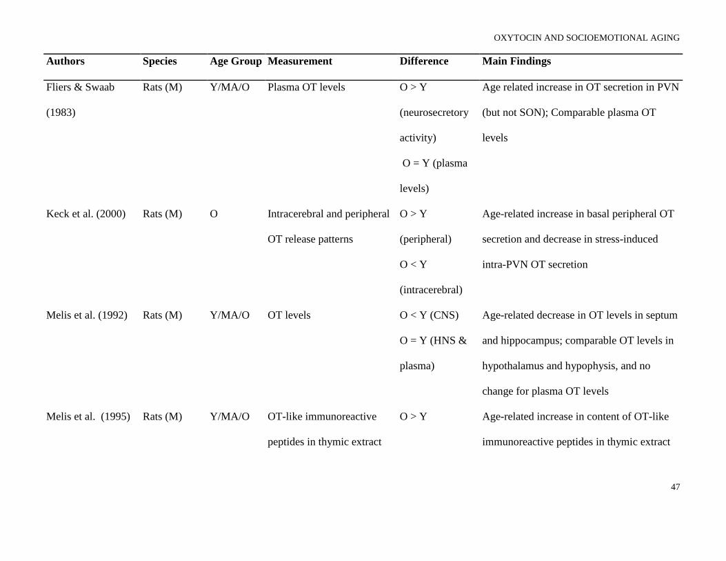
OXYTOCIN AND SOCIOEMOTIONAL AGING
47
Authors Species Age Group Measurement Difference Main Findings
Fliers & Swaab
(1983)
Rats (M) Y/MA/O Plasma OT levels O > Y
(neurosecretory
activity)
O = Y (plasma
levels)
Age related increase in OT secretion in PVN
(but not SON); Comparable plasma OT
levels
Keck et al. (2000) Rats (M) O Intracerebral and peripheral
OT release patterns
O > Y
(peripheral)
O < Y
(intracerebral)
Age-related increase in basal peripheral OT
secretion and decrease in stress-induced
intra-PVN OT secretion
Melis et al. (1992) Rats (M) Y/MA/O OT levels O < Y (CNS)
O = Y (HNS &
plasma)
Age-related decrease in OT levels in septum
and hippocampus; comparable OT levels in
hypothalamus and hypophysis, and no
change for plasma OT levels
Melis et al. (1995) Rats (M) Y/MA/O OT-like immunoreactive
peptides in thymic extract
O > Y Age-related increase in content of OT-like
immunoreactive peptides in thymic extract

OXYTOCIN AND SOCIOEMOTIONAL AGING
48
Authors Species Age Group Measurement Difference Main Findings
Parker et al. (2010) Rhesus
monkeys (F)
Y/O CSF OT levels O > Y CSF OT levels positively correlated with
adult female age (but negatively correlated
with infant age)
Zbuzek et al. (1988) Rats (M) O Plasma and hypothalamic
OT concentration
O = Y (plasma,
hypothalamic
concentration)
O > Y (secretory
release)
Comparable OT concentration in plasma and
hypothalamus; age-related increase in
secretory release of OT
Note. Y = Young subjects; MA = Middle-aged subjects; O = Older subjects; M = Male; F = Female; OT = Oxytocin; PVN = Paraventricular
nuclei of hypothalamus; SON = Supraoptic nuclei (SON) of hypothalamus; AVP = Arginine vasopressin; NOS = Nitric oxide synthase; CSF =
Cerebrospinal fluid

OXYTOCIN AND SOCIOEMOTIONAL AGING
49
Box 1. Questions for future research
1. Is aging accompanied by increases or decreases in central and peripheral release of OT?
2. Does the dynamic activity of the OT system change with age and, if so, how and why?
3. Do age-related differences in OT system dynamics underlie age-related differences in socioemotional
functioning? If so, how do these changes frame our understanding of the age-associated changes in
important social skills (i.e., reading facial emotions, face memory, approach and avoidance behavior)?
4. How do OT-related individual genetic (and epigenetic) differences interact with neural and behavior
age-related changes in socioemotional domains?
5. Does the OT system mediate some of the effects of adverse early experience on health and well-being?
How does this play out in old age?
6. Does the OT system mediate some of the salutary psychological and health effects of ongoing social
relationships (both intimate and larger social networks)? To what extent do age-related changes in social
relationships influence these effects?
7. How do sex differences in OT system dynamics play out in the context of aging? For example, what is
the role of age-related changes in estrogen and testosterone?
8. Does the OT system have a role in age-related changes in cognition and memory?
9. Might OT be an effective treatment for conditions like social anxiety or depression in the elderly?
Would such treatment improve quality of life?
10. Might older adults be at increased risk of OT-related side effects (i.e., hyponatremia) with chronic
dosing?

OXYTOCIN AND SOCIOEMOTIONAL AGING
50
Figure Legends
Figure 1. (A) Emotion Identification [Ebner et al., 2010]; (B) Face Memory [Ebner & Johnson, 2009];
(C) Emotion Identification: Ventromedial Prefrontal Cortex [Ebner et al., 2012]; (D) Emotion
Identification: Dorsomedial Prefrontal Cortex [Ebner et al., 2012]
Figure 2. Age-Related Genetic, Neurobiological, Sociobehavioral Model of Oxytocin (AGeNeS-OT model)
Figure 3. Schematic Representation of Guiding Working Hypotheses
Figure 4. Area of Anterior Cingulate Cortex (ACC) showing Happy Faces > Angry Faces (T-contrast):
(A) Left ACC (BA 32, 10; MNI: x = -3, y = 51, z = 0; cluster size: 26 voxels; maximum T-value for
cluster: 4.19). The region of activation represents the T-map of the contrast; it is displayed on the
standard reference brain in SPM. The crosshair indicates the peak voxel (local maximum) within the
region of activation. (B) Bar graphs show the mean left ACC parameter estimates (beta values)
separately for OXTR rs237887 AA and GA/GG carriers. (C) Bar graphs show the mean left ACC
parameter estimates (beta values) separately for OXTR rs237887 AA and GA/GG carriers and young and
older participants, respectively; betas depicted were extracted for each individual from a 5-mm sphere
around the local maximum within the region of activation and averaged to produce a single value for
each condition of interest, respectively.

Figure 1.
0%
20%
40%
60%
80%
100%
YA OA
(A) Emotion Identification: % Correct
(D) Dorsomedial prefrontal cortex: Brain Map, Parameter Estimates
(C) Ventromedial prefrontal cortex: Brain Map, Parameter Estimates *
(B) Face Memory: % Correct (Hits – False Alarms)
0%
20%
40%
60%
80%
100%
YA OA
*
* p < .05
* p < .05
YA OA
YA OA
Acti
vati
on
(Para
mete
r E
sti
mate
s)
Acti
vati
on
(Para
mete
r E
sti
mate
s)
Note. YA = Young adults, OA = Older adults; Error bars represent standard errors of age-group (A and B) and within-group (C and D) mean
differences.

Figure 2.
Genetic/Epigenetic
e.g.,
OXT
OXTR
CD38
Neurobiologicale.g.,
Amygdala
Frontal Cortex
Hippocampus
Brainstem
Ventral Tegmental Area
Sociobehavioral
e.g.,
Emotion Recognition
Social Memory
Prosociality
Social Relationships
Approach/Avoidance
Aging

OXYTOCIN AND SOCIOEMOTIONAL AGING
53
Figure 3.
(B) Main Effect: OT*
(C) Interaction: OT X Age
(A) Main Effect: Age*
OT
+
−
*
OT OTP P
P
Bra
in
Fu
ncti
on
/
Beh
avio
r
+
−
Bra
in
Fu
ncti
on
/
Beh
avio
r
+
−
Bra
in
Fu
ncti
on
/
Beh
avio
r
Note. YA = Young adults, OA = Older adults; OT = Oxytocin, P = Placebo

OXYTOCIN AND SOCIOEMOTIONAL AGING
54
Figure 4.
(A) MNI coordinates: -3, 51, 0;
p < .05 (FDR corrected)(B) Polymorphism
-1.0
-0.6
-0.2
0.2
0.6
1.0
Young Participants Older Participants
Ac
tiva
tio
n(P
ara
me
ter
Es
tim
ate
s)
AA GA/GG*
-1.0
-0.6
-0.2
0.2
0.6
1.0
Total SampleA
cti
va
tio
n(P
ara
me
ter
Es
tim
ate
s)
AA GA/GG
*
(C) Polymorphism by Participant Age
Note. * p < .05. Error bars represent standard errors of the between-group differences.