Expression, stability, and replacement of glucan- remodeling ...
14
Volume 22 May 1, 2011 1585 Expression, stability, and replacement of glucan- remodeling enzymes during developmental transitions in Saccharomyces cerevisiae Eleonora Rolli a, * ,† , Enrico Ragni a, *, María de Medina-Redondo b,‡ , Javier Arroyo c , Carlos R. Vázquez de Aldana b , and Laura Popolo a a Dipartimento di Scienze Biomolecolari e Biotecnologie, Università degli Studi di Milano, Milan, Italy; b Departamento de Microbiologia y Genetica, Instituto de Microbiologia Bioquimica, Consejo Superior de Investigaciones Científicas/ Universidad de Salamanca, Salamanca, Spain; c Departamento de Microbiologia II, Universidad Complutense de Madrid, Madrid, Spain This article was published online ahead of print in MBoC in Press (http://www .molbiolcell.org/cgi/doi/10.1091/mbc.E10-03-0268) on March 9, 2011. *These authors contributed equally to this work. Present addresses: † Dipartimento di Scienze e Tecnologie Agroalimentari e Mi- crobiologiche, Università degli Studi di Milano, Milan, Italy; ‡ Institute of Biochem- istry, ETH Zurich, Zurich, Switzerland. Address correspondence to: Laura Popolo ([email protected]). Abbreviations used: BSA, bovine serum albumin; C T , cycle threshold; DAPI, 4,6-di- amidine-phenylindole; dH 2 O, distilled water; ER, endoplasmic reticulum; GFP, green fluorescent protein; GPI, glycosylphosphatidylinositol; HA, hemagglutinin; Ig, immunoglobulin; mAb, monoclonal antibody; mRFP, monomeric red fluores- cent protein; OD, optical density; PAK, p21-activated kinase; PBS, phosphate-buff- ered saline; PM, plasma membrane; PSM, prospore membrane; qRT-PCR, quanti- tative reverse transcriptase PCR; SA, semidefined presporulation medium; SPM, sporulation medium; YPA, yeast peptone acetate; YPD, yeast peptone dextrose. © 2011 Rolli et al. This article is distributed by The American Society for Cell Biol- ogy under license from the author(s). Two months after publication it is available to the public under an Attribution–Noncommercial–Share Alike 3.0 Unported Creative Commons License (http://creativecommons.org/licenses/by-nc-sa/3.0). “ASCB ® ,“ “The American Society for Cell Biology ® ,” and “Molecular Biology of the Cell ® ” are registered trademarks of The American Society of Cell Biology. ABSTRACT Sporulation is a developmental variation of the yeast life cycle whereby four spores are produced within a diploid cell, with proliferation resuming after germination. The GAS family of glycosylphosphatidylinositol-anchored glucan-remodeling enzymes exempli- fies functional interplay between paralogous genes during the yeast life cycle. GAS1 and GAS5 are expressed in vegetative cells and repressed during sporulation while GAS2 and GAS4 exhibit a reciprocal pattern. GAS3 is weakly expressed in all the conditions and en- codes an inactive protein. Although Gas1p functions in cell wall formation, we show that it persists during sporulation but is relocalized from the plasma membrane to the epiplasm in a process requiring End3p-mediated endocytosis and the Sps1 protein kinase of the p21-acti- vated kinase family. Some Gas1p is also newly synthesized and localized to the spore mem- brane, but this fraction is dispensable for spore formation. By way of contrast, the Gas2–Gas4 proteins, which are essential for spore wall assembly, are rapidly degraded after spore forma- tion. On germination, Gas1p is actively synthesized and concentrated in the growing part of the spore, which is essential for its elongation. Thus Gas1p is the primary glucan-remodeling enzyme required in vegetative growth and during reentry into the proliferative state. The dynamic interplay among Gas proteins is crucial to couple glucan remodeling with morpho- genesis in developmental transitions. INTRODUCTION The life cycle of the budding yeast Saccharomyces cerevisiae is a fascinating example of alternative modes of reproduction and de- velopment of a simple eukaryotic cell. Yeast cells can exist in ge- netically stable haploid or diploid states, and they reproduce asexu- ally by budding. In addition, haploid cells of opposite mating type (a or α) can mate. In the absence of a nitrogen source and in the presence of a nonfermentable carbon source, the resulting diploid a/α cell enters the alternative pathway of meiosis and sporulation. This pathway consists of a round of DNA replication followed by two nuclear divisions (meiosis I and II), which culminate in the formation of four haploid nuclei, each representing a meiotic segregant. Then each nucleus is surrounded by a spore wall to generate mature spores, which remain inside the diploid cell, forming a structure called an ascus. The four haploid spores can be released from the asci by treatment with hydrolytic enzymes that remove the ascal wall. Finally, spores can germinate under favorable nutritional Monitoring Editor Sandra K. Lemmon University of Miami Received: Mar 31, 2010 Revised: Feb 11, 2011 Accepted: Mar 2, 2011 MBoC | ARTICLE
-
Upload
khangminh22 -
Category
Documents
-
view
0 -
download
0
Transcript of Expression, stability, and replacement of glucan- remodeling ...
Volume 22 May 1, 2011 1585
Expression, stability, and replacement of glucan-remodeling enzymes during developmental transitions in Saccharomyces cerevisiaeEleonora Rollia,*,†, Enrico Ragnia,*, María de Medina-Redondob,‡, Javier Arroyoc, Carlos R. Vázquez de Aldanab, and Laura Popoloa
aDipartimento di Scienze Biomolecolari e Biotecnologie, Università degli Studi di Milano, Milan, Italy; bDepartamento de Microbiologia y Genetica, Instituto de Microbiologia Bioquimica, Consejo Superior de Investigaciones Científicas/Universidad de Salamanca, Salamanca, Spain; cDepartamento de Microbiologia II, Universidad Complutense de Madrid, Madrid, Spain
This article was published online ahead of print in MBoC in Press (http://www .molbiolcell.org/cgi/doi/10.1091/mbc.E10-03-0268) on March 9, 2011.*These authors contributed equally to this work.Present addresses: †Dipartimento di Scienze e Tecnologie Agroalimentari e Mi-crobiologiche, Università degli Studi di Milano, Milan, Italy; ‡Institute of Biochem-istry, ETH Zurich, Zurich, Switzerland.Address correspondence to: Laura Popolo ([email protected]).Abbreviations used: BSA, bovine serum albumin; CT, cycle threshold; DAPI, 4,6-di-amidine-phenylindole; dH2O, distilled water; ER, endoplasmic reticulum; GFP, green fluorescent protein; GPI, glycosylphosphatidylinositol; HA, hemagglutinin; Ig, immunoglobulin; mAb, monoclonal antibody; mRFP, monomeric red fluores-cent protein; OD, optical density; PAK, p21-activated kinase; PBS, phosphate-buff-ered saline; PM, plasma membrane; PSM, prospore membrane; qRT-PCR, quanti-tative reverse transcriptase PCR; SA, semidefined presporulation medium; SPM, sporulation medium; YPA, yeast peptone acetate; YPD, yeast peptone dextrose. © 2011 Rolli et al. This article is distributed by The American Society for Cell Biol-ogy under license from the author(s). Two months after publication it is available to the public under an Attribution–Noncommercial–Share Alike 3.0 Unported Creative Commons License (http://creativecommons.org/licenses/by-nc-sa/3.0).“ASCB®,“ “The American Society for Cell Biology®,” and “Molecular Biology of the Cell®” are registered trademarks of The American Society of Cell Biology.
ABSTRACT Sporulation is a developmental variation of the yeast life cycle whereby four spores are produced within a diploid cell, with proliferation resuming after germination. The GAS family of glycosylphosphatidylinositol-anchored glucan-remodeling enzymes exempli-fies functional interplay between paralogous genes during the yeast life cycle. GAS1 and GAS5 are expressed in vegetative cells and repressed during sporulation while GAS2 and GAS4 exhibit a reciprocal pattern. GAS3 is weakly expressed in all the conditions and en-codes an inactive protein. Although Gas1p functions in cell wall formation, we show that it persists during sporulation but is relocalized from the plasma membrane to the epiplasm in a process requiring End3p-mediated endocytosis and the Sps1 protein kinase of the p21-acti-vated kinase family. Some Gas1p is also newly synthesized and localized to the spore mem-brane, but this fraction is dispensable for spore formation. By way of contrast, the Gas2–Gas4 proteins, which are essential for spore wall assembly, are rapidly degraded after spore forma-tion. On germination, Gas1p is actively synthesized and concentrated in the growing part of the spore, which is essential for its elongation. Thus Gas1p is the primary glucan-remodeling enzyme required in vegetative growth and during reentry into the proliferative state. The dynamic interplay among Gas proteins is crucial to couple glucan remodeling with morpho-genesis in developmental transitions.
INTRODUCTIONThe life cycle of the budding yeast Saccharomyces cerevisiae is a fascinating example of alternative modes of reproduction and de-velopment of a simple eukaryotic cell. Yeast cells can exist in ge-netically stable haploid or diploid states, and they reproduce asexu-ally by budding. In addition, haploid cells of opposite mating type (a or α) can mate. In the absence of a nitrogen source and in the presence of a nonfermentable carbon source, the resulting diploid a/α cell enters the alternative pathway of meiosis and sporulation. This pathway consists of a round of DNA replication followed by two nuclear divisions (meiosis I and II), which culminate in the formation of four haploid nuclei, each representing a meiotic segregant. Then each nucleus is surrounded by a spore wall to generate mature spores, which remain inside the diploid cell, forming a structure called an ascus. The four haploid spores can be released from the asci by treatment with hydrolytic enzymes that remove the ascal wall. Finally, spores can germinate under favorable nutritional
Monitoring EditorSandra K. Lemmon University of Miami
Received: Mar 31, 2010Revised: Feb 11, 2011Accepted: Mar 2, 2011
MBoC | ARTICLE
1586 | E. Rolli et al. Molecular Biology of the Cell
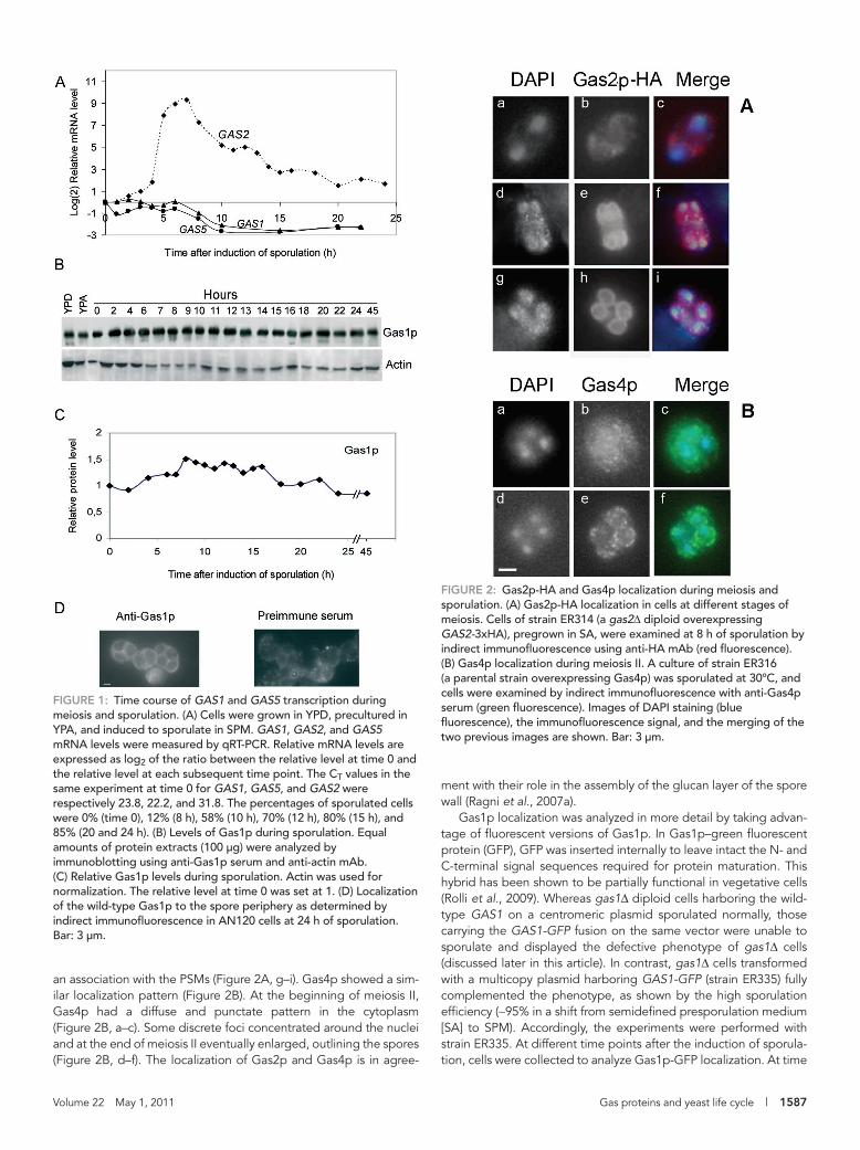
RESULTSExpression profiles of GAS1 and GAS5 during meiosis and sporulationGAS1 and GAS5 mRNA levels were monitored in the strain AN120, derived from the sporulation-proficient strain SK-1 (Kane and Roth, 1974). Total RNA was extracted at different time intervals after the induction of sporulation and used for a quantitative reverse tran-scriptase PCR (qRT-PCR) analysis. Actin mRNA was chosen as a ref-erence transcript because the expression of ACT1 does not fluctu-ate significantly in sporulating cells (Primig et al., 2000). As illustrated in Figure 1A, the GAS1 and GAS5 transcripts showed very similar profiles. They were detectable at time 0 and remained roughly con-stant until 6 h after induction of sporulation, when they started to decrease, and at 10 h the levels were reduced by more than four-fold. These results are in accordance with previous Northern hybrid-ization and transcriptome analyses (Popolo et al., 1993a; Chu et al., 1998; Primig et al., 2000).
As an internal control, we compared the expression profiles of GAS1 and GAS5 with that of GAS2 as a representative member of the GAS2-GAS4 gene pair (Figure 1A). Consistent with a previous analysis, we detected a peak of expression at 7 h (Ragni et al., 2007a). In conclusion, upon the induction of sporulation, either the expression of GAS1 and GAS5 is switched off and/or GAS1-GAS5 mRNAs are degraded.
Gas1 protein is stable during meiosis and sporulationTotal protein extracts were prepared at different time intervals after induction of sporulation and analyzed by immunoblot (Figure 1B). Gas1p was detected as a 130-kDa polypeptide. As shown in Figure 1B, the relative Gas1p level was constant throughout the sporulation process except for a slight increase in the first 10 h, which was probably due to completion of maturation of the precur-sors. The level of Gas1p remained constant during sporulation also in the W303 genetic background (Supplemental Figure S1). Thus the Gas1p present at time 0 appears to persist after the shift to the sporulation medium (SPM). Surprisingly, Gas1p was still detected at 45 h after sporulation induction, indicating that it is a highly stable protein (Figure 1, B and C). The persistence of Gas1p prompted us to examine its localization. As shown in Figure 1D, Gas1p was local-ized to the spore periphery in mature asci. In conclusion, although GAS1 transcription is repressed, the product persists over time and its localization changes.
Localization of Gas proteins during meiosis and spore formationTo study the localization of other Gas proteins during meiosis and sporulation, Gas2p was tagged with three hemagglutinin (HA) epitopes, whereas Gas4p was recognized by an anti-Gas4p serum. Sporulating cells were collected during sporulation and processed for indirect immunofluorescence. Gas2 and Gas4 proteins could be visualized only when overexpressed. Previous studies showed that their overexpression had no effect on sporulation (Ragni et al., 2007a). At time 0, no fluorescence was detected (data not shown), in agreement with the absence of expression of GAS2 and GAS4 during vegetative growth (Ragni et al., 2007a). During meiosis I, Gas2p-HA exhibited a diffuse fluorescence in the cytoplasm that was more intense around the two nuclei (Figure 2A, a–c), in agree-ment with the localization to the endoplasmic reticulum (ER) and PSM vesicle precursors (Neiman, 2005). During meiosis II, Gas2p-HA localized to elongated ellipsoidal structures corresponding to the newly assembled PSMs (Figure 2A, d–f). At the completion of meiosis II, Gas2p-HA expanded into ringlike structures, indicating
conditions and reenter the mitotic cycle or mate again and restore the diploid state.
Over the years, the process of meiosis and sporulation has been studied in depth (Neiman, 2005). More recently a molecular charac-terization of the spore wall assembly pathway has been undertaken (Coluccio et al., 2004). The spore wall has a four-layer organization comprising from inside to outside 1) mannoproteins, 2) β(1,3)-glucan, 3) chitosan, and 4) dityrosine. The outer spore wall layers confer the spore much of its resistance to environmental stress. The deposition of spore wall components begins at the end of meiosis II when the prospore membrane (PSM) closure occurs. The PSM is a double membrane sac that originates from an initial priming struc-ture, the meiotic outer plaque of the spindle pole body, to which the vesicles are redirected from the Golgi apparatus through a develop-mentally modified branch of the constitutive secretory pathway (Neiman, 1998; Morishita et al., 2007). The PSM extends progres-sively until it engulfs a haploid nucleus. The leading edges of the PSM fuse to form a double membrane compartment inside whose lumen spore wall material is deposited in a tightly regulated man-ner. At the end of the process, the outer membrane is dissolved. Spore germination is another important morphogenetic process. Under favorable conditions, nondividing haploid spores grow and reenter the mitotic cycle. At the germinating pole, a new wall with the typical three-layer organization of the vegetative cell wall—from inside to outside, chitin, glucan, and mannoprotein—is synthesized (Neiman, 2005).
Gas proteins belong to family 72 of glucanases/transglycosi-dases (Cantarel et al., 2009). Their β(1,3)-glucanase/transglycosi-dase activity is crucial for the incorporation and remodeling of β(1,3)-glucan into the cell wall and for creating attachment sites for the anchoring of mannoproteins and chitin (Mouyna et al., 2000; Ragni et al., 2007b; Rolli et al., 2009). In the S. cerevisiae genome, the GAS family is constituted by five paralogous genes: 1) GAS1 and GAS5, which are expressed during vegetative growth; 2) GAS2 and GAS4, which are repressed in vegetative growth and specifically induced during sporulation; and 3) GAS3, a weakly ex-pressed gene encoding an inactive member of the family (Rolli et al., 2010). The Gas2 and Gas4 proteins are essential for proper spore assembly and viability (Ragni et al., 2007a). Gas1p plays a major role in cell wall construction during vegetative growth, whereas Gas5p presumably plays an ancillary function. GAS1 mRNA is also four times more abundant than GAS5 mRNA (Rolli et al., 2010). Overexpression of GAS genes has no affect on the phenotype of vegetative or sporulating yeast cells. GAS products are glycosylphosphatidylinositol (GPI)–anchored proteins, and while Gas2p and Gas4p are poorly mannosylated, Gas1, Gas3, and Gas5 are highly mannosylated proteins (Ragni et al., 2007a, 2007b; Rolli et al., 2010). Gas1p is predominantly anchored to the plasma membrane (PM) through GPI, but a fraction is also cova-lently linked to the cell wall (Yin et al., 2005). Accordingly, Gas1p localizes to microdomains of the PM, but it is also cross-linked to the chitin ring and bud scars, where it remains for many genera-tions (Rolli et al., 2009). Moreover, at cytokinesis Gas1p localizes to the primary septum. At the bud neck and septum region, Gas1p is involved in the maintenance of the neck size and in cell separa-tion (Rolli et al., 2009).
Here we investigated the dynamic changes occurring in the lo-calization of Gas1p during the yeast life cycle with a view to analyze the functional interplay among Gas paralogous proteins. Our results provide novel information about the dynamics of Gas proteins and the roles of glucan-remodeling enzymes during spore morphogen-esis and germination.
Volume 22 May 1, 2011 Gas proteins and yeast life cycle | 1587
ment with their role in the assembly of the glucan layer of the spore wall (Ragni et al., 2007a).
Gas1p localization was analyzed in more detail by taking advan-tage of fluorescent versions of Gas1p. In Gas1p–green fluorescent protein (GFP), GFP was inserted internally to leave intact the N- and C-terminal signal sequences required for protein maturation. This hybrid has been shown to be partially functional in vegetative cells (Rolli et al., 2009). Whereas gas1Δ diploid cells harboring the wild-type GAS1 on a centromeric plasmid sporulated normally, those carrying the GAS1-GFP fusion on the same vector were unable to sporulate and displayed the defective phenotype of gas1Δ cells (discussed later in this article). In contrast, gas1Δ cells transformed with a multicopy plasmid harboring GAS1-GFP (strain ER335) fully complemented the phenotype, as shown by the high sporulation efficiency (∼95% in a shift from semidefined presporulation medium [SA] to SPM). Accordingly, the experiments were performed with strain ER335. At different time points after the induction of sporula-tion, cells were collected to analyze Gas1p-GFP localization. At time
an association with the PSMs (Figure 2A, g–i). Gas4p showed a sim-ilar localization pattern (Figure 2B). At the beginning of meiosis II, Gas4p had a diffuse and punctate pattern in the cytoplasm (Figure 2B, a–c). Some discrete foci concentrated around the nuclei and at the end of meiosis II eventually enlarged, outlining the spores (Figure 2B, d–f). The localization of Gas2p and Gas4p is in agree-
FiGuRE 1: Time course of GAS1 and GAS5 transcription during meiosis and sporulation. (A) Cells were grown in YPD, precultured in YPA, and induced to sporulate in SPM. GAS1, GAS2, and GAS5 mRNA levels were measured by qRT-PCR. Relative mRNA levels are expressed as log2 of the ratio between the relative level at time 0 and the relative level at each subsequent time point. The CT values in the same experiment at time 0 for GAS1, GAS5, and GAS2 were respectively 23.8, 22.2, and 31.8. The percentages of sporulated cells were 0% (time 0), 12% (8 h), 58% (10 h), 70% (12 h), 80% (15 h), and 85% (20 and 24 h). (B) Levels of Gas1p during sporulation. Equal amounts of protein extracts (100 μg) were analyzed by immunoblotting using anti-Gas1p serum and anti-actin mAb. (C) Relative Gas1p levels during sporulation. Actin was used for normalization. The relative level at time 0 was set at 1. (D) Localization of the wild-type Gas1p to the spore periphery as determined by indirect immunofluorescence in AN120 cells at 24 h of sporulation. Bar: 3 μm.
FiGuRE 2: Gas2p-HA and Gas4p localization during meiosis and sporulation. (A) Gas2p-HA localization in cells at different stages of meiosis. Cells of strain ER314 (a gas2Δ diploid overexpressing GAS2-3xHA), pregrown in SA, were examined at 8 h of sporulation by indirect immunofluorescence using anti-HA mAb (red fluorescence). (B) Gas4p localization during meiosis II. A culture of strain ER316 (a parental strain overexpressing Gas4p) was sporulated at 30ºC, and cells were examined by indirect immunofluorescence with anti-Gas4p serum (green fluorescence). Images of DAPI staining (blue fluorescence), the immunofluorescence signal, and the merging of the two previous images are shown. Bar: 3 μm.

1588 | E. Rolli et al. Molecular Biology of the Cell
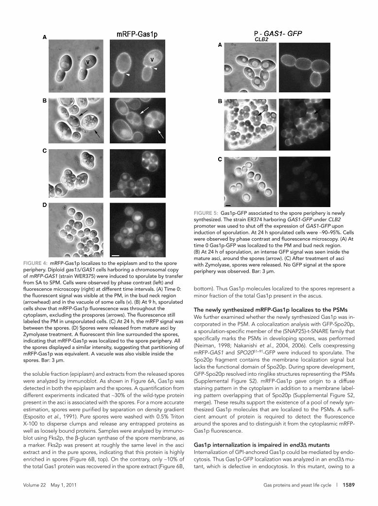
cence at the PM steadily started to decrease, and the protein was observed inside the cell (Figure 3A, cell 3). At later stages, a diffuse staining pattern was observed except for four round, black areas, corresponding to the developing spores, indicating that the fluo-rescence was excluded from these structures (Figure 3A, cell 4). In immature asci, only weak or no labeling was observed at the level of the PM of the mother cell and Gas1p-GFP was found between as well as around the spores (Figure 3A, cell 5). At 24 h, mature asci showed a bright Gas1p-GFP fluorescence in the epiplasm, which was compressed after the collapse of the wall on the spores (Figure 3A, cell 6). To determine whether the fluorescence was associated with the spores, asci were mildly treated with Zymolyase. As shown in Figure 3B, Gas1p-GFP decorated the spore periphery, indicating that the fluorescent labeling was associated with the spores. How-ever, the fluorescence was less intense than in the intact ascus, sug-gesting that a fraction of Gas1p-GFP was lost during ascus wall di-gestion. Consistent with the removal of Gas1p-GFP from the mother PM during sporulation, the ascus ghosts were negative except for the presence of very bright rings (Figure 3C, right). These structures contained Gas1p-GFP cross-linked to the bud scars produced dur-ing vegetative growth (Rolli et al., 2009). Thus during sporulation the GPI-anchored Gas1p is completely removed from the PM of the mother cell and internalized, whereas the Gas1p cross-linked to the chitin of the bud scars remains static.
To exclude any artificial effect due to overexpression or type of fusion, we used a hybrid of Gas1p with the monomeric red fluores-cent protein (mRFP-Gas1p). In this chimera, mRFP was inserted im-mediately after the signal peptide and the hybrid protein was fully functional (Rolli et al., 2009). One copy of the fusion gene was inte-grated at the LEU2 locus in the W303 genetic background (strain WER375). During vegetative growth, mRFP-Gas1p showed the typi-cal localization sites of Gas1p at the PM, bud neck (Figure 4A), and scars (not shown in the figure). During sporulation, mRFP-Gas1p was found inside the sporulating cells and around the prospores in addi-tion to the PM staining that was detected in cells with not yet visible spores (Figure 4B). In mature asci, an intense staining was observed in the epiplasm (Figure 4C). The protein uniformly labeled the pe-riphery of the released spores (Figure 4D). Thus both the relocaliza-tion of Gas1p from the PM to the epiplasm and the association to the spore periphery are independent of the fusion protein used and the genetic background. In addition, because the mRFP-GAS1 fu-sion was carried by only one parental strain, from which the diploid was generated and an equivalent partitioning of the hybrid protein to the spores was observed (fluorescent spores > 99%), we can con-clude that transport of either the preexisting or the new synthesized protein could occur.
To answer the question of the origin of the spore-associated Gas1p, the GAS1 promoter of the GAS1-GFP construct was re-placed by the CLB2 promoter, which is repressed upon induction of sporulation. In this strain, the fate of the vegetative protein present at the PM can be analyzed without the interference of the protein that can be synthesized in the first hours of sporulation. In vegeta-tive cells Gas1p-GFP was detected at the typical sites (Figure 5A). At 24 h of sporulation, a bright fluorescence was present around and between the spores (Figure 5B). However, no labeling was observed at the periphery of the released spores (Figure 5C). Taken together, these data support the idea that Gas1p localization at the spore depends on newly made protein, whereas the protein present at the PM during vegetative growth is transported to the cytoplasm but excluded from the developing spores.
To confirm these results, we also examined the level of untagged Gas1p present in the epiplasm and spore extracts. Mature asci from a wild-type diploid strain (AN120) were treated with Zymolyase, and
0, Gas1p-GFP localized to the PM (Figure 3A, cell 1) and still labeled the PM at meiosis II (cell 2). At the completion of meiosis II, a re-markable change in Gas1p-GFP localization occurred: The fluores-
FiGuRE 3: Gas1p-GFP localization changes during sporulation. A gas1Δ/gas1Δ mutant expressing Gas1p-GFP (strain ER337) was shifted from SA to SPM and allowed to sporulate. (A) At different time points, sporulating cells were stained with DAPI and observed under a fluorescence microscope to detect both the DAPI and Gas1p-GFP fluorescence. Bar: 3 μm. (B) Spores released from the asci by treatment with Zymolyase at 24 h after the induction of sporulation. Bar: 3 μm. (C) An ascus ghost left after Zymolyase treatment of asci. No fluorescence is detectable except for the two bright rings corresponding to the Gas1p cross-linked to the bud scars. Bar: 5 μm.
Volume 22 May 1, 2011 Gas proteins and yeast life cycle | 1589
bottom). Thus Gas1p molecules localized to the spores represent a minor fraction of the total Gas1p present in the ascus.
The newly synthesized mRFP-Gas1p localizes to the PSMsWe further examined whether the newly synthesized Gas1p was in-corporated in the PSM. A colocalization analysis with GFP-Spo20p, a sporulation-specific member of the (SNAP25)-t-SNARE family that specifically marks the PSMs in developing spores, was performed (Neiman, 1998; Nakanishi et al., 2004, 2006). Cells coexpressing mRFP-GAS1 and SPO2051–91-GFP were induced to sporulate. The Spo20p fragment contains the membrane localization signal but lacks the functional domain of Spo20p. During spore development, GFP-Spo20p resolved into ringlike structures representing the PSMs (Supplemental Figure S2). mRFP-Gas1p gave origin to a diffuse staining pattern in the cytoplasm in addition to a membrane label-ing pattern overlapping that of Spo20p (Supplemental Figure S2, merge). These results support the existence of a pool of newly syn-thesized Gas1p molecules that are localized to the PSMs. A suffi-cient amount of protein is required to detect the fluorescence around the spores and to distinguish it from the cytoplasmic mRFP-Gas1p fluorescence.
Gas1p internalization is impaired in end3Δ mutantsInternalization of GPI-anchored Gas1p could be mediated by endo-cytosis. Thus Gas1p-GFP localization was analyzed in an end3Δ mu-tant, which is defective in endocytosis. In this mutant, owing to a
the soluble fraction (epiplasm) and extracts from the released spores were analyzed by immunoblot. As shown in Figure 6A, Gas1p was detected in both the epiplasm and the spores. A quantification from different experiments indicated that ∼30% of the wild-type protein present in the asci is associated with the spores. For a more accurate estimation, spores were purified by separation on density gradient (Esposito et al., 1991). Pure spores were washed with 0.5% Triton X-100 to disperse clumps and release any entrapped proteins as well as loosely bound proteins. Samples were analyzed by immuno-blot using Fks2p, the β-glucan synthase of the spore membrane, as a marker. Fks2p was present at roughly the same level in the asci extract and in the pure spores, indicating that this protein is highly enriched in spores (Figure 6B, top). On the contrary, only ∼10% of the total Gas1 protein was recovered in the spore extract (Figure 6B,
FiGuRE 4: mRFP-Gas1p localizes to the epiplasm and to the spore periphery. Diploid gas1Δ/GAS1 cells harboring a chromosomal copy of mRFP-GAS1 (strain WER375) were induced to sporulate by transfer from SA to SPM. Cells were observed by phase contrast (left) and fluorescence microscopy (right) at different time intervals. (A) Time 0: the fluorescent signal was visible at the PM, in the bud neck region (arrowhead) and in the vacuole of some cells (v). (B) At 9 h, sporulated cells show that mRFP-Gas1p fluorescence was throughout the cytoplasm, excluding the prospores (arrows). The fluorescence still labeled the PM in unsporulated cells. (C) At 24 h, the mRFP signal was between the spores. (D) Spores were released from mature asci by Zymolyase treatment. A fluorescent thin line surrounded the spores, indicating that mRFP-Gas1p was localized to the spore periphery. All the spores displayed a similar intensity, suggesting that partitioning of mRFP-Gas1p was equivalent. A vacuole was also visible inside the spores. Bar: 3 μm.
FiGuRE 5: Gas1p-GFP associated to the spore periphery is newly synthesized. The strain ER374 harboring GAS1-GFP under CLB2 promoter was used to shut off the expression of GAS1-GFP upon induction of sporulation. At 24 h sporulated cells were ∼90–95%. Cells were observed by phase contrast and fluorescence microscopy. (A) At time 0 Gas1p-GFP was localized to the PM and bud neck region. (B) At 24 h of sporulation, an intense GFP signal was seen inside the mature asci, around the spores (arrow). (C) After treatment of asci with Zymolyase, spores were released. No GFP signal at the spore periphery was observed. Bar: 3 μm.

1590 | E. Rolli et al. Molecular Biology of the Cell
calization during sporulation in an sps1Δ null diploid mutant. Gas1p-GFP fluorescence was detected in the PM in most cells that had completed meiosis II, and it was also slightly diffused throughout the cytoplasm, although it was never observed at the PSMs or in spore-like compartments (Figure 7, g–g′′). Thus the sps1Δ mutant appears to be defective in triggering the internalization step of PM-associated Gas1p-GFP.
Functional characterization of GAS1 during sporulationThe presence of a fraction of Gas1p in the spore suggests a po-tential involvement of the protein in spore wall assembly or in subsequent germination. We first examined the effect of GAS1 inac-tivation on sporulation. The phenotypic analysis of gas1Δ/gas1Δ cells during sporulation was complicated by the fact that the mutant was unable to grow in presporulation media containing acetate as a carbon source. Cells appeared swollen and underwent lysis. A spot assay on solid medium indicated that the viability of gas1Δ/gas1Δ cells was very low on acetate, glycerol, and ethanol with respect to
failure in the localization of the glucan and chitin synthase to the PSMs the deposition of glucan and chitosan layers does not occur (Morishita and Engebrecht, 2005). At 30°C, the sporulation effi-ciency of the end3Δ mutant was only ∼45% (n = 200), with an enrich-ment of asci with one, two, or three spores, in agreement with previ-ously reported data (Morishita and Engebrecht, 2005). At 24 h after the induction of sporulation, Gas1p-GFP localized to the periphery of the spores and between the spores in the wild-type strain (Figure 7, a and a′), whereas it was mislocalized in the majority of end3Δ cells (Figure 7, b–f). Gas1p-GFP was detected mainly in the cytoplasm of abnormally shaped cells and excluded from the spores (Figure 7, b and b′). In some cells, Gas1p-GFP remained at the PM of the mother cells even when refractive spores were already visible (Figure 7, c–f). In ∼35% of the sporulated cells, Gas1p-GFP showed only a faint fluorescence on the spores, indicating that some new synthesis of the spore-associated protein occurred (Figure 7, e′ and d′). In conclusion, End3p-mediated endocytosis contributes to the efficient removal of Gas1p from the PM although other pathways are likely to be involved.
Gas1p is not internalized in an sps1Δ mutantSPS1 encodes a sporulation-specific serine/threonine protein kinase with homology to members of the p21-activated kinases (PAKs) (Fri-esen et al., 1994; Park and Bi, 2007). It has previously been shown that sps1Δ mutants are not affected in the progression of meiosis or PSMs formation but form only aberrant spore-like compartments (Friesen et al., 1994; Iwamoto et al., 2005). Sps1p does not affect the developmentally regulated branch of the secretory pathway but regulates the trafficking of the enzymes involved in spore wall bio-genesis to the PSMs (Iwamoto et al., 2005). To determine whether Gas1p internalization is regulated by Sps1p, we analyzed Gas1p lo-
FiGuRE 6: Gas1p associated to the spores is a minor fraction of the total Gas1p present in the ascus. Wild-type cells (AN120) were induced to sporulate by transfer from SA to SPM. (A) At 24 h efficiency of sporulation was ∼90–95%. Cells were treated with Zymolyase 20T to release spores as in Figure 3. The immunoblot showed the presence of Gas1p in total asci (lane 1), in spores (lane 2), and in the supernatant obtained after Zymolyase digestion (lane 3). The volume of loaded Zymolyase supernatant corresponded to the loaded amount of spores and asci extract. (B) A sporulated culture (efficiency ∼77%) was collected at 24 h and processed to isolate pure spores as described in Materials and Methods. Total extracts (∼100 μg proteins) from asci (lane 1) and from Triton X-100–washed purified spores (lane 2) were analyzed by immunoblot using anti-Fks2p antibodies and anti-Gas1p serum.
FiGuRE 7: Gas1p-GFP internalization is defective in end3Δ and sps1Δ mutants. The images show Gas1p-GFP expressed in the control strain (ER337), in an end3Δ mutant (ER351), and in an sps1Δ mutant (WER352) at 24 h after induction of sporulation. WT cells show the typical fluorescence around the spores (a and a′). The end3Δ cells show partially internalized Gas1p-GFP (b and b′, d and d′, e and e′, and f and f′) or Gas1p-GFP at the PM of the mother cells (c and c′). Moreover, in some spores a weak fluorescence around the spores was detectable, indicating that newly synthesized Gas1p properly localized in some spores (arrow in b′ and d′). The sps1Δ cells displayed Gas1p-GFP at the PM and a weak cytoplasmic fluorescence. In detail, differential interference contrast image (g), DAPI fluorescence (g′), and Gas1p-GFP fluorescence (g′′). Note the granular aspect of the cytoplasm. Bar: 3 μm.
Volume 22 May 1, 2011 Gas proteins and yeast life cycle | 1591
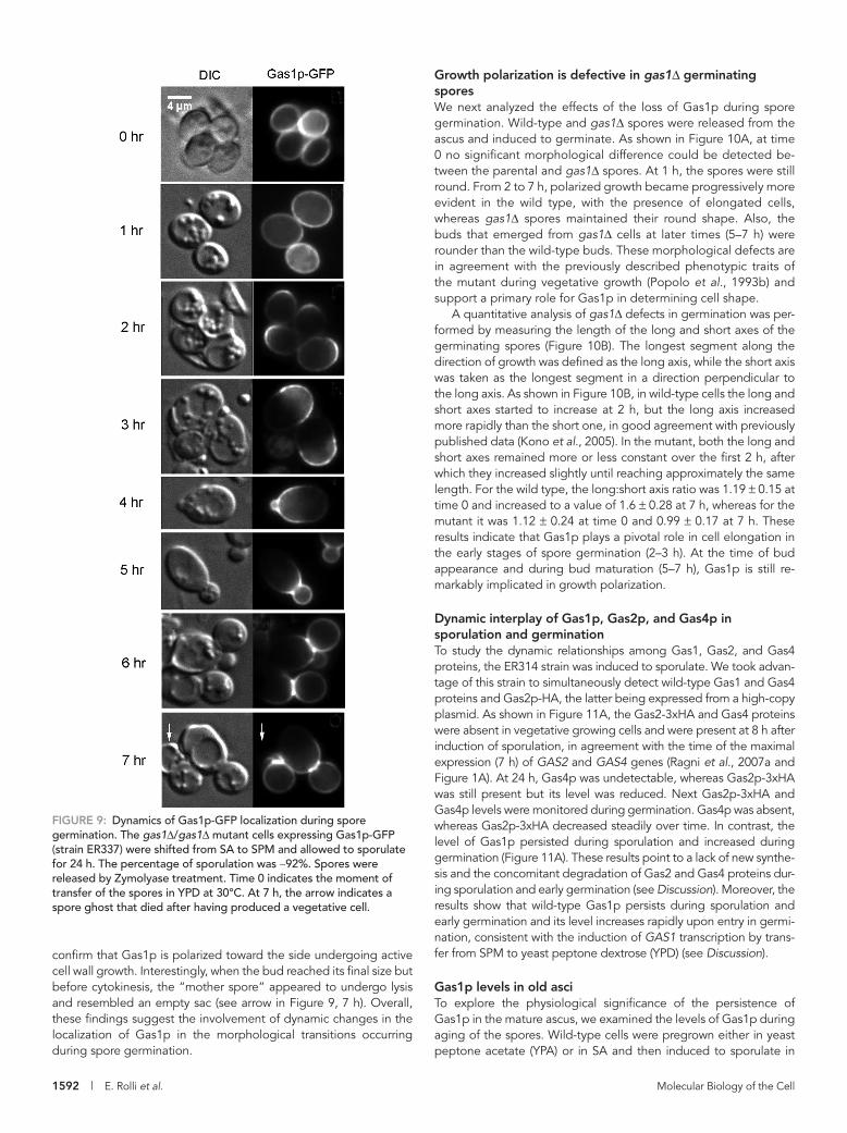
like aspect. Notably, the fluorescent protein marked the site of growth polarization. As germination proceeded, the Gas1p-GFP signal was almost undetectable in the nongrowing half, and it con-centrated in the growing half as a bright crescent (3 h). At 4 h, bud emergence was observed, and Gas1p-GFP was still highly polarized and decorated the periphery of small new buds. From this stage, it was possible to recognize the Gas1p-GFP localization pattern typical of vegetative growth (Rolli et al., 2009) (5 h). These results
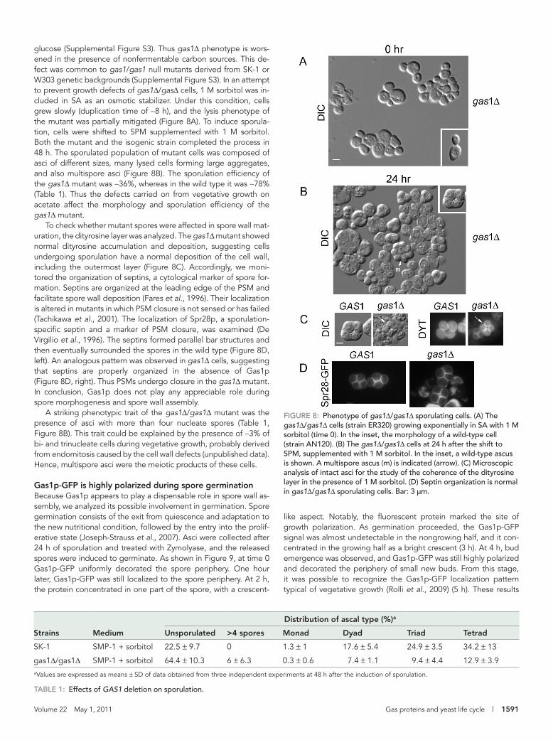
glucose (Supplemental Figure S3). Thus gas1Δ phenotype is wors-ened in the presence of nonfermentable carbon sources. This de-fect was common to gas1/gas1 null mutants derived from SK-1 or W303 genetic backgrounds (Supplemental Figure S3). In an attempt to prevent growth defects of gas1Δ/gasΔ cells, 1 M sorbitol was in-cluded in SA as an osmotic stabilizer. Under this condition, cells grew slowly (duplication time of ∼8 h), and the lysis phenotype of the mutant was partially mitigated (Figure 8A). To induce sporula-tion, cells were shifted to SPM supplemented with 1 M sorbitol. Both the mutant and the isogenic strain completed the process in 48 h. The sporulated population of mutant cells was composed of asci of different sizes, many lysed cells forming large aggregates, and also multispore asci (Figure 8B). The sporulation efficiency of the gas1Δ mutant was ∼36%, whereas in the wild type it was ∼78% (Table 1). Thus the defects carried on from vegetative growth on acetate affect the morphology and sporulation efficiency of the gas1Δ mutant.
To check whether mutant spores were affected in spore wall mat-uration, the dityrosine layer was analyzed. The gas1Δ mutant showed normal dityrosine accumulation and deposition, suggesting cells undergoing sporulation have a normal deposition of the cell wall, including the outermost layer (Figure 8C). Accordingly, we moni-tored the organization of septins, a cytological marker of spore for-mation. Septins are organized at the leading edge of the PSM and facilitate spore wall deposition (Fares et al., 1996). Their localization is altered in mutants in which PSM closure is not sensed or has failed (Tachikawa et al., 2001). The localization of Spr28p, a sporulation-specific septin and a marker of PSM closure, was examined (De Virgilio et al., 1996). The septins formed parallel bar structures and then eventually surrounded the spores in the wild type (Figure 8D, left). An analogous pattern was observed in gas1Δ cells, suggesting that septins are properly organized in the absence of Gas1p (Figure 8D, right). Thus PSMs undergo closure in the gas1Δ mutant. In conclusion, Gas1p does not play any appreciable role during spore morphogenesis and spore wall assembly.
A striking phenotypic trait of the gas1Δ/gas1Δ mutant was the presence of asci with more than four nucleate spores (Table 1, Figure 8B). This trait could be explained by the presence of ∼3% of bi- and trinucleate cells during vegetative growth, probably derived from endomitosis caused by the cell wall defects (unpublished data). Hence, multispore asci were the meiotic products of these cells.
Gas1p-GFP is highly polarized during spore germinationBecause Gas1p appears to play a dispensable role in spore wall as-sembly, we analyzed its possible involvement in germination. Spore germination consists of the exit from quiescence and adaptation to the new nutritional condition, followed by the entry into the prolif-erative state (Joseph-Strauss et al., 2007). Asci were collected after 24 h of sporulation and treated with Zymolyase, and the released spores were induced to germinate. As shown in Figure 9, at time 0 Gas1p-GFP uniformly decorated the spore periphery. One hour later, Gas1p-GFP was still localized to the spore periphery. At 2 h, the protein concentrated in one part of the spore, with a crescent-
FiGuRE 8: Phenotype of gas1Δ/gas1Δ sporulating cells. (A) The gas1Δ/gas1Δ cells (strain ER320) growing exponentially in SA with 1 M sorbitol (time 0). In the inset, the morphology of a wild-type cell (strain AN120). (B) The gas1Δ/gas1Δ cells at 24 h after the shift to SPM, supplemented with 1 M sorbitol. In the inset, a wild-type ascus is shown. A multispore ascus (m) is indicated (arrow). (C) Microscopic analysis of intact asci for the study of the coherence of the dityrosine layer in the presence of 1 M sorbitol. (D) Septin organization is normal in gas1Δ/gas1Δ sporulating cells. Bar: 3 μm.
Distribution of ascal type (%)a
Strains Medium unsporulated >4 spores Monad Dyad Triad Tetrad
SK-1 SMP-1 + sorbitol 22.5 ± 9.7 0 1.3 ± 1 17.6 ± 5.4 24.9 ± 3.5 34.2 ± 13
gas1Δ/gas1Δ SMP-1 + sorbitol 64.4 ± 10.3 6 ± 6.3 0.3 ± 0.6 7.4 ± 1.1 9.4 ± 4.4 12.9 ± 3.9aValues are expressed as means ± SD of data obtained from three independent experiments at 48 h after the induction of sporulation.
TABLE 1: Effects of GAS1 deletion on sporulation.
1592 | E. Rolli et al. Molecular Biology of the Cell
Growth polarization is defective in gas1Δ germinating sporesWe next analyzed the effects of the loss of Gas1p during spore germination. Wild-type and gas1Δ spores were released from the ascus and induced to germinate. As shown in Figure 10A, at time 0 no significant morphological difference could be detected be-tween the parental and gas1Δ spores. At 1 h, the spores were still round. From 2 to 7 h, polarized growth became progressively more evident in the wild type, with the presence of elongated cells, whereas gas1Δ spores maintained their round shape. Also, the buds that emerged from gas1Δ cells at later times (5–7 h) were rounder than the wild-type buds. These morphological defects are in agreement with the previously described phenotypic traits of the mutant during vegetative growth (Popolo et al., 1993b) and support a primary role for Gas1p in determining cell shape.
A quantitative analysis of gas1Δ defects in germination was per-formed by measuring the length of the long and short axes of the germinating spores (Figure 10B). The longest segment along the direction of growth was defined as the long axis, while the short axis was taken as the longest segment in a direction perpendicular to the long axis. As shown in Figure 10B, in wild-type cells the long and short axes started to increase at 2 h, but the long axis increased more rapidly than the short one, in good agreement with previously published data (Kono et al., 2005). In the mutant, both the long and short axes remained more or less constant over the first 2 h, after which they increased slightly until reaching approximately the same length. For the wild type, the long:short axis ratio was 1.19 ± 0.15 at time 0 and increased to a value of 1.6 ± 0.28 at 7 h, whereas for the mutant it was 1.12 ± 0.24 at time 0 and 0.99 ± 0.17 at 7 h. These results indicate that Gas1p plays a pivotal role in cell elongation in the early stages of spore germination (2–3 h). At the time of bud appearance and during bud maturation (5–7 h), Gas1p is still re-markably implicated in growth polarization.
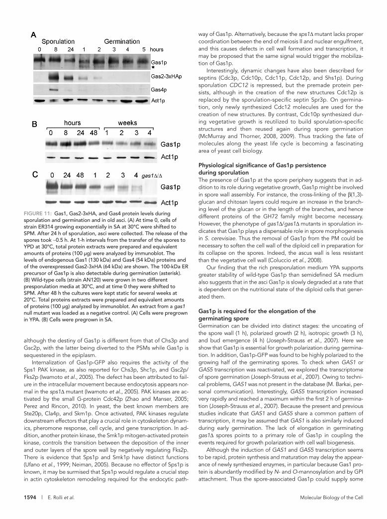
Dynamic interplay of Gas1p, Gas2p, and Gas4p in sporulation and germinationTo study the dynamic relationships among Gas1, Gas2, and Gas4 proteins, the ER314 strain was induced to sporulate. We took advan-tage of this strain to simultaneously detect wild-type Gas1 and Gas4 proteins and Gas2p-HA, the latter being expressed from a high-copy plasmid. As shown in Figure 11A, the Gas2-3xHA and Gas4 proteins were absent in vegetative growing cells and were present at 8 h after induction of sporulation, in agreement with the time of the maximal expression (7 h) of GAS2 and GAS4 genes (Ragni et al., 2007a and Figure 1A). At 24 h, Gas4p was undetectable, whereas Gas2p-3xHA was still present but its level was reduced. Next Gas2p-3xHA and Gas4p levels were monitored during germination. Gas4p was absent, whereas Gas2p-3xHA decreased steadily over time. In contrast, the level of Gas1p persisted during sporulation and increased during germination (Figure 11A). These results point to a lack of new synthe-sis and the concomitant degradation of Gas2 and Gas4 proteins dur-ing sporulation and early germination (see Discussion). Moreover, the results show that wild-type Gas1p persists during sporulation and early germination and its level increases rapidly upon entry in germi-nation, consistent with the induction of GAS1 transcription by trans-fer from SPM to yeast peptone dextrose (YPD) (see Discussion).
Gas1p levels in old asciTo explore the physiological significance of the persistence of Gas1p in the mature ascus, we examined the levels of Gas1p during aging of the spores. Wild-type cells were pregrown either in yeast peptone acetate (YPA) or in SA and then induced to sporulate in
FiGuRE 9: Dynamics of Gas1p-GFP localization during spore germination. The gas1Δ/gas1Δ mutant cells expressing Gas1p-GFP (strain ER337) were shifted from SA to SPM and allowed to sporulate for 24 h. The percentage of sporulation was ∼92%. Spores were released by Zymolyase treatment. Time 0 indicates the moment of transfer of the spores in YPD at 30°C. At 7 h, the arrow indicates a spore ghost that died after having produced a vegetative cell.
confirm that Gas1p is polarized toward the side undergoing active cell wall growth. Interestingly, when the bud reached its final size but before cytokinesis, the “mother spore” appeared to undergo lysis and resembled an empty sac (see arrow in Figure 9, 7 h). Overall, these findings suggest the involvement of dynamic changes in the localization of Gas1p in the morphological transitions occurring during spore germination.

Volume 22 May 1, 2011 Gas proteins and yeast life cycle | 1593
tion. Consistently, Gas2p and Gas4p are es-sential for spore wall formation, suggesting that Gas1p and Gas5p cannot compensate for their absence (Ragni et al., 2007a). In this work, we demonstrate that Gas1 protein persists during sporulation, but its localiza-tion changes. The fluorescent Gas1 protein marked the PM of the diploid cell but was relocalized to the epiplasm of the ascus. The PM-epiplasm transport of the protein seems a rather late event, probably beginning at the end of meiosis II and after PSM closure, as suggested by the exclusion of the fluo-rescence from the developing prospores. Additionally, in mature asci, Gas1p (∼10% of the total) was also associated to the PM of the spores. The spore-associated Gas1p is newly made. The synthesis probably occurs during early sporulation, taking into account that a steady-state level of transcript is pres-ent until 6 h of sporulation. This protein is probably processed through the secretory pathway and equally distributed to the PSMs, as suggested by the experiment per-formed with the integrated copy of mRFP-GAS1 (Figure 4).
Dynamic interplay of proteins involved in spore wall formationOverall, the formation of the spore wall re-quires a specific set of enzymes, some of which are reused from vegetative growth while others are replaced by sporulation-specific isoforms. Moreover, spatial and temporal regulation of protein trafficking is crucial for proper spore formation. Remark-ably, Fks1p, the predominant vegetative form of the β(1,3)-glucan synthase during growth on glucose, is replaced by Gsc2p/
Fks2p (Ishihara et al., 2007). GSC2/FKS2 expression is induced dur-ing sporulation (Mazur et al., 1995). Gsc2/Fks2p reaches the PM before being recycled to the PSMs (Morishita and Engebrecht, 2005). Similarly to Fks proteins, Gas2p and Gas4p replace Gas1p function in spore wall assembly and are degraded after execution of their function (Figure 11A). With regard to chitosan synthesis, CHS3, encoding the catalytic subunit of chitin synthase III and responsible for the synthesis of chitosan (Pammer et al., 1992), is not transcrip-tionally up-regulated during sporulation, but the protein is recycled (Iwamoto et al., 2005), and this is consistent with the notion of it be-ing an enzyme that is mainly regulated at the posttranslational level (Choi et al., 1994; Valdivia and Schekman, 2003). On the contrary, its activator, Chs4p, is quickly degraded upon induction of sporulation and is replaced by the sporulation-specific Shc1p, which is also di-rected to the PSMs (Sanz et al., 2002; Iwamoto et al., 2005).
Interestingly, in recent years END3-mediated endocytosis has been shown to play an important role in membrane trafficking events required for spore wall formation (Morishita and Enge-brecht, 2005). Chs3p and Gsc2p/Fks2p require End3p for their relocalization to the PSMs. In the present work, we demonstrate that in the absence of End3p, Gas1p is also inefficiently internal-ized. Thus a partially common mechanism could mediate the transport of Gas1p, Chs3p, and Gsc2p inside the sporulating cell,
SPM. At 48 h sporulated cultures were kept static at 20°C for several weeks. Figure 11B shows that the level of Gas1p from YPA cells did not change until 24 h of sporulation. A quantification of the relative levels using actin for normalization indicated that from 48 h to 4 wk Gas1p was present at a level corresponding to ∼60–70% of the pro-tein present at time 0, indicating that Gas1p slowly decays in the asci. Figure 11C shows that Gas1p from SA-grown cells had a faster kinetics of decay. At 24 h the residual fraction was ∼60% the amount of Gas1p present at time 0, and from 48 h to 4 wk the protein steadily decreased until reaching a value of ∼10% the amount present at time 0. Thus Gas1p has a high stability in asci from cells pregrown in YPA, whereas the protein appears less stable in asci derived from cells pregrown in SA. Spores from 4-wk-old asci, both from YPA or SA, were still able to germinate, indicating that they were viable (data not shown). Interestingly, actin appears to be a long-lived pro-tein because its level was unaffected in both conditions.
DISCUSSIONMultigene families offer a good model to study the interplay among paralogous proteins and their interactions for the fine-tuning of dif-ferent biological processes. One such family is the Gas family of S. cerevisiae. Gas2 and Gas4 isoforms replace the vegetative Gas1 and Gas5 proteins in glucan remodeling during spore wall forma-
FiGuRE 10: Germinating gas1Δ spores are defective in the elongation of the outgrowth portion. (A) Time course of the morphological changes of germinating wild-type and gas1Δ spores in YPD at 30°C. (B) Elongation of the spores at different time intervals during germination. Plots report the length of the long and short axes at different time intervals during germination. At least 20 spores were measured for each strain, and the data are shown as mean (μm) ± SD. , long axis; , short axis.
1594 | E. Rolli et al. Molecular Biology of the Cell
way of Gas1p. Alternatively, because the sps1Δ mutant lacks proper coordination between the end of meiosis II and nuclear engulfment, and this causes defects in cell wall formation and transcription, it may be proposed that the same signal would trigger the mobiliza-tion of Gas1p.
Interestingly, dynamic changes have also been described for septins (Cdc3p, Cdc10p, Cdc11p, Cdc12p, and Shs1p). During sporulation CDC12 is repressed, but the premade protein per-sists, although in the creation of the new structures Cdc12p is replaced by the sporulation-specific septin Spr3p. On germina-tion, only newly synthesized Cdc12 molecules are used for the creation of new structures. By contrast, Cdc10p synthesized dur-ing vegetative growth is reutilized to build sporulation-specific structures and then reused again during spore germination (McMurray and Thorner, 2008, 2009). Thus tracking the fate of molecules along the yeast life cycle is becoming a fascinating area of yeast cell biology.
Physiological significance of Gas1p persistence during sporulationThe presence of Gas1p at the spore periphery suggests that in ad-dition to its role during vegetative growth, Gas1p might be involved in spore wall assembly. For instance, the cross-linking of the β(1,3)-glucan and chitosan layers could require an increase in the branch-ing level of the glucan or in the length of the branches, and hence different proteins of the GH72 family might become necessary. However, the phenotype of gas1Δ/gas1Δ mutants in sporulation in-dicates that Gas1p plays a dispensable role in spore morphogenesis in S. cerevisiae. Thus the removal of Gas1p from the PM could be necessary to soften the cell wall of the diploid cell in preparation for its collapse on the spores. Indeed, the ascus wall is less resistant than the vegetative cell wall (Coluccio et al., 2008).
Our finding that the rich presporulation medium YPA supports greater stability of wild-type Gas1p than semidefined SA medium also suggests that in the asci Gas1p is slowly degraded at a rate that is dependent on the nutritional state of the diploid cells that gener-ated them.
Gas1p is required for the elongation of the germinating sporeGermination can be divided into distinct stages: the uncoating of the spore wall (1 h), polarized growth (2 h), isotropic growth (3 h), and bud emergence (4 h) (Joseph-Strauss et al., 2007). Here we show that Gas1p is essential for growth polarization during germina-tion. In addition, Gas1p-GFP was found to be highly polarized to the growing half of the germinating spores. To check when GAS1 or GAS5 transcription was reactivated, we explored the transcriptome of spore germination (Joseph-Strauss et al., 2007). Owing to techni-cal problems, GAS1 was not present in the database (M. Barkai, per-sonal communication). Interestingly, GAS5 transcription increased very rapidly and reached a maximum within the first 2 h of germina-tion (Joseph-Strauss et al., 2007). Because the present and previous studies indicate that GAS1 and GAS5 share a common pattern of transcription, it may be assumed that GAS1 is also similarly induced during early germination. The lack of elongation in germinating gas1Δ spores points to a primary role of Gas1p in coupling the events required for growth polarization with cell wall biogenesis.
Although the induction of GAS1 and GAS5 transcription seems to be rapid, protein synthesis and maturation may delay the appear-ance of newly synthesized enzymes, in particular because Gas1 pro-tein is abundantly modified by N- and O-mannosylation and by GPI attachment. Thus the spore-associated Gas1p could supply some
although the destiny of Gas1p is different from that of Chs3p and Gsc2p, with the latter being diverted to the PSMs while Gas1p is sequestered in the epiplasm.
Internalization of Gas1p-GFP also requires the activity of the Sps1 PAK kinase, as also reported for Chs3p, Shc1p, and Gsc2p/Fks2p (Iwamoto et al., 2005). The defect has been attributed to fail-ure in the intracellular movement because endocytosis appears nor-mal in the sps1Δ mutant (Iwamoto et al., 2005). PAK kinases are ac-tivated by the small G-protein Cdc42p (Zhao and Manser, 2005; Perez and Rincon, 2010). In yeast, the best known members are Ste20p, Cla4p, and Skm1p. Once activated, PAK kinases regulate downstream effectors that play a crucial role in cytoskeleton dynam-ics, pheromone response, cell cycle, and gene transcription. In ad-dition, another protein kinase, the Smk1p mitogen-activated protein kinase, controls the transition between the deposition of the inner and outer layers of the spore wall by negatively regulating Fks2p. There is evidence that Sps1p and Smk1p have distinct functions (Ufano et al., 1999; Neiman, 2005). Because no effector of Sps1p is known, it may be surmised that Sps1p would regulate a crucial step in actin cytoskeleton remodeling required for the endocytic path-
FiGuRE 11: Gas1, Gas2-3xHA, and Gas4 protein levels during sporulation and germination and in old asci. (A) At time 0, cells of strain ER314 growing exponentially in SA at 30°C were shifted to SPM. After 24 h of sporulation, asci were collected. The release of the spores took ∼0.5 h. At 1-h intervals from the transfer of the spores to YPD at 30°C, total protein extracts were prepared and equivalent amounts of proteins (100 μg) were analyzed by immunoblot. The levels of endogenous Gas1 (130 kDa) and Gas4 (54 kDa) proteins and of the overexpressed Gas2-3xHA (64 kDa) are shown. The 100-kDa ER precursor of Gas1p is also detectable during germination (asterisk). (B) Wild-type cells (strain AN120) were grown in two different presporulation media at 30°C, and at time 0 they were shifted to SPM. After 48 h the cultures were kept static for several weeks at 20°C. Total proteins extracts were prepared and equivalent amounts of proteins (100 μg) analyzed by immunoblot. An extract from a gas1 null mutant was loaded as a negative control. (A) Cells were pregrown in YPA. (B) Cells were pregrown in SA.
Volume 22 May 1, 2011 Gas proteins and yeast life cycle | 1595
and uracil and 100 mg/l for adenine. To induce sporulation, loga-rithmic cultures were shifted from YPD to YPA (1% yeast extract, 2% Bacto peptone, 2% acetate) or to semidefined presporulation medium (SA) (10 g potassium acetate, 6.7 g yeast nitrogen base without amino acids, 1 g yeast extract in 1 l of 0.05 M phthalate buf-fer, pH 5) at an initial optical density of 0.2 OD450. Cells were grown at 30°C, collected the following morning, and washed with sterile dH2O before being shifted to SPM (1% potassium acetate) at a cell density of ∼107 cells/ml (OD450 between 1 and 1.5) at a 1:10 ratio of culture volume to flask volume. Cultures were allowed to sporulate under vigorous shaking at 30ºC. The percentage of sporulation was
activity at the onset of spore germination before the new molecules become available.
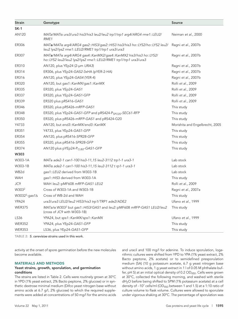
MATERIALS AND METHODSYeast strains, growth, sporulation, and germination conditionsThe strains are listed in Table 2. Cells were routinely grown at 30°C in YPD (1% yeast extract, 2% Bacto peptone, 2% glucose) or in syn-thetic dextrose minimal medium (Difco yeast nitrogen base without amino acids at 6.7 g/l, 2% glucose) to which the required supple-ments were added at concentrations of 50 mg/l for the amino acids
Strain Genotype Source
SK-1
AN120 MATa/MATα ura3/ura3 his3/his3 leu2/leu2 trp1/trp1 arg4/ARG4 rme1::LEU2/RME1
Neiman et al., 2000
ER306 MATa/MATα arg4/ARG4 gas2::HIS3/gas2::HIS3 his3/his3 ho::LYS2/ho::LYS2 leu2/leu2 lys2/lys2 rme1::LEU2/RME1 trp1/trp1 ura3/ura3
Ragni et al., 2007b
ER307 MATa/MATα arg4/ARG4 gas4::KanMX2/gas4::KanMX2 his3/his3 ho::LYS2/ho::LYS2 leu2/leu2 lys2/lys2 rme1::LEU2/RME1 trp1/trp1 ura3/ura3
Ragni et al., 2007b
ER310 AN120, plus YEp24 (2-μm URA3) Ragni et al., 2007b
ER314 ER306, plus YEp24-GAS2-3xHA (pYER-2-HA) Ragni et al., 2007b
ER316 AN120, plus YEp24-GAS4 (YER-4) Ragni et al., 2007b
ER320 AN120, but gas1::KanMX/gas1::KanMX Rolli et al., 2009
ER335 ER320, plus YEp24-GAS1 Rolli et al., 2009
ER337 ER320, plus YEp24-GAS1-GFP Rolli et al., 2009
ER339 ER320 plus pRS416-GAS1 Rolli et al., 2009
ER346 ER320, plus pRS426-mRFP-GAS1 This study
ER348 ER320, plus YEp24-GAS1-GFP and pRS424-PSPO20-SEC61-RFP This study
ER350 ER320, plus pRS426-mRFP-GAS1 and pRS424-G20 This study
Y4733 AN120, but end3::KanMX/end3::KanMX Morishita and Engelbrecht, 2005
ER351 Y4733, plus YEp24-GAS1-GFP This study
ER354 AN120, plus pRS416-SPR28-GFP This study
ER355 ER320, plus pRS416-SPR28-GFP This study
ER374 AN120 plus pYEp24-PCLB2-GAS1-GFP This study
W303
W303-1A MATa ade2-1 can1-100 his3-11,15 leu2-3112 trp1-1 ura3-1 Lab stock
W303-1B MATα ade2-1 can1-100 his3-11,15 leu2-3112 t rp1-1 ura3-1 Lab stock
WB2d gas1::LEU2 derived from W303-1B Lab stock
WAH gas1::HIS3 derived from W303-1A This study
JC9 WAH leu2::pMF608 mRFP-GAS1 LEU2 Rolli et al., 2009
W3032 Cross of W303-1A and W303-1B Ragni et al., 2007a
W30322-gas1Δ Cross of WB-2d and WAH This study
YPA24 ura3/ura3 LEU2/leu2 HIS3/his3 trp1/TRP1 ade2/ADE2 Ufano et al., 1999
WER375 MATa/α W3032 but gas1::HIS3/GAS1 and leu2::pMF608 mRFP-GAS1 LEU2/leu2 (cross of JC9 with W303-1B)
This study
LS36 YPA24, but sps1::KanMX/sps1::KanMX Ufano et al., 1999
WER352 YPA24, plus YEp24-GAS1-GFP This study
WER353 LS36, plus YEp24-GAS1-GFP This study
TABLE 2: S. cerevisiae strains used in this work.
1596 | E. Rolli et al. Molecular Biology of the Cell
moter was swapped with the PCLB2 by cloning the 1-kb DNA frag-ment of the −1000/−1 CLB2 region upstream from the GAS1 cod-ing sequence. The pRS416-PCLB2-GAS1-GFP was obtained. The DNA fragment was generated by PCR using the SK-1 genomic DNA as template and the primers CLB2Prom-SmaI (GCTACTCCCGG-GACCCTGTTCTTGACCGTC, SmaI restriction site underlined, CLB2 sequence −1000/−983 in italics) and CLB2Prom-BstAPI (AAAAAGCAGCAGCGGTTGCTAACTTTGAAAGGGATTTAAA-CAACATCTATAAGATCAATGAAGA, BstAPI restriction site under-lined, CLB2 sequence −18/−1 in italics, GAS1 coding sequence +1/+46 in bold). Finally, the SmaI-BamHI fragment of pRS416-PCLB2-GAS1-GFP containing PCLB2-GAS1-GFP was transferred into a simi-larly cut YEp24 to create YEp24-PCLB2-GAS1-GFP.
MicroscopyCells were observed by phase-contrast microscopy, and sporulation was scored by counting at least 200 cells after a mild sonication. For Gas1p-GFP visualization, 1 ml sporulating culture was centrifuged at 8000 rpm for 2 min at 4°C, washed twice with cold PBS, and in-cubated for 15 min on ice. If required, 8.3 μg/ml 4,6-diamidine-phenylindole (DAPI) was added to the cells. Samples were then in-cubated for 15 min at room temperature in the dark before microscopy observation. Cells were observed as wet mounts using an Eclipse 90i (Nikon, Tokyo, Japan) or a DMRXA (Leica, Wetzlar, Germany) microscope equipped with epifluorescence, Nomarski optics, and a Hamamatsu ORCA-ER camera (Nuhsbaum, McHenry, IL). The setup, including the microscope and camera, was controlled by MetaMorph software (Molecular Devices, Sunnyvale, CA). Alter-natively, cells were examined with an Olympus BX60 microscope (Olympus Optical, Tokyo, Japan) connected to a DC290 Kodak digital camera. The images were analyzed using ImageJ-BMF soft-ware (McMaster Biophotonics Facility, Hamilton, ON, Canada) and Adobe Photoshop (San Jose, CA). The observation of the natural fluorescence of dityrosine was performed as described previously (Briza et al., 1986).
For indirect immunofluorescence, sporulating cells (20 OD450) were fixed in 3.7% formaldehyde–0.1 M KPO4 buffer for 30 min. Af-ter a 3-min centrifugation at 1500 rpm, the cells were resuspended in the same volume of fixing solution (0.1 M KPO4, 3.7% formalde-hyde) for 2–4 h. Fixed cells were washed and resuspended in SHA buffer (1 M sorbitol, 0.1 M HEPES-KOH, pH 7.5, and 5 mM NAN3) at a concentration of 108 cells/ml. A small aliquot of fixed cells (150 μl) was centrifuged and resuspended in SHA supplemented
∼75–85% when cells were pregrown in YPA and ∼95% when pre-grown in SA. Spore purification was performed as previously described (Esposito et al., 1991). To induce germination, a 10-ml aliquot of a sporulated culture was harvested, washed once with phosphate-buffered saline (PBS; pH 7.2), and then resuspended in PBS containing 20 μg/ml Zymolyase 20T (ICN Biomedicals, Irvine, CA). Cells were incubated for 15 min at 37ºC to release the spores. Then the samples were washed twice with PBS, resuspended in YPD at a final concentration of 1–2 × 10⁷ cells/ml, and allowed to germi-nate at 30ºC. For the release of gas1Δ spores, ascus digestion was performed in PBS containing 4 μg/ml Zymolyase 20T. To obtain old asci, cultures at 48 h of sporulation were kept static at 20°C for sev-eral weeks. To check spore germination, spores were resuspended in YPD at 30°C and increase in OD450 was monitored.
Quantification of mRNA using real-time qRT-PCRTotal RNA extraction, cDNA preparation, and real-time PCR were performed as previously described (Ragni et al., 2007a). The primer pairs for GAS1 and GAS5 were GAS1 forward 5′-AGGTAGTGTT-GATTTGGGTTCC and gas1 reverse 5′-AGAAGACCCCGAAGCGTTA; GAS5 forward 5′-TCCTCGACTTCAAAGGCATC and GAS5 reverse 5′-ATGCGGCAGATTTTAGCAAG. In the case of ACT1 and GAS2, the primers have been described previously (Ragni et al., 2007a). Each cDNA was assayed in at least duplicate PCRs for two indepen-dent experiments. Basic analysis was performed using SDS 1.9.1 software (Applied Biosystems, Carlsbad, CA). For further elabora-tion of the data, the Livak method (Livak and Schmittgen, 2001) was used. Briefly, from each duplicate reaction, a ΔCT was calculated by subtracting the average cycle threshold (CT) value of ACT1 from the average CT value of the gene of interest for the same time. Then the difference between the ΔCT at any time and the ΔCT at time 0 was calculated (ΔΔCT). The plotted values are 2-Δ Δ CT.
Plasmid constructionThe plasmids are listed in Table 3. pRS426-mRFP-GAS1 was con-structed by cloning the SacI-BamHI fragment from pMF608, kindly provided by Y. Jigami (National Institute of Advanced Industrial Sci-ence and Technology, Tsukuba, Japan) and described previously (Rolli et al., 2009), into similarly cut pRS426. To place the GAS1-GFP fusion under the control of the CLB2 promoter (PCLB2), the BamHI/SmaI fragment of YEp24-GAS1-GFP containing the GAS1-GFP cassette (including GAS1 promoter and terminator) was cloned in pRS416, generating pRS416-GAS1-GFP. Then the GAS1 pro-
Name Markers Cloned gene Source
pRS416-GAS1 CEN URA3 GAS1 Rolli et al., 2009
pRS416-GAS1-GFP CEN URA3 GAS1-GFP Rolli et al., 2009
YEp24-GAS1 URA3, 2 μ GAS1 Rolli et al., 2009
YEp24-GAS1-GFP URA3, 2 μ GAS1-GFP Rolli et al., 2009
YEp24-PCLB2-GAS1-GFP URA3, 2 μ PCLB2-GAS1-GFP This study
pRS426-mRFP-GAS1 URA3, 2 μ mRFP-GAS1 This study
pRS424-G20 TRP1, 2 μ PTEF2-GFP-SPO2051–91 Nakanishi et al., 2004
pRS426-SPO20 pr-SEC61-mRFP URA3, 2 μ PSPO20-SEC61-mRFP Nakanishi et al., 2007
pSB19 URA3, CEN SPR28-GFP Virgilio et al., 1996
pYER-2-HA URA3, 2 μ GAS2-3xHA Ragni et al., 2007b
pYER-4 URA3, 2 μ GAS4 Ragni et al., 2007b
TABLE 3: Yeast plasmids used in this study.
Volume 22 May 1, 2011 Gas proteins and yeast life cycle | 1597
with 25 μg/ml Zymolyase 20T (ICN Biomedicals) and 0.2% β-mercaptoethanol. After a 30-min incubation at 37ºC, removal of the ascus sac was checked before proceeding. Spheroplasts were then permeabilized by incubating in SHA and 0.1% Triton X-100 for 5 min. After attaching to glass slides, cells were plunged into −20°C methanol for 6 min, followed by −20°C acetone for 30 s. After a blocking step in PBS + block (1% milk, 0.5% bovine serum albumin [BSA] in PBS) for 10 min, slides were incubated in primary antibody diluted in PBS + block for 2 h. Primary antibodies were anti-Gas1p rabbit serum (diluted 1:500), anti-HA monoclonal antibody (mAb) (diluted 1:1000; Covance, Berkeley, CA), and anti-Gas4p serum. Rabbit immunoglobulin (Ig) G was purified from anti-Gas4p serum using a protein A microspin column and diluted 1:50. Slides were washed 12 times with PBS + 0.5 mg/ml BSA and then incubated in Alexa Fluor 594 goat anti–mouse IgG (1:1000 dilution) or Alexa Fluor 488 goat anti–rabbit IgG (1:1500 dilution) for 1 h. After 12 washes with PBS, or with PBS–0.5% BSA–0.1% Triton X-100 for Gas4p, a mounting media (Gel Mount; Biomeda Corporation, Foster City, CA) was added to the slides, which were kept at 4ºC for 1 h and observed under an epifluorescence microscope.
Extract preparation, electrophoresis, and immunoblottingSporulating cells (2 × 10⁸) were collected by filtration, washed, and resuspended in ice-cold dH2O. After a 2-min centrifugation at 4°C, the pellet was rapidly frozen and stored at −20°C. Extract prepara-tion, determination of protein concentrations, and immunoblotting were performed as described previously (Gatti et al., 1994; Ragni et al., 2007a). Anti-Gas1p serum, anti-actin, or anti-HA mAbs and an anti-Gas4p serum, diluted 1:1000, were used (Rolli et al., 2009). Peroxidase-conjugated, affinity-purified F(ab′)2 fragment donkey anti–rabbit or anti–mouse IgG were used (1:10,000 dilution; Jackson Laboratory, Bar Harbor, ME). Bound antibodies were developed us-ing enhanced chemiluminescence Western blotting detection re-agents (Amersham Pharmacia Biotech, Piscataway, NJ). Densitom-etry measurements were performed using the Scion Image program (Scion, Frederick, MD).
ACKNOWLEDGMENTSThe authors thank Roberto Cavatorta for the preparation of the figures and Nic Skinner for English revision of the manuscript, Aaron Neiman and JoAnne Engebrecht for plasmids and strains, the Unidad de Genómica–Universidad Complutense de Madrid/Parque Científico de Madrid for help with the qRT-PCR, and Con-cetta Compagno for helpful discussions. This work was partially supported by P.U.R grants 2009 and 2010 from Università degli Studi di Milano to L.P., EU RTN project N. 512481 “CanTrain” to L.P. and by grants from the Comisión Interministerial de Ciencia y Tecnología (Spain): BFU2004-0078 to C.R.V.-A. and BIO2007-67821 and BIO2010-22146 to J.A. M.M.-R. held a fellowship from the Ministerio de Educación y Ciencia (Spain), and E.R. was a re-cipient of a type A contract from Università degli Studi di Milano.
REFERENCESBriza P, Winkler G, Kalchhauser H, Breitenbach M (1986). Dityrosine is a
prominent component of the yeast ascospore wall. A proof of its struc-ture. J Biol Chem 261, 4288–4294.
Cantarel BL, Coutinho PM, Rancurel C, Bernard T, Lombard V, Henrissat B (2009). The Carbohydrate-Active EnZymes database (CAZy): an expert resource for glycogenomics. Nucleic Acids Res 37, D233–D238.
Choi WJ, Santos B, Duran A, Cabib E (1994). Are yeast chitin synthases regulated at the transcriptional or the posttranslational level? Mol Cell Biol 14, 7685–7694.
Chu S, DeRisi J, Eisen M, Mulholland J, Botstein D, Brown PO, Herskowitz I (1998). The transcriptional program of sporulation in budding yeast. Science 282, 699–705.
Coluccio A, Bogengruber E, Conrad MN, Dresser ME, Briza P, Neiman AM (2004). Morphogenetic pathway of spore wall assembly in Saccharomy-ces cerevisiae. Eukaryot Cell 3, 1464–1475.
Coluccio AE, Rodriguez RK, Kernan MJ, Neiman AM (2008). The yeast spore wall enables spores to survive passage through the digestive tract of Drosophila. PLoS One 3, e2873.
De Virgilio C, DeMarini DJ, Pringle JR (1996). SPR28, a sixth member of the septin gene family in Saccharomyces cerevisiae that is expressed specifi-cally in sporulating cells. Microbiology 142, 2897–2905.
Esposito RE, Dresser M, Breitenbach M (1991). Identifying sporulation genes, visualizing synaptonemal complexes, and large-scale spore and spore wall purification. Methods Enzymol 194, 110–131.
Fares H, Goetsch L, Pringle JR (1996). Identification of a developmentally regulated septin and involvement of the septins in spore formation in Saccharomyces cerevisiae. J Cell Biol 132, 399–411.
Friesen H, Lunz R, Doyle S, Segall J (1994). Mutation of the SPS1-encoded protein kinase of Saccharomyces cerevisiae leads to defects in transcrip-tion and morphology during spore formation. Genes Dev 8, 2162–2175.
Gatti E, Popolo L, Vai M, Rota N, Alberghina L (1994). O-linked oligosac-charides in yeast glycosyl phosphatidylinositol-anchored protein gp115 are clustered in a serine-rich region not essential for its function. J Biol Chem 269, 19695–19700.
Ishihara S, Hirata A, Nogami S, Beauvais A, Latge JP, Ohya Y (2007). Ho-mologous subunits of 1,3-beta-glucan synthase are important for spore wall assembly in Saccharomyces cerevisiae. Eukaryot Cell 6, 143–156.
Iwamoto MA, Fairclough SR, Rudge SA, Engebrecht J (2005). Saccharomy-ces cerevisiae Sps1p regulates trafficking of enzymes required for spore wall synthesis. Eukaryot Cell 4, 536–544.
Joseph-Strauss D, Zenvirth D, Simchen G, Barkai N (2007). Spore germina-tion in Saccharomyces cerevisiae: global gene expression patterns and cell cycle landmarks. Genome Biol 8, R241.
Kane SM, Roth R (1974). Carbohydrate metabolism during ascospore devel-opment in yeast. J Bacteriol 118, 8–14.
Kono K, Matsunaga R, Hirata A, Suzuki G, Abe M, Ohya Y (2005). Involve-ment of actin and polarisome in morphological change during spore germination of Saccharomyces cerevisiae. Yeast 22, 129–139.
Livak KJ, Schmittgen TD (2001). Analysis of relative gene expression data using real-time quantitative PCR and the 2−ΔΔC
T method. Methods 25, 402–408.
Mazur P, Morin N, Baginsky W, el-Sherbeini M, Clemas JA, Nielsen JB, Foor F (1995). Differential expression and function of two homolo-gous subunits of yeast 1,3-beta-D-glucan synthase. Mol Cell Biol 15, 5671–5681.
McMurray MA, Thorner J (2008). Septin stability and recycling during dynamic structural transitions in cell division and development. Curr Biol 18, 1203–1208.
McMurray MA, Thorner J (2009). Reuse, replace, recycle. Specificity in subunit inheritance and assembly of higher-order septin structures during mitotic and meiotic division in budding yeast. Cell Cycle 8, 195–203.
Morishita M, Engebrecht J (2005). End3p-mediated endocytosis is required for spore wall formation in Saccharomyces cerevisiae. Genetics 170, 1561–1574.
Morishita M, Mendonsa R, Wright J, Engebrecht J (2007). Snc1p v-SNARE transport to the prospore membrane during yeast sporulation is depen-dent on endosomal retrieval pathways. Traffic 8, 1231–1245.
Mouyna I, Fontaine T, Vai M, Monod M, Fonzi WA, Diaquin M, Popolo L, Hartland RP, Latge JP (2000). Glycosylphosphatidylinositol-anchored glucanosyltransferases play an active role in the biosynthesis of the fungal cell wall. J Biol Chem 275, 14882–14889.
Nakanishi H, de los Santos P, Neiman AM (2004). Positive and negative regulation of a SNARE protein by control of intracellular localization. Mol Biol Cell 15, 1802–1815.
Nakanishi H, Morishita M, Schwartz CL, Coluccio A, Engebrecht J, Neiman AM (2006). Phospholipase D and the SNARE Sso1p are necessary for vesicle fusion during sporulation in yeast. J Cell Sci 119, 1406–1415.
Nakanishi H, Suda Y, Neiman AM (2007). Erv14 family cargo receptors are necessary for ER exit during sporulation in Saccharomyces cerevisiae. J Cell Sci 120, 908–916.
Neiman AM (1998). Prospore membrane formation defines a developmen-tally regulated branch of the secretory pathway in yeast. J Cell Biol 140, 29–37.
1598 | E. Rolli et al. Molecular Biology of the Cell
Ragni E, Fontaine T, Gissi C, Latge JP, Popolo L (2007b). The Gas family of proteins of Saccharomyces cerevisiae: characterization and evolutionary analysis. Yeast 24, 297–308.
Rolli E, Ragni E, Calderon J, Porello S, Fascio U, Popolo L (2009). Immo-bilization of the glycosylphosphatidylinositol-anchored Gas1 protein into the chitin ring and septum is required for proper morphogenesis in yeast. Mol Biol Cell 20, 4856–4870.
Rolli E, Ragni E, Rodriguez-Pena JM, Arroyo J, Popolo L (2010). GAS3, a devel-opmentally regulated gene, encodes a highly mannosylated and inactive protein of the Gas family of Saccharomyces cerevisiae. Yeast 27, 597–610.
Sanz M, Trilla JA, Duran A, Roncero C (2002). Control of chitin synthesis through Shc1p, a functional homologue of Chs4p specifically induced during sporulation. Mol Microbiol 43, 1183–1195.
Tachikawa H, Bloecher A, Tatchell K, Neiman AM (2001). A Gip1p-Glc7p phosphatase complex regulates septin organization and spore wall formation. J Cell Biol 155, 797–808.
Ufano S, San-Segundo P, del Rey F, Vazquez de Aldana CR (1999). SWM1, a developmentally regulated gene, is required for spore wall assembly in Saccharomyces cerevisiae. Mol Cell Biol 19, 2118–2129.
Valdivia RH, Schekman R (2003). The yeasts Rho1p and Pkc1p regulate the transport of chitin synthase III (Chs3p) from internal stores to the plasma membrane. Proc Natl Acad Sci USA 100, 10287–10292.
Yin QY, de Groot PW, Dekker HL, de Jong L, Klis FM, de Koster CG (2005). Comprehensive proteomic analysis of Saccharomyces cerevisiae cell walls: identification of proteins covalently attached via glycosylphos-phatidylinositol remnants or mild alkali-sensitive linkages. J Biol Chem 280, 20894–20901.
Zhao ZS, Manser E (2005). PAK and other Rho-associated kinases—effectors with surprisingly diverse mechanisms of regulation. Biochem J 386, 201–214.
Neiman AM (2005). Ascospore formation in the yeast Saccharomyces cer-evisiae. Microbiol Mol Biol Rev 69, 565–584.
Neiman AM, Katz L, Brennwald PJ (2000). Identification of domains required for developmentally regulated SNARE function in Saccharomyces cerevi-siae. Genetics 155, 1643–1655.
Pammer M, Briza P, Ellinger A, Schuster T, Stucka R, Feldmann H, Breitenbach M (1992). DIT101 (CSD2, CAL1), a cell cycle-regulated yeast gene required for synthesis of chitin in cell walls and chitosan in spore walls. Yeast 8, 1089–1099.
Park HO, Bi E (2007). Central roles of small GTPases in the develop-ment of cell polarity in yeast and beyond. Microbiol Mol Biol Rev 71, 48–96.
Perez P, Rincon SA (2010). Rho GTPases: regulation of cell polarity and growth in yeasts. Biochem J 426, 243–253.
Popolo L, Cavadini P, Vai M, Alberghina L (1993a). Transcript accumula-tion of the GGP1 gene, encoding a yeast GPI-anchored glycoprotein, is inhibited during arrest in the G1 phase and during sporulation. Curr Genet 24, 382–387.
Popolo L, Vai M, Gatti E, Porello S, Bonfante P, Balestrini R, Alberghina L (1993b). Physiological analysis of mutants indicates involvement of the Saccharomyces cerevisiae GPI-anchored protein gp115 in morphogenesis and cell separation. J Bacteriol 175, 1879–1885.
Primig M, Williams RM, Winzeler EA, Tevzadze GG, Conway AR, Hwang SY, Davis RW, Esposito RE (2000). The core meiotic transcriptome in bud-ding yeasts. Nat Genet 26, 415–423.
Ragni E, Coluccio A, Rolli E, Rodriguez-Pena JM, Colasante G, Arroyo J, Neiman AM, Popolo L (2007a). GAS2 and GAS4, a pair of developmen-tally regulated genes required for spore wall assembly in Saccharomyces cerevisiae. Eukaryot Cell 6, 302–316.