Oxytocin and social motivation
41
Oxytocin and Social Motivation Ilanit Gordon a,* , Carina Martin a,* , Ruth Feldman a,b , and James F. Leckman a a Child Study Center, Yale University, New Haven, CT, USA b Psychology Department and Gonda Brain Research Center, Bar Ilan University, Israel Abstract Humans are fundamentally social creatures who are ‘motivated’ to be with others. In this review we examine the role of oxytocin (OT) as it relates to social motivation. OT is synthesized in the brain and throughout the body, including in the heart, thymus, gastrointestinal tract, as well as reproductive organs. The distribution of the OT receptor (OTR) system in both the brain and periphery is even more far-reaching and its expression is subject to changes over the course of development. OTR expression is also sensitive to changes in the external environment and the internal somatic world. The OT system functions as an important element within a complex, developmentally sensitive biobehavioral system. Other elements include sensory inputs, the salience, reward, and threat detection pathways, the hypothalamic-pituitary-gonadal axis, and the hypothalamic-pituitary-adrenal stress response axis. Despite an ever expanding scientific literature, key unresolved questions remain concerning the interplay of the central and peripheral components of this complex biobehavioral system that dynamically engages the brain and the body as humans interact with social partners over the course of development. Keywords motivation; dyadic social interactions; parenting; sexual behavior; oxytocin; vasopressin; salience; reward; dopamine; estrogen; testosterone; stress; cortisol; epigenetics Every variety of love…is born, lives, dies, or attains immortality in accordance with the same laws. Henri Marie Beyle (Stendhal), 1822 1. Introduction ‘Motivation’ is defined in the Merriam Webster dictionary as being: “1.a: the act or process of giving someone a reason for doing something; b: the condition of being eager to act or work; 2: a force or influence that causes someone to do something.” In this review, we begin with the observation that humans are fundamentally social creatures who are ‘motivated’ to be with others. The various forms and contexts through which we can ‘be with others’ in today’s world are ever expanding and include, for example, being ‘friends’ on Facebook and © 2011 Elsevier Ltd. All rights reserved. Correspondence to James F. Leckman, M.D., Child Study Center, Yale University School of Medicine, 230 South Frontage Road, New Haven, CT 06520-7900; [email protected]. * These authors contributed equally to the preparation of this article. Publisher's Disclaimer: This is a PDF file of an unedited manuscript that has been accepted for publication. As a service to our customers we are providing this early version of the manuscript. The manuscript will undergo copyediting, typesetting, and review of the resulting proof before it is published in its final citable form. Please note that during the production process errors may be discovered which could affect the content, and all legal disclaimers that apply to the journal pertain. NIH Public Access Author Manuscript Dev Cogn Neurosci. Author manuscript; available in PMC 2012 October 01. Published in final edited form as: Dev Cogn Neurosci. 2011 October 1; 1(4): 471–493. doi:10.1016/j.dcn.2011.07.007. NIH-PA Author Manuscript NIH-PA Author Manuscript NIH-PA Author Manuscript
-
Upload
independent -
Category
Documents
-
view
2 -
download
0
Transcript of Oxytocin and social motivation

Oxytocin and Social Motivation
Ilanit Gordona,*, Carina Martina,*, Ruth Feldmana,b, and James F. Leckmana
aChild Study Center, Yale University, New Haven, CT, USAbPsychology Department and Gonda Brain Research Center, Bar Ilan University, Israel
AbstractHumans are fundamentally social creatures who are ‘motivated’ to be with others. In this reviewwe examine the role of oxytocin (OT) as it relates to social motivation. OT is synthesized in thebrain and throughout the body, including in the heart, thymus, gastrointestinal tract, as well asreproductive organs. The distribution of the OT receptor (OTR) system in both the brain andperiphery is even more far-reaching and its expression is subject to changes over the course ofdevelopment. OTR expression is also sensitive to changes in the external environment and theinternal somatic world. The OT system functions as an important element within a complex,developmentally sensitive biobehavioral system. Other elements include sensory inputs, thesalience, reward, and threat detection pathways, the hypothalamic-pituitary-gonadal axis, and thehypothalamic-pituitary-adrenal stress response axis. Despite an ever expanding scientificliterature, key unresolved questions remain concerning the interplay of the central and peripheralcomponents of this complex biobehavioral system that dynamically engages the brain and thebody as humans interact with social partners over the course of development.
Keywordsmotivation; dyadic social interactions; parenting; sexual behavior; oxytocin; vasopressin; salience;reward; dopamine; estrogen; testosterone; stress; cortisol; epigenetics
Every variety of love…is born, lives, dies, or attains immortality in accordancewith the same laws.
Henri Marie Beyle (Stendhal), 1822
1. Introduction‘Motivation’ is defined in the Merriam Webster dictionary as being: “1.a: the act or processof giving someone a reason for doing something; b: the condition of being eager to act orwork; 2: a force or influence that causes someone to do something.” In this review, we beginwith the observation that humans are fundamentally social creatures who are ‘motivated’ tobe with others. The various forms and contexts through which we can ‘be with others’ intoday’s world are ever expanding and include, for example, being ‘friends’ on Facebook and
© 2011 Elsevier Ltd. All rights reserved.Correspondence to James F. Leckman, M.D., Child Study Center, Yale University School of Medicine, 230 South Frontage Road,New Haven, CT 06520-7900; [email protected].*These authors contributed equally to the preparation of this article.Publisher's Disclaimer: This is a PDF file of an unedited manuscript that has been accepted for publication. As a service to ourcustomers we are providing this early version of the manuscript. The manuscript will undergo copyediting, typesetting, and review ofthe resulting proof before it is published in its final citable form. Please note that during the production process errors may bediscovered which could affect the content, and all legal disclaimers that apply to the journal pertain.
NIH Public AccessAuthor ManuscriptDev Cogn Neurosci. Author manuscript; available in PMC 2012 October 01.
Published in final edited form as:Dev Cogn Neurosci. 2011 October 1; 1(4): 471–493. doi:10.1016/j.dcn.2011.07.007.
NIH
-PA Author Manuscript
NIH
-PA Author Manuscript
NIH
-PA Author Manuscript

Twitter. More broadly, the concept of social motivation considers the basic human need tobecome a member of groups organized around one’s familial, cultural, religious, national,community, political, occupational, scholastic, and/or recreational identity.
In this review, we first focus on the importance of monitoring dyadic social interactions. Wethen review the ever-expanding scientific literature concerning the biobehavioral processesthat underlie the motivation to be with others. The complex neural and somatic systems thatare influenced by the neuropeptide oxytocin (OT) in the emergence of intimate dyadicrelationships over the course of development are a major focus of this review. Next, wepresent a conceptual model and close by posing questions for future research. Beforereviewing any specific findings, we offer our evolutionary point-of-view concerning thelikely existence of common biobehavioral processes in attachment and bonding among allmammals.
Humankind and especially the human brain are remarkable products of evolution. While thebasic machinery of the vertebrate brain has been in place for more than 450 million years,our subspecies (Homo sapiens sapiens) emerged between 100,000 and 200,000 years ago. Inthe struggle for life, certain traits have come to predominate. It is likely that many of theelements in our mental and behavioral repertoire related to successful mammalianreproduction have been the focus of intense selective pressures ever since the first lactatingproto-mammals emerged some 300 million years ago. The selection of a mate, bearing ofviable offspring, and the formation of parental commitments that will sustain an infantthrough a period of dependency (especially lengthy for humans) are just a few of thedevelopmentally sensitive, complex, interdependent processes needed for individual survivaland species viability. Although most of our biological and behavioral potentialities are likelyto be called upon at one point or another in the service of these goals, there must be highlyconserved brain- and body-based systems that are specifically activated at developmentallyappropriate moments to achieve and sustain these processes. We hypothesize that a thoroughunderstanding of these “normal” processes will also lead to deeper insights into ourvulnerability to develop a range of psychopathological outcomes (Leckman and Mayes,1998).
This point of view is consistent with the work of Darwin (1872), Tinbergen (1963), Lorenz(1978), Hinde (1970, 1986), Bowlby (1969; 1973; 1980; 1988) and Ainsworth et al. (1978),as well as scientists currently engaged in the study of bond formation (e.g., Carter et al.,2005) or reproductive strategies (e.g., Belsky et al., 1991) from an evolutionary perspective.As noted by Tinbergen (1963), this evolutionary perspective challenges us to examine boththe “proximal” and “ultimate” causes of these conserved behaviors. Consequently, we willexamine the biobehavioral mechanisms that stimulate and support social interactions as wellas how these mechanisms change over the course of development. We will also considerhow this complex biobehavioral system compares across species and how it directly impactsan individual’s survival and ability to reproduce.
2. Characterization of dyadic social behaviors over the course ofdevelopment
One important mechanism by which mammalian parents bond with their infants (Hrdy,2005) and adults bond with each other (Sacher, 2005) involves approach and socialengagement followed by a specific set of highly repetitive and predictable behaviors that areoften species-specific. Consequently, this area of research depends upon the ability ofinvestigators to record and empirically code the exact nature of these species-specific dyadicsocial interactions. Coding schemes to monitor dyadic interactions in rodents are wellestablished (Myers et al., 1989; Hammock and Young, 2005). For example, Myers et al.
Gordon et al. Page 2
Dev Cogn Neurosci. Author manuscript; available in PMC 2012 October 01.
NIH
-PA Author Manuscript
NIH
-PA Author Manuscript
NIH
-PA Author Manuscript

(1989) developed a now-widely used intensive observation system in which dyadicinteractions are scored 25 times for 3 min blocks five times per day for a total of 125 dailyobservation blocks per mother (Champagne et al., 2001: Francis et al., 1999). The codeddyadic repertoire includes such items as the mother licking and grooming any pup, nursingof pups in an arched-back posture, engaging in a “blanket” posture in which the mother laysover the pups or a passive posture while the pups nurse, as well as the absence of pup-mother contact. Similar paradigms are available for coding human behavior (Ainsworth etal., 1978; Feldman, 1998; Koos and Gergely, 2001; Tronick et al., 1980).
As with the animal work, these studies have begun to untangle the specific types of micro-level behaviors that typify different attachment relationships, such as interactions withmothers versus fathers, dyadic parent-child interactions versus whole family exchanges,interactions between parent and child as compared to those expressed by romantic partnersat the initiation of pair bonding, as well as how the expression of these behaviors may differby environment, e.g., in industrial versus more traditional societies. For example, usingCoding Interactive Behavior (CIB) codes, Feldman and colleagues have been able tomonitor minute, subtle, repetitive and spontaneous elements of dyadic and triadic behaviorsacross the life span (Feldman, 1998; 2003; 2007; Feldman et al., 2101c; 2011). The codesused to characterize these dyadic and triadic interactions include mutual gaze, affect,vocalization, arousal indicators, lead-lag relationships, joint focus, exploratory behavior,proximity position, and type of touch. These behaviors are observed and coded throughoutthe interaction in multiple players within different contexts (for example, free play versustoy exploration paradigms, paternal versus maternal play, or dyadic versus triadic contexts)and can be consequently integrated into meaningful behavioral composites. By assessing thedegree of coordination of various micro-level codes between partners, an exchange canfurther be classified as “synchronous” (interactions where partners coordinate their behaviorin relation to the other’s social signals) versus “intrusive” (interactions in which one partnerover-stimulates and disregards the other’s focus of interest or need for rest) (Feldman et al.2010a).
3. Biological determinants of motivation in a social context during thecourse of development
There are several comprehensive reviews on this topic that focus either on animal studies,human studies, or both (Barrett and Fleming, 2011; Bartz et al., 2011; Bos et al., 2011;Goodson and Thompson, 2010; Heinrichs et al., 2009; Ross and Young, 2009). In thisreview, we have used a systems approach to survey important findings relevant to neural andsomatic systems integral to social motivation. In addition to the biology and ontogeny of theOT and arginine vasopressin (AVP) system, we focus on how these nanopeptide systemsinteract with the salience and reward pathways, the hypothalamic-pituitary-gonadal axis, thehypothalamic-pituitary-adrenal stress response axis, the immune system and other peripheralorgan systems.
3.1. Molecular and anatomical framework: The oxytocin (OT) and arginine vasopressin(AVP) axis
OT and AVP are nine amino acid peptides that are closely related structurally, differing atonly two amino acids. Phylogenetically the genes regulating both OT and AVP can be tracedto invertebrates. However, the specific amino acid sequences of OT and AVP are with fewexceptions, mostly present only in placental mammals. In the central nervous system (CNS),synthesis of OT and AVP occurs primarily in the supraoptic nuclei (SON) andparaventricular nuclei (PVN) of the hypothalamus, although the spinal cord, bed nucleus ofthe stria terminalis (BNST) and the anterior commissural nucleus are also central sources of
Gordon et al. Page 3
Dev Cogn Neurosci. Author manuscript; available in PMC 2012 October 01.
NIH
-PA Author Manuscript
NIH
-PA Author Manuscript
NIH
-PA Author Manuscript

OT (Sofroniew, 1983) and the suprachiasmatic nucleus, medial amygdala, BNST and othercaudal brain stem areas are other central sources of AVP (Carter, 1998).
As depicted in Figure 1, OT fibers are present in a number of brain regions including themedial preoptic area (MPOA), the BNST, the lateral septum, the nucleus accumbens(NAcc), the amgydala, the hippocampus, as well as caudally in the ventral tegmental area(VTA) of the midbrain and the spinal cord (Sofroniew, 1983; Wang et al., 1996).Magnocellular neurons in both the SON and PVN project to the posterior pituitary to releaseOT and AVP into the bloodstream, where they exert peripheral effects.
Based on tract tracing studies performed in prairie voles (Microtus ochrogaster), Ross andYoung (2009), argue that OT release is not limited to the synaptic cleft. They hypothesizethat OT projections coursing through the forebrain could either be direct projections or becollaterals from magnocellular neurons projecting to the posterior pituitary from either theSON or the PVN or both (Ross et al., 2009). If the OT fibers seen in the forebrain arecollaterals of magnocellular hypothalamic neurons, this would provide a direct mechanismfor coordination of central release in the forebrain with peripheral release under specificphysiological states, such as vaginocervical stimulation during mating or parturition, orsensory stimulation during breast feeding.
It also appears likely that neurons in the PVN and SON can release OT from their entiresurface area (Pow and Morris, 1989) and that OT can diffuse through the extracellular space.Remarkably, dendritic release of OT has been well characterized and is independent ofneuronal firing (for review see Ludwig and Leng, 2006) and likely contributes to brain OTconcentrations. The complexities of this somatodendritic release are still only partiallyunderstood, but this mechanism could contribute to the ability of these compounds tocommunicate more dynamically and holistically with the body and brain (Bos et al., 2011;Landgraf and Neumann, 2004; Veening and Barendregt, 2010; Veening, et al., 2010). Futureresearch is needed to address the mechanisms that may underlie the synchronous release ofOT in the brain and periphery.
OT receptors (OTR) are widely distributed through the CNS in a highly species-specificfashion (Donaldson and Young, 2008; Goodson and Thompson, 2010). A large number oftaxa have been examined. Within species, the both pattern of OTR distribution across brainregions and the density of OTRs within specific brain regions can be affected by rearingenvironments and developmental stage (Campbell et al. 2009; Leung et al., 2009).
3.1.1. Ontogeny of the OT and AVP systems: Sensitive periods and the impactof environmental pertubations—From the fetal to adult stage, several developmentalalterations take place in the OT and AVP systems. These changes presumably underlie thematuration of social behaviors and facilitate the various changing physiologic functions ofthese systems across the lifespan. For example, the weaning period is an importantdevelopmental transition in rodents, marked by reduced maternal investment, alterations indiet and increased independent socialization and exploration (Martin, 1984). The availableevidence suggests that it is also a critical period in the function of the OT system (Higashidaet al., 2010; Lopatina et al., 2011). Prior to weaning, exogenous sources of OT are providedto the young via the fetal placenta and maternal breast milk. Following weaning, theoffspring become dependent upon the endogenous synthesis of OT, and consequently,deficits in the function of the OT system become more apparent and different pathologicalphenotypes may emerge. As reviewed below in the genetic section, Higashida andcolleagues found that following weaning, mice pups need to be self sufficient in theproduction and release of OT in order to sustain maternal behavior and male socialrecognition (Higashida, 2010; Lopatina et al., 2011).
Gordon et al. Page 4
Dev Cogn Neurosci. Author manuscript; available in PMC 2012 October 01.
NIH
-PA Author Manuscript
NIH
-PA Author Manuscript
NIH
-PA Author Manuscript

In adulthood, pregnancy and parturition mark another major transition point in the pattern ofneuropeptide secretion and availability for females. During pregnancy, central basal releaseof OT is transiently suppressed by high levels of progesterone (Keverne and Kendrick,1992). This is thought to sensitize the brain to the effects of OT released in the MPOA,BNST and olfactory bulb during parturition to facilitate the induction of maternal behavior(Kendrick et al., 1988a; 1988b; Kendrick et al., 1992).
In the brain, the expression of both the OTR and AVP (V1aR) receptor is sensitive to severalimportant developmental transitions. The pre-weaning period in rats is characterized byreductions in OTR binding in the cingulate cortex and dorsal hippocampus, along with theappearance of OTR in the ventral hippocampus (Tribollet et al., 1989). At the onset ofpuberty in the rat, Tribollet and colleagues (1989) also reported increases in OTR binding inthe olfactory tubercle and the ventromedial hypothalamic nucleus—an area with a highdensity of estrogen receptors in the adult brain—suggesting expression in this region may becontingent upon increased gonadal function accompanying the onset of puberty. In fact,estradiol-related and postpartum increases in OTR binding have been identified in severalstudies (Insel, 1986; De Kloet et al., 1986). Lukas and colleagues (2010) report decreases inOTR binding in the caudate-putamen and lateral septum and increases in OTR binding in theventromedial hypothalamus and increased V1aR binding in the lateral septum withincreasing age in male rats. In contrast, expression of OTR in some regions appears to beconstant; for example, OTR binding in the dorsal motor nucleus of the vagus nerve is firstidentified during fetal life and persists into adulthood (Tribollet et al., 1989).
Studies suggest the developmental trajectory of the OT and AVP systems is not completelyhardwired can be modulated by both early life experience and the rearing environment. Forexample, maternal separation induces dramatic and long-lasting changes in OT and AVPreceptor binding in the juvenile, adolescent and adult brain. In male rats, 3 hours of dailymaternal separation between postnatal days 1 to 14 is associated with age-dependentalterations in receptor binding; specifically, increased V1aR binding in the lateral septum ofjuveniles and piriform cortex of adolescents and adults, decreased OTR binding in theagranular cortex of juveniles and adolescents and lateral septum and caudate putamen ofadults and increased OTR binding in the ventromedial hypothalamus of adults (Lukas et al.,2010). The authors speculate that the altered expression of these receptor systems mayunderlie the long-term behavioral alterations associated with reduced maternal contact,including altered expression of olfaction-dependent behaviors such as social recognition(possibly related to changes in the piriform cortex or agranular cortex, regions associatedwith olfactory processing) or anxiety and aggression (presumably due to changes in thelateral septum). Similarly, in highly social prairie voles, differing amounts of manipulationfrom postnatal day 1 to 7 are also associated with alterations in the development of the OTsystem and social function. Compared to those not handled at all or handled once onpostnatal day 7, early handling (once daily for the first 7 postnatal days or once on postnatalday 1) is associated with lower OTR binding in the BNST and NAcc, higher OT cell bodydensity in the SON and behaviorally, increased time engaging in non-huddling contact andalloparental behavior (Bales et al., 2011).
Importantly, these effects seem to be sexually dimorphic and species-dependent. Forexample, early maternal separation is associated with decreased OT immunoreactivity in thePVN of lactating females but increased AVP immunoreactivity in the PVN of males ofCB57BL/6 mice (Veenema et al., 2007). In this population, maternal separation is associatedwith decreased latency of maternal aggression but increased latency of inter-maleaggression, whereas in rats, the opposite effects are reported for both adult inter-maleaggression (Veenema et al., 2006) and maternal aggression (Boccia and Pedersen, 2001).Offspring age at weaning is also associated with sex-specific alterations in receptor
Gordon et al. Page 5
Dev Cogn Neurosci. Author manuscript; available in PMC 2012 October 01.
NIH
-PA Author Manuscript
NIH
-PA Author Manuscript
NIH
-PA Author Manuscript

expression and behavior. While no sex-related differences are noted for mice weaned onpostnatal day 21, weaning on postnatal day 28 is associated with increased OTR binding infemales and decreased OTR binding in males within the ventral medial hypothalamus(VMH; Curley et al., 2009). Given that the VMH is associated with estradiol-inducedregulation of sociosexual behaviors, it is possible that these alterations underlie changes inreproductive strategies observed with varying parental investment, such as the increasedlordosis response of females receiving low maternal care in the early postnatal period(Curley et al 2009). These environmentally driven effects are likely driven by epigeneticmechanisms, discussed subsequently.
Other structural developmental changes in the brain have been identified, including thegradual emergence of projections between brain regions, such as AVP projections from theBNST and medial amygdala to the lateral habenula and septum (De Vries et al., 1981).Parturition, lactation and the induction of maternal behavior have also been associated withseveral morphological changes to oxytocinergic neurons in the rat SON, including dendriticbundling, the emergence of large areas of somatic appositions, increases in the number ofpresynaptic terminals making contact with multiple postsynaptic elements and dye coupling(an indicator of electrotonic coupling) (Hatton et al., 1992).
Outside of the brain, alterations in the peripheral OT system have also been noted; forexample, OTR density in the porcine endometrium has been found to vary with themenstrual cycle, increasing during oestrus and decreasing during early pregnancy and theluteal phase (Okano, 1996). In humans, OTR density in the myometrium and deciduaincrease with pregnancy and peak with labor, presumably in association with OT’s role inthe stimulation of uterine contractions in the initiation of labor (Fuchs et al., 1982).
3.1.2. The role of oxytocin and vasopressin in parental behaviors—OT has longbeen considered a maternal hormone, traditionally known for its role in milk-let downduring breastfeeding and uterine contraction during childbirth. A synthetic form of OT isused for birth induction and to prevent bleeding from uterine tissue after delivery.Breastfeeding, a fundamental maternal behavior, relies on OT secretion and OT is abundantin maternal milk and serves as an exogenous source of OT for the nursing infant (Carter,2003).
Infant behaviors during breastfeeding (i.e., suckling and infant hand stimulation of thenipple) induce OT release in the mother (Amico and Finley, 1986) and this response issensitive both to mechanisms of conditional learning and a range of infant reminders(McNeilly et al., 1983). Repeated assessments of maternal salivary OT around the time ofbreastfeeding show that OT concentrations peak a few minutes prior to breastfeeding inpreparation for milk let-down (White-Traut et al., 2009).
Animal research describes a specific time-window most suitable for the initiation ofbonding. For instance, during the estrous cycle in females, varying levels of estrogensensitize the female brain to the effects of OT on pair bonding. Specifically, estradiolpotentiates the OT system by increasing synthesis of the neuropeptide (Miller et al., 1989;Caldwell et al., 1989) and its receptor (Insel 1986). In prairie voles, the removal of the ovarydoes not eliminate the capacity to form pair bonds (Williams et al., 1992); yet, females formbonds more quickly if they mate. This finding led researchers to the hypothesis that OT isthe critical element in pair bonding (Carter, S. Personal communication, 2011). The effect ofOT on timing is also observed in the initiation of maternal behaviors; in virgin rats andsheep that are “primed” with estrogen, intracerebroventricular injections of OT quicklyinduce the full repertoire of maternal behavior in a dose-dependent fashion (Lévy et al.,1992; Pedersen et al., 1982). Importantly, this effect is facilitated by opioids, which are
Gordon et al. Page 6
Dev Cogn Neurosci. Author manuscript; available in PMC 2012 October 01.
NIH
-PA Author Manuscript
NIH
-PA Author Manuscript
NIH
-PA Author Manuscript

known to increase with parturition, act in synergy with OT in the induction of maternalbehaviors (Keverne and Kendrick, 1992) and play a role in the development of maternal-infant touch and contact (Weller and Feldman, 2003). Conversely, administration of an OTRantagonist during this critical time supresses the onset of maternal behaviors (Van Leengoedet al., 1987). In addition, in virgin female wild-type mice, central OT administration inhibitsinfanticide (McCarthy, 1990). It appears that timing creates both the willingness to partakein reward-seeking behaviors and marks a redefined motivational salience for participating inthese bonding behaviors.
Production of OT during breastfeeding is associated with traits that support responsive andsensitive mothering, such as calmness and sociability (Uvnas-Moberg, 1998a, 1998b), and adecrease in feelings that might hinder maternal function like anxiety, aggression, guilt andsuspicion (Uvnas-Moberg et al., 1990; 1993). Similarly, the amount of breast milk mothersexpressed following premature birth was found to predict the amount of micro-levelmaternal behaviors during mother-infant interactions, especially affectionate touch (Feldmanand Eidelman, 2003).
Numerous studies assessing the involvement of OT in human bonding examined theexpression of micro-level social behavior in each partner during dyadic or triadicinteractions along the dimensions of gaze, proximity, arousal, touch, affect, exploratorybehavior, and vocalizations. The expression of these behaviors in various social contexts –such as face-to-face interactions, exploratory play, triadic contexts, interactions betweenchildren or adolescents with their best friends or a peer group, and exchanges betweenromantic partners – are assessed in relation to peripheral measures of OT. Such micro-levelbehaviors are integrated into meaningful behavioral constellations with distinct temporalpatterns and can advance our understanding of the intricate relationships between theoxytocinergic system and attachment processes in humans. While human studies are limitedto peripheral measurements (plasma, saliva, urine, or CSF) due to obvious constraints in themeasurement of brain OT, studies testing the correlations between the behaviors of parentsand infants and other social partners may show consistency with the animal studies and thusvalidate the use of peripheral measures. Neuroimaging studies of new mothers also point tothe involvement of OT-related circuitry in sensitive care giving (Kim et al., 2011; Strathearnet al., 2009).
Measuring plasma OT in pregnant mothers throughout pregnancy and in the immediatepostpartum period, Feldman and colleagues found that higher OT concentrations in the firsttrimester of pregnancy and the first postpartum month predicted maternal bondingbehaviors, including gaze to infant, “motherese” high-pitched vocalizations, positive affect,and affectionate touch (Feldman et al., 2007). In this sample, the increase in OTconcentrations from the first to the third trimester of pregnancy was associated with themother’s self-reported attachment to her fetus prior to birth (Levine et al., 2007).
In a study of mothers and fathers during the first six months after the birth of their first child,maternal plasma OT concentrations were correlated with the mother’s affectionate-styleparenting, including maternal gaze, positive affect, “motherese” vocalizations, andaffectionate touch (Gordon et al., 2010a). On the other hand, fathers’ OT levels wereassociated with their stimulatory play style, which included stimulatory and proprioceptivetouch, high positive arousal, and object focus—measures thought to capture the father’stendency to induce positive arousal and orient the infant towards the external environment(Gordon, et al., 2010a). In a social play context, paternal OT was also associated with affectsynchrony, which was indexed by the coordination of paternal gaze, vocalizations, andpositive affect during moments of infant positive vocalizations (Gordon et al., 2010c). Closeproximity and warmth during mother-father-infant triadic family interactions were similarly
Gordon et al. Page 7
Dev Cogn Neurosci. Author manuscript; available in PMC 2012 October 01.
NIH
-PA Author Manuscript
NIH
-PA Author Manuscript
NIH
-PA Author Manuscript

linked with both maternal and paternal OT. At six months, the amount of triadic synchrony— defined as moments of physical proximity and affectionate touch between mother, father,and child while parents synchronize social gaze— was predicted by the peripheral levels ofmaternal and paternal OT (Gordon et al., 2010b).
To examine the effects of maternal and paternal touch patterns on the OT response, a groupof 112 mothers and fathers (not couples) interacted in 15-minute “play-and-touch” sessionswith their 4-6 month old infants. Mothers who provided high levels of affectionate contactshowed an increase in salivary OT following dyadic interactions, but mothers who providedminimal touch did not show an OT response. Among fathers, those who provided highlevels of stimulatory contact also exhibited an OT response, whereas those displayingminimal contact showed no OT increase following the parent-infant session (Feldman et al.,2010b).
Another study tested the cross-generational transmission of peripheral OT levels fromparents to infant. Mothers and fathers were observed in the “play-and-touch” paradigm andsalivary OT was assessed in both parent and infant before and after play. Correlations werefound between the OT levels of mothers and fathers and that of their infant in both the pre-play baseline and the post-contact period. Importantly, we found that the degree of affectsynchrony moderated the cross-generational transmission of OT levels from parent to child.Dyads engaging in synchronous play also exhibited a significantly higher cross-generationallink between the parent and infant’s OT levels (Feldman et al., 2010c). Finally, we foundthat maternal and paternal plasma and salivary OT were inter-related and were associatedwith affect synchrony as well as with self-reported measures of romantic attachment andbonding to own parents, indicating that OT may mediate the individual’s multipleattachments across the lifespan (Feldman et al., 2011). Interestingly, peripheral OT levelsmeasured in young adults who were currently not involved in a romantic relationship werefound to be correlated with the level of affectionate care they reported receiving as a childfrom their mothers as well as their fathers (Mother Care, r = 0.42, p<.01; Father Care, r =0.36, p<.05) (Gordon et al., 2008; Parker, et al., 1979).
OT reactivity measured in urine after a “cuddle play” session in which foster mother andchild were asked to play together in an interaction that included a lot of warm physicalcontact was associated with the amount of “maternal delight” foster mothers displayedduring the interactions (Bick and Dozier, 2010).
These findings linking parental behaviors with relatively stable peripheral levels of OT raisea number of issues that are as yet unresolved. Most notably, how are stable OTconcentrations maintained in the periphery and how are they related to events in the brain?Is somatodendritic release and diffusion sufficient? Or is it due to release from axoncollaterals extending into the forebrain from magnocellular hypothalamic neurons? Or, aswe discuss in a later section of the review, are there, as yet unexplored mechanisms bywhich peripheral sources of OT contribute to this stability?
3.1.3. The role of oxytocin and vasopressin in sexual behavior and theformation and maintenance of adult pair bonds—Evidence from microinjection,double immunofluorescence and intra-cerebral microdialysis studies suggests CNS OT mayplay an important role in both the motivational and consummatory phases of sexual behaviorby virtue of its connections with mesolimbic dopaminergic neurons (for a review, see Melisand Argiolas, 2011). OT injection into the PVN and extra-hypothalamic sites facilitatespenile erections and improves male copulatory performance in monkeys, mice, rats, andrabbits (Melis and Argiolas, 2011; Argiolas and Gessa, 1991; Carter, 1992; Pedersen et al1992; Argiolas and Melis, 1995, 2004; Argiolas, 1999, Winslow and Insel, 1991; Arletti et
Gordon et al. Page 8
Dev Cogn Neurosci. Author manuscript; available in PMC 2012 October 01.
NIH
-PA Author Manuscript
NIH
-PA Author Manuscript
NIH
-PA Author Manuscript

al., 1985). In females, OT is thought to increase sexual receptivity. Intracerebroventricularand intraperitoneal injections of OT significantly increase lordosis behavior in estrogen-treated female rats in a dose-dependent manner (Arletti and Bertolini, 1985; Caldwell et al.,1986).
Consistent with these findings, OT is released during sexual activity in both men and women(Carmichael et al., 1987). Plasma OT levels measured in men during sexual climax werefive times higher than at basal levels, but subsequently returned to basal levels within a 30minute period (Murphy et al., 1987; Uckert et al., 2003). Loving affectionate touch betweenromantic partners, such as hugs and provision of social support, are related to higher levelsof OT (Grewen et a., 2005; Light et al., 2004; 2005; Turner et al., 1999).
The effects of AVP on sexual behavior and partner preference formation seem to be moreimportant in males, especially considering the fact that AVP has been found to be moreabundant in males (De Vries et al., 1994; Insel and Hulihan, 1995). In the monogamousprairie vole, administration of a V1aR antagonist inhibits the formation of partner preferencethat normally follows mating, while AVP administration alone can induce partner preferencein the absence of mating (Cho et al., 1999). Consistent with these findings, differences in theneuroanatomical distribution of V1aR correlate with differences in pair-bonding behaviorsbetween different rodent species (Insel et al., 1994). In human males, AVP is secretedduring sexual arousal (Murphy, et al., 1987). Additionally, as in rodent species, the V1aRalso seems to be important in regulation of sexual and bonding behaviors. Walum et al.(2008) report an association between a polymorphism in the gene encoding V1aR(AVPR1a) and various dimensions of male partner bonding, including perceived maritalproblems, marital status and marital quality.
Intranasal administration of OT and AVP is one mechanism whereby the central effects ofthese peptides can be manipulated and studied in humans. Intranasal administration candeliver AVP, and almost certainly OT, to the brain without uptake into the peripheralcirculation (Riekkinen et al., 1987; Born et al., 2002). Two routes have been proposed forthe passage of these peptides from the nose to the brain: an intraneuronal and anextraneuronal pathway (Chen et al., 1998; Illum, 2000). Intraneuronal transport requires theinternalization of the peptide into olfactory neurons, followed by axonal transport. However,the likelihood of lysosomal degradation and lengthy period for delivery to the olfactory bulb(Illum, 2000) suggest this is an unlikely route. It therefore seems more plausible that AVPand OT travel by the extracellular route, passing through patent intercellular clefts in theolfactory epithelium to diffuse into the subarachnoid space (Illum, 2000).
Several studies suggest intranasal OT administration may increase prosocial behaviors andimprove social cognition in humans under certain circumstances—an effect that wouldpresumably enhance engagement in both parental and romantic affiliative behaviors. Forexample, intranasal OT administration is associated with increased perceptions ofattractiveness (Theodoridou et al., 2009), trustworthiness (Baumgartner et al., 2008; Kosfeldet al., 2005; Theodoridou et al., 2009), and approachability (Rimmele et al., 2009). OTadministration has also been found to enhance various dimensions of social cognition suchas emotion recognition in both healthy adults (Domes et al., 2007) and individuals withautism (Guastella et al., 2010) and social memory (Rimmele et al 2009, Savaskan et al.,2008). Importantly, not all studies of intranasal OT administration report a significant maineffect of OT on the aforementioned dimensions, and a few studies actually report negativeeffects (Declerck et al 2010; Bartz et al., 2010 a; 2010b) or an increase in behaviors that arenot considered prosocial, like jealousy and gloating (Shamay-Tsoory et al., 2009). A reviewof the extant literature by Bartz et al. (2011) concludes that the prosocial effects of OT in
Gordon et al. Page 9
Dev Cogn Neurosci. Author manuscript; available in PMC 2012 October 01.
NIH
-PA Author Manuscript
NIH
-PA Author Manuscript
NIH
-PA Author Manuscript

humans are likely highly context- and person-specific and likely the result of OT’scombined effects on anxiety reduction, affiliative motivation and perceptual selectivity.
Recent work from the Feldman laboratory, suggests that intranasal OT can dramaticallyincrease peripheral levels of OT and may also influence the peripheral levels of OT ofindividuals involved in dyadic interactions (Weisman et al., 2011). If replicated, this findingagain raises the question of the nature of the interrelationship between the factors thatinfluence and co-determine central and peripheral OT levels.
Administration of intranasal AVP in humans appears to have sex-specific effects (Thompsonet al., 2006). Specifically, in men, AVP stimulates agonistic facial motor patterns inresponse to the faces of unfamiliar men and decreases perceptions of the friendliness ofthose faces. In contrast, in women, AVP stimulates affiliative facial motor patterns inresponse to the faces of unfamiliar women and increases perceptions of the friendliness ofthose faces. More recently, intranasal AVP administration to men was also found toselectively prime cognitive processing towards sexual words over other types of words(Guastella et al., 2011) and to increase stress reactivity only in situations in which menexperienced social evaluations (Shalev et al., 2011).
3.2. Interactions with dopamine and related salience and reward pathwaysThe dopaminergic system and its interactions with OT have been implicated in variousaspects of motivated states and affiliative behavior. Central to dopamine’s (DA) role insupporting affiliative behaviors is its involvement in the process of hedonic transformation,linking sensory cues from the object of recognition and bonding with the reward system toenable and sustain the formation of selective social bonds (Leckman et al., 2005).
DA is synthesized from L-dihydroxyphenylalanine (L-DOPA) in the soma of neurons in thesubstantia nigra, hypothalamus, arcuate nucleus, VTA and zona incerta. The axons of theseneurons project widely within the CNS via six main pathways with involvement in distinctfunctions: mesocortical (motivational and emotional responses), mesolimbic (reward andreinforcement behaviors), nigrostriatal (production of movement), tuberinfiundibular(regulation of prolactin secretion from the anterior pituitary), incertohypothalamic(innervation of the SON and PVN and related regions for control of endocrine function andsexual behavior) and diencephalospinal (spinal reflexes) (Baskerville and Douglas, 2010).Five DA receptor subtypes (D1, D2, D3, D4 and D5) have been identified. Of thesereceptors, D1 and D2 are the most abundant and are found in the striatum, cortex,hypothalamus, olfactory bulbs and substantia nigra.
There is substantial anatomical overlap between the OT and dopamine neuron populations.For example, OT is released from the MPOA, SON, and PVN of the hypothalamus (whichmediates sociosexual behavior in rodents) and these regions are also rich in D2 receptors,suggesting DA released from the incertohypothalamic system may play a regulatoryfunction in OT release in this region and consequently, in the regulation of associatedsociosexual behaviors (Baskerville et al., 2009; Buijs et al., 1984; Decavel et al., 1987).Overlap in the OT-DA circuit also exists between the OT output from the PVN of thehypothalamus to the VTA, hippocampus and amygdala and the dopaminergic output fromthe VTA to the hippocampus, amygdala and NAcc, suggesting bidirectional interactionbetween the two systems (Baskerville and Douglas, 2010).
The prefrontal cortex and NAcc, which receive input from both OT and DA neurons and arerich in receptors for both systems, have been proposed as additional integrative sites thatmay underlie social attachment behaviors (Baskerville and Douglas, 2010; Gingrich et al.,1992; Smeltzer et al., 2006; Young, 1999). Dopaminergic stimulation has also been shown
Gordon et al. Page 10
Dev Cogn Neurosci. Author manuscript; available in PMC 2012 October 01.
NIH
-PA Author Manuscript
NIH
-PA Author Manuscript
NIH
-PA Author Manuscript
to induce OT secretion in both in vitro and in vivo studies (Argiolas, 1999; Bridges et al.,1976; Cameron et al., 1992; Melis et al., 1990; 1992; Succu et al., 2007) and stimulation ofDA receptors in electrophysiological studies induces depolarization of OT hypothalamiccells (Mason, 1983; Yang et al., 1991).
Evidence suggests DA-OT interactions are involved in the neuromodulation of variousaspects of mate-specific affiliative behavior, from the activation of the sexual response tothe subsequent post-copulatory induction of bonding and partner preference. Both OT-DAand DA-OT pathways seem to be involved in the activation of penile erection. For example,intracerebroventricular administration of OT and DA independently facilitate penile erection(Arletti et al., 1985; Hull and Dominguez, 2007; Martino et al., 2005; Melis et al., 2005,2006, 2007, 2009; Paredes and Agmo, 2004). Cross-deactivation of these two systems hasalso been observed, as penile erection following administration of OT or a DA agonist canbe blocked by OTR blockade (Argiolas et al., 1987) or DA receptor antagonist (Martino, etal., 2005), respectively.
Projections of OT neurons in the MPOA to the VTA (the origin of the mesocorticolimbicDA pathway that mediates behavioral responses to stimuli of salience) are important in theregulation and maintenance of social bonds, including human parenting. In the case ofpartner preference, mating-induced OT release may link sexual arousal and bonding byactivating the mesolimbic dopamine circuit and causing release of DA from the NAcc(Aragona et al., 2003; Insel, 2003; Liu and Wang, 2003; Wang et al., 1999; Young andWang, 2004). There is also evidence to suggest these effects may be reciprocal (Hammockand Young, 2006; Liu and Wang, 2003). Consistent with these suggestions, administrationof OT or DA antagonists into the NAcc can block or attenuate the formation of partnerpreference (Waldherr and Neumann, 2007). Connections between the related neuropeptide,AVP, and DA in the NAcc may influence pair bonding perhaps in a gender specific fashion(Carter, 2007; Hammock and Young, 2006; Insel and Young, 2001).
The DA mesocorticolimbic pathway and its connections with the OT system are alsoimportant for the anticipatory (e.g., pup seeking) and consummatory (e.g., pup retrieval, pupLG) aspects of maternal behavior. Pups are an extremely salient stimuli for lactating dams –elevated DA in the NAcc is observed in response to pup suckling (Hansen et al., 1993) aswell as in anticipation of LG (Champagne et al., 2004) and pup suckling elicits greateractivation of the mesocorticolimbic DA system than either cocaine or food (Ferris et al.,2005; Turchan et al., 2001). Furthermore, following separation from pups, lactating damswill bar press many times for access to pups, but if the MPOA is lesioned, a significantreduction in bar-pressing behavior is observed (Lee et al., 2000). Human structural andfunctional neuroimaging studies also support the involvement of regions of thehypothalamus and the mesocorticolimbic DA system in perceived early attachmentexperience, specifically the quality of maternal care in childhood (Kim et al., 2010).
Local increases in DA levels within the NAcc are observed during nursing bouts(Champagne et al., 2004; Hansen, et al., 1993) and variations in the magnitude of the NAccDA signal correlate with the duration of pup LG (Champagne et al., 2004). Furthermore,injections of DA receptor antagonists result in deficits in maternal behavior (Keer and Stern,1999; Numan et al., 2005), whereas improvements in dopaminergic signaling within theNAcc of dams with olfactory bulbectomies positively impacted their deficits in observedmaternal behaviors (Sato et al., 2011). Evidence from Shahrokh et al. (2010) suggests thisDA signal is OT-dependent. For example, infusions of OT into the VTA increases the DAsignal observed in the NAcc, while infusion of an OT antagonist into the VTA obliteratesdifferences in the magnitude of the NAcc DA signal observed between high and low LGdams. A significantly higher number of OT projections from the MPOA and PVN of the
Gordon et al. Page 11
Dev Cogn Neurosci. Author manuscript; available in PMC 2012 October 01.
NIH
-PA Author Manuscript
NIH
-PA Author Manuscript
NIH
-PA Author Manuscript

hypothalamus to VTA has been identified in high compared to low LG dams, suggestingindividual differences in appetitive behaviors directed towards pups may at least in part beexplained by differences in the number of MPOA-PVN projections and activity in themesocorticolimbic pathway during interactions with pups (Shahrokh et al., 2010).
Along with DA, noradrenergic and serotonergic pathways are also involved in the salienceand reward systems. The enzyme dopamine beta hydroxylase (Dbh) synthesizes two ligandsfor adrenergic receptors, epinephrine and norepinephrine. As reviewed below, Dbh knockoutmice exhibit widespread deficits in maternal behavior (Thomas and Palmiter, 1997; Thomaset al., 1995, 1998).
3.3. Interactions with sex hormones and the hypothalamic-pituitary-gonadal axisThe gonadal steroid hormones estradiol, the main estrogen in humans, and testosterone arelikewise thought to figure prominently in the regulation of social behavior, in part throughtheir developmental and direct effects on OT and AVP expression. Testosterone andestradiol are present in both sexes to varying degrees and synthesized from a commonprecursor, cholesterol. Testosterone is synthesized primarily in the Leydig cells of testes andto a lesser extent, in the adrenal gland. Estradiol is synthesized from the combined action ofthe theca (which produce testosterone) and granulosa (which convert testosterone to 17β-estradiol with the enzyme aromatase) cells of the ovaries. In men and postmenopausalwomen, extragonadal sites, including the brain, mesenchymal cells of adipose tissue,vascular endothelium and smooth muscle cells, as well as bone osteoblasts andchondrocytes, are more important sources of estradiol (Simpson, 2003).
Gonadal function, and consequently the synthesis of estradiol and testosterone, variesmarkedly over the lifespan and is driven by the release of hypothalamic GonadotropinReleasing Hormone (GnRH), which in turn causes the anterior pituitary to release FollicleStimulating Hormone (FSH) and Luteneizing Hormone (LH). In males, FSH inducesspermatogenesis in Sertolli cells, while LH induces testosterone production by Leydig cells(which in turn also exerts positive feedback on the spermatogenic effects of Sertolli cells).The male HPG axis is regulated by negative inhibition at both the level of the anteriorpituitary and hypothalamus by testosterone, as well as negative feedback on the anteriorpituitary by Inhibin, a product of the Sertolli cells. In females, regulation of the HPG axis iscycle-dependent. During the follicular phase, FSH and LH secreted by the anterior pituitaryinduce estradiol secretion by follicular cells, which in turn exerts negative feedback on theanterior pituitary. Mid-cycle, rising estradiol levels exceed a critical concentration andsubsequently begin to exert positive feedback on the anterior pituitary, ultimately leading tothe FSH/LH surge associated with ovulation. During the luteal phase, LH and FSH primarilyinduce the secretion of progesterone from the ovaries, which then regulates the female HPGaxis via negative feedback on the anterior pituitary. Variations in patterns and levels ofgonadal hormonal secretions across the lifespan set into motion cascades of events withvarious important developmental consequences.
Gonadal steroids have profound biological and behavioral effects. In the periphery, prenataland neonatal exposure to sex steroids guides key events in fetal phenotypic sexualdifferentiation, including differentiation of internal and external genitalia. Puberty is markedby the pulsatile and increased secretion of GnRH, which in turn drives greater FSH, LH,estradiol and testosterone secretion. These hormonal changes are responsible for thedevelopment and maintenance of secondary sex characteristics and later, ovumdevelopment, pregnancy maintenance and preparation of the female breast for lactationduring the reproductive years. Finally, senescence is marked by increases in the levels andpulsatility of GnRH, LH and FSH, and decreases in estradiol.
Gordon et al. Page 12
Dev Cogn Neurosci. Author manuscript; available in PMC 2012 October 01.
NIH
-PA Author Manuscript
NIH
-PA Author Manuscript
NIH
-PA Author Manuscript

In the brain, the conversion of testosterone into estradiol is catalyzed by the enzymearomatase, whose expression in avian and mammalian species is restricted to thehypothalamic MPOA, limbic system and scattered neuronal populations in the cerebralcortex (Cornil et al., 2006). Sex hormones have important effects on the mammalian brainduring development and can also alter brain function via both short and longer-termpathways later on. In the short-term (seconds to minutes), estradiol and testosterone canexert transient non-genomic effects via induction of various second messenger signaltransduction cascades (Balthazart et al., 2006; Heinlein and Chang, 2002). Binding toandrogen or estrogen receptors can initiate transcription of target genes, resulting in alonger-term genomic effect.
Unlike OT and AVP, estradiol and testosterone can both readily cross the blood-brainbarrier to exert central effects. Prenatal exposure to gonadal steroids (from both the fetus’circulating hormones and the hormones that enter fetal circulation via the placenta) guidespatterns of connectivity between neurons in different brain regions and neuron death andsurvival, with the consequence that early exposure to androgens impacts sex-typicalbehavior across the lifespan (Arnold and Gorski, 1984; Collaer and Hines, 1995; De Vriesand Simerly, 2002). One prominent example of the effects of prenatal androgen exposure isseen in the case of Congenital Adrenal Hyperplasia, whereby girls exposed to high levels oftestosterone in the womb exhibit, for example, male-typical play behavior and drawings(Iijima et al., 2001; Nordenstrom et al., 2002; Swaab, 2007).
During adult life, estradiol and testosterone have been linked to the rapid activation ofvarious socio-emotional behavioral profiles (many of which have also been linked to OT andAVP), including reproductive behaviors (sexual receptivity, frequency of copulatorybehavior) and aggression (Balthazart and Ball, 2006; Mehta and Beer, 2010). Consistentwith these findings, evidence suggests these steroid hormones directly modulate the functionof the OT system, with estradiol potentiating the OT system, testosterone potentiating theAVP system and also affecting OT via aromatization of testosterone to estradiol. Forexample, estradiol administration is associated with increases in electrical excitability ofOT-producing neurons in the PVN (Akaishi and Sakuma, 1985) and increases in the rate oftranscription of the OTR gene (via the effect of estradiol on the gene’s promoter region)(Quinones-Jenab et al., 1997). Fluctuations in estradiol levels during the estrous cycleproduce parallel fluctuations in plasma OT and OTR mRNA levels (Bale et al., 1995; Hoand Lee, 1992; Sarkar et al., 1992; for a review, see Choleris et al., 2008).
3.3.1. Interactions between the OT/AVP and HPG axes and parental behaviors—It has been suggested that the pregnancy-related increases in estradiol and testosteronemay help prime maternal behavior via the effects of these hormones on the OT system assteroid hormones have also been implicated in parental behaviors. In humans, administrationof testosterone in women without children increases neural responsiveness towards infantcrying (Bos et al., 2010) and increases in the levels of testosterone have been observed inpregnant women (Fleming et al., 1997). In fathers, infant crying is associated with increasesin peripheral testosterone concentrations (Fleming et al., 2002).
Increased parental care in mice is also associated with increased aromatase activity(indicating higher conversion of testosterone to estradiol) in the brain (Trainor and Marler,2002). In ewes, this parturition-induced increase in OT has also been shown to bepotentiated by estradiol (Bridges, 1984; Keverne and Kendrick, 1992; Miller et al., 1989).Drawing from this evidence, it has been proposed that testosterone-mediated increases inAVP function and/or aromatization of testosterone to estradiol (which would increasecentral estradiol levels, in turn causing increases in OT synthesis) may activate caringbehavior in the mother and father.
Gordon et al. Page 13
Dev Cogn Neurosci. Author manuscript; available in PMC 2012 October 01.
NIH
-PA Author Manuscript
NIH
-PA Author Manuscript
NIH
-PA Author Manuscript

However, the most compelling evidence suggesting the HPG axis is crucial for maternalbehavior comes from studies of mice that lack either aromatase (Ogawa et al., 1998; Spiteriet al., 2010a, 2010b) or estrogen receptor (ER) alpha (Pierman et al., 2008), which arediscussed in the genetics section.
3.3.2 Interactions between the OT/AVP and HPG axes and sexual behavior andthe formation and maintenance of adult pair bonds—Reduced expression of ERalpha in the ventro medial nucleus of the hypothalamus is associated with deficits in theentire repertoire of female rat sexual behavior, including absence of sexual incentivemotivation (mate approach) as well as deficits in copulatory behaviors (failure to exhibit thelordosis response and proceptive behaviors such as ear wiggling, hopping and darting, andincreased rejection of sexual advances by male stud mice) (Ogawa, et al., 1998; Spiteri, etal., 2010a, 2010b).
Bos et al. (2011) propose a model whereby gonadal steroids and neuropeptides jointlyinfluence the motivation for social behavior via their action on the amygdala. Specifically, incontexts characterized by social challenge, testosterone acts in concert with AVP to reducefear and increase sympathetic efference, amygdala output to the brainstem and motivation toact; in contrast, in contexts deemed to be safe, estradiol and OT increase parasympatheticefference and inhibit amygdala output to the brainstem, enabling greater prefrontal activityand OT-dopamine interactions, which would function to facilitate bonding (Bos et al.,2011).
3.4. Arousal, anxiety, threat detection and the stress response in social bondingThe interaction between stress and affiliation is complex. Anatomically, the PVN - one ofthe nuclei where OT is produced in the brain - is also a key structure in the HPA axis.Specifically, the PVN is also the sole site of corticotropin releasing hormone (CRH)production. From the PVN, CRH is transported to the anterior pituitary, stimulatingadrenocorticotropic hormone (ACTH) release, thereby activating the HPA axis andprompting the release of cortisol (CORT) from the adrenal gland. As in the case of the HPGaxis, HPA axis function is regulated by negative feedback effects, in this case exerted bycorticosteroids at the level of both the hypothalamus and pituitary. Indeed, Dabrowska andcolleagues (2011) recently presented neuroanatomical evidence for reciprocal regulation ofthe CRH and OT systems in the hypothalamus and the bed nucleus of the stria terminalis inrats.
Elevated concentrations of stress hormones have been reported in humans during periods offalling in love and during the transition to parenthood (Carter, 1998; Marazziti and Canale,2004). The HPA axis steroid CORT has been consistently implicated in human maternalbehavior and responsiveness to the infant (Fleming et al., 1993, 1997; Maestripieri, 2001;Stallings, 2001), however, the associations between CORT and parenting have been shownto be complex, depending on multiple factors including maternal age, prior experience, andfeeding patterns (Krpan et al., 2005). OT has been reported to have negative (Altemus et al.,1995; Heinrichs and Domes, 2008; Heinrichs and Gaab, 2007; Meinlschmidt and Heim,2007), positive (Hoge et al., 2008; Marazziti et al., 2006; Taylor et al., 2006; Tops et al.,2007) as well as non-significant (Gordon and Feldman, 2008; Levine, et al., 2007)correlations with CORT.
One mechanism to explain the prosocial effects of OT is that the OT system reduces anxiety,especially social anxiety (McCarthy et al., 1996; Heinrichs and Domes, 2008). Thetransition to parenthood is known to be one of the most stressful periods in parents’ livesaccompanied by worries and preoccupation around the newborn that is introduced into theemerging family system and requires substantial caretaking and resources from both parents
Gordon et al. Page 14
Dev Cogn Neurosci. Author manuscript; available in PMC 2012 October 01.
NIH
-PA Author Manuscript
NIH
-PA Author Manuscript
NIH
-PA Author Manuscript

(Cowan, 1992; Cox and Paley, 1997). Similarly, bio-behavioral as well as mental aspects ofstress come into play during periods of falling in love with a romantic partner, whichincludes preoccupations, worries about reciprocity, and increased stress and anxiety(Leckman et al., 1999). Activation of the stress response is not considered pathologicalduring bond formation periods; in fact, lack of preoccupation or singular focus on the lovedone during these times is considered abnormal. To some extent, stress may be an integralpart of bonding and it may facilitate idealization of the other and increased focus andattention on the emerging attachment. Evidence in support of this hypothesis comes fromwork on male-female pair bond formation in male prairie voles (DeVries et al., 1996).Winnicott’s (1956) notion of “primary maternal preoccupation” is a prime example of atheoretical construct that incorporates aspects of mental stress in normative maternalbonding to the infant. OT is also thought to mediate the calm state associated withbreastfeeding (Neumann and Landgraf, 2008; Uvnas-Moberg, 1997, 1998a, 1998b),
An anxiety reduction hypothesis has also been proposed to explain some of the prosocialeffects of intranasal OT, such as increasing trust and social approach (Heinrichs and Domes,2008). This reduction in anxiety might also account for some of the selectively beneficialeffects of intranasal OT for individuals on the autism spectum, given that social anxiety isfrequently an important part of their clinical presentation (Amaral et al., 2008; Bartz et al.,2010).
3.5. Interactions with the immune systemSome preliminary evidence suggests that the OT system may play a role in the modulationof immune function, perhaps by acting as a link between the neuroendocrine and immunesystems (Hansenne, 2005; Macciò et al., 2010; Melis et al., 1993). Specifically, OT isproduced in the thymus (Elands et al., 1990). Within the thymus, OT is expressed in thymicnurse cells and is thought to play a role in the maturation and differentiation of T-celllymphocytes by acting as the self-antigen of the neurophypophysial family (Geenen et al.,1986, 1988).
Administration of OT has been found to increase the mitogenic response of peripheral bloodmononuclear cells to administered phytohemagglutinin (PHA), as well as the expression ofCD25 (a transmembrane protein present on activated T-cells) and CD95 (a receptor proteinthat mediates apoptotic signaling). When administered in conjunction with estradiol, OT cancounteract the immunosuppressive effects of estradiol on PHA-induced mitosis of peripheralblood mononuclear cells (Macciò et al., 2010). OT may also be involved in the regulation ofT-cell lymphocytes. For example, Yamaguchi et al. (2004) found that OT can attenuate theneuroendocrine and cytokine response to a bacterial endotoxin (Clodi et al, 2008). Otherinvestigators have reported that high levels of OT are associated with accelerated woundhealing (Gouin, et al., 2010). In addition, Ndiaye and colleagues (2008) identified both OTand the OTR on all major T-cell populations within the bovine corpus luteum anddemonstrated that administration of OT induces calcium influxes in T-cells, suggesting OTmay be involved in the regulation of immune cell activity within the corpus luteum.
3.6. Involvement of other peripheral organ systemsAs depicted in Figure 1, in addition to the thymus (Elands et al., 1990), OT is synthesized ina number of somatic organ systems including the gastrointestinal tract (Ohlsson et al., 2006),heart (Danalache et al., 2010;Jankowski et al., 1998), pregnant intrauterine tissue (Chibbar etal., 1993; Mitchell et al., 1998), ovaries (Ivell and Richter, 1984), as well as the testis,epididymis and prostate (Ivell et al., 1997).
Gordon et al. Page 15
Dev Cogn Neurosci. Author manuscript; available in PMC 2012 October 01.
NIH
-PA Author Manuscript
NIH
-PA Author Manuscript
NIH
-PA Author Manuscript

OT secreted from the posterior pituitary as well as these peripheral sources engage receptorsin multiple sites, enabling the neuropeptide to play diverse roles in the maintenance ofhomeostasis. Peripheral OT targets include the mammary gland (Kimura and Ivell, 1999),ovary (Fuchs et al., 1990), uterine endometrium (Kimura et al., 1992) and myometrium(Fuchs et al., 1984), amnion, chorion and decidua (Chibbar et al., 1993), testis (Ivell et al.,1998), epididymis and prostate gland (Frayne and Nicholson, 1998), vascular endothelium(Thibonnier et al., 1999), heart (Gutkowska et al., 1997), and kidney (Schmidt et al., 1990),
Consistent with this pattern of distribution, OT exerts diverse peripheral effects, includingmilk ejection (Moos and Richard, 1989; Nishimori et al., 1996), initiation of parturition,contraction of ejaculatory tissues (Thackare et al., 2006), analgesia (Kordower and Bodnar,1984), as well as trophic effects on myometrial cells (Devost et al.,2005). OT also regulatesbody fluid levels and cardiovascular homeostasis via its interactions with atrial natriureticpeptide (Gutkowska, et al., 1997), influences pancreatic hormone secretions and circulatinglevels of glucagon, glucose and insulin (Bjorkstrand et al., 1996) and attenuates thesynthesis or secretion of CORT by the adrenal cortex (Legros et al., 1988); for a review, see(Gimpl and Fahrenholz, 2001; Landgraf and Neumann, 2004; Neumann and Landgraf,2008). OT may also be involved in regulation of gastric motility (McCann and Rogers,1990), and along with AVP, thermoregulation (Lipton and Glyn, 1980). AVP also hasseveral peripheral targets and is involved in diverse homeostatic functions includingregulation of water reabsorption by the kidney and peripheral vascular resistance.
4. Genetic and epigenetic influences4.1. Genetic influences
The gene for the OT peptide is located in chromosome 20 in humans (Summar et al., 1990)and in chromosome 2 in the mouse (Hara et al., 1990). It consists of three exons, whichtogether encode for the signal peptide, the nonapeptide, a tripeptide processing peptide andthe carrier molecule for OT, neurophysin I (for a review, see (Winslow and Insel, 2002).
The development of OT knockout (OTKO) mice (Gross et al., 1998; Nishimori, et al., 1996;Young et al., 1996) was an important contribution to the existing pharmacologically basedOT literature. While OTKO mice lack the milk ejection reflex and thus cannot lactate, otheraspects of maternal behavior, associated with the OT system, such as parturition, initiallyappeared to be intact (Nishimori, et al. 1996; Young et al. 1996). However, subsequent andmore in depth studies of these mice revealed that nulliparous OTKO mice showed decreasedlevels of pup retrieval and LG (Pedersen et al., 2006) and were significantly more likely toengage in infanticidal behavior than wild-type mice in the same environment (Ragnauth etal., 2005). Compared to wildtype mice, OTKO mice also exhibit increased aggression inboth isolation and resident-intruder paradigms (Winslow et al., 2000) and fail to recognizefamiliar conspecifics (Ferguson et al., 2000; 2001), an effect which may result fromabnormal processing of olfactory social stimuli in the medial amygdala (Winslow and Insel,2002).
It is also clear from the work of Higashida and colleagues that the endogenous productionand proper secretion of OT is key to the maintenance of social recognition and maternalbehavior (Higashida 2010; Lopatina et al., 2011). They studied animals that lacked the genefor CD38, a transmembrane glycoprotein with ADP ribosyl cyclase activity, whose properfunction is required for calcium-induced calcium release for OT secretion in hypothalamicneurons. Prior to the weaning period, no behavioral differences were noted between CD38knockout and wildtype mice. However, post-weaning, CD38 knockout mice exhibiteddecreases in plasma OT and elevations in hypothalamic and pituitary OT concurrent withimpaired social recognition (Higashida et al 2010). In a subsequent study they also reported
Gordon et al. Page 16
Dev Cogn Neurosci. Author manuscript; available in PMC 2012 October 01.
NIH
-PA Author Manuscript
NIH
-PA Author Manuscript
NIH
-PA Author Manuscript

deficits in maternal behavior that were in part conditioned by whether the CD38 knockoutdams were primiparous or multiparous (Lopatina et al., 2011).
The gene for the OTR is found on chromosome 3 in humans (Kimura et al., 1992) andchromosome 6 in mice. It consists of 3 introns and 4 exons and codes for a 388-amino acidpolypeptide with seven transmembrane domains typical of G protein-coupled receptors.OTR knockout (OTRKO) mice demonstrate deficits in lactation. In the postpartum period,the OTRKO dams also display longer latencies to retrieve pups and spend less timecrouching over their pups (Takayanagi et al., 2005). Interestingly, when the OTR gene wasconditionally knocked out in the forebrain only, females were able to lactate (Lee et al.,2008); this may in part be due to the low selectivity of the OTR and the capacity of AVP tofunction as a partial agonist for the receptor in the absence of the preferred ligand (Chini etal., 1996; Kimura et al., 1994; Postina et al., 1996). The male adult OTRKO mice alsodisplay deficits in social discrimination and elevated aggressive behavior, obesity anddysfunctions in temperature regulation (Nishimori et al., 2008). Importantly, the OTRKOpups make fewer vocalizations in response to social isolation (Nishimori et al., 2008).Similar findings have been reported for pups lacking the mu-opioid receptor gene (Moles etal., 2004).
In human studies variations in the OTR gene have been associated with observed maternalsensitivity during a series of problem-solving tasks between mother and 2 year old toddlers(Bakermans-Kranenburg and Van Ijzendoorn, 2008). Allelic variations in OTR gene havealso been associated with women’s tendency to give birth at a relatively earlier age (Prichardet al., 2007). Replication of these studies is needed as are studies to determine of these areindeed functional polymorphisms.
In addition to the genes coding for OT and the OTR, several other genes related to the OTcircuitry have been identified as important for affiliative behaviors. For example, damslacking the alpha-subunits of the two main members of the G-protein family needed for theOTR to function [Galpha(q/11) and Galpha(11)] also show profound deficits in maternalbehavior in that they do not display nest building, pup retrieval, crouching or nursing(Wettschureck et al., 2004).
Concerning the HPG axis, gene knockout studies have found that mice lacking a keyenzyme involved in estrogen synthesis, aromatase, display deficits in social recognition(essential for establishing the mother-infant bond) and AVP activation (Pierman et al.,2008). They also report that the exogenous administration of estrogen recovers socialrecognition and AVP activation. In addition, ER alpha knockout mice are less likely toengage in appropriate sexual behaviors (e.g., lordosis) and typical parental behavior (e.g.,pup retrieval) (Ogawa et al., 1996, 1998). These dams were also more likely to engage ininfanticide. In a subsequent study, Spiteri et al (2010a; 2010b) localized these effects,finding that decreased receptor expression in the posterodorsal amygdala is associated withdeficits in social recognition and decreased anxiety, while decreased receptor expression inthe ventromedial nucleus of the hypothalamus results in increased aggression towards noveljuveniles.
With regard to the social salience pathway, mice lacking the dopamine transporter (DAT)gene have been characterized by high extracellular DA levels, spontaneous hyperlocomotionand marked deficits in maternal behavior (Spielewoy et al., 2000). These DAT knockoutmice were fertile, but the percentage of pregnant females was lower and they had smallerlitters compared to wildtype dams. Although there were no genotype differences in the firstcontact or first retrieval, the knockout mothers spent less time in the nest, took a longer timeto regroup all the pups in the nest and spent more time between retrievals.
Gordon et al. Page 17
Dev Cogn Neurosci. Author manuscript; available in PMC 2012 October 01.
NIH
-PA Author Manuscript
NIH
-PA Author Manuscript
NIH
-PA Author Manuscript

Similarly, the loss of a key enzyme in the synthesis of norepinephrine, Dbh, is alsoassociated with deficits in maternal behavior (Thomas and Palmiter, 1997; Thomas et al.,1995; 1998). Mice homozygous for the Dbh mutation die in utero of apparent cardiovascularfailure (Thomas et al., 1995). However, these mice could be rescued by provision ofadrenergic agonists or a synthetic precursor of norepinephrine, L-threo-3, 4-dihydroxyphenylserine (DOPS), in the maternal drinking water (Thomas et al., 1995). Themajority of these rescued animals became viable adults. In a subsequent study, Thomas andPalmiter (1997) demonstrated impaired maternal behavior across virtually all domainsevaluated. Pups were observed scattered within the bedding around the nest. Often pupswere not cleaned, and their placentas remained attached. Milk was not detected in thestomachs of most pups born to these knockout dams, which suggests that the pups were notnursing despite the presence of normal mammary gland tissue.
Timing is critical to the relationship between the presence of Dbh and the induction ofmaternal behaviors. Specifically, while administration of a norepinephrine precursor, L-threo-3,4-dihydroxyphenylserine (DOPS), in the evening prior to birth is associated withsome recovery of maternal behavior, administration of DOPS in the morning after birth doesnot result in recovery. The greatest recovery is observed when DOPS is administered bothbefore and after birth, suggesting norepinephrine may play a role in continuously realigningthe dam’s sense of what is salient as the object of salience evolves (Thomas and Palmiter,1997). Interestingly, in 85% of the mutant females, the rescue of maternal behavior byDOPS extended to the mother’s subsequent pregnancies even in the absence of DOPSinjections (Thomas et al., 1998). This observation again reinforces the view that initialconditions are critically important and that once the neural processes associated withmaternal behavior are initiated, they are, to some degree, self-sustaining.
Another gene crucial for the emergence of maternal behavior is FosB, an immediately earlygene. Postpartum dams lacking FosB exhibit decreased pup retrieval, nursing and nestbuilding, an effect presumably mediated at least in part by decreased Fos B proteinexpression in the MPOA, an OT, estradiol and prolactin-sensitive region involved inmaternal behavior (Brown, et al., 1996). In addition genes coding for specific transcriptionfactors (Peg3, Fkh5), enzymes (Mest/Peg1, neuronal nitric oxide synthase) and the prolactinreceptor have also been shown to be important for diverse aspects of maternal behavior(Gammie and Nelson, 1999; Lefebvre et al., 1998; Li et al., 1999; Lucas et al., 1998;Ormandy et al 1997; Wehr et al., 1997).
In sum, more than a dozen genes have been identified that disrupt various aspects ofmaternal behavior in rodents and there are at least two genes that reduce the level ofultrasonic vocalizations in response to maternal separation. They include the genes for OTand the OTR as well as genes that are essential for the function of the HPG axis and DA andnoradrenergic central salience pathways. These findings offer compelling proof that theneurobiological systems that depend on the normal expression of these genes are, indeed,essential for normal patterns of dam-pup dyadic behavior to appear at a key point indevelopment. Some of these findings from the DBH knockout studies also indicate that thecircuitry required for the initiation of maternal behavior may not be crucial for itsmaintenance or re-initiation at the birth of subsequent litters (Thomas et al., 1998).
4.2. Epigenetic influencesAspects of the intrauterine and early postnatal environments can have an enduring impact onthe social development of mammals. A growing literature documents the existence ofmaternally-induced epigenetic effects, whereby differences in maternal care received inearly life induce structural alternations in DNA that permanently alter gene expression inspecific brain regions (for a review, see Kappeler and Meaney, 2010). This environmental
Gordon et al. Page 18
Dev Cogn Neurosci. Author manuscript; available in PMC 2012 October 01.
NIH
-PA Author Manuscript
NIH
-PA Author Manuscript
NIH
-PA Author Manuscript
programming of gene expression is thought to occur via the mechanism of DNAmethylation. Presumably, this phenotypic plasticity is an adaptive response that has beenshaped by natural selection, activating and matching different phenotypic programs toparental cues that are supposedly reflective of likely future environmental demands (Hinde,1986).
Thus far, epigenetic changes have been documented in several genes that play key roles inthe HPG and HPA axes. For example, Weaver et al. (2004) report that increased pup lickingand grooming (LG) and arched-back nursing (ABN) by rat mothers altered the methylationof the promoter region of the glucocorticoid receptor (GR) gene in the hippocampus, suchthat significant differences in methylation patterns emerged between offspring of low versushigh LG and ABN dams. Of note, these differences emerged over the first week of life andpersisted into adulthood, but could be reversed with cross-fostering. They were associatedwith altered histone acetylation and transcription factor (NGFI-A) binding to the GRpromoter. The exact mechanisms whereby pup LG induces epigenetic effects remain to befully elucidated, but cytosine methylation in the pups of high LG mothers has been directlyobserved during the first week of life (Champagne and Curley, 2008, 2009; Champagne etal., 2003).
Similar epigenetic alterations have been reported in the promoter region of the GR gene inthe hippocampus of humans who were suicide victims with a history of childhood abuse(McGowan et al., 2009). Compared to controls, these individuals exhibited increasedmethylation of the neuron-specific glucocorticoid receptor (NR3C1) promoter gene, alongwith decreased levels of GR mRNA and GR 1F splice variant mRNA transcripts. Thesefindings bridge the gap between the animal and human literature, suggesting commonepigenetic mechanisms mediate the effects of parental care on function of the HPA stressresponse system of the offspring.
Other epigenetic modifications may also have an impact on hippocampal function. Forexample, Zhang and colleagues (2010) reported changes in the methylation of the glutamicacid decarboxylase 1 (GAD1) gene in the hippocampus. Compared with the offspring oflow-LG mothers, those reared by high-LG dams showed enhanced hippocampal GAD1mRNA expression, decreased cytosine methylation, and increased histone 3-lysineacetylation of the GAD1 promoter.
Roth and colleagues (2009) have also reported that early-life adversity can leave lastingepigenetic marks in the brain-derived neurotrophic factor (BDNF) gene in the adultprefrontal cortex. They also observed a transgenerational effect such that altered BDNFDNA methylation was also observed in the offspring of females that had previouslyexperienced the maltreatment regimen.
Epigenetic modifications have also been reported in some of same genes needed to initiateand sustain maternal behavior. Specifically, Champagne et al. (2006) found that the femaleoffspring of high LG mothers showed increased ER alpha expression in the MPOA. Cross-fostering studies confirmed this association between maternal care and ER alpha expressionin the MPOA. These findings suggest that maternal care is associated with cytosinemethylation of the promoter, providing a potential mechanism for the programming ofindividual differences in ER alpha expression and maternal behavior in the female offspring.
These data from animal and human studies indicate that the interval surrounding birth is acritical period in the life of mammals – that is likely to have enduring neurobiological andbehavioral consequences, especially since these epigenetic changes can be passed on fromgeneration to generation. If epigenetic programming of these genes does indeed take place,then the early epigenetic programming of many more genes is likely to occur some of which
Gordon et al. Page 19
Dev Cogn Neurosci. Author manuscript; available in PMC 2012 October 01.
NIH
-PA Author Manuscript
NIH
-PA Author Manuscript
NIH
-PA Author Manuscript

will influence the emergence of a wide variety of social behaviors. For example, there isnow preliminary data to suggest that the OTR is differentially expressed and methylated insome individuals with autism. Specifically, Gregory et al. (2009) found statisticallysignificant increases in the DNA methylation of the OTR in the peripheral blood cells andtemporal cortex of individuals with autism as compared to age and sex matched controls.This increase in methylation was associated with reduced OTR mRNA levels in thetemporal cortex tissue derived from these individuals. If replicated, this could be a majoradvance in our understanding of the biological factors underlying this disorder.
5. An emerging systems modelDrawing on the extant literature, we offer a tentative bio-behavioral model that focuses onthe interface of several highly conserved neural systems involved in perception, arousal,maintenance of physiological homeostasis, and stress regulation, as well as reward andthreat detection and response pathways (Figure 2). Motivation and our OT systems are keyingredients in this model, but they do not work alone. We propose that in the course ofevolution, natural selection has led to a species-specific integration of these systemsproducing the diversity of affiliative behaviors observable today. In our estimation, theutility of this integrative model comes in identification of which of these biobehavioralcomponents are active in any one of a range of adaptive and maladaptive outcomes. Finally,we encourage investigators to explore how these systems are interacting with each other ateach level of analysis, from the genome and epigenome to social behavior to metacognitiverepresentations (Figure 2). We believe this emerging model will provide a fruitful point oforientation for scientists from a range of disciplines to integrate their work and evaluate newexperimental and therapeutic interventions as they develop this model and seek to informsocial policy.
6. Critique of model and questions for future researchWhile the extant literature provides strong support for this model, many questions remainregarding the role of the OT system in social motivation. Moving forward, we must continueto disentangle the genomic and epigenomic mechanisms through which the HPA and HPGaxes and the other systems that we examined interact with the OT/AVP system. We alsoneed to examine how environmental conditions influence dyadic social interactions at keypoints in development and contribute to mold our social repertoires. Noting that theserelationships are dynamic and may vary widely across species, it will be important toinvestigate these effects more thoroughly in humans and at different points in time in theformation and maturation of parental and romantic bonds. A clear understanding of themolecular events that underlie the various components of social motivation and affiliativebehaviors, as well as how central and peripheral OT release are coordinated is also missingfrom the literature. Box 1 offers specific questions for future research. The various gaps inthe literature and questions elicited by our model are challenges for future researchers andwe hope that our emerging model will provide a framework from which new hypotheses canbe formulated and evaluated. Eventually, an understanding of these processes could beinstructive in the treatment of various disorders characterized by altered or absent socialmotivation including social phobia, post-traumatic stress disorder, depression, obsessive-compulsive disorder, schizophrenia and autism, and also in the development of preventativeinterventions and social policy (Leckman and Mayes, 2007).
7. ConclusionsAs briefly summarized in Box 2, for over 100 years, a growing literature has implicated OTin a wide variety of physiological and behavioral functions that sustain mammalian survival
Gordon et al. Page 20
Dev Cogn Neurosci. Author manuscript; available in PMC 2012 October 01.
NIH
-PA Author Manuscript
NIH
-PA Author Manuscript
NIH
-PA Author Manuscript

and social behavior across all phases of the life cycle. Drawing from a vast literature thatincorporates findings from both animal and human data and various investigativetechniques, we propose a biobehavioral model whereby OT interfaces with various highlyconserved neural systems – the dopaminergic reward pathway, the HPG and HPA axes, thesocial engagement system for arousal and threat detection, and various perceptual systems—to modulate social behavior and cognition in mammalian species. At birth and in early life,the evidence suggests that these interactions mediate key events in reproductive physiology(parturition, milk letdown) as well as the bonding and parenting behaviors necessary for oursurvival. As we emerge into adulthood, OT seems to underlie other critical elements of oursociobehavioral repertoire, including mate affiliation and bonding, social recognition, sexualincentive and copulatory behaviors, and aggression.
Importantly, in addition to its involvement in the activation of these various timing-dependent and stage-specific behavioral profiles, the OT system also seems to play a centralrole in ongoing homeostatic processes, interacting with the HPG and HPA axes and thereward circuitry at multiple levels, and exerting its own effects in the periphery to maintainbasal homeostasis in response to changing environmental demands. Literature on thegenetics of the OT system suggests interspecies and inter-individual differences in thefunction of the OT system and affiliative behaviors may be to a large degree hardwired.However, it is also evident that the OTR system is highly nuanced and exhibits substantialplasticity, being responsive to early environment via epigenetic feedback effects that havelong-lasting consequences.
What is the origin of the factors that motivate us to engage in social bonds? Apart from theevolutionary importance of successful reproduction, it is also important to reflect on ourorigins and the “oneness” that characterized the earliest periods of our biological andemotional development. It is possible that the forceful motivation and desire to bond lies inthe longing to return in some ways to that initial state when we are a part of another, seenand experienced by our parents and under optimal conditions having our needs met,intimately and fully. Just as we are programmed to bond, an integral part of the humancondition is also to try and understand the origins of our own motivation to engage in socialbehavior.
Behavioral, neurobiological, genetic, and epigenetic data relevant to social bonding in ourhuman species and in model mammalian systems have the potential to inform ourunderstanding of social motivation as well as to guide aspects of clinical practice,particularly early intervention programs for high-risk expectant parents. “Good enough”genes combined with “good enough” parental care are needed to ensure positive outcomes inchildhood and beyond. Among these positive outcomes is the development of resiliency tosubsequent adversities in life, the capacity to be a good enough parent for the nextgeneration, and possibly improved physical health. Consequently, it is possible that effectiveearly intervention programs may have enduring positive consequences extending tosubsequent generations.
Close collaborations between clinicians and the designers of model intervention programshave been long standing. These collaborations are now beginning to include neuroimagers,developmental and behavioral neuroscientists, geneticists, and immunologists. Our capacityto study genes, the development of the brain and the determinants of immunological healthhas never been stronger. Future studies should permit the examination of how successfulearly intervention programs influence brain development, problem solving abilities, andstress responses and vulnerability to mental and physical illnesses.
Gordon et al. Page 21
Dev Cogn Neurosci. Author manuscript; available in PMC 2012 October 01.
NIH
-PA Author Manuscript
NIH
-PA Author Manuscript
NIH
-PA Author Manuscript

While OT is a key element in this unfolding story, it is surely not the only one. Movingforward, we must continue to untangle the relationships between OT and other players in ourhighly complex internal biological and emotional worlds and external social environment,and how they produce the rich and highly nuanced, dynamic dyadic interactions that arecharacteristic of our species.
AcknowledgmentsThe authors thank C. Sue Carter for her detailed critique and suggested edits to this review. Lee Foundation,Singapore (IG); the National Institute of Mental Health (CM, JFL: R25MH077823; JFL: K05MH076273); the US-Israel Binational Science Foundation (2005-273, RF, JFL); the Institute for the Study of Unlimited Love (JFL); theNational Alliance for Research on Schizophrenia and Depression (RF); and the Associates of the Yale Child StudyCenter.
ReferencesAinsworth, MDS.; Blehar, MC.; Waters, E.; Wall, S. Patterns of Attachment: A Psychological Study
of the Strange Situation. Erlbaum; Hillsdale, NJ: 1978.Akaishi T, Sakuma Y. Estrogen excites oxytocinergic, but not vasopressinergic cells in the
paraventricular nucleus of female rat hypothalamus. Brain Res. 1985; 335(2):302–305. [PubMed:4005557]
Altemus M, Deuster PA, Galliven E, Carter CS, Gold PW. Suppression of hypothalmic-pituitary-adrenal axis responses to stress in lactating women. J. Clin. Endocr. Metab. 1995; 80(10):2954–2959. [PubMed: 7559880]
Amaral DG, Schumann CM, Nordahl CW. Neuroanatomy of autism. Trends Neurosci. 2008; 31(3):137–145. [PubMed: 18258309]
Amico JA, Finley BE. Breast stimulation in cycling women, pregnant women and a woman withinduced lactation: pattern of release of oxytocin, prolactin and luteinizing hormone. Clin.Endocrinol. 1986; 25(2):97–106.
Aragona BJ, Liu Y, Curtis JT, Stephan FK, Wang Z. A critical role for nucleus accumbens dopaminein partner-preference formation in male prairie voles. J. Neurosci. 2003; 23(8):3483–3490.[PubMed: 12716957]
Argiolas A. Neuropeptides and sexual behaviour. Neurosci. Biobehav. Rev. 1999; 23(8):1127–1142.[PubMed: 10643821]
Argiolas A, Gessa GL. Central functions of oxytocin. Neurosci. Biobehav. 1991; 15:217–231.Argiolas A, Melis MR. Neuromodulation of penile erection: an overview of the role of
neurotransmitters and neuropeptides. Prog. Neurobiol. 1995; 47:235–255.Argiolas A, Melis MR. The role of oxytocin and the paraventricular neucleus in the sexual behavior of
male mammals. Physiol. Behav. 2004; 83:309–317. [PubMed: 15488547]Argiolas A, Melis MR, Vargiu L, Gessa GL. d(CH2)5Tyr(Me)-[Orn8]vasotocin, a potent oxytocin
antagonist, antagonizes penile erection and yawning induced by oxytocin and apomorphine, butnot by ACTH-(1-24). Eur. J. Pharmacol. 1987; 134(2):221–224. [PubMed: 3032653]
Arletti R, Bazzani C, Castelli M, Bertolini A. Oxytocin improves male copulatory performance in rats.Horm. Behav. 1985; 19(1):14–20. [PubMed: 2984103]
Arletti R, Bertolini A. Oxytocin stimulates lordosis behavior in female rats. Neuropeptides. 1985; 6(3):247–253. [PubMed: 4022280]
Arnold AP, Gorski RA. Gonadal steroid induction of structural sex differences in the central nervoussystem. Annu. Rev. Neurosci. 1984; 7:413–442. [PubMed: 6370082]
Bakermans-Kranenburg MJ, Van IJzendoorn MH. Oxytocin receptor (OXTR) and serotonintransporter (5-HTT) genes associated with observed parenting. Soc. Cogn. Affect. Neur. 2008;3:128–134.
Bale TL, Dorsa DM, Johnston CA. Oxytocin receptor mRNA expression in the ventromedialhypothalamus during the estrous cycle. J. Neurosci. 1995; 15(7 Pt 1):5058–5064. [PubMed:7623134]
Gordon et al. Page 22
Dev Cogn Neurosci. Author manuscript; available in PMC 2012 October 01.
NIH
-PA Author Manuscript
NIH
-PA Author Manuscript
NIH
-PA Author Manuscript

Bales KL, Boone E, Epperson P, Hoffman G, Carter CS. Are behavioral effects of early experiencemediated by oxytocin? Frontiers Psych. 2011; 2:24.
Balthazart J, Ball GF. Is brain estradiol a hormone or a neurotransmitter? Trends Neurosci. 2006;29(5):241–249. [PubMed: 16580076]
Balthazart J, Cornil CA, Taziaux M, Charlier TD, Baillien M, Ball GF. Rapid changes in productionand behavioral action of estrogens. Neurosci. 2006; 138(3):783–791.
Barrett J, Fleming AS. All mothers are not created equal: neural and psychobiological perspectives onmothering and the importance of individual differences. Journal of Child Psychology andPsychiatry. 2011; 52(4):368–397. [PubMed: 20925656]
Bartz J, Simeon D, Hamilton H, Kim S, Crystal S, Braun A, Vicens V, Hollander E. Oxytocin canhinder trust and cooperation in borderline personality disorder. Soc. Cogn. Affect. Neurosci. Nov29.2010a 2010. [Epub ahead of print].
Bartz JA, Zaki J, Bolger N, Hollander E, Ludwig NN, Kolevzon A, Ochsner KN. Oxytocin selectivelyimproves empathic accuracy. Psychological Science. 2010b; 21(10):1426–1428. [PubMed:20855907]
Bartz JA, Zaki J, Bolger N, Ochsner KN. Social effects of oxytocin in humans context and personmatter. Trends Cogn. Sci. 2011; 15(7):301–309. [PubMed: 21696997]
Baskerville TA, Allard J, Wayman C, Douglas AJ. Dopamine-oxytocin interactions in penile erection.Eur. J. Neurosci. 2009; 30(11):2151–2164. [PubMed: 20128851]
Baskerville TA, Douglas AJ. Dopamine and oxytocin interactions underlying behaviors: potentialcontributions to behavioral disorders. CNS Neurosci.Therapeutics. 2010; 16(3):e92–123.
Baumgartner T, Heinrichs M, Vonlanthen A, Fischbacher U, Fehr E. Oxytocin shapes the neuralcircuitry of trust and trust adaptation in humans. Neuron. 2008; 58:639–650. [PubMed: 18498743]
Belsky J, Steinberg L, Draper P. Childhood experience, interpersonal development, and reproductivestrategy: and evolutionary theory of socialization. Child Dev. 1991; 62(4):647–670. [PubMed:1935336]
Bick J, Dozier M. Mothers’ and Children’s Concentrations of Oxytocin Following Close, PhysicalInteractions with Biological and Non-biological Children. Dev. Psychobiol. 2010; 52(1):100–107.[PubMed: 20953313]
Bjorkstrand E, Eriksson M, Uvnas-Moberg K. Evidence of a peripheral and a central effect of oxytocinon pancreatic hormone release in rats. Neuroendocrinology. 1996; 63(4):377–383. [PubMed:8739893]
Boccia ML, Pedersen CA. Brief vs. long maternal separations in infancy: contrasting relationshipswith adult maternal behavior and lactation levels of aggression and anxiety.Psychoneuroendocrinology. 2001; 26(7):657–572. [PubMed: 11500248]
Born J, Lange T, Kern W, McGregor GP, Bickel U, Fehm HL. Sniffing neuropeptides: a transnasalapproach to the human brain. Nature Neuroscience. 2002; 5(6):514–516.
Bos PA, Hermans EJ, Montoya ER, Ramsey NF, van Honk J. Testosterone administration modulatesneural responses to crying infants in young females. Psychoneuroendocrinology. 2010; 35(1):114–121. [PubMed: 19819079]
Bos PA, Panksepp J, Bluthe RM, Honk JV. Acute effects of steroid hormones and neuropeptides onhuman social-emotional behavior: A review of single administration studies. Front.Neuroendocrin. 2011
Bowlby, J. Attachment and Loss. Basic Books; New York: 1969.Bowlby, J. Separation. Anxiety and Anger. Attachment and Loss. Vol. Vol. II. Basic Books; New
York: 1973.Bowlby, J. Loss. Attachment and Loss. Vol. Vol. III. Basic Books; New York: 1980.Bowlby, J. A Secure Base: Parent-Child Attachment and Healthy Human Development. Basic Books;
New York: 1988.Bridges RS. A quantitative analysis of the roles of dosage, sequence and duration of estradiol and
progresterone exposure in the regulation of maternal behavior in the rat. Endocrinol. 1984;114:930–940.
Gordon et al. Page 23
Dev Cogn Neurosci. Author manuscript; available in PMC 2012 October 01.
NIH
-PA Author Manuscript
NIH
-PA Author Manuscript
NIH
-PA Author Manuscript

Bridges TE, Hillhouse EW, Jones MT. The effect of dopamine on neurohypophysial hormone releasein vivo and from the rat neural lobe and hypothalamus in vitro. J. Physiol. 1976; 260(3):647–666.[PubMed: 988183]
Brown JR, Ye H, Bronson RT, Dikkes P, Greenberg ME. A defect in nurturing in mice lacking theimmediate early gene fosB. Cell. 1996; 86(2):297–309. [PubMed: 8706134]
Buijs RM, Geffard M, Pool CW, Hoorneman EM. The dopaminergic innervation of the supraoptic andparaventricular nucleus. A light and electron microscopical study. Brain Res. 1984; 323(1):65–72.[PubMed: 6395937]
Caldwell JD, Brooks PJ, Jirkikowski GR, Barakat AS, Lund PK, Pedersen CA. Estrogen altersoxytocin mRNA levels in the preoptic area. J. Neuroendocrinol. 1989; 1:273–278. [PubMed:19210440]
Caldwell JD, Prange AJ, Pedersen CA. Oxytocin facilitates the sexual receptivity of oestrogen-treatedfemale rats. Neuropeptides. 1986; 7:175–189. [PubMed: 3703183]
Cameron JL, Pomerantz SM, Layden LM, Amico JA. Dopaminergic stimulation of oxytocinconcentrations in the plasma of male and female monkeys by apomorphine and a D2 receptoragonist. J. Clin. Endocr. Metab. 1992; 75(3):855–860. [PubMed: 1355489]
Campbell P, Ophir AG, Phelps SM. Central vasopressin and oxytocin receptor distributions in twospecies of singing mice. Journal of Comparative Neurology. 2009; 516:321–333. [PubMed:19637308]
Carmichael MS, Humbert R, Dixen J, Palmisano G, Greenleaf W, Davidson JM. Plasma oxytocinincreases in the human sexual response. J. Clin. Endocrinol. Metab. 1987; 64(1):27–31. [PubMed:3782434]
Carter CS. Oxytocin and sexual behavior. Neurosci. Biobehav, Rev. 1992; 6:131–144. [PubMed:1630727]
Carter CS. Neuroendocrine perspectives on social attachment and love. Psychoneuroendocrinol. 1998;23(8):779–818.
Carter CS. Developmental consequences of oxytocin. Physiol. Behav. 2003; 79(3):383–397. [PubMed:12954433]
Carter CS. Sex differences in oxytocin and vasopressin: implications for autism spectrum disorders?Behav Brain Res. 2007; 176(1):170–186. [PubMed: 17000015]
Carter, CS.; Ahnert, L.; Grossmann, KE.; Hrdy, SB.; Lamb, ME.; S., W.; Porges, SW.; Sachser, N.,editors. Attachment and Bonding: A New Synthesis. The MIT Press; Boston, MA: 2005.
Champagne F, Diorio J, Sharma S, Meaney MJ. Naturally occurring variations in maternal behavior inthe rat are associated with differences in estrogen-inducible central oxytocin receptors. P. Natl.Acad. Sci. USA. 2001; 98(22):12736–12741.
Champagne FA, Chretien P, Stevenson CW, Zhang TY, Gratton A, Meaney MJ. Variations in nucleusaccumbens dopamine associated with individual differences in maternal behavior in the rat. J.Neurosci. 2004; 24(17):4113–4123. [PubMed: 15115806]
Champagne FA, Curley JP. Maternal regulation of estrogen receptor alpha methylation. Curr. Opin.Pharmacol. 2008; 8(6):735–739. [PubMed: 18644464]
Champagne FA, Curley JP. Epigenetic mechanisms mediating the long-term effects of maternal careon development. Neurosci. Biobehav. Rev. 2009; 33(4):593–600. [PubMed: 18430469]
Champagne FA, Weaver IC, Diorio J, Dymov S, Szyf M, Meaney MJ. Maternal care associated withmethylation of the estrogen receptor-alpha1b promoter and estrogen receptor-alpha expression inthe medial preoptic area of female offspring. Endocrinology. 2006; 147(6):2909–2915. [PubMed:16513834]
Champagne FA, Weaver IC, Diorio J, Sharma S, Meaney MJ. Natural variations in maternal care areassociated with estrogen receptor alpha expression and estrogen sensitivity in the medial preopticarea. Endocrinology. 2003; 144(11):4720–4724. [PubMed: 12959970]
Chen XQ, Fawcett JR, Rahman YE, Ala TA, Frey WH II. Delivery of nerve growth factor to the brainvia the olfactory pathway. J. Alzheimers Disease. 1998; 1(1):35–44. [PubMed: 12214010]
Chibbar R, Miller FD, Mitchell BF. Synthesis of oxytocin in amnion, chorion, and decidua mayinfluence the timing of human parturition. J. Clin. Investi. 1993; 91(1):185–192.
Gordon et al. Page 24
Dev Cogn Neurosci. Author manuscript; available in PMC 2012 October 01.
NIH
-PA Author Manuscript
NIH
-PA Author Manuscript
NIH
-PA Author Manuscript
Chini B, Mouillac B, Balestre MN, Trumpp-Kallmeyer S, Hoflack J, Hibert M, Andriolo M, Pupier S,Jard S, Barberts C. Two aromatic residues regulate the response of the human oxytocin receptor tothe partial agonist arginine vasopressin. FEBS letters. 1996; 397(2-3):201–206. [PubMed:8955347]
Cho MM, DeVries AC, Williams JR, Carter CS. The effects of oxytocin and vasopressin on partnerpreferences in male and female prairie voles (Microtus ochrogaster). Behav. Neurosci. 1999;113(5):1071–1079. [PubMed: 10571489]
Choleris E, Devidze N, Kavaliers M, Pfaff DW. Steroidal/neuropeptide interactions in hypothalamusand amygdala related to social anxiety. Prog. Brain Res. 2008; 170:291–303. [PubMed: 18655890]
Clodi M, Vila G, Geyeregger R, Riedl M, Stulnig TM, Struck J, Luger TA, Luger A. Oxytocinalleviates the neuroendocrine and cytokine response to bacterial endotoxin in healthy men. Am JPhysiol. Endocrinol. Metab. 2008; 295(3):E686–E691. [PubMed: 18593851]
Collaer ML, Hines M. Human behavioral sex differences: a role for gonadal hormones during earlydevelopment? Psychol. Bull. 1995; 118(1):55–107. [PubMed: 7644606]
Cornil CA, Ball GF, Balthazart J. Functional significance of the rapid regulation of brain estrogenaction: where do the estrogens come from? Brain Res. 2006; 1126(1):2–26. [PubMed: 16978590]
Cowan, CP.; Cowan, PA. When partners become parents: The Big Life Change for Couples. US BasicBooks; New York, NY: 1992.
Cox MJ, Paley B. Families as systems. Annu. Rev. Psychol. 1997; 48:243–267. [PubMed: 9046561]Curley JP, Jordan ER, Swaney WT, Izraelit A, Kammel S, Champagne FA. The meaning of weaning:
Influence of the weaning period on behavioral development in mice. Dev. Neurosci. 2009;31:318–331. [PubMed: 19546569]
Dabrowska J, Hazra R, Ahern TH, Guo JD, McDonald AJ, Mascagni F, Muller JF, Young LJ, RainnieDG. Neuroanatomical evidence for reciprocal regulation of the corticotrophin-releasing factor andoxytocin systems in the hypothalamus and the bed nucleus of the stria terminalis of the rat.Psychoneuroendocrinol. Apr 8.2011 [Epub ahead of print].
Darwin, C. The expression of the emotions in man and animals. John Murray; London: 1872.Declerck CH, Boone C, Kiyonari T. Oxytocin and cooperation under conditions of uncertainty: the
modulating role of incentives and social information. Horm. Behav. 2010; 57:368–371. [PubMed:20080100]
De Kloet ER, Voorhuis DAM, Boschma Y, Elands J. Estradiol modulates density of putative oxytocinsites for neurohypophyseal hormones in the rat brain. Eur. J. Pharmacol. 1986; 11:113–119.
De Vries GJ, Buijs RM, Swaab DF. Ontogeny of the vasopressinergic neurons of the suprachiasmaticnucleus and their extrahypothalamic projections in the rat brain – presence of a sex difference inthe lateral septum. Brain Res. 1981; 218:67–78. [PubMed: 7023607]
De Vries, GJ.; Simerly, RB. Anatomy, development, and function of sexually dimorphic neuralcircuits in the mammalian brain. In: Pfaff, DW.; Arnold, AP.; Etgen, AM.; Fahrbach, SE.; Moss,RL.; Rubin, RT., editors. Hormones, Brain, and Behavior. Vol. Vol. 4. Academic Press; SanDiego: 2002. p. 137-191.
De Vries GJ, Wang Z, Bullock NA, Neuman S. Sex differences in the effects of testosterone and itsmetabolites on vasopressin messenger RNA levels in the bed nucleus of the stria terminalis of rats.J Neurosci. 1994; 14(3 Pt 2):1789–1794. [PubMed: 8126571]
Decavel C, Geffard M, Calas A. Comparative study of dopamine- and noradrenaline-immunoreactiveterminals in the paraventricular and supraoptic nuclei of the rat. Neurosci. Lett. 1987; 77(2):149–154. [PubMed: 3601226]
Devost D, Girotti M, Carrier ME, Russo C, Zingg HH. Oxytocin induces dephosphorylation ofeukaryotic elongation factor 2 in human myometrial cells. Endocrinology. 2005; 146(5):2265–2270. [PubMed: 15661857]
Donaldson ZR, Young LJ. Oxytocin, vasopressin, and the neurogenetics of sociality. Science. 2008;322:900–904. [PubMed: 18988842]
Domes G, Heinrichs M, Michel A, Berger C, Herpertz SC. Oxytocin improves “mind-reading” inhumans. Biol. Psychiatry. 2007; 61:731–733. [PubMed: 17137561]
Elands J, Resink A, De Kloet ER. Neurohypophyseal hormone receptors in the rat thymus, spleen, andlymphocytes. Endocrinology. 1990; 126(5):2703–2710. [PubMed: 2158433]
Gordon et al. Page 25
Dev Cogn Neurosci. Author manuscript; available in PMC 2012 October 01.
NIH
-PA Author Manuscript
NIH
-PA Author Manuscript
NIH
-PA Author Manuscript

Feldman, R. Coding Interactive Behavior (CIB) Manual. Bar-Ilan University; 1998. Unpublishedmanual
Feldman R. Infant-mother and infant-father synchrony: The coregulation of positive arousal. InfantMen. Hlth J. 2003; 24:1–23.
Feldman R. Parent-infant synchrony and the construction of shared timing; physiological precursors,developmental outcomes, and risk conditions. J. Child Psychol. Psyc. 2007; 48:329–354.
Feldman R, Eidelman AI. Direct and indirect effects of breast milk on the neurobehavioral andcognitive development of premature infants. Dev. Psychobiol. 2003; 43(2):109–119. [PubMed:12918090]
Feldman R, Gordon I, Schneiderman I, Weisman O, Zagoory-Sharon O. Natural variations in maternaland paternal care are associated with systematic changes in oxytocin following parent-infantcontact. Psychoneuroendocrinology. 2010b; 35(8):1133–1141. [PubMed: 20153585]
Feldman R, Gordon I, Zagoory-Sharon O. The cross-generation transmission of oxytocin in humans.Horm. Behav. 2010c; 58(4):669–676. [PubMed: 20558167]
Feldman R, Gordon I, Zagoory-Sharon O. Maternal and paternal plasma, salivary, and urinaryoxytocin and parent–infant synchrony: considering stress and affiliation components of humanbonding. Developmental Sci. 2011 in press.
Feldman R, Singer M, Zagoory O. Touch attenuates infants’ physiological reactivity to stress.Developmental Sci. 2010a; 13(2):271–278.
Feldman R, Weller A, Zagoory-Sharon O, Levine A. Evidence for a neuroendocrinological foundationof human affiliation: plasma oxytocin levels across pregnancy and the postpartum period predictmother-infant bonding. Psychol. Sci. 2007; 18(11):965–970. [PubMed: 17958710]
Ferguson JN, Aldag JM, Insel TR, Young LJ. Oxytocin in the medial amygdala is essential for socialrecognition in the mouse. J. Neurosci. 2001; 21(20):8278–8285. [PubMed: 11588199]
Ferguson JN, Young LJ, Hearn EF, Matzuk MM, Insel TR, Winslow JT. Social amnesia in micelacking the oxytocin gene. Nat. Genet. 2000; 25(3):284–288. [PubMed: 10888874]
Ferris CF, Kulkarni P, Sullivan JM Jr. Harder JA, Messenger TL, Febo M. Pup suckling is morerewarding than cocaine: evidence from functional magnetic resonance imaging and three-dimensional computational analysis. J. Neurosci. 2005; 25(1):149–156. [PubMed: 15634776]
Fleming AS, Corter C, Franks P, Surbey M, Schneider B, Steiner M. Postpartum factors related tomother’s attraction to newborn infant odors. Dev. Psychobiol. 1993; 26(2):115–132. [PubMed:8467961]
Fleming AS, Corter C, Stallings J, Steiner M. Testosterone and prolactin are associated with emotionalresponses to infant cries in new fathers. Horm. Behav. 2002; 42(4):399–413. [PubMed: 12488107]
Fleming AS, Steiner M, Corter C. Cortisol, hedonics, and maternal responsiveness in human mothers.Hormones and Behavior. 1997; 32(2):85–98. [PubMed: 9367716]
Francis D, Diorio J, Liu D, Meaney MJ. Nongenomic transmission across generations of maternalbehavior and stress responses in the rat. Science. 1999a; 286(5442):1155–1158. [PubMed:10550053]
Frayne J, Nicholson HD. Localization of oxytocin receptors in the human and macaque monkey malereproductive tracts: evidence for a physiological role of oxytocin in the male. Mol. Hum. Reprod.1998; 4(6):527–532. [PubMed: 9665335]
Fuchs AR, Behrens O, Helmer H, Vangsted A, Ivanisevic M, Grifo J, Barros C, Fields M. Oxytocinand vasopressin binding sites in human and bovine ovaries. Am. J. Obstet. Gynecol. 1990; 163(6Pt 1):1961–1967. [PubMed: 2175150]
Fuchs AR, Fuchs F, Husslein P, Soloff MS. Oxytocin receptors in the human uterus during pregnancyand parturition. Am. J. Obstet. Gynecol. 1984; 150(6):734–741. [PubMed: 6093538]
Fuchs AR, Fuchs F, Husslein P, Soloff MS, Fernstrom MJ. Oxytocin receptors and human parturition:a dual role for oxytocin in the initiation of labor. Science. 1982; 215:1396–1398. [PubMed:6278592]
Gainer, H.; Wray, S. Cellular and molecular biology of oxytocin and vasopressin. In: Knobil, E.; Neill,J., editors. The Physiology of Reproduction. Vol. Vol. 1. Raven Press; New York: 1994. p.1099-1130.
Gordon et al. Page 26
Dev Cogn Neurosci. Author manuscript; available in PMC 2012 October 01.
NIH
-PA Author Manuscript
NIH
-PA Author Manuscript
NIH
-PA Author Manuscript

Gammie SC, Nelson RJ. Maternal aggression is reduced in neuronal nitric oxide synthase-deficientmice. J. Neurosci. 1999; 19:8027–8035. [PubMed: 10479702]
Geenen V, Defresne MP, Robert F, Legros JJ, Franchimont P, Boniver J. The neurohormonal thymicmicroenvironment: immunocytochemical evidence that thymic nurse cells are neuroendocrinecells. Neuroendocrinology. 1988; 47(4):365–368. [PubMed: 3374762]
Geenen V, Legros JJ, Franchimont P, Baudrihaye M, Defresne MP, Boniver J. The neuroendocrinethymus: coexistence of oxytocin and neurophysin in the human thymus. Science. 1986;232(4749):508–511. [PubMed: 3961493]
Gimpl G, Fahrenholz F. The oxytocin receptor system: structure, function, and regulation. Physiol.Rev. 2001; 81(2):629–683. [PubMed: 11274341]
Gingrich JA, Dearry A, Falardeau P, Bates MD, Fremeau RT Jr. Caron MG. Location and molecularcloning of D1 dopamine receptor. Neurochem. Int. 1992; 20(Suppl):9S–15S. [PubMed: 1365464]
Goodson JL, Thompson RR. Nonapeptide mechanisms of social cognition, behavior and specieis-specific social systems. Curr. Opinion Neurobiol. 2010; 20:784–794.
Gordon I, Feldman R. Synchrony in the triad: a microlevel process model of coparenting and parent-child interactions. Family Process. 2008; 47(4):465–479. [PubMed: 19130788]
Gordon I, Zagoory-Sharon O, Leckman JF, Feldman R. Oxytocin and the development of parenting inhumans. Biol. Psychiatry. 2010a; 68(4):377–82. [PubMed: 20359699]
Gordon I, Zagoory-Sharon O, Leckman JF, Feldman R. Oxytocin, cortisol, and triadic familyinteractions. Physiology and Behavior. 2010b; 101(5):679–684. [PubMed: 20723553]
Gordon I, Zagoory-Sharon O, Leckman JF, Feldman R. Prolactin, Oxytocin, and the development ofpaternal behavior across the first six months of fatherhood. Horm. Behav. 2010c; 58(3):513–518.[PubMed: 20399783]
Gouin JP, Carter CS, Pournajafi-Nazarloo H, Glaser R, Malarkey WB, Loving TJ, Stowell J, Kiecolt-Glaser JK. Marital behavior, oxytocin, vasopressin, and wound healing.Psychoneuroendocrinology. 2010; 35(7):1082–1090. [PubMed: 20144509]
Gregory SG, Connelly JJ, Towers AJ, Johnson J, Biscocho D, Markunas CA, Lintas C, Abramson RK,Wright HH, Ellis P, et al. Genomic and epigenetic evidence for oxytocin receptor deficiency inautism. BMC Medicine. 2009; 22(7):62. [PubMed: 19845972]
Grewen KM, Girdler SS, Amico J, Light KC. Effects of partner support on resting oxytocin, cortisol,norepinephrine, and blood pressure before and after warm partner contact. Psychosom. Med.2005; 67(4):531–538. [PubMed: 16046364]
Gross GA, Imamura T, Luedke C, Vogt SK, Olson LM, Nelson DM, Sadovsky Y, Muglia JJ.Opposing actions of prostaglandins and oxytocin determine the onset of murine labor. P. Natl.Acad. Sci. USA. 1998; 95(20):11875–11879.
Guastella AJ, Kenyon AR, Unkelbach C, Alvares GA, Hickie IB. Arginine Vasopressin selectivelyenhances recognition of sexual cues in male humans. Psychoneuroendocrinology. 2011; 36(2):294–297. [PubMed: 20729000]
Guastella AJ, Einfeld SL, Gray KM, Rinehart NJ, Tonge BJ, Lambert TJ, Hickie IB. Intranasaloxytocin improves emotion recognition for youth with autism spectrum disorders. Biol.Psychiatry. 2010; 67:692–694. [PubMed: 19897177]
Gutkowska J, Jankowski M, Lambert C, Mukaddam-Daher S, Zingg HH, McCann SM. Oxytocinreleases atrial natriuretic peptide by combining with oxytocin receptors in the heart. P. Natl.Acad. Sci. USA. 1997; 94(21):11704–11709.
Hammock EA, Young LJ. Microsatellite instability generates diversity in brain and sociobehavioraltraits. Science. 2005; 308(5728):1630–1634. [PubMed: 15947188]
Hammock EA, Young LJ. Oxytocin, vasopressin and pair bonding: implications for autism. Philos. T.R. Soc. Lon. B. 2006; 361(1476):2187–2198.
Hansenne I. Thymic transcription of neurohypophysial and insulin-related genes: impact upon T-celldifferentiation and self-tolerance. J. Neuroendocrinol. 2005; 17(5):321–327. [PubMed:15869568]
Hansen S, Bergvall AH, Nyiredi S. Interaction with pups enhances dopamine release in the ventralstriatum of maternal rats: a microdialysis study. Pharmacol. Biochem. Behav. 1993; 45(3):673–676. [PubMed: 7687357]
Gordon et al. Page 27
Dev Cogn Neurosci. Author manuscript; available in PMC 2012 October 01.
NIH
-PA Author Manuscript
NIH
-PA Author Manuscript
NIH
-PA Author Manuscript
Hara Y, Battey J, Gainer H. Structure of mouse vasopressin and oxytocin genes. Brain research. Mol.Brain Res. 1990; 8(4):319–324. [PubMed: 2176709]
Heinlein CA, Chang C. The roles of androgen receptors and androgen-binding proteins in nongenomicandrogen actions. Mol. Endocrinol. 2002; 16(10):2181–2187. [PubMed: 12351684]
Hatton GI, Modney BK, Salm AK. Increases in dendritic bundling and dye coupling of supraopticneurons after the induction of maternal behavior. Ann NY Acad. 1992; 652:142–155.
Heinrichs M, Domes G. Neuropeptides and social behaviour: effects of oxytocin and vasopressin inhumans. Prog. Brain Res. 2008; 170:337–350. [PubMed: 18655894]
Heinrichs M, Gaab J. Neuroendocrine mechanisms of stress and social interaction: implications formental disorders. Curr. Opin. Psychiatr. 2007; 20(2):158–162.
Higashida H, Lopatina O, Yoshihara T, Pichugina YA, Soumarokov AA, Munesue T, Minabe Y,Kikuchi M, Ono Y, Korshunova N, Salmina AB. Oxytocin signal and social behaviour:comparison among adult and infant oxytocin, oxytocin receptor and CD38 gene knockout mice.Journal of Neuroendocrinology. 2010; 22(5):373–379. [PubMed: 20141571]
Hinde, RA. Animal Behaviour. McGraw Hill; New York: 1970.Hinde, RA. Some implications of evolutionary theory and comparative data for the study of human
prosocial and aggressive behavior. In: Olweus, D.; Block, J.; Radke-Yarrow, M., editors.Development of Anti-social and Prosocial Behavior. Academic Press; 1986. p. 13-32.
Ho ML, Lee JN. Ovarian and circulating levels of oxytocin and arginine vasopressin during the estrouscycle in the rat. Acta Endocrinol-cop. 1992; 126(6):530–534.
Hoge EA, Pollack MH, Kaufman RE, Zak PJ, Simon NM. Oxytocin levels in social anxiety disorder.CNS Neuroscience and Therapuetics. 2008; 14(3):165–170.
Hrdy, SB. Evolutionary context of human development. In: Carter, CS.; Ahnert, L.; Grossmann, KE.;Hrdy, SB.; Lamb, ME.; Porges, SW.; Sachser, N., editors. Attachment and bonding: A newsynthesis. The MIT Press; Boston, MA: 2005. p. 9-32.
Hull EM, Dominguez JM. Sexual behavior in male rodents. Horm. Behav. 2007; 52(1):45–55.[PubMed: 17499249]
Iijima M, Arisaka O, Minamoto F, Arai Y. Sex differences in children’s free drawings: a study on girlswith congenital adrenal hyperplasia. Horm. Behav. 2001; 40(2):99–104. [PubMed: 11534969]
Illum L. Transport of drugs from the nasal cavity to the central nervous system. Eur. J Pharmacol. Sci.2000; 11(1):1–18.
Insel TR. Postpartum increases in brain oxytocin binding. Neuroendocrinol. 1986; 206:419–436.Insel TR. Is social attachment an addictive disorder? Physiol. Behav. 2003; 79(3):351–357. [PubMed:
12954430]Insel TR, Hulihan TJ. A gender-specific mechanism for pair bonding: oxytocin and partner preference
formation in monogamous voles. Behav. Neurosci. 1995; 109(4):782–789. [PubMed: 7576222]Insel TR, Wang ZX, Ferris CF. Patterns of brain vasopressin receptor distribution associated with
social organization in microtine rodents. J Neurosci. 1994; 14(9):5381–5392. [PubMed:8083743]
Insel TR, Young LJ. The neurobiology of attachment. Nat. Rev. Neurosci. 2001; 2(2):129–136.[PubMed: 11252992]
Ivell R, Balvers M, Rust W, Bathgate R, Einspanier A. Oxytocin and male reproductive function. Adv.Exp. Med.Biol. 1997; 424:253–264. [PubMed: 9361803]
Ivell R, Bathgate RA, Walther N, Kimura T. The molecular basis of oxytocin and oxytocin receptorgene expression in reproductive tissues. Adv. Exp. Med. Biol. 1998; 449:297–306. [PubMed:10026817]
Ivell R, Richter D. The gene for the hypothalamic peptide hormone oxytocin is highly expressed in thebovine corpus luteum: biosynthesis, structure and sequence analysis. EMBO J. 1984; 3(10):2351–2354. [PubMed: 6209133]
Jankowski M, Hajjar F, Kawas SA, Mukaddam-Daher S, Hoffman G, McCann SM, Gutkowska J. Ratheart: a site of oxytocin production and action. P. Natl. Acad. Sci. USA. 1998; 95(24):14558–14563.
Gordon et al. Page 28
Dev Cogn Neurosci. Author manuscript; available in PMC 2012 October 01.
NIH
-PA Author Manuscript
NIH
-PA Author Manuscript
NIH
-PA Author Manuscript

Kappeler L, Meaney MJ. Epigenetics and parental effects. BioEssays. 2010; 32(9):818–827. [PubMed:20652892]
Keer SE, Stern JM. Dopamine receptor blockade in the nucleus accumbens inhibits maternal retrievaland licking, but enhances nursing behavior in lactating rats. Physiol. Behav. 1999; 67(5):659–669. [PubMed: 10604835]
Kendrick KM, Keverne EB, Chapmam C, Baldwin BA. Intracranial dialysis measurement of oxytocin,monoamine and uric acid release from the olfactory bulb and substantia nigra of sheep duringparturition, suckling, separation from lambs and eating. Brain Res. 1988b; 439:1–10. [PubMed:3359176]
Kendrick KM, Keverne EB, Chapman C, Baldwin BA. Microdialysis measurement of oxytocin,aspartate, y-aminobutyric acid and glutamate release from the olfactory bulb of the sheep duringvaginocervical stimulation. Brain Res. 1988a; 442:171–174. [PubMed: 2896051]
Kendrick KM, Keverne EB, Hinton MR, Goode JA. Oxytocin, amino acids and monoamine release inthe medial pre-optic area and bed nucleus of stria terminalis of the sheep during parturition andsuckling. Brain Res. 1992; 569:199–209. [PubMed: 1540826]
Keverne EB, Kendrick KM. Oxytocin Facilitation of Maternal Behavior in Sheep. Annals New YorkAcad. Sci. 1992; 652:83–101.
Kim P, Feldman R, Mayes LC, Eicher V, Thompson N, Leckman JF, Swain JE. Breastfeeding, brainactivation to own infant cry, and maternal sensitivity. J. Child Psychol. Psychiatr. 2011 doi:10.1111/j.1469-7610.2011.02406.x.
Kim P, Leckman JF, Mayes LC, Newman M, Feldman R, Swain JE. Perceived quality of maternal carein childhood and structure and function of mothers’ brain. Developmental Sci. 2010; 13(4):662–673.
Kimura T, Ivell R. The oxytocin receptor. Results and Problems in Cell Differentiation. 1999; 26:135–168. [PubMed: 10453463]
Kimura T, Makino Y, Saji F, Takemura M, Inoue T, Kikuchi T, Kubota Y, Azuma C, Nobunaga T,Tokugawa Y, Tanizawa O. Molecular characterization of a cloned human oxytocin receptor. Eur.J. Endocrinol. 1994; 131(4):385–390. [PubMed: 7921228]
Kimura T, Tanizawa O, Mori K, Brownstein MJ, Okayama H. Structure and expression of a humanoxytocin receptor. Nature. 1992; 356(6369):526–529. [PubMed: 1313946]
Koos O, Gergely G. A contingency-based approach to the etiology of ‘disorganized’ attachment: the‘flickering switch’ hypothesis. Bulletin Menninger Clinic. 2001; 65:397–410.
Kordower JH, Bodnar RJ. Vasopressin analgesia: specificity of action and non-opioid effects.Peptides. 1984; 5(4):747–756. [PubMed: 6494025]
Kosfeld M, Heinrichs M, Zak PJ, Fischbacher U, Fehr E. Oxytocin increases trust in humans. Nature.2005; 435:673–676. [PubMed: 15931222]
Krpan KM, Coombs R, Zinga D, Steiner M, Fleming AS. Experiential and hormonal correlates ofmaternal behavior in teen and adult mothers. Horm. Behav. 2005; 47(1):112–122. [PubMed:15579272]
Landgraf R, Neumann ID. Vasopressin and oxytocin release within the brain: a dynamic concept ofmultiple and variable modes of neuropeptide communication. Front. Neuroendocrin. 2004;25(3-4):150–176.
Leckman, JF.; Carter, CS.; Hennessy, MB.; Hrdy, SB.; Keverne, EB.; Klann-Delius, C.; Schradin, C.;Todt, D.; von Holst, D. Biobehavioral processes in attachment and bonding. In: Carter, CS.;Ahnert, L.; Grossmann, KE.; Hrdy, SB.; Lamb, ME.; Porges, SW.; Sachser, N., editors.Attachment and bonding: A new synthesis. The MIT Press; Boston, MA: 2005. p. 301-347.
Leckman JF, Mayes LC. Understanding developmental psychopathology: How useful are evolutionaryperspectives? J. Am. Acad. Child Adolesc. Psychiatry. 1998; 37:1011–1021. [PubMed: 9785712]
Leckman JF, Mayes LC. Nurturing resilient children. J. Child Psychol. Psyc. 2007; 48:221–223.Leckman JF, Mayes LC, Feldman R, Evans DW, King RA, Cohen DJ. Early parental preoccupations
and behaviors and their possible relationship to the symptoms of obsessive-compulsive disorder.Acta Psychiat. Scand. Supplement. 1999; 396:1–26.
Gordon et al. Page 29
Dev Cogn Neurosci. Author manuscript; available in PMC 2012 October 01.
NIH
-PA Author Manuscript
NIH
-PA Author Manuscript
NIH
-PA Author Manuscript

Lee A, Clancy S, Fleming AS. Mother rats bar-press for pups: effects of lesions of the mpoa andlimbic sites on maternal behavior and operant responding for pup-reinforcement. Behav. BrainRes. 2000; 108(2):215–231. [PubMed: 10701665]
Lee HJ, Caldwell HK, Macbeth AH, Young WS 3rd. Behavioural studies using temporal and spatialinactivation of the oxytocin receptor. Prog. Brain Res. 2008; 170:73–77. [PubMed: 18655873]
Lefebvre L, Viville S, Barton SC, Ishino F, Keverne EB, Surani MA. Abnormal maternal behavior andgrowth retardation associated with loss of the imprinted gene. Mest. Nature Genetics. 1998;20:163–169.
Legros JJ, Chiodera P, Geenen V. Inhibitory action of exogenous oxytocin on plasma cortisol innormal human subjects: evidence of action at the adrenal level. Neuroendocrinology. 1988;48(2):204–206. [PubMed: 2851753]
Leung CH, Goode CT, Young LJ, Maney DL. Neural distribution of nonapeptide binding sites in twospecies of songbird. J. Comparative Neurology. 2009; 513:321–333.
Levine A, Zagoory-Sharon O, Feldman R, Weller A. Oxytocin during pregnancy and earlypostpartum: individual patterns and maternal-fetal attachment. Peptides. 2007; 28(6):1162–1169.[PubMed: 17513013]
Lévy F, Kendrick KM, Keverne EB, Piketty V, Poindron P. Intracerebral oxytocin is important for theonset of maternal behavior in inexperienced ewes delivered under peridural anesthesia. BehavNeurosci. 1992; 106(24):27–32.
Li LL, Keverne EB, Aparicio SA, Ishino F, Barton SC, Surani MA. Regulation of maternal behaviorand offspring growth by paternally expressed Peg3. Science. 1999; 284:330–333. [PubMed:10195900]
Light KC, Grewen KM, Amico JA. More frequent partner hugs and higher oxytocin levels are linkedto lower blood pressure and heart rate in premenopausal women. Biol. Psychol. 2005; 69(1):5–21. [PubMed: 15740822]
Light KC, Grewen KM, Amico JA, Boccia M, Brownley KA, Johns JM. Deficits in plasma oxytocinresponses and increased negative affect, stress, and blood pressure in mothers with cocaineexposure during pregnancy. Addict. Behav. 2004; 29(8):1541–1564. [PubMed: 15451123]
Lipton JM, Glyn JR. Central administration of peptides alters thermoregulation in the rabbit. Peptides.1980; 1(1):15–18. [PubMed: 7243607]
Liu Y, Wang ZX. Nucleus accumbens oxytocin and dopamine interact to regulate pair bond formationin female prairie voles. Neurosci. 2003; 121(3):537–544.
Lopatina O, Inzhutova A, Pichugina YA, Okamoto H, Salmina AB, Higashida H. ReproductiveExperience Affects Parental Retrieval Behaviour Associated with Increased Plasma OxytocinLevels in Wild-Type and Cd38-Knockout Mice. Journal of Neuroendocrinology. 2011 [Epubahead of print].
Lorenz, K. Behind the Mirror. Mariner Books; New York: 1978.Lucas BK, Ormandy CJ, Binart N, Bridges RS, Kelly PA. Null mutation of the prolactin receptor gene
produces a defect in maternal behavior. Endocrinology. 1998; 139:4102–4107. [PubMed:9751488]
Lukas M, Bredewold R, Neumann ID, Veenema AH. Maternal separation interferes withdevelopmental changes in brain vasopressin and oxytocin receptor binding in male rats.Neuropharmacol. 2010; 58:78–87.
Ludwig M, Leng G. Dendritic pepitide release and peptide-dependent behaviors. Nature ReviewsNeuroscience. 2006; 7:126–136.
Macciò A, Madeddu C, Chessa P, Panzone F, Lissoni P, Mantovani G. Oxytocin both increasesproliferative response of peripheral blood lymphomonocytes to phytohemagglutinin and reversesimmunosuppressive estrogen activity. In Vivo. 2010; 24(2):157–63. [PubMed: 20363988]
Maestripieri D. Biological bases of maternal attachment. Current Directions in Psychol. Sci. 2001;10:79–83.
Marazziti D, Canale D. Hormonal changes when falling in love. Psychoneuroendocrinology. 2004;29(7):931–936. [PubMed: 15177709]
Marazziti D, Dell’Osso B, Baroni S, Mungai F, Catena M, Rucci P, Albanese F, Giannaccini G, BettiL, Fabbrini L, Italiani P, DelDebbio Al, Luccacchini A, Dell’Osso L. A relationship between
Gordon et al. Page 30
Dev Cogn Neurosci. Author manuscript; available in PMC 2012 October 01.
NIH
-PA Author Manuscript
NIH
-PA Author Manuscript
NIH
-PA Author Manuscript

oxytocin and anxiety of romantic attachment. Clinical Practice and Epidemology in MentalHealth. 2006; 2:28.
Martin P. The meaning of weaning. Anim. Behav. 1984; 32:1257–1259.Martino B, Hsieh GC, Hollingsworth PR, Mikusa JP, Moreland RB, Bitner RS. Central oxytocinergic
and dopaminergic mechanisms regulating penile erection in conscious rats. Pharma. Biochem.Behav. 2005; 81(4):797–804.
Mason WT. Excitation by dopamine of putative oxytocinergic neurones in the rat supraoptic nucleus invitro: evidence for two classes of continuously firing neurones. Brain Res. 1983; 267(1):113–121. [PubMed: 6860938]
McCann MJ, Rogers RC. Oxytocin excites gastric-related neurones in rat dorsal vagal complex. J.Physiol. 1990; 428:95–108. [PubMed: 2231433]
McCarthy MM. Oxytocin inhibits infanticide in female house mice (Mus domesticus). Horm Behav.1990; 24(3):365–375. [PubMed: 2227849]
McCarthy MM, McDonald CH, Brooks PJ, Goldman D. An anxiolytic action of oxytocin is enhancedby estrogen in the mouse. Physiology and Behavior. 1996; 60(5):1209–1215. [PubMed:8916173]
McGowan PO, Sasaki A, D’Alessio AC, Dymov S, Labonté B, Szyf M, Turecki G, Meaney MJ.Epigenetic regulation of the glucocorticoid receptor in human brain associates with childhoodabuse. Nature Neurosci. 2009; 12(3):342–348. [PubMed: 19234457]
McNeilly AS, Robinson IC, Houston MJ, Howie PW. Release of oxytocin and prolactin in response tosuckling. British Med. J. 1983; 286(6361):257–259.
Mehta PH, Beer J. Neural mechanisms of the testosterone-aggression relation: the role of orbitofrontalcortex. J. Cog. Neurosci. 2010; 22(10):2357–2368.
Meinlschmidt G, Heim C. Sensitivity to intranasal oxytocin in adult men with early parentalseparation. Biol. Psychiat. 2007; 61(9):1109–1111. [PubMed: 17141739]
Melis MR, Argiolas A. Central control of penile erection: A re-visitation of the role of oxytocin and itsinteraction with dopamine and glutamic acid in male rats. Neurosci. Biobehav. Rev. 2011;35:939–955. [PubMed: 21050872]
Melis MR, Argiolas A, Stancampiano R, Gessa GL. Effect of apomorphine on oxytocin concentrationsin different brain areas and plasma of male rats. Eur. J. Pharmacol. 1990; 182(1):101–107.[PubMed: 2205505]
Melis MR, Melis T, Cocco C, Succu S, Sanna F, Pillolla G, Boi A, Ferri GL, Argiolas A. Oxytocininjected into the ventral tegmental area induces penile erection and increases extracellulardopamine in the nucleus accumbens and paraventricular nucleus of the hypothalamus of malerats. Eur. J. Neurosci. 2007; 26(4):1026–1035. [PubMed: 17672853]
Melis MR, Stancampiano R, Argiolas A. Hippocampal oxytocin mediates apomorphine-induced penileerection and yawning. Pharm. Biochem. Behav. 1992; 42(1):61–66.
Melis MR, Stancampiano R, Argiolas A. Oxytocin- and vasopressin-like immunoreactivity in the ratthymus: characterization and possible involvement in the immune response. Regul. Peptides.1993; 45(1-2):269–272.
Melis MR, Succu S, Mascia MS, Argiolas A. PD-168077, a selective dopamine D4 receptor agonist,induces penile erection when injected into the paraventricular nucleus of male rats. Neurosci.Lett. 2005; 379(1):59–62. [PubMed: 15814200]
Melis MR, Succu S, Sanna F, Boi A, Argiolas A. Oxytocin injected into the ventral subiculum or theposteromedial cortical nucleus of the amygdala induces penile erection and increasesextracellular dopamine levels in the nucleus accumbens of male rats. Eur. J. Neurosci. 2009;30(7):1349–1357. [PubMed: 19769589]
Melis MR, Succu S, Sanna F, Melis T, Mascia MS, Enguehard-Gueiffier C, Hugner H, Gmeiner P,Gueiffier A, Argiolas A. PIP3EA and PD-168077, two selective dopamine D4 receptor agonists,induce penile erection in male rats: site and mechanism of action in the brain. Euro. J. Neurosci.2006; 24(7):2021–2030.
Miller FD, Ozimek G, Milner RJ, Bloom FE. Regulation of neuronal oxytocin mRNA by ovariansterioids in the mature and developing hypothalamus. Proc. Natl. Acad. Si. 1989; S6:2468–2472.
Gordon et al. Page 31
Dev Cogn Neurosci. Author manuscript; available in PMC 2012 October 01.
NIH
-PA Author Manuscript
NIH
-PA Author Manuscript
NIH
-PA Author Manuscript

Miller L, Kramer R, Warner V, Wickramaratne P, Weissman M. Intergenerational transmission ofparental bonding among women. J Am. Acad. Psy. 1997; 36(8):1134–1139.
Mitchell BF, Fang X, Wong S. Oxytocin: a paracrine hormone in the regulation of parturition? Rev.Reprod. 1998; 3(2):113–122. [PubMed: 9685190]
Moles A, Kieffer BL, D’Amato FR. Deficit in attachment behavior in mice lacking the mu-opioidreceptor gene. Science. 2004; 304(5679):1983–1986. [PubMed: 15218152]
Moos F, Richard P. Paraventricular and supraoptic bursting oxytocin cells in rat are locally regulatedby oxytocin and functionally related. J. Physiol. 1989; 408:1–18. [PubMed: 2778722]
Murphy MR, Seckl JR, Burton S, Checkley SA, Lightman SL. Changes in oxytocin and vasopressinsecretion during sexual activity in men. J. Clin. Endocrinol. Metab. 1987; 65(4):738–741.[PubMed: 3654918]
Myers MM, Bruenelli SA, Squire JM, Shindeldecker RD, Hofer MA. Maternal behavior of SHR ratsand its relationship to offspring blood pressures. Developmental Psychobiology. 1989; 22:29–53.[PubMed: 2912812]
Ndiaye K, Poole DH, Pate JL. Expression and Regulation of Functional Oxytocin Receptors in BovineT Lymphocytes. Biol. Reprod. 2008; 78(4):786–793. [PubMed: 18094352]
Neumann ID, Landgraf R. Advances in vasopressin and oxytocin--from genes to behaviour to disease.Preface. Prog. Brain Res. 2008; 170:xi–xiii.
Nishimori K, Takayanagi Y, Yoshida M, Kasahara Y, Young LJ, Kawamata M. New aspects ofoxytocin receptor function revealed by knockout mice: sociosexual behavior and control ofenergy balance. Progress Brain Res. 2008; 170:79–90.
Nishimori K, Young LJ, Guo Q, Wang Z, Insel TR, Matzuk MM. Oxytocin is required for nursing butis not essential for parturition or reproductive behavior. P. Natl. Acad. Sci. USA. 1996; 93(21):11699–11704.
Nordenstrom A, Servin A, Bohlin G, Larsson A, Wedell A. Sex-typed toy play behavior correlateswith the degree of prenatal androgen exposure assessed by CYP21 genotype in girls withcongenital adrenal hyperplasia. J. Clin. Endocrinol. Metab. 2002; 87(11):5119–5124. [PubMed:12414881]
Numan M, Numan MJ, Pliakou N, Stolzenberg DS, Mullins OJ, Murphy JM, Smith CD. The effects ofD1 or D2 dopamine receptor antagonism in the medial preoptic area, ventral pallidum, or nucleusaccumbens on the maternal retrieval response and other aspects of maternal behavior in rats.Behav. Neurosci. 2005; 119(6):1588–1604. [PubMed: 16420162]
Ogawa S, Eng V, Taylor J, Lubahn DB, Korach KS, Pfaff DW. Roles of estrogen receptor-alpha geneexpression in reproduction-related behaviors in female mice. Endocrinology. 1998; 139(12):5070–5081. [PubMed: 9832446]
Ogawa S, Taylor JA, Lubahn DB, Korach KS, Pfaff DW. Reversal of sex roles in genetic female miceby disruption of estrogen receptor gene. Neuroendocrinology. 1996; 64:467–470. [PubMed:8990081]
Ohlsson B, Truedsson M, Djerf P, Sundler F. Oxytocin is expressed throughout the humangastrointestinal tract. Regul. Peptides. 2006; 135(1-2):7–11.
Okano A, Okuda K, Takahashi M, Schams D. Oxytocin receptors in the porcine endometrium duringthe estrous cycle and early pregnancy. Anim. Reprod. Sci. 1996; 41:61–70.
Ormandy CJ, Camus A, Barra J, Damotte D, Lucas B, Buteau H, Edery M, Brousse N, Babinet C,Binart N, Kelly PA. Null mutation of the prolactin receptor gene produces multiple reproductivedefects in the mouse. Genes Develop. 1997; 11:167–178. [PubMed: 9009200]
Paredes RG, Agmo A. Has dopamine a physiological role in the control of sexual behavior? A criticalreview of the evidence. Prog. Neurobiol. 2004; 73(3):179–226. [PubMed: 15236835]
Parker G, Tulping H, Brown LB. A parental bonding instrument. Brit. J. Med. Psychology. 1979;52:1–10.
Pedersen CA, Ascher JA, Monroe YL, Prange AJ Jr. Oxytocin induces maternal behavior in virginfemale rats. Science. 1982; 216(4546):648–650. [PubMed: 7071605]
Pedersen, CA.; Caldwell, JD.; Jirikowski, GF.; Insel, TR. Annals of the New York Academy ofSciences. Vol. vol. 652. The New York Academy of Sciences; New York: 1992. Oxytocin inMaternal, Sexual and Social Behaviors.
Gordon et al. Page 32
Dev Cogn Neurosci. Author manuscript; available in PMC 2012 October 01.
NIH
-PA Author Manuscript
NIH
-PA Author Manuscript
NIH
-PA Author Manuscript

Pedersen CA, Vadlamudi SV, Boccia ML, Amico JA. Maternal behavior deficits in nulliparousoxytocin knockout mice. Genes, Brain and Behavior. 2006; 5:274–281.
Pierman S, Sica M, Allieri F, Viglietti-Panzica C, Panzica GC, Bakker J. Activational effects ofestradiol and dihydrotestosterone on social recognition and the arginine-vasopressinimmunoreactive system in male mice lacking a functional aromatase gene. Horm. Behav. 2008;54(1):98–106. [PubMed: 18346740]
Postina R, Kojro E, Fahrenholz F. Separate agonist and peptide antagonist binding sites of the oxytocinreceptor defined by their transfer into the V2 vasopressin receptor. J. Biol. Chem. 1996; 271(49):31593–31601. [PubMed: 8940177]
Pow DV, Morris JF. Dendrites of hypothalamic magnocellular neurons release neurohypophysialpeptides by exocytosis. Neuroscience. 1989; 32:435–439. [PubMed: 2586758]
Prichard ZM, Mackinnon AJ, Jorm AF, Easteal S. AVPR1A and OXTR polymorphisms are associatedwith sexual and reproductive behavioral phenotypes in humans. Mutation in brief no. 981.Online. Hum. Mutat. 2007; 28(11):1150. [PubMed: 17939166]
Quinones-Jenab V, Jenab S, Ogawa S, Adan RA, Burbach JP, Pfaff DW. Effects of estrogen onoxytocin receptor messenger ribonucleic acid expression in the uterus, pituitary, and forebrain ofthe female rat. Neuroendocrinology. 1997; 65(1):9–17. [PubMed: 9032770]
Ragnauth AK, Devidze N, Mot V, Finley K, Goodwillie A, Kow LM, Muglia LJ, Pfaff DW. Femaleoxytocin gene-knockot mice, in a semi-natural environement, display exaggerated aggressivebehavior. Genes, Brain and Behavior. 2005; 4:229–239.
Riekkinen P, Legros JJ, Sennef C, Jolkkonen J, Smitz S, Soininen H. Penetration of DGAVP (Org5667) across the blood-brain barrier in human subjects. Peptides. 1987; 8(2):261–265. [PubMed:3588346]
Rimmele U, Hediger K, Heinrichs M, Klaver P. Oxytocin makes a face in memory familiar. J.Neurosci. 2009; 29:38–42. [PubMed: 19129382]
Ross HE, Young LJ. Oxytocin and the neural mechanisms regulating social cognition and affiliativebehavior. Front. Neuroendocrinol. 2009; 30(4):534–547. [PubMed: 19481567]
Ross HE, Cole CD, Smith Y, Neumann ID, Landgraf R, Murphy AZ, Young LJ. Characterization ofthe oxytocin system regulating affiliative behavior in female prairie voles. Neuroscience. 2009;162(4):892–903. [PubMed: 19482070]
Roth TL, Lubin FD, Funk AJ, Sweatt JD. Lasting epigenetic influence of early-life adversity on theBDNF gene. Biol. Psych. 2009; 65(9):760–769.
Sacher, N. Adult social bonding: insights fromstudes of nonhuman mammals. In: Carter, CS.; Ahnert,L.; Grossmann, KE.; Hrdy, SB.; Lamb, ME.; Porges, SW.; Sachser, N., editors. Attachment andbonding: A new synthesis. The MIT Press; Boston, MA: 2005. p. 119-135.
Sarkar DK, Frautschy SA, Mitsugi N. Pituitary portal plasma levels of oxytocin during the estrouscycle, lactation, and hyperprolactinemia. Ann. NY Acad. Sci. 1992; 652:397–410. [PubMed:1626839]
Sato A, Nakagawasai O, Tan-No K, Onogi H, Niijima F, Tadano T. Influence of olfactory bulbectomyon maternal behavior and dopaminergic function in nucleus accumbens in mice. Behav. BrainRes. 2011; 215(1):141–145. [PubMed: 20638419]
Savaskan E, Ehrhardt R, Schulz A, Walter M, Schächinger H. Post-learning intranasal oxytocinmodulates human memory for facial identity. Psychoneuroendocrinol. 2008; 33:368–377.
Schmidt A, Jard S, Dreifuss JJ, Tribollet E. Oxytocin receptors in rat kidney during development. Am.J. Physiol. 1990; 259(6 Pt 2):F872–881. [PubMed: 2175561]
Shahrokh DK, Zhang TY, Diorio J, Gratton A, Meaney MJ. Oxytocin-dopamine interactions mediatevariations in maternal behavior in the rat. Endocrinology. 2010; 151(5):2276–2286. [PubMed:20228171]
Shalev I, Israel S, Uzefovsky F, Gritsenko I, Kaitz M, Ebstein RP. Vasopressin needs an audience:Neuropeptide elicited stress responses are contingent upon perceived social evaluative threats.Horm. Behav. 2011; 60:121–127. [PubMed: 21554881]
Shamay-Tsoory SG, Fischer M, Dvash J, Harari H, Perach-Bloom N, Levkovitz Y. Intranasaladministration of oxytocin increases envy and schadenfreude (gloating). Biol. Psych. 2009;66(9):864–870.
Gordon et al. Page 33
Dev Cogn Neurosci. Author manuscript; available in PMC 2012 October 01.
NIH
-PA Author Manuscript
NIH
-PA Author Manuscript
NIH
-PA Author Manuscript

Simpson ER. Sources of estrogen and their importance. J. Steroid Biochem. 2003; 86(3-5):225–230.Smeltzer MD, Curtis JT, Aragona BJ, Wang Z. Dopamine, oxytocin, and vasopressin receptor binding
in the medial prefrontal cortex of monogamous and promiscuous voles. Neurosci. Lett. 2006;394(2):146–151. [PubMed: 16289323]
Sofroniew MV. Vasopressin and oxytocin in the mammalian brain and spinal cord. Trends Neurosci.1983; 6:467–472.
Spielewoy C, Roubert C, Hamon M, Nosten-Bertrand M, Betancur C, Giros B. Behaviouraldisturbances associated with hyperdopaminergia in dopamine-transporter knockout mice. Behav.Pharmacol. 2000; 11(3-4):279–290. [PubMed: 11103882]
Spiteri T, Musatov S, Ogawa S, Ribeiro A, Pfaff DW, Agmo A. Estrogen-induced sexual incentivemotivation, proceptivity and receptivity depend on a functional estrogen receptor alpha in theventromedial nucleus of the hypothalamus but not in the amygdala. Neuroendocrinology. 2010a;91(2):142–154. [PubMed: 19887773]
Spiteri T, Musatov S, Ogawa S, Ribeiro A, Pfaff DW, Agmo A. The role of the estrogen receptor alphain the medial amygdala and ventromedial nucleus of the hypothalamus in social recognition,anxiety and aggression. Behav. Brain Res. 2010b; 210(2):211–220. [PubMed: 20184922]
Stallings J, Fleming AS, Corter C, Worthman C, Steiner M. The Effects of Infant Cries and Odors onSympathy, Cortisol, and Autonomic Responses in New Mothers and Nonpostpartum Women.Parenting. 2001; 1(1):71–100.
Strathearn L, Fonagy P, Amico J, Montague PR. Adult attachment predicts maternal brain andoxytocin response to infant cues. Neuropsychopharmacol. 2009; 34(13):2655–2666.
Succu S, Sanna F, Melis T, Boi A, Argiolas A, Melis MR. Stimulation of dopamine receptors in theparaventricular nucleus of the hypothalamus of male rats induces penile erection and increasesextra-cellular dopamine in the nucleus accumbens: Involvement of central oxytocin.Neuropharmacology. 2007; 52(3):1034–1043. [PubMed: 17164075]
Summar ML, Phillips JA 3rd, Battey J, Castiglione CM, Kidd KK, Maness KJ, Weiffenbach B,Gravius TC. Linkage relationships of human arginine vasopressin-neurophysin-II and oxytocin-neurophysin-I to prodynorphin and other loci on chromosome 20. Molec. Endocrinol. 1990; 4(6):947–950. [PubMed: 1978246]
Swaab DF. Sexual differentiation of the brain and behavior. Best practice & research. Clin.Endocrinol. Metabo. 2007; 21(3):431–444.
Takayanagi Y, Yoshida M, Bielsky IF, Ross HE, Kawamata M, Onaka T, Yanagisawa T, Kimura T,Matzuk MM, Young LJ, Nishimori K. Pervasive social deficits, but normal parturition, inoxytocin receptor-deficient mice. P. Natl. Acad. Sci. USA. 2005; 102(44):16096–16101.
Taylor SE, Gonzaga GC, Klein LC, Hu P, Greendale GA, Seeman TE. Relation of oxytocin topsychological stress responses and hypothalamic-pituitary-adrenocortical axis activity in olderwomen. Psychosom. Med. 2006; 68(2):238–245. [PubMed: 16554389]
Thackare H, Nicholson HD, Whittington K. Oxytocin--its role in male reproduction and new potentialtherapeutic uses. Hum. Reprod. Update. 2006; 12(4):437–448. [PubMed: 16436468]
Theodoridou A, Rowe AC, Penton-Voak IS, Rogers PJ. Oxytocin and social perception: Oxytocinincreases perceived facial trust and attractiveness. Hormones Behav. 2009; 56:128–132.
Thibonnier M, Conarty DM, Preston JA, Plesnicher CL, Dweik RA, Erzurum SC. Human vascularendothelial cells express oxytocin receptors. Endocrinology. 1999; 140(3):1301–1309. [PubMed:10067857]
Thomas SA, Matsumoto AM, Palmiter RD. Noradrenaline is essential for mouse fetal development.Nature. 1995; 374(6523):643–646. [PubMed: 7715704]
Thomas SA, Marck BT, Palmiter RD, Matsumoto AM. Restoration of norepinephrine and reversal ofphenotypes in mice lacking dopamine beta-hydroxylase. J Neurochem. 1998; 70(6):2468–2476.[PubMed: 9603211]
Thomas SA, Palmiter RD. Impaired maternal behavior in mice lacking norepinephrine andepinephrine. Cell. 1997; 91:583–592. [PubMed: 9393852]
Thompson RR, George K, Walton JC, Orr SP, Benson J. Sex-specific influences of vasopressin onhuman social communication. Proceedings of the National Academy of Sciences USA. 2006;103(20):7889–7894.
Gordon et al. Page 34
Dev Cogn Neurosci. Author manuscript; available in PMC 2012 October 01.
NIH
-PA Author Manuscript
NIH
-PA Author Manuscript
NIH
-PA Author Manuscript

Tinbergen N. On aims and methods of ethology. Zeitschrift für Tierpsychologie. 1963; 20:410–433.Tops M, van Peer JM, Korf J, Wijers AA, Tucker DM. Anxiety, cortisol, and attachment predict
plasma oxytocin. Psychophysiology. 2007; 44(3):444–449. [PubMed: 17371496]Trainor BC, Marler CA. Testosterone promotes paternal behaviour in a monogamous mammal via
conversion to oestrogen. Proceedings. Biol. Sci. / Royal Soc. 2002; 269(1493):823–829.Tribollet E, Charpak S, Schmidt A, Dubois-Dauphin M, Dreifuss JJ. Appearance and transiet
expression of oxytocin receptors in fetal, infant, and peripubertal rat brain studied byautoradiography and electrophysiology. J Neurosci. 1989; 9(5):1764–1773. [PubMed: 2542479]
Tronick EZ, Als H, Brazelton TB. Monadic phases: A structural descriptive analysis of infant–mothernormal interaction. Merrill-Palmer Q. 1980; 26:3–24.
Turchan J, Anderson C, Hauser KF, Sun Q, Zhang J, Liu Y, Wise PM, Kruman I, Maragos W, MattsonMP, Booze R, Nath A. Estrogen protects against the synergistic toxicity by HIV proteins,methamphetamine and cocaine. BMC Neurosci. 2001; 2:3. [PubMed: 11252157]
Turner RA, Altemus M, Enos T, Cooper B, McGuinness T. Preliminary research on plasma oxytocinin normal cycling women: investigating emotion and interpersonal distress. Psychiatry. 1999;62(2):97–113. [PubMed: 10420425]
Uckert S, Becker AJ, Ness BO, Stief CG, Scheller F, Knapp WH, Jonas U. Oxytocin plasma levels inthe systemic and cavernous blood of healthy males during different penile conditions. World J.Uro. 2003; 20(6):323–326.
Uvnas-Moberg K. Oxytocin linked antistress effects--the relaxation and growth response. ActaPhysiol. Scan. Suppl. 1997; 640:38–42.
Uvnas-Moberg K. Antistress Pattern Induced by Oxytocin. News Physiol. Sci. 1998a; 13:22–25.[PubMed: 11390754]
Uvnas-Moberg K. Oxytocin may mediate the benefits of positive social interaction and emotions.Psychoneuroendocrinol. 1998b; 23(8):819–835.
Uvnas-Moberg K, Arn I, Jonsson CO, Ek S, Nilsonne A. The relationships between personality traitsand plasma gastrin, cholecystokinin, somatostatin, insulin, and oxytocin levels in healthy women.J. Psychosom. Res. 1993; 37(6):581–588. [PubMed: 8105080]
Uvnas-Moberg K, Widstrom AM, Werner S, Matthiesen AS, Winberg J. Oxytocin and prolactin levelsin breast-feeding women. Correlation with milk yield and duration of breast-feeding. ActaObstet. Gyn. Scan. 1990; 69(4):301–306.
Veenema AH, Blume A, Niederie D, Buwalda B, Neumann ID. Effects of early life stress on adultmale aggression and hypothalamic vasopressin and serotonin. Eur. J. Neurosci. 2006; 24:1711–1720. [PubMed: 17004935]
Veenema AH, Bredewold R, Neumann ID. Opposite effects of maternal separation on intermale andmaternal aggression in C57BL/6 mice. Link to hypothalamic vasopressin and oxytocinimmunocreactivy. Psychoeuroendocrinol. 2007; 32:437–450.
Veening JG, Barendregt HP. The regulation of brain states by neuroactive substances distributed viathe cerebrospinal fluid; a review. Cerebrospinal Fluid Res. 2010; 7:1. [PubMed: 20157443]
Veening JG, de Jong T, Barendregt HP. Oxytocin-messages via the cerebrospinal fluid: behavioraleffects; a review. Physiol. Behav. 2010; 101(2):193–210. [PubMed: 20493198]
Waldherr M, Neumann ID. Centrally released oxytocin mediates mating-induced anxiolysis in malerats. P. Natl. Acad. Sci. USA. 2007; 104(42):16681–16684.
Walum H, Westberg L, Henningsson S, Neiderhiser JM, Reiss D, Igl W, Ganiban JM, Spotts EL,Pedersen NL, Eriksson E, Lichtenstein P. Genetic variation in the vasopressin receptor 1a gene(AVPR1A) associates with pair-bonding behavior in humans. Proc. Natl. Acad. Sci. U. S. A.2008; 105(37):14153–14156. [PubMed: 18765804]
Wang Z, Yu G, Cascio C, Liu Y, Gingrich B, Insel TR. Dopamine D2 receptor-mediated regulation ofpartner preferences in female prairie voles (Microtus ochrogaster): a mechanism for pairbonding? Behav. Neurosci. 1999; 113(3):602–611. [PubMed: 10443786]
Wang Z, Zhou L, Hulihan TJ, Insel TR. Immunoreactivity of central vasopressin and oxytocinpathways in microtine rodents: a quantitative comparative study. Journal of ComparativeNeurology. 1996; 366:726–737. [PubMed: 8833119]
Gordon et al. Page 35
Dev Cogn Neurosci. Author manuscript; available in PMC 2012 October 01.
NIH
-PA Author Manuscript
NIH
-PA Author Manuscript
NIH
-PA Author Manuscript
Weaver IC, Cervoni N, Champagne FA, D’Alessio AC, Sharma S, Seckl JR, Dymov S, Szyf M,Meaney MJ. Epigenetic programming by maternal behavior. Nat. Neurosci. 2004; 7(8):847–854.[PubMed: 15220929]
Wehr R, Mansouri A, de Maeyer T, Gruss P. Fkh5-deficient mice show dysgenesis in the caudalmidbrain and hypothalamic mammillary body. Development. 1997; 124:4447–4456. [PubMed:9409663]
Weisman, O.; Zagoory-Sharon, O.; Feldman, R. Intranasal oxytocin improves the autonomic,hormonal and social basis of fatherhood. Poster presentation at the 10th World Congress ofBiological Psychiatry; Prague, Czech Republic. 2011.
Weller A, Feldman R. Emotion regulation and touch in infants: The role of cholecystokinin andopioids. Peptides. 2003; 24:779–780. [PubMed: 12895666]
Wettschureck N, Moers A, Hamalainen T, Lemberger T, Schutz G, Offermanns S. Heterotrimeric Gproteins of the Gq/11 family are crucial for the induction of maternal behavior in mice. Molec.Cell Biol. 2004; 24:8048–8054. [PubMed: 15340067]
White-Traut R, Watanabe K, Pournajafi-Nazarloo H, Schwertz D, Bell A, Carter CS. Detection ofsalivary oxytocin levels in lactating women. Dev. Psychobiol. 2009; 51(4):367–373. [PubMed:19365797]
Williams JR, Catania KC, Carter SC. Development of partner preferences in female prairie voles(Microtus ochrogaster): the role of social and sexual experience. Horm Behav. 1992; 26(3):339–349. [PubMed: 1398553]
Winnicott, D. Primary Maternal Preoccupation. Hogarth; London: 1956.Winslow JT, Hearn EF, Ferguson J, Young LJ, Matzuk MM, Insel TR. Infant vocalization, adult
aggression, and fear behavior of an oxytocin null mutant mouse. Horm. Behav. 2000; 37(2):145–155. [PubMed: 10753584]
Winslow JT, Insel TR. The social deficits of the oxytocin knockout mouse. Neuropeptides. 2002;36(2-3):221–229. [PubMed: 12359512]
Winslow JT, Insel TR. Social status in pairs of male squirrel monkeys determines the behavioralresponse to central oxytocin administration. J. Neurosci. 1991; 11:2032–2038. [PubMed:1648603]
Yamaguchi Y, Yamada K, Suzuki T, Wu YP, Kita K, Takahashi S, Ichinose M, Suzuki N. Induction ofuPA release in human peripheral blood lymphocytes by [deamino-cysl,D-Arg8]-vasopressin(dDAVP). Am. J. Phys. Endocrinol. Metab. 2004; 287:E970–E976.
Yang CR, Bourque CW, Renaud LP. Dopamine D2 receptor activation depolarizes rat supraopticneurones in hypothalamic explants. J. Physiol. 1991; 443:405–419. [PubMed: 1688025]
Young LJ. Frank A. Beach Award. Oxytocin and vasopressin receptors and species-typical socialbehaviors. Horm. Behav. 1999; 36(3):212–221. [PubMed: 10603285]
Young LJ, Wang Z. The neurobiology of pair bonding. Nat. Neurosci. 2004; 7(10):1048–1054.[PubMed: 15452576]
Young WS 3rd, Shepard E, Amico J, Hennighausen L, Wagner KU, LaMarca ME, McKinney C,Ginns EI. Deficiency in mouse oxytocin prevents milk ejection, but not fertility or parturition. J.Neuroendocrinol. 1996; 8(11):847–853. [PubMed: 8933362]
Zhang TY, Hellstrom IC, Bagot R.C,, Wen, X. Diorio J, Meaney MJ. Maternal care and DNAmethylation of a glutamic acid decarboxylase 1 promoter in rat hippocampus. J. Neurosci. 2010;30(39):13130–13137. [PubMed: 20881131]
Gordon et al. Page 36
Dev Cogn Neurosci. Author manuscript; available in PMC 2012 October 01.
NIH
-PA Author Manuscript
NIH
-PA Author Manuscript
NIH
-PA Author Manuscript

Box 1
Questions for future research
1. 1. How do the peripheral and central oxytocin (OT) systems interact moment-to-moment over the course of development to influence social motivation?Although several research groups are reporting relatively stable levels ofperipheral levels of OT, what does this mean in specific social anddevelopmental contexts (falling in love vs. early parenting)? To date, individualstability has been reported over the course of several months but can it beobserved over several years and across developmental transitions? How does theOT system function during periods of rapid brain reorganization (e.g., puberty)versus those of relative developmental stability? Is the source of OT beingmeasured in the plasma and saliva entirely derived from the posterior pituitaryor do somatic sources such as the GI tract also contribute? If peripheral sourcesdo contribute, what regulatory mechanisms guide this process and do they differdepending on gender, age or social context? What role do sensory afferents andthe autonomic nervous system play as we seek to understand the brain-bodyinterface and the role of OT in social motivation within dyadic relationships?
2. 2. Are the effects of OT on prosocial and afiliative behaviors causal or are theysecondary to the anxiolytic effects of OT or the increased saliency of social cuesrelated to OT? As reviewed by Bartz et al. (2011), the acute effects of OT onsocial cognition and prosocial behaviors are variable depending on context andthe individual’s relevant attributes. Additional research is needed to understandthese individual differences.
3. 3. Are clinical trials of intranasal OT justified in children with autism or otherneuropsychiatric disorders? Several studies are underway to examine the safetyand efficacy of intranasal OT. Given the known risks and benefits, are thereadditional preliminary studies that should be performed? Are there any long-term risks associated with repeated OT exposure?
4. 4. Can OT administration be used to improve parenting in conditions associatedwith disruptions to the maternal-infant bonding, such as post-partum depressionand premature birth? The numbers of women suffering postpartum depressionand those of prematurely-born infants increase significantly each decade andcarry known risks to the initiation of parenting. Disruptions to OT functioning inthese conditions have also been reported. What are the risks and benefits tousing OT administration as a therapeutic agent in such conditions?
5. 5. What are the effects of trauma on the functioning of the OT system? Exposureto trauma carries a multi-dimensional effect on well-being and stress pronenessbut its impact on OT is still unknown. What are the differential effects ofcontinuous versus acute trauma? Trauma experienced in early childhood versusthat experienced at later stages?
6. 6. What are the effects of differing levels (central and peripheral) of OT oninterpersonal interactions within the family and beyond? Recently, the MotherChild Education Foundation (Anne Çocuk Eğitim Vakfı), based in Turkey,suggested that family- and community-based programs designed to enhanceearly child development can alter patterns of decision making within thefamilies and communities, so that there is greater “peace” and less conflictwithin families as well as within the larger community. Based on the
Gordon et al. Page 37
Dev Cogn Neurosci. Author manuscript; available in PMC 2012 October 01.
NIH
-PA Author Manuscript
NIH
-PA Author Manuscript
NIH
-PA Author Manuscript

preliminary report by Weisman et al. (2011), it may be reasonable to monitorperipheral levels of OT before and after such interventions.
7. 7. We have described the involvement of OT in bond formation but what are theeffects of bond dissolution on OT? If OT functions to maintain long-termaffiliative bonds, what happens when we fall “out of love” or when individualslose partners due to divorce or death? Alternatively, what happens if someoneremains ‘in love’ despite being rejected by the other?
8. 8. How variable is the distribution and density of OT receptors in humans anddo individual differences vary depending on gender, developmental stage andthe initiation of intimate dyadic relationships? In various animal species thenumber and distribution varies by sex and developmental stage. To what extentis this true of our human species, particularly when new dyadic parent-child oradult romantic relationships are being formed?
9. 9. To what extent are central or peripheral OT receptors sensitive to epigeneticmodification? Given the preliminary findings in the temporal cortices andperipheral blood of a small number of autistic subjects (Gregory et al., 2009), doepigenetic modifications of the OT receptor matter? Can these changes betransmitted from generation to generation? What is the impact of differingpatterns of OT receptor methylation in specific brain regions and somatic tissueson social behaviors and motivations? Under what circumstances can theseepigenetic changes be reversed?
Gordon et al. Page 38
Dev Cogn Neurosci. Author manuscript; available in PMC 2012 October 01.
NIH
-PA Author Manuscript
NIH
-PA Author Manuscript
NIH
-PA Author Manuscript
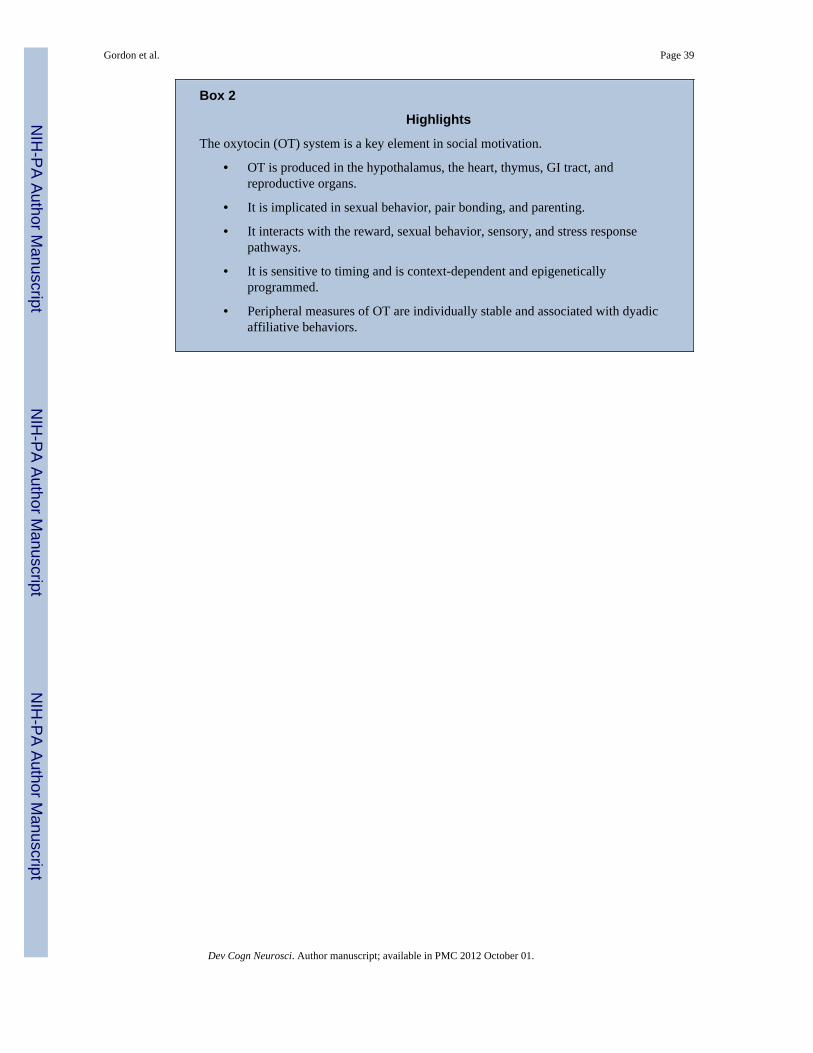
Box 2
Highlights
The oxytocin (OT) system is a key element in social motivation.
• OT is produced in the hypothalamus, the heart, thymus, GI tract, andreproductive organs.
• It is implicated in sexual behavior, pair bonding, and parenting.
• It interacts with the reward, sexual behavior, sensory, and stress responsepathways.
• It is sensitive to timing and is context-dependent and epigeneticallyprogrammed.
• Peripheral measures of OT are individually stable and associated with dyadicaffiliative behaviors.
Gordon et al. Page 39
Dev Cogn Neurosci. Author manuscript; available in PMC 2012 October 01.
NIH
-PA Author Manuscript
NIH
-PA Author Manuscript
NIH
-PA Author Manuscript

Figure 1. Central and Peripheral Sites of Oxytocin (OT) ReleaseWithin the brain, OT is released from the paraventricular (PVN) and supraoptic (SON)nuclei of the hypothalamus, and to a lesser extent, the bed nucleus of the stria terminalis,spinal cord and anterior commissural nucleus. Central OT projections are pictured in green.Peripheral sources of OT include OT released into circulation via the posterior pituitary aswell as numerous sites outside of the brain, including the heart, thymus, gastrointestinaltract, testis, epididymis, prostate, pregnant intrauterine tissue, ovaries, and adrenal medulla.The breast, pancreas and kidney are peripheral OT targets. NAcc = nucleus accumbens;MPOA = medial preoptic area; BNST= bed nucleus of stria terminalis; VTA = ventraltegmental area; A = amygdala.
Gordon et al. Page 40
Dev Cogn Neurosci. Author manuscript; available in PMC 2012 October 01.
NIH
-PA Author Manuscript
NIH
-PA Author Manuscript
NIH
-PA Author Manuscript

Figure 2. Integrative ModelAn integrative model of biobehavioral processes involved in the formation and maintenanceof caretaker-child bonds and related attachment phenomena. “Responsiveness” includes: a) arange of perceptual, autonomic, behavioral, emotional and cognitive responses conditionedby the absence, presence, or merely cues of the other; b) responses conditioned by thecaretaker’s experience of being cared for as an infant; and c) responses conditioned byperceived threats and adversity. By entering into a synchronous affective dyadiccommunication with the infant, the caregiver provides an external support for the infant’sdeveloping bioregulatory abilities and thereby conveys lifelong resilience to stress andenhanced coping ability. The experience of the caregiver and child’s micro-level matchingof affective states and level of arousal during face-to-face dyadic interactions beginningaround the second month of life provides the basis for children’s self awareness, socialdevelopment, empathy, and moral internalization. Maternal gaze matching, facialexpressions, vocalizations, and regulation of arousal states during face-to-face play providecritical environmental inputs during the sensitive period of maturation of the visual cortex.Furthermore, by synchronizing with infant arousal state, mothers entrain the infant’sbiological rhythms, providing a “resonance” of internal and external experience, self andother, brain and behavior. Metacognitive representations and mental states correspond tointernal working models and are continually being updated and revised. However, one’searliest experiences with caregivers can powerfully shape the landscape of possible andexpected interactions with others. For example, stranger anxiety appears when infantsexperience a mismatch between expected caregiver stimuli and stimuli that the infantencounters when exposed to an unfamiliar person. These metacognitive representations andmental states can be both conscious and unconscious and can directly influence both patternsof responsiveness in the self and others as well as the core neural circuitry and associatedperipheral organs that dynamically link (in a context specific fashion) the oxytocin (OT) andarginine vasopressin (AVP) and the dopamine dependent reward pathways along with theHypothalamic-Pituitary-Adrenal (HPA) and the Hypothalamic-Pituitary-Gonadal (HPG)axes (see text).
Gordon et al. Page 41
Dev Cogn Neurosci. Author manuscript; available in PMC 2012 October 01.
NIH
-PA Author Manuscript
NIH
-PA Author Manuscript
NIH
-PA Author Manuscript