Histamine and motivation
14
REVIEW ARTICLE published: 04 July 2012 doi: 10.3389/fnsys.2012.00051 Histamine and motivation Fernando Torrealba 1,2 *, Maria E. Riveros 1,2 , Marco Contreras 1,2 and Jose L. Valdes 3 1 Facultad de Ciencias Biológicas, Departamento de Fisiología, Pontificia Universidad Católica de Chile, Santiago, Chile 2 Millenium Nucleus in stress and addiction, Pontificia Universidad Católica de Chile, Santiago, Chile 3 Facultad de Medicina, Departamento de Fisiología y Biofísica, Instituto de Ciencias Biomédicas, Universidad de Chile, Santiago, Chile Edited by: Pertti Panula, University of Helsinki, Finland Reviewed by: Ezio Tirelli, Université de Liège, Belgium Ritchie E. Brown, VA Boston Healthcare System, USA Saara Nuutinen, University of Helsinki, Finland José-Antonio Arias-Montaño, Centro de Investigación y de Estudios Avanzados, Mexico *Correspondence: Fernando Torrealba, Facultad de Ciencias Biológicas, Departamento de Fisiología, Pontificia Universidad Católica de Chile, Alameda 340, Santiago, Chile. e-mail: [email protected] Brain histamine may affect a variety of different behavioral and physiological functions; however, its role in promoting wakefulness has overshadowed its other important functions. Here, we review evidence indicating that brain histamine plays a central role in motivation and emphasize its differential involvement in the appetitive and consummatory phases of motivated behaviors. We discuss the inputs that control histaminergic neurons of the tuberomamillary nucleus (TMN) of the hypothalamus, which determine the distinct role of these neurons in appetitive behavior, sleep/wake cycles, and food anticipatory responses. Moreover, we review evidence supporting the dysfunction of histaminergic neurons and the cortical input of histamine in regulating specific forms of decreased motivation (apathy). In addition, we discuss the relationship between the histamine system and drug addiction in the context of motivation. Keywords: addiction, apathy, appetite, histamine, infralimbic cortex, motivation, tuberomamillary nucleus INTRODUCTION Motivations are essential for the preservation of genes, and they provide a rational and structured explanation of behavior and brain organization. Normal and pathological appetites provide guidance; for example, they provide direction in decision-making and influence subsequent behavioral and physiological expres- sion. The scientific study of behavior took an important step forward when the the concept of motivation (sometimes called instinctive behavior, drive or self-regulatory behavior) became widely accepted. It is important to understand why, of all the many possible behaviors in a given context, animals usually per- form only a few or just one. In addition, animals in a specific context perform one action and, at other times in the same exact context, perform another action, which may sometimes be opposite to the former. A simple and fixed relationship between the stimulus and response is not a satisfactory model for the behavior of animals with a nervous system. Motivation can be affected by the internal state of the animal, and as other theorists have suggested, arousal or the energizing of behavior is a central component of motivation. D. Hebb (Hebb, 1955) presented a model of motivation that is particularly useful for discussion; this work is accessi- ble, along with other classic papers in Psychology, at http:// psychclassics.yorku.ca/. Briefly, Hebb suggested that motivation provides direction and intensity for behavior; he was prob- ably referring exclusively to higher vertebrates. Motivation is directed toward or away from reinforcers (a functional term for rewards, associated only with positive reinforcers). Positive reinforcers may attract appetitive behavior, while aversions drive behavior away from reinforcers. A historical account of the emer- gence of the concept of motivation is worth reading (Stellar, 1990). An additional useful distinction that enriched the concept of motivated behaviors was proposed by Craig in 1917 (Craig, 1918); this distinction is a key element when considering the role of the brain histamine system in behavior. Craig suggested a dis- tinction between the appetitive and consummatory phase of a motivated behavior. Craig stated that “an appetite, so far as exter- nally observable, is a state of agitation which continues so long as a certain stimulus is absent. When the appeted stimulus is at length received it releases a consummatory reaction, after which the appetitive behavior ceases and is succeeded by a state of rel- ative rest, a state of satisfaction.” This statement emphasizes that the arousal component characterizes only the appetitive phase of a motivated behavior. These phases of motivated behavior have counterparts in the physiology of the somatic and visceral output systems that pre- pare the organism and allow it to better maintain bodily home- ostasis and to perpetuate genes. The key role of behavior in long-term homeostatic balance and the preservation of genes has been thoughtfully addressed by Garcia (Garcia et al., 1974) and Blessing (Blessing, 1997), among others. We will discuss the involvement of brain histamine in appeti- tive and aversive behaviors; in these behaviors, high arousal and an increased readiness to act and to spend energy predominate in parallel with neuroendocrine and sympathetic activation (Akins and Bealer, 1993). We will also argue that brain histamine may have a negative effect on consummatory behavior. Frontiers in Systems Neuroscience www.frontiersin.org July 2012 | Volume 6 | Article 51 | 1 SYSTEMS NEUROSCIENCE
-
Upload
independent -
Category
Documents
-
view
1 -
download
0
Transcript of Histamine and motivation
REVIEW ARTICLEpublished: 04 July 2012
doi: 10.3389/fnsys.2012.00051
Histamine and motivationFernando Torrealba 1,2*, Maria E. Riveros 1,2, Marco Contreras 1,2 and Jose L. Valdes 3
1 Facultad de Ciencias Biológicas, Departamento de Fisiología, Pontificia Universidad Católica de Chile, Santiago, Chile2 Millenium Nucleus in stress and addiction, Pontificia Universidad Católica de Chile, Santiago, Chile3 Facultad de Medicina, Departamento de Fisiología y Biofísica, Instituto de Ciencias Biomédicas, Universidad de Chile, Santiago, Chile
Edited by:
Pertti Panula, University of Helsinki,Finland
Reviewed by:
Ezio Tirelli, Université de Liège,BelgiumRitchie E. Brown, VA BostonHealthcare System, USASaara Nuutinen, University ofHelsinki, FinlandJosé-Antonio Arias-Montaño, Centrode Investigación y de EstudiosAvanzados, Mexico
*Correspondence:
Fernando Torrealba, Facultad deCiencias Biológicas, Departamentode Fisiología, Pontificia UniversidadCatólica de Chile, Alameda 340,Santiago, Chile.e-mail: [email protected]
Brain histamine may affect a variety of different behavioral and physiological functions;however, its role in promoting wakefulness has overshadowed its other importantfunctions. Here, we review evidence indicating that brain histamine plays a central role inmotivation and emphasize its differential involvement in the appetitive and consummatoryphases of motivated behaviors. We discuss the inputs that control histaminergic neuronsof the tuberomamillary nucleus (TMN) of the hypothalamus, which determine the distinctrole of these neurons in appetitive behavior, sleep/wake cycles, and food anticipatoryresponses. Moreover, we review evidence supporting the dysfunction of histaminergicneurons and the cortical input of histamine in regulating specific forms of decreasedmotivation (apathy). In addition, we discuss the relationship between the histaminesystem and drug addiction in the context of motivation.
Keywords: addiction, apathy, appetite, histamine, infralimbic cortex, motivation, tuberomamillary nucleus
INTRODUCTIONMotivations are essential for the preservation of genes, and theyprovide a rational and structured explanation of behavior andbrain organization. Normal and pathological appetites provideguidance; for example, they provide direction in decision-makingand influence subsequent behavioral and physiological expres-sion. The scientific study of behavior took an important stepforward when the the concept of motivation (sometimes calledinstinctive behavior, drive or self-regulatory behavior) becamewidely accepted. It is important to understand why, of all themany possible behaviors in a given context, animals usually per-form only a few or just one. In addition, animals in a specificcontext perform one action and, at other times in the sameexact context, perform another action, which may sometimes beopposite to the former. A simple and fixed relationship betweenthe stimulus and response is not a satisfactory model for thebehavior of animals with a nervous system. Motivation can beaffected by the internal state of the animal, and as other theoristshave suggested, arousal or the energizing of behavior is a centralcomponent of motivation.
D. Hebb (Hebb, 1955) presented a model of motivationthat is particularly useful for discussion; this work is accessi-ble, along with other classic papers in Psychology, at http://psychclassics.yorku.ca/. Briefly, Hebb suggested that motivationprovides direction and intensity for behavior; he was prob-ably referring exclusively to higher vertebrates. Motivation isdirected toward or away from reinforcers (a functional termfor rewards, associated only with positive reinforcers). Positivereinforcers may attract appetitive behavior, while aversions drive
behavior away from reinforcers. A historical account of the emer-gence of the concept of motivation is worth reading (Stellar,1990).
An additional useful distinction that enriched the concept ofmotivated behaviors was proposed by Craig in 1917 (Craig, 1918);this distinction is a key element when considering the role ofthe brain histamine system in behavior. Craig suggested a dis-tinction between the appetitive and consummatory phase of amotivated behavior. Craig stated that “an appetite, so far as exter-nally observable, is a state of agitation which continues so longas a certain stimulus is absent. When the appeted stimulus is atlength received it releases a consummatory reaction, after whichthe appetitive behavior ceases and is succeeded by a state of rel-ative rest, a state of satisfaction.” This statement emphasizes thatthe arousal component characterizes only the appetitive phase ofa motivated behavior.
These phases of motivated behavior have counterparts in thephysiology of the somatic and visceral output systems that pre-pare the organism and allow it to better maintain bodily home-ostasis and to perpetuate genes. The key role of behavior inlong-term homeostatic balance and the preservation of genes hasbeen thoughtfully addressed by Garcia (Garcia et al., 1974) andBlessing (Blessing, 1997), among others.
We will discuss the involvement of brain histamine in appeti-tive and aversive behaviors; in these behaviors, high arousal andan increased readiness to act and to spend energy predominate inparallel with neuroendocrine and sympathetic activation (Akinsand Bealer, 1993). We will also argue that brain histamine mayhave a negative effect on consummatory behavior.
Frontiers in Systems Neuroscience www.frontiersin.org July 2012 | Volume 6 | Article 51 | 1
SYSTEMS NEUROSCIENCE
Torrealba et al. Brain histamine and appetitive behavior
EFFERENT CONNECTIONS OF HISTAMINERGIC NEURONSIn the mammalian brain, neuronal histamine is exclusivelypresent in the tuberomamillary nucleus (TMN), a loosely packedset of magnocellular neurons located in the posterior and ventralregion of the hypothalamus, in close proximity to the posteriorrecess of the third ventricle (Panula et al., 1989). Histaminergicaxons innervate many brain regions, from the prefrontal cortexto the spinal cord, providing an excitatory tone to postsynapticneurons through H1 and H2 receptors (H1R, H2R) and mod-ulating the release of histamine and other transmitters throughH3 receptors (H3R). This widespread distribution of histamine-containing axon terminals and histaminergic receptors helps toclarify the involvement of histamine in many brain functions.However, there have been indications that subsets of TMN neu-rons, some of which have a specific sensitivity to pharmacologicalagents, project to defined brain regions (Giannoni et al., 2009)or influence particular brain functions (Miklos and Kovacs, 2003;Valdes et al., 2010).
WHAT INPUTS DRIVE TMN ACTIVITY?To begin unraveling the roles of histaminergic neurons in a varietyof brain functions, it is important to consider the main inputsthat might drive, by either increasing or decreasing the activity ofTMN neurons. Driver inputs, as opposed to modulatory inputs,are those inputs that are central to the functions of a given cell,whereas modulators may change the expression of those functions(Sherman and Guillery, 1998); for example, a synaptic input maybe either a driver or modulator depending on the conditions. Wewill show the importance of some of these inputs in defining theseparate roles of histamine in wake/sleep cycles, feeding-relatedanticipatory activity, and motivation.
Neuroanatomical studies have addressed the origin of TMNafferents using axonal tract-tracing methods. In general, one cansimplify the many inputs ascribed to the ventral TMN by focus-ing on the most robust (Ericson et al., 1991), considering thatsome of them may be driver inputs. TMN afferents originatefrom rostral forebrain limbic structures that include hypothala-mic regions involved in food anticipatory activity (Acosta-Galvanet al., 2011), the infralimbic cortex (IL) (Wouterlood et al., 1987),the hypothalamic preoptic region and the lateral septum (Ericsonet al., 1991; Sherin et al., 1998). Afferents from other aminer-gic nuclei and hypothalamic regions, including areas involved incircadian rhythmicity (Deurveilher and Semba, 2005) and theorexin/hypocretin neurons from the lateral/perifornical hypotha-lamic area, are likely modulatory rather than driver inputs(Ericson et al., 1989; Torrealba et al., 2003) (Figure 1).
FOOD ENTRAINABLE CIRCADIAN OSCILLATOR INPUTThe relationship between the motivation for food, arousal, andTMN activity was first studied using a model of restricted feeding.Rats placed on a feeding schedule that is restricted to a few day-time hours wake up in anticipation of mealtime. This anticipatorybehavior has an important adaptive value because in nature,food may be available during the same few daily hours (Stephan,2001), and the anticipatory physiological and behavioral activa-tion prepares the animals to take advantage of this predictablephenomena. It was shown that this anticipatory waking up is
related to transient activation of the TMN (Figure 2A) but not ofother ascending arousal system (AAS) nuclei (Inzunza et al., 2000;Angeles-Castellanos et al., 2004), including the orexin neuronsfrom the lateral hypothalamic/perifornical area (LHA) (Meynardet al., 2005). Increased TMN activation begins approximately onehour before the scheduled mealtime (Meynard et al., 2005), asevidenced by fos mRNA expression; however, the precise rela-tionship between fos expression and electric activity of the TMNneurons remains to be determined. LHA neurons, including non-orexin neurons, become active well after TMN peak activation.We hypothesized that a signal from a food-entrainable circadianoscillator (Meynard et al., 2005) other than the suprachiasmaticnucleus (Mistlberger, 1994) should excite the TMN. Of interestfor the present study is the distinction between the histaminergiceffects on appetitive versus consummatory behavior. This antici-patory TMN activation quickly disappears (Figure 2C) when theanimals begin eating (Meynard et al., 2005). Our preliminarydata suggest that a bilateral TMN lesion impairs the anticipa-tory arousal induced by scheduled restricted feeding (Recabarrenet al., 2003). Together, these pieces of evidences support theidea discussed that increased histamine levels are important forappetitive behavior and that decreased histamine levels facilitateconsummatory behavior. It is likely that the food-entrained cir-cadian input that activates the TMN arises from a component ofthe intrahypothalamic network that determines food anticipatoryactivity (Acosta-Galvan et al., 2011) and provides afferents to theTMN. The dorsomedial hypothalamic nucleus, subparaventricu-lar zone, and medial preoptic area are possible candidates for thiscomponent (Deurveilher and Semba, 2005).
THE INFRALIMBIC CORTICAL AREA INPUTExposure to appetitive food stimuli activates many brain regionsin humans (Wang et al., 2004) and rats (Valdes et al., 2010).In both species, there is a marked activation of the frontalcortex, including the anterior insula and orbitofrontal cortices(Figure 3), both of which are closely involved in motivation andsend projections to the LHA in rats (Gabbott et al., 2005). Theactivation of these frontal cortices was impaired by a TMN lesion(Valdes et al., 2010).
We view the cortical input to the TMN as essential for theappetitive function of histaminergic neurons, which will subse-quently be discussed in more detail. Layer 5 pyramidal cells fromthe medial prefrontal cortex, which mostly originate from the IL,are the main cortical inputs to the TMN. The close proximity ofthe axon terminals from the IL to the histaminergic neurons andthe glutamatergic phenotype of the pyramidal cells strongly sug-gest that IL afferents have an excitatory effect on histaminergicneurons. However, it is possible that the IL afferents may havean inhibitory effect if they make synaptic connections with localinhibitory neurons. An electron microscopic study elucidating thetype of synaptic contact between the IL and TMN, which is pre-sumably asymmetric and excitatory, similar to the other IL targets(Torrealba and Muller, 1999), is still lacking.
We reasoned that the prefrontal cortex, which is central indecision-making and motivation, is the input that activates theTMN during the appetitive procurement of reinforcers such asfood. In fact, Goldman-Rakic demonstrated that the prefrontal
Frontiers in Systems Neuroscience www.frontiersin.org July 2012 | Volume 6 | Article 51 | 2

Torrealba et al. Brain histamine and appetitive behavior
Master circadianoscillator Food entrainable
oscillator
Appetitive behaviorSleep/wakecycle
Food anticipatoryactivity
Behavioral and vegetative arousal
TMNAminergic
Orexin/LHA
VLPOVLPO
Infralimbic cortex
FIGURE 1 | Diagram of the putative drivers (blue, green, and gray) and
modulatory (light orange) inputs to the TMN. The presumed role of thedriver inputs in the distinct arousal functions of the TMN is indicatedon top; in brief, inputs from the prefrontal cortex or the food-entrainableoscillator activate the TMN during appetitive behavior or circadiananticipatory activity, respectively, while input from the VLPO inhibits
TMN activity and promotes sleep. The green arrow indicatesexcitatory input; the black arrow designates presumed excitatoryinput; the red T-line indicates inhibitory input; the orange arrowdesignates mixed input to aminergic neurons. Double-headed arrowsindicate bidirectional input between the TMN and aminergic andorexin neurons.
cortex is the only source of cortical connections to arousal nucleiand that as such, the prefrontal cortex may control its own levelof activity (GoldmanRakic, 1987). To study the functional rela-tionship between IL and TMN neurons in motivated behavior,we devised a method to prolong the appetitive phase by enticingthe rats with a wire-mesh box filled with food that was placedwithin their home cage. The rats may attempt to open the box,but they are unable to obtain the food [see Methods and video inValdes et al. (Valdes et al., 2010)]. This procedure clearly separatesappetitive from consummatory behavior.
Of particular relevance is that a subpopulation of rat IL neu-rons becomes active during enticement, as demonstrated in singleunit recordings in freely moving animals and by c-fos expression(Valdes et al., 2006). A relatively large proportion, 33.3% of ILneurons, increase their firing rate during this enticing stage, while10% become excited immediately after eating, and 3.3% becameactive in both conditions. A small percentage (6.7%) of IL neu-rons decreased their firing rate in response to enticement andafter eating. This high proportion of IL neurons that respondedto enticement suggests that the IL might use a population code torepresent a given behavioral state. Taking into account the manyspecific functions of the IL during, for example, fear extinction
(Quirk and Beer, 2006), stress (Amat et al., 2005), or enticement,and its numerous subcortical targets (Gabbott et al., 2005), onecan imagine that subpopulations of IL neurons may participatein several of those behavioral states and that the global “visceralmotor” IL (Terreberry and Neafsey, 1987) output may reflect sucha combinatorial effect.
IL-TMN AXIS-DEPENDENT MECHANISMS OF BEHAVIORALRESPONSES TO ENTICEMENTThe increased arousal state made apparent during enticement wasmeasured by polysomnographic recordings (Valdes et al., 2005)or was evaluated by rat motor activity, which in this case andas observed in the video provided by Valdes et al. (Valdes et al.,2010), is goal-directed and not at all non-specific. Animals aremotivated to obtain a reward when they detect homeostatic needs(hunger in the present example) and/or to obtain pleasure. Oneday of fasting ostensibly increased the rats’ motivation to openthe box more than feeding them ad libitum. Additionally, fastedrats familiar with a mixture of plain food pellets and palatablemorsels (salami and chocolate cookies) made even more intenseattempts to obtain the food compared to rats that were onlyoffered plain pellets (Valdes et al., 2010) (Figure 2B). The increase
Frontiers in Systems Neuroscience www.frontiersin.org July 2012 | Volume 6 | Article 51 | 3
Torrealba et al. Brain histamine and appetitive behavior
highlymotivated (60’FE)
% of TMN active neurons Fos+/ADA+)0 10 20 30 40 50
0
200
400
600
800
1000
1200
Motoractivity(counts/1h)
motivatedunmotivated
highly motivated
P=0.007CC=0.513
B
0
10
20
30
40
50
60
*
naive before aftermeal-time
circadian stimulus
C
0
10
20
30
40
50
60 sensory stimuli
naïve
unmotivated (FE)
hungry
FW(1tap/h)
FW(12taps/h)
motivated (30’ FE)
motivated (60’FE)
motivated (120’FE)
120’feed
A
**
**
%ofTMNactiveneurons
(Fos+/ADA+)
%ofTMNactiveneurons
(Fos+/ADA+)
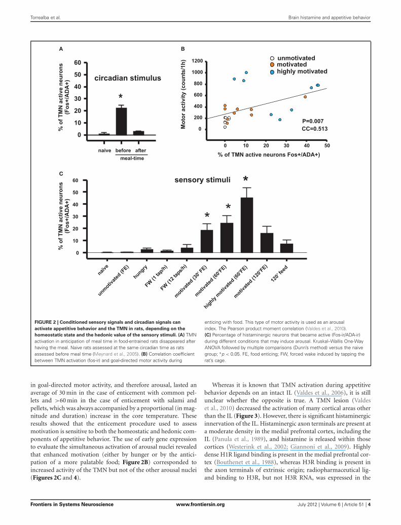
FIGURE 2 | Conditioned sensory signals and circadian signals can
activate appetitive behavior and the TMN in rats, depending on the
homeostatic state and the hedonic value of the sensory stimuli. (A) TMNactivation in anticipation of meal time in food-entrained rats disappeared afterhaving the meal. Naive rats assessed at the same circadian time as ratsassessed before meal time (Meynard et al., 2005). (B) Correlation coefficientbetween TMN activation (fos-ir) and goal-directed motor activity during
enticing with food. This type of motor activity is used as an arousalindex. The Pearson product moment correlation (Valdes et al., 2010).(C) Percentage of histaminergic neurons that became active (Fos-ir/ADA-ir)during different conditions that may induce arousal. Kruskal–Wallis One-WayANOVA followed by multiple comparisons (Dunn’s method) versus the naivegroup; ∗p < 0.05. FE, food enticing; FW, forced wake induced by tapping therat’s cage.
in goal-directed motor activity, and therefore arousal, lasted anaverage of 30 min in the case of enticement with common pel-lets and >60 min in the case of enticement with salami andpellets, which was always accompanied by a proportional (in mag-nitude and duration) increase in the core temperature. Theseresults showed that the enticement procedure used to assessmotivation is sensitive to both the homeostatic and hedonic com-ponents of appetitive behavior. The use of early gene expressionto evaluate the simultaneous activation of arousal nuclei revealedthat enhanced motivation (either by hunger or by the antici-pation of a more palatable food; Figure 2B) corresponded toincreased activity of the TMN but not of the other arousal nuclei(Figures 2C and 4).
Whereas it is known that TMN activation during appetitivebehavior depends on an intact IL (Valdes et al., 2006), it is stillunclear whether the opposite is true. A TMN lesion (Valdeset al., 2010) decreased the activation of many cortical areas otherthan the IL (Figure 3). However, there is significant histaminergicinnervation of the IL. Histaminergic axon terminals are present ata moderate density in the medial prefrontal cortex, including theIL (Panula et al., 1989), and histamine is released within thosecortices (Westerink et al., 2002; Giannoni et al., 2009). Highlydense H1R ligand binding is present in the medial prefrontal cor-tex (Bouthenet et al., 1988), whereas H3R binding is present inthe axon terminals of extrinsic origin; radiopharmaceutical lig-and binding to H3R, but not H3R RNA, was expressed in the
Frontiers in Systems Neuroscience www.frontiersin.org July 2012 | Volume 6 | Article 51 | 4

Torrealba et al. Brain histamine and appetitive behavior
IL SS1 MO1
naïve (n=5)sham (n=12)TMN lesion (n=7)
* *
#
#
NºofFos-irneurons/ mm
2
0
25
50
75
100
125
150
175
200
�
MO2MO1
SS1
cc
VL
CP
-0.26 mm
Sham TMN lesion
IL
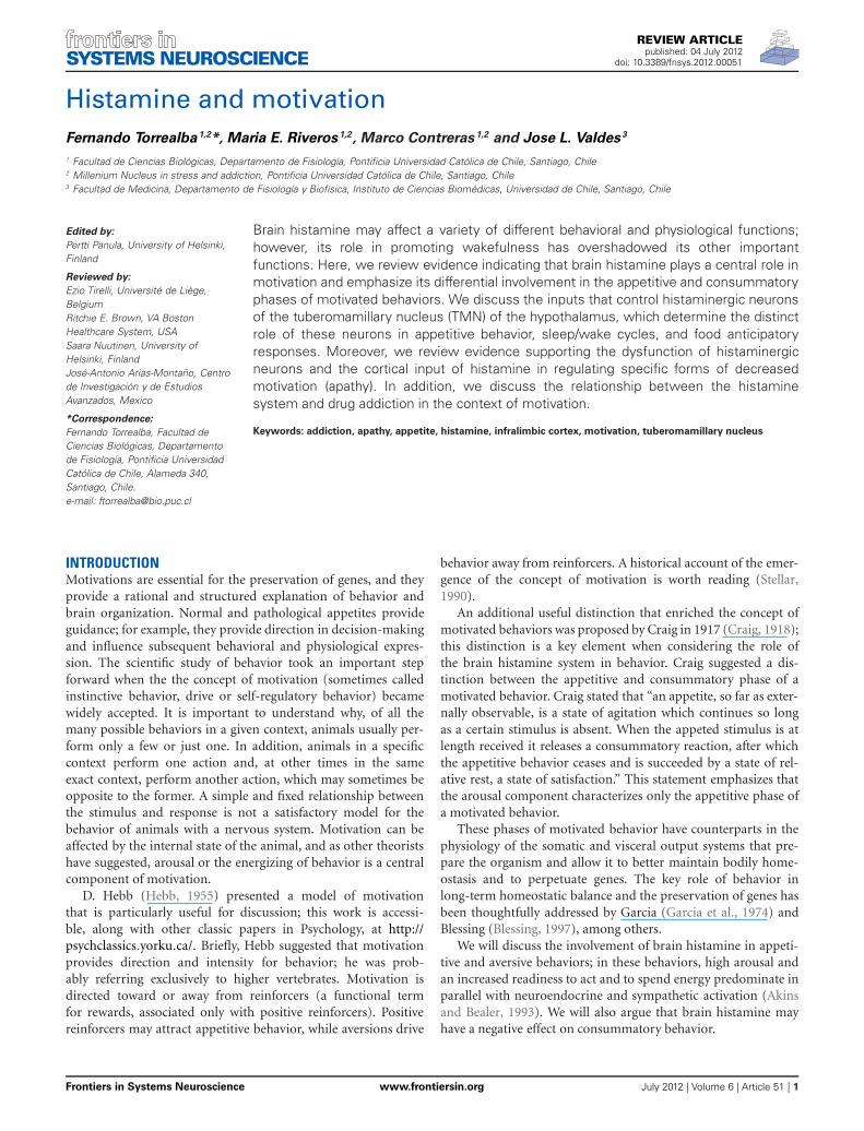
FIGURE 3 | Enticing intact (sham) hungry rats with food strongly
activated the frontal cortex (left column). This activation was impaired bya TMN lesion (right column), except for the IL. Lower graph compares theactivation of three cortices in the sham and TMN lesioned rats. The naïvegroup corresponded to the circadian control, i.e., to rats fed ad libitum, notenticed and sacrificed at the same time during the day as the other groups(Valdes et al., 2006).
IL (Pillot et al., 2002). It has been shown that H3R activation inthe medial prefrontal cortex decreases histamine release in boththe TMN and prefrontal cortex of freely moving rats, which is aform of negative feedback (Flik et al., 2011). This finding mayhelp to explain why TMN lesions, which should decrease his-taminergic release in the prefrontal cortex, have little effect on ILactivity during enticement. When using enticement as a model
30` Pellet (n= 5)30` Pellet + Salami (n= 6)
TMN / ADA-ir LHA / orexin-ir
%ofdouble-irneurons
0
10
20
30
40
50
60
*
30` Pellet, ad lib
#
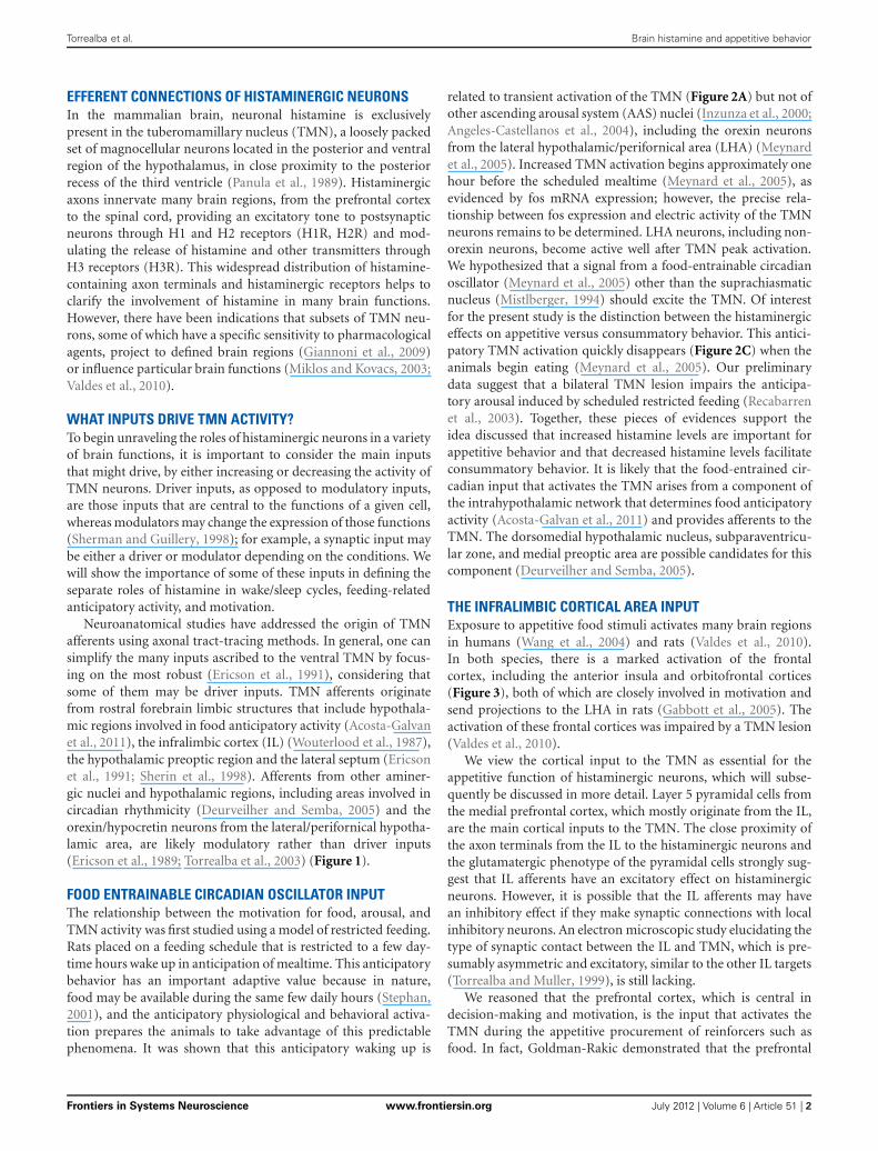
FIGURE 4 | The number of TMN active neurons that responded to
enticing was dependent on the homeostatic state and palatability of
the stimulus: Orexin neurons from the LHA did not respond to
enticing. #Different from the other conditions; ∗different from the pelletplus salami. Modified from Valdes et al. (2010).
for appetitive behavior, we found that TMN lesions preventedincreases in goal-directed motor activity (Valdes et al., 2010); thisdecreased appetitive behavior was correlated with the size of thelesion in the dorsal TMN. A bilateral IL lesion also impaired TMNactivation during enticement (Valdes et al., 2006). Both the ILand TMN lesions blocked the delayed activation of other AASnuclei, but not the locus coeruleus (LC) and orexin neurons, sug-gesting that these neurons contribute to arousal maintenance butnot appetitive behavior. It is conceivable that the maintenanceof IL activation during enticement after a TMN lesion might bethe result of both decreased H3R inhibition and this spared LCactivity and the minor activation of orexin neurons (Valdes et al.,2010). In fact, the IL receives a more substantial overlap of nora-drenergic and orexinergic input (Baldo et al., 2003) comparedwith other cortical regions. It is possible that these two inputsmaintain IL activity while the other cortical regions are depressedby TMN lesions.
The IL, initially described as a visceral motor cortex(Terreberry and Neafsey, 1987), appears to be essential for theactivation of the body and brain that takes place during theanticipation of an event and during appetitive behavior. The ILis a cortical area with more direct connections to subcorticalsites involved in neuroendocrine and somatic responses relatedto appetitive or aversive behaviors (Gabbott et al., 2005). One ofthose responses is the arousal function of the IL during appeti-tive behavior, which is importantly mediated by its connection tothe TMN. The TMN becomes active and increases arousal (Valdeset al., 2005) before the other AAS nuclei (Figure 5), and togetherwith the IL, contributes to the delayed activation of the other AASnuclei, which in turn maintain and potentially enrich the brainfunctions that optimize the expression of appetitive behaviors.For example, using the same behavioral task (Robbins and Everitt,1995), it was shown that cortical cholinergic functions contribute
Frontiers in Systems Neuroscience www.frontiersin.org July 2012 | Volume 6 | Article 51 | 5

Torrealba et al. Brain histamine and appetitive behavior
TMN
Orx
LDT
LCDR
0 30’ 60’ 120’
Neuronalactivity(Fos-ir)
Neuronalactivity(Fos-ir)
High
Low
Medium
Enticing time (min)Enticing time (min)
FIGURE 5 | Temporal course of ascending arousal system nuclei
activation during appetitive behavior induced by enticing hungry rats
with unreachable food. The level of neuronal activity, evaluated by Fosexpression, is indicated by the grayscale. The four bar graphs correspond torats that have fasted for 24 h and were sacrificed immediately before thebeginning of the enticing procedure (time 0) or killed after the indicatedtimes (30–120 min). Note that the first nucleus to increase Fos-ir 30 minafter the beginning of enticing was the TMN. The other arousal nucleiincreased Fos-ir after 60 min and continued with medium to high Fos-ir at120 min. DR, dorsal raphe; LC, locus coeruleus; LDT, laterodorsal tegmentalnucleus; Orx, orexin neurons from the lateral/perifornical hypothalamicarea; TMN, tuberomamillary nucleus. Modified from Valdes et al. (2005).
to the accuracy of behavioral responses; these functions includethe action of dopamine in delaying responses, the action of nora-drenaline from the LC in distraction, and the action of serotoninthat is related to rats’ response impulsivity.
Novelty has reinforcing properties that motivate the explo-ration of new environments or novel objects. Evidence indicatesthat this exploration depends on an intact prefrontal cortex(Daffner et al., 2000) and histamine system. It has been shownthat mice lacking the enzyme histidine decarboxylase (HDC)show decreased spatial novelty-induced arousal (Parmentier et al.,2002) and reduced exploratory activity in an open field butnormal habituation to the same open field (Dere et al., 2004).However, another study found no differences between HDC-KOand wild-type mice in locomotor activity during a 2 h exposureto a new chamber (Nuutinen et al., 2010). Mice lacking H1R(Inoue et al., 1996) show reduced exploratory behavior (ambu-lation and rearings) in a novel environment. Furthermore, micelacking H1Rs show reduced emotional responses to a novel envi-ronment and do not generate a place preference conditioned bythe novel object; however, they do explore (consummatory behav-ior?) new objects in a familiar setting (Zlomuzica et al., 2008). Thenucleus accumbens (NAcc) is one site where histamine increasesexploratory behavior through the activation of H1 and H2Rs(Orofino et al., 1999). In contrast, histamine administration intothe ventral hippocampus decreased exploration and associatedemotionality in an H1- and H2-dependent manner (Ruarte et al.,1997). However, the existence of small regions in the NAcc thatmay increase or decrease the hedonic response (hotspots) (Smith
and Berridge, 2007) makes it difficult to interpret the responsesto a NAcc injection, whose precise target within this nucleus isunknown.
IL-TMN AXIS-DEPENDENT MECHANISMS OF VEGETATIVERESPONSES TO ENTICEMENTThe complexity of the mechanisms underlying the vegetativearousal that characterizes appetitive behavior is illustrated byemphasizing the thermogenic response to enticement. A cell-specific lesion that involves more than 50% of the bilateral IL(Valdes et al., 2006) or a large TMN lesion (Valdes et al., 2010)prevents the thermogenic response to enticement. This effectwas correlated with the size of a ventral but not a dorsal TMNlesion (Valdes et al., 2010). However, the normal pattern of ther-moregulatory nucleus activation was differentially disrupted byIL versus TMN lesions. A lesion to the IL prevented the acti-vation of all of the thermoregulatory nuclei examined, such asthe preoptic area, dorsomedial nucleus of the hypothalamus,and raphe pallidus. In contrast, a TMN lesion only preventedthe activation of the raphe pallidus (where sympathetic premo-tor neurons to brown adipose tissue are located). Other ther-moregulatory nuclei were very active despite the absence of athermogenic response. Importantly, while an IL lesion preventedTMN activation, a TMN lesion did not change IL activation dur-ing enticement. Taken together, these results indicate that thethermogenic response during appetitive behavior is controlledby the IL and TMN and that histaminergic input to the raphepallidus plays a gating or permissive role in the IL thermogenic-related signals to the raphe pallidus. The temperature increaseinduced by activation of the IL-TMN axis is, in part, mediatedby the action of histamine on H1Rs and H3Rs expressed by non-GABAergic and GABAergic neurons of the hypothalamic preopticregion (Lundius et al., 2010). This thermogenic response to his-tamine injection into the preoptic area is largely mediated by theactivation of brown adipose tissue and its sympathetic innerva-tion (Cannon and Nedergaard, 2004). Interestingly and similarto enticement, olfactory stimulation with grapefruit oil excitedsympathetic innervation of the brown adipose tissue and adrenalgland and reduced the appetite for food. Moreover, all of theseeffects were blocked by the systemic administration of an H1antagonist (Shen et al., 2005).
VENTROLATERAL PREOPTIC AREA INPUTThe best-studied driver input to the TMN is the one central tothe wake/sleep function of histaminergic neurons (Saper et al.,2005). This input arises from the ventrolateral preoptic area(VLPO) and adjacent regions in the anterior and ventral hypotha-lamus (Sherin et al., 1998). These neurons express the inhibitorytransmitters GABA and galanin and make symmetrical GABAimmunoreactive synaptic connections with the dendrites and cellbodies of TMN neurons and other neurons from the AAS (Sherinet al., 1998). VLPO cells are one of the few cell groups that aremore active during sleep than wakefulness (Sherin et al., 1996).In addition, they inhibit AAS neurons, including the TMN, viaGABA release, which results in sleep initiation and maintenance(Saper et al., 2005). This VLPO-TMN pathway is essential tounderstanding the well-described role of histaminergic neurons,
Frontiers in Systems Neuroscience www.frontiersin.org July 2012 | Volume 6 | Article 51 | 6

Torrealba et al. Brain histamine and appetitive behavior
which is to promote wakefulness during the active phase of theday.
The lateral septum is another source of robust, and mostlyGABAergic, input to the TMN region. However, there are nopublished studies on the functional significance of this afferentconnection. Nonetheless, histamine in the lateral septum has ananxiogenic effect that is mediated by H1 and H2Rs (Zarrindastet al., 2008).
OREXINERGIC INPUTOrexin (hypocretin) neurons from the LHA, which are involvedin appetitive behavior, in part because of their role in incentivesaliency (Harris et al., 2005), form a well studied excitatory (Bayeret al., 2001; Eriksson et al., 2001) input to histaminergic neu-rons [while histamine has little effect on orexin neurons (Li et al.,2002)]. Orexin neurons may be considered a modulatory inputto the TMN. For example, animals unable to synthesize orexinshow intense electric activity of histaminergic neurons during cat-aplexia (John et al., 2004), indicating that inputs other than orexinmaintain this high activity of histaminergic neurons. Orexin neu-rons have a variety of postulated roles, including arousal (Huanget al., 2001), in which histaminergic neurons appear to be theeffector. Orexinergic neurons, perhaps under cortical influence(Monda et al., 2004), operate through type B receptors andhistamine to increase brown adipose tissue sympathetic nerveactivity (Yasuda et al., 2005) and thermogenesis.
HISTAMINE IN AVERSIVE BEHAVIORWhile the focus of this review is centered on the role of his-tamine in appetitive behaviors, it is useful to note that, in amore general sense, the role of histamine in the activation of thebrain (behavioral arousal) and body (increased motor, neuroen-docrine, and sympathetic activity) extends to behavioral changesassociated with threat situations. Brown and colleagues (Brownet al., 2001) reviewed evidence supporting the idea that the brainhistamine system is widely engaged in situations of physiologi-cal or existential danger. The anti-nociceptive action of centralhistamine and its role in water retention and adaptive anorexiahighlight the relevance of the histaminergic system in situationsof physiological threat, in which licking a wound, drinking, oreating is disadvantageous. For example, axon terminals express-ing the hypothalamic anorectic peptide α-MSH, which perhapsoriginates in the arcuate nucleus, make synaptic contacts withTMN neurons (Fekete and Liposits, 2003) and may contributeto adaptive anorexia. Similarly, the histamine system is involvedin situations of potential existential danger, i.e., stressful situa-tions (Westerink et al., 2002; Miklos and Kovacs, 2003), whichmay elicit anxiety and a behavioral inhibition response (Gray andMcNaughton, 2003) that is characterized by increased arousal,changes in behavioral focus, and increased exploratory behavior.For these reasons, Brown and colleagues proposed that brain his-tamine has a role as a danger response system. We offer a versionof Brown’s danger hypothesis that extends the range of goal-directed behavior from aversive to appetitive behavior. Histamineis released during goal-directed actions and results in an increasein behavioral and vegetative arousal and a decrease in the drive toconsume, which allows the optimal progression of a motivated
behavior. This short-lived adaptive anorexia is most likely animportant feature of motivated behaviors.
HISTAMINE INVOLVEMENT IN APATHYReduced motivation or apathy has been defined as a reductionof self-generated, voluntary, and purposeful behaviors (Levy andDubois, 2006). There are normal and pathological forms of apa-thy, depending on the cause, co-morbidity, and duration. Damageto the systems that generate and control voluntary actions locatedin the prefrontal cortex and basal ganglia will produce apathy.Apathy may be present in some forms of depression, dementia,schizophrenia, Huntington’s or Alzheimer’s disease, and frontallobe injury, among others. In addition, apathy itself can also be asyndrome (Marin, 1991).
Levy and Dubois (Levy and Dubois, 2006) distinguish threetypes of pathological apathy based on the underlying psycho-logical process and neural mechanism. (1) Emotional-affectiveapathy characterized by difficulties in behavioral modulation onthe basis of emotional value, associated with damage to theorbitomedial prefrontal cortex, (2) cognitive apathy characterizedby problems in planning and executing goal-directed behavior,associated with dysfunction of the lateral prefrontal cortex, and(3) auto-activation apathy characterized by the loss of spontane-ity and a need of an external impulse to initiate actions, associatedwith injury to the medial dorsal prefrontal cortex, basal ganglia(internal globus pallidus), and/or paramedial thalamus. We thinkthat the auto-activation type of apathy is characterized by a deficitin the energization of motivated behaviors, which we proposedepend on an intact IL-TMN axis. Consistent with this idea, thistype of apathy is caused by a lesion of the medial Brodmann areas9/10, 24, 25, and 32; it is thought that the rat infralimbic corticalarea is homologous to primate area 25 (Ongur and Price, 2000).
Drug abuse severity is associated with apathy scores (Verdejo-Garcia et al., 2006). Apathy is also associated with poor decision-making and the shortening of self-defined future in patients withfrontal lobe damage (Fellows and Farah, 2005). The latter impair-ments may diminish the likelihood of success for psychologicalprograms that manage drug addiction. Thus, treating apathy withmethylphenidate (see below) might be beneficial in the treat-ment of addiction, as it has proven effective in weight-reducingprograms (Desouza et al., 2011).
Interestingly, pathologies that exhibit co-morbidity with apa-thy have been characterized, from a neurochemical perspective,by dopaminergic (Bressan and Crippa, 2005) and histaminergic(see below) dysfunctions. However, none of these studies focusedon the presence of apathy in the subjects analyzed; thus, the linkbetween apathy and histaminergic or dopaminergic dysfunctionis tenuous at present.
Regarding the histaminergic system, a reduction in H1R lig-and binding in the frontal lobe of depressed patients (Kano et al.,2004) and schizophrenic patients (Iwabuchi et al., 2005) andthe frontal and temporal regions of Alzheimer’s disease patients(Higuchi et al., 2000) has been observed. However, chronicschizophrenic patients have increased levels of histamine metabo-lites in their cerebrospinal fluid (Prell et al., 1995). Parkinson’sdisease patients have increased levels of histamine but do nothave increased levels of its metabolite, telemethylhistamine, in
Frontiers in Systems Neuroscience www.frontiersin.org July 2012 | Volume 6 | Article 51 | 7

Torrealba et al. Brain histamine and appetitive behavior
the putamen, substantia nigra compacta, and both divisionsof the globus pallidus (Rinne et al., 2002); they also haveincreased histamine fibers in both divisions of the substantianigra (Anichtchik et al., 2000). However, no increase in HDCmRNA expression was found in the TMN (Shan et al., 2012b)of Parkinson’s disease patients, suggesting that there is no changein histamine production. Reduced H3R mRNA expression andincreased histamine methyltransferase mRNA levels in the susb-tantia nigra were also found in Parkinson’s disease patients (Shanet al., 2012a). In Huntington’s disease patients, HDC mRNAis increased in the inferior frontal gyrus and caudate nucleus,while H1 and H3R expression is increased in the inferior frontalgyrus and decreased in the caudate nucleus (van Wamelen et al.,2011). It remains to be determined whether these changes inthe brain regions involved in appetitive behavior are a conse-quence of adaptations to the primary illness or whether theyhave a causal link to some of the symptoms common to thesediseases.
Furthermore, it was found that apathy scores are higher inhealthy males than in healthy females (cited in Verdejo-Garciaet al., 2006), while histamine H1R ligand binding is lower in thelimbic system of males (Yoshizawa et al., 2009), which further sug-gests a link between brain histamine and motivation in human.In the same study, the authors found an increase in H1R lig-and binding in the amygdala, putamen, and globus pallidus ofanorexia nervosa patients (Yoshizawa et al., 2009). It is thoughtthat anorexia nervosa patients are motivated to work around food(some may enjoy cooking, for example) but show a strong aver-sion to consume it. It was recently suggested that anorexia nervosapatients have a lower sensitivity to natural pleasurable reinforcers(not only to food), a trait that is modulated by cognitive processesfocused on thinness (Soussignan et al., 2011).
Pharmacological evidence also indicates a causal relation-ship between histamine dysfunction and apathy. Methylphenidatestrongly increases extracellular levels of dopamine, noradrenaline(Berridge et al., 2006), and histamine in the rat prefrontal cor-tex (Horner et al., 2007) and improves apathy scores in patientswith Alzheimer’s disease (Padala et al., 2010), stroke (Spiegel et al.,2009), and dementia (Dolder et al., 2010). It is possible that theincrease in the histamine levels by methylphenidate is secondaryto the increased extracellular concentration of dopamine, as isthe case for systemic methamphetamine administration (Morissetet al., 2002), because D2 brain receptor activation enhances theTMN neuronal firing frequency, histamine release, and wakeful-ness in freely moving rats (Yanovsky et al., 2011).
We have shown that the IL activity and a consequent increasein histamine release and arousal are necessary for appetitivebehavior. We, therefore, hypothesize that damage to the IL-TMNaxis could induce a state of apathy that is characterized by a reduc-tion in the disposition to work for rewards or avoid danger. Infact, conditions resulting in high apathy scores are associated withprefrontal damage linked to stroke, depression, or drug addiction(Verdejo-Garcia et al., 2006). The exploration of novel stimuli isa motivated behavior that is dependent on the histamine system(discussed above), which has been shown to be negatively corre-lated with high apathy scores in patients with Alzheimer’s diseaseor frontal lobe damage (Daffner et al., 2000).
Anorexia may also be a normal response to a variety of physi-ological or pathological conditions. A well studied example is thereliable and phasic decrease in food intake that follows a cyclicincrease in estrogen in rodents and primates, including humans(Geary et al., 2001). While several hypothalamic nuclei, includ-ing the TMN, express estrogen receptors, the anorectic effect ofestradiol appears to depend on its direct action on TMN neurons,in addition to the effects of corticotropin-releasing hormone onTMN neurons (Gotoh et al., 2005). Estradiol also acts on the par-aventricular hypothalamic nucleus (an important anorexigenicregion), which releases corticotropin-releasing hormone (Gotohet al., 2009). Histaminergic neurons may act on ventromedialhypothalamic nucleus (VMH) neurons via H1R to decrease foodintake (King, 2006). Female hamsters show changes in brain his-tamine content following the estrous cycle (Hine et al., 1986); thehighest level is reached on proestrous day (day 3) in the hypotha-lamus and on day 2 in the rest of the brain. During pregnancy,there was an overall decline in histamine content and an increasein food intake. Female rats and humans are more vulnerable todrug abuse in general but particularly on the days with higherestrogen levels (Anker and Carroll, 2011). This vulnerability isfacilitated by estrogens.
The site of brain histamine-mediated suppression of foodintake is likely the VMH (Ookuma et al., 1989; Malick et al.,2001; King, 2006). The VMH, a hypothalamic site that containsglucose-responsive neurons and descending axonal projectionsto hindbrain regions that contain premotor sympathetic neu-rons, is the somatomotor center for motivated behavior-relatedactivities (Simerly, 2004). Blockade of H1R within the VMH,but not in other hypothalamic nuclei such as the paraventricu-lar hypothalamic nucleus or the LHA, increases both meal sizeand duration and suppresses the activity of glucose-responsiveneurons (Fukagawa et al., 1989).
HISTAMINE INVOLVEMENT IN ADDICTIONAddiction is a chronic behavioral disorder characterized by acompulsive and relapsing pattern of drug-seeking and drug-taking behavior that occurs despite the awareness of seriousnegative consequences. Thus, the behavior and cognitive pro-cesses of addicts are centered on drugs, and they have enormousdifficulties in attending to other activities (Everitt and Robbins,2005; Hyman et al., 2006).
Addiction develops after a prolonged period of drug intakein vulnerable individuals (Deroche-Gamonet et al., 2004;Vanderschuren and Everitt, 2004) and appears to depend on per-sistent neuroplastic changes (Kalivas and O’Brien, 2008) thatinclude impairment in the synaptic plasticity of relevant braincircuits such as the NAcc (Kasanetz et al., 2010). Synaptic plastic-ity (long-term depression) at cortico-striatal synapses is criticallycontrolled by dopamine (Surmeier et al., 2007) and acetylcholine(Wang et al., 2006), and it is thought to underlie the formation ofgoal-directed behaviors and habits.
It has been proposed that, as with habit formation in gen-eral, addiction involves a progression from action-outcometo stimulus-response mechanisms; drug use in the addict iscontrolled by automatic action patterns interacting with non-automatic cognitive processes (Tiffany, 1990; Everitt and Robbins,
Frontiers in Systems Neuroscience www.frontiersin.org July 2012 | Volume 6 | Article 51 | 8

Torrealba et al. Brain histamine and appetitive behavior
2005; Pierce and Vanderschuren, 2010). This sequence, frombehavior controlled by outcome to habitual drug seeking, is par-alleled by the gradual recruitment of ventral to dorsal striatalregions (Porrino et al., 2002) that have an anatomical substrate inthe spiraling connections between the medial prefrontal cortex,NAcc, and ventral tegmental area (VTA) and the dorsal prefrontalcortex, dorsal striatum, and substantia nigra (Haber et al., 2000;Pierce and Vanderschuren, 2010). The histaminergic system sendsprojections to the prefrontal cortex and striatum (Panula et al.,1989; Giannoni et al., 2009) and receives inputs from the medialprefrontal cortex, particularly from the IL. This cortical inputdrives TMN activity and seems to be important in sustaininga high level of arousal during motivated behavior, as discussedabove. Because histaminergic axon terminals are present in theprefrontal cortex, dorsal striatum, and NAcc, it is possible thathistamine may participate in the initial and late phases of addic-tion. Consistent with the idea presented in this review, histaminewould amplify the incentive salience (Berridge and Robinson,1998) of reinforcers and reinforcer-associated cues and supportthe arousal of the appetitive phase of motivated behavior directedtoward obtaining drugs.
A link between the activity of the histamine system and therisk of becoming addicted to drugs has been reported. Noveltyseeking is a personality trait that is related to an increased riskof addictive behavior in human and animal models (Kampov-Polevoy et al., 2004; Belin et al., 2011). Females have higher levelsof brain histamine (Hine et al., 1986; Prell and Green, 1991) andare more vulnerable to addiction than males. High gonadal hor-mone estrogen levels during the menstrual cycle may facilitatedrug abuse in women (Anker and Carroll, 2011). Anorexia ner-vosa patients show a significantly higher density of histamine H1Rthan controls, suggesting that the alteration of central histamin-ergic activity is involved in eating disorders (Yoshizawa et al.,2009). Interestingly, there is a co-morbidity between bulimia ner-vosa or anorexia nervosa and addiction (Baker et al., 2010). Ratswith a strong preference for alcohol have elevated levels of brainhistamine and its metabolites, as well as a higher density of his-taminergic nerve fibers than rats with a lower preference foralcohol (Lintunen et al., 2001).
Despite these correlation studies, a clear association betweenhistamine activity and the likelihood of engaging in drug seekingbehavior is still lacking. The systemic administration of thiop-eramide to normal mice, which increases the firing of TMNneurons and histamine release in the prefrontal cortex and poste-rior hypothalamus (Flik et al., 2011) is effective in facilitating thedevelopment of conditioned place preference induced by a small(but not a larger) dose of cocaine (Brabant et al., 2005). However,thioperamide interferes with psychostimulant metabolism andmaintains higher concentrations (Brabant et al., 2009). In addi-tion, antagonizing H3R alters the release of other neurotrans-mitters through heterosynaptic mechanisms, making it difficultto ascribe its effects to histamine transmission only. Histamineinhibits the development of morphine-induced conditioned placepreference (Gong et al., 2007, 2010); however, HDC-KO mice arenot more responsive to the stimulant effect of cocaine and requirecocaine doses similar to those required by WT mice to developconditioned place preference (Brabant et al., 2007).
Drugs that are abused disrupt the neural circuitry involved inmotivational processes such as pleasure, incentive saliency, andlearning (Robinson and Berridge, 2008). The mesocorticolim-bic dopaminergic system arising from the VTA and substantianigra compacta is important in acquiring a conditioned inclina-tion to stimuli that have been associated with obtaining primaryreinforcers and in maintaining habits once a motivation-relatedtask has been learned (Wise, 2004). Addictive drugs share theproperty of activating this mesocorticolimbic dopaminergic sys-tem, and the increase in dopaminergic concentration in the NAccand frontal cortex appears to be an essential mechanism of drugaddiction (Roberts et al., 1980; Koob, 1992). The frontal cortexand NAcc receive input from limbic structures and brainstemautonomic centers related to affective and motivational function(Kelley, 2004). The NAcc is a key region involved in the process-ing of stimuli reinforcement information and in the selection ofthe appropriate motor action toward the selected goal (Kelley,2004; Salamone et al., 2007). Dopaminergic transmission in theNAcc appears to be important for assigning incentive salienceto rewards and to conditioned stimulus-reward associations butseems to be irrelevant to hedonic processes (Smith et al., 2011).Brain histamine can also affect the neural operation of struc-tures involved in motivation/reward processes and influence themesocorticolimbic dopamine system in opposite ways by actingat different levels.
The nuclei of origin of the mesocorticolimbic system (VTAand substantia nigra compacta) receive high to moderate his-taminergic fibers (Panula et al., 1989). H1R are present in the VTAand substantia nigra compacta (Bouthenet et al., 1988). In addi-tion, H2R mRNA and protein are present (Vizuete et al., 1997)in the VTA and substantia nigra compacta at moderate levels.H3R mRNA is expressed in substantia nigra compacta but not inVTA neurons (Pillot et al., 2002), suggesting a presynaptic effectof histamine on nigrostriatal but not on VTA axon terminals inthe NAcc or the prefrontal cortex. Histamine inhibits the activ-ity of these dopaminergic neurons indirectly via the excitation ofGABAergic neurons (Korotkova et al., 2002), although it is notknown whether those GABAergic neurons are local or projecting.
The NAcc has a moderate number of histaminergic fibers(Panula et al., 1989) and high densities of H1, H2, and H3R(Bouthenet et al., 1988; Vizuete et al., 1997; Pillot et al., 2002),suggesting that histamine has the potential to exert complexeffects on NAcc function but exhibits no effect on VTA dopamin-ergic terminals, which lack H3R. Intracerebroventricular (icv)administration of histamine stimulates mesolimbic dopamineneurons projecting to the NAcc through an action on H1R, whileno such effect was found in the dorsal striatum (Fleckensteinet al., 1993). Local administration of histamine into the NAccincreases or decreases the firing rate of the accumbens neurons(Shoblock and O’Donnell, 2000) and increases local extracellu-lar dopamine via H1 activation of cholinergic interneurons (Prastet al., 1999), which act on presynaptic nicotinic receptors toincrease dopamine release (Wonnacott et al., 2000; Galosi et al.,2001). In addition, acetylcholine facilitates the inhibition pro-vided by fast-spiking GABAergic interneurons on medium spinyneurons (MSNs) by activating postsynaptic nicotinic receptors(de Rover et al., 2002).
Frontiers in Systems Neuroscience www.frontiersin.org July 2012 | Volume 6 | Article 51 | 9
Torrealba et al. Brain histamine and appetitive behavior
The dorsal striatum (caudate-putamen) receives a low to mod-erate density of histaminergic fibers (Panula et al., 1989) andcontains high densities of H2R and H3R (Vizuete et al., 1997;Pillot et al., 2002) and a moderate density of H1R (Bouthenetet al., 1988). The substantia nigra sends projections to the dor-sal striatum (Beckstead et al., 1979), receives a moderate densityof histaminergic fibers (Panula et al., 1989) and has a high den-sity of H3R, particularly in the pars reticulata (Pillot et al.,2002). Histamine H3R are located on nigrostriatal terminals,and their activation inhibits striatal dopamine synthesis (Molina-Hernandez et al., 2000). Histamine excites dissociated cholinergicstriatal interneurons through H1 and H2R, which subsequentlyexcite the MSNs, most likely via muscarinic receptors. In contrast,histamine has no direct effect on dissociated MSNs (Munakataand Akaike, 1994). In anesthetized rats, the local iontophoreticinjection of histamine into the striatum increased the firingrate in 40% of the neurons (Sittig and Davidowa, 2001). Usinga slice preparation from the dorsal striatum of mice, bath-applied histamine depolarized MSNs by acting on H2R andsuppressed both cortical (Doreulee et al., 2001; Ellender et al.,2011) and thalamic excitatory inputs (studied by single-pulsestimulation) to MSNs acting on presynaptic H3R (Ellender et al.,2011). Histaminergic depression of excitatory input to the MSNsthrough H3R can also result from inhibition of glutamatergicrelease (Brown and Haas, 1999). Interestingly, histamine facili-tates the short-term dynamics (studied by paired-pulse stimula-tion) of thalamostriatal more than the dynamics of corticostriatalsynapses, leading to a larger facilitation of thalamic input in thiscondition (Ellender et al., 2011). Histamine has no effect on fast-spiking GABAergic interneurons but abolishes lateral inhibitionbetween MSNs. Histamine also depolarizes cholinergic interneu-rons acting at H1Rs in the dorsal striatum (Bell et al., 2000),which increases MSN inhibition, as has been described in theNAcc. These results suggest that increased activity of the his-taminergic system in the striatum in general may lead to anamplification of the feed-forward inhibition by interneurons onprojection neurons and a facilitated response to thalamostriatalinput.
The evidence presented here suggests that the histaminergicsystem contributes to the regulation of the normal function ofthe dorsal striatum and highlights the possibility that histaminemay exert control over the behavioral and motor output of thisregion. The effects of histamine on the dorsal striatum suggestthat histamine has a role in the later stages of addiction (Everittand Robbins, 2005), which is when drug intake is driven by habit.While the mechanisms of action are not entirely clear and mayinfluence the intake of specific drugs instead of interfering withaddiction in general, it is interesting that pyrilamine significantly
reduces nicotine self-administration (Levin et al., 2011) and thatH3R antagonists reduce alcohol intake (Lintunen et al., 2001;Galici et al., 2011) and methamphetamine self-administration(Munzar et al., 2004) in animal models of addiction.
Aminergic neurotransmitters act on postsynaptic structuresmostly through volume transmission, resulting in longer-lastingand more widespread effects (Torrealba and Carrasco, 2004)compared to fast neurotransmitters. Fast-scan cyclic voltamme-try studies report that the half-life in the extracellular space ofhistamine released by electrical stimulation of the medial fore-brain bundle is 4.1 ± 0.9 s (Hashemi et al., 2011). Similar studieshave determined that the dopaminergic half-life is 1.12 ± 0.05 s(Park et al., 2010). No high-affinity uptake system for histaminehas been reported; the termination of histamine synaptic actionappears to require its catabolism to telemethylhistamine by theenzyme histamine N-methyl transferase in the extracellular space(Haas and Panula, 2003). Taken together, these findings sug-gest that in physiological conditions, histamine signaling couldbring about longer-lasting effects compared to dopamine in thesame brain region (i.e., NAcc shell), which could contribute todopaminergic action on incentive salience and learning.
The data reviewed here suggest that the histaminergic systemis involved in the motivation for reward and reward-associatedstimuli that are relevant to the neural process that mediate addic-tion through the amplification of the physiological effects ofdopamine and by its direct effects on the dorsal striatum andNAcc. However, the functional relationship between the his-taminergic and the dopaminergic systems have not been studiedin detail, in part because we lack of a clear conceptual frameworkof the links between the parallel and convergent functions of thesetwo systems in motivated behavior.
CONCLUSIONSThe brain histaminergic system is essential for the appetitiveand the aversive phases of motivated behaviors. Such behaviorsinclude the maintenance of homeostatic balance, the explorationof new environments, reproduction and caring of the progeny,and responses to threatening situations. The brain histaminergicsystem also contributes to successful goal-directed behaviors bydecreasing the drive to consume. In addition, dysfunction of thehistamine system may underlie some forms of apathy and feedingdisorders. Finally, the histaminergic system may have a potentiallyimportant function in abnormal appetites for drugs, although thisrole has not yet been explored in detail.
ACKNOWLEDGMENTSThis study was supported by Anillo Conicyt ACT-66, Fondecyt1100465, and Nucleo Milenio P10-063-F.
REFERENCESAcosta-Galvan, G., Yi, C. X., van
der Vliet, J., Jhamandas, J. H.,Panula, P., Angeles-Castellanos, M.,Del Carmen, B. M., Escobar, C.,and Buijs, R. M. (2011). Interactionbetween hypothalamic dorsomedialnucleus and the suprachiasmaticnucleus determines intensity of food
anticipatory behavior. Proc. Natl.Acad. Sci. U.S.A. 108, 5813–5818.
Akins, V. F., and Bealer, S. L. (1993).Hypothalamic histamine release,neuroendocrine and cardiovascularresponses during tuberomammil-lary nucleus stimulation in theconscious rat. Neuroendocrinology57, 849–855.
Amat, J., Baratta, M. V., Paul, E., Bland,S. T., Watkins, L. R., and Maier,S. F. (2005). Medial prefrontal cor-tex determines how stressor con-trollability affects behavior and dor-sal raphe nucleus. Nat. Neurosci. 8,365–371.
Angeles-Castellanos, M., Aguilar-Roblero, R., and Escobar, C. (2004).
c-Fos expression in hypothalamicnuclei of food-entrained rats. Am. J.Physiol. Regul. Integr. Comp. Physiol.286, R158–R165.
Anichtchik, O. V., Rinne, J. O., Kalimo,H., and Panula, P. (2000). An alteredhistaminergic innervation of thesubstantia nigra in Parkinson’s dis-ease. Exp. Neurol. 163, 20–30.
Frontiers in Systems Neuroscience www.frontiersin.org July 2012 | Volume 6 | Article 51 | 10

Torrealba et al. Brain histamine and appetitive behavior
Anker, J. J., and Carroll, M. E. (2011).Females are more vulnerable to drugabuse than males: evidence frompreclinical studies and the role ofovarian hormones. Curr. Top. Behav.Neurosci. 8, 73–96.
Baker, J. H., Mitchell, K. S., Neale, M.C., and Kendler, K. S. (2010). Eatingdisorder symptomatology and sub-stance use disorders: prevalence andshared risk in a population basedtwin sample. Int. J. Eat. Disord. 43,648–658.
Baldo, B. A., Daniel, R. A., Berridge,C. W., and Kelley, A. E. (2003).Overlapping distributions oforexin/hypocretin- and dopamine-beta- hydroxylase immunoreactivefibers in rat brain regions mediatingarousal, motivation, and stress.J. Comp. Neurol. 464, 220–237.
Bayer, L., Eggermann, E., Serafin, M.,Saint-Mleux, B., Machard, D., Jones,B., and Muhlethaler, M. (2001).Orexins (hypocretins) directlyexcite tuberomammillary neurons.Eur. J. Neurosci. 14, 1571–1575.
Beckstead, R. M., Domesick, V. B., andNauta, W. J. (1979). Efferent con-nections of the substantia nigra andventral tegmental area in the rat.Brain Res. 175, 191–217.
Belin, D., Berson, N., Balado, E., Piazza,P. V., and Deroche-Gamonet, V.(2011). High-novelty-preferencerats are predisposed to compul-sive cocaine self-administration.Neuropsychopharmacology 36,569–579.
Bell, M. I., Richardson, P. J., and Lee,K. (2000). Histamine depolarizescholinergic interneurones in the ratstriatum via a H(1)-receptor medi-ated action. Br. J. Pharmacol. 131,1135–1142.
Berridge, C. W., Devilbiss, D. M.,Andrzejewski, M. E., Arnsten, A.F., Kelley, A. E., Schmeichel, B.,Hamilton, C., and Spencer, R. C.(2006). Methylphenidate prefer-entially increases catecholamineneurotransmission within theprefrontal cortex at low doses thatenhance cognitive function. Biol.Psychiatry 60, 1111–1120.
Berridge, K. C., and Robinson, T.E. (1998). What is the role ofdopamine in reward: hedonicimpact, reward learning, or incen-tive salience? Brain Res. Rev. 28,309–369.
Blessing, W. W. (1997). The LowerBrainstem and Bodily Homeostasis.New York, NY: Oxford UniversityPress.
Bouthenet, M. L., Ruat, M., Sales,N., Garbarg, M., and Schwartz,J. C. (1988). A detailed map-ping of histamine H1-receptors
in guinea-pig central nervous sys-tem established by autoradiogra-phy with [125I]iodobolpyramine.Neuroscience 26, 553–600.
Brabant, C., Alleva, L., Grisar, T.,Quertemont, E., Lakaye, B., Ohtsu,H., Lin, J. S., Jatlow, P., Picciotto,M. R., and Tirelli, E. (2009). Effectsof the H3 receptor inverse agonistthioperamide on cocaine-inducedlocomotion in mice: role of thehistaminergic system and potentialpharmacokinetic interactions.Psychopharmacology (Berl.) 202,673–687.
Brabant, C., Charlier, Y., Quertemont,E., and Tirelli, E. (2005). TheH3 antagonist thioperamide revealsconditioned preference for a contextassociated with an inactive smalldose of cocaine in C57BL/6J mice.Behav. Brain Res. 160, 161–168.
Brabant, C., Quertemont, E., Anaclet,C., Lin, J. S., Ohtsu, H., and Tirelli,E. (2007). The psychostimulant andrewarding effects of cocaine in his-tidine decarboxylase knockout micedo not support the hypothesis of aninhibitory function of histamine onreward. Psychopharmacology (Berl.)190, 251–263.
Bressan, R. A., and Crippa, J. A. (2005).The role of dopamine in rewardand pleasure behaviour–reviewof data from preclinical research.Acta Psychiatr. Scand. Suppl. 111,14–21.
Brown, R. E., and Haas, H. L. (1999).On the mechanism of histaminer-gic inhibition of glutamate releasein the rat dentate gyrus. J. Physiol.515(Pt 3), 777–786.
Brown, R. E., Stevens, D. R., and Haas,H. L. (2001). The physiology ofbrain histamine. Prog. Neurobiol. 63,637–672.
Cannon, B., and Nedergaard, J. (2004).Brown adipose tissue: function andphysiological significance. Physiol.Rev. 84, 277–359.
Craig, W. (1918). Appetites and aver-sions as constituents of instincts.Biol. Bull. 34, 91–107.
Daffner, K. R., Mesulam, M. M., Scinto,L. F., Acar, D., Calvo, V., Faust,R., Chabrerie, A., Kennedy, B., andHolcomb, P. (2000). The central roleof the prefrontal cortex in direct-ing attention to novel events. Brain123(Pt 5), 927–939.
Dere, E., De Souza-Silva, M. A., Spieler,R. E., Lin, J. S., Ohtsu, H., Haas,H. L., and Huston, J. P. (2004).Changes in motoric, exploratoryand emotional behaviours andneuronal acetylcholine contentand 5-HT turnover in histidinedecarboxylase-KO mice. Eur. J.Neurosci. 20, 1051–1058.
Deroche-Gamonet, V., Belin, D., andPiazza, P. V. (2004). Evidence foraddiction-like behavior in the rat.Science 305, 1014–1017.
de Rover, M., Lodder, J. C., Kits, K. S.,Schoffelmeer, A. N., and Brussaard,A. B. (2002). Cholinergic modula-tion of nucleus accumbens mediumspiny neurons. Eur. J. Neurosci. 16,2279–2290.
Desouza, C. V., Padala, P. R., Haynatzki,G., Anzures, P., Demarsi, C., andShivaswamy, V. (2011). Role of apa-thy in the effectiveness of weightmanagement programmes. DiabetesObes. Metab. 14, 419–423.
Deurveilher, S., and Semba, K. (2005).Indirect projections from thesuprachiasmatic nucleus to majorarousal-promoting cell groups inrat: implications for the circa-dian control of behavioural state.Neuroscience 130, 165–183.
Dolder, C. R., Davis, L. N., andMcKinsey, J. (2010). Use of psychos-timulants in patients with dementia.Ann. Pharmacother. 44, 1624–1632.
Doreulee, N., Yanovsky, Y., Flagmeyer,I., Stevens, D. R., Haas, H. L., andBrown, R. E. (2001). HistamineH(3) receptors depress synaptictransmission in the corticostriatalpathway. Neuropharmacology 40,106–113.
Ellender, T. J., Huerta-Ocampo, I.,Deisseroth, K., Capogna, M., andBolam, J. P. (2011). Differentialmodulation of excitatory andinhibitory striatal synaptic trans-mission by histamine. J. Neurosci.31, 15340–15351.
Ericson, H., Blomqvist, A., and Kohler,C. (1989). Brainstem afferents tothe tuberomammillary nucleus inthe rat brain with special refer-ence to monoaminergic innerva-tion. J. Comp. Neurol. 281, 169–192.
Ericson, H., Blomqvist, A., and Kohler,C. (1991). Origin of neuronal inputsto the region of the tuberomam-millary nucleus of the rat brain.J. Comp. Neurol. 311, 45–64.
Eriksson, K. S., Sergeeva, O., Brown,R. E., and Haas, H. L. (2001).Orexin/hypocretin excites the his-taminergic neurons of the tubero-mammillary nucleus. J. Neurosci. 21,9273–9279.
Everitt, B. J., and Robbins, T. W. (2005).Neural systems of reinforcement fordrug addiction: from actions tohabits to compulsion. Nat. Neurosci.8, 1481–1489.
Fekete, C., and Liposits, Z. (2003).Histamine-immunoreactive neu-rons of the tuberomammil-lary nucleus are innervatedby alpha-melanocyte stimulat-ing hormone-containing axons.
Generation of a new histamine anti-serum for ultrastructural studies.Brain Res. 969, 70–77.
Fellows, L. K., and Farah, M. J. (2005).Dissociable elements of humanforesight: a role for the ventro-medial frontal lobes in framingthe future, but not in discountingfuture rewards. Neuropsychologia43, 1214–1221.
Fleckenstein, A. E., Lookingland, K. J.,and Moore, K. E. (1993). Activationof mesolimbic dopaminergic neu-rons following central administra-tion of histamine is mediated by H1receptors. Naunyn SchmiedebergsArch. Pharmacol. 347, 50–54.
Flik, G., Dremencov, E., Cremers, T. I.,Folgering, J. H., and Westerink, B.H. (2011). The role of cortical andhypothalamic histamine-3 receptorsin the modulation of central his-tamine neurotransmission: an invivo electrophysiology and micro-dialysis study. Eur. J. Neurosci. 34,1747–1755.
Fukagawa, K., Sakata, T., Shiraishi,T., Yoshimatsu, H., Fujimoto,K., Ookuma, K., and Wada, H.(1989). Neuronal histamine mod-ulates feeding behavior throughH1-receptor in rat hypothalamus.Am. J. Physiol. 256, R605–R611.
Gabbott, P. L., Warner, T. A., Jays, P. R.,Salway, P., and Busby, S. J. (2005).Prefrontal cortex in the rat: pro-jections to subcortical autonomic,motor, and limbic centers. J. Comp.Neurol. 492, 145–177.
Galici, R., Rezvani, A. H., Aluisio, L.,Lord, B., Levin, E. D., Fraser, I.,Boggs, J., Welty, N., Shoblock, J.R., Motley, S. T., Letavic, M. A.,Carruthers, N. I., Dugovic, C.,Lovenberg, T. W., and Bonaventure,P. (2011). JNJ-39220675, a novelselective histamine H3 receptorantagonist, reduces the abuse-related effects of alcohol in rats.Psychopharmacology (Berl.) 214,829–841.
Galosi, R., Lenard, L., Knoche, A., Haas,H., Huston, J. P., and Schwarting,R. K. (2001). Dopaminergic effectsof histamine administration inthe nucleus accumbens and theimpact of H1-receptor blockade.Neuropharmacology 40, 624–633.
Garcia, J., Hankins, W. G., andRusiniak, K. W. (1974). Behavioralregulation of the milieu interne inman and rat. Science 185, 824–831.
Geary, N., Asarian, L., Korach, K.S., Pfaff, D. W., and Ogawa, S.(2001). Deficits in E2-dependentcontrol of feeding, weight gain,and cholecystokinin satiation in ER-alpha null mice. Endocrinology 142,4751–4757.
Frontiers in Systems Neuroscience www.frontiersin.org July 2012 | Volume 6 | Article 51 | 11

Torrealba et al. Brain histamine and appetitive behavior
Giannoni, P., Passani, M. B., Nosi,D., Chazot, P. L., Shenton, F. C.,Medhurst, A. D., Munari, L., andBlandina, P. (2009). Heterogeneityof histaminergic neurons in thetuberomammillary nucleus of therat. Eur. J. Neurosci. 29, 2363–2374.
GoldmanRakic, P. S. (1987). “Circuitryof primate prefrontal cortexand regulation of behavior byrepresentational memory,” inHandbook of Physiology: Sec. 1.The Nervous System: Vol 5. HigherFunction of the Brain (Part 1), edF. Plum (Bethesda, MD: AmericanPhysiological Society), 373–417.
Gong, Y. X., Lv, M., Zhu, Y. P., Zhu, Y.Y., Wei, E. Q., Shi, H., Zeng, Q. L.,and Chen, Z. (2007). Endogenoushistamine inhibits the developmentof morphine-induced conditionedplace preference. Acta Pharmacol.Sin. 28, 10–18.
Gong, Y. X., Zhang, W. P., Shou,W. T., Zhong, K., and Chen, Z.(2010). Morphine induces condi-tioned place preference behaviorin histidine decarboxylase knockoutmice. Neurosci. Lett. 468, 115–119.
Gotoh, K., Fukagawa, K., Fukagawa,T., Noguchi, H., Kakuma, T.,Sakata, T., and Yoshimatsu, H.(2005). Glucagon-like peptide-1,corticotropin-releasing hormone,and hypothalamic neuronalhistamine interact in the leptin-signaling pathway to regulatefeeding behavior. FASEB J. 19,1131–1133.
Gotoh, K., Masaki, T., Chiba,S., Higuchi, K., Kakuma, T.,Shimizu, H., Mori, M., Sakata,T., and Yoshimatsu, H. (2009).Hypothalamic neuronal histaminesignaling in the estrogen deficiency-induced obesity. J. Neurochem. 110,1796–1805.
Gray, J. A., and McNaughton, N.(2003). The Neuropsychology ofAnxiety. Oxford: Oxford UniversityPress.
Haas, H., and Panula, P. (2003). Therole of histamine and the tubero-mamillary nucleus in the nervoussystem. Nat. Rev. Neurosci. 4,121–130.
Haber, S. N., Fudge, J. L., andMcFarland, N. R. (2000).Striatonigrostriatal pathways inprimates form an ascending spiralfrom the shell to the dorsolateralstriatum. J. Neurosci. 20, 2369–2382.
Harris, G. C., Wimmer, M., and Aston-Jones, G. (2005). A role for lat-eral hypothalamic orexin neuronsin reward seeking. Nature 437,556–559.
Hashemi, P., Dankoski, E. C., Wood, K.M., Ambrose, R. E., and Wightman,
R. M. (2011). In vivo electrochem-ical evidence for simultaneous 5-HT and histamine release in therat substantia nigra pars reticu-lata following medial forebrain bun-dle stimulation. J. Neurochem. 118,749–759.
Hebb, D. O. (1955). Drives andthe C.N.S. (Conceptual NervousSystem). Psychol. Rev. 62, 243–254.
Higuchi, M., Yanai, K., Okamura, N.,Meguro, K., Arai, H., Itoh, M.,Iwata, R., Ido, T., Watanabe, T.,and Sasaki, H. (2000). HistamineH-1 receptors in patients withAlzheimer’sdisease assessed bypositron emission tomography.Neuroscience 99, 721–729.
Hine, R. J., Orsini, M. W., andHegstrand, L. R. (1986). Brainhistamine levels in the hamsterduring the estrous cycle andon selected days of pregnancy.Neuroendocrinology 43, 273–276.
Horner, W. E., Johnson, D. E., Schmidt,A. W., and Rollema, H. (2007).Methylphenidate and atomoxetineincrease histamine release in rat pre-frontal cortex. Eur. J. Pharmacol.558, 96–97.
Huang, Z.-L., Qu, W.-M., Li, W.-D.,Mochizuki, T., Eguchi, N.,Watanabe, T., Urade, Y., andHayaishi, O. (2001). Arousal effectof orexin A depends on activation ofthe histaminergic system. Proc. Natl.Acad. Sci. U.S.A. 98, 9965–9970.
Hyman, S. E., Malenka, R. C., andNestler, E. J. (2006). Neural mech-anisms of addiction: the roleof reward-related learning andmemory. Annu. Rev. Neurosci. 29,565–598.
Inoue, I., Yanai, K., Kitamura, D.,Taniuchi, I., Kobayashi, T., Niimura,K., and Watanabe, T. (1996).Impaired locomotor activity andexploratory behavior in micelacking histamine H1 receptors.Proc. Natl. Acad. Sci. U.S.A. 93,13316–13320.
Inzunza, O., Seron-Ferre, M. J., Bravo,H., and Torrealba, F. (2000).Tuberomammillary nucleus acti-vation anticipates feeding under arestricted schedule in rats. Neurosci.Lett. 293, 139–142.
Iwabuchi, K., Ito, C., Tashiro, M., Kato,M., Kano, M., Itoh, M., Iwata, R.,Matsuoka, H., Sato, M., and Yanai,K. (2005). Histamine H1 recep-tors in schizophrenic patients mea-sured by positron emission tomog-raphy. Eur. Neuropsychopharmacol.15, 185–191.
John, J., Wu, M. F., Boehmer, L. N.,and Siegel, J. M. (2004). Cataplexy-active neurons in the hypotha-lamus: implications for the role
of histamine in sleep and wakingbehavior. Neuron 42, 619–634.
Kalivas, P. W., and O’Brien, C. (2008).Drug addiction as a pathologyof staged neuroplasticity. Neuro-psychopharmacology 33, 166–180.
Kampov-Polevoy, A. B., Eick, C.,Boland, G., Khalitov, E., and Crews,F. T. (2004). Sweet liking, noveltyseeking, and gender predict alco-holic status. Alcohol. Clin. Exp. Res.28, 1291–1298.
Kano, M., Fukudo, S., Tashiro, A.,Utsumi, A., Tamura, D., Itoh, M.,Iwata, R., Tashiro, M., Mochizuki,H., Funaki, Y., Kato, M., Hongo, M.,and Yanai, K. (2004). Decreased his-tamine H1 receptor binding in thebrain of depressed patients. Eur. J.Neurosci. 20, 803–810.
Kasanetz, F., Deroche-Gamonet, V.,Berson, N., Balado, E., Lafourcade,M., Manzoni, O., and Piazza, P. V.(2010). Transition to addiction isassociated with a persistent impair-ment in synaptic plasticity. Science328, 1709–1712.
Kelley, A. E. (2004). Ventral stri-atal control of appetitive motiva-tion: role in ingestive behavior andreward-related learning. Neurosci.Biobehav. Rev. 27, 765–776.
King, B. M. (2006). The rise, fall,and resurrection of the ventrome-dial hypothalamus in the regula-tion of feeding behavior and bodyweight. Physiol. Behav. 87, 221–244.
Koob, G. F. (1992). Drugs of abuse:anatomy, pharmacology and func-tion of reward pathways. TrendsPharmacol. Sci. 13, 177–184.
Korotkova, T. M., Haas, H. L., andBrown, R. E. (2002). Histamineexcites GABAergic cells in therat substantia nigra and ventraltegmental area in vitro. Neurosci.Lett. 320, 133–136.
Levin, E. D., Slade, S., Wells, C.,Pruitt, M., Cousins, V., Cauley, M.,Petro, A., Hampton, D., and Rose,J. (2011). Histamine H(1) antag-onist treatment with pyrilaminereduces nicotine self-administrationin rats. Eur. J. Pharmacol. 650,256–260.
Levy, R., and Dubois, B. (2006). Apathyand the functional anatomy of theprefrontal cortex-basal ganglia cir-cuits. Cereb. Cortex 16, 916–928.
Li, Y., Gao, X. B., Sakurai, T., andvan den Pol, A. N. (2002).Hypocretin/orexin excites hypocre-tin neurons via a local glutamateneuron–A potential mechanismfor orchestrating the hypothala-mic arousal system. Neuron 36,1169–1181.
Lintunen, M., Hyytia, P., Sallmen, T.,Karlstedt, K., Tuomisto, L., Leurs,
R., Kiianmaa, K., Korpi, E. R.,and Panula, P. (2001). Increasedbrain histamine in an alcohol-preferring rat line and modulationof ethanol consumption by H(3)receptor mechanisms. FASEB J. 15,1074–1076.
Lundius, E. G., Sanchez-Alavez,M., Ghochani, Y., Klaus, J., andTabarean, I. V. (2010). Histamineinfluences body temperature byacting at H1 and H3 receptors ondistinct populations of preopticneurons. J. Neurosci. 30, 4369–4381.
Malick, A., Jakubowski, M., Elmquist,J. K., Saper, C. B., and Burstein,R. (2001). A neurohistochemicalblueprint for pain-induced loss ofappetite. Proc. Natl. Acad. Sci. U.S.A.98, 9930–9935.
Marin, R. S. (1991). Apathy: aneuropsychiatric syndrome. J.Neuropsychiatry Clin. Neurosci. 3,243–254.
Meynard, M. M., Valdes, J. L.,Recabarren, M., Seron-Ferre, M.,and Torrealba, F. (2005). Specificactivation of histaminergic neuronsduring daily feeding anticipatorybehavior in rats. Behav. Brain Res.158, 311–319.
Miklos, I. H., and Kovacs, K. J.(2003). Functional heterogeneity ofthe responses of histaminergic neu-ron subpopulations to various stresschallenges. Eur. J. Neurosci. 18,3069–3079.
Mistlberger, R. E. (1994). Circadianfood-anticipatory activity: formalmodels and physiological mecha-nisms. Neurosci. Biobehav. Rev. 18,171–195.
Molina-Hernandez, A., Nunez, A.,and Arias-Montano, J. A. (2000).Histamine H3-receptor activationinhibits dopamine synthesis in ratstriatum. Neuroreport 11, 163–166.
Monda, M., Viggiano, A. N., Viggiano,A. L., Fuccio, F., and De Luca,V. (2004). Cortical spreadingdepression blocks the hyperthermicreaction induced by orexin A.Neuroscience 123, 567–574.
Morisset, S., Pilon, C., Tardivel-Lacombe, J., Weinstein, D., Rostene,W., Betancur, C., Sokoloff, P.,Schwartz, J. C., and Arrang, J. M.(2002). Acute and chronic effectsof methamphetamine on tele-methylhistamine levels in mousebrain: selective involvement ofthe D-2 and not D-3 receptor. J.Pharmacol. Exp. Ther. 300, 621–628.
Munakata, M., and Akaike, N. (1994).Regulation of K+ conductance byhistamine H1 and H2 receptorsin neurones dissociated from ratneostriatum. J. Physiol. 480(Pt 2),233–245.
Frontiers in Systems Neuroscience www.frontiersin.org July 2012 | Volume 6 | Article 51 | 12

Torrealba et al. Brain histamine and appetitive behavior
Munzar, P., Tanda, G., Justinova,Z., and Goldberg, S. R.(2004). Histamine h3 receptorantagonists potentiate metham-phetamine self-administrationand methamphetamine-inducedaccumbal dopamine release.Neuropsychopharmacology 29,705–717.
Nuutinen, S., Karlstedt, K., Aitta-Aho,T., Korpi, E. R., and Panula, P.(2010). Histamine and H3 receptor-dependent mechanisms regulateethanol stimulation and condi-tioned place preference in mice.Psychopharmacology (Berl.) 208,75–86.
Ongur, D., and Price, J. L. (2000). Theorganization of networks within theorbital and medial prefrontal cor-tex of rats, monkeys and humans.Cereb. Cortex 10, 206–219.
Ookuma, K., Yoshimatsu, H., Sakata,T., Fujimoto, K., and Fukagawa,F. (1989). Hypothalamic sites ofneuronal histamine action on foodintake by rats. Brain Res. 490,268–275.
Orofino, A. G., Ruarte, M. B., andAlvarez, E. O. (1999). Exploratorybehaviour after intra-accumbenshistamine and/or histamine antag-onists injection in the rat. Behav.Brain Res. 102, 171–180.
Padala, P. R., Burke, W. J., Shostrom,V. K., Bhatia, S. C., Wengel, S. P.,Potter, J. F., and Petty, F. (2010).Methylphenidate for apathy andfunctional status in dementia ofthe Alzheimer type. Am. J. Geriatr.Psychiatry 18, 371–374.
Panula, P., Pirvola, U., Auvinen, S.,and Airaksinen, M. S. (1989).Histamine-immunoreactive nervefibers in the rat brain. Neuroscience28, 585–610.
Park, J., Aragona, B. J., Kile, B. M.,Carelli, R. M., and Wightman, R. M.(2010). In vivo voltammetric mon-itoring of catecholamine releasein subterritories of the nucleusaccumbens shell. Neuroscience 169,132–142.
Parmentier, R., Ohtsu, H., Djebbara-Hannas, Z., Valatx, J. L., Watanabe,T., and Lin, J. S. (2002). Anatomical,physiological, and pharmacologicalcharacteristics of histidine decar-boxylase knock-out mice: evidencefor the role of brain histamine inbehavioral and sleep-wake control.J. Neurosci. 22, 7695–7711.
Pierce, R. C., and Vanderschuren, L.J. (2010). Kicking the habit: theneural basis of ingrained behav-iors in cocaine addiction. Neurosci.Biobehav. Rev. 35, 212–219.
Pillot, C., Heron, A., Cochois, V.,Tardivel-Lacombe, J., Ligneau, X.,
Schwartz, J. C., and Arrang, J.M. (2002). A detailed mapping ofthe histamine H(3) receptor andits gene transcripts in rat brain.Neuroscience 114, 173–193.
Porrino, L. J., Lyons, D., Miller, M.D., Smith, H. R., Friedman, D. P.,Daunais, J. B., and Nader, M. A.(2002). Metabolic mapping of theeffects of cocaine during the initialphases of self-administration in thenonhuman primate. J. Neurosci. 22,7687–7694.
Prast, H., Tran, M. H., Lamberti,C., Fischer, H., Kraus, M., Grass,K., and Philippu, A. (1999).Histaminergic neurons modu-late acetylcholine release in theventral striatum: role of H1 andH2 histamine receptors. NaunynSchmiedebergs Arch. Pharmacol.360, 552–557.
Prell, G. D., and Green, J. P. (1991).Histamine metabolites and pros-methylimidazoleacetic acid inhuman cerebrospinal fluid. AgentsActions Suppl. 33, 343–363.
Prell, G. D., Green, J. P., Kaufmann, C.A., Khandelwal, J. K., Morrishow,A. M., Kirch, D. G., Linnoila, M.,and Wyatt, R. J. (1995). Histaminemetabolites in cerebrospinal fluid ofpatients with chronic schizophrenia:their relationships to levels of otheraminergic transmitters and ratingsof symptoms. Schizophr. Res. 14,93–104.
Quirk, G. J., and Beer, J. S. (2006).Prefrontal involvement in the reg-ulation of emotion: convergence ofrat and human studies. Curr. Opin.Neurobiol. 16, 723–727.
Recabarren, M. P., Valdes, J. L.,Seron-Ferre, M., and Torrealba,F. (2003). “Tuberomammillarynucleus lesion decreases the antici-patory events inducad by restrictedfeeding in rats,” in Society forNeuroscience, (New York, NY:Society for Neuroscience).
Rinne, J. O., Anichtchik, O. V.,Eriksson, K. S., Kaslin, J., Tuomisto,L., Kalimo, H., Roytta, M., andPanula, P. (2002). Increased brainhistamine levels in Parkinson’sdisease but not in multiple systematrophy. J. Neurochem. 81, 954–960.
Robbins, T. W., and Everitt, B. J. (1995).“Arousal systems and attention,”in The Cognitive Neurosciences, edM. S. Gazzaniga (Cambridge, MA:MIT Press), 703–720.
Roberts, D. C., Koob, G. F., Klonoff,P., and Fibiger, H. C. (1980).Extinction and recovery of co-caine self-administration following6-hydroxydopamine lesions of thenucleus accumbens. Pharmacol.Biochem. Behav. 12, 781–787.
Robinson, T. E., and Berridge, K.C. (2008). Review. The incentivesensitization theory of addiction:some current issues. Philos. Trans.R. Soc. Lond. B Biol. Sci. 363,3137–3146.
Ruarte, M. B., Orofino, A. G., andAlvarez, E. O. (1997). Hippocampalhistamine receptors and conflic-tive exploration in the rat: studiesusing the elevated asymmetric plus-maze. Braz. J. Med. Biol. Res. 30,1451–1461.
Salamone, J. D., Correa, M., Farrar, A.,and Mingote, S. M. (2007). Effort-related functions of nucleus accum-bens dopamine and associated fore-brain circuits. Psychopharmacology(Berl.) 191, 461–482.
Saper, C. B., Scammell, T. E., and Lu, J.(2005). Hypothalamic regulation ofsleep and circadian rhythms. Nature437, 1257–1263.
Shan, L., Bossers, K., Luchetti, S.,Balesar, R., Lethbridge, N., Chazot,P. L., Bao, A. M., and Swaab, D.F. (2012a). Alterations in the his-taminergic system in the substantianigra and striatum of Parkinson’spatients: a postmortem study.Neurobiol. Aging 33, 343–344.
Shan, L., Liu, C. Q., Balesar, R.,Hofman, M. A., Bao, A. M., andSwaab, D. F. (2012b). Neuronal his-tamine production remains unal-tered in Parkinson’s disease despitethe accumulation of Lewy bodiesand Lewy neurites in the tubero-mamillary nucleus. Neurobiol. Aging33, 1488.e1–1488.e13.
Shen, J., Niijima, A., Tanida, M., Horii,Y., Maeda, K., and Nagai, K. (2005).Olfactory stimulation with scentof grapefruit oil affects autonomicnerves, lipolysis and appetite in rats.Neurosci. Lett. 380, 289–294.
Sherin, J. E., Elmquist, J. K., Torrealba,F., and Saper, C. B. (1998).Innervation of histaminergictuberomammillary neurons byGABAergic and galaninergic neu-rons in the ventrolateral preopticnucleus of the rat. J. Neurosci. 18,4705–4721.
Sherin, J. E., Shiromani, P. J., McCarley,R. W., and Saper, C. B. (1996).Activation of ventrolateral preopticneurons during sleep. Science 271,216–219.
Sherman, S. M., and Guillery, R. W.(1998). On the actions that onenerve cell can have on another: dis-tinguishing “drivers” from “modu-lators”. Proc. Natl. Acad. Sci. U.S.A.95, 7121–7126.
Shoblock, J., and O’Donnell, P. (2000).Histamine modulation of nucleusaccumbens neurons. Ann. N.Y.Acad. Sci. 909, 270–272.
Simerly, R. B. (2004). “Anatomicalsubstrates of hypothalamic integra-tion,” in The Rat Nervous System,ed G. Paxinos (San Diego, CA,USA: Elsevier Academic Press),335–368.
Sittig, N., and Davidowa, H. (2001).Histamine reduces firing and burst-ing of anterior and intralaminarthalamic neurons and activates stri-atal cells in anesthetized rats. Behav.Brain Res. 124, 137–143.
Smith, K. S., and Berridge, K. C.(2007). Opioid limbic circuit forreward: interaction between hedo-nic hotspots of nucleus accumbensand ventral pallidum. J. Neurosci.27, 1594–1605.
Smith, K. S., Berridge, K. C.,and Aldridge, J. W. (2011).Disentangling pleasure fromincentive salience and learningsignals in brain reward circuitry.Proc. Natl. Acad. Sci. U.S.A. 108,E255–E264.
Soussignan, R., Schaal, B., Rigaud, D.,Royet, J. P., and Jiang, T. (2011).Hedonic reactivity to visual andolfactory cues: rapid facial elec-tromyographic reactions are alteredin anorexia nervosa. Biol. Psychol.86, 265–272.
Spiegel, D. R., Kim, J., Greene, K.,Conner, C., and Zamfir, D. (2009).Apathy due to cerebrovascularaccidents successfully treated withmethylphenidate: a case series. J.Neuropsychiatry Clin. Neurosci. 21,216–219.
Stellar, E. (1990). “Brain and behavior,”in Neurobiology of Food and FluidIntake, ed E. M. Stricker (New York,NY: Plenum Press), 3–22.
Stephan, F. K. (2001). “Food – entrain-able oscillators in mammals,”in Circadian Clocks, eds J. S.Takahashi, F. W. Turek, and R. Y.Moore (New York, NY: KluwerAcademic/Plenum Publishers),223–246.
Surmeier, D. J., Ding, J., Day, M., Wang,Z., and Shen, W. (2007). D1 andD2 dopamine-receptor modulationof striatal glutamatergic signalingin striatal medium spiny neurons.Trends Neurosci. 30, 228–235.
Terreberry, R. R., and Neafsey, E. J.(1987). The rat medial frontal cor-tex projects directly to autonomicregions of the brainstem. Brain Res.Bull. 19, 639–649.
Tiffany, S. T. (1990). A cognitive modelof drug urges and drug-use behav-ior: role of automatic and nonau-tomatic processes. Psychol. Rev. 97,147–168.
Torrealba, F., and Carrasco, M. A.(2004). A review on electronmicroscopy and neurotransmitter
Frontiers in Systems Neuroscience www.frontiersin.org July 2012 | Volume 6 | Article 51 | 13

Torrealba et al. Brain histamine and appetitive behavior
systems. Brain Res. Brain Res. Rev.47, 5–17.
Torrealba, F., and Muller, C. (1999).Ultrastructure of glutamate andGABA immunoreactive axon ter-minals of the rat nucleus tractussolitarius, with a note on infralim-bic cortex afferents. Brain Res. 820,20–30.
Torrealba, F., Yanagisawa, M., andSaper, C. B. (2003). Colocalizationof orexin and glutamate immunore-activity in axon terminals in thetuberomammillary nucleus in rats.Neuroscience 119, 1033–1044.
Valdes, J. L., Farias, P., Ocampo-Garces,A., Cortes, N., Seron-Ferre, M., andTorrealba, F. (2005). Arousal anddifferential Fos expression in his-taminergic neurons of the ascend-ing arousal system during a feeding-related motivated behaviour. Eur. J.Neurosci. 21, 1931–1942.
Valdes, J. L., Maldonado, P.,Recabarren, M., Fuentes, R.,and Torrealba, F. (2006). The infral-imbic cortical area commands thebehavioral and vegetative arousalduring appetitive behavior in therat. Eur. J. Neurosci. 23, 1352–1364.
Valdes, J. L., Sanchez, C., Riveros,M. E., Blandina, P., Contreras, M.,Farias, P., and Torrealba, F. (2010).The histaminergic tuberomammil-lary nucleus is critical for moti-vated arousal. Eur. J. Neurosci. 31,2073–2085.
Vanderschuren, L. J., and Everitt, B.J. (2004). Drug seeking becomescompulsive after prolonged cocaineself-administration. Science 305,1017–1019.
van Wamelen, D. J., Shan, L., Aziz,N. A., Anink, J. J., Bao, A. M.,Roos, R. A., and Swaab, D. F.(2011). Functional increase ofbrain histaminergic signaling inHuntington’s disease. Brain Pathol.21, 419–427.
Verdejo-Garcia, A., Rivas-Perez, C.,Lopez-Torrecillas, F., and Perez-Garcia, M. (2006). Differentialimpact of severity of drug useon frontal behavioral symptoms.Addict. Behav. 31, 1373–1382.
Vizuete, M. L., Traiffort, E., Bouthenet,M. L., Ruat, M., Souil, E., Tardivel-Lacombe, J., and Schwartz, J.C. (1997). Detailed mappingof the histamine H2 recep-tor and its gene transcripts inguinea-pig brain. Neuroscience 80,321–343.
Wang, G. J., Volkow, N. D., Telang,F., Jayne, M., Ma, J., Rao, M.,Zhu, W., Wong, C. T., Pappas,N. R., Geliebter, A., and Fowler,J. S. (2004). Exposure to appeti-tive food stimuli markedly activatesthe human brain. Neuroimage 21,1790–1797.
Wang, Z., Kai, L., Day, M., Ronesi,J., Yin, H. H., Ding, J., Tkatch, T.,Lovinger, D. M., and Surmeier, D.J. (2006). Dopaminergic controlof corticostriatal long-term synap-tic depression in medium spinyneurons is mediated by cholin-ergic interneurons. Neuron 50,443–452.
Westerink, B. H., Cremers, T. I., DeVries, J. B., Liefers, H., Tran, N., andDe, B. P. (2002). Evidence for activa-tion of histamine H3 autoreceptors
during handling stress in the pre-frontal cortex of the rat. Synapse 43,238–243.
Wise, R. A. (2004). Dopamine, learningand motivation. Nat. Rev. Neurosci.5, 483–494.
Wonnacott, S., Kaiser, S., Mogg, A.,Soliakov, L., and Jones, I. W. (2000).Presynaptic nicotinic receptorsmodulating dopamine release in therat striatum. Eur. J. Pharmacol. 393,51–58.
Wouterlood, F. G., Steinbusch, H.W., Luiten, P. G., and Bol, J.G. (1987). Projection from theprefrontal cortex to histaminer-gic cell groups in the posteriorhypothalamic region of the rat.Anterograde tracing with Phaseolusvulgaris leucoagglutinin combinedwith immunocytochemistry of his-tidine decarboxylase. Brain Res. 406,330–336.
Yanovsky, Y., Li, S., Klyuch, B. P., Yao,Q., Blandina, P., Passani, M. B., Lin,J. S., Haas, H. L., and Sergeeva,O. A. (2011). L-Dopa activates his-taminergic neurons. J. Physiol. 589,1349–1366.
Yasuda, T., Masaki, T., Kakuma, T.,Hara, M., Nawata, T., Katsuragi, I.,and Yoshimatsu, H. (2005). Dualregulatory effects of orexins onsympathetic nerve activity innervat-ing brown adipose tissue in rats.Endocrinology 146, 2744–2748.
Yoshizawa, M., Tashiro, M., Fukudo,S., Yanai, K., Utsumi, A., Kano, M.,Karahasi, M., Endo, Y., Morisita, J.,Sato, Y., Adachi, M., Itoh, M., andHongo, M. (2009). Increased brainhistamine H1 receptor binding in
patients with anorexia nervosa. Biol.Psychiatry 65, 329–335.
Zarrindast, M. R., Valizadegan, F.,Rostami, P., and Rezayof, A. (2008).Histaminergic system of the lat-eral septum in the modulation ofanxiety-like behaviour in rats. Eur. J.Pharmacol. 583, 108–114.
Zlomuzica, A., Viggiano, D., de SouzaSilva, M. A., Ishizuka, T., GironiCarnevale, U. A., Ruocco, L. A.,Watanabe, T., Sadile, A. G., Huston,J. P., and Dere, E. (2008). Thehistamine H1-receptor mediatesthe motivational effects of novelty.Eur. J. Neurosci. 27, 1461–1474.
Conflict of Interest Statement: Theauthors declare that the researchwas conducted in the absence of anycommercial or financial relationshipsthat could be construed as a potentialconflict of interest.
Received: 03 February 2012; accepted:08 June 2012; published online: 04 July2012.Citation: Torrealba F, Riveros ME,Contreras M and Valdes JL (2012)Histamine and motivation. Front. Syst.Neurosci. 6:51. doi: 10.3389/fnsys.2012.00051Copyright © 2012 Torrealba, Riveros,Contreras and Valdes. This is an open-access article distributed under the termsof the Creative Commons AttributionNon Commercial License, which per-mits non-commercial use, distribution,and reproduction in other forums, pro-vided the original authors and source arecredited.
Frontiers in Systems Neuroscience www.frontiersin.org July 2012 | Volume 6 | Article 51 | 14