The analysis of advanced glycation endproducts - Maastricht ...
Upload
independentCategory
view
1download
0
ARTICLE
Advanced glycation of apolipoprotein A-I impairsits anti-atherogenic properties
A. Hoang & A. J. Murphy & M. T. Coughlan &
M. C. Thomas & J. M. Forbes & R. O’Brien &
M. E. Cooper & J. P. F. Chin-Dusting & D. Sviridov
Received: 2 April 2007 /Accepted: 8 May 2007 / Published online: 20 June 2007# Springer-Verlag 2007
AbstractAims/hypothesis AGE contribute to the pathogenesis ofdiabetic complications, including dyslipidaemia and athero-sclerosis. However, the precise mechanisms remain to beestablished. In the present study, we examined whether AGEmodification of apolipoprotein A-I (apoA-I) affects itsfunctionality, thus altering its cardioprotective profile.Materials and methods The ability of AGE-modified apoA-Ito facilitate cholesterol and phospholipid efflux, stabiliseATP-binding cassette transporter A1 (ABCA1) and inhibitexpression of adhesion molecules in human macrophages andmonocytes was studied.Results The ability of AGE-modified apoA-I to promotecholesterol efflux from THP-1 macrophages, isolated humanmonocytes and from ABCA1-transfected HeLa cells wassignificantly reduced (>70%) compared with unmodifiedapoA-I. This effect was reversed by preventing AGEformation with aminoguanidine or reversing AGE modifica-tion using the cross-link breaker alagebrium chloride. AGE-modification of HDL also reduced its capacity to promotecholesterol efflux. AGE–apoA-I was also less effective thanapoA-I in stabilising ABCA1 in THP-1 cells as well as ininhibiting expression of CD11b in human monocytes.Conclusions/interpretation AGE modification of apoA-Iconsiderably impairs its cardioprotective, antiatherogenicproperties, including the ability to promote cholesterol efflux,stabilise ABCA1 and inhibit the expression of adhesion
molecules. These findings provide a rationale for targetingAGE in the management of diabetic dyslipidaemia.
Keywords AGE . Atherosclerosis . Diabetes .
High-density lipoprotein . Inflammation .
Reverse cholesterol transport
AbbreviationsABCA1 ATP-binding cassette transporter A1ABCG1 ATP-binding cassette transporter G1AcLDL Acetylated low density lipoproteinapoA-I Apolipoprotein A-ICAD Coronary artery diseaseCML CarboxymethyllysineFITC Fluorescein isothiocyanateHSA Human serum albuminLXR Liver X receptorMGO MethylglyoxalPMA Phorbol myristate acetatePOPC Palmitoyloleoyl phosphatidylcholineRCT Reverse cholesterol transport
Introduction
Both type 1 and type 2 diabetes are associated with increasedrisk of developing atherosclerosis and coronary arterydisease (CAD) [1, 2]. While diabetic dyslipidaemia appearsto be an important pro-atherogenic factor [3, 4], changes inthe lipid profile are not sufficient to explain the increasedcardiovascular risk observed in diabetes. Moreover, diabe-tes per se has both independent and additive contributionsover and above that of dyslipidaemia towards the develop-ment and progression of atherosclerotic lesions [5, 6]. Thissuggests that not only quantitative, but also functional
Diabetologia (2007) 50:1770–1779DOI 10.1007/s00125-007-0718-9
A. Hoang :A. J. Murphy :M. T. Coughlan :M. C. Thomas :J. M. Forbes : R. O’Brien :M. E. Cooper : J. P. F. Chin-Dusting :D. Sviridov (*)Baker Heart Research Institute,P.O. Box 6492, St Kilda Rd Central,Melbourne, Victoria 8008, Australiae-mail: [email protected]

changes in lipid metabolism induced by diabetes maycontribute significantly to the accelerated atherosclerosisassociated with diabetes. Hence, not only plasma concen-trations of lipoproteins, but also their functional propertiesare affected by glucose-induced modifications and may becritical for diabetes-induced atherosclerosis.
Functional changes in both forward and reverse cholesteroltransport may be important in mediating diabetes-associatedatherosclerosis. While the role of diabetes-induced changes inthe forward cholesterol transport branch of lipoproteinmetabolism has been thoroughly investigated [3], informationon the effect of diabetes on reverse cholesterol transport(RCT) is limited. Type 2 diabetes is consistently associatedwith reduced levels of HDL [7], and the composition, andpossibly the functionality, of HDL particles are also alteredin diabetic patients [7]. The rate-limiting step of RCT ischolesterol efflux, which contributes both to the regulation ofplasma HDL levels [8] and to maintaining macrophagecholesterol homeostasis [9]. Cholesterol efflux in diabeticpatients can be impaired by a number of mechanisms,including the effects of increased concentration of fatty acids[10] or glucose [11] on ATP-binding cassette, transporter A1(ABCA1). Further, AGE modification of ABCA1 inhibits itsfunction as well as its abundance in the macrophages ofdiabetic patients [12]. Another factor likely to contribute tothe impairment of cholesterol efflux is the modification ofHDL and apolipoprotein A-I (apoA-I) induced by factorsassociated with diabetes, such as nitration [13]. Simpleglycation of HDL apparently does not affect its ability topromote cholesterol efflux [14]; however, non-enzymaticglycosylation of HDL has been reported to impair its abilityto bind to the cell surface receptors on human fibroblasts[15] and to support the efflux of intracellular cholesterol[16]. The potential effects of advanced glycation of apoA-Iand the majority of the elements of RCT have not beenexamined previously. In the present study, we assess for thefirst time the effect of AGE modification of HDL and apoA-Ion RCT and other anti-atherogenic properties.
Materials and methods
Cells THP-1 cells (a human monocyte–macrophage cellline) were cultured in RPMI 1640, 10% fetal bovine serum,100 units/ml penicillin, 100 μg/ml streptomycin and2 mmol/l glutamine (all reagents were from JRH Biosciences,Brooklyn, VIC, Australia). Cells were seeded at a density of106 cells per well in 12-well tissue culture plates and culturedfor 48 h. Phorbol myristate acetate (PMA) was added todifferentiate the cells at the final concentration of 100 ng/ml.When indicated, cells were treated for 18 h with the liver Xreceptor (LXR) agonist T0-901317 (Sigma, St Louis, MO,USA) at the final concentration of 1 μmol/l.
HeLa cells were transfected with ABCA1 or ATP-binding cassette transporter G1 (ABCG1) (a kind gift ofA. Remaley) using Lipofectamine Plus Reagent (Invitrogen,Mount Waverley, VIC, Australia) according to the manu-facturer’s recommendations. Cells were treated for 18 h with1 μmol/l 5′-azacytidine to prevent methylation of the cyto-megalovirus (CMV) promoter [17].
Resting human monocytes were isolated from wholeblood using the Dynal negative isolation kit (Invitrogen) [18].
Cholesterol acceptors HDL was isolated by sequentialcentrifugation from plasma obtained from healthy volun-teers (n=5) and patients with diabetes and kidney disease(n=10). Samples were provided with the written consent ofall participants and the approval of the Austin Healthhuman ethics committee. HDL for in vitro experiments wasisolated from pooled plasma provided by the Red Cross.ApoA-I was isolated as described previously [19]. Recon-stituted HDL (rHDL) (apoA-I/POPC 1:80 mol/mol) wasprepared as described previously [20].
AGE modification Lipid-free apoA-I, isolated plasma HDLand human serum albumin (HSA) from healthy volunteerswere modified by incubation with ribose (final concentra-tion 0.5 mol/l) at 37°C for 18 h followed by dialysis withPBS, and further incubated for 5 days unless indicatedotherwise. Glycated samples were dialysed extensively andsterilised by filtration.
To account for changes introduced by prolongedincubation, control preparations were also treated underidentical conditions but without ribose (designated here as‘treated’ proteins). Carboxymethyllysine (CML) levelswere measured by indirect ELISA and expressed as μgCML/mg protein. Because of considerable variation in theAGE levels in human samples, results are expressed as theirgeometric mean. In vitro modification with methylglyoxal(MGO) has also been suggested to provide a good repre-sentation of physiological AGE modification [21, 22].Purified apoA-I was modified by incubation with theindicated concentration of MGO at 37°C for 18 h. ExcessMGO was removed by dialysis before samples were furtherincubated at 37°C for 72 h. Samples were then dialysedextensively and sterilised by filtration. In additional experi-ments, human apoA-I was also glycated by incubation withD-glucose (final concentration 0.5 mol/l) in the presence orabsence of aminoguanidine (450 nmol/l) at 37°C for theindicated period of time.
Cholesterol and phospholipid efflux Cellular cholesterol orphospholipids were labelled by incubation in serum-containing medium with [1α,2α(n)-3H]cholesterol (GEHealthcare-Amersham, Rydalmere, NSW, Australia; specif-ic radioactivity 1.81 TBq/mmol, final radioactivity
Diabetologia (2007) 50:1770–1779 1771
0.5 MBq/ml) or [methyl-14C]choline (0.2 MBq/ml) for 48 hat 37°C in a CO2 incubator. After labelling, cells werewashed and further incubated for 18 h in serum-freemedium. Cells were then washed and incubated for 2 h at37°C in serum-free medium containing either lipid-freeapoA-I or rHDL (final concentration 30 μg/ml) or isolatedHDL (final concentration 40 μg/ml). The medium wascollected and centrifuged to remove cellular debris, and theradioactivity in the supernatant fractions was measured.Cells were harvested and dispersed in 0.5 ml distilled water,and aliquots were counted. For phospholipid efflux, phos-pholipids were isolated from medium and cells by thin-layerchromatography as described previously [23], and radio-activity was measured. Cholesterol and phospholipids effluxwere expressed as the percentage of labelled cholesterol orphospholipid transferred from cells to the medium.
Cholesterol esterification To assess cholesterol esterifica-tion, cells were incubated for 2 h at 37°C with [14C]oleicacid (GE Healthcare-Amersham; specific activity2.22 GBq/mmol; final radioactivity 0.185 MBq/ml) com-plexed to BSA (Sigma; essentially fatty-acid-free). Whereindicated, cells were preloaded with cholesterol by incuba-tion for 24 h with acetylated LDL (AcLDL) (finalconcentration 50 μg/ml). Cells were washed and lipidswere extracted and analysed by thin-layer chromatographyas described previously [24]. Spots of cholesterol andcholesteryl oleate were identified by standards (Sigma),scraped and counted in a beta-counter.
ABCA1 stability Cells were grown as described above andABCA1 expression was induced by incubation for 18 h withthe LXR agonist T0-901317 (1 μmol/l). Cells were thenwashed and incubated for 18 h at 37°C in the presence orabsence of apoA-I or AGE-apoA-I (final concentration30 μg/ml). Cells were then lysed by incubation in 5 mmol/lTris–HCl (pH 7.5) containing protease inhibitor cocktail(Roche, Kew, VIC, Australia) at 4°C and soluble membraneprotein fractions were isolated as described in [25]. ABCA1abundance was then analysed by western blot usingmonoclonal anti-ABCA1 antibody and quantitated usingthe Quantity One documentation system (Bio-Rad, RegentsPark, NSW, Australia).
Expression of CD11b on human monocytes Resting humanmonocytes were isolated from whole blood and resus-pended at 106 cells/ml. One hundred microlitres of thesuspension was stimulated with PMA (final concentration100 ng/ml) in the presence or absence of apoA-I, treatedapoA-I or AGE-apoA-I (40 μg/ml) and incubated withfluorescein isothiocyanate (FITC)-conjugated antibodyagainst the active epitope of CD11b for 15 min at 37°C.Cells were then fixed with 4% paraformaldehyde. Controls
used were unstimulated monocytes and the isotype-matchedirrelevant antibody (FITC-anti-mouse IgG). CD11b expres-sion was measured as fluorescence intensity by the use of aFACSCalibur flow cytometer (Beckman, Glagesville, NSW,Australia). Analysis was conducted using the Cell QuestPro software (BD Biosciences, San Jose, CA, USA).
Confocal microscopy THP-1 cells were differentiated andcultured for 72 h on sterile collagen-coated glass coverslips.Cells were washed with PBS then incubated with apoA-I orAGE-apoA-I in serum-free media for 18 h at 37°C, washedwith PBS and fixed in acetone for 10 min at –20°C. Thiswas followed by incubation for 1 h with purified monoclo-nal anti-ABCA1 antibody (5 μg/ml), and with secondaryAlexa Fluor anti-mouse IgM antibodies (5 μg/ml). Cellswere observed using a Zeiss Meta confocal microscope.
Statistics All experiments were replicated two to four timesand representative experiments are shown unless indicatedotherwise. Means±SD of quadruplicate determinations areshown. Student’s t test was used to determine the statisticalsignificance of the differences. Correlation was analysedusing Spearman rank order correlation.
Results
AGE modification of apoA-I and HDL To assess the effectsof AGE modification on apoA-I and HDL functionality,lipid-free apoA-I, isolated plasma HDL and HSA weremodified by incubation with ribose (see Methods). Toaccount for changes introduced by prolonged incubation,the preparations were also treated under identical conditionswithout ribose (designated here as ‘treated’ proteins).Treatment of apoA-I and HDL in the absence of ribosedid not affect their size as assessed by PAGE, while AGEmodification slightly increased the size of apoA-I and HDL(Fig. 1a, b). Importantly, AGE modification uniformlyaffected all apoA-I molecules and HDL particles withoutformation of separate subpopulations of modified particles.The AGE CML was found both in apoA-I and HDL treatedwith ribose in physiological concentrations, but was absentfrom untreated samples and samples incubated in theabsence of ribose (Fig. 1a, b).
To assess the possible level of AGE modification ofHDL in diabetic patients, HDL was isolated from plasmasof ten diabetic subjects with kidney disease (a group ofpatients known to have high levels of circulating AGE);CML levels in plasma protein and isolated HDL werecompared with those of five healthy individuals. The levelsof CML in patient plasmas were 1.7-fold higher than those inthe plasmas of healthy subjects (3.0±1.7 vs 1.8±0.5 μg/mg
1772 Diabetologia (2007) 50:1770–1779

protein). By contrast, the concentration of CML in HDLfractions was almost tenfold higher in diabetic subjects withkidney disease compared with healthy subjects and similar tothe levels of CML observed in apoA-I and HDL modified invitro (Fig. 1c).
Cholesterol and phospholipid efflux to AGE-modifiedapoA-I Cholesterol efflux from THP-1 human macrophagesto AGE-modified apoA-I was inhibited by 70% comparedwith the untreated apoA-I added at the same concentration(Fig. 2a). ApoA-I incubated in the absence of ribose was notsignificantly altered in its ability to promote cholesterolefflux from that observed in untreated samples (Fig. 2a).Further, we confirmed these changes in cholesterol efflux toapoA-I and AGE-modified apoA-I in experiments usingisolated human blood monocytes instead of THP-1 cells. Inthese studies, cholesterol efflux from human monocytes to
AGE-modified apoA-I was also threefold lower than tounmodified apoA-I (Fig. 2b).
To investigate whether impairment of cholesterol effluxto AGE-modified apoA-I affects the ABCA1-dependentpathway, ABCA1 expression in the cells was boosted byovernight treatment with the LXR agonist T0-901317. Thistreatment significantly increased the rate of cholesterolefflux. However, AGE modification of apoA-I with ribosereduced LXR agonist-induced cholesterol efflux by 70%(Fig. 2a). ApoA-I incubated without ribose retained had anability to support cholesterol efflux from activated cellssimilar to that of untreated apoA-I (Fig. 2a). To furtherconfirm the involvement of the ABCA1-dependent choles-terol efflux pathway, the effect of AGE modification ofapoA-I on phospholipid efflux was tested. The efflux ofphospholipid to AGE-apoA-I from THP-1 cells activatedwith T0-901317 was halved compared with the efflux tounmodified apoA-I (Fig. 2c).
Fig. 2 Cholesterol and phospholipid efflux from macrophages toapoA-I. ApoA-I was AGE-modified by incubation with ribose asdescribed in Materials and methods. Cholesterol efflux experimentswere performed as described in Materials and methods. T-apoA-Idenotes treated apoA-I, i.e. apoA-I incubated under conditionsidentical to those used for AGE modification but in the absence ofcarbohydrate. a Cholesterol efflux from THP-1 cells activated (closedbars) or not (open bars) with T0-901317 (18 h, final concentration1 μmol/l) to the unmodified, treated or AGE-modified apoA-I (finalconcentration 30 μg/ml). b Resting human monocytes were plated into12-well plates and allowed to adhere and spread on the surface. Cellswere then labelled with [14C]cholesterol and tested in cholesterolefflux experiments exactly as described for THP-1 cells (seeMethods). c Phospholipid efflux from THP-1 cells activated withT0-901317 to unmodified apoA-I or AGE-apoA-I (final concentration30 μg/ml). d Cholesterol efflux to apoA-I (left) or AGE-apoA-I (right)either untreated (open bars) or treated with alagebrium chloride(0.1 mg/ml) (hatched bars, ALT) or aminoguanidine (450 nmol/l)(closed bars, AG). *p<0.01
T-apoA-IAGE-apoA-IapoA-I
26 kDa
size (nm)a b
45.5NDNDCML (μg/mgprotein)
T-HDLAGE-HDLHDL
122.1NDNDCML (μg/mgprotein)
c
1712.210.48.2
7.5
CM
L (μ
g/m
g pr
otei
n)
0.1
1
10
100
1,000
Controls
Patients
AGE-apoA-I (RB)
AGE-apoA-I (GL)
*
AGE-HDL
Fig. 1 Characterisation of AGE-modified apoA-I andHDL. a, b ApoA-Iand HDL were AGE-modified using incubation with ribose asdescribed in the Materials and methods. T-apoA-I and T-HDL denotetreated apoA-I or HDL, which were incubated under conditionsidentical to those used for AGE modification but in the absence ofcarbohydrate. Tables in a and b show concentration of CMLdetermined by ELISA as described in Materials and methods. ND,not detected. a 12% SDS-PAGE of unmodified, treated and AGE-modified apoA-I (Coomassie Blue staining). b Native 4–20% PAGEof unmodified, treated and AGE-modified HDL (Coomassie Bluestaining). c HDL was isolated from plasma of five healthy subjects(controls) and ten patients with renal deficiency (patients). AGE-apoA-I and AGE-HDL were prepared as described in Materials andmethods. AGE-apoA-I (RB), AGE-apoA-I modified with ribose;AGE-apoA-I (GL), AGE-apoA-I modified with glucose (12 weeksincubation). CML in HDL was determined as described in Materialsand methods. Geometrical means±SD are shown. *p<0.01 vs CML inHDL of healthy subjects
Diabetologia (2007) 50:1770–1779 1773
To exclude the possibility of a non-specific effect ofAGE moiety on cholesterol efflux, THP-1 cells were pre-incubated for 18 h with AGE-modified HSA. No effect ofAGE-modified HSA on cholesterol efflux was observedwhen HSAwas either removed from the incubation mixturebefore adding apoA-I or when AGE HSA was left in theincubation mixture during the efflux (3.9±0.5 and 3.8±0.2%for unmodified and AGE-modified HSA respectively).
Effects of inhibitors of advanced glycation on cholesterolefflux The effect of AGE modification of apoA-I oncholesterol efflux was further tested using two compoundsthat attenuate AGE modification. First, we used the putativeAGE crosslink breaker alagebrium chloride (ALT-711)[26]. When AGE-modified apoA-I was pretreated with0.1 mg/ml alagebrium chloride, its ability to support choles-terol efflux was fully restored (Fig. 2d). Second, we usedaminoguanidine, which prevents the formation of AGE [27].Prolonged incubation of apoA-I with aminoguanidine en-hanced the ability of apoA-I to promote cholesterol efflux;however, cholesterol efflux was similar for apoA-I andapoA-I modified in the presence of aminoguanidine(Fig. 2d).
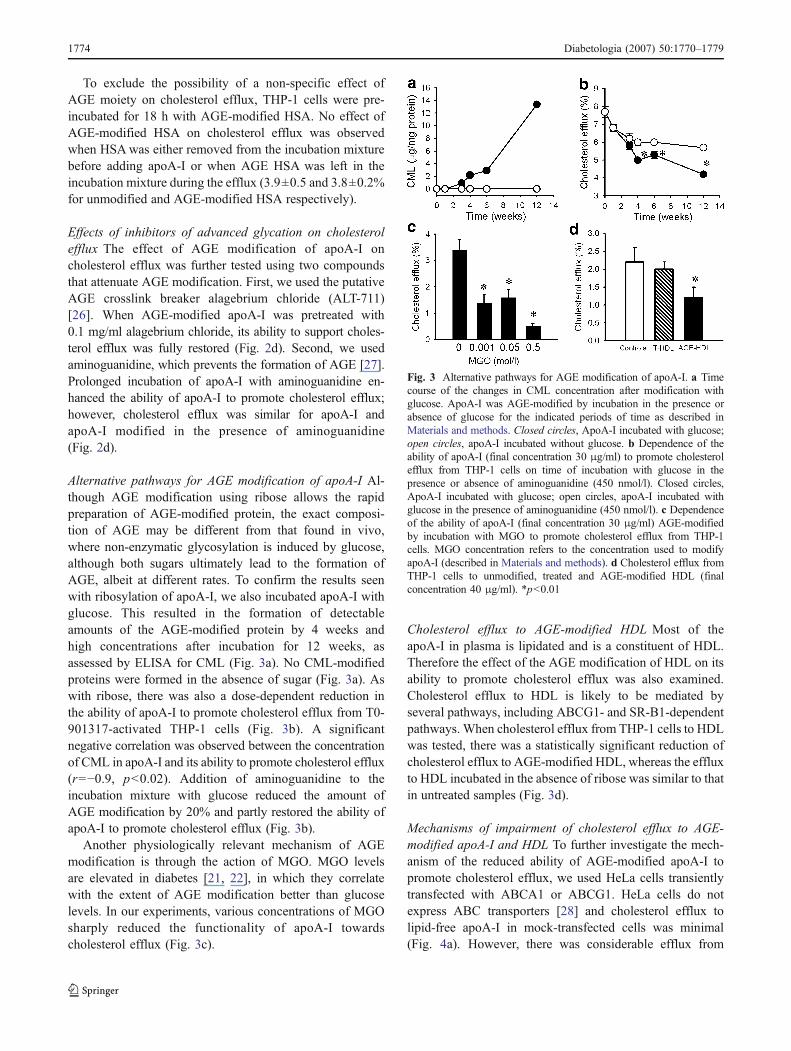
Alternative pathways for AGE modification of apoA-I Al-though AGE modification using ribose allows the rapidpreparation of AGE-modified protein, the exact composi-tion of AGE may be different from that found in vivo,where non-enzymatic glycosylation is induced by glucose,although both sugars ultimately lead to the formation ofAGE, albeit at different rates. To confirm the results seenwith ribosylation of apoA-I, we also incubated apoA-I withglucose. This resulted in the formation of detectableamounts of the AGE-modified protein by 4 weeks andhigh concentrations after incubation for 12 weeks, asassessed by ELISA for CML (Fig. 3a). No CML-modifiedproteins were formed in the absence of sugar (Fig. 3a). Aswith ribose, there was also a dose-dependent reduction inthe ability of apoA-I to promote cholesterol efflux from T0-901317-activated THP-1 cells (Fig. 3b). A significantnegative correlation was observed between the concentrationof CML in apoA-I and its ability to promote cholesterol efflux(r=−0.9, p<0.02). Addition of aminoguanidine to theincubation mixture with glucose reduced the amount ofAGE modification by 20% and partly restored the ability ofapoA-I to promote cholesterol efflux (Fig. 3b).
Another physiologically relevant mechanism of AGEmodification is through the action of MGO. MGO levelsare elevated in diabetes [21, 22], in which they correlatewith the extent of AGE modification better than glucoselevels. In our experiments, various concentrations of MGOsharply reduced the functionality of apoA-I towardscholesterol efflux (Fig. 3c).
Cholesterol efflux to AGE-modified HDL Most of theapoA-I in plasma is lipidated and is a constituent of HDL.Therefore the effect of the AGE modification of HDL on itsability to promote cholesterol efflux was also examined.Cholesterol efflux to HDL is likely to be mediated byseveral pathways, including ABCG1- and SR-B1-dependentpathways. When cholesterol efflux from THP-1 cells to HDLwas tested, there was a statistically significant reduction ofcholesterol efflux to AGE-modified HDL, whereas the effluxto HDL incubated in the absence of ribose was similar to thatin untreated samples (Fig. 3d).
Mechanisms of impairment of cholesterol efflux to AGE-modified apoA-I and HDL To further investigate the mech-anism of the reduced ability of AGE-modified apoA-I topromote cholesterol efflux, we used HeLa cells transientlytransfected with ABCA1 or ABCG1. HeLa cells do notexpress ABC transporters [28] and cholesterol efflux tolipid-free apoA-I in mock-transfected cells was minimal(Fig. 4a). However, there was considerable efflux from
Fig. 3 Alternative pathways for AGE modification of apoA-I. a Timecourse of the changes in CML concentration after modification withglucose. ApoA-I was AGE-modified by incubation in the presence orabsence of glucose for the indicated periods of time as described inMaterials and methods. Closed circles, ApoA-I incubated with glucose;open circles, apoA-I incubated without glucose. b Dependence of theability of apoA-I (final concentration 30 μg/ml) to promote cholesterolefflux from THP-1 cells on time of incubation with glucose in thepresence or absence of aminoguanidine (450 nmol/l). Closed circles,ApoA-I incubated with glucose; open circles, apoA-I incubated withglucose in the presence of aminoguanidine (450 nmol/l). c Dependenceof the ability of apoA-I (final concentration 30 μg/ml) AGE-modifiedby incubation with MGO to promote cholesterol efflux from THP-1cells. MGO concentration refers to the concentration used to modifyapoA-I (described in Materials and methods). d Cholesterol efflux fromTHP-1 cells to unmodified, treated and AGE-modified HDL (finalconcentration 40 μg/ml). *p<0.01
1774 Diabetologia (2007) 50:1770–1779

HeLa cells to both rHDL (apoA-I/POPC complex) andnative HDL. The efflux from HeLa cells to rHDL assembledwith AGE-modified apoA-I was 25% lower than rHDLcontaining unmodified apoA-I (Fig. 4a). The efflux to AGE-modified HDL was fourfold lower than unmodified HDL(Fig. 4a). Transfection of HeLa cells with ABCA1dramatically increased the efflux to apoA-I, but not toAGE-modified apoA-I (Fig. 4b). Furthermore, there was nodifference in cholesterol efflux to apoA-I in ABCG1-transfected cells (Fig. 4c), indicating that advanced glyca-tion of apoA-I specifically affects ABCA1-dependentcholesterol efflux. Transfection of HeLa cells with ABCG1elevated cholesterol efflux to HDL, but not to rHDL(Fig. 4c). There was 40% reduction of cholesterol effluxto AGE-modified HDL compared with unmodified HDLand 10% reduction in the efflux to AGE-rHDL comparedwith unmodified rHDL (Fig. 4c). These findings indicatethat advanced glycation of HDL affects both the ABCG1-dependent and ABCG1-independent pathways. The experi-ments with HDL were only done with HDL modified byribose, as long incubations of HDL with glucose resulted indisintegration of HDL particles independently of thepresence of glucose, precluding use of this method to studyAGE modification of HDL.
Intracellular cholesterol content Differences in the rate ofcholesterol efflux are likely to be reflected in the steady-state concentration of intracellular cholesterol. A sensitivemeasure of cellular cholesterol content is the rate ofcholesteryl ester synthesis [29]. We measured the rate ofcholesterol ester synthesis in THP-1 cells before and afterloading the cells with cholesterol by incubation withAcLDL. As expected, loading cells with cholesterolincreased the rate of cholesteryl ester synthesis, whereasaddition of apoA-I to the incubation mixture decreased it(Fig. 5). AGE-modified apoA-I was ineffective in decreas-ing the rate of cholesteryl ester synthesis in unloaded cells
and less effective in cholesterol-loaded cells compared withnative apoA-I (Fig. 5).
AGE modification of apoA-I and ABCA1 stability Thecontribution of apoA-I to RCT is not limited to it being acholesterol acceptor. Another important function of apoA-Iis its ability to enhance ABCA1 abundance by inhibitingcalpain-dependent degradation of ABCA1 [30, 31]. To testwhether this function of apoA-I is affected by AGEmodification, we boosted ABCA1 expression in differenti-ated THP-1 cells using the LXR agonist T0-901317. Theagonist was then removed and cells were incubated for 18 hin the presence or absence of apoA-I or AGE-apoA-I inserum-free media. The abundance of ABCA1 was mea-sured by western blot followed by densitometry and relatedto the abundance of β-actin. Most of the ABCA1 dis-appeared after 18 h incubation in the absence of apoA-I
Fig. 5 Effect of apoA-I and AGE-modified apoA-I on the rate ofcholesterol esterification. THP-1 cells were incubated for 24 h in theabsence (left) or presence (right) of AcLDL (50 μg/ml). The rate ofcholesteryl ester synthesis was then measured as described inMaterials and methods in the absence (open bars) or presence ofapoA-I (hatched bars) or AGE-modified apoA-I (closed bars; finalconcentration 30 μg/ml). *p<0.01 vs no apoA-I
Fig. 4 Cholesterol efflux from HeLa cells mock-transfected (a) ortransfected with ABCA1 (b) or ABCG1 (c). HeLa cells weretransfected with pCMV empty plasmid (a), ABCA1 (b) or ABCG1(c) as described in Materials and methods. Cholesterol efflux assay to
apoA-I or AGE-apoA-I, rHDL or AGE-rHDL and HDL and AGE-HDL was performed as described in Materials and methods. Openbars, ApoA-I, rHDL or HDL; closed bars, AGE-modified apoA-I,rHDL or HDL. *p<0.01
Diabetologia (2007) 50:1770–1779 1775
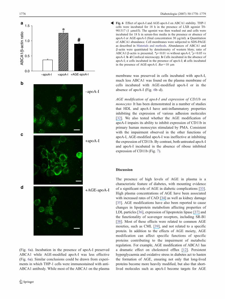
(Fig. 6a). Incubation in the presence of apoA-I preservedABCA1 while AGE-modified apoA-I was less effective(Fig. 6a). Similar conclusions could be drawn from experi-ments in which THP-1 cells were immunostained with anti-ABCA1 antibody. While most of the ABCA1 on the plasma
membrane was preserved in cells incubated with apoA-I,much less ABCA1 was found on the plasma membrane ofcells incubated with AGE-modified apoA-I or in theabsence of apoA-I (Fig. 6b–d).
AGE modification of apoA-I and expression of CD11b onmonocytes It has been demonstrated in a number of studiesthat HDL and apoA-I have anti-inflammatory propertiesinhibiting the expression of various adhesion molecules[32]. We also tested whether the AGE modification ofapoA-I impairs its ability to inhibit expression of CD11b inprimary human monocytes stimulated by PMA. Consistentwith the impairment observed in the other functions ofapoA-I, AGE-modified apoA-I was ineffective at inhibitingthe expression of CD11b. By contrast, both untreated apoA-Iand apoA-I incubated in the absence of ribose inhibitedexpression of CD11b (Fig. 7).
Discussion
The presence of high levels of AGE in plasma is acharacteristic feature of diabetes, with mounting evidenceof a significant role of AGE in diabetic complications [33].High plasma concentrations of AGE have been associatedwith increased rates of CAD [34] as well as kidney damage[35]. AGE modifications have also been reported to causechanges in lipoprotein metabolism affecting properties ofLDL particles [36], expression of lipoprotein lipase [37] andthe functionality of scavenger receptors, including SR-B1[38]. Most of these effects were related to common AGEmoieties, such as CML [39], and not related to a specificprotein. In addition to the effects of AGE moiety, AGEmodification can affect specific functions of specificproteins contributing to the impairment of metabolicregulation. For example, AGE modification of ABCA1 hasa dramatic effect on cholesterol efflux [12]. Persistenthyperglycaemia and oxidative stress in diabetes act to hastenthe formation of AGE, ensuring not only that long-livedproteins become more heavily modified, but also that short-lived molecules such as apoA-I become targets for AGE
b
–apoA-I
+apoA-I
+AGE-apoA-I
a
d
c
–apoA-I
AB
CA
1/β-
actin
rat
io
0.0
0.5
1.0
1.5
*
#
+apoA-I +AGE-apoA-I
Fig. 6 Effect of apoA-I and AGE-apoA-I on ABCA1 stability. THP-1cells were incubated for 18 h in the presence of LXR agonist T0-901317 (1 μmol/l). The agonist was then washed out and cells wereincubated for 18 h in serum-free media in the presence or absence ofapoA-I or AGE-apoA-I (final concentration 30 μg/ml). a Quantitationof ABCA1 abundance. Cell membranes were subjected to SDS-PAGEas described in Materials and methods. Abundances of ABCA1 andβ-actin were quantitated by densitometry of western blots; ratio ofABCA1/β-actin is presented. *p<0.01 vs without apoA-I; #p<0.05 vsapoA-I. b–d Confocal microscopy. b Cells incubated in the absence ofapoA-I; c cells incubated in the presence of apoA-I; d cells incubatedin the presence of AGE-apoA-I. Bar=10 μm
R
1776 Diabetologia (2007) 50:1770–1779

modification [40]. In addition to their role in RCT, apoA-Iand HDL have other anti-atherogenic functions, includinganti-inflammatory [32], antioxidant [41] and antithromboticactions [42]. Hence, AGE modification of apoA-I may havewide-ranging implications for the development of athero-sclerosis, particularly in the setting of diabetes. In this studywe investigated the effect of AGE modification of apoA-I onseveral of its anti-atherogenic functions.
The main finding of this study is that a number of theknown anti-atherogenic functions of apoA-I were impairedby a physiologically relevant level of AGE modification ofthis specific protein. These functions include cholesterolefflux. The cholesterol efflux pathway affected was likelyto be mainly, but not exclusively, the ABCA1-dependentpathway. This conclusion is supported by the greaterinhibition of cholesterol efflux to AGE-apoA-I comparedwith AGE–HDL, concomitant inhibition of phospholipidefflux and almost complete inhibition of cholesterol effluxto AGE-apoA-I from ABCA1-transfected HeLa cells,where ABCA1-dependent cholesterol efflux is the onlypathway for removing cholesterol to apoA-I. However,inhibition of cholesterol efflux to AGE–HDL indicates thatthe other cholesterol efflux pathways, including theABCG1-dependent pathway, are also affected. The changesin cholesterol efflux were specific for AGE-modification of
apoA-I as AGE-HSA had no effect on cholesterol efflux.The reduced ability of AGE-apoA-I to support cholesterolefflux was reflected in its reduced capacity to decrease cellcholesterol content as assessed by the rate of cholesterolesterification. The impairment of cholesterol efflux wasreversed by preventing or reversing AGE modification ofapoA-I. Interestingly, simple glycation of apoA-I does notseem to affect its ability to promote cholesterol efflux [14],emphasising that further modification with the formation ofAGE is necessary for impairment of apoA-I function. Ourfinding is consistent with that of Brubaker et al. [43]showing that reductive methylation of lysine residuesimpairs the ability of apoA-I to support cholesterol efflux,and with that of Duell et al. [16] showing that non-enzymatic glycosylation of HDL impairs its function. Thus,AGE-modification of apoA-I and HDL results in asignificant loss of its ability to support cholesterol efflux,which can be reversed by either inhibiting or reversingAGE-modification. These findings provide a rationale fortargeting AGE as part of the management of diabeticdyslipidaemia and cardioprotection.
Other functions of apoA-I impaired by AGE-modificationincluded its ability to stabilise ABCA1 [31] and to inhibitexpression of adhesion molecules [32]. The former furtherimpairs RCT, and the latter is a reflection of the anti-inflammatory function of apoA-I. Inflammation is animportant element in the pathogenesis of atherosclerosisand the anti-inflammatory properties of apoA-I are likely tocontribute to its overall anti-atherosclerotic effect [44].
The clinical relevance of AGE modification to thefunctions of apoA-I in vivo remains to be tested. Althoughthe concentration of CML found in the in vitro-modifiedapoA-I used in this study was similar to that found in vivo,it is conceivable that other AGE-modifications maycontribute to protein dysfunction, and may be differentfrom that observed in vivo. Moreover, AGE modification isnot the only apoA-I modification observed in diabetes thatpotentially contributes to impaired apoA-I functions. Inparticular, nitration and chlorination of apoA-I also occur indiabetes, damaging apoA-I functionality [13, 45, 46]. Whilethe relative contribution of AGE modification to the overalleffect of diabetes on apoA-I dysfunction is yet to beestablished, it is clear that interventions to reduce AGElevels in diabetes are anti-atherosclerotic [47].
In conclusion, we demonstrated that AGE modificationof apoA-I and HDL results in a significant loss of theirability to support cholesterol efflux, stabilise ABCA1 andinhibit the expression of CD11b. These changes appear tobe specific to the AGE modification of apoA-I and can beattenuated by preventing AGE modification. The impairedfunctioning of AGE-modified apoA-I may contributesignificantly to the increased risk of atherosclerosis associ-ated with diabetes.
Fig. 7 Effect of apoA-I and AGE apoA-I on CD11b expression onhuman monocytes. Resting human monocytes were resuspended at106 cells/ml and 100 μl of this suspension was stimulated with PMA(final concentration 100 ng/ml) in the presence or absence of apoA-I,treated apoA-I (T-apoA-I) or AGE-apoA-I (40 μg/ml). Cells wereincubated with the FITC-conjugated antibody against CD11b for 15 minat 37°C and fixed with 4% paraformaldehyde. CD11b expression wasmeasured as fluorescence intensity using flow cytometry. Values shownare percentage increases in the expression of CD11b over non-stimulatedmonocytes. Means±SEM for four independent experiments are shown.*p<0.01 vs PMA alone; #p<0.05 vs PMA+apoA-I
Diabetologia (2007) 50:1770–1779 1777

Acknowledgements We are grateful to A. Remaley for the ABCA1and ABCG1 plasmids. This study was supported by a grant from theDiabetes Australia Research Trust (D. Sviridov, R. O’Brien) and fromthe Juvenile Diabetes Research Foundation (J. M. Forbes, M. T.Coughlan, M. C. Thomas). D. Sviridov and J. P. F. Chin-Dusting arefellows of the National Health and Medical Research Council ofAustralia.
Duality of interest The authors declare that there is no duality ofinterest associated with this manuscript.
References
1. Nathan DM, Cleary PA, Backlund JY et al (2005) Intensivediabetes treatment and cardiovascular disease in patients withtype 1 diabetes. N Engl J Med 353:2643–2653
2. Haffner SM, Lehto S, Ronnemaa T, Pyorala K, Laakso M (1998)Mortality from coronary heart disease in subjects with type 2diabetes and in nondiabetic subjects with and without priormyocardial infarction. N Engl J Med 339:229–234
3. Beckman JA, Creager MA, Libby P (2002) Diabetes andatherosclerosis: epidemiology, pathophysiology, and management.JAMA 287:2570–2581
4. Goldberg IJ (2004) Why does diabetes increase atherosclerosis? Idon’t know! J Clin Invest 114:613–615
5. Renard CB, Kramer F, Johansson F et al (2004) Diabetes anddiabetes-associated lipid abnormalities have distinct effects oninitiation and progression of atherosclerotic lesions. J Clin Invest114:659–668
6. Kanter JE, Johansson F, LeBoeuf RC, Bornfeldt KE (2007) Doglucose and lipids exert independent effects on atheroscleroticlesion initiation or progression to advanced plaques? Circ Res100:769–781
7. Berthezene F (1996) Non-insulin dependent diabetes and reversecholesterol transport. Atherosclerosis 124 (Suppl):S39–S42
8. Brewer HB Jr, Remaley AT, Neufeld EB, Basso F, Joyce C (2004)Regulation of plasma high-density lipoprotein levels by theABCA1 transporter and the emerging role of high-densitylipoprotein in the treatment of cardiovascular disease. ArteriosclerThromb Vasc Biol 24:1755–1760
9. Oram JF, Lawn RM (2001) ABCA1: the gatekeeper foreliminating excess tissue cholesterol. J Lipid Res 42:1173–1179
10. Wang Y, Oram JF (2002) Unsaturated fatty acids inhibit cholesterolefflux from macrophages by increasing degradation of ATP-binding cassette transporter A1. J Biol Chem 277:5692–5697
11. Albrecht C, Simon-Vermot I, Elliott JIJ, Higgins CF, JohnstonDG, Valabhji J (2004) Leukocyte ABCA1 gene expression isassociated with fasting glucose concentration in normoglycemicmen. Metabolism 53:17–21
12. Passarelli M, Tang C, McDonald TO et al (2005) Advancedglycation end product precursors impair ABCA1-dependentcholesterol removal from cells. Diabetes 54:2198–2205
13. Hermo R, Mier C, Mazzotta M, Tsuji M, Kimura S, Gugliucci A(2005) Circulating levels of nitrated apolipoprotein A-I areincreased in type 2 diabetic patients. Clin Chem Lab Med43:601–606
14. Rashduni DL, Rifici VA, Schneider SH, Khachadurian AK (1999)Glycation of high-density lipoprotein does not increase itssusceptibility to oxidation or diminish its cholesterol effluxcapacity. Metabolism 48:139–143
15. Duell PB, Oram JF, Bierman EL (1990) Nonenzymatic glycosyl-ation of HDL resulting in inhibition of high-affinity binding tocultured human fibroblasts. Diabetes 39:1257–1263
16. Duell PB, Oram JF, Bierman EL (1991) Nonenzymatic glycosyl-ation of HDL and impaired HDL-receptor-mediated cholesterolefflux. Diabetes 40:377–384
17. Escher G, Hoang A, Georges S et al (2005) Demethylation usingthe epigenetic modifier, 5-azacytidine, increases the efficiency oftransient transfection of macrophages. J Lipid Res 46:356–365
18. Woollard KJ, Phillips DC, Griffiths HR (2002) Direct modulatoryeffect of C-reactive protein on primary human monocyte adhesionto human endothelial cells. Clin Exp Immunol 130:256–262
19. Morrison JR, Fidge NH, Grego B (1990) Studies on the formation,separation, and characterization of cyanogen bromide fragments ofhuman AI apolipoprotein. Anal Biochem 186:145–152
20. Sviridov D, Pyle L, Fidge N (1996) Efflux of cellular cholesteroland phospholipid to apolipoprotein A-I mutants. J Biol Chem271:33277–33283
21. Baynes JW, Thorpe SR (1999) Role of oxidative stress in diabeticcomplications: a new perspective on an old paradigm. Diabetes 48:1–9
22. McLellan AC, Thornalley PJ, Benn J, Sonksen PH (1994)Glyoxalase system in clinical diabetes mellitus and correlationwith diabetic complications. Clin Sci (Lond) 87:21–29
23. Sviridov D, Hoang A, Huang W, Sasaki J (2002) Structure-function studies of apoA-I variants: site-directed mutagenesis andnatural mutations. J Lipid Res 43:1283–1292
24. Sviridov D, Fidge N (1995) Efflux of intracellular vs plasmamembrane cholesterol in HepG2 cells: different availability andregulation by apolipoprotein A-I. J Lipid Res 36:1887–1896
25. Yamauchi Y, Abe-Dohmae S, Yokoyama S (2002) Differentialregulation of apolipoprotein A-I/ATP binding cassette transporterA1-mediated cholesterol and phospholipid release. BiochimBiophys Acta 1585:1–10
26. Forbes JM, Thallas V, Thomas MC et al (2003) The breakdown ofpreexisting advanced glycation end products is associated withreduced renal fibrosis in experimental diabetes. FASEB J17:1762–1764
27. Brownlee M, Vlassara H, Kooney A, Ulrich P, Cerami A (1986)Aminoguanidine prevents diabetes-induced arterial wall proteincross-linking. Science 232:1629–1632
28. Remaley AT, Stonik JA, Demosky SJ et al (2001) Apolipoproteinspecificity for lipid efflux by the human ABCAI transporter.Biochem Biophys Res Commun 280:818–823
29. Chirinos JA, Zambrano JP, Chakko S et al (2005) Ability of serumto decrease cellular AcylCoA:cholesterol acyl transferase activitypredicts cardiovascular outcomes. Circulation 112:2446–2453
30. Wang N, Chen W, Linsel-Nitschke P et al (2003) A PESTsequence in ABCA1 regulates degradation by calpain proteaseand stabilization of ABCA1 by apoA-I. J Clin Invest 111:99–107
31. Martinez LO, Agerholm-Larsen B, Wang N, Chen W, Tall AR(2003) Phosphorylation of a pest sequence in ABCA1 promotescalpain degradation and is reversed by ApoA-I. J Biol Chem278:37368–37374
32. Barter PJ, Nicholls S, Rye K-A, Anantharamaiah GM, Navab M,Fogelman AM (2004) Antiinflammatory properties of HDL. CircRes 95:764–772
33. Forbes JM, Thallas-Bonke V, Cooper ME, Thomas MC (2004)Advanced glycation: how are we progressing to combat this web ofsugar anomalies in diabetic nephropathy. Curr PharmDes 10:3361–3372
34. Kilhovd BK, Juutilainen A, Lehto S et al (2005) High serumlevels of advanced glycation end products predict increasedcoronary heart disease mortality in nondiabetic women but notin nondiabetic men: a population-based 18-year follow-up study.Arterioscler Thromb Vasc Biol 25:815–820
35. Thomas MC, Tsalamandris C, MacIsaac R et al (2004) Low-molecular-weight AGEs are associated with GFR and anemia inpatients with type 2 diabetes. Kidney Int 66:1167–1172
36. Cai W, He JC, Zhu L et al (2004) High levels of dietary advancedglycation end products transform low-density lipoprotein into a
1778 Diabetologia (2007) 50:1770–1779
potent redox-sensitive mitogen-activated protein kinase stimulantin diabetic patients. Circulation 110:285–291
37. Beauchamp M-C, Michaud S-E, Li L, Sartippour MR, Renier G(2004) Advanced glycation end products potentiate the stimula-tory effect of glucose on macrophage lipoprotein lipase expres-sion. J Lipid Res 45:1749–1757
38. Ohgami N, Nagai R, Miyazaki A et al (2001) Scavenger receptorclass B type I-mediated reverse cholesterol transport is inhibited byadvanced glycation end products. J Biol Chem 276:13348–13355
39. Horiuchi S, Araki N, Morino Y (1991) Immunochemical approachto characterize advanced glycation end products of the Maillardreaction. Evidence for the presence of a common structure. J BiolChem 266:7329–7332
40. Brownlee M (2001) Biochemistry and molecular cell biology ofdiabetic complications. Nature 414:813–820
41. Garner B, Witting PK, Waldeck AR, Christison JK, Raftery M,Stocker R (1998) Oxidation of high density lipoproteins. I.Formation of methionine sulfoxide in apolipoproteins AI andAII is an early event that accompanies lipid peroxidation and canbe enhanced by α-tocopherol. J Biol Chem 273:6080–6087
42. Nofer J-R, Walter M, Kehrel B et al (1998) HDL(3)-mediatedinhibition of thrombin-induced platelet aggregation and fibrinogen
binding occurs via decreased production of phosphoinositide-derived second messengers 1,2-diacylglycerol and inositol 1,4,5-tris-phosphate. Arterioscler Thromb Vasc Biol 18:861–869
43. Brubaker G, Peng DQ, Somerlot B, Abdollahian DJ, Smith JD(2006) Apolipoprotein A-I lysine modification: effects on helicalcontent, lipid binding and cholesterol acceptor activity. BiochimBiophys Acta 1761:64–72
44. Moore RE, Navab M, Millar JS et al (2005) Increasedatherosclerosis in mice lacking apolipoprotein A-I attributable toboth impaired reverse cholesterol transport and increased inflam-mation. Circ Res 97:763–771
45. Shao B, Bergt C, Fu X et al (2005) Tyrosine 192 in apolipoproteinA-I is the major site of nitration and chlorination by myeloper-oxidase, but only chlorination markedly impairs ABCA1-dependentcholesterol transport. J Biol Chem 280:5983–5993
46. Shao B, Oda MN, Bergt C et al (2006) Myeloperoxidase impairsABCA1-dependent cholesterol efflux through methionine oxida-tion and site-specific tyrosine chlorination of apolipoprotein A-I.J Biol Chem 281:9001–9004
47. Forbes JM, Yee LT, Thallas V et al (2004) Advanced glycationend product interventions reduce diabetes-accelerated atheroscle-rosis. Diabetes 53:1813–1823
Diabetologia (2007) 50:1770–1779 1779
Copyright © 2022 FDOKUMEN