The estimation of patient-specific cardiac diastolic functions from clinical measurements
Upload
independentCategory
view
3download
0
Diastolic Dysfunction of Aging Is Independent ofMyocardial Structure but Associated with PlasmaAdvanced Glycation End-Product LevelsDuncan J. Campbell1,2*, Jithendra B. Somaratne3, Alicia J. Jenkins2, David L. Prior2,3, Michael Yii4,5,
James F. Kenny5, Andrew E. Newcomb4,5, Casper G. Schalkwijk6, Mary Jane Black7, Darren J. Kelly2
1 St. Vincent’s Institute of Medical Research, Fitzroy, Australia, 2 Department of Medicine, The University of Melbourne, St. Vincent’s Health, Fitzroy, Australia,
3 Department of Cardiology, St. Vincent’s Health, Fitzroy, Australia, 4 Department of Surgery, University of Melbourne, St. Vincent’s Health, Fitzroy, Australia, 5 Department
of Cardiothoracic Surgery, St. Vincent’s Health, Fitzroy, Australia, 6 Department of Internal Medicine, University of Maastricht, Maastricht, The Netherlands, 7 Department
of Anatomy and Developmental Biology, Monash University, Clayton, Australia
Abstract
Background: Heart failure is associated with abnormalities of myocardial structure, and plasma levels of the advancedglycation end-product (AGE) Ne-(carboxymethyl)lysine (CML) correlate with the severity and prognosis of heart failure. Agingis associated with diastolic dysfunction and increased risk of heart failure, and we investigated the hypothesis that diastolicdysfunction of aging humans is associated with altered myocardial structure and plasma AGE levels.
Methods: We performed histological analysis of non-ischemic left ventricular myocardial biopsies and measured plasmalevels of the AGEs CML and low molecular weight fluorophores (LMWFs) in 26 men undergoing coronary artery bypass graftsurgery who had transthoracic echocardiography before surgery. None had previous cardiac surgery, myocardial infarction,atrial fibrillation, or heart failure.
Results: The patients were aged 43–78 years and increasing age was associated with echocardiographic indices of diastolicdysfunction, with higher mitral Doppler flow velocity A wave (r = 0.50, P = 0.02), lower mitral E/A wave ratio (r = 0.64,P = 0.001), longer mitral valve deceleration time (r = 0.42, P = 0.03) and lower early diastolic peak velocity of the mitral septalannulus, e’ (r = 0.55, P = 0.008). However, neither mitral E/A ratio nor mitral septal e’ was correlated with myocardial total,interstitial or perivascular fibrosis (picrosirius red), immunostaining for collagens I and III, CML, and receptor for AGEs (RAGE),cardiomyocyte width, capillary length density, diffusion radius or arteriolar dimensions. Plasma AGE levels were notassociated with age. However, plasma CML levels were associated with E/A ratio (r = 0.44, P = 0.04) and e’ (r = 0.51, P = 0.02)and LMWF levels were associated with E/A ratio (r = 0.49, P = 0.02). Moreover, the mitral E/A ratio remained correlated withplasma LMWF levels in all patients (P = 0.04) and the mitral septal e’ remained correlated with plasma CML levels in non-diabetic patients (P = 0.007) when age was a covariate.
Conclusions: Diastolic dysfunction of aging was independent of myocardial structure but was associated with plasma AGElevels.
Citation: Campbell DJ, Somaratne JB, Jenkins AJ, Prior DL, Yii M, et al. (2012) Diastolic Dysfunction of Aging Is Independent of Myocardial Structure butAssociated with Plasma Advanced Glycation End-Product Levels. PLoS ONE 7(11): e49813. doi:10.1371/journal.pone.0049813
Editor: Rajesh Gopalrao Katare, University of Otago, New Zealand
Received June 12, 2012; Accepted October 17, 2012; Published November 26, 2012
Copyright: � 2012 Campbell et al. This is an open-access article distributed under the terms of the Creative Commons Attribution License, which permitsunrestricted use, distribution, and reproduction in any medium, provided the original author and source are credited.
Funding: This work was funded by grants from the National Health and Medical Research Council of Australia and from the National Heart Foundation ofAustralia. D.J.C. (Grant ID 395508) and D.J.K. (Grant ID 566867) are recipients of Senior Research Fellowships from the National Health and Medical ResearchCouncil of Australia. St Vincent’s Institute of Medical Research is supported in part by the Victorian Government’s Operational Infrastructure Support Program. Thefunders had no role in study design, data collection and analysis, decision to publish, or preparation of the manuscript.
Competing Interests: The authors have declared that no competing interests exist.
* E-mail: [email protected]
Introduction
Aging is accompanied by increased vascular and ventricular
stiffness, diastolic dysfunction and an increased risk of heart failure
[1–4]. Heart failure, with either reduced or preserved ejection
fraction, is associated with abnormalities of myocardial structure
and microvasculature including increased fibrosis, cardiomyocyte
hypertrophy and reduced microvascular density [5–9], and animal
models suggest that these abnormalities precede the development
of heart failure in older age. Senescent animals have reduced
cardiomyocyte number, hypertrophy of surviving cardiomyocytes,
increased cardiac fibrosis, reduced capillary density and increased
diffusion radius [10–13]. In addition, advanced glycation end-
products (AGEs) are proposed to contribute to the increased
myocardial stiffening of aging by cross-linking collagen and elastin
and promoting collagen accumulation [14], and by promoting
inflammation and oxidative stress mediated by the receptor for
AGEs (RAGE) [15]. Moreover, plasma AGE levels correlate with
PLOS ONE | www.plosone.org 1 November 2012 | Volume 7 | Issue 11 | e49813
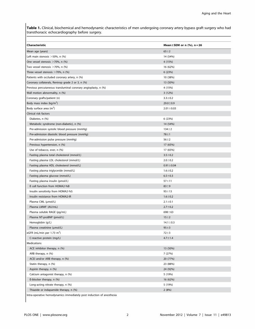
Table 1. Clinical, biochemical and hemodynamic characteristics of men undergoing coronary artery bypass graft surgery who hadtransthoracic echocardiography before surgery.
Characteristic Mean±SEM or n (%), n = 26
Mean age (years) 6562
Left main stenosis .50%, n (%) 14 (54%)
One vessel stenosis .70%, n (%) 4 (15%)
Two vessel stenosis .70%, n (%) 16 (62%)
Three vessel stenosis .70%, n (%) 6 (23%)
Patients with occluded coronary artery, n (%) 10 (38%)
Coronary collaterals, Rentrop grade 2 or 3, n (%) 13 (50%)
Previous percutaneous transluminal coronary angioplasty, n (%) 4 (15%)
Wall motion abnormality, n (%) 3 (12%)
Coronary grafts/patient (n) 3.360.2
Body mass index (kg/m2) 29.060.9
Body surface area (m2) 2.0160.03
Clinical risk factors
Diabetes, n (%) 6 (23%)
Metabolic syndrome (non-diabetic), n (%) 14 (54%)
Pre-admission systolic blood pressure (mmHg) 13462
Pre-admission diastolic blood pressure (mmHg) 7861
Pre-admission pulse pressure (mmHg) 5662
Previous hypertension, n (%) 17 (65%)
Use of tobacco, ever, n (%) 17 (65%)
Fasting plasma total cholesterol (mmol/L) 3.560.2
Fasting plasma LDL cholesterol (mmol/L) 2.060.2
Fasting plasma HDL cholesterol (mmol/L) 0.9160.04
Fasting plasma triglyceride (mmol/L) 1.660.2
Fasting plasma glucose (mmol/L) 6.560.3
Fasting plasma insulin (pmol/L) 57611
ß cell function from HOMA2-%B 8369
Insulin sensitivity from HOMA2-%S 93613
Insulin resistance from HOMA2-IR 1.660.2
Plasma CML (mmol/L) 2.160.1
Plasma LMWF (AU/mL) 2.760.2
Plasma soluble RAGE (pg/mL) 698663
Plasma NT-proBNP (pmol/L) 1562
Hemoglobin (g/L) 14.160.3
Plasma creatinine (mmol/L) 9563
eGFR (mL/min per 1.73 m2) 7263
C-reactive protein (mg/L) 4.761.4
Medications
ACE inhibitor therapy, n (%) 13 (50%)
ARB therapy, n (%) 7 (27%)
ACEI and/or ARB therapy, n (%) 20 (77%)
Statin therapy, n (%) 23 (88%)
Aspirin therapy, n (%) 24 (92%)
Calcium antagonist therapy, n (%) 5 (19%)
ß-blocker therapy, n (%) 16 (62%)
Long-acting nitrate therapy, n (%) 5 (19%)
Thiazide or indapamide therapy, n (%) 2 (8%)
Intra-operative hemodynamics immediately post induction of anesthesia
Aging and the Heart
PLOS ONE | www.plosone.org 2 November 2012 | Volume 7 | Issue 11 | e49813

Table 1. Cont.
Characteristic Mean±SEM or n (%), n = 26
Central venous pressure (mmHg) 8.660.9
Pulmonary capillary wedge pressure (mmHg) 10.360.7
Cardiac index (L/min/m2) 2.760.2
Stroke Volume index (mL/m2) 4762
Coronary collaterals were scored according to Rentrop et al. [58]. ACE, angiotensin converting enzyme; ARB, angiotensin receptor blocker; eGFR, estimated glomerularfiltration rate calculated using the Modification of Diet in Renal Disease study equation [30]; CML, Ne-(carboxymethyl)lysine; HDL, high density lipoprotein; HOMA,Homeostasis Model Assessment calculator version 2.2 [31]; LDL, low density lipoprotein; LMWF, low molecular weight fluorophore; NT-proBNP, amino-terminal-pro-B-type natriuretic peptide; RAGE, receptor for advanced glycation end-products.doi:10.1371/journal.pone.0049813.t001
Figure 1. Correlations between echocardiographic parameters and age in men with coronary artery disease. Age was not correlatedwith mitral Doppler flow velocity E wave velocity (A), but was correlated with mitral Doppler flow velocity A wave velocity (B), E/A ratio (C), mitralearly inflow deceleration time (D) and mitral early diastolic peak velocity of the septal mitral annulus, e’ (E). However, the correlation of age with E/e’ratio was not statistically significant (F).doi:10.1371/journal.pone.0049813.g001
Aging and the Heart
PLOS ONE | www.plosone.org 3 November 2012 | Volume 7 | Issue 11 | e49813
the severity and prognosis of heart failure and predict all-cause and
cardiovascular disease mortality in older adults [16,17]. There is,
however, only limited information about the effects of aging on the
structure and microvasculature of the human myocardium, which
comes mainly from autopsy studies that may have been influenced
by comorbidities [18–20].
We investigated the hypothesis that diastolic dysfunction of
aging humans is associated with altered myocardial structure and
plasma AGE levels. We performed histological analysis of non-
ischemic left ventricular (LV) myocardial biopsies from patients
without heart failure or previous myocardial infarction who were
undergoing coronary artery bypass graft surgery and had
transthoracic echocardiography before surgery. We measured
total, interstitial and perivascular myocardial fibrosis, cardiomy-
ocyte size, capillary length density, diffusion radius and arteriolar
dimensions. We also measured myocardial expression of the AGE
Ne-(carboxymethyl)lysine (CML) and RAGE, and plasma levels of
CML, low molecular weight fluorophores (LMWFs) and soluble
RAGE. Although we obtained LV biopsies from both men and
women, preliminary analysis showed gender-specific differences in
myocardial structure [21]; therefore, given the smaller number of
women recruited to this study, the present analysis was confined to
men. We previously reported that neither diabetes nor the
metabolic syndrome was associated with altered myocardial total
or interstitial fibrosis, cardiomyocyte width, capillary length
density, diffusion radius, arteriolar dimensions or immunostaining
for collagens I and III, CML, or RAGE in this patient population,
although diabetic and metabolic syndrome patients had reduced
perivascular fibrosis [22].
Methods
The St. Vincent’s Health Human Research Ethics Committee
(Fitzroy, Australia) approved the study protocol. Each participant
provided written informed consent to be included in the study.
PatientsDetails of the Cardiac Tissue Bank have been previously
described [21–23]. From the Tissue Bank we selected all of 26
male patients having coronary artery bypass graft surgery alone
who had transthoracic echocardiography before surgery; none had
heart failure or atrial fibrillation, had received loop diuretic
therapy or had evidence of previous myocardial infarction.
Absence of previous myocardial infarction was established from
the clinical history, electrocardiogram and troponin measure-
ments, and was confirmed by inspection of the ventriculogram,
transthoracic and transesophageal echocardiography and exami-
nation of the heart at surgery. All patients had normal or near-
normal LV systolic function as assessed by pre-operative
transthoracic echocardiography and ventriculogram, with LV
ejection fraction $50%. A partial-thickness wedge-shaped biopsy
was taken during surgery, immediately after cardioplegia, from a
region of the lateral wall of the LV near the base of the heart,
between the territories of the left anterior descending and
circumflex arteries, that was free of any macroscopic pathology,
without evidence of ischemia or wall motion abnormality on pre-
operative or intra-operative imaging studies.
The metabolic syndrome was defined according to the
International Diabetes Federation [24]. For patients in whom
abdominal circumference was not measured, based on the
relationship between abdominal circumference and BMI [25],
those with BMI .25 kg/m2 were considered to exceed the
abdominal circumference threshold for the metabolic syndrome. A
patient had diabetes if a history of diabetes was evident from use of
glucose-lowering medications and/or insulin or if fasting plasma
glucose was $7 mmol/L [26]. All 6 diabetic patients had type 2
diabetes; one was newly diagnosed and treated with diet alone, two
were treated with insulin alone, one with insulin and metformin,
and two with metformin and gliclazide. The mean duration of
diabetes was 13 (range 0–30) years and the mean HbA1c was 7.5%
(range 5.3–9.8%, n = 5).
Preoperative Transthoracic Echocardiography and Intra-operative Hemodynamics
Preoperative transthoracic echocardiography was performed
and reported by either the referring institution or by St. Vincent’s
Health according to the American Society of Echocardiography
guidelines [27]. Left ventricular mass was calculated using the
Devereux equation and left ventricular mass index was calculated
by dividing left ventricular mass by height2.7 [28,29]. All patients
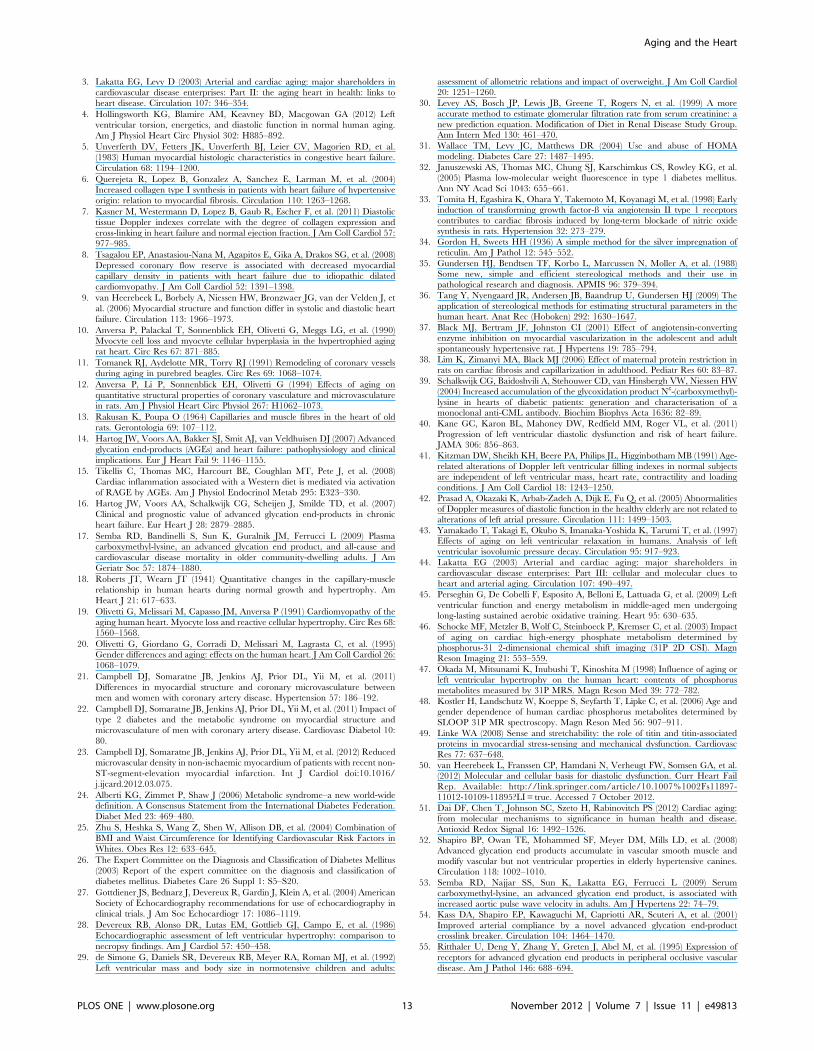
Figure 2. Picrosirius-red staining of collagen, reticulin stainingof cardiomyocyte membranes, and CD31 immunostaining ofcapillaries. Representative sections of left ventricular biopsies fromcoronary artery bypass graft surgery patients stained with picrosirius-red demonstrating interstitial and perivascular fibrosis (stained red) andarteriolar dimensions (A), reticulin stain demonstrating cardiomyocytemembranes (B), and immunostained for CD31 demonstrating capillaries(C).doi:10.1371/journal.pone.0049813.g002
Aging and the Heart
PLOS ONE | www.plosone.org 4 November 2012 | Volume 7 | Issue 11 | e49813

had Swan-Ganz catheters inserted before surgery that provided
measures of pulmonary artery and pulmonary capillary wedge
pressures and cardiac output recorded immediately after induction
of anesthesia.
BiochemistryBlood hemoglobin and hemoglobin A1c and plasma levels of
creatinine were measured as part of the routine pre-surgery
workup. All other variables were measured on fasting blood
collected on the day of surgery, before induction of anesthesia.
Estimated glomerular filtration rate (eGFR) was calculated from
the Modification of Diet in Renal Disease formula [30]. Insulin
resistance (HOMA2-IR), insulin sensitivity (HOMA2-%S) and ß-
cell function (HOMA2-%B) were calculated using the HOMA
calculator version 2.2 [31]. CML was measured by ELISA
(Microcoat, Penzberg, Germany). LMWFs were measured by
fluorescence spectroscopy [32]. Soluble RAGE was measured by
ELISA (R&D Systems Inc., Minneapolis, MN). Amino-terminal-
pro-B-type natriuretic peptide (NT-proBNP) was measured by
electrochemiluminescence immunoassay using an Elecsys instru-
ment (Roche Diagnostics, Basel, Switzerland).
Histological AnalysisDetails of tissue collection, fixation and histology have been
previously described [21–23]. All histological analyses were
performed blind to patient identity and age. Picrosirius red-
Figure 3. Correlations between myocardial fibrosis and echocardiographic parameters of diastolic dysfunction, mitral E/A ratio andmitral septal e’ velocity, in men with coronary artery disease. Myocardial total fibrosis, interstitial fibrosis and perivascular fibrosis were notcorrelated with mitral E/A ratio (A, B, C) or with mitral septal e’ velocity (D, E, F).doi:10.1371/journal.pone.0049813.g003
Aging and the Heart
PLOS ONE | www.plosone.org 5 November 2012 | Volume 7 | Issue 11 | e49813
stained 4 mm sections of paraffin-embedded tissue were analyzed
for total, interstitial and perivascular fibrosis and arteriolar
dimensions by quantitative morphometry of digitized images of
the whole myocardial section (Aperio Technologies, Inc., CA) as
previously described [21–23]. Myocardial total fibrosis was
calculated using the positive pixel count algorithm as the area of
collagen staining expressed as a percentage of the total myocardial
tissue area, after excluding the pericardium, whereas interstitial
fibrosis was calculated as described for total fibrosis, with exclusion
of perivascular fibrosis.
Arterioles were identified by the presence of a layer of media
and immunohistochemical staining for elastin showed the blood
vessels were relaxed. The tissue was immersion fixed and the
arterioles were usually oval in shape because of deformation and/
or because they were cut at an oblique angle. We did not attempt
to analyze arterioles in longitudinal section and only arterioles in
approximate cross- or oblique-section were analyzed for perivas-
cular fibrosis and arteriolar dimensions; these arterioles had
diameters (average of maximum and minimum diameter of each
arteriole) of 12–106 mm. Perivascular fibrosis was calculated as the
ratio of the area of perivascular fibrosis to the total vessel area
(area of vessel wall plus lumen) [33]. Arteriolar wall area/
circumference ratio was measured for arterioles with average
diameters of 20–80 mm, which represented 88% of all arterioles
analyzed.
Cardiomyocyte width, determined on 4 mm sections of paraffin-
embedded tissue (one section per patient) stained for reticulin [34],
was the mean of .100 measurements for each section of the
shortest diameter of cardiomyocyte profiles containing a nucleus.
Capillary length density, which is the length of capillaries per
unit volume of tissue, and diffusion radius were determined by
analysis of 4 mm sections of paraffin-embedded tissue (one section
per patient) immunostained for CD31 (mouse anti-human CD31
monoclonal antibody, Dako Denmark A/S, Glostrup, Denmark)
using standard stereological techniques [35–38], as previously
described [21–23].
Immunohistochemistry for collagens I and III was performed in
frozen sections using mouse monoclonal antibodies ab6308 and
ab6310 (Abcam, Cambridge, UK), respectively. Myocardial total
collagen I and III densities were calculated using the positive pixel
count algorithm (Aperio Technologies, Inc., CA) as the area of
collagen staining expressed as a percentage of the total myocardial
tissue area, after excluding the pericardium. Immunohistochem-
istry for CML was performed in paraffin sections using a mouse
monoclonal antibody as described by Schalkwijk et al. [39].
Immunohistochemistry for RAGE was performed with goat
polyclonal antibody AB5484 (Millipore, Billerica, MA). Immuno-
staining of arteriolar media and intima for CML and of arteriolar
media, intima and capillaries for RAGE was individually scored by
its intensity as 0+, 1+, 2+, or 3+, after inspection of the digitized
image of the whole of each section.
Statistical AnalysisData are shown as means6SEM or n (%). Correlations were
estimated using Pearson correlation coefficients and were consid-
ered significant at P,0.05.
Results
Study PatientsThe clinical, biochemical and hemodynamic characteristics of
the study patients (age range 43–78 years) are shown in Table 1.
Older age was associated with increased plasma NT-proBNP
levels (P = 0.001) and reduced eGFR (P = 0.03). The extent of
coronary artery disease (proportions of patients with left main
stenosis, three-vessel stenosis, occluded coronary arteries, coronary
collaterals and wall motion abnormalities) and number of bypass
grafts performed were unrelated to age. In addition, diabetes,
metabolic syndrome, medication use and hemodynamics, includ-
ing pulmonary capillary wedge pressure, were not associated with
age.
EchocardiographyAge was not associated with LV ejection fraction, left atrial
dimensions, LV mass (not shown) or mitral E wave velocity
(Figure 1). However, increasing age was associated echocardio-
graphic indices of LV diastolic dysfunction, with higher mitral
Doppler flow velocity A wave, lower E/A wave ratio, longer mitral
valve deceleration time, and lower early diastolic peak velocity of
the mitral septal annulus, e’, although the association between age
and E/e’ ratio did not achieve statistical significance (Figure 1).
None of the echocardiographic parameters was associated with the
presence of wall motion abnormalities or with pulse pressure.
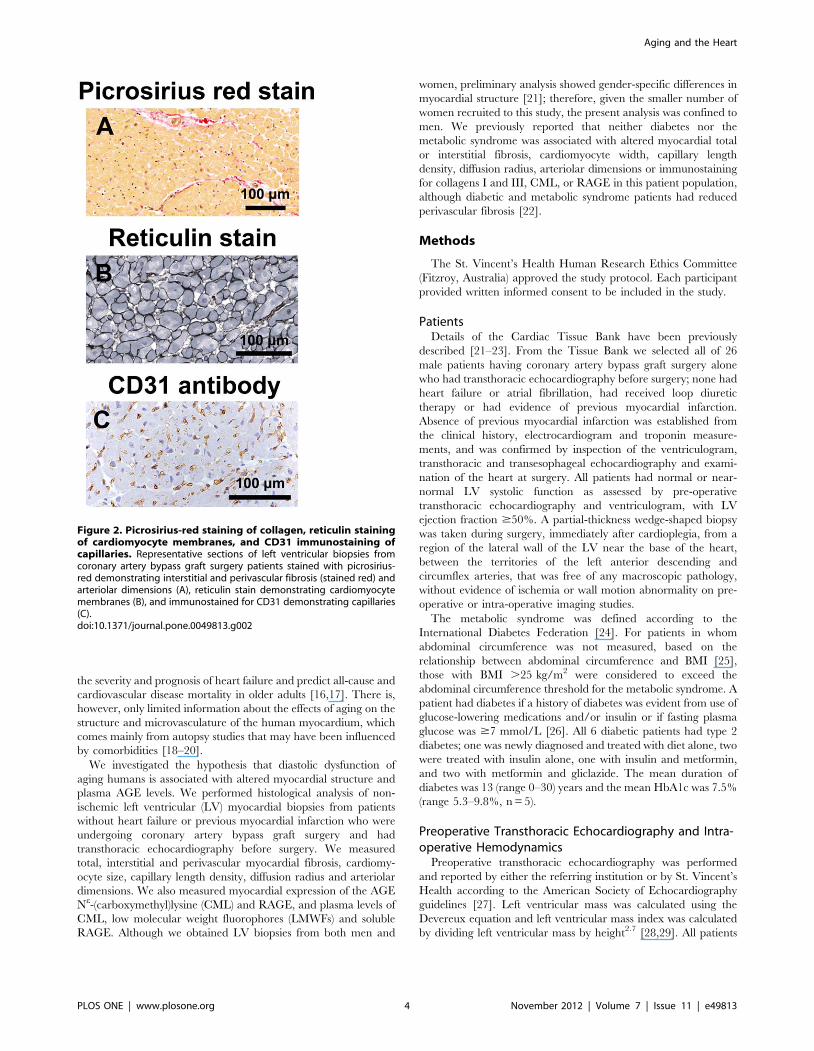
Figure 4. Immunostaining for collagens I and III. Representativesections of a left ventricular biopsy from a coronary artery bypass graftsurgery patient immunostained for collagen I (A), collagen III (B), and anegative control section without primary antibody (C).doi:10.1371/journal.pone.0049813.g004
Aging and the Heart
PLOS ONE | www.plosone.org 6 November 2012 | Volume 7 | Issue 11 | e49813
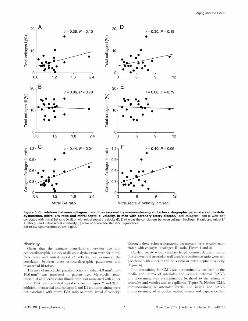
HistologyGiven that the strongest correlations between age and
echocardiographic indices of diastolic dysfunction were for mitral
E/A ratio and mitral septal e9 velocity, we examined the
correlation between these echocardiographic parameters and
myocardial histology.
The area of myocardial paraffin sections (median 4.3 mm2, 1.7–
10.6 mm2) was unrelated to patient age. Myocardial total,
interstitial and perivascular fibrosis were not associated with either
mitral E/A ratio or mitral septal e’ velocity (Figure 2 and 3). In
addition, myocardial total collagen I and III immunostaining were
not associated with mitral E/A ratio or mitral septal e’ velocity,
although these echocardiographic parameters were weakly asso-
ciated with collagen I/collagen III ratio (Figure 4 and 5).
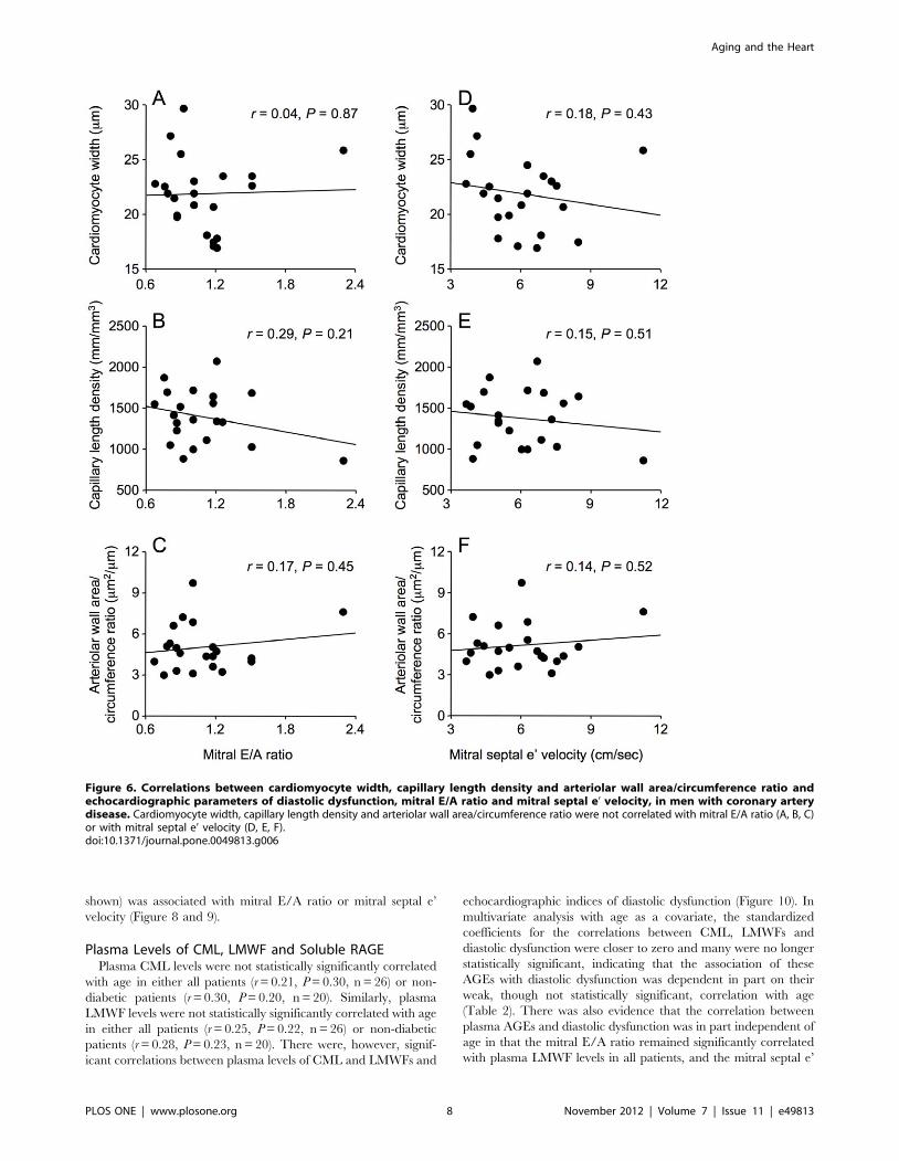
Cardiomyocyte width, capillary length density, diffusion radius
(not shown) and arteriolar wall area/circumference ratio were not
associated with either mitral E/A ratio or mitral septal e’ velocity
(Figure 6).
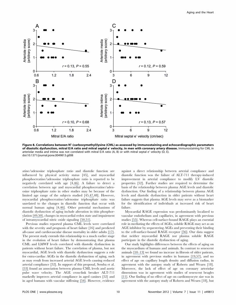
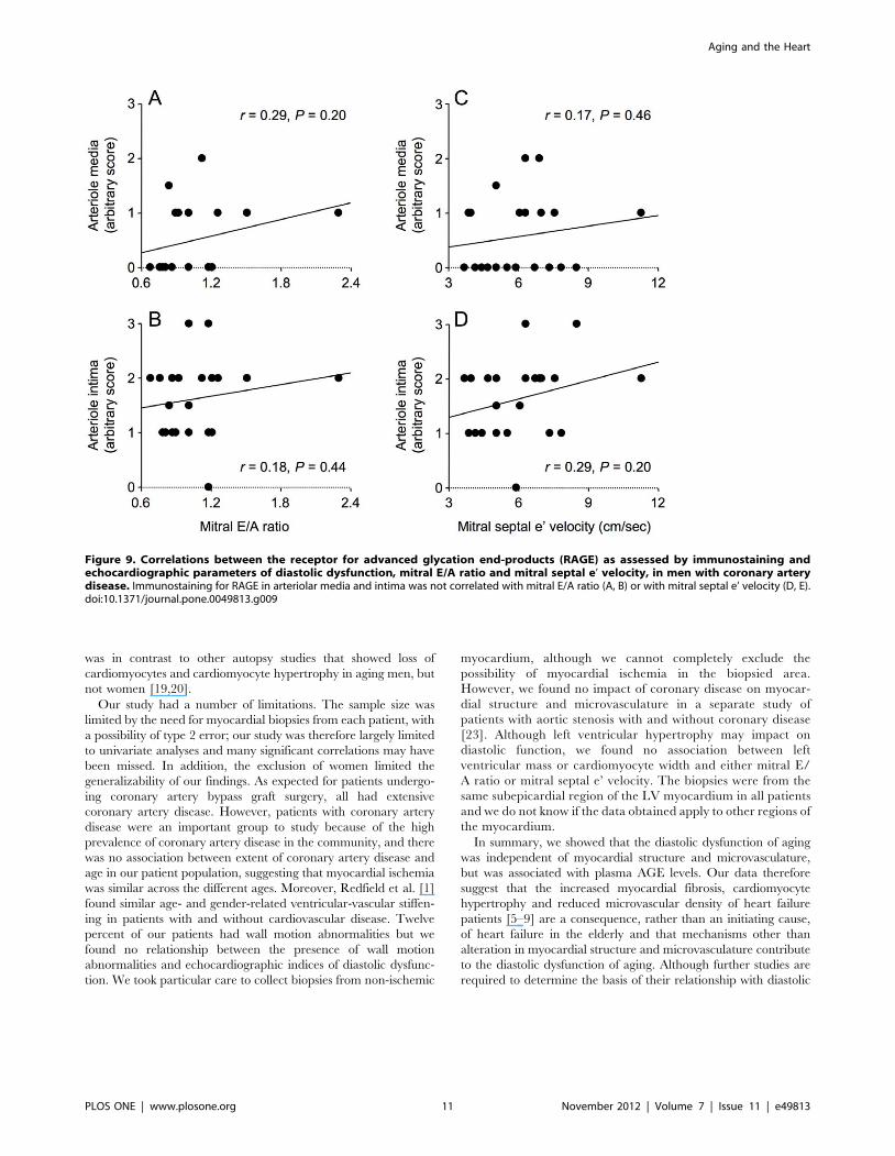
Immunostaining for CML was predominantly localized to the
media and intima of arterioles and venules, whereas RAGE
immunostaining was predominantly localized to the intima of
arterioles and venules and to capillaries (Figure 7). Neither CML
immunostaining of arteriolar media and intima nor RAGE
immunostaining of arteriolar media, intima and capillaries (not
Figure 5. Correlations between collagens I and III as assessed by immunostaining and echocardiographic parameters of diastolicdysfunction, mitral E/A ratio and mitral septal e’ velocity, in men with coronary artery disease. Total collagens I and III were notcorrelated with mitral E/A ratio (A, B) or with mitral septal e’ velocity (D, E) whereas the correlations between collagen I/collagen III ratio and mitral E/A ratio (C) and mitral septal e’ velocity (F) were of borderline statistical significance.doi:10.1371/journal.pone.0049813.g005
Aging and the Heart
PLOS ONE | www.plosone.org 7 November 2012 | Volume 7 | Issue 11 | e49813
shown) was associated with mitral E/A ratio or mitral septal e’
velocity (Figure 8 and 9).
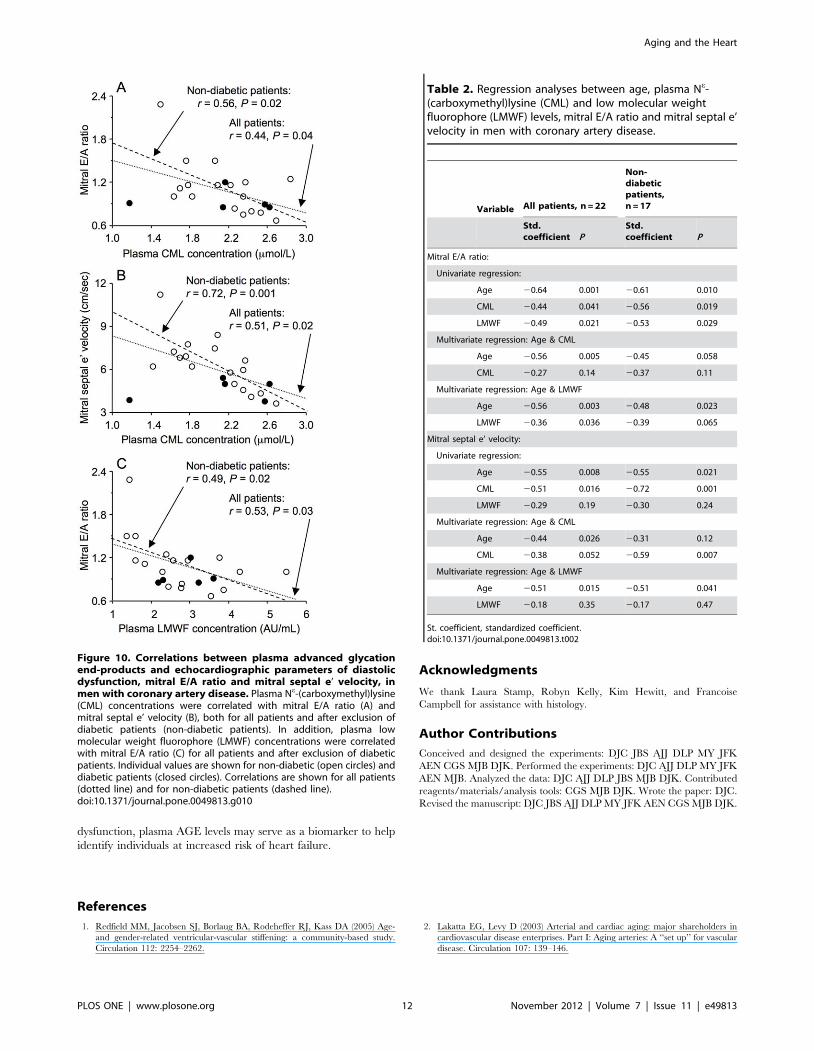
Plasma Levels of CML, LMWF and Soluble RAGEPlasma CML levels were not statistically significantly correlated
with age in either all patients (r = 0.21, P = 0.30, n = 26) or non-
diabetic patients (r = 0.30, P = 0.20, n = 20). Similarly, plasma
LMWF levels were not statistically significantly correlated with age
in either all patients (r = 0.25, P = 0.22, n = 26) or non-diabetic
patients (r = 0.28, P = 0.23, n = 20). There were, however, signif-
icant correlations between plasma levels of CML and LMWFs and
echocardiographic indices of diastolic dysfunction (Figure 10). In
multivariate analysis with age as a covariate, the standardized
coefficients for the correlations between CML, LMWFs and
diastolic dysfunction were closer to zero and many were no longer
statistically significant, indicating that the association of these
AGEs with diastolic dysfunction was dependent in part on their
weak, though not statistically significant, correlation with age
(Table 2). There was also evidence that the correlation between
plasma AGEs and diastolic dysfunction was in part independent of
age in that the mitral E/A ratio remained significantly correlated
with plasma LMWF levels in all patients, and the mitral septal e’
Figure 6. Correlations between cardiomyocyte width, capillary length density and arteriolar wall area/circumference ratio andechocardiographic parameters of diastolic dysfunction, mitral E/A ratio and mitral septal e’ velocity, in men with coronary arterydisease. Cardiomyocyte width, capillary length density and arteriolar wall area/circumference ratio were not correlated with mitral E/A ratio (A, B, C)or with mitral septal e’ velocity (D, E, F).doi:10.1371/journal.pone.0049813.g006
Aging and the Heart
PLOS ONE | www.plosone.org 8 November 2012 | Volume 7 | Issue 11 | e49813

velocity remained significantly correlated with plasma CML levels
in non-diabetic patients when age was a covariate (Table 2).
Plasma levels of soluble RAGE did not correlate with any
echocardiographic parameter or with eGFR or pulse pressure (not
shown).
Discussion
The key finding of this study was that the diastolic dysfunction
of aging men undergoing coronary artery bypass graft surgery was
not associated with myocardial fibrosis or alteration in cardiomy-
ocyte width, capillary length density, diffusion radius, arteriolar
dimensions or myocardial expression of CML and RAGE, but was
associated with plasma levels of CML and LMWFs, and these
associations were dependent, in part, on age. In addition to
diastolic dysfunction, older patients had higher plasma NT-
proBNP levels and lower eGFR, consistent with the effects of
aging. Our patient population included men with type 2 diabetes
and the metabolic syndrome, and in a separate analysis we showed
that neither condition affected myocardial structure, microvascu-
lature, or expression of CML and RAGE, apart from reduced
perivascular fibrosis of diabetic and metabolic syndrome patients
[22]. Although diabetic patients showed evidence of impaired
diastolic function [22], the association between plasma AGE levels
and diastolic dysfunction in the present study remained after
exclusion of diabetic patients. Our findings suggest that the
increased myocardial fibrosis, cardiomyocyte hypertrophy and
reduced microvascular density of heart failure patients [5–9] are a
consequence, rather than an initiating cause, of heart failure in the
elderly and, in addition, that mechanisms other than alteration in
myocardial structure and microvasculature contribute to the
diastolic dysfunction of aging.
Echocardiographic indices of diastolic dysfunction are associat-
ed with an increased risk of heart failure [40]. Our finding that age
was associated with echocardiographic indices of diastolic
dysfunction but not with pulmonary capillary wedge pressure
was in agreement with previous studies reporting that although
associated with alterations with LV filling, age is not associated
with invasive measures of pulmonary capillary wedge pressure or
with LV isovolumic pressure decay [41–43]. Many different
mechanisms other than alteration in myocardial structure and
microvasculature may contribute to alterations in LV filling and
increased heart failure risk of aging individuals [2,3,44], including
alteration in myocardial energetics. Both myocardial phosphocre-
Figure 7. Immunostaining for Ne-(carboxymethyl)lysine (CML) and the receptor for advanced glycation end-products (RAGE).Representative sections of left ventricular biopsies from coronary artery bypass graft surgery patients immunostained for CML (A) or RAGE (C) andtheir corresponding negative control sections (B, D).doi:10.1371/journal.pone.0049813.g007
Aging and the Heart
PLOS ONE | www.plosone.org 9 November 2012 | Volume 7 | Issue 11 | e49813
atine/adenosine triphosphate ratio and diastolic function are
influenced by physical activity status [45], and myocardial
phosphocreatine/adenosine triphosphate ratio is reported to be
negatively correlated with age [4,46]. A failure to detect a
correlation between age and myocardial phosphocreatine/aden-
osine triphosphate ratio in other studies may be because of the
limited age range of the subjects studied [45,47,48]. However,
myocardial phosphocreatine/adenosine triphosphate ratio was
unrelated to the changes in diastolic function that occur with
normal human aging [4,46]. Other potential mechanisms of
diastolic dysfunction of aging include alteration in titin phosphor-
ylation [49,50], changes in myocardial redox state and impairment
of intramyocardial nitric oxide signaling [50,51].
Previous studies reported plasma CML levels were correlated
with the severity and prognosis of heart failure [16] and predicted
all-cause and cardiovascular disease mortality in older adults [17].
The present study extends this relationship to a much earlier stage
in the evolution of heart failure by demonstrating that plasma
CML and LMWF levels correlated with diastolic dysfunction in
patients without heart failure. The correlation of plasma, but not
myocardial, AGE levels with diastolic dysfunction suggests a role
for extra-cardiac AGEs in the diastolic dysfunction of aging, such
as may result from increased arterial AGE levels causing reduced
arterial compliance [52]. In support of this proposal, Semba et al.
[53] found an association between plasma CML levels and aortic
pulse wave velocity. The AGE cross-link breaker ALT-711
markedly improves arterial compliance in aged canines [52] and
in aged humans with vascular stiffening [54]. However, evidence
against a direct relationship between arterial compliance and
diastolic function was the failure of ALT-711 therapy-induced
improvement in arterial compliance to modify LV diastolic
properties [52]. Further studies are required to determine the
basis of the relationship between plasma AGE levels and diastolic
dysfunction. Our finding of a relationship between plasma AGE
levels and diastolic dysfunction in older patients without heart
failure suggests that plasma AGE levels may serve as a biomarker
for the identification of individuals at increased risk of heart
failure.
Myocardial RAGE expression was predominantly localized to
vascular endothelium and capillaries, in agreement with previous
studies [55]. Whereas cell-surface-bound RAGE plays an essential
role in mediating the effects of AGEs, soluble RAGE may act as an
AGE inhibitor by sequestering AGEs and preventing their binding
to the cell-surface-bound RAGE receptor [56]. Our data suggest
that neither myocardial RAGE nor plasma soluble RAGE
participate in the diastolic dysfunction of aging.
Our study highlights differences between the effects of aging on
the myocardium of humans and animals. In contrast to senescent
animals [10–13] we found no increase in fibrosis of older patients,
in agreement with previous studies in humans [19,57], and no
effect of age on capillary length density and diffusion radius, in
agreement with the autopsy study of Roberts and Wearn [18].
Moreover, the lack of effect of age on coronary arteriolar
dimensions was in agreement with studies of senescent beagles
[11]. Our finding of no effect of age on cardiomyocyte size was in
agreement with the autopsy study of Roberts and Wearn [18], but
Figure 8. Correlations between Ne-(carboxymethyl)lysine (CML) as assessed by immunostaining and echocardiographic parametersof diastolic dysfunction, mitral E/A ratio and mitral septal e’ velocity, in men with coronary artery disease. Immunostaining for CML inarteriolar media and intima was not correlated with mitral E/A ratio (A, B) or with mitral septal e’ velocity (D, E).doi:10.1371/journal.pone.0049813.g008
Aging and the Heart
PLOS ONE | www.plosone.org 10 November 2012 | Volume 7 | Issue 11 | e49813
was in contrast to other autopsy studies that showed loss of
cardiomyocytes and cardiomyocyte hypertrophy in aging men, but
not women [19,20].
Our study had a number of limitations. The sample size was
limited by the need for myocardial biopsies from each patient, with
a possibility of type 2 error; our study was therefore largely limited
to univariate analyses and many significant correlations may have
been missed. In addition, the exclusion of women limited the
generalizability of our findings. As expected for patients undergo-
ing coronary artery bypass graft surgery, all had extensive
coronary artery disease. However, patients with coronary artery
disease were an important group to study because of the high
prevalence of coronary artery disease in the community, and there
was no association between extent of coronary artery disease and
age in our patient population, suggesting that myocardial ischemia
was similar across the different ages. Moreover, Redfield et al. [1]
found similar age- and gender-related ventricular-vascular stiffen-
ing in patients with and without cardiovascular disease. Twelve
percent of our patients had wall motion abnormalities but we
found no relationship between the presence of wall motion
abnormalities and echocardiographic indices of diastolic dysfunc-
tion. We took particular care to collect biopsies from non-ischemic
myocardium, although we cannot completely exclude the
possibility of myocardial ischemia in the biopsied area.
However, we found no impact of coronary disease on myocar-
dial structure and microvasculature in a separate study of
patients with aortic stenosis with and without coronary disease
[23]. Although left ventricular hypertrophy may impact on
diastolic function, we found no association between left
ventricular mass or cardiomyocyte width and either mitral E/
A ratio or mitral septal e’ velocity. The biopsies were from the
same subepicardial region of the LV myocardium in all patients
and we do not know if the data obtained apply to other regions of
the myocardium.
In summary, we showed that the diastolic dysfunction of aging
was independent of myocardial structure and microvasculature,
but was associated with plasma AGE levels. Our data therefore
suggest that the increased myocardial fibrosis, cardiomyocyte
hypertrophy and reduced microvascular density of heart failure
patients [5–9] are a consequence, rather than an initiating cause,
of heart failure in the elderly and that mechanisms other than
alteration in myocardial structure and microvasculature contribute
to the diastolic dysfunction of aging. Although further studies are
required to determine the basis of their relationship with diastolic
Figure 9. Correlations between the receptor for advanced glycation end-products (RAGE) as assessed by immunostaining andechocardiographic parameters of diastolic dysfunction, mitral E/A ratio and mitral septal e’ velocity, in men with coronary arterydisease. Immunostaining for RAGE in arteriolar media and intima was not correlated with mitral E/A ratio (A, B) or with mitral septal e’ velocity (D, E).doi:10.1371/journal.pone.0049813.g009
Aging and the Heart
PLOS ONE | www.plosone.org 11 November 2012 | Volume 7 | Issue 11 | e49813
dysfunction, plasma AGE levels may serve as a biomarker to help
identify individuals at increased risk of heart failure.
Acknowledgments
We thank Laura Stamp, Robyn Kelly, Kim Hewitt, and Francoise
Campbell for assistance with histology.
Author Contributions
Conceived and designed the experiments: DJC JBS AJJ DLP MY JFK
AEN CGS MJB DJK. Performed the experiments: DJC AJJ DLP MY JFK
AEN MJB. Analyzed the data: DJC AJJ DLP JBS MJB DJK. Contributed
reagents/materials/analysis tools: CGS MJB DJK. Wrote the paper: DJC.
Revised the manuscript: DJC JBS AJJ DLP MY JFK AEN CGS MJB DJK.
References
1. Redfield MM, Jacobsen SJ, Borlaug BA, Rodeheffer RJ, Kass DA (2005) Age-
and gender-related ventricular-vascular stiffening: a community-based study.
Circulation 112: 2254–2262.
2. Lakatta EG, Levy D (2003) Arterial and cardiac aging: major shareholders in
cardiovascular disease enterprises. Part I: Aging arteries: A ‘‘set up’’ for vascular
disease. Circulation 107: 139–146.
Figure 10. Correlations between plasma advanced glycationend-products and echocardiographic parameters of diastolicdysfunction, mitral E/A ratio and mitral septal e’ velocity, inmen with coronary artery disease. Plasma Ne-(carboxymethyl)lysine(CML) concentrations were correlated with mitral E/A ratio (A) andmitral septal e’ velocity (B), both for all patients and after exclusion ofdiabetic patients (non-diabetic patients). In addition, plasma lowmolecular weight fluorophore (LMWF) concentrations were correlatedwith mitral E/A ratio (C) for all patients and after exclusion of diabeticpatients. Individual values are shown for non-diabetic (open circles) anddiabetic patients (closed circles). Correlations are shown for all patients(dotted line) and for non-diabetic patients (dashed line).doi:10.1371/journal.pone.0049813.g010
Table 2. Regression analyses between age, plasma Ne-(carboxymethyl)lysine (CML) and low molecular weightfluorophore (LMWF) levels, mitral E/A ratio and mitral septal e’velocity in men with coronary artery disease.
Variable All patients, n = 22
Non-diabeticpatients,n = 17
Std.coefficient P
Std.coefficient P
Mitral E/A ratio:
Univariate regression:
Age 20.64 0.001 20.61 0.010
CML 20.44 0.041 20.56 0.019
LMWF 20.49 0.021 20.53 0.029
Multivariate regression: Age & CML
Age 20.56 0.005 20.45 0.058
CML 20.27 0.14 20.37 0.11
Multivariate regression: Age & LMWF
Age 20.56 0.003 20.48 0.023
LMWF 20.36 0.036 20.39 0.065
Mitral septal e’ velocity:
Univariate regression:
Age 20.55 0.008 20.55 0.021
CML 20.51 0.016 20.72 0.001
LMWF 20.29 0.19 20.30 0.24
Multivariate regression: Age & CML
Age 20.44 0.026 20.31 0.12
CML 20.38 0.052 20.59 0.007
Multivariate regression: Age & LMWF
Age 20.51 0.015 20.51 0.041
LMWF 20.18 0.35 20.17 0.47
St. coefficient, standardized coefficient.doi:10.1371/journal.pone.0049813.t002
Aging and the Heart
PLOS ONE | www.plosone.org 12 November 2012 | Volume 7 | Issue 11 | e49813
3. Lakatta EG, Levy D (2003) Arterial and cardiac aging: major shareholders in
cardiovascular disease enterprises: Part II: the aging heart in health: links to
heart disease. Circulation 107: 346–354.
4. Hollingsworth KG, Blamire AM, Keavney BD, Macgowan GA (2012) Left
ventricular torsion, energetics, and diastolic function in normal human aging.
Am J Physiol Heart Circ Physiol 302: H885–892.
5. Unverferth DV, Fetters JK, Unverferth BJ, Leier CV, Magorien RD, et al.
(1983) Human myocardial histologic characteristics in congestive heart failure.
Circulation 68: 1194–1200.
6. Querejeta R, Lopez B, Gonzalez A, Sanchez E, Larman M, et al. (2004)
Increased collagen type I synthesis in patients with heart failure of hypertensive
origin: relation to myocardial fibrosis. Circulation 110: 1263–1268.
7. Kasner M, Westermann D, Lopez B, Gaub R, Escher F, et al. (2011) Diastolic
tissue Doppler indexes correlate with the degree of collagen expression and
cross-linking in heart failure and normal ejection fraction. J Am Coll Cardiol 57:
977–985.
8. Tsagalou EP, Anastasiou-Nana M, Agapitos E, Gika A, Drakos SG, et al. (2008)
Depressed coronary flow reserve is associated with decreased myocardial
capillary density in patients with heart failure due to idiopathic dilated
cardiomyopathy. J Am Coll Cardiol 52: 1391–1398.
9. van Heerebeek L, Borbely A, Niessen HW, Bronzwaer JG, van der Velden J, et
al. (2006) Myocardial structure and function differ in systolic and diastolic heart
failure. Circulation 113: 1966–1973.
10. Anversa P, Palackal T, Sonnenblick EH, Olivetti G, Meggs LG, et al. (1990)
Myocyte cell loss and myocyte cellular hyperplasia in the hypertrophied aging
rat heart. Circ Res 67: 871–885.
11. Tomanek RJ, Aydelotte MR, Torry RJ (1991) Remodeling of coronary vessels
during aging in purebred beagles. Circ Res 69: 1068–1074.
12. Anversa P, Li P, Sonnenblick EH, Olivetti G (1994) Effects of aging on
quantitative structural properties of coronary vasculature and microvasculature
in rats. Am J Physiol Heart Circ Physiol 267: H1062–1073.
13. Rakusan K, Poupa O (1964) Capillaries and muscle fibres in the heart of old
rats. Gerontologia 69: 107–112.
14. Hartog JW, Voors AA, Bakker SJ, Smit AJ, van Veldhuisen DJ (2007) Advanced
glycation end-products (AGEs) and heart failure: pathophysiology and clinical
implications. Eur J Heart Fail 9: 1146–1155.
15. Tikellis C, Thomas MC, Harcourt BE, Coughlan MT, Pete J, et al. (2008)
Cardiac inflammation associated with a Western diet is mediated via activation
of RAGE by AGEs. Am J Physiol Endocrinol Metab 295: E323–330.
16. Hartog JW, Voors AA, Schalkwijk CG, Scheijen J, Smilde TD, et al. (2007)
Clinical and prognostic value of advanced glycation end-products in chronic
heart failure. Eur Heart J 28: 2879–2885.
17. Semba RD, Bandinelli S, Sun K, Guralnik JM, Ferrucci L (2009) Plasma
carboxymethyl-lysine, an advanced glycation end product, and all-cause and
cardiovascular disease mortality in older community-dwelling adults. J Am
Geriatr Soc 57: 1874–1880.
18. Roberts JT, Wearn JT (1941) Quantitative changes in the capillary-muscle
relationship in human hearts during normal growth and hypertrophy. Am
Heart J 21: 617–633.
19. Olivetti G, Melissari M, Capasso JM, Anversa P (1991) Cardiomyopathy of the
aging human heart. Myocyte loss and reactive cellular hypertrophy. Circ Res 68:
1560–1568.
20. Olivetti G, Giordano G, Corradi D, Melissari M, Lagrasta C, et al. (1995)
Gender differences and aging: effects on the human heart. J Am Coll Cardiol 26:
1068–1079.
21. Campbell DJ, Somaratne JB, Jenkins AJ, Prior DL, Yii M, et al. (2011)
Differences in myocardial structure and coronary microvasculature between
men and women with coronary artery disease. Hypertension 57: 186–192.
22. Campbell DJ, Somaratne JB, Jenkins AJ, Prior DL, Yii M, et al. (2011) Impact of
type 2 diabetes and the metabolic syndrome on myocardial structure and
microvasculature of men with coronary artery disease. Cardiovasc Diabetol 10:
80.
23. Campbell DJ, Somaratne JB, Jenkins AJ, Prior DL, Yii M, et al. (2012) Reduced
microvascular density in non-ischaemic myocardium of patients with recent non-
ST-segment-elevation myocardial infarction. Int J Cardiol doi:10.1016/
j.ijcard.2012.03.075.
24. Alberti KG, Zimmet P, Shaw J (2006) Metabolic syndrome–a new world-wide
definition. A Consensus Statement from the International Diabetes Federation.
Diabet Med 23: 469–480.
25. Zhu S, Heshka S, Wang Z, Shen W, Allison DB, et al. (2004) Combination of
BMI and Waist Circumference for Identifying Cardiovascular Risk Factors in
Whites. Obes Res 12: 633–645.
26. The Expert Committee on the Diagnosis and Classification of Diabetes Mellitus
(2003) Report of the expert committee on the diagnosis and classification of
diabetes mellitus. Diabetes Care 26 Suppl 1: S5–S20.
27. Gottdiener JS, Bednarz J, Devereux R, Gardin J, Klein A, et al. (2004) American
Society of Echocardiography recommendations for use of echocardiography in
clinical trials. J Am Soc Echocardiogr 17: 1086–1119.
28. Devereux RB, Alonso DR, Lutas EM, Gottlieb GJ, Campo E, et al. (1986)
Echocardiographic assessment of left ventricular hypertrophy: comparison to
necropsy findings. Am J Cardiol 57: 450–458.
29. de Simone G, Daniels SR, Devereux RB, Meyer RA, Roman MJ, et al. (1992)
Left ventricular mass and body size in normotensive children and adults:
assessment of allometric relations and impact of overweight. J Am Coll Cardiol20: 1251–1260.
30. Levey AS, Bosch JP, Lewis JB, Greene T, Rogers N, et al. (1999) A more
accurate method to estimate glomerular filtration rate from serum creatinine: a
new prediction equation. Modification of Diet in Renal Disease Study Group.Ann Intern Med 130: 461–470.
31. Wallace TM, Levy JC, Matthews DR (2004) Use and abuse of HOMA
modeling. Diabetes Care 27: 1487–1495.
32. Januszewski AS, Thomas MC, Chung SJ, Karschimkus CS, Rowley KG, et al.(2005) Plasma low-molecular weight fluorescence in type 1 diabetes mellitus.
Ann NY Acad Sci 1043: 655–661.
33. Tomita H, Egashira K, Ohara Y, Takemoto M, Koyanagi M, et al. (1998) Earlyinduction of transforming growth factor-ß via angiotensin II type 1 receptors
contributes to cardiac fibrosis induced by long-term blockade of nitric oxidesynthesis in rats. Hypertension 32: 273–279.
34. Gordon H, Sweets HH (1936) A simple method for the silver impregnation of
reticulin. Am J Pathol 12: 545–552.
35. Gundersen HJ, Bendtsen TF, Korbo L, Marcussen N, Moller A, et al. (1988)Some new, simple and efficient stereological methods and their use in
pathological research and diagnosis. APMIS 96: 379–394.
36. Tang Y, Nyengaard JR, Andersen JB, Baandrup U, Gundersen HJ (2009) Theapplication of stereological methods for estimating structural parameters in the
human heart. Anat Rec (Hoboken) 292: 1630–1647.
37. Black MJ, Bertram JF, Johnston CI (2001) Effect of angiotensin-convertingenzyme inhibition on myocardial vascularization in the adolescent and adult
spontaneously hypertensive rat. J Hypertens 19: 785–794.
38. Lim K, Zimanyi MA, Black MJ (2006) Effect of maternal protein restriction inrats on cardiac fibrosis and capillarization in adulthood. Pediatr Res 60: 83–87.
39. Schalkwijk CG, Baidoshvili A, Stehouwer CD, van Hinsbergh VW, Niessen HW
(2004) Increased accumulation of the glycoxidation product Ne-(carboxymethyl)-
lysine in hearts of diabetic patients: generation and characterisation of amonoclonal anti-CML antibody. Biochim Biophys Acta 1636: 82–89.
40. Kane GC, Karon BL, Mahoney DW, Redfield MM, Roger VL, et al. (2011)
Progression of left ventricular diastolic dysfunction and risk of heart failure.JAMA 306: 856–863.
41. Kitzman DW, Sheikh KH, Beere PA, Philips JL, Higginbotham MB (1991) Age-
related alterations of Doppler left ventricular filling indexes in normal subjectsare independent of left ventricular mass, heart rate, contractility and loading
conditions. J Am Coll Cardiol 18: 1243–1250.
42. Prasad A, Okazaki K, Arbab-Zadeh A, Dijk E, Fu Q, et al. (2005) Abnormalitiesof Doppler measures of diastolic function in the healthy elderly are not related to
alterations of left atrial pressure. Circulation 111: 1499–1503.
43. Yamakado T, Takagi E, Okubo S, Imanaka-Yoshida K, Tarumi T, et al. (1997)Effects of aging on left ventricular relaxation in humans. Analysis of left
ventricular isovolumic pressure decay. Circulation 95: 917–923.
44. Lakatta EG (2003) Arterial and cardiac aging: major shareholders incardiovascular disease enterprises: Part III: cellular and molecular clues to
heart and arterial aging. Circulation 107: 490–497.
45. Perseghin G, De Cobelli F, Esposito A, Belloni E, Lattuada G, et al. (2009) Left
ventricular function and energy metabolism in middle-aged men undergoinglong-lasting sustained aerobic oxidative training. Heart 95: 630–635.
46. Schocke MF, Metzler B, Wolf C, Steinboeck P, Kremser C, et al. (2003) Impact
of aging on cardiac high-energy phosphate metabolism determined byphosphorus-31 2-dimensional chemical shift imaging (31P 2D CSI). Magn
Reson Imaging 21: 553–559.
47. Okada M, Mitsunami K, Inubushi T, Kinoshita M (1998) Influence of aging orleft ventricular hypertrophy on the human heart: contents of phosphorus
metabolites measured by 31P MRS. Magn Reson Med 39: 772–782.
48. Kostler H, Landschutz W, Koeppe S, Seyfarth T, Lipke C, et al. (2006) Age andgender dependence of human cardiac phosphorus metabolites determined by
SLOOP 31P MR spectroscopy. Magn Reson Med 56: 907–911.
49. Linke WA (2008) Sense and stretchability: the role of titin and titin-associatedproteins in myocardial stress-sensing and mechanical dysfunction. Cardiovasc
Res 77: 637–648.
50. van Heerebeek L, Franssen CP, Hamdani N, Verheugt FW, Somsen GA, et al.(2012) Molecular and cellular basis for diastolic dysfunction. Curr Heart Fail
Rep. Available: http://link.springer.com/article/10.1007%1002Fs11897-
11012-10109-11895?LI = true. Accessed 7 October 2012.
51. Dai DF, Chen T, Johnson SC, Szeto H, Rabinovitch PS (2012) Cardiac aging:from molecular mechanisms to significance in human health and disease.
Antioxid Redox Signal 16: 1492–1526.
52. Shapiro BP, Owan TE, Mohammed SF, Meyer DM, Mills LD, et al. (2008)Advanced glycation end products accumulate in vascular smooth muscle and
modify vascular but not ventricular properties in elderly hypertensive canines.Circulation 118: 1002–1010.
53. Semba RD, Najjar SS, Sun K, Lakatta EG, Ferrucci L (2009) Serum
carboxymethyl-lysine, an advanced glycation end product, is associated withincreased aortic pulse wave velocity in adults. Am J Hypertens 22: 74–79.
54. Kass DA, Shapiro EP, Kawaguchi M, Capriotti AR, Scuteri A, et al. (2001)
Improved arterial compliance by a novel advanced glycation end-productcrosslink breaker. Circulation 104: 1464–1470.
55. Ritthaler U, Deng Y, Zhang Y, Greten J, Abel M, et al. (1995) Expression of
receptors for advanced glycation end products in peripheral occlusive vascular
disease. Am J Pathol 146: 688–694.
Aging and the Heart
PLOS ONE | www.plosone.org 13 November 2012 | Volume 7 | Issue 11 | e49813
56. Barlovic DP, Soro-Paavonen A, Jandeleit-Dahm KA (2011) RAGE biology,
atherosclerosis and diabetes. Clin Sci (Lond) 121: 43–55.57. Unverferth DV, Baker PB, Arn AR, Magorien RD, Fetters J, et al. (1986) Aging
of the human myocardium: a histologic study based upon endomyocardial
biopsy. Gerontology 32: 241–251.
58. Rentrop KP, Cohen M, Blanke H, Phillips RA (1985) Changes in collateral
channel filling immediately after controlled coronary artery occlusion by an
angioplasty balloon in human subjects. J Am Coll Cardiol 5: 587–592.
Aging and the Heart
PLOS ONE | www.plosone.org 14 November 2012 | Volume 7 | Issue 11 | e49813
Copyright © 2022 FDOKUMEN