Determinants of Left Ventricular Hypertrophy and Systolic Dysfunction in Chronic Renal Failure
Sfwid
laddtpft
FM
a
Journal of the American College of Cardiology Vol. 49, No. 1, 2007© 2007 by the American College of Cardiology Foundation ISSN 0735-1097/07/$32.00P
EXPEDITED REVIEWS
Systolic and Diastolic Dyssynchrony in Patients WithDiastolic Heart Failure and the Effect of Medical Therapy
Jianwen Wang, MD, PHD, Karla M. Kurrelmeyer, MD, Guillermo Torre-Amione, MD, PHD,Sherif F. Nagueh, MD
Houston, Texas
Objectives The purpose of this study was to determine the prevalence of systolic and diastolic dyssynchrony in diastolicheart failure (DHF) patients and identify the effects of medical therapy.
Background The prevalence of systolic and diastolic dyssynchrony in DHF patients is unknown with no data on the effects ofmedical therapy on dyssynchrony.
Methods Patients presenting with DHF (n � 60; 61 � 9 years old, 35 women) underwent echocardiographic imaging si-multaneous with invasive measurements. An age-matched control group of 35 subjects and 60 patients withsystolic heart failure (SHF) were included for comparison. Systolic and diastolic dyssynchrony were assessed bytissue Doppler and defined using mean and SD values in the control group.
Results Systolic dyssynchrony was present in 20 patients (33%) with DHF and 24 patients (40%) with SHF and was asso-ciated in both groups with significantly worse left ventricular (LV) systolic and diastolic properties (p � 0.05 vs.control group and patients without systolic dyssynchrony). Diastolic dyssynchrony was present in 35 patients(58%) with DHF and 36 patients (60%) with SHF and had significant inverse correlations with mean wedge pres-sure and time constant of LV relaxation. In DHF patients, medical therapy resulted in significant shortening ofdiastolic time delay (39 � 23 ms to 28 � 20 ms; p � 0.02) but no significant change in systolic interval (p �
0.15). Shortening of diastolic time delay correlated well with � shortening after therapy (r � 0.85; p � 0.001).
Conclusions Systolic dyssynchrony occurs in 33% of DHF patients, and diastolic dyssynchrony occurs in 58%. Medical therapy re-sults in significant shortening of the diastolic intraventricular time delay which is closely related to improvement in LVrelaxation. (J Am Coll Cardiol 2007;49:88–96) © 2007 by the American College of Cardiology Foundation
ublished by Elsevier Inc. doi:10.1016/j.jacc.2006.10.023
EpcEcpdstcddt
tdtrod
ystolic dyssynchrony in patients with congestive heartailure and depressed ejection fraction (EF) is associatedith increased morbidity and mortality, even when it occurs
n the presence of a normal QRS duration (1). Treatment ofyssynchrony by atrial synchronized biventricular pacing
See page 106
eads to an improvement in left ventricular (LV) functionnd symptomatic status (2). The presence of mechanicalyssynchrony in patients with normal EF has not beenirectly examined, but there have been 2 published studieshat are relevant to this topic. In the first report, theresence of dyssynchrony in patients with congestive heartailure and EF �40% was investigated. That study showedhat systolic dyssynchrony is not uncommon in patients with
rom the Department of Cardiology and Methodist DeBakey Heart Center, Theethodist Hospital, Houston, Texas.
aManuscript received June 8, 2006; revised manuscript received July 25, 2006,
ccepted August 14, 2006.
F �40% (3). In the second study, the presence of arolonged QRS duration was associated with worse out-ome in patients with congestive heart failure and normalF (4). However, those 2 studies are limited in their
onclusions, as they pertain to congestive heart failureatients with normal EF: one study included patients withepressed EF and the other used QRS duration as aurrogate for mechanical dyssynchrony (3,4). Therefore, theopic of systolic and diastolic dyssynchrony in patients withongestive heart failure and normal EF remains to beirectly addressed, including the impact of mechanicalyssynchrony on cardiac systolic and diastolic function inhis population.
In addition, it is important to identify safe and effectiveherapeutic measures for this abnormality in patients withiastolic heart failure, given the high prevalence as well ashe morbidity and mortality of this disease (5). In thategard, there are few studies that have examined the effectsf medical and nonmedical treatment in patients withiastolic dysfunction and normal EF. Those studies evalu-
ted patients with coronary artery disease (6), aortic stenosis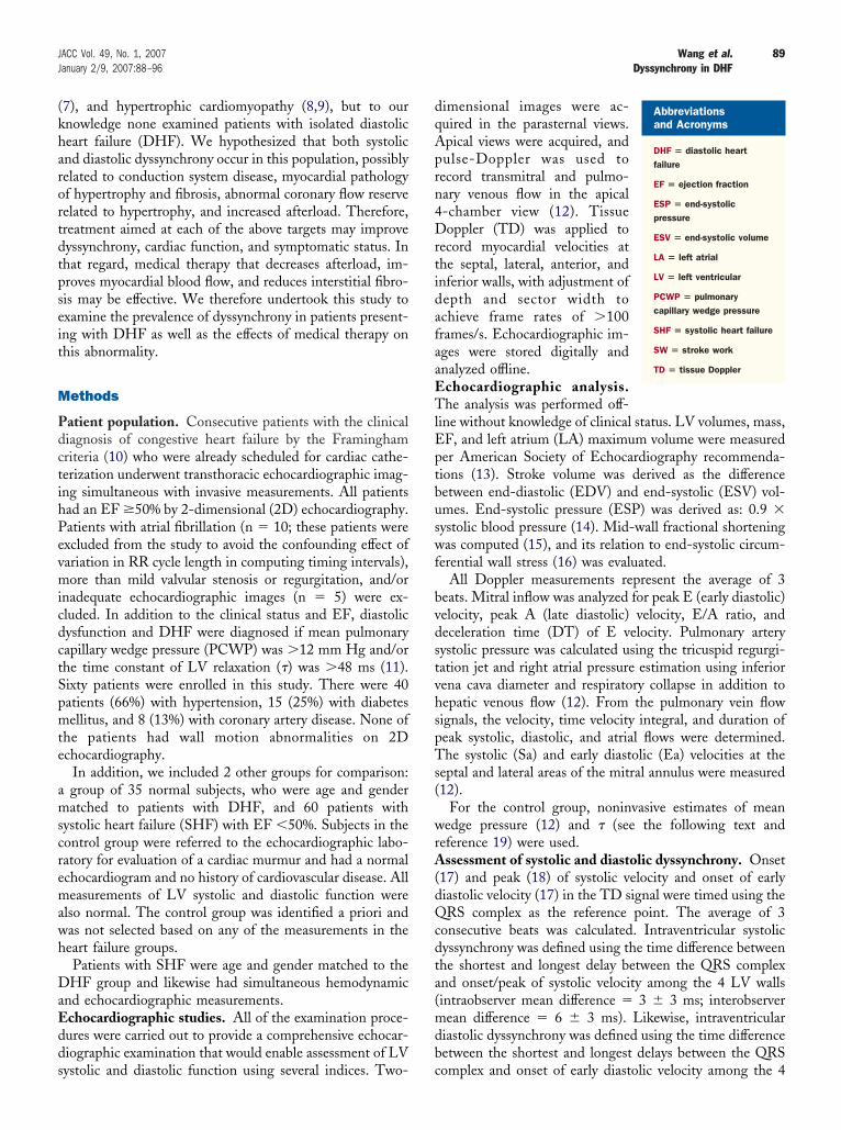
(kharortdtpseit
M
PdctihPevmicdctSpmte
amscremawh
DaEdds
dqAprn4DrtidafaaETlEptbuswf
bvdstvhspTs(
wrA(dQcdta(mdb
89JACC Vol. 49, No. 1, 2007 Wang et al.January 2/9, 2007:88–96 Dyssynchrony in DHF
7), and hypertrophic cardiomyopathy (8,9), but to ournowledge none examined patients with isolated diastoliceart failure (DHF). We hypothesized that both systolicnd diastolic dyssynchrony occur in this population, possiblyelated to conduction system disease, myocardial pathologyf hypertrophy and fibrosis, abnormal coronary flow reserveelated to hypertrophy, and increased afterload. Therefore,reatment aimed at each of the above targets may improveyssynchrony, cardiac function, and symptomatic status. Inhat regard, medical therapy that decreases afterload, im-roves myocardial blood flow, and reduces interstitial fibro-is may be effective. We therefore undertook this study toxamine the prevalence of dyssynchrony in patients present-ng with DHF as well as the effects of medical therapy onhis abnormality.
ethods
atient population. Consecutive patients with the clinicaliagnosis of congestive heart failure by the Framinghamriteria (10) who were already scheduled for cardiac cathe-erization underwent transthoracic echocardiographic imag-ng simultaneous with invasive measurements. All patientsad an EF �50% by 2-dimensional (2D) echocardiography.atients with atrial fibrillation (n � 10; these patients werexcluded from the study to avoid the confounding effect ofariation in RR cycle length in computing timing intervals),ore than mild valvular stenosis or regurgitation, and/or
nadequate echocardiographic images (n � 5) were ex-luded. In addition to the clinical status and EF, diastolicysfunction and DHF were diagnosed if mean pulmonaryapillary wedge pressure (PCWP) was �12 mm Hg and/orhe time constant of LV relaxation (�) was �48 ms (11).ixty patients were enrolled in this study. There were 40atients (66%) with hypertension, 15 (25%) with diabetesellitus, and 8 (13%) with coronary artery disease. None of
he patients had wall motion abnormalities on 2Dchocardiography.
In addition, we included 2 other groups for comparison:group of 35 normal subjects, who were age and genderatched to patients with DHF, and 60 patients with
ystolic heart failure (SHF) with EF �50%. Subjects in theontrol group were referred to the echocardiographic labo-atory for evaluation of a cardiac murmur and had a normalchocardiogram and no history of cardiovascular disease. Alleasurements of LV systolic and diastolic function were
lso normal. The control group was identified a priori andas not selected based on any of the measurements in theeart failure groups.Patients with SHF were age and gender matched to theHF group and likewise had simultaneous hemodynamic
nd echocardiographic measurements.chocardiographic studies. All of the examination proce-ures were carried out to provide a comprehensive echocar-iographic examination that would enable assessment of LV
ystolic and diastolic function using several indices. Two- cimensional images were ac-uired in the parasternal views.pical views were acquired, andulse-Doppler was used toecord transmitral and pulmo-ary venous flow in the apical-chamber view (12). Tissueoppler (TD) was applied to
ecord myocardial velocities athe septal, lateral, anterior, andnferior walls, with adjustment ofepth and sector width tochieve frame rates of �100rames/s. Echocardiographic im-ges were stored digitally andnalyzed offline.chocardiographic analysis.he analysis was performed off-
ine without knowledge of clinical status. LV volumes, mass,F, and left atrium (LA) maximum volume were measureder American Society of Echocardiography recommenda-ions (13). Stroke volume was derived as the differenceetween end-diastolic (EDV) and end-systolic (ESV) vol-mes. End-systolic pressure (ESP) was derived as: 0.9 �ystolic blood pressure (14). Mid-wall fractional shorteningas computed (15), and its relation to end-systolic circum-
erential wall stress (16) was evaluated.All Doppler measurements represent the average of 3
eats. Mitral inflow was analyzed for peak E (early diastolic)elocity, peak A (late diastolic) velocity, E/A ratio, andeceleration time (DT) of E velocity. Pulmonary arteryystolic pressure was calculated using the tricuspid regurgi-ation jet and right atrial pressure estimation using inferiorena cava diameter and respiratory collapse in addition toepatic venous flow (12). From the pulmonary vein flowignals, the velocity, time velocity integral, and duration ofeak systolic, diastolic, and atrial flows were determined.he systolic (Sa) and early diastolic (Ea) velocities at the
eptal and lateral areas of the mitral annulus were measured12).
For the control group, noninvasive estimates of meanedge pressure (12) and � (see the following text and
eference 19) were used.ssessment of systolic and diastolic dyssynchrony. Onset
17) and peak (18) of systolic velocity and onset of earlyiastolic velocity (17) in the TD signal were timed using theRS complex as the reference point. The average of 3
onsecutive beats was calculated. Intraventricular systolicyssynchrony was defined using the time difference betweenhe shortest and longest delay between the QRS complexnd onset/peak of systolic velocity among the 4 LV wallsintraobserver mean difference � 3 � 3 ms; interobserverean difference � 6 � 3 ms). Likewise, intraventricular
iastolic dyssynchrony was defined using the time differenceetween the shortest and longest delays between the QRS
Abbreviationsand Acronyms
DHF � diastolic heartfailure
EF � ejection fraction
ESP � end-systolicpressure
ESV � end-systolic volume
LA � left atrial
LV � left ventricular
PCWP � pulmonarycapillary wedge pressure
SHF � systolic heart failure
SW � stroke work
TD � tissue Doppler
omplex and onset of early diastoli
c velocity among the 4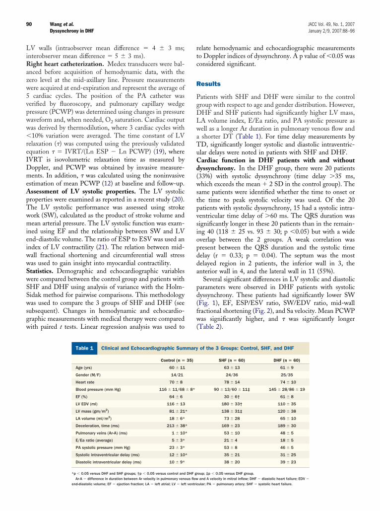
LiRazw5vpww�reIDmeApTwmieiwwSwSSwsgw
rtc
R
PgDLwaTuCd(wstpvsiopdda
pd(fw(
90 Wang et al. JACC Vol. 49, No. 1, 2007Dyssynchrony in DHF January 2/9, 2007:88–96
V walls (intraobserver mean difference � 4 � 3 ms;nterobserver mean difference � 5 � 3 ms).
ight heart catheterization. Medex transducers were bal-nced before acquisition of hemodynamic data, with theero level at the mid-axillary line. Pressure measurementsere acquired at end-expiration and represent the average ofcardiac cycles. The position of the PA catheter was
erified by fluoroscopy, and pulmonary capillary wedgeressure (PCWP) was determined using changes in pressureaveform and, when needed, O2 saturation. Cardiac outputas derived by thermodilution, where 3 cardiac cycles with10% variation were averaged. The time constant of LV
elaxation (�) was computed using the previously validatedquation � � IVRT/(Ln ESP � Ln PCWP) (19), whereVRT is isovolumetric relaxation time as measured byoppler, and PCWP was obtained by invasive measure-ents. In addition, � was calculated using the noninvasive
stimation of mean PCWP (12) at baseline and follow-up.ssessment of LV systolic properties. The LV systolicroperties were examined as reported in a recent study (20).he LV systolic performance was assessed using strokeork (SW), calculated as the product of stroke volume andean arterial pressure. The LV systolic function was exam-
ned using EF and the relationship between SW and LVnd-diastolic volume. The ratio of ESP to ESV was used anndex of LV contractility (21). The relation between mid-all fractional shortening and circumferential wall stressas used to gain insight into myocardial contractility.tatistics. Demographic and echocardiographic variablesere compared between the control group and patients withHF and DHF using analysis of variance with the Holm-idak method for pairwise comparisons. This methodologyas used to compare the 3 groups of SHF and DHF (see
ubsequent). Changes in hemodynamic and echocardio-raphic measurements with medical therapy were comparedith paired t tests. Linear regression analysis was used to
Clinical and Echocardiographic Summary of the
Table 1 Clinical and Echocardiographic Sum
Control (n �
Age (yrs) 60 � 11
Gender (M/F) 14/21
Heart rate 70 � 8
Blood pressure (mm Hg) 116 � 11/68
EF (%) 64 � 6
LV EDV (ml) 116 � 13
LV mass (gm/m2) 81 � 21
LA volume (ml/m2) 18 � 6*
Deceleration, time (ms) 213 � 38
Pulmonary veins (Ar-A) (ms) 1 � 10
E/Ea ratio (average) 5 � 3*
PA systolic pressure (mm Hg) 23 � 3*
Systolic intraventricular delay (ms) 12 � 10
Diastolic intraventricular delay (ms) 10 � 9*
*p � 0.05 versus DHF and SHF groups; †p � 0.05 versus control and
Ar-A � difference in duration between Ar velocity in pulmonary venous flowend-diastolic volume; EF � ejection fraction; LA � left atrial; LV � left ventric
elate hemodynamic and echocardiographic measurementso Doppler indices of dyssynchrony. A p value of �0.05 wasonsidered significant.
esults
atients with SHF and DHF were similar to the controlroup with respect to age and gender distribution. However,HF and SHF patients had significantly higher LV mass,A volume index, E/Ea ratio, and PA systolic pressure asell as a longer Ar duration in pulmonary venous flow andshorter DT (Table 1). For time delay measurements byD, significantly longer systolic and diastolic intraventric-lar delays were noted in patients with SHF and DHF.ardiac function in DHF patients with and without
yssynchrony. In the DHF group, there were 20 patients33%) with systolic dyssynchrony (time delay �35 ms,hich exceeds the mean � 2 SD in the control group). The
ame patients were identified whether the time to onset orhe time to peak systolic velocity was used. Of the 20atients with systolic dyssynchrony, 15 had a systolic intra-entricular time delay of �60 ms. The QRS duration wasignificantly longer in these 20 patients than in the remain-ng 40 (118 � 25 vs. 93 � 30; p �0.05) but with a wideverlap between the 2 groups. A weak correlation wasresent between the QRS duration and the systolic timeelay (r � 0.33; p � 0.04). The septum was the mostelayed region in 2 patients, the inferior wall in 3, thenterior wall in 4, and the lateral wall in 11 (55%).
Several significant differences in LV systolic and diastolicarameters were observed in DHF patients with systolicyssynchrony. These patients had significantly lower SWFig. 1), EF, ESP/ESV ratio, SW/EDV ratio, mid-wallractional shortening (Fig. 2), and Sa velocity. Mean PCWPas significantly higher, and � was significantly longer
Table 2).
ups: Control, SHF, and DHF
of the 3 Groups: Control, SHF, and DHF
SHF (n � 60) DHF (n � 60)
63 � 13 61 � 9
24/36 25/35
78 � 14 74 � 10
90 � 13/60 � 11‡ 145 � 28/86 � 19
30 � 6† 61 � 8
180 � 33† 110 � 35
138 � 31‡ 120 � 38
73 � 28 65 � 10
169 � 23 189 � 30
53 � 10 48 � 5
21 � 4 18 � 5
53 � 8 46 � 5
35 � 21 31 � 25
38 � 20 39 � 23
roup; ‡p � 0.05 versus DHF group.
3 Gro
mary
35)
� 8*
*
*
*
*
DHF g
and A velocity in mitral inflow; DHF � diastolic heart failure; EDV �ular; PA � pulmonary artery; SHF � systolic heart failure.
(ctAawi
oDddscsdaD�r0Cd(ciwdd
aeds
(db05twcimmEsf(n2iTSsvimsti�
91JACC Vol. 49, No. 1, 2007 Wang et al.January 2/9, 2007:88–96 Dyssynchrony in DHF
There were 35 patients (58%) with diastolic dyssynchronytime delay �35 ms). The 20 patients with systolic dyssyn-hrony had evidence of diastolic dyssynchrony. In addition,here were 15 patients with isolated diastolic dyssynchrony.
weak correlation was present between the QRS durationnd diastolic time delay (r � 0.3; p � 0.05). The septumas the most delayed region in 3 patients, the inferior wall
n 6, the anterior wall in 8, and the lateral wall in 18 (51%).In general, hemodynamic and echocardiographic indices
f LV systolic properties were similar between patients withHF and no dyssynchrony and those with isolated diastolic
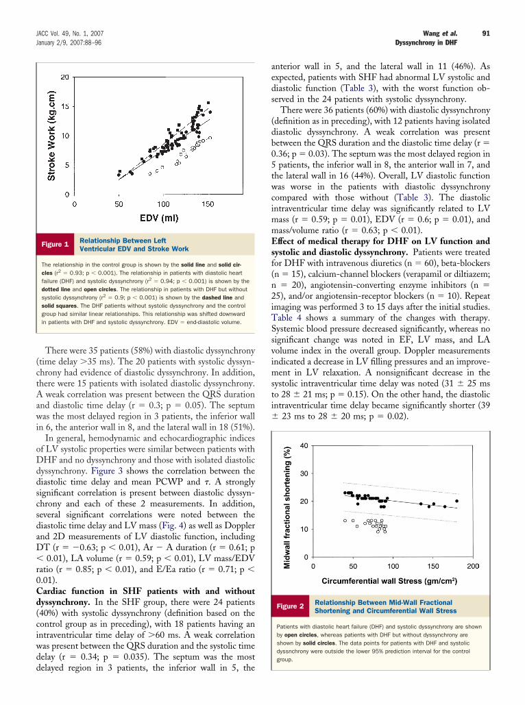
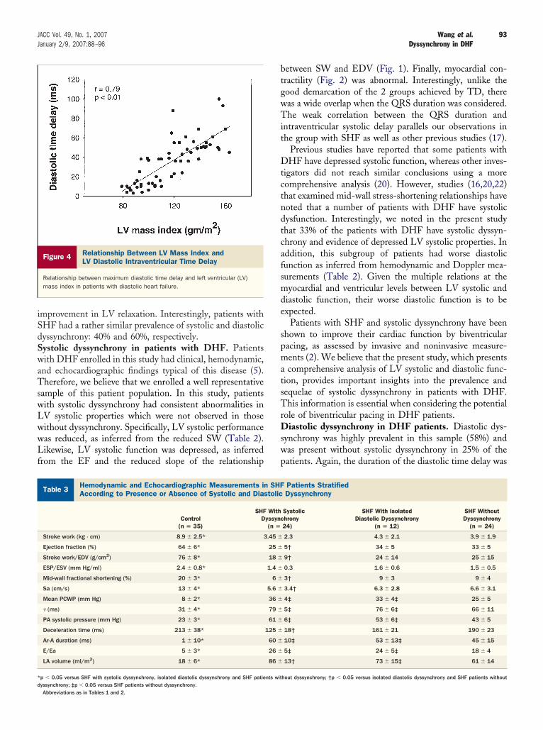
yssynchrony. Figure 3 shows the correlation between theiastolic time delay and mean PCWP and �. A stronglyignificant correlation is present between diastolic dyssyn-hrony and each of these 2 measurements. In addition,everal significant correlations were noted between theiastolic time delay and LV mass (Fig. 4) as well as Dopplernd 2D measurements of LV diastolic function, includingT (r � �0.63; p � 0.01), Ar � A duration (r � 0.61; p0.01), LA volume (r � 0.59; p � 0.01), LV mass/EDV
atio (r � 0.85; p � 0.01), and E/Ea ratio (r � 0.71; p �.01).ardiac function in SHF patients with and without
yssynchrony. In the SHF group, there were 24 patients40%) with systolic dyssynchrony (definition based on theontrol group as in preceding), with 18 patients having anntraventricular time delay of �60 ms. A weak correlationas present between the QRS duration and the systolic timeelay (r � 0.34; p � 0.035). The septum was the most
Figure 1 Relationship Between LeftVentricular EDV and Stroke Work
The relationship in the control group is shown by the solid line and solid cir-cles (r2 � 0.93; p � 0.001). The relationship in patients with diastolic heartfailure (DHF) and systolic dyssynchrony (r2 � 0.94; p � 0.001) is shown by thedotted line and open circles. The relationship in patients with DHF but withoutsystolic dyssynchrony (r2 � 0.9; p � 0.001) is shown by the dashed line andsolid squares. The DHF patients without systolic dyssynchrony and the controlgroup had similar linear relationships. This relationship was shifted downwardin patients with DHF and systolic dyssynchrony. EDV � end-diastolic volume.
elayed region in 3 patients, the inferior wall in 5, the
nterior wall in 5, and the lateral wall in 11 (46%). Asxpected, patients with SHF had abnormal LV systolic andiastolic function (Table 3), with the worst function ob-erved in the 24 patients with systolic dyssynchrony.
There were 36 patients (60%) with diastolic dyssynchronydefinition as in preceding), with 12 patients having isolatediastolic dyssynchrony. A weak correlation was presentetween the QRS duration and the diastolic time delay (r �.36; p � 0.03). The septum was the most delayed region inpatients, the inferior wall in 8, the anterior wall in 7, and
he lateral wall in 16 (44%). Overall, LV diastolic functionas worse in the patients with diastolic dyssynchrony
ompared with those without (Table 3). The diastolicntraventricular time delay was significantly related to LV
ass (r � 0.59; p � 0.01), EDV (r � 0.6; p � 0.01), andass/volume ratio (r � 0.63; p � 0.01).ffect of medical therapy for DHF on LV function and
ystolic and diastolic dyssynchrony. Patients were treatedor DHF with intravenous diuretics (n � 60), beta-blockersn � 15), calcium-channel blockers (verapamil or diltiazem;
� 20), angiotensin-converting enzyme inhibitors (n �5), and/or angiotensin-receptor blockers (n � 10). Repeatmaging was performed 3 to 15 days after the initial studies.able 4 shows a summary of the changes with therapy.ystemic blood pressure decreased significantly, whereas noignificant change was noted in EF, LV mass, and LAolume index in the overall group. Doppler measurementsndicated a decrease in LV filling pressures and an improve-
ent in LV relaxation. A nonsignificant decrease in theystolic intraventricular time delay was noted (31 � 25 mso 28 � 21 ms; p � 0.15). On the other hand, the diastolicntraventricular time delay became significantly shorter (39
23 ms to 28 � 20 ms; p � 0.02).
Figure 2 Relationship Between Mid-Wall FractionalShortening and Circumferential Wall Stress
Patients with diastolic heart failure (DHF) and systolic dyssynchrony are shownby open circles, whereas patients with DHF but without dyssynchrony areshown by solid circles. The data points for patients with DHF and systolicdyssnchrony were outside the lower 95% prediction interval for the controlgroup.
RLwstc5trot
D
Tupsidati
HS
*‡
ressure
92 Wang et al. JACC Vol. 49, No. 1, 2007Dyssynchrony in DHF January 2/9, 2007:88–96
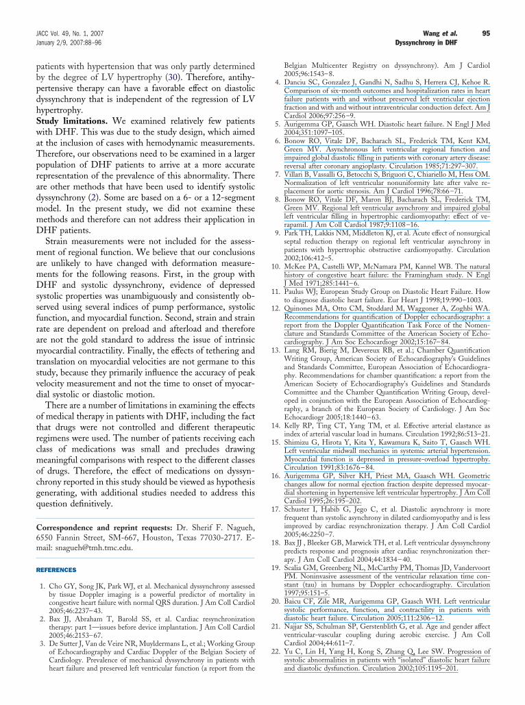
elation of the change in LV diastolic dyssynchrony toV relaxation and filling. A significant positive correlationas noted between the decrease in LV ESP and the
hortening in diastolic time delay (r � 0.71; p � 0.01). Inurn, the change in diastolic time delay correlated signifi-antly with the shortening in � (r � 0.85; p � 0.001) (Fig.), the increase in Ea velocity (r � �0.75; p � 0.01), andhe decrease in E/Ea ratio (r � 0.8; p � 0.01). On multipleegression analysis, the decrease in ESP and the shorteningf the diastolic time delay were the main determinants ofhe improvement in � (R2 � 0.84; p � 0.01).
Figure 3 Relation Between LV Diastolic Intraventricular Time D
Relationship between maximum diastolic time delay and relaxation time constantin patients with diastolic heart failure. LV � left ventricular; PCWP � pulmonary ca
emodynamic and Echocardiographic Measurements in DHF Patientratified According to Presence or Absence of Systolic and Diasto
Table 2 Hemodynamic and Echocardiographic Measurements inStratified According to Presence or Absence of Systoli
Control(n � 35)
DHFD
Stroke work (kg · cm) 8.9 � 2.5
Ejection fraction (%) 64 � 6
Stroke work/EDV (g/cm2) 76 � 8
ESP/ESV (mm Hg/ml) 2.4 � 0.8
Mid-wall fractional shortening (%) 20 � 3
Sa (cm/s) 13 � 4
Mean PCWP (mm Hg) 8 � 2*
� (ms) 31 � 4*
PA systolic pressure (mm Hg) 23 � 3*
Deceleration time (ms) 213 � 38 1
Ar-A duration (ms) 1 � 10*
E/Ea 5 � 3*
LA volume (ml/m2) 18 � 6*
p � 0.05 versus systolic dyssynchrony, isolated diastolic dyssynchrony, and DHF patients withoutp � 0.05 versus DHF patients without dyssynchrony; §p � 0.05 versus control group, isolated dESP � end-systolic pressure; ESV � end-systolic volume; PCWP � pulmonary capillary wedge p
iscussion
he present study shows that systolic dyssynchrony is notncommon in patients with DHF, occurring in 33% of theatients. These patients were characterized by abnormal LVystolic properties and had invasive and echocardiographicndices indicative of the most advanced stage of diastolicysfunction. At 58%, diastolic dyssynchrony appears to havehigher prevalence in this population. Importantly, medical
herapy results in a significant shortening of the diastolicntraventricular time delay, which is closely related to
and LV Relaxation and Mean PCWP
and mean wedge pressure (right)wedge pressure.
yssynchrony
PatientsDiastolic Dyssynchrony
Systolichrony20)
DHF With IsolatedDiastolic Dyssynchrony
(n � 15)
DHF WithoutDyssynchrony
(n � 25)
1.8§ 9.8 � 2.3 10 � 2.8
3§ 65 � 4 66 � 6
8§ 90 � 10 88 � 11
0.3§ 2.8 � 0.8 2.9 � 1.0
1§ 19 � 4 19 � 3
4.3§ 13.5 � 4.3 12.8 � 4.6
4† 22 � 4 20 � 4
7† 61 � 4‡ 57 � 4
8† 41 � 6 38 � 6
21† 190 � 28 200 � 25
13† 49 � 10 40 � 11
5† 18 � 4 16 � 6
11† 66 � 6‡ 56 � 8
chrony; †p � 0.05 versus isolated diastolic dyssynchrony and DHF patients without dyssynchrony;dyssynchrony and DHF patients without dyssynchrony.; Sa � systolic mitral annular velocity; other abbreviations as in Table 1.
elay
� (left)pillary
tslic D
DHFc and
Withyssync(n �
7.1 �
57 �
55 �
1.9 �
12 �
8.4 �
28 �
69 �
51 �
63 �
58 �
23 �
81 �
dyssyniastolic
iSdSwaTswLwwLf
btgwTit
Dtctndtcafsmde
spmatsTrDswp
HA
*d
93JACC Vol. 49, No. 1, 2007 Wang et al.January 2/9, 2007:88–96 Dyssynchrony in DHF
mprovement in LV relaxation. Interestingly, patients withHF had a rather similar prevalence of systolic and diastolicyssynchrony: 40% and 60%, respectively.ystolic dyssynchrony in patients with DHF. Patientsith DHF enrolled in this study had clinical, hemodynamic,
nd echocardiographic findings typical of this disease (5).herefore, we believe that we enrolled a well representative
ample of this patient population. In this study, patientsith systolic dyssynchrony had consistent abnormalities inV systolic properties which were not observed in thoseithout dyssynchrony. Specifically, LV systolic performanceas reduced, as inferred from the reduced SW (Table 2).ikewise, LV systolic function was depressed, as inferred
rom the EF and the reduced slope of the relationship
Figure 4 Relationship Between LV Mass Index andLV Diastolic Intraventricular Time Delay
Relationship between maximum diastolic time delay and left ventricular (LV)mass index in patients with diastolic heart failure.
emodynamic and Echocardiographic Measurements in SHF Patienccording to Presence or Absence of Systolic and Diastolic Dyssyn
Table 3 Hemodynamic and Echocardiographic Measurements inAccording to Presence or Absence of Systolic and Dia
Control(n � 35)
SHFD
Stroke work (kg · cm) 8.9 � 2.5* 3
Ejection fraction (%) 64 � 6*
Stroke work/EDV (g/cm2) 76 � 8*
ESP/ESV (mm Hg/ml) 2.4 � 0.8*
Mid-wall fractional shortening (%) 20 � 3*
Sa (cm/s) 13 � 4*
Mean PCWP (mm Hg) 8 � 2*
� (ms) 31 � 4*
PA systolic pressure (mm Hg) 23 � 3*
Deceleration time (ms) 213 � 38*
Ar-A duration (ms) 1 � 10*
E/Ea 5 � 3*
LA volume (ml/m2) 18 � 6*
p � 0.05 versus SHF with systolic dyssynchrony, isolated diastolic dyssynchrony and SHF patie
yssynchrony; ‡p � 0.05 versus SHF patients without dyssynchrony.Abbreviations as in Tables 1 and 2.etween SW and EDV (Fig. 1). Finally, myocardial con-ractility (Fig. 2) was abnormal. Interestingly, unlike theood demarcation of the 2 groups achieved by TD, thereas a wide overlap when the QRS duration was considered.he weak correlation between the QRS duration and
ntraventricular systolic delay parallels our observations inhe group with SHF as well as other previous studies (17).
Previous studies have reported that some patients withHF have depressed systolic function, whereas other inves-
igators did not reach similar conclusions using a moreomprehensive analysis (20). However, studies (16,20,22)hat examined mid-wall stress-shortening relationships haveoted that a number of patients with DHF have systolicysfunction. Interestingly, we noted in the present studyhat 33% of the patients with DHF have systolic dyssyn-hrony and evidence of depressed LV systolic properties. Inddition, this subgroup of patients had worse diastolicunction as inferred from hemodynamic and Doppler mea-urements (Table 2). Given the multiple relations at theyocardial and ventricular levels between LV systolic and
iastolic function, their worse diastolic function is to bexpected.
Patients with SHF and systolic dyssynchrony have beenhown to improve their cardiac function by biventricularacing, as assessed by invasive and noninvasive measure-ents (2). We believe that the present study, which presentscomprehensive analysis of LV systolic and diastolic func-
ion, provides important insights into the prevalence andequelae of systolic dyssynchrony in patients with DHF.his information is essential when considering the potential
ole of biventricular pacing in DHF patients.iastolic dyssynchrony in DHF patients. Diastolic dys-
ynchrony was highly prevalent in this sample (58%) andas present without systolic dyssynchrony in 25% of theatients. Again, the duration of the diastolic time delay was
ratifiedny
Patients StratifiedDyssynchrony
Systolichrony24)
SHF With IsolatedDiastolic Dyssynchrony
(n � 12)
SHF WithoutDyssynchrony
(n � 24)
2.3 4.3 � 2.1 3.9 � 1.9
5† 34 � 5 33 � 5
9† 24 � 14 25 � 15
0.3 1.6 � 0.6 1.5 � 0.5
3† 9 � 3 9 � 4
3.4† 6.3 � 2.8 6.6 � 3.1
4‡ 33 � 4‡ 25 � 5
5‡ 76 � 6‡ 66 � 11
6‡ 53 � 6‡ 43 � 5
18† 161 � 21 190 � 23
10‡ 53 � 13‡ 45 � 15
5‡ 24 � 5‡ 18 � 4
13† 73 � 15‡ 61 � 14
hout dyssynchrony; †p � 0.05 versus isolated diastolic dyssynchrony and SHF patients without
ts Stchro
SHFstolic
Withyssync(n �
.45 �
25 �
18 �
1.4 �
6 �
5.6 �
36 �
79 �
61 �
125 �
60 �
26 �
86 �
nts wit
st(tDms1svddac
atatp
iwsddrdcTlpbdHdsbdbwbpe
madao
rrtcbdegfu
nonin
94 Wang et al. JACC Vol. 49, No. 1, 2007Dyssynchrony in DHF January 2/9, 2007:88–96
imilar to that observed in patients with SHF enrolled inhis study and previously reported by other investigators17). There are a number of pathophysiologic mechanismshat can account for diastolic dyssynchrony in patients withHF. They include systolic dyssynchrony, because seg-ents with delayed contraction also show delayed expan-
ion. Coronary artery disease is another potential reason. Inreport, coronary artery disease was associated with dia-
tolic dyssynchrony, which improved after mechanical re-ascularization (6). However, epicardial coronary arteryisease was present in only 4 of the 35 patients with diastolicyssynchrony in the present study. Therefore, other mech-nisms likely play a role in the etiology of diastolic dyssyn-hrony in this population.
The strong association between TD diastolic time delaynd LV mass (and mass/volume ratio) raises the possibilityhat LV hypertrophy and interstitial fibrosis may be caus-tive factors. This hypothesis, however, remains to beested. Finally, increased afterload appears to play an im-ortant role. Previous animal studies showed that an acute
Figure 5 Relation Between the Change in LV Disatolic Dyssyn-chrony Index and LV Relaxation With Medical Therapy
Relationship between the percentage change in diastolic time delay and thecorresponding percentage change in relaxation time constant � after medicaltherapy for diastolic heart failure. LV � left ventricular.
Changes With Medical Therapy for DHF
Table 4 Changes With Medical Therapy for
Heart rate
Systolic/diastolic blood pressure (mm Hg)
EF (%)
Deceleration time (ms)
Pulmonary veins (Ar-A) (ms)
E/Ea ratio (average of septal and lateral)
PA systolic pressure (mm Hg)
�† (ms)
Ea (average of septal and lateral) (cm/s)
*p � 0.05 versus baseline; †� was derived using IVRT by Doppler, andAbbreviations as in Table 1.
p
ncrease in LV afterload leads to increased dyssynchronyhich was linearly correlated with � (23). In the present
tudy, a significant correlation was present between theecrease in LV ESP and the shortening in diastolic timeelay after medical therapy for congestive heart failure,aising the possibility that increased afterload may have aetrimental effect on LV relaxation, in part through in-reased diastolic dyssynchrony.reatment of diastolic dyssynchrony in DHF. There are
imited data on the treatment of diastolic dyssynchrony inatients with normal EF (6–9). For patients with SHF,iventricular pacing, which reduces the extent of systolicyssynchrony, can result in an improvement in LV filling.owever, the favorable effect of biventricular pacing on LV
iastolic function and dyssynchrony has been observed inome but not all patients (17,24), highlighting the contri-ution of factors other than systolic dyssynchrony to theiastolic abnormality noted in these subjects. The effects ofiventricular pacing on diastolic dyssynchrony in patientsith DHF is speculative at this time. However, it may beeneficial, similar to the observations in those with de-ressed EF, to some but not all patients, given the multipletiologies of diastolic dyssynchrony in patients with DHF.
The correlation between diastolic dyssynchrony and LVass raises the possibility that regression of LV hypertrophy
nd interstitial fibrosis, which is feasible with a number ofrugs (25–29), may be of value as well. We could notddress this possible etiology owing to the short follow-upf the present group.On the other hand, it is reasonable to conclude that
eduction of systemic blood pressure, in and of itself, caneduce diastolic dyssynchrony, particularly in patients wherehe QRS duration is not prolonged. Increased afterload mayontribute to dyssynchrony through its effects on myocardiallood flow. Increased systolic wall stress increases myocar-ial oxygen demand, which in the presence of abnormalndothelial function and coronary vasculature leads to re-ional heterogeneity in coronary blood flow and regionalunction. This mechanism is supported by previous studiessing positron emission tomography which reported on the
Baseline After Medical Therapy
74 � 10 69 � 11
� 28/86 � 19 125 � 21/68 � 16*
61 � 8 63 � 9
189 � 30 218 � 41*
48 � 5 28 � 8*
18 � 5 11 � 8*
46 � 5 34 � 8*
60 � 6 49 � 8*
6 � 3 7.5 � 1.9*
vasive estimation of ESP and mean PCWP.
DHF
145
resence of abnormal regional myocardial blood flow in
pbpdhSwaTpradmmD
mamDssframtsvd
otrcmocgq
C6m
R
1
1
1
1
1
1
1
1
1
1
2
2
2
95JACC Vol. 49, No. 1, 2007 Wang et al.January 2/9, 2007:88–96 Dyssynchrony in DHF
atients with hypertension that was only partly determinedy the degree of LV hypertrophy (30). Therefore, antihy-ertensive therapy can have a favorable effect on diastolicyssynchrony that is independent of the regression of LVypertrophy.tudy limitations. We examined relatively few patientsith DHF. This was due to the study design, which aimed
t the inclusion of cases with hemodynamic measurements.herefore, our observations need to be examined in a largeropulation of DHF patients to arrive at a more accurateepresentation of the prevalence of this abnormality. Therere other methods that have been used to identify systolicyssynchrony (2). Some are based on a 6- or a 12-segmentodel. In the present study, we did not examine theseethods and therefore can not address their application inHF patients.Strain measurements were not included for the assess-ent of regional function. We believe that our conclusions
re unlikely to have changed with deformation measure-ents for the following reasons. First, in the group withHF and systolic dyssynchrony, evidence of depressed
ystolic properties was unambiguously and consistently ob-erved using several indices of pump performance, systolicunction, and myocardial function. Second, strain and strainate are dependent on preload and afterload and thereforere not the gold standard to address the issue of intrinsicyocardial contractility. Finally, the effects of tethering and
ranslation on myocardial velocities are not germane to thistudy, because they primarily influence the accuracy of peakelocity measurement and not the time to onset of myocar-ial systolic or diastolic motion.There are a number of limitations in examining the effects
f medical therapy in patients with DHF, including the facthat drugs were not controlled and different therapeuticegimens were used. The number of patients receiving eachlass of medications was small and precludes drawingeaningful comparisons with respect to the different classes
f drugs. Therefore, the effect of medications on dyssyn-hrony reported in this study should be viewed as hypothesisenerating, with additional studies needed to address thisuestion definitively.
orrespondence and reprint requests: Dr. Sherif F. Nagueh,550 Fannin Street, SM-667, Houston, Texas 77030-2717. E-ail: [email protected].
EFERENCES
1. Cho GY, Song JK, Park WJ, et al. Mechanical dyssynchrony assessedby tissue Doppler imaging is a powerful predictor of mortality incongestive heart failure with normal QRS duration. J Am Coll Cardiol2005;46:2237–43.
2. Bax JJ, Abraham T, Barold SS, et al. Cardiac resynchronizationtherapy: part 1—issues before device implantation. J Am Coll Cardiol2005;46:2153–67.
3. De Sutter J, Van de Veire NR, Muyldermans L, et al.; Working Groupof Echocardiography and Cardiac Doppler of the Belgian Society of
Cardiology. Prevalence of mechanical dyssynchrony in patients withheart failure and preserved left ventricular function (a report from theBelgian Multicenter Registry on dyssynchrony). Am J Cardiol2005;96:1543–8.
4. Danciu SC, Gonzalez J, Gandhi N, Sadhu S, Herrera CJ, Kehoe R.Comparison of six-month outcomes and hospitalization rates in heartfailure patients with and without preserved left ventricular ejectionfraction and with and without intraventricular conduction defect. Am JCardiol 2006;97:256–9.
5. Aurigemma GP, Gaasch WH. Diastolic heart failure. N Engl J Med2004;351:1097–105.
6. Bonow RO, Vitale DF, Bacharach SL, Frederick TM, Kent KM,Green MV. Asynchronous left ventricular regional function andimpaired global diastolic filling in patients with coronary artery disease:reversal after coronary angioplasty. Circulation 1985;71:297–307.
7. Villari B, Vassalli G, Betocchi S, Briguori C, Chiariello M, Hess OM.Normalization of left ventricular nonuniformity late after valve re-placement for aortic stenosis. Am J Cardiol 1996;78:66–71.
8. Bonow RO, Vitale DF, Maron BJ, Bacharach SL, Frederick TM,Green MV. Regional left ventricular asynchrony and impaired globalleft ventricular filling in hypertrophic cardiomyopathy: effect of ve-rapamil. J Am Coll Cardiol 1987;9:1108–16.
9. Park TH, Lakkis NM, Middleton KJ, et al. Acute effect of nonsurgicalseptal reduction therapy on regional left ventricular asynchrony inpatients with hypertrophic obstructive cardiomyopathy. Circulation2002;106:412–5.
0. McKee PA, Castelli WP, McNamara PM, Kannel WB. The naturalhistory of congestive heart failure: the Framingham study. N EnglJ Med 1971;285:1441–6.
1. Paulus WJ; European Study Group on Diastolic Heart Failure. Howto diagnose diastolic heart failure. Eur Heart J 1998;19:990–1003.
2. Quinones MA, Otto CM, Stoddard M, Waggoner A, Zoghbi WA.Recommendations for quantification of Doppler echocardiography: areport from the Doppler Quantification Task Force of the Nomen-clature and Standards Committee of the American Society of Echo-cardiography. J Am Soc Echocardiogr 2002;15:167–84.
3. Lang RM, Bierig M, Devereux RB, et al.; Chamber QuantificationWriting Group, American Society of Echocardiography’s Guidelinesand Standards Committee, European Association of Echocardiogra-phy. Recommendations for chamber quantification: a report from theAmerican Society of Echocardiography’s Guidelines and StandardsCommittee and the Chamber Quantification Writing Group, devel-oped in conjunction with the European Association of Echocardiog-raphy, a branch of the European Society of Cardiology. J Am SocEchocardiogr 2005;18:1440–63.
4. Kelly RP, Ting CT, Yang TM, et al. Effective arterial elastance asindex of arterial vascular load in humans. Circulation 1992;86:513–21.
5. Shimizu G, Hirota Y, Kita Y, Kawamura K, Saito T, Gaasch WH.Left ventricular midwall mechanics in systemic arterial hypertension.Myocardial function is depressed in pressure-overload hypertrophy.Circulation 1991;83:1676–84.
6. Aurigemma GP, Silver KH, Priest MA, Gaasch WH. Geometricchanges allow for normal ejection fraction despite depressed myocar-dial shortening in hypertensive left ventricular hypertrophy. J Am CollCardiol 1995;26:195–202.
7. Schuster I, Habib G, Jego C, et al. Diastolic asynchrony is morefrequent than systolic asynchrony in dilated cardiomyopathy and is lessimproved by cardiac resynchronization therapy. J Am Coll Cardiol2005;46:2250–7.
8. Bax JJ , Bleeker GB, Marwick TH, et al. Left ventricular dyssynchronypredicts response and prognosis after cardiac resynchronization ther-apy. J Am Coll Cardiol 2004;44:1834–40.
9. Scalia GM, Greenberg NL, McCarthy PM, Thomas JD, VandervoortPM. Noninvasive assessment of the ventricular relaxation time con-stant (tau) in humans by Doppler echocardiography. Circulation1997;95:151–5.
0. Baicu CF, Zile MR, Aurigemma GP, Gaasch WH. Left ventricularsystolic performance, function, and contractility in patients withdiastolic heart failure. Circulation 2005;111:2306–12.
1. Najjar SS, Schulman SP, Gerstenblith G, et al. Age and gender affectventricular-vascular coupling during aerobic exercise. J Am CollCardiol 2004;44:611–7.
2. Yu C, Lin H, Yang H, Kong S, Zhang Q, Lee SW. Progression ofsystolic abnormalities in patients with “isolated” diastolic heart failure
and diastolic dysfunction. Circulation 2002;105:1195–201.
2
2
2
2
2
2
2
3
96 Wang et al. JACC Vol. 49, No. 1, 2007Dyssynchrony in DHF January 2/9, 2007:88–96
3. Schafer S, Fiedler VB, Thamer V. Afterload dependent prolongationof left ventricular relaxation: importance of asynchrony. CardiovascRes 1992;26:631–7.
4. Waggoner AD, Faddis MN, Gleva MJ, de las Fuentes L, Davila-Roman VG. Improvements in left ventricular diastolic function aftercardiac resynchronization therapy are coupled to response in systolicperformance. J Am Coll Cardiol 2005;46:2244–9.
5. Brilla CG, Funck RC, Rupp H. Lisinopril-mediated regression ofmyocardial fibrosis in patients with hypertensive heart disease. Circu-lation 2000;102:1388–93.
6. Diez J, Querejeta R, Lopez B, Gonzalez A, Larman M, MartinezUbago JL. Losartan-dependent regression of myocardial fibrosis isassociated with reduction of left ventricular chamber stiffness in
hypertensive patients. Circulation 2002;105:2512–7.7. Lopez B, Querejeta R, Gonzalez A, Sanchez E, Larman M, Diez J.Effects of loop diuretics on myocardial fibrosis and collagen type Iturnover in chronic heart failure. J Am Coll Cardiol 2004;43:2028 –35.
8. Patel R, Nagueh SF, Tsybouleva N, et al. Simvastatin inducesregression of cardiac hypertrophy and fibrosis and improves cardiacfunction in a transgenic rabbit model of human hypertrophic cardio-myopathy. Circulation 2001;104:317–24.
9. Fukuta H, Sane DC, Brucks S, Little WC. Statin therapy may beassociated with lower mortality in patients with diastolic heart failure:a preliminary report. Circulation 2005;112:357–63.
0. Kaufmann PA, Camici PG. Myocardial blood flow measurement byPET: technical aspects and clinical applications. J Nucl Med
2005;46:75–88.Copyright © 2022 FDOKUMEN