Tallinna Teataja Nr 205 (1069) Tallinnas, esmaspäewal 9 st2 ...

Non-Myocardial Production of ST2 Protein in Human Hypertrophyand Failure is Related to Diastolic Load
Jozef Bartunek, MD, PhD1,2,+, Leen Delrue, PhD1,2,+, Frederik Van Durme, MD1, OlivierMuller, MD, PhD1, Filip Casselman, MD, PhD3, Bart De Wiest, RN1, Romaric Croes, MD4,Sofie Verstreken, MD1, Marc Goethals, MD1, Herbert de Raedt, MD1, Jaydeep Sarma, MD,PhD, Lija Joseph, MD5, Marc Vanderheyden, MD1,2, and Ellen O Weinberg, PhD5
1Cardiovascular Center and Cardiovascular Research Center, OLV Hospital, Aalst, Belgium
2Translational Cardiology Unit, OLV Hospital, Aalst, Belgium
3Dpt of Cardiovascular Surgery, OLV Hospital, Aalst, Belgium
4Dpt of Pathology, St. Blasium Hospital, Dendermonde, Belgium
5Boston Medical Center, Boston, MA, USA
AbstractObjectives—Aims were to investigate 1) relationships between serum ST2 levels andhemodynamic/neurohormonal variables; 2) myocardial ST2 production; 3) expression of ST2,membrane-anchored ST2L, and its ligand, IL-33, in myocardium, endothelium and leukocytes frompatients with LV pressure overload and congestive cardiomyopathy.
Background—Serum levels of ST2 are elevated in heart failure. Relationship of ST2 withhemodynamic variables, source of ST2, and expression of ST2L and IL-33 in the cardiovascularsystem are unknown.
Methods—Serum ST2 (pg/mL; median [25th-75th]) was measured in patients with LV hypertrophy(aortic stenosis, AS, N=45), congestive cardiomyopathy (CCM N=53), and Controls (N=23). ST2was correlated to NT-pro BNP, CRP and hemodynamic variables. Coronary sinus and arterial bloodsampling determined myocardial gradient (production) of ST2. ST2, ST2L and IL-33 were measured(RT-PCR) in myocardial biopsies and leukocytes; ST2 protein production was evaluated in humanendothelial cells. IL-33 protein expression was determined (immunohistochemistry) in coronaryartery endothelium
Results—ST2 was elevated in AS (103[65-165], p<0.05) and CCM (194[69-551], p<0.01) vs.Controls (49[4-89]) and correlated with BNP (r=0.5; p<0.05), CRP (r=0.6; p<0.01) and LV EDP(r=0.38, p<0.03). LV ST2 mRNA was similar in AS and CCM vs. Control (NS). No myocardial ST2protein gradient was observed. Endothelial cells secreted ST2. IL-33 protein was expressed in
Correspondence to: 1. Jozef Bartunek, MD, PhD, Cardiovascular Center and Cardiovascular Research Center, Moorselbaan 164, 9300Aalst Belgium. Phone: +32 53 72 447; Fax:+32 53 72 4550; Email: [email protected] 2. Ellen O Weinberg, PhD, BostonMedical Center, EBRC Room 704, 650 Albany Street, Boston, MA 02118 USA. Phone: 617-638-7677; FAX: 617-638-8081; Email:[email protected].+authors contributed equallyAll authors state no conflicts of interestPublisher's Disclaimer: This is a PDF file of an unedited manuscript that has been accepted for publication. As a service to our customerswe are providing this early version of the manuscript. The manuscript will undergo copyediting, typesetting, and review of the resultingproof before it is published in its final citable form. Please note that during the production process errors may be discovered which couldaffect the content, and all legal disclaimers that apply to the journal pertain.
NIH Public AccessAuthor ManuscriptJ Am Coll Cardiol. Author manuscript; available in PMC 2009 December 16.
Published in final edited form as:J Am Coll Cardiol. 2008 December 16; 52(25): 2166–2174. doi:10.1016/j.jacc.2008.09.027.
NIH
-PA Author Manuscript
NIH
-PA Author Manuscript
NIH
-PA Author Manuscript

coronary artery endothelium. Leukocyte ST2L and IL-33 levels were highly correlated (r=0.97,p<0.001).
Conclusions—In human hypertrohy and failure, serum ST2 correlates with diastolic load. Thoughthe heart, endothelium, and leukocytes express components of ST2/ST2L/IL-33 pathway, the sourceof circulating serum ST2 is extra-myocardial.
Keywordsinterleukins; ST2; hypertrophy; heart failure; natriuretic peptides; endothelium-derived factors
IntroductionInflammatory/innate immune signaling contributes to pathophysiology of human hypertrophyand heart failure. Recently, serum ST2 protein has emerged as a new prognostic biomarker inpatients with recent myocardial infarction and congestive heart failure(1-6). ST2, and itsmembrane-anchored counterpart, ST2L, belong to the Toll-Interleukin-1 receptor family andproduction of both isoforms from a single mRNA is transcriptionally regulated(7,8).Extracellular engagement of ST2L with its ligand, IL-33, leads to the induction of Th2cytokines in T cells(9) and downregulation of Toll-like receptor 4 signaling(10-12). SolubleST2 protein is secreted in response to inflammatory signals. It inhibits the binding of IL-33 toST2L(13) and has also been shown to bind to macrophages leading to downregulation ofproinflammatory cytokines(14-16). Separate studies identified IL-33 as an intracellular proteinlocalizing to the nucleus in inflamed high endothelial venules(17,18)
Apart from prognostic information obtained from soluble ST2 serum levels, little is knownabout myocardial production of ST2 or regulation and expression of ST2, ST2L, and IL-33(9), in the cardiovascular system. Serum ST2 was strongly correlated with serum B-typenatriuretic peptide (BNP) and proANP(3), which are released from myocardium in responseto pressure or volume overload and relate to indices of hemodynamic load(19). ST2 levels werealso correlated with serum levels of norepinephrine(3), a marker of systemic neurohormonalactivation in the failing heart. However, whether hemodynamic factors are directly related toserum ST2 levels in human hypertrophy and failure is unknown.
We tested the hypothesis that serum ST2 levels correlate to hemodynamic variables in patientswith pressure overload hypertrophy and congestive cardiomyopathy. Both conditions representtwo distinct pathophysiological mechanisms of heart failure allowing to pin downhemodynamic variables related to elevated serum ST2. Furthermore, we evaluated themyocardium as the origin of serum ST2 by investigating expression levels in myocardialbiopsies as well as protein production of ST2. Finally, we report new information on secretionof ST2 protein in endothelial cells, and expression of ST2L and IL-33 in the myocardium andperipheral leukocytes from patients with pressure overload hypertrophy and congestivecardiomyopathy.
MethodsPatients
A total of 223 patients were studied. Serum ST2 levels were determined in 163 patients: 50consecutive patients with symptomatic aortic valve stenosis (AS), 87 patients with congestiveheart failure referred for elective diagnostic left-right heart catheterization (CCM), and acontrol group consisted of 26 subjects in whom diagnostic cardiac catheterization showednormal coronary angiogram and LV function (Control). In addition, 14 patients presenting withdiastolic heart failure defined by elevated filling pressures and preserved LV systolic function
Bartunek et al. Page 2
J Am Coll Cardiol. Author manuscript; available in PMC 2009 December 16.
NIH
-PA Author Manuscript
NIH
-PA Author Manuscript
NIH
-PA Author Manuscript

were included for ST2 determination. Patients presenting with acute coronary syndrome,systemic inflammation or active infection were excluded. For determination of myocardialBNP and ST2 production, coronary sinus and arterial blood were sampled during cardiaccatheterization from 24 patients with advanced congestive heart failure. In 38 patients (detailsbelow) LV myocardial gene expression for ST2, ST2L, IL-33, and BNP was performed. Thestudy was approved by the institutional ethics committee and all patients gave informedconsent.
HemodynamicsAt catheterization, pulmonary capillary wedge pressure was measured by a Swan-Ganzcatheter whereas LV pressure was recorded with a catheter positioned in the LV cavity. LVvolumes and ejection fraction (EF) were derived from the single plane angiogram using thearea-length method. Aortic valve area was calculated using the Gorlin formula.
Echo-Doppler was performed immediately before or after cardiac catheterization according toguidelines of the American Society of Echocardiography(20). LV end-diastolic and end-systolic meridional wall stress (WS) were calculated from M-mode in combination withpressure data as previously described(21). LV mass was calculated by equation of Devereux(22). Increased diastolic load is defined as LV end-diastolic pressure (EDP) > 16 mmHg(23).
Serum MeasurementsST2 was measured by ELISA (MBL International, Inc, Woburn, MA). The ELISA coefficientof variation and stability of ST2 have been described(1,4-6). N-terminal-pro-brain natriureticpeptide (NT-pro-BNP) was measured using automated enzyme immunoassay (RocheDiagnositcs, Basel, Switzerland). High sensitive-CRP (hs-CRP) was determined usingimmunoturbidimetric method with latex beads coated with anti-CRP-specific antibody (RocheDiagnostics, Basel, Switzerland).
LV biopsiesLV endomyocardial biopsies were obtained from 38 patients (9 Control, 12 AS and 17 CCM).Control biopsies were obtained from patients with normal LV function and stable anginapectoris referred to elective cardiac bypass surgery. In Control and AS, LV biopsies wereobtained during cardiac surgery and in patients with congestive CCM during routine cardiaccatheterization.
Cultured CellsHuman saphenous vein endothelial cells were cultured in endothelial media at passage 3-5.Human coronary artery endothelial cells were obtained from Lonza (Allendale, NJ). Culturesupernatant was assayed for ST2 16 hours after treatment, or, for time course, when indicated.Phorbol ester (200 nmol/L), H2O2 (50 μmol/L), and brefeldin A (5 μmol/L) were obtainedfrom Sigma (St. Louis, MO); IL-1β (5 ng/mL), and TNF-α (10 ng/mL) were obtained from Rand D Systems (Minneapolis, MN).
RNA extractionRNA was extracted using RNeasy Mini Kit (Qiagen, Venlo, Netherlands) and was DNasedigested and reverse transcribed using High-Capacity cDNA Archive System (AppliedBiosystems, Foster City, CA).
Reverse Transcriptase-Polymerase Chain Reaction (RT-PCR)Real-time RT-PCR on cDNAs was performed in triplicate using Assay-on-Demand (ST2) orspecific primers and TaqMan MGB probes (ST2L-F: 5’-CAGGCGGCACATTTTCATC-3’,
Bartunek et al. Page 3
J Am Coll Cardiol. Author manuscript; available in PMC 2009 December 16.
NIH
-PA Author Manuscript
NIH
-PA Author Manuscript
NIH
-PA Author Manuscript

ST2L-R 5’-TGCTCGTAGGCAAACTCCTTATT-3’, ST2L-probe 6-FAM-ACCCCTCAGATCACTC-MGB and IL-33-F: 5’-CCAGGCCTTCTTTGTCCTTCATAAT-3’, IL-33-R 5’-CTCCAGGATCAGTCTTGCATTCAA-3’, IL-33-probe 6-FAM-CACTCCAACTGTGTTTCAT-MGB) (ABI Prism 7000 Sequence Detection System, AppliedBiosystems, Foster City, CA). Expression of ST2, ST2L and IL-33 was normalized to GAPDH(Applied Biosystems, Foster City, CA) in the same sample.
ImmunohistochemistryImmunohistochemistry for IL-33 was performed on a human coronary artery using affinitypurified rabbit polyclonal antibody against an IL-33 peptide antigen (Affinity Bioreagents,Golden, CO) and DAB staining with antigen retrieval (Vector Laboratories, Burlingame, CA).
Statistical analysesData were analyzed using GraphPad Prism (La Jolla, CA) and are presented as mean±SD fornormally distributed variables and as median [25th-75th] when non-Gaussian distributed. Forthree groups, data were analyzed by ANOVA for parametric analysis or Kruskal-Wallis fornonparametric analysis and, when significant (P<0.05), was followed by Dunn’s or Bonferronimultiple comparison testing, respectively. For two groups, data were analyzed using Student’st-test with p<0.05 considered significant. When variables displayed heterogeneity of variance,data were transformed and are presented on a log scale. Pearson correlation was used fornormally distributed variables and Spearman correlation was used for nonparametric variables.
ResultsPatient characteristics
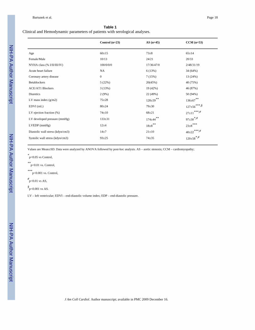
Baseline characteristics of Control, AS and CCM patients undergoing serological analyses areshown in Table 1. LV mass index was higher in AS and CCM patients compared to Controls.CCM patients had larger end-diastolic volumes (EDVI), lower ejection fractions (LV EF), andhigher diastolic and systolic wall stress compared to Controls. LV EDP was elevated in ASand CCM groups compared to Controls. LV developed pressure was increased in the AS groupand decreased in the CCM group compared to Controls. There were no differences in clinicalor hemodynamic characteristics between both subpopulations. In both populations, 64% and52% of CCM patients, respectively, presented with acute decompensation or worsening ofcongestive heart failure symptoms.
Serum ST2, hs-CRP and NT-pro BNP levelsSerum levels of ST2 (Figure 1A, left) were significantly increased in patients with AS (103[65-165] pg/mL, p<0.05) and CCM (194[69-551] pg/mL, p<0.01) compared to Controls (49[4-89] pg/mL). NT-pro-BNP (Figure 1A, middle) was significantly increased in AS (537[253-1936] pg/mL, p<0.05) and CCM patients (3088[629-7846] pg/mL, p<0.01) vs. Controls(43[28-100] pg/mL). Hs-CRP (Figure 1A, right) was significantly increased in patients withAS (6.1[1.3-15] mg/L, p<0.05) and CCM (12.0[3.3-29.2] mg/L, p<0.01) vs. Controls (2.2[1.2-3.0]). In the entire population, a significant positive relationship was noted between ST2and NT-pro-BNP (Figure 1B, left, r=0.47, p <0.05) and between ST2 and hs-CRP (Figure 1B,right, r=0.55, p<0.001).
Serum ST2 and hemodynamic parametersNo significant relationship was noted between serum ST2 levels and indices of LV remodellingincluding LV end-diastolic or end-systolic volume, LV mass index and LV relative wallthickness. Furthermore, ST2 levels did not correlate with LV systolic developed pressure or
Bartunek et al. Page 4
J Am Coll Cardiol. Author manuscript; available in PMC 2009 December 16.
NIH
-PA Author Manuscript
NIH
-PA Author Manuscript
NIH
-PA Author Manuscript

systolic wall stress. However, in the entire study population, a weak but significant, inverserelationship was noted between ST2 levels and LV ejection fraction(r= -0.21,p<005). On theother hand, ST2 levels showed stronger relationship with LV EDP(r= 0.37, p<0.01).
ST2 and Diastolic LoadIn the entire cohort (Figure 2, left), serum ST2 levels were higher in patients with moderatelyor severely elevated LV EDP as compared to patients with normal or mildly increased LVEDP. ST2 levels were increased proportionally with extent of diastolic load as assessed fromelevated levels of LV EDP (Figure 2, right panel).
To further determine whether diastolic load is associated with ST2 levels independent of LVsystolic function (ejection fraction), ST2 was examined in patients (N=14) presenting withisolated diastolic heart failure (HF) and normal LV systolic function (Figure 3A, left). SerumST2 was higher in these patients compared to Controls (383±149 vs. 75±12 pg/mL, p<0.001).Likewise, NT-pro-BNP was also increased compared to Controls (901±293 vs. 88±31 pg/mL,p=0.001, Figure 3A, right). The corresponding LV EDP (18±10 vs. 12±4 mmHg,p=0.03)and ejection fraction (73±13 vs. 74±7%,NS) are shown (Figure 3B). These results suggest thatdiastolic load is a hemodynamic factor that modulates ST2 production.
Myocardial production of BNP and ST2To determine myocardial production of BNP and ST2, the coronary sinus minus arterial bloodconcentration gradient was determined in 24 patients with chronic heart failure undergoingimplantation of biventricular pacemakers. Hemodynamic characteristics of these patients withcorresponding LV EDP (22±9 vs. 23±9 mmHg, p=NS) and ejection fraction (27±9 vs. 27±9%,NS) were similar to CCM group undergoing serological analyses. Arterial and coronary sinuslevels of NT-pro-BNP were 1069±933 and 1798±1178 pg/mL respectively, with a differenceof 729±410 pg/mL, p<0.0001 sinus vs. arterial (Figure 4A, left). Arterial and sinus levels ofST2 were 275±195 and 264±171 pg/mL respectively, with a differences of −11±39 pg/mL,p=NS (Figure 4A, right). These results demonstrate myocardial production of BNP but not ofST2. In LV myocardial biopsies (characteristics in Table 2), BNP mRNA levels were 4.4-foldincreased in AS (log AU, 0.98±1.16) and 43 fold in CCM (log AU, 2.71±0.41) compared toControl (log AU, 0.27±1.11, p<0.001) (Figure 4B, left). Serum NT-pro-BNP levels correlatedwith their corresponding myocardial BNP mRNA levels (samples from the same patients, r =0.54,p=0.01). ST2 mRNA levels were not significantly increased in AS (log AU 0.73±0.66)and CCM patients (log AU, 0.85±0.39) compared to Controls (log AU, 0.55±0.20, p=NS)(Figure 4B, right) with no correlation between serum ST2 and myocardial ST2 mRNA.
ST2 secretion by venous and arterial endothelial cellsPhorbol-ester induced ST2 secretion in venous endothelial cells compared to Control (299±22vs. 92±6 pg/mL, p<0.001)) (Figure 5). Interleukin-1β induced ST2 secretion in venous andarterial endothelial cells (venous, 125±6 vs. 92±6 pg/mL, p<0.001); arterial, 103±48 vs. 50±24pg/mL, p<0.05). TNF-α also induced ST2 secretion (115±4 pg/mL) compared to Control (96±6 pg/mL p<0.001). H2O2 blocked basal ST2 secretion in endothelial cells (16±4 vs. 50±24pg/mL, p<0.05) demonstrating that local oxidative stress can inhibit ST2 secretion. ST2secretion was elevated by 3 hours compared to Control (150±20 vs. 52±12 pg/mL, p<0.001),reached peak levels at 6 hours (304±14 vs. 51±10 pg/mL, p<0.001) (Figure 5, right) anddeclined afterwards (not shown). Brefeldin A, a blocker of protein secretion through the ER/Golgi, in the presence of phorbol ester completely blocked ST2 secretion (Figure 5, right).Data represent 3-4 experiments with duplicate samples.
Bartunek et al. Page 5
J Am Coll Cardiol. Author manuscript; available in PMC 2009 December 16.
NIH
-PA Author Manuscript
NIH
-PA Author Manuscript
NIH
-PA Author Manuscript
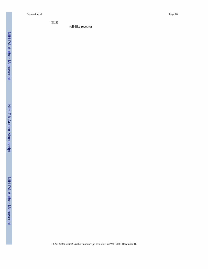
Expression of IL-33 and ST2L in the heart and endotheliumIL-33 mRNA levels were modestly lower in LV biopsies from AS patients (0.80±0.33 AU)compared to CCM patients (1.33±0.48 AU,p=0.005, Figure 6A, left). Levels of membrane-anchored ST2L were significantly decreased in AS compared to Control (0.27±0.22 vs. 1.00±0.60 AU; p<0.05) but levels were similar between CCM (0.92±0.72 AU) and Control(NS,Figure 6A, left). IL-33 protein localizes to endothelial cells in the human coronary artery(Figure 6B). A weak correlation between IL-33 and ST2L mRNA expression in myocardialbiopsies was observed (r=0.35, p=0.045).
Peripheral leukocyte expression of ST2, ST2L and IL-33Levels of soluble ST2 mRNA in peripheral leukocytes were similar in all groups (Control, 2.0±1.4; AS, 4.3±3.6; CCM, 4.2±2.8 AU; NS) as were levels of ST2L (Control, 20.1±15.9; AS,23.8±17; CCM, 16.0±14 AU, NS) and IL-33 (Control, 0.94±0.84; AS, 1.13±1,1; CCM, 0.63±0.77 AU; NS). However, a strong correlation was noted between IL33 and ST2L mRNA inleukocytes over the range of all values (Figure 7. r = 0.996; p<0.001).
DiscussionThe present study shows that serum ST2 protein is elevated in patients with chronic LV pressureoverload due to aortic stenosis as well as in patients with congestive heart failure. ST2positively correlated with NT-pro-BNP and the inflammatory marker CRP. ST2 levels wereproportionally related to extent of diastolic load. A dominant role of diastolic load inrelationship to ST2 levels is further supported by findings in a subgroup of patients with normalsystolic function and isolated diastolic failure. In this group, ST2 levels were increased as wereNT-pro-BNP levels. Thus, ST2 induction appears to be sensitive primarily to diastolic load.However, it is interesting that unlike BNP induction(19,23), ST2 production did not show asigificant relationship with systolic wall stress.
The source of serum ST2 in cardiovascular disease was presumed to be myocardial followingin vitro data showing load induction of ST2 mRNA in neonatal rat cardiac myocytes(24). Weprovide direct evidence that the adult, human myocardium is not the source of increased serumST2 in pressure overload hypertrophy and congestive cardiomyopathy. First, it is well-established that ST2 is transcriptionally regulated (mRNA induction)(7,8), yet ST2 mRNAlevels were not significantly increased in myocardial biopsies. This is in contrast with BNPexpression, which was increased in the same RNA pool of LV samples reflecting increasedlevels of serum NT-pro-BNP in the same patients. Second, direct measurements of the BNPand ST2 protein concentration in coronary sinus and arterial blood samples demonstrated atransmyocardial gradient (production) of BNP, but no transmyocardial gradient of ST2. Thesesurprising findings prompted us to examine ST2 protein production in extra-myocardial, non-myocyte cell types. We present the novel findings that human venous and arterial endothelialcells secrete ST2 protein. ST2 protein synthesis and release was rapid, peaked within 6 hoursand was blocked by brefeldin, which blocks intracellular protein transport(25), confirming thatST2 exits endothelial cells through a secretion pathway responsive to inflammatory signals.This finding, together with the relationship between serum ST2 and indices of diastolic load,suggest that the vascular endothelium, sensing hemodynamic and inflammatory status, is apotential source of elevated serum ST2 levels in hemodynamic overload and heart failure.
In this regard, it is noteworthy that ST2 levels failed to provide prognostic information inpatients with acute coronary syndrome(26). Interestingly, authors noticed highest serum ST2levels in chronic alcoholics, patients with systemic sepsis, and concomittant lung diseases,consistent with studies reporting elevated serum ST2 in pulmonary diseases(13,27). SerumST2 is also elevated in patients with dengue virus infection(28), sepsis and trauma(29), and in
Bartunek et al. Page 6
J Am Coll Cardiol. Author manuscript; available in PMC 2009 December 16.
NIH
-PA Author Manuscript
NIH
-PA Author Manuscript
NIH
-PA Author Manuscript

CSF fluid following subarachnoid hemorrhage(30). The broad specificity of ST2 inductioncorroborates its nonmyocardial production and may explain why ST2 performs well as anindependent biomarker in multi-marker studies, providing information distinct from BNP incardiovascular disease.
In addition to a cardiovascular biomarker, ST2/ST2L signaling is likely to be important incardiovascular disease modulation. At the cellular level, soluble ST2 binds to macrophagesand downregulates proinflammatory cytokines, IL-1, IL-6 and TNF-α(14,15). Soluble ST2 canbind to IL-33, the ST2L ligand, to diminish MAP kinase and NFkB signaling(13). Membrane-anchored ST2L inhibits also IL-1 receptor and innate immunity TLR4 signaling (11,12,31).Hence, both ST2L and soluble ST2 negatively regulate IL-1 receptor and TLR4 signaling(12,32). We present the novel findings that ST2L and IL-33 mRNA are expressed in normaland diseased human myocardium and both are modestly but significantly decreased in patientswith aortic stenosis. The mechanism is unknown, although it is recognized that soluble vs.membrane-anchored ST2L are regulated separately(7,8). In addiiton, we found that IL-33protein is expressed in endothelial cells within coronary arteries suggesting that IL-33 mayhave a similar function in vascular endothelium that may be regulated by soluble ST2. Wereport a remarkable pronounced correlation between ST2L and IL-33 levels in peripheral bloodleukocytes, suggesting that they are highly co-regulated in these cells. Further studies areneeded to determine the biological relevance of these observations.
LimitationsThe present study is limited to patients with chronic aortic stenosis or heart failure. It wouldbe interesting to interrogate myocardial production of ST2 protein in acute myocardialinfarction following which serum ST2 levels are also increased. Though the lack oftransmyocardial ST2 gradient argues against cardiac production of serum ST2, ST2 proteinproduction in human adult cardiac myocytes was not directly tested.
ImplicationsThis study describes elevated serum ST2 in patients with pressure overload hypertrophy andcongestive or diastolic heart failure. Our data support diastolic load as a predominanthemodynamic factor that contributes to ST2 production in heart disease. Note, our data indicatethat overloaded myocardium is not a major source of increased serum ST2. We showed thatendothelial cells possess a functional ST2 secretory system in vitro, providing proof-of-conceptfor ST2 production by the vascular endothelium in vivo, which can be investigated in futurestudies.
AcknowledgementsThis study was funded in part by NIH (Bethesda, MD) grant HL069484 (EOW), by 3M Pharma Prize Award (JB andMV), and by Meijer Lavino Foundation for Cardiac Research, Aalst, Belgium.
References1. Januzzi JL Jr, Peacock WF, Maisel AS, et al. Measurement of the interleukin family member ST2 in
patients with acute dyspnea: results from the PRIDE (Pro-Brain Natriuretic Peptide Investigation ofDyspnea in the Emergency Department) study. J Am Coll Cardiol 2007;50:607–13. [PubMed:17692745]
2. Shimpo M, Morrow DA, Weinberg EO, et al. Serum levels of the interleukin-1 receptor family memberST2 predict mortality and clinical outcome in acute myocardial infarction. Circulation 2004;109:2186–90. [PubMed: 15117853]
3. Weinberg EO, Shimpo M, Hurwitz S, Tominaga S, Rouleau JL, Lee RT. Identification of serum solubleST2 receptor as a novel heart failure biomarker. Circulation 2003;107:721–6. [PubMed: 12578875]
Bartunek et al. Page 7
J Am Coll Cardiol. Author manuscript; available in PMC 2009 December 16.
NIH
-PA Author Manuscript
NIH
-PA Author Manuscript
NIH
-PA Author Manuscript

4. Mueller T, Dieplinger B, Gegenhuber A, Poelz W, Pacher R, Haltmayer M. Increased plasmaconcentrations of soluble ST2 are predictive for 1-year mortality in patients with acute destabilizedheart failure. Clin Chem 2008;54:752–6. [PubMed: 18375488]
5. Sabatine MS, Morrow DA, Higgins LJ, et al. Complementary roles for biomarkers of biomechanicalstrain ST2 and N-terminal prohormone B-type natriuretic peptide in patients with ST-elevationmyocardial infarction. Circulation 2008;117:1936–44. [PubMed: 18378613]
6. Rehman SU, Martinez-Rumayor A, Mueller T, Januzzi JL Jr. Independent and incremental prognosticvalue of multimarker testing in acute dyspnea: results from the ProBNP Investigation of Dyspnea inthe Emergency Department (PRIDE) study. Clin Chim Acta 2008;392:41–5. [PubMed: 18387360]
7. Bergers G, Reikerstorfer A, Braselmann S, Graninger P, Busslinger M. Alternative promoter usage ofthe Fos-responsive gene Fit-1 generates mRNA isoforms coding for either secreted or membrane-bound proteins related to the IL-1 receptor. Embo J 1994;13:1176–88. [PubMed: 8131748]
8. Iwahana H, Yanagisawa K, Ito-Kosaka A, et al. Different promoter usage and multiple transcriptioninitiation sites of the interleukin-1 receptor-related human ST2 gene in UT-7 and TM12 cells. Eur JBiochem 1999;264:397–406. [PubMed: 10491084]
9. Schmitz J, Owyang A, Oldham E, et al. IL-33, an interleukin-1-like cytokine that signals via the IL-1receptor-related protein ST2 and induces T helper type 2-associated cytokines. Immunity2005;23:479–90. [PubMed: 16286016]
10. Yanagisawa K, Takagi T, Tsukamoto T, Tetsuka T, Tominaga S. Presence of a novel primary responsegene ST2L, encoding a product highly similar to the interleukin 1 receptor type 1. Lett FEBS1993;318:83–7.
11. Brint EK, Xu D, Liu H, et al. ST2 is an inhibitor of interleukin 1 receptor and Toll-like receptor 4signaling and maintains endotoxin tolerance. Nat Immunol 2004;5:373–9. [PubMed: 15004556]
12. Liew FY, Xu D, Brint EK, O’Neill LA. Negative regulation of toll-like receptor-mediated immuneresponses. Nat Rev Immunol 2005;5:446–58. [PubMed: 15928677]
13. Hayakawa H, Hayakawa M, Kume A, Tominaga S. Soluble ST2 blocks interleukin-33 signaling inallergic airway inflammation. J Biol Chem 2007;282:26369–80. [PubMed: 17623648]
14. Sweet MJ, Leung BP, Kang D, et al. A novel pathway regulating lipopolysaccharide-induced shockby ST2/T1 via inhibition of Toll-like receptor 4 expression. J Immunol 2001;166:6633–9. [PubMed:11359817]
15. Takezako N, Hayakawa M, Hayakawa H, et al. ST2 suppresses IL-6 production via the inhibition ofIkappaB degradation induced by the LPS signal in THP-1 cells. Biochem Biophys Res Commun2006;341:425–32. [PubMed: 16426569]
16. Yanagisawa K, Naito Y, Kuroiwa K, et al. The expression of ST2 gene in helper T cells and thebinding of ST2 protein to myeloma-derived RPMI8226 cells. J Biochem (Tokyo) 1997;121:95–103.[PubMed: 9058198]
17. Baekkevold ES, Roussigne M, Yamanaka T, et al. Molecular characterization of NF-HEV, a nuclearfactor preferentially expressed in human high endothelial venules. Am J Pathol 2003;163:69–79.[PubMed: 12819012]
18. Carriere V, Roussel L, Ortega N, et al. IL-33, the IL-1-like cytokine ligand for ST2 receptor, is achromatin-associated nuclear factor in vivo. Proc Natl Acad Sci U S A 2007;104:282–7. [PubMed:17185418]
19. Vanderheyden M, Goethals M, Verstreken S, et al. Wall stress modulates brain natriuretic peptideproduction in pressure overload cardiomyopathy. J Am Coll Cardiol 2004;44:2349–54. [PubMed:15607397]
20. ACC/AHA guidelines for the clinical application of echocardiography. A report of the AmericanCollege of Cardiology/American Heart Association Task Force on Assessment of Diagnostic andTherapeutic Cardiovascular Procedures. Circulation 1990;82:2323–45. [PubMed: 2242558]
21. Douglas PS, Reichek N, Plappert T, Muhammad A, St John Sutton MG. Comparison ofechocardiographic methods for assessment of left ventricular shortening and wall stress. J Am CollCardiol 1987;9:945–51. [PubMed: 3558991]
22. Devereux RB, Alonso DR, Lutas, et al. Echocardiographic assessment of left ventricular hypertrophy:comparison to necropsy findings. Am J Cardiol 1986;57:450–8. [PubMed: 2936235]
Bartunek et al. Page 8
J Am Coll Cardiol. Author manuscript; available in PMC 2009 December 16.
NIH
-PA Author Manuscript
NIH
-PA Author Manuscript
NIH
-PA Author Manuscript

23. Maeda K, Tsutamoto T, Wada A, Hisanaga T, Kinoshita M. Plasma brain natriuretic peptide as abiochemical marker of high left ventricular end-diastolic pressure in patients with symptomatic leftventricular dysfunction. Am Heart J 1998;135:825–32. [PubMed: 9588412]
24. Weinberg EO, Shimpo M, De Keulenaer GW, et al. Expression and regulation of ST2, an interleukin-1receptor family member, in cardiomyocytes and myocardial infarction. Circulation 2002;106:2961–6. [PubMed: 12460879]
25. Xie L, Boyle D, Sanford D, Scherer PE, Pessin JE, Mora S. Intracellular trafficking and secretion ofadiponectin is dependent on GGA-coated vesicles. J Biol Chem 2006;281:7253–9. [PubMed:16407204]
26. Brown AM, Wu AH, Clopton P, Robey JL, Hollander JE. ST2 in emergency department chest painpatients with potential acute coronary syndromes. Ann Emerg Med 2007;50:153–8. 158 e1. [PubMed:17466411]
27. Oshikawa K, Yanagisawa K, Tominaga S, Sugiyama Y. ST2 protein induced by inflammatory stimulican modulate acute lung inflammation. Biochem Biophys Res Commun 2002;299:18–24. [PubMed:12435383]
28. Becerra A, Warke RV, de Bosch N, Rothman AL, Bosch I. Elevated levels of soluble ST2 protein indengue virus infected patients. Cytokine 2008;41:114–20. [PubMed: 18226917]
29. Brunner M, Krenn C, Roth G, et al. Increased levels of soluble ST2 protein and IgG1 production inpatients with sepsis and trauma. Intensive Care Med 2004;30:1468–73. [PubMed: 14991091]
30. Kanda M, Ohto-Ozaki H, Kuroiwa K, Tominaga S, Watanabe E, Iwahana H. Elevation of ST2 proteinlevels in cerebrospinal fluid following subarachnoid hemorrhage. Acta Neurol Scand 2006;113:327–33. [PubMed: 16629769]
31. Dunne A, O’Neill LA. The interleukin-1 receptor/Toll-like receptor superfamily: signal transductionduring inflammation and host defense. Sci STKE 2003 2003:re3.
32. Trajkovic V, Sweet MJ, Xu D. T1/ST2--an IL-1 receptor-like modulator of immune responses.Cytokine Growth Factor Rev 2004;15:87–95. [PubMed: 15110792]
List of abbreviationsAS
aortic stenosis
CCM cardiomyopathy
LV left ventricular
BNP B-type natriuretic peptide
NT-pro-BNP N-terminal-pro-brain natriuretic peptide
EDP end-diastolic pressure
EF ejection fraction
IL interleukin
hsCRP high sensitivity C-reactive protein
Bartunek et al. Page 9
J Am Coll Cardiol. Author manuscript; available in PMC 2009 December 16.
NIH
-PA Author Manuscript
NIH
-PA Author Manuscript
NIH
-PA Author Manuscript
TLR toll-like receptor
Bartunek et al. Page 10
J Am Coll Cardiol. Author manuscript; available in PMC 2009 December 16.
NIH
-PA Author Manuscript
NIH
-PA Author Manuscript
NIH
-PA Author Manuscript

Figure 1. Serum levels of humoral markersA. Serum levels of ST2, CRP and BNP in AS, CCM and control patients. Box-plots showmedian [25th-75th]. B. Scatterplot between NT-pro-BNP and ST2 (left) and CRP and ST2(right) in all patients.
Bartunek et al. Page 11
J Am Coll Cardiol. Author manuscript; available in PMC 2009 December 16.
NIH
-PA Author Manuscript
NIH
-PA Author Manuscript
NIH
-PA Author Manuscript

Figure 2. Relationship between serum ST2 levels and extent of diastolic load as assessed from leftventricular end diastolic pressure (LVEDP)Serum ST2 levels according to LVEDP in all study subjects(left panel), and in Controls andin CCM patients(right panel). Box-plots show median [25th-75th].
Bartunek et al. Page 12
J Am Coll Cardiol. Author manuscript; available in PMC 2009 December 16.
NIH
-PA Author Manuscript
NIH
-PA Author Manuscript
NIH
-PA Author Manuscript

Figure 3. Serum ST2 and BNP in patients with isolated diastolic heart failureA. Serum ST2 (left) and BNP (right) levels in patients with isolated diastolic failure. Data weretransformed due to heterogeneity of variance into the log scale. B. LVEDP (left) and LVejection fraction in patients with diastolic failure compared to control patients.
Bartunek et al. Page 13
J Am Coll Cardiol. Author manuscript; available in PMC 2009 December 16.
NIH
-PA Author Manuscript
NIH
-PA Author Manuscript
NIH
-PA Author Manuscript

Figure 4. Transmyocardial gradient of and myocardial RNA levels of BNP and ST2A. Transmyocardial production of BNP (left) and ST2 (right) in individual patients (N=24)with heart failure. Mean±SD are also shown in addition to individual values. B. LV BNPmRNA levels (left) in Control, AS and CCM cardiac biopsies. LV ST2 mRNA levels (right)are not significantly different in all groups. AU: arbitrary units.
Bartunek et al. Page 14
J Am Coll Cardiol. Author manuscript; available in PMC 2009 December 16.
NIH
-PA Author Manuscript
NIH
-PA Author Manuscript
NIH
-PA Author Manuscript

Figure 5. ST2 protein secretion by human venous and arterial endothelial cellsPhorbol ester, IL-1β and tumor necrosis factor-1 induced ST2 secretion in venous endothelialcells (left). IL-1β induced secretion while H2O2 blocked secretion in arterial endothelial cells(middle). Time course shows ST2 is rapidly synthesized and secreted with significantlyelevated levels at 3 hours and reaches peak values at 6 hours (right). Brefeldin A blocked ST2secretion (right).
Bartunek et al. Page 15
J Am Coll Cardiol. Author manuscript; available in PMC 2009 December 16.
NIH
-PA Author Manuscript
NIH
-PA Author Manuscript
NIH
-PA Author Manuscript
Figure 6. IL33 and ST2 mRNA levels in myocardial biopsiesA. IL-33 mRNA levels were decreased in AS hearts compared to CCM hearts. ST2L mRNAlevels were significantly decreased in AS hearts compared to Control. ST2L levels were similarin CCM hearts compared to Control (middle). B. IL-33 protein was expressed in humancoronary artery endothelium (brown staining).
Bartunek et al. Page 16
J Am Coll Cardiol. Author manuscript; available in PMC 2009 December 16.
NIH
-PA Author Manuscript
NIH
-PA Author Manuscript
NIH
-PA Author Manuscript

Figure 7. Correlation between ST2L and IL-33 in leukocytesRelationship between ST2L vs. IL-33 mRNA levels in leukocytes from Control, AS and CCMpatients.
Bartunek et al. Page 17
J Am Coll Cardiol. Author manuscript; available in PMC 2009 December 16.
NIH
-PA Author Manuscript
NIH
-PA Author Manuscript
NIH
-PA Author Manuscript
NIH
-PA Author Manuscript
NIH
-PA Author Manuscript
NIH
-PA Author Manuscript
Bartunek et al. Page 18
Table 1Clinical and Hemodynamic parameters of patients with serological analyses.
Control (n=23) AS (n=45) CCM (n=53)
Age 60±15 73±8 65±14
Female/Male 10/13 24/21 20/33
NYHA class (% I/II/III/IV) 100/0/0/0 17/36/47/0 2/48/31/19
Acute heart failure NA 6 (13%) 34 (64%)
Coronary artery disease 0 7 (15%) 13 (24%)
Betablockers 5 (22%) 20(45%) 40 (75%)
ACE/AT1 Blockers 3 (13%) 19 (42%) 46 (87%)
Diuretics 2 (9%) 22 (49%) 50 (94%)
LV mass index (g/m2) 75±28 128±59** 136±67**
EDVI (mL) 80±24 79±30 127±56***,§
LV ejection fraction (%) 74±10 68±21 27±11***,#
LV developed pressure (mmHg) 133±31 174±44** 97±30*,#
LVEDP (mmHg) 12±4 18±8** 23±8***
Diastolic wall stress (kdyn/cm3) 14±7 21±10 40±22***,#
Systolic wall stress (kdyn/cm3) 93±25 74±35 120±58*,#
Values are Mean±SD. Data were analyzed by ANOVA followed by post-hoc analysis. AS – aortic stenosis; CCM – cardiomyopathy;
*p<0.05 vs Control,
**p<0.01 vs. Control,
***p<0.001 vs. Control,
#p<0.01 vs AS,
§p<0.001 vs AS.
LV – left ventricular; EDVI – end-diastolic volume index; EDP – end-diastolic pressure.
J Am Coll Cardiol. Author manuscript; available in PMC 2009 December 16.

NIH
-PA Author Manuscript
NIH
-PA Author Manuscript
NIH
-PA Author Manuscript
Bartunek et al. Page 19
Table 2Clinical and hemodynamic parameters of patients with gene expression analysis
Control (n=9) AS (n=12) CCM (n=17)
Age 61±13 71±9 58±16
Female/Male 1/8 6/6 4/13
NYHA class 100/0/0/0 25/67/8/0 6/42/35/17
Acute heart failure NA 2 (12%) 9 (52%)
Coronary artery disease 6 (66%) 3 (25%) 3 (17%)
Betablockers 4 (44%) 1 (8%) 10 (57%)
ACE/AT1 Blockers 4 (44%) 5 (42%) 17 (100%)
Diuretics 2 (22%) 5 (42%) 10 (57%)
LV mass index (g/m2) 52±18 117±40** 120±25**
EDVI (mL) 77±14 83±25 145±58***,§
LV ejection fraction (%) 72±11 67±18 24±11***,#
LV developed pressure (mmHg) 117±26 178±46* 90±25*,#
LVEDP (mmHg) 13±3 17±6** 21±7***
Diastolic wall stress (kdyn/cm3) 18±9 19±9 38±15***,#
Systolic wall stress (kdyn/cm3) 70±25 69±32 134±27*,#
Values are Mean±SD. Data were analyzed by ANOVA followed by post-hoc analysis. AS – aortic stenosis; CCM – cardiomyopathy;
*p<0.05 vs Control,
**p<0.01 vs. Control,
***p<0.001 vs. Control,
#p<0.01 vs AS,
§p<0.001 vs AS.
LV – left ventricular; EDVI – end-diastolic volume index; EDP – end-diastolic pressure.
J Am Coll Cardiol. Author manuscript; available in PMC 2009 December 16.
Copyright © 2022 FDOKUMEN