Left and right ventricular diastolic dysfunction as predictors of difficult separation from...
10
1020 CANADIAN JOURNAL OF ANESTHESIA Purpose: As the evaluation of diastolic function can be complex in the setting of a busy cardiac operating room, its assessment may benefit from an algorithmic approach using transesopha- geal echocardiography. We developed a diagnostic algorithm which was then applied in a series of cardiac surgery patients to determine whether moderate to severe left ventricular diastolic dysfunction (LVDD) and right ventricular diastolic dysfunction (RVDD) can predict difficult separation from cardiopulmonary bypass (DSB). Methods: An algorithm using pulsed-wave Doppler interroga- tion of the mitral and tricuspid valve, the pulmonary and hepatic venous flow, and tissue Doppler interrogation of the mitral and tricuspid annulus was developed. The study was divided in two phases involving two groups of patients undergoing cardiac sur- gery. In phase I, echocardiographic evaluations of patients (n = 74) were used to test the reproducibility of the algorithm and to evaluate inter-observer variability using Cohen’s kappa values which were calculated in three specific periods. In phase II, the algorithm was applied to a second group of patients (validation group, n = 179) to explore its prognostic significance. The pri- mary end-point in phase II was DSB. Results: In phase I, the kappa coefficients for LVDD and RVDD algorithms were 0.77 and 0.82, respectively. In phase II, moderate or severe degrees of LVDD were observed in 29 patients (16%) and moderate to severe RVDD was observed in 18 patients (10%) before cardiac surgery. Both moderate and severe LVDD (P = 0.017) and RVDD (P = 0.049) before sur- gery were observed more frequently in patients with DSB. Conclusion: Moderate and severe LVDD and RVDD can be identified with very good reproducibility, and both degrees of diastolic dysfunction are associated with DSB. Objectif: L’évaluation de la fonction diastolique pouvant être com- plexe dans le contexte d’un bloc opératoire très actif en chirurgie cardiaque, on pourrait tirer profit d’un algorithme pour son évalua- tion avec l’échocardiographie transœsophagienne. Nous avons développé un algorithme en cardiochirurgie pour déterminer si la dysfonction ventriculaire diastolique gauche (DVDG) modérée ou sévère et la dysfonction ventriculaire diastolique droite (DVDD) pouvaient être des prédicteurs de difficultés de sevrage de la circu- lation extracorporelle (DSCE). Méthode : Un algorithme utilisant l’examen Doppler pulsé pour évaluer la vélocité des valvules mitrale et tricuspide, la vélocité des veines pulmonaire et hépatique et l’examen Doppler tissulaire 1020 CAN J ANESTH 2006 / 53: 10 / pp 1020–1029 Cardiothoracic Anesthesia, Respiration and Airway Left and right ventricular diastolic dysfunction as predictors of difficult separation from cardiopul- monary bypass [La dysfonction ventriculaire diastolique gauche et droite comme prédicteur des difficultés de sevrage de la circulation extracorporelle] André Y. Denault MD FRCPC,* Pierre Couture MD FRCPC,* Jean Buithieu MD FRCPC,¶ Francois Haddad MD FRCPC,* Michel Carrier MD FRCPC,‡ Denis Babin MSc,* Sylvie Levesque MSc,§ Jean-Claude Tardif MD FRCPC† From the Departments of Anesthesia,* Medicine,† Cardiac Surgery,‡ and Biostatistics,§ Montreal Heart Institute; Université de Montréal; and the Department of Medicine,¶ McGill Health Center, Montréal, Québec, Canada. Address correspondence to: Dr. André Y. Denault, Department of Anesthesia, Montreal Heart Institute, 5000 Belanger Street East, Montréal, Québec H1T 1C8, Canada. Phone: 514-376-3330; Fax: 514-376-8784; E-mail: [email protected] Acknowledgement of funding: Supported in part by by the Dr. Earl Wynands Research Award in Cardiovascular Anesthesia and/or Peri- operative Blood Conservation, le Fonds de la recherche en santé du Québec, les Instituts de recherche en santé du Canada et la Fondation de l’Institut de Cardiologie de Montréal. Dr .Tardif holds the Pfizer and Canadian Institutes of Health Research Chair in atherosclerosis. Presented in part at the 2004 Annual Meeting of the Canadian Anesthesiologists’ Society. Accepted for publication October 1, 2005. Revision accepted May 15, 2006. Final revision accepted June 15, 2006. Competing interests: None declared.
Transcript of Left and right ventricular diastolic dysfunction as predictors of difficult separation from...

1020 CANADIAN JOURNAL OF ANESTHESIA
Purpose: As the evaluation of diastolic function can be complex in the setting of a busy cardiac operating room, its assessment may benefit from an algorithmic approach using transesopha-geal echocardiography. We developed a diagnostic algorithm which was then applied in a series of cardiac surgery patients to determine whether moderate to severe left ventricular diastolic dysfunction (LVDD) and right ventricular diastolic dysfunction (RVDD) can predict difficult separation from cardiopulmonary bypass (DSB).
Methods: An algorithm using pulsed-wave Doppler interroga-tion of the mitral and tricuspid valve, the pulmonary and hepatic venous flow, and tissue Doppler interrogation of the mitral and tricuspid annulus was developed. The study was divided in two phases involving two groups of patients undergoing cardiac sur-gery. In phase I, echocardiographic evaluations of patients (n = 74) were used to test the reproducibility of the algorithm and to evaluate inter-observer variability using Cohen’s kappa values which were calculated in three specific periods. In phase II, the algorithm was applied to a second group of patients (validation group, n = 179) to explore its prognostic significance. The pri-mary end-point in phase II was DSB.
Results: In phase I, the kappa coefficients for LVDD and RVDD algorithms were 0.77 and 0.82, respectively. In phase
II, moderate or severe degrees of LVDD were observed in 29 patients (16%) and moderate to severe RVDD was observed in 18 patients (10%) before cardiac surgery. Both moderate and severe LVDD (P = 0.017) and RVDD (P = 0.049) before sur-gery were observed more frequently in patients with DSB.
Conclusion: Moderate and severe LVDD and RVDD can be identified with very good reproducibility, and both degrees of diastolic dysfunction are associated with DSB.
Objectif: L’évaluation de la fonction diastolique pouvant être com-plexe dans le contexte d’un bloc opératoire très actif en chirurgie cardiaque, on pourrait tirer profit d’un algorithme pour son évalua- tion avec l’échocardiographie transœsophagienne. Nous avons développé un algorithme en cardiochirurgie pour déterminer si la dysfonction ventriculaire diastolique gauche (DVDG) modérée ou sévère et la dysfonction ventriculaire diastolique droite (DVDD) pouvaient être des prédicteurs de difficultés de sevrage de la circu-lation extracorporelle (DSCE).
Méthode : Un algorithme utilisant l’examen Doppler pulsé pour évaluer la vélocité des valvules mitrale et tricuspide, la vélocité des veines pulmonaire et hépatique et l’examen Doppler tissulaire
1020
CAN J ANESTH 2006 / 53: 10 / pp 1020–1029
Cardiothoracic Anesthesia, Respiration and Airway
Left and right ventricular diastolic dysfunction as predictors of difficult separation from cardiopul-monary bypass [La dysfonction ventriculaire diastolique gauche et droite comme prédicteur des
difficultés de sevrage de la circulation extracorporelle] André Y. Denault MD FRCPC,* Pierre Couture MD FRCPC,* Jean Buithieu MD FRCPC,¶ Francois Haddad MD FRCPC,* Michel Carrier MD FRCPC,‡ Denis Babin MSc,* Sylvie Levesque MSc,§ Jean-Claude Tardif MD FRCPC†
From the Departments of Anesthesia,* Medicine,† Cardiac Surgery,‡ and Biostatistics,§ Montreal Heart Institute; Université de Montréal; and the Department of Medicine,¶ McGill Health Center, Montréal, Québec, Canada.
Address correspondence to: Dr. André Y. Denault, Department of Anesthesia, Montreal Heart Institute, 5000 Belanger Street East, Montréal, Québec H1T 1C8, Canada. Phone: 514-376-3330; Fax: 514-376-8784; E-mail: [email protected] of funding: Supported in part by by the Dr. Earl Wynands Research Award in Cardiovascular Anesthesia and/or Peri-operative Blood Conservation, le Fonds de la recherche en santé du Québec, les Instituts de recherche en santé du Canada et la Fondation de l’Institut de Cardiologie de Montréal. Dr .Tardif holds the Pfizer and Canadian Institutes of Health Research Chair in atherosclerosis.
Presented in part at the 2004 Annual Meeting of the Canadian Anesthesiologists’ Society.Accepted for publication October 1, 2005.Revision accepted May 15, 2006.Final revision accepted June 15, 2006.Competing interests: None declared.
Denault et al.: DIASTOLIC DYSFUNCTION AND CARDIAC SURGERY 1021
des anneaux mitral et tricuspide a été mis au point. L’étude, en deux phases, a comporté deux groupes de patients devant subir une opération cardiaque. Pendant la phase I, des évaluations échocardiographiques de patients (n = 74) ont permis de véri-fier la reproductibilité de l’algorithme et d’évaluer la variabilité inter-observateur d’après les valeurs Kappa de Cohen qui ont été calculées à trois moments spécifiques. Pendant la phase II, l’algorithme a été appliqué au second groupe de patients (groupe de validation, n = 179) pour explorer sa portée pronostique. Le principal paramètre de la phase II était les DSCE.
Résultats : Pendant la phase I, les coefficients kappa pour les algorithmes de DVDG et DVDD ont été respectivement de 0,77 et 0,82. Pendant la phase II, des DVDG modérées ou sévères ont été observées chez 29 patients (16 %) et des DVDD chez 18 patients (10 %) avant l’opération cardiaque. Des DVDG modérées et sévères (P = 0,017) et des DVDD (P = 0,049) préchirurgicales ont été observées plus souvent chez les patients qui présentaient des DSCE.
Conclusion : Des DVDG et des DVDD modérées et sévères peu-vent être observées avec une très bonne reproductibilité et les deux degrés de dysfonction diastolique sont associés à des DSCE.
THERE is growing interest in evaluating diastolic function1 in cardiology and in cardiac surgery. Left ventricular diastolic dysfunction (LVDD) is associated with a
poorer prognosis in patients with left-sided heart fail-ure,2 right-sided heart failure3 and septic shock.4 We and others have reported previously that LVDD is an independent predictor of difficult separation from car-diopulmonary bypass (DSB)5 and postoperative com-plications.5–7 However, in several studies, the severity of diastolic dysfunction was not examined, and newer modalities such as tissue Doppler imaging8 were not applied to the evaluation of LVDD.5–7
With respect to evaluating the importance of right ventricular diastolic dysfunction (RVDD), currently available data are limited. An abnormal hepatic venous flow (HVF) pattern is commonly observed after cardiac surgery9–12 and may suggest right atrial and ventricular dysfunction. Abnormal HVF, suggesting abnormal right ventricular filling, is the most common diastolic abnormality observed in hemodynamically unstable patients after cardiac surgery.13 Furthermore, it has been shown that an abnormal preoperative HVF pattern is associated with hemodynamic instabil-ity after cardiac surgery.14 While there are different degrees of LVDD and RVDD,15 their incidence and prognostic importance related to cardiac surgery have not been established. As assessment of LVDD and
RVDD can be complex and particularly challenging in the environment of a busy cardiac operating room, we identified that patients might benefit from adop-tion of an algorithmic approach to evaluation of their cardiac function. We hypothesized that a simple algo-rithm can be used to stratify the severity of LVDD and RVDD with good reproducibility, and that diastolic dysfunction (DD) grading of this algorithm as marker of abnormal ventricular filling would be predictive of DSB after cardiac surgery.
MethodsTo evaluate the reliability and the prognostic value of a new algorithm for the evaluation of bi-ventricu-lar diastolic function, we performed a study in two phases. In phase I, following approval of the research protocol from the Ethics Institutional Review Board, informed consent was obtained from patients for their participation in a randomized trial evaluating the intraoperative use of amiodarone. For each patient, a transesophageal echocardiography (TEE) examination was performed to determine the reproducibility of an algorithm for assessment of biventricular diastolic function. In phase II, the prognostic significance of this algorithm was tested in a larger cohort of patients (validation group, n = 179) who underwent cardiac surgery during the time period December, 2001 - August, 2003. Written informed consent was obtained from each patient prior to his/her participation in the data-collection process (i.e., before TEE insertion and pulmonary artery catheterization), using the consent form approved by the Research Ethics Committee.
In phase I, patients of either sex undergoing valvu-lar surgery alone or in combination with other types of cardiac surgery were included. The phase II vali-dation group included consecutive male and female patients undergoing elective coronary revasculariza-tion, valvular surgery, thoracic aortic surgery, heart transplantation or congenital heart disease surgery. A complex operation was defined as a combination of two or more procedures. Patients were excluded if there were specific contraindications to the use of TEE. Such contraindications included, but were not limited to: esophageal disease, weight < 40 kg or inability to insert the probe. In addition, patients with atrial fibrillation, paced or non-sinus rhythms were excluded from the analysis. Left ventricular diastolic dysfunction was not evaluated in patients with mitral stenosis or severe mitral or aortic regurgitation. Right ventricular diastolic dysfunction evaluation was not performed in the presence of severe tricuspid regur-gitation and tricuspid annuloplasty. The evaluation of LVDD and RVDD was also not performed if the
Denault et al.: DIASTOLIC DYSFUNCTION AND CARDIAC SURGERY 1021

1022 CANADIAN JOURNAL OF ANESTHESIA
Doppler signals were not obtained, or if the signal quality was judged by the anesthesiologist performing the examination or the reviewer, to be inadequate for interpretation.
The anesthetic management of this population has been described previously5,14 and was similar for all patients. Patients were monitored with a pulmonary artery catheter, electrocardiogram, pulse oximeter, capnograph and radial artery catheter. A tidal volume of 6–8 mL·kg–1 with an appropriate respiratory fre-quency was set to achieve a PaCO2 of 40 ± 5 mmHg. Anesthesia was induced with sufentanil and midazol-am, then maintained with either isoflurane or sevo-flurane according to the preference of the attending anesthesiologist. Thereafter, a multiplane TEE probe (Hewlett Packard Sonos 5500, Omniplane 3.5-5.0 MHz, Andover, MA, USA) was inserted. A standard TEE examination (see below ) was performed during a period of hemodynamic stability prior to chest opening, before cardiopulmonary bypass (CPB), (during phase I) and again during sternal closure (in phases I and II). Baseline hemodynamic profiles were obtained from a radial artery catheter, a pulmonary artery catheter, and the TEE examination was performed following induc-tion of anesthesia prior to median sternotomy. The following hemodynamic variables were recorded: heart rate, mean arterial pressures (MAP), mean pulmonary artery pressure (MPAP), central venous pressure, pul-monary capillary wedge pressure (PCWP) and cardiac output. Cardiac index (CI) was calculated.
Difficult separation from cardiopulmonary bypass was defined as systolic blood pressure < 80 mmHg confirmed with central measurement (femoral or aortic), in association with either diastolic pulmonary artery pressure or PCWP > 15 mmHg, during pro-gressive weaning from CPB and requiring the use of inotropic or vasopressive support (norepinephrine > 4 µg·min–1, epinephrine > 2 µg·min–1, dobutamine > 2 µg·kg–1·min–1, milrinone bolus > 50 µg·kg–1, then > 0.5 µg·kg–1·min–1 , intra-aortic balloon pump or mechanical support)5,13,14 to enable weaning from CPB. The same definition was used for patients in whom off-pump bypass was used and associated with hemodynamic instability at the end of the procedure.
All intraoperative TEE examinations were per-formed by anesthesiologists with National Board Certification in perioperative echocardiography or more than ten years of experience in TEE. The TEE examination included 2D examination in the mid-esophageal 4-, 2- and long-axis views and transgastric short-axis view at the mid-papillary level, with addi-tional colour flow imaging of the mitral, aortic and tricuspid valves in order to detect any significant val-
vular abnormality. This was followed by a pulsed-wave Doppler examination of the pulmonary venous flow (PVF) and transmitral flow in the mid-esophageal view at 0º. Mitral annulus interrogation with tissue Doppler imaging (TDI) was performed according to published guidelines.16
Tissue Doppler interrogation of the mitral annulus can be performed at several sites: antero-lateral at 0º, inferior and anterior at 90º and infero-lateral at 120º. We measured the lateral velocity as it has been shown to be more reproducible.8 Early mitral annular tissue Doppler velocities (Em) below 8 cm·sec–1 are consis-tent with DD and above 12.5 cm·sec–1 are considered normal.17 However, these values are mostly derived from awake patients undergoing transthoracic echo-cardiography. Normal values in patients under general anesthesia are unknown. Furthermore, tissue Doppler data is affected by the angle between the moving target and the Doppler beam, which can be quite different between transthoracic and transesophageal examination. This is why 8 cm·sec–1 was selected as the cut-off for an abnormal value, but in the algorithm legend a value between 8–12.5 cm·sec–1 could be con-sidered within normal range.
The classification of LVDD was based on the Canadian consensus guidelines18 and the newer crite-ria.19 Mild LVDD was defined by E/A (early filling to late or atrial filling ratio) < 1 in transmitral flow, or 1< E/A < 2, with S/D (systolic to diastolic ration) > 1 in PVF and Em < 8 cm·sec–1 or Em < Am (atrial com-ponent of the mitral annular tissue Doppler velocity). Moderate LVDD was considered present when E/A
1022 CANADIAN JOURNAL OF ANESTHESIA
FIGURE 1 A) Deep transgastric right ventricular long axis view. This view allows the evaluation of both the pulsed-wave Doppler interrogation of the tricuspid valve and tissue Doppler imaging of the tricuspid annulus along the dotted line. B) The darker line on the right side of the triangular sketch is matched to the thicker line on the triangular slice of the 3D icon. IVC = inferior vena cava; MPA = mean pul-monary artery; PV = pulmonic valve; RA = right atrium; RV = right ventricle; SVC = superior vena cava; TV = tricuspid valve.
Denault et al.: DIASTOLIC DYSFUNCTION AND CARDIAC SURGERY 1023
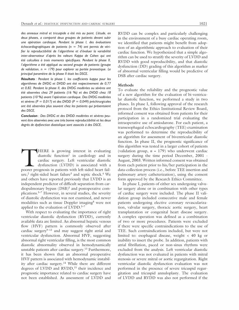
> 1 and ≤ 2 with S/D < 1 and Em < 8 cm·sec–1, or Em < Am. Severe LVDD was diagnosed when E/A > 2 with S/D < 1.
The transtricuspid pulsed Doppler flow was obtained from a mid-esophageal view between 40–70º. The transtricuspid Doppler and tricuspid annular velocities were also obtained with a deep transgastric long axis view at 120–145º with right-sided rotation (Figure 1). In this view, the tricuspid annulus interrogation axis is parallel to the Doppler axis. Furthermore, the HVF can be visualized in some patients with this view. A lower esophageal view with right sided rotation was also used to obtain the HVF. Normal right ventricular diastolic function15 was defined using normal values reported for Doppler transtricuspid flow,20 HVF11,21,22 and TDI of the tricuspid annulus.23,24 A normal HVF was defined as a ratio of systolic to diastolic velocities greater than 1 with the atrial wave reversal less than half the maximum systolic wave velocity.21 Mild RVDD was defined by E/A < 1 in transtricuspid flow velocities, or 1 < E/A < 2, with S/D > 1 in HVF and Et (early com-ponent of the tricuspid annular tissue Doppler veloc-ity) < At (atrial component of the tricuspid annular tissue Doppler velocity) or an atrial reversal wave more than half of the systolic wave of the HVF. Moderate or severe RVDD was present if a reduced or inverted systolic waveform was present on the Doppler HVF signal, respectively.
An algorithm for the evaluation of LVDD and RVDD was established using a consensus between two cardiac anesthesiologists and two cardiologists who were experts in TEE (Figures 2 and 3). The devel-opment of the algorithm was based on our previous reports5,13,14,25,26 and tissue Doppler examinations19 for the evaluation of DD. In phase I, the Doppler signals obtained intraoperatively from three examina-tions (two before, and one after CPB) were reviewed and analyzed off-line in a random and blinded fashion, by two anesthesiologists. In phase II, the attending anesthesiologist classified left and right diastolic func-tion on-line, using the algorithm. Two examinations were performed (before and after CPB), but only the pre-CPB or baseline TEE examination (as a predic-tor of DSB) was entered into the data set for subse-quent analysis of DSB. Abnormal diastolic profiles were separated in two groups: normal or relaxation abnormalities were classified as normal to mild DD, pseudonormal, and restrictive patterns were classified as moderate and severe DD.
Statistical analysisIn phase I, agreement on the algorithm between the two cardiac anesthesiologists (A.D., P.C.) was
assessed using Cohen’s kappa27 at three time points: before and after chest opening, and after CPB. Values between 0.6 and 1.0 were considered to indicate good agreement, while values from 0.40 to 0.59, 0.2 to 0.39 and 0 to 0.19 defined moderate, fair, and poor agreement respectively. Phase II data are presented as mean ± SD for continuous variables and n (percentage) for categorical variables. Pearson Chi-square tests were used to compare categorical variables according to grades (normal-mild vs moderate-severe). For continuous variables, a Student’s t test was used to compare grades (normal-mild vs moderate-severe). Sample size was calculated based on the prevalence
FIGURE 2 Algorithm used in the diagnosis and classifi-cation of left ventricular diastolic dysfunction (DD). The diastolic function is classified using pulsed-wave Doppler of the transmitral flow (TMF), pulmonary venous flow (PVF) and tissue Doppler examination of mitral annular velocity (MAV). Patients with pacemaker, atrial fibrillation, non-sinus rhythm, mitral stenosis, severe mitral and aortic insuf-ficiency are excluded from analysis. A = atrial component of the TMF; Am = atrial component of the MAV; D = diastolic component of the PVF; E = early filling of the TMF; Em = early component of the MAV; S = systolic component of the PVF.*Normal Em is within an 8–12.5 cm·sec–1 interval (see text).

1024 CANADIAN JOURNAL OF ANESTHESIA
of DSB observed in previous studies.5,14 For LVDD, a two-group χ2 test with a two-sided significance level of 0.05 would achieve 66% power to detect a difference between a group 1 proportion, π1, of 0.409 and a group 2 proportion, π2, of 0.655 (odds ratio of 2.743) when the sample sizes are 115 and 29, respectively (a total sample size of 144). For RVDD, a two-group χ2 test with a 0.05 two-sided significance level would have 50% power to detect a difference between a group 1 proportion, π1, of 0.476 and a group 2 proportion, π2, of 0.722 (odds ratio of 2.859) when sample sizes are 145 and 18, respectively (a total sample size of 163). Statistical analyses were
done with SAS version 8.02 (SAS Institute Inc., Cary, NC, USA). A P value < 0.05 (two tailed) was considered significant.
ResultsPhase ISeventy-four patients were studied including 43 men and 31 women with a mean age of 67 ± 11 years. A total of 50 isolated valvular surgeries and 24 com-bined valvular and revascularization procedures were performed. The valvular surgical procedures consisted of 53 aortic valve surgeries, 22 mitral valve surgeries and one tricuspid valve annuloplasty. Twenty-nine measurements for LVDD were excluded because the Doppler signals could not be obtained. The LVDD algorithm was used to evaluate 193 measurements (74, 51 and 68 patients for the three evaluation time points: before chest opening, after chest opening and after CPB respectively). The kappa values were 0.82, 0.57 and 0.77. Fifty measurements for RVDD were excluded for the same reasons as mentioned above. The RVDD algorithm was used to evaluate 182 measurements (70, 52 and 60 patients for the three evaluation time points). The kappa values were 0.69, 0.82 and 0.91. When the three evaluation time points
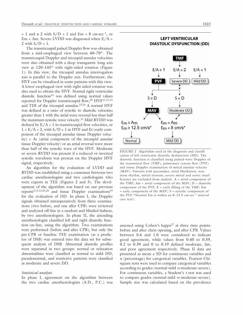
FIGURE 3 Algorithm used in the diagnosis and clas-sification of right ventricular diastolic dysfunction (DD). Diastolic function is classified by pulsed-wave Doppler of the transtricuspid flow (TTF) and hepatic venous flow (HVF) and tissue Doppler imaging of the tricuspid annulus or tricuspid annular velocity (TAV). Patients with pace-maker, atrial fibrillation, non-sinus rhythm, moderate to severe tricuspid regurgitation and tricuspid annuloplasty are excluded from analysis. A = atrial component of the TTF; AR = atrial reversal component of the HVF; At = atrial component of the TAV; D = diastolic component of the HVF; E = early filling of the TTF; Et = early component of the TAV; S = systolic component of the HVF.
FIGURE 4 Pooled analysis of left ventricular diastolic function evaluation between reviewers #1 and #2 for phase I of the study. The numbers in parentheses indicate the measurements done during three periods: before chest opening, before cardiopulmonary bypass (CPB) and after CPB.

Denault et al.: DIASTOLIC DYSFUNCTION AND CARDIAC SURGERY 1025
were pooled, the LVDD and RVDD algorithm kappa values were 0.77 and 0.82. In the evaluation of LVDD 26/193 (13%) and 3/193 (1.6%) time points were respectively excluded by reviewers #1 and #2 because the Doppler signals were insufficient or of unaccept-able quality (Figure 4). In the evaluation of RVDD 23/182 (13%) and 4/182 (2%) time points were excluded by reviewers #1 and #2 (Figure 5).
Phase II: validation groupOne hundred seventy-nine patients were studied, including 57 women and 122 men with a mean age of 67 ± 10 yr (Table I). Coronary revascularization procedures were performed in 129 patients, of which 23 were done off-pump. There were seven re-opera-tions, 42 complex procedures, with 54 aortic, nine mitral and three pulmonic valve replacements as well as 19 mitral valve repairs, four thoracic aortic surger-ies, one cardiac transplantation, closure of five atrial septal defects, and one ventricular septal defect. Of the 179 patients, 35 (20%) and 16 (9%) patients were respectively excluded from the analysis of left and right ventricular diastolic function using the pre-speci-fied exclusion criteria.
The hemodynamic values and outcome data are shown in Table II. Patients with moderate to severe
LVDD at baseline tended to have higher PCWP (15 ± 5 mmHg vs 13 ± 4 mmHg, P = 0.0597) compared to those with normal to mild LVDD. The presence of moderate to severe RVDD at baseline was also associ-ated with lower MPAP (19 ± 6 mmHg vs 22 ± 6, P = 0.0551) and lower CI (1.8 ± 0.4 vs 2.0 ± 0.4, P = 0.0458) compared to patients with normal to mild RVDD. Patients with moderate to severe LVDD and RVDD tended to have longer intensive care unit and hospital stays but this did not reach statistical signifi-cance (Table II). There were five (2.8%) deaths and no difference between groups was observed. Difficult separation from cardiopulmonary bypass was observed in 92 patients (51.4%) and was more frequent in patients with moderate to severe LVDD and RVDD. Difficult separation from cardiopulmonary bypass was present in 19 patients (65.5%) with moderate or severe LVDD compared with 47 patients (40.9%) with nor-mal left ventricular diastolic function or mild LVDD (P = 0.0173). Difficult separation from cardiopulmo-nary bypass was also observed in the 13 (72%) patients with moderate to severe RVDD compared with the 69 patients (48%) who had normal right ventricular diastolic function or mild RVDD (P = 0.0486).
DiscussionThe algorithms proposed in this study for the assess-ment of left and right ventricular diastolic function are simple, while their application is reproducible and useful in the stratification of patients at risk of devel-oping DSB. In this discussion, we will first analyze the technical aspects of the echocardiographic evaluation of LVDD and RVDD and then present the clinical implications of this study along with its limitations. Obtaining the echocardiographic signals for the evalu-ation of diastolic function using TEE is relatively easy and its interpretation is reproducible. Available data were considered adequate to classify LVDD and RVDD in the majority of patients. One reviewer consistently excluded more time points than the other; however, these patients were classified as normal by the other reviewer. In addition, the identification of moderate and severe LVDD and RVDD was almost identical for both reviewers and this population could represent the most important group at risk for DSB.6,7
In a multivariate analysis, we have previously observed that LVDD was a better predictor of hemo-dynamic instability after cardiac surgery compared to systolic dysfunction.5 However in that study, patients were not graded according to the severity of DD. Moreover, newer modalities such as tissue Doppler were not used at that time. Two groups6,7 have recently observed that more severe forms of LVDD
FIGURE 5 Pooled analysis of right ventricular diastolic function evaluation between reviewers #1 and #2. The numbers in parentheses indicate the measurements done during three periods: before chest opening, before cardio-pulmonary bypass (CPB) and after CPB.
1026 CANADIAN JOURNAL OF ANESTHESIA
are associated with complications after cardiac surgery. This is consistent with our clinical observations and with large population studies in cardiology28 showing the relationship between LVDD and outcomes.
Right ventricular diastolic dysfunction could rep-resent an additional marker to identify populations at higher risk of requiring vasoactive support and poten-tially other adverse clinical outcomes. We have previ-
ously documented that in hemodynamically unstable patients in the intensive care unit, abnormal right ventricular filling abnormalities were the most com-mon echocardiographic observation.13 We also noted in a pilot study that abnormal HVF, when present before cardiac surgery, was associated with increased need for vasoactive support after cardiac surgery.14 Again, in these two previous studies, patients were not
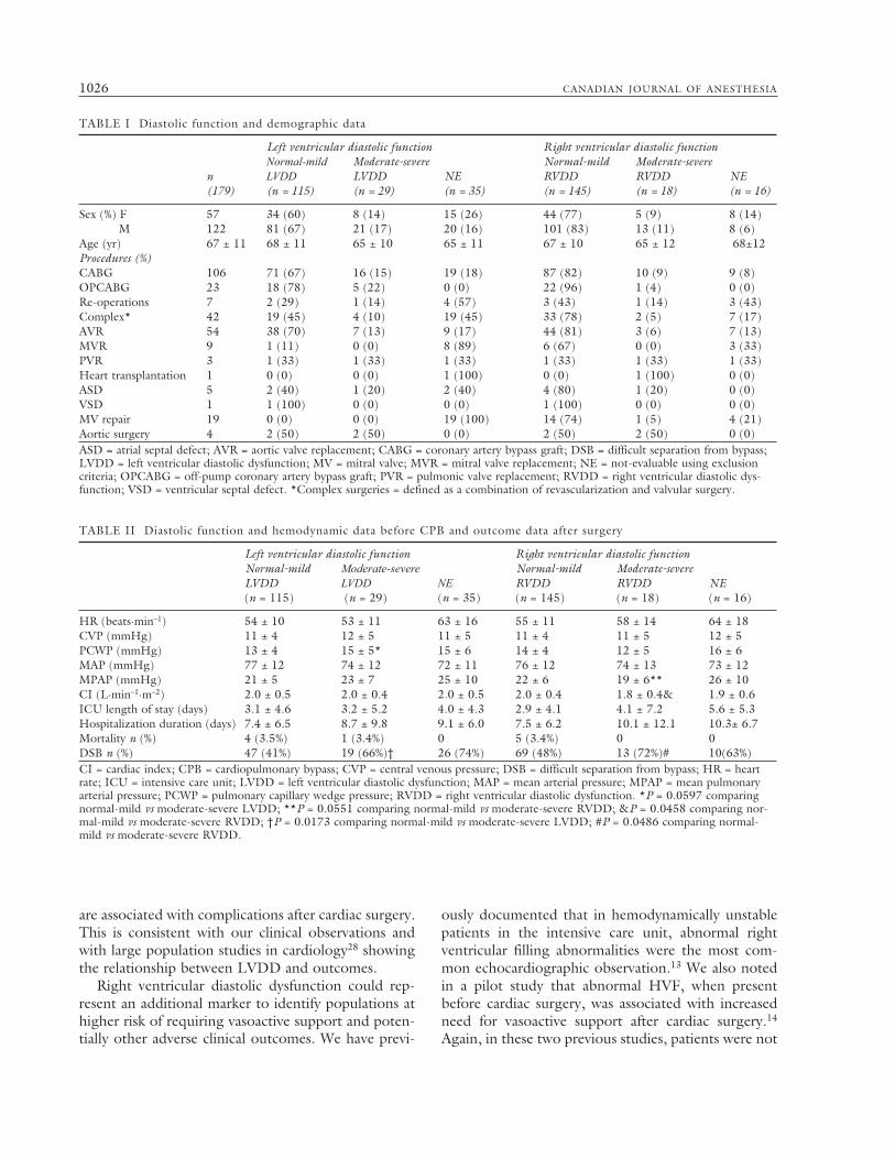
TABLE I Diastolic function and demographic data
Left ventricular diastolic function Right ventricular diastolic function Normal-mild Moderate-severe Normal-mild Moderate-severe n LVDD LVDD NE RVDD RVDD NE (179) (n = 115) (n = 29) (n = 35) (n = 145) (n = 18) (n = 16)
Sex (%) F 57 34 (60) 8 (14) 15 (26) 44 (77) 5 (9) 8 (14) M 122 81 (67) 21 (17) 20 (16) 101 (83) 13 (11) 8 (6) Age (yr) 67 ± 11 68 ± 11 65 ± 10 65 ± 11 67 ± 10 65 ± 12 68±12Procedures (%) CABG 106 71 (67) 16 (15) 19 (18) 87 (82) 10 (9) 9 (8)OPCABG 23 18 (78) 5 (22) 0 (0) 22 (96) 1 (4) 0 (0)Re-operations 7 2 (29) 1 (14) 4 (57) 3 (43) 1 (14) 3 (43)Complex* 42 19 (45) 4 (10) 19 (45) 33 (78) 2 (5) 7 (17)AVR 54 38 (70) 7 (13) 9 (17) 44 (81) 3 (6) 7 (13)MVR 9 1 (11) 0 (0) 8 (89) 6 (67) 0 (0) 3 (33)PVR 3 1 (33) 1 (33) 1 (33) 1 (33) 1 (33) 1 (33)Heart transplantation 1 0 (0) 0 (0) 1 (100) 0 (0) 1 (100) 0 (0)ASD 5 2 (40) 1 (20) 2 (40) 4 (80) 1 (20) 0 (0)VSD 1 1 (100) 0 (0) 0 (0) 1 (100) 0 (0) 0 (0)MV repair 19 0 (0) 0 (0) 19 (100) 14 (74) 1 (5) 4 (21)Aortic surgery 4 2 (50) 2 (50) 0 (0) 2 (50) 2 (50) 0 (0)ASD = atrial septal defect; AVR = aortic valve replacement; CABG = coronary artery bypass graft; DSB = difficult separation from bypass; LVDD = left ventricular diastolic dysfunction; MV = mitral valve; MVR = mitral valve replacement; NE = not-evaluable using exclusion criteria; OPCABG = off-pump coronary artery bypass graft; PVR = pulmonic valve replacement; RVDD = right ventricular diastolic dys-function; VSD = ventricular septal defect. *Complex surgeries = defined as a combination of revascularization and valvular surgery.
TABLE II Diastolic function and hemodynamic data before CPB and outcome data after surgery
Left ventricular diastolic function Right ventricular diastolic function Normal-mild Moderate-severe Normal-mild Moderate-severe LVDD LVDD NE RVDD RVDD NE (n = 115) (n = 29) (n = 35) (n = 145) (n = 18) (n = 16)
HR (beats·min–1) 54 ± 10 53 ± 11 63 ± 16 55 ± 11 58 ± 14 64 ± 18CVP (mmHg) 11 ± 4 12 ± 5 11 ± 5 11 ± 4 11 ± 5 12 ± 5PCWP (mmHg) 13 ± 4 15 ± 5* 15 ± 6 14 ± 4 12 ± 5 16 ± 6MAP (mmHg) 77 ± 12 74 ± 12 72 ± 11 76 ± 12 74 ± 13 73 ± 12MPAP (mmHg) 21 ± 5 23 ± 7 25 ± 10 22 ± 6 19 ± 6** 26 ± 10CI (L·min–1·m–2) 2.0 ± 0.5 2.0 ± 0.4 2.0 ± 0.5 2.0 ± 0.4 1.8 ± 0.4& 1.9 ± 0.6ICU length of stay (days) 3.1 ± 4.6 3.2 ± 5.2 4.0 ± 4.3 2.9 ± 4.1 4.1 ± 7.2 5.6 ± 5.3Hospitalization duration (days) 7.4 ± 6.5 8.7 ± 9.8 9.1 ± 6.0 7.5 ± 6.2 10.1 ± 12.1 10.3± 6.7Mortality n (%) 4 (3.5%) 1 (3.4%) 0 5 (3.4%) 0 0DSB n (%) 47 (41%) 19 (66%)† 26 (74%) 69 (48%) 13 (72%)# 10(63%)CI = cardiac index; CPB = cardiopulmonary bypass; CVP = central venous pressure; DSB = difficult separation from bypass; HR = heart rate; ICU = intensive care unit; LVDD = left ventricular diastolic dysfunction; MAP = mean arterial pressure; MPAP = mean pulmonary arterial pressure; PCWP = pulmonary capillary wedge pressure; RVDD = right ventricular diastolic dysfunction. *P = 0.0597 comparing normal-mild vs moderate-severe LVDD; **P = 0.0551 comparing normal-mild vs moderate-severe RVDD; &P = 0.0458 comparing nor-mal-mild vs moderate-severe RVDD; †P = 0.0173 comparing normal-mild vs moderate-severe LVDD; #P = 0.0486 comparing normal-mild vs moderate-severe RVDD.

Denault et al.: DIASTOLIC DYSFUNCTION AND CARDIAC SURGERY 1027
graded according to the severity of RVDD whereas in the present study we confirm that moderate to severe RVDD is associated with lower CI and increased risk of DSB.
In this study, normal (n = 33) to mild RVDD (n = 112) was present in 145 patients (81%), moderate to severe RVDD existed in 18 patients (10%) whereas RVDD function was not evaluable in 16 patients (9%). The overall incidence of RVDD was 74%, which is higher than that reported by Mishra who observed abnormal HVF in 11% of patients undergoing coro-nary revascularization.29 We have previously observed that abnormal HVF suggestive of RVDD is less com-mon in patients undergoing coronary revasculariza-tion as compared to valvular surgery, and reported the presence of abnormal HVF in 41% of patients under-going valvular surgery14 (including mild, moderate and severe RVDD). The higher percentage of patients with RVDD in the current study is most likely related to the use of tissue Doppler which provided for greater sensitivity in detection of mild RVDD. This echocar-diographic modality was not available in the previous study.14 The elevated incidence of RVDD in patients with valvular disease may reflect maladaptation to pulmonary hypertension, which is frequently present in this population. This factor alone could explain why DSB was more frequently observed in patients with abnormal RVDD as our pilot study suggested. Interestingly, in the present study, CI was lower in patients with moderate to severe RVDD whereas MPAP was not “abnormally elevated”. The absence of an observed association between pulmonary hyperten-sion and moderate to severe RVDD may be related to the large number of patients who underwent coronary artery bypass grafting in the current series, in contrast to the Carricart study14 where only valvular surgical patients were selected.
There are several limitations to this study. First, we grouped LVDD into two categories instead of the four standard grades. This grouping was necessary for the analysis because a left ventricular restrictive pat-tern is uncommon before surgery. We observed that restrictive LVDD was indeed present in two patients (1%) in the phase II validation group and in six patients (1.2%) in our TEE database of 500 consecu-tive patients. The same observations applied to RVDD where a restrictive pattern was also present in only two patients (1%) in the phase II validation group. It would have been futile to have enrolled the large number of patients that would have been necessary to evaluate the independent significance of restrictive LVDD and RVDD on our primary outcome of interest. Mild RVDD, on the other hand, is relatively common, and
was observed in the majority of our population (81%) since we began incorporation of tissue Doppler evalu-ation of our cardiac surgery patients.
The primary goal of the study was to develop and validate a simple algorithm of diastolic dysfunction and explore its predictive value with respect to dif-ficulty in weaning from cardiopulmonary bypass. This is why a phase II validation group was used with DSB as a primary end-point. This study represents the larg-est published series of patients in whom biventricular diastolic function was assessed in the cardiac surgical setting using the newer Doppler modalities and where the results were also correlated with hemodynamic data. We observed an association between moderate to severe LVDD and RVDD and DSB. However, the number of patients with these diastolic filling abnor-malities was insufficient to subject to a multivariable analysis. The relative importance of DD compared to other variables in relation to DSB and mortal-ity should be investigated in a larger population of cardiac surgery patients. The results from the pres-ent study are consistent with our earlier work which demonstrated that both abnormal left5 and right13,14 ventricular diastolic profiles are associated with hemo-dynamic instability.
The two anesthesiologists responsible for clinical management of the study patients were not blinded to the echocardiographic data for ethical and practi-cal reasons, and this generates potential for some bias. However, intraoperative anesthetic management was directed towards maintenance of adequate MAP rather than optimization of diastolic parameters and ventricular filling patterns. We also identify that evalu-ation of LVDD and RVDD is not possible in up to 10–20% of cardiac surgical patients for several reasons including rhythm abnormality, severe valvular disease, and inability to obtain a complete set of Doppler images. Finally, several diastolic parameters change with increasing age, including mitral and tricuspid annular velocities,30 and the algorithm was not age-adjusted accordingly for the sake of simplification. However, as most age-related changes in ventricular function involve relaxation abnormalities, patients with normal function and mild diastolic abnormalities were analyzed together, which would have minimized the potential confounding influence of age-related dif-ferences in cardiac function.
In summary, the severity of LVDD and RVDD can be determined before cardiac surgery using a simple algorithm. Moderate to severe LVDD and RVDD are associated with a greater risk of DSB and with great-er hemodynamic abnormalities. Further multicentre studies with larger populations will be necessary to
1028 CANADIAN JOURNAL OF ANESTHESIA
explore the value of the identification of LVDD and RVDD and the therapeutic implications of incorpo-rating this information into the routine perioperative management of cardiac surgery patients.
References 1 Vasan RS, Benjamin EJ. Diastolic heart failure--no
time to relax. N Engl J Med 2001; 344: 56–9. 2 Pinamonti B, Di Lenarda A, Sinagra G, Camerini F.
Restrictive left ventricular filling pattern in dilated car-diomyopathy assessed by Doppler echocardiography: clinical, echocardiographic and hemodynamic cor-relations and prognostic implications. Heart Muscle Disease Study Group. J Am Coll Cardiol 1993; 22: 808–15.
3 Sun JP, James KB, Yang XS, et al. Comparison of mor-tality rates and progression of left ventricular dysfunc-tion in patients with idiopathic dilated cardiomyopathy and dilated versus nondilated right ventricular cavities. Am J Cardiol 1997; 80: 1583–7.
4 Poelaert J, Declerck C, Vogelaers D, Colardyn F, Visser CA. Left ventricular systolic and diastolic function in septic shock. Intensive Care Med 1997; 23: 553–60.
5 Bernard F, Denault A, Babin D, et al. Diastolic dys-function is predictive of difficult weaning from cardio-pulmonary bypass. Anesth Analg 2001; 92: 291–8.
6 Vaskelyte J, Stoskute N, Kinduris S, Ereminiene E. Coronary artery bypass grafting in patients with severe left ventricular dysfunction: predictive signifi-cance of left ventricular diastolic filling pattern. Eur J Echocardiogr 2001; 2: 62–7.
7 Liu J, Tanaka N, Murata K, et al. Prognostic value of pseudonormal and restrictive filling patterns on left ventricular remodeling and cardiac events after coro-nary artery bypass grafting. Am J Cardiol 2003; 91: 550–4.
8 Khouri SJ, Maly GT, Suh DD, Walsh TE. A practi-cal approach to the echocardiographic evaluation of diastolic function. J Am Soc Echocardiogr 2004; 17: 290–7.
9 Wranne B, Pinto FJ, Hammarstrom E, St Goar FG, Puryear J, Popp RL. Abnormal right heart filling after cardiac surgery: time course and mechanisms. Br Heart J 1991; 66: 435–42.
10 Pinto FJ, Wranne B, St Goar FG, et al. Systemic venous flow during cardiac surgery examined by intraoperative transesophageal echocardiography. Am J Cardiol 1992; 69: 387–93.
11 Nomura T, Lebowitz L, Koide Y, Keehn L, Oka Y. Evaluation of hepatic venous flow using transesopha-geal echocardiography in coronary artery bypass surgery: an index of right ventricular function. J Cardiothorac Vasc Anesth 1995; 9: 9–17.
12 Gardeback M, Settergren G, Brodin LA. Hepatic blood flow and right ventricular function during cardiac sur-gery assessed by transesophageal echocardiography. J Cardiothorac Vasc Anesth 1996; 10: 318–22.
13 Costachescu T, Denault A, Guimond JG, et al. The hemodynamically unstable patient in the intensive care unit: hemodynamic vs. transesophageal echocardio-graphic monitoring. Crit Care Med 2002; 30: 1214–23.
14 Carricart M, Denault AY, Couture P, et al. Incidence and significance of abnormal hepatic venous Doppler flow velocities before cardiac surgery. J Cardiothorac Vasc Anesth 2005; 19: 751–8.
15 Klein AL, Hatle LK, Burstow DJ, et al. Comprehensive Doppler assessment of right ventricular diastolic func-tion in cardiac amyloidosis. J Am Coll Cardiol 1990; 15: 99–108.
16 Quinones MA, Otto CM, Stoddard M, Waggoner A, Zaghbi WA; Doppler Quantification Task Force of the Nomenclature and Standards Committee of the American Society of Echocardiography. Recommendations for quantification of Doppler echocardiography: a report from the Doppler Quantification Task Force of the Nomenclature and Standards Committee of the American Society of Echocardiography. J Am Soc Echocardiogr 2002; 15: 167–84.
17 Yamada H, Oki T, Mishiro Y, et al. Effect of aging on diastolic left ventricular myocardial velocities measured by pulsed tissue Doppler imaging in healthy subjects. J Am Soc Echocardiogr 1999; 12: 574–81.
18 Rakowski H, Appleton C, Chan KL, et al. Canadian consensus recommendations for the measurement and reporting of diastolic dysfunction by echocar-diography: from the Investigators of Consensus on Diastolic Dysfunction by Echocardiography. J Am Soc Echocardiogr 1996; 9: 736–60.
19 Garcia MJ, Thomas JD, Klein AL. New Doppler echo-cardiographic applications for the study of diastolic function. J Am Coll Cardiol 1998; 32: 865–75.
20 Pye MP, Pringle SD, Cobbe SM. Reference values and reproducibility of Doppler echocardiography in the assessment of the tricuspid valve and right ventricular diastolic function in normal subjects. Am J Cardiol 1991; 67: 269–73.
21 Appleton CP, Hatle LK, Popp RL. Superior vena cava and hepatic vein Doppler echocardiography in healthy adults. J Am Coll Cardiol 1987; 10: 1032–9.
22 Pinto FJ, Wranne B, St Goar FG, Schnittger I, Popp RL. Hepatic venous flow assessed by transesophageal echo-cardiography. J Am Coll Cardiol 1991; 17: 1493–8.
23 Caso P, Galderisi M, Cicala S, et al. Association between myocardial right ventricular relaxation time
Denault et al.: DIASTOLIC DYSFUNCTION AND CARDIAC SURGERY 1029
and pulmonary arterial pressure in chronic obstructive lung disease: analysis by pulsed Doppler tissue imaging. J Am Soc Echocardiogr 2001; 14: 970–7.
24 Ozer N, Tokgozoglu L, Coplu L, Kes S. Echocardiographic evaluation of left and right ventricu-lar diastolic function in patients with chronic obstruc-tive pulmonary disease. J Am Soc Echocardiogr 2001; 14: 557–61.
25 Lattik R, Couture P, Denault AY, et al. Mitral Doppler indices are superior to two-dimensional echocardio-graphic and hemodynamic variables in predicting responsiveness of cardiac output to a rapid intravenous infusion of colloid. Anesth Analg 2002; 94: 1092–9.
26 Hache M, Denault A, Belisle S, et al. Inhaled epopro-stenol (prostacyclin) and pulmonary hypertension before cardiac surgery. J Thorac Cardiovasc Surg 2003; 125: 642–9.
27 Fleiss JL. Statistical Methods for Rates and Proportions, 2nd ed. New York: Wiley; 1981.
28 Redfield MM, Jacobsen SJ, Burnett JC Jr, Mahoney DW, Bailey KR, Rodeheffer RJ. Burden of systolic and dia-stolic ventricular dysfunction in the community: appre-ciating the scope of the heart failure epidemic. JAMA 2003; 289: 194–202.
29 Mishra M, Chauhan R, Sharma KK, et al. Real-time intraoperative transesophageal echocardiography--how useful? Experience of 5,016 cases. J Cardiothorac Vasc Anesth 1998; 12: 625–32.
30 Nikitin NP, Witte KK, Thackray SD, de Silva R, Clark AL, Cleland JG. Longitudinal ventricular func-tion: normal values of atrioventricular annular and myocardial velocities measured with quantitative two-dimensional color Doppler tissue imaging. J Am Soc Echocardiogr 2003; 16: 906–21.