Practice Guidelines for Management of the Difficult Airway
88
Embargoed for release until approved by ASA House of Delegates. No part of this document may be released, distributed or reprinted until approved. Any unauthorized copying, reproduction, appropriation or communication of the contents of this document without the express written consent of the American Society of Anesthesiologists is subject to civil and criminal prosecution to the fullest extent possible, including punitive damages. 2022 American Society of Anesthesiologists Practice Guidelines for Management of the Difficult Airway * [MS #ALN-D-21-00272] Jeffrey L. Apfelbaum, MD, Carin A. Hagberg, MD, Richard T. Connis, PhD, Basem B. Abdelmalak, MD, Madhulika Agarkar, MPH, Richard P. Dutton, MD, John E. Fiadjoe MD, Robert Greif, MD, P. Allan Klock, Jr., MD, David Mercier, MD, Sheila N. Myatra, MD, Ellen P. O'Sullivan, MD, William H. Rosenblatt, MD, Massimiliano Sorbello, MD, Avery Tung, MD. ABSTRACT: The American Society of Anesthesiologists, All India Difficult Airway Association; European Airway Management Society; European Society of Anaesthesiology and Intensive Care † ; Italian Society of Anesthesiology, Analgesia, Resuscitation and Intensive Care, Learning, Teaching and Investigation Difficult Airway Group ‡ ; Society for Airway Management; Society for Ambulatory Anesthesia; Society for Head and Neck Anesthesia; Society for Pediatric Anesthesia; § Society of Critical Care Anesthesiologists; and the Trauma Anesthesiology Society present an updated report of the Practice Guidelines for Management of the Difficult Airway. PRACTICE Guidelines are systematically developed recommendations that assist the 1 practitioner and patient in making decisions about health care. These recommendations may be 2 adopted, modified, or rejected according to clinical needs and constraints, and are not intended 3 to replace local institutional policies. In addition, Practice Guidelines developed by the 4 American Society of Anesthesiologists (ASA) are not intended as standards or absolute 5 requirements, and their use cannot guarantee any specific outcome. Practice Guidelines are 6 subject to revision as warranted by the evolution of medical knowledge, technology, and 7 * An updated report by the American Society of Anesthesiologists Task Force on Management of the Difficult Airway. Received from the American Society of Anesthesiologists, Schaumburg, Illinois. Submitted for publication October __, 2021. Accepted for publication October __, 2021. Supported by the American Society of Anesthesiologists and developed under the direction of the Committee on Standards and Practice Parameters, Jeffrey L. Apfelbaum, M.D. (Chair). Approved by the ASA House of Delegates on October __, 2021. These Guidelines have been endorsed by the Difficult Airway Society. Address correspondence to the American Society of Anesthesiologists: 1061 American Lane, Schaumburg, Illinois 60173. This Practice Guideline, as well as all published ASA Practice Parameters, may be obtained at no cost through the Journal Web site, https://pubs.asahq.org/anesthesiology. † Formacion Internacional en Docencia e Investacion en Via Aerea. ‡ Società Italiana di Anestesia Analgesia Rianimazione e Terapia Intensiva. § A pediatric infographic and algorithm are included in these Guidelines, developed in collaboration with the Society for Pediatric Anesthesia Pediatric Difficult Intubation Collaborative.
-
Upload
khangminh22 -
Category
Documents
-
view
6 -
download
0
Transcript of Practice Guidelines for Management of the Difficult Airway
Embargoed for release until approved by ASA House of Delegates. No part of this document may be released, distributed or reprinted until approved. Any unauthorized copying, reproduction, appropriation or communication of the contents of this document without the express written consent of the American Society of Anesthesiologists is subject to civil and criminal prosecution to the fullest extent possible, including punitive damages.
2022 American Society of Anesthesiologists Practice Guidelines for Management of the Difficult Airway* [MS #ALN-D-21-00272] Jeffrey L. Apfelbaum, MD, Carin A. Hagberg, MD, Richard T. Connis, PhD, Basem B. Abdelmalak, MD, Madhulika Agarkar, MPH, Richard P. Dutton, MD, John E. Fiadjoe MD, Robert Greif, MD, P. Allan Klock, Jr., MD, David Mercier, MD, Sheila N. Myatra, MD, Ellen P. O'Sullivan, MD, William H. Rosenblatt, MD, Massimiliano Sorbello, MD, Avery Tung, MD. ABSTRACT: The American Society of Anesthesiologists, All India Difficult Airway Association; European
Airway Management Society; European Society of Anaesthesiology and Intensive Care†; Italian Society of Anesthesiology, Analgesia, Resuscitation and Intensive Care, Learning, Teaching
and Investigation Difficult Airway Group‡; Society for Airway Management; Society for
Ambulatory Anesthesia; Society for Head and Neck Anesthesia; Society for Pediatric Anesthesia;§ Society of Critical Care Anesthesiologists; and the Trauma Anesthesiology Society
present an updated report of the Practice Guidelines for Management of the Difficult Airway.
PRACTICE Guidelines are systematically developed recommendations that assist the 1
practitioner and patient in making decisions about health care. These recommendations may be 2
adopted, modified, or rejected according to clinical needs and constraints, and are not intended 3
to replace local institutional policies. In addition, Practice Guidelines developed by the 4
American Society of Anesthesiologists (ASA) are not intended as standards or absolute 5
requirements, and their use cannot guarantee any specific outcome. Practice Guidelines are 6
subject to revision as warranted by the evolution of medical knowledge, technology, and 7
* An updated report by the American Society of Anesthesiologists Task Force on Management of the Difficult Airway.
Received from the American Society of Anesthesiologists, Schaumburg, Illinois. Submitted for publication October __, 2021. Accepted for publication October __, 2021. Supported by the American Society of Anesthesiologists and developed under the direction of the Committee on Standards and Practice Parameters, Jeffrey L. Apfelbaum, M.D. (Chair). Approved by the ASA House of Delegates on October __, 2021. These Guidelines have been endorsed by the Difficult Airway Society. Address correspondence to the American Society of Anesthesiologists: 1061 American Lane, Schaumburg, Illinois 60173. This Practice Guideline, as well as all published ASA Practice Parameters, may be obtained at no cost through the Journal Web site, https://pubs.asahq.org/anesthesiology. † Formacion Internacional en Docencia e Investacion en Via Aerea. ‡ Società Italiana di Anestesia Analgesia Rianimazione e Terapia Intensiva. § A pediatric infographic and algorithm are included in these Guidelines, developed in collaboration with the Society for Pediatric Anesthesia Pediatric Difficult Intubation Collaborative.

2
practice. They provide basic recommendations that are supported by a synthesis and analysis 8
of the current literature, expert and practitioner opinion, open forum commentary, and clinical 9
feasibility data. 10
This document is a revision of the “Practice Guidelines for Management of the Difficult 11
Airway: A Report by the American Society of Anesthesiologists Task Force on Management of 12
the Difficult Airway,” adopted by the ASA in 2012 and published in 2013.1 13
Methodology 14
Definition of Difficult Airway 15
For these Practice Guidelines, a difficult airway includes the clinical situation in which 16
anticipated or unanticipated difficulty or failure is experienced by a physician trained in 17
anesthesia care, including but not limited to one or more of the following: facemask ventilation, 18
laryngoscopy, ventilation using a supraglottic airway, tracheal intubation, extubation, or invasive 19
airway. These clinical situations are further defined as follows: 20
Difficult facemask ventilation – It is not possible to provide adequate ventilation (e.g., 21
confirmed by end-tidal carbon dioxide detection), because of one or more of the following 22
problems: inadequate mask seal, excessive gas leak, or excessive resistance to the ingress or 23
egress of gas. 24
Difficult laryngoscopy – It is not possible to visualize any portion of the vocal cords after 25
multiple attempts at laryngoscopy. 26
Difficult supraglottic airway ventilation – It is not possible to provide adequate ventilation 27
because of one or more of the following problems: difficult supraglottic airway placement, 28
supraglottic airway placement requiring multiple attempts, inadequate supraglottic airway seal, 29
excessive gas leak, or excessive resistance to the ingress or egress of gas. 30
Difficult or failed tracheal intubation – Tracheal intubation requires multiple attempts, or 31
tracheal intubation fails after multiple attempts. 32
Difficult or failed tracheal extubation – The loss of airway patency and 33
adequate ventilation after removal of a tracheal tube or supraglottic airway from a patient with a 34
known or suspected difficult airway (i.e., an “at risk” extubation). 35
Difficult or failed invasive airway – Anatomic features or abnormalities reducing or 36
preventing the likelihood of successfully placing an airway into the trachea through the front of 37
the neck. 38
Inadequate ventilation: Indicators of inadequate ventilation include absent or inadequate 39
exhaled carbon dioxide, absent or inadequate chest movement, absent or inadequate breath 40

3
sounds, auscultatory signs of severe obstruction, cyanosis, gastric air entry or dilatation, 41
decreasing or inadequate oxygen saturation, absent or inadequate exhaled gas flow as 42
measured by spirometry, anatomic lung abnormalities as detected by lung ultrasound, and 43
hemodynamic changes associated with hypoxemia or hypercarbia (e.g., hypertension, 44
tachycardia, bradycardia, arrhythmia). Additional clinical symptoms may include changed 45
mental status or somnolence. 46
Purposes of the Guidelines 47
The purposes of these Guidelines are to guide the management of patients with difficult 48
airways, optimize first attempt success of airway management, improve patient safety during 49
airway management and minimize/avoid adverse events. The principal adverse outcomes 50
associated with the difficult airway include (but are not limited to) death, brain injury, 51
cardiopulmonary arrest, airway trauma, and damage to the teeth. The appropriate choice of 52
medications and techniques for anesthesia care and airway management is dependent upon 53
the experience, training, and preference of the individual practitioner, requirements or 54
constraints imposed by associated medical issues of the patient, type of procedure or 55
environment in which airway management takes place. The choice of agents, techniques and 56
devices may be limited by federal, state, or municipal regulations or statutes. 57
Focus 58
These Guidelines focus specifically on the management of the difficult airway encountered 59
with mask ventilation, tracheal intubation or supraglottic airway placement during procedures 60
requiring general anesthesia, deep sedation, moderate sedation or regional anesthesia, or 61
elective airway management without a procedure. Procedures include diagnostic, elective and 62
emergency procedures, and invasive airway access. Airway management during 63
cardiopulmonary resuscitation is not addressed by these Guidelines. The Guidelines are 64
intended for adult and pediatric patients with either anticipated or unanticipated difficult airways, 65
obstetric patients, intensive care (ICU) patients, and critically ill patients. The Guidelines do not 66
address patients at risk of aspiration without anatomically difficult airways, patients where 67
difficult airways are not encountered, or physiologically difficult airways that are not anatomically 68
difficult.** 69
These Guidelines do not address education, training, or certification requirements for 70
practitioners who provide anesthesia and airway management. Some aspects of the Guidelines 71
** These include, but are not limited to patients at increased risk for cardiorespiratory deterioration with
airway management due to underlying conditions such as hypoxemia, hypotension, severe metabolic acidosis, or right ventricular failure.

4
may be relevant in other clinical contexts. The Guidelines do not represent an exhaustive 72
consideration of all manifestations of the difficult airway or all possible approaches to airway 73
management. 74
Application 75
These Guidelines are intended for use by anesthesiologists and all other individuals who 76
perform anesthesia care or airway management. The Guidelines are intended to apply to all 77
airway management and anesthetic care delivered in inpatient (e.g., perioperative, non-78
operating room, emergency department, and critical care settings); and ambulatory settings 79
(e.g., ambulatory surgery centers, office-based surgery and procedure centers performing 80
invasive airway procedures). Excluded are prehospital settings and individuals who do not 81
deliver anesthetic care or perform airway management. 82
These guidelines are also intended to serve as a resource for other physicians and patient 83
care personnel who are involved in the care of difficult airway patients, including those involved 84
in local policy development. 85
Task Force Members 86
In 2019, the ASA Committee on Standards and Practice Parameters requested that these 87
Guidelines be updated. This update is a revision developed by an ASA-appointed task force of 88
15 members, including physician anesthesiologists in both private and academic practices from 89
the United States, India, Ireland, Italy, and Switzerland, an independent consulting 90
methodologist and an ASA staff methodologist. Conflict of interest documentation regarding 91
current or potential financial and other interests pertinent to the practice guideline were 92
disclosed by all task force members and managed.†† 93
Process and Evaluation of Evidence 94
These updated Guidelines were developed by means of a six-step process. First, 95
consensus was reached on the criteria for evidence. Second, a comprehensive literature 96
search was conducted by an independent librarian to identify citations relevant to the evidence 97
criteria. Third, original published articles from peer-reviewed journals relevant to difficult airway 98
management were evaluated and added to literature included in the previous update. Fourth, 99
consultants who had expertise or interest in difficult airway management and who practiced or 100
worked in various settings (e.g., private and academic practice) were asked to participate in 101
opinion surveys addressing the appropriateness, completeness, and feasibility of 102
†† Additional conflict of interest information is located after Appendix 2 in this document.

5
implementation of the draft recommendations and to review and comment on a draft of the 103
Guidelines. Fifth, additional opinions were solicited from random samples of active members of 104
the ASA and participating organizations. Sixth, all available information was used to build 105
consensus to finalize the Guidelines. A summary of recommendations is provided in appendix 1. 106
Preparation of these updated Guidelines followed a rigorous methodological process, 107
described in more detail in appendix 2 and other related publications.2-5 108
Criteria for literature acceptance included randomized controlled trials, prospective 109
nonrandomized comparative studies (e.g., quasi-experimental, cohort), retrospective 110
comparative studies (e.g., case-control), observational studies (e.g., correlational or descriptive 111
statistics), and case reports or case series from peer-review journals. Literature exclusion 112
criteria included: (1) patients or practitioners described in the study who were specifically 113
excluded or not identified by evidence criteria in the evidence model; (2) interventions not 114
identified or specifically excluded in the evidence model; (3) studies with insufficient or no 115
outcome data, or reported outcomes not relevant to the evidence model; (4) articles with no 116
original data, including review articles, descriptive letters or editorials, (5) systematic reviews, 117
secondary data, meta-analysis,‡‡ or other articles with no original data, (6) abstracts, letters or 118
articles not published in a peer-review journal, (7) studies outside of designated search dates, 119
(8) duplicate data presented in a different reviewed article, or (9) retracted publications. 120
Within the text of these Guidelines, literature classifications are reported for each 121
intervention as follows: Category A level 1, meta-analysis of randomized controlled trials; 122
Category A level 2, multiple randomized controlled trials; and Category A level 3, a single 123
randomized controlled trial. Category B level 1, nonrandomized studies with group comparisons; 124
Category B level 2, nonrandomized studies with associative findings; Category B level 3, 125
nonrandomized studies with descriptive findings, and Category B level 4, case series or case 126
reports. Statistically significant outcomes (P < 0.01) are designated as either beneficial (B) or 127
harmful (H) to the patient; statistically nonsignificant findings are designated as equivocal (E).§§ 128
When available, category A evidence is given precedence over category B evidence for any 129
particular outcome. The lack of sufficient scientific evidence in the literature is reported in the 130
‡‡ All meta-analyses are conducted by the ASA methodology group. Meta-analyses from other sources are reviewed but not included as evidence in this document. A minimum of five independent randomized controlled trials (i.e., sufficient for fitting a random-effects model) is required for meta-analysis. §§ A complete bibliography used to develop this updated Advisory, arranged alphabetically by author, is available as Supplemental Digital Content 1, http://links.lww.com/ALN/XXXXXXXX.

6
text of the Guidelines as “insufficient evidence.”*** Opinions regarding the scientific quality of the 131
studies or opinion ratings of the strength of recommendations are not reported in this document. 132
Survey findings from task force appointed expert consultants, and samples of the 133
memberships of ASA and participating organizations††† are reported in appendix 2. Survey 134
responses for each recommendation are reported using a 5-point scale based on median values 135
from strongly agree to strongly disagree. 136
Guidelines 137
Evaluation of the Airway 138
Airway evaluation topics include (1) risk assessment to predict a difficult airway or risk of 139
aspiration, and (2) airway examination (bedside and advanced). 140
Risk assessment includes evaluation of information obtained from a patient’s history or 141
medical records, including demographic information, clinical conditions, diagnostic tests, and 142
patient/family interviews or questionnaires. An airway examination is intended to identify the 143
presence of upper airway pathologies or anatomical anomalies. Issues addressed in these 144
Guidelines include: (1) measurement of facial and jaw features, (2) anatomical measurements 145
and landmarks, (3) imaging with ultrasound or virtual laryngoscopy/bronchoscopy, (4) three 146
dimensional printing, and (5) bedside endoscopy. 147
Literature Findings. 148
Patient demographic and personal characteristics evaluated for difficult airway risk 149
prediction included age, sex, body mass index, weight and height. Clinical characteristics 150
assessed included a history of difficult intubation, distorted airway anatomy, snoring, obstructive 151
sleep apnea, diabetes mellitus, or findings from diagnostic tests (e.g., radiography, computed 152
tomography), patient interviews and questionnaires. Measurement of facial and jaw features 153
included mouth opening, the ability to prognath, head and neck mobility, prominent upper 154
incisors, presence of a beard, and an upper lip bite test. Anatomical measures included 155
Mallampati and Modified Mallampati scores, thyromental distance, sternomental distance, 156
interincisor distance, neck circumference, ratio of neck circumference to thyromental distance, 157
ratio of height to thyromental distance, hyomental distance, and hyomental distance ratio. 158
Measurements obtained from ultrasound included skin-to-hyoid distance, tongue volume, and 159
distance from skin to epiglottis. 160
*** A more detailed description of the definition of insufficient evidence is described in Appendix 2. ††† See Appendix 2 for tables reporting a complete listing of survey findings.

7
Observational studies reported comparative demographic findings for difficult versus non-161
difficult airway patients as well as sensitivity, specificity, positive predictive, negative predictive 162
and accuracy values for difficult laryngoscopy, supraglottic airway use, and tracheal intubation. 163
Findings for the above patient characteristics were shown to have very high predictive and 164
comparative variability, with sensitivity, specificity and significance values ranging from low to 165
very high across all patient demographic measures (Category B2-E evidence).6-70 No single 166
characteristic was identified as consistently being more predictive than another, and multivariate 167
measures intended to predict difficult airways were too few and diverse among the studies to 168
determine a common set of predictors. 169
Case reports identified difficult laryngoscopy or difficult intubation occurring among patients 170
with a variety of acquired or congenital disease states (e.g., ankylosing spondylitis, 171
degenerative osteoarthritis, Treacher-Collins, Klippel-Feil, Down syndrome, 172
mucopolysaccharidosis, and airway masses) (Category B4-H evidence).71-122 173
Observational studies reported comparative findings for facial and jaw features and 174
anatomical measurement for difficult versus non-difficult airway patients as well as sensitivity, 175
specificity, positive predictive, negative predictive and accuracy values for difficult laryngoscopy 176
and intubation. Findings for facial and jaw features,7-11,13,14,18,27,33,38-40,42,43,45-47,49,51-54,57,58,64,68,123-177 159 anatomical measurements,7-11,13-15,18,22,23,27-30,33,35,37-40,45-47,49,51-54,57,58,60,64,65,68,70,123-132,134-178 154,156,158-203 and ultrasound anatomical measurements69,139,162,170,194,196,203-213 were shown to have 179
very high predictive and comparative variability, with sensitivity, specificity and significance 180
values ranging from low to very high across all patient measures (Category B2-E evidence). No 181
single characteristic was identified as consistently being more predictive than another, and 182
multivariate measures intended to predict difficult airways were too few and diverse among the 183
studies to determine a common set of predictors. 184
A prospective cohort study reported improved laryngeal views (during tongue protrusion) 185
when transnasal endoscopy was added to the preoperative bedside evaluation (Category B2-B 186
evidence),214 and an observational study utilizing preoperative endoscopic examination as an 187
added airway assessment tool reported that airway management plans were revised in 26% of 188
patients based on the results of this examination (Category B3-B evidence).215 Observational 189
studies and case reports indicated that radiography and computed tomography scans identified 190
anatomical characteristics such as laryngeal deviations, cervical abnormalities, fractures, and 191
abscesses that may suggest a potential difficult airway (Category B3-B and B4-B 192
evidence).90,216-219 Observational studies indicated that patient questionnaires may identify 193
patients at risk of difficult ventilation and intubation (Category B3-B evidence).163,220,221 194

8
The literature was insufficient to evaluate the predictive value of virtual laryngoscopy/ 195
bronchoscopy or three dimensional printing. 196
Survey Findings. The consultants and members of participating organizations strongly 197
agree with recommendations to assure that an airway risk assessment is performed by the 198
person(s) responsible for airway management whenever feasible before the initiation of 199
anesthetic care or airway management, and with the recommendation to conduct an airway 200
physical examination before the initiation of anesthetic care or airway management. 201
Recommendations for Evaluation of the Airway. 202
• Before the initiation of anesthetic care or airway management, assure that an airway risk 203
assessment is performed by the person(s) responsible for airway management 204
whenever feasible to identify patient, medical, surgical, environmental, and anesthetic 205
factors (e.g., risk of aspiration) that may indicate the potential for a difficult airway. 206
o When available in the patient’s medical records, evaluate demographic information, 207
clinical conditions, diagnostic test findings, patient/family interviews, and 208
questionnaire responses. 209
o Assess multiple demographic and clinical characteristics to determine a patient’s 210
potential for a difficult airway or aspiration. 211
• Before the initiation of anesthetic care or airway management, conduct an airway 212
physical examination to further identify physical characteristics that may indicate the 213
potential for a difficult airway. 214
o The physical examination may include assessment of facial features,‡‡‡ and 215
assessment of anatomical measurements and landmarks.§§§ 216
o Additional evaluation to characterize the likelihood or nature of the anticipated airway 217
difficulty may include bedside endoscopy, virtual laryngoscopy/bronchoscopy or 218
three dimensional printing.**** 219
• Assess multiple airway features to determine a patient’s potential for a difficult airway or 220
aspiration. 221
‡‡‡ Examples of facial features include mouth opening, the ability to prognath, head and neck mobility, prominent upper incisors, presence of a beard, and the upper lip bite test. §§§ Examples of anatomical measures include Mallampati and Modified Mallampati scores, thyromental distance, sternomental distance, interincisor distance, neck circumference, ratio of neck circumference to thyromental distance, ratio of height to thyromental distance, hyomental distance, and hyomental distance ratio. Measurements obtained from ultrasound included skin-to-hyoid distance, tongue volume, and distance from skin to epiglottis. **** In addition to airway evaluation, three dimensional printing may be a useful means of testing methods for device insertion or for practitioner training.

9
Preparation for Difficult Airway Management 222
Topics related to interventions intended to prepare for difficult airway management include: 223
(1) the availability of equipment for airway management (e.g., items for anesthetizing locations, 224
portable storage unit, cart or trolley for difficult airway management), (2) informing the patient 225
with a known or suspected difficult airway, (3) preoxygenation, (4) patient positioning, (5) 226
sedative administration, (6) local anesthesia, (7) supplemental oxygen during difficult airway 227
management, (8) patient monitoring, and (9) human factors.†††† 228
Literature Findings. Although the need for immediate access to difficult airway 229
management equipment is a well-accepted practice, the literature is insufficient to directly 230
evaluate outcomes associated with the availability of such equipment. In addition, the literature 231
is insufficient to evaluate outcomes associated with informing the patient of a known or 232
suspected difficult airway, preoxygenation, administration of sedatives or local anesthesia, or 233
patient monitoring. One randomized controlled trial comparing the ramped with sniffing 234
positions reported equivocal findings (p > 0.01) for laryngoscopic view and intubation success 235
(Category A3-E evidence).222 A nonrandomized study comparing the sniffing position with head 236
and neck raised beyond the sniffing position reported improved laryngeal views with the raised 237
position (Category B-2 B evidence).223 238
Survey Findings. The consultants and members of participating organizations strongly 239
agree with recommendations to: assure that a skilled individual is present or immediately 240
available to assist with airway management if a difficult airway is known or suspected; inform 241
the patient or responsible person of the special risks and procedures pertaining to management 242
of the difficult airway; and administer oxygen before initiating management of the difficult airway, 243
and to deliver supplemental oxygen throughout the process of difficult airway management, 244
including extubation. 245
Recommendations for Preparation for Difficult Airway Management. 246
• Assure that airway management equipment is available in the room. ‡‡‡‡ 247
• Assure that a portable storage unit that contains specialized equipment for difficult 248
airway management is immediately available.§§§§ 249
• If a difficult airway is known or suspected: 250
†††† Human factors are generally considered part of airway preparation as well as management and post event airway care (see Table 3 for additional human factors information) ‡‡‡‡ See Table 1 for examples of appropriate airway equipment. §§§§ See Table 2 for an example of specialized equipment for a portable storage unit.

10
o Assure that a skilled individual is present or immediately available to assist with 251
airway management when feasible. 252
o Inform the patient or responsible person of the special risks and procedures 253
pertaining to management of the difficult airway. 254
o Properly position the patient, administer supplemental oxygen before initiating 255
management of the difficult airway, *****
†††††
and continue to deliver supplemental oxygen 256
whenever feasible throughout the process of difficult airway management, including 257
extubation. 258
• Assure that, at a minimum, monitoring according to the ASA Standards for Basic 259
Anesthesia Monitoring are followed immediately before, during and after airway 260
management of all patients.‡‡‡‡‡ 261
Anticipated Difficult Airway Management 262
Airway management of an anticipated difficult airway consists of interventions addressing 263
awake tracheal intubation, anesthetized tracheal intubation, or both awake and anesthetized 264
intubation. 265
Literature Findings for Awake Tracheal Intubation. Studies with observational findings 266
reported successful awake intubation in 88–100% of anticipated difficult airway patients 267
(Category B3-B evidence).224-227 Case reports for awake intubation (e.g., blind tracheal 268
intubation, intubation through supraglottic devices, optically guided intubation) also observed 269
success with anticipated difficult airway patients (Category B4-B evidence).228-230 270
Literature Findings for Anesthetized Tracheal Intubation. The literature is insufficient to 271
evaluate the benefit or harm of the following interventions: use of cricoid pressure (i.e., Sellick 272
maneuver), pressure limited mask ventilation versus ablation of spontaneous ventilation, 273
maintenance of spontaneous ventilation versus ablation of spontaneous ventilation, 274
administration of neuromuscular blockade to improve mask ventilation, or rocuronium with 275
sugammadex versus suxamethonium or succinylcholine for airway management of anticipated 276
difficult airway patients. 277
Literature Findings for both Awake and Anesthetized Intubation. Interventions 278
addressed for anticipated difficult airway patients receiving either awake or anesthetized airway 279
***** The uncooperative or pediatric patient may impede opportunities for oxygen administration. ††††† Opportunities for supplemental oxygen administration include (but are not limited to) oxygen delivery by nasal cannulae, facemask, or supraglottic insufflation. ‡‡‡‡‡ This recommendation does not preclude local or institutional policies that require more stringent monitoring.

11
management include (1) airway maneuvers, (2) noninvasive airway management devices, (3) 280
combination techniques, (4) invasive airway management interventions, and (5) extracorporeal 281
membrane oxygenation. 282
Two case reports indicated that use of a backward-upward-rightward pressure of the larynx 283
(BURP) maneuver resulted in successful intubation of difficult airway patients (Category B4-B 284
evidence). 231,232 One case report observed successful intubation using external cricoid 285
manipulation after failed direct intubation (Category B4-B evidence).233 286
Noninvasive devices for airway management of patients with anticipated difficult airways 287
include rigid laryngoscopic blades of alternative design and size; adjuncts (e.g., introducers, 288
bougies, stylets, and alternative tracheal tubes), videolaryngoscopes; flexible intubation scopes; 289
supraglottic airway devices; lighted or optical stylets; and rigid bronchoscopes. The literature is 290
insufficient to evaluate which devices are most effective when attempted first after failed 291
intubation, nor is the literature sufficient to evaluate the most effective order of devices to be 292
used for attempted intubation of an anticipated difficult airway. 293
Rigid laryngoscopic blades of alternative design and size: A randomized controlled trial 294
comparing levering laryngoscopes to standard laryngoscopes reported no differences in 295
laryngoscopic view, but shorter times to intubation and fewer intubation maneuvers were 296
needed for successful intubation with the levering laryngoscope (Category A3-B evidence).234 297
Case reports observed intubation success with levering laryngoscopic blades (Category B4-B 298
evidence).235,236 Case reports of mechanical failure and arytenoid dislocation have been noted 299
with levering blades (Category B4-H evidence).237-239 300
Adjuncts (e.g., introducers, bougies, stylets, alternative tracheal tubes, intubating stylets or 301
tube changers). Observational studies reported intubation success ranging from 87%-100% of 302
patients (Category B3-B evidence),240-242 and case reports observed intubation success with 303
bougies and stylets (Category B4-B evidence).243-248 304
Videolaryngoscopes: Meta-analyses of randomized controlled trials comparing video-305
assisted laryngoscopy with direct laryngoscopy in patients with predicted difficult airways 306
reported improved laryngeal views, a higher frequency of successful intubations, a higher 307
frequency of first attempt intubations and fewer intubation maneuvers with video-assisted 308
laryngoscopy (Category A1-B evidence);249-259 findings for time to intubation were equivocal 309
(Category A1-E evidence).250,253-255,258-261§§§§§ Randomized controlled trials comparing video-310
assisted laryngoscopy with awake laryngoscopy with a flexible intubation scope reported 311
equivocal findings for laryngeal view, visualization time, first attempt intubation success, and 312
§§§§§ See Appendix 2 for meta-analysis details.

12
time to intubation (Category A2-E evidence).262-265 Randomized controlled trials comparing 313
channel-guided videolaryngoscopes with non-channel-guided videolaryngoscopes reported 314
equivocal findings for laryngeal view, intubation success, first-attempt intubation, time to 315
intubation, and needed intubation maneuvers (Category A3-E evidence).256,266 Randomized 316
controlled trials reported equivocal findings for laryngoscopic view, intubation success, first 317
attempt intubation success, and time to intubation when hyperangulated videolaryngoscopes 318
were compared with non-angulated videolaryngoscopes for anticipated difficult airways 319
(Category A2-E evidence).257,259 320
Observational studies indicated intubation success rates for videolaryngoscopes ranging 321
from 85% to 100% of patients267-275 and first attempt successful intubation rates ranging from 322
51% to 100%267,269,271-275 (Category B3-B evidence). Case reports observed videolaryngoscope 323
intubation successes with a wide range of difficult airway conditions (Category B4-B 324
evidence).160,276-297 Adverse outcomes that may occur include sore throat, laryngospasm, lip, 325
dental, or mucosal injuries (Category B4-H evidence).278,298 326
Flexible intubation scopes: A nonrandomized comparative study comparing intubation with 327
a flexible bronchoscope versus direct laryngoscopy reported equivocal findings for complicated 328
intubations (Category B2-E evidence).299 Studies with observational findings for flexible 329
intubation scopes indicated success rates ranging from 78%-100% (Category B3-B 330
evidence).224-227,300-303 Case reports also observed successful intubation with flexible intubation 331
scopes (Category B4-B evidence).304-356 332
Supraglottic airway devices: 333
Observational studies indicated successful supraglottic airway insertion and intubation 334
ranging from 65%-100% of anticipated difficult airway patients (Category B3-B evidence).357-367 335
Three observational studies reported oxygen desaturation occurring in 1.8%-3.3% of patients 336
after supraglottic airway placement (Category B3-H evidence). 362,363,368 Case reports observed 337
successful ventilation and intubation with various supraglottic airways (Category B4-B 338
evidence).369-413 339
Randomized controlled trials comparing flexible intubation through supraglottic airways 340
versus flexible intubation scopes alone reported a higher frequency of first attempt intubation 341
success with the supraglottic airway, (Category A2-B evidence);414-417 findings were equivocal 342
for overall successful intubation and time to intubation (Category A2-E evidence).415-417 A 343
randomized controlled trial comparing second generation supraglottic airways with first 344
generation supraglottic airways reported faster times to intubation with second generation 345

13
supraglottic airways (Category A2-B evidence).418 Randomized controlled trials reported 346
equivocal findings for overall successful intubation (Category A2-E evidence).418,419 347
Lighted or optical stylets: A randomized controlled trial comparing intubation with a 348
lightwand versus blind intubation for patients with anticipated difficult airways reported a 349
significantly higher frequency of successful intubations and shorter intubation times for the 350
lightwand (Category A3-B evidence).420 Two randomized controlled trials reported shorter 351
intubation times when lighted stylets were compared with direct laryngoscopy (Category A2-B 352
evidence); findings were equivocal for successful intubation and first attempt success (Category 353
A2-E evidence).255,421 Randomized controlled trials comparing lighted stylets with flexible 354
bronchoscopes reported shorter intubation times with lighted stylets (Category A3-B 355
evidence).422,423 356
Observational studies reported successful intubation ranging from 84.9%-100% of 357
anticipated difficult airway patients when lighted stylets were used (Category B3-B evidence).424-358 428 Case reports observed successful intubations with lighted and optical stylets (Category B4-B 359
evidence).429-437 360
Rigid bronchoscope: The literature is insufficient to evaluate the benefit or harm of the rigid 361
bronchoscope for patients with anticipated difficult airways. 362
Combination techniques: Examples of combination techniques include: (1) direct or video 363
laryngoscopy combined with either optical/video stylet, flexible intubation scope, airway 364
exchange catheter, retrograde placed guide wire or supraglottic airway placement, and 365
(2) supraglottic airway combined with either optical/video stylet, flexible intubation scope (with or 366
without hollow guide catheter). A randomized controlled trial comparing a lightwand combined 367
with direct laryngoscopy versus a lightwand alone for intubation reported equivocal findings for 368
successful intubation, first attempt success, time to intubation, and number of intubation 369
attempts (Category A3-E evidence).438 A randomized controlled trial comparing a 370
videolaryngoscope combined with a flexible bronchoscope reported a greater first attempt 371
success rate with the combination technique than with a videolaryngoscope alone (Category 372
A3-B evidence).439 373
Observational studies indicated successful intubation with combination techniques ranging 374
from 80%-90%440-445 and first attempt success rates ranging from 50%-100% of anticipated 375
difficult airway patients440-442,446 (Category B3-B evidence). Case reports also observed 376
successful intubation occurring with various combinations of techniques (Category B4-B 377
evidence).447-468 378

14
Invasive airway management interventions for anticipated difficult airway management 379
include retrograde wire-guided intubation, front-of-neck percutaneous or surgical 380
cricothyrotomy/tracheostomy, awake cricothyrotomy/tracheostomy, and extracorporeal 381
membrane oxygenation. Case reports observed successful intubations when retrograde wire-382
graded intubation was performed for patients with anticipated difficult airways (Category B4-B 383
evidence).469-473 A case report observes successful percutaneous tracheostomy for an 384
anticipated difficult airway patient as an alternative after unsuccessful surgical tracheostomy 385
(Category B3-B evidence).474 The literature is insufficient to evaluate awake 386
cricothyrotomy/tracheostomy, and Extracorporeal Membrane Oxygenation for anticipated 387
difficult airway patients. 388
Survey Findings. The consultants and members of participating organizations strongly 389
agree with the recommendation to identify a strategy for (1) awake intubation, (2) the patient 390
who can be adequately ventilated but is difficult to intubate, (3) the patient who cannot be 391
ventilated or intubated, and (4) alternative approaches to airway management failure. The 392
consultants strongly agree, and members of participating organizations agree or strongly agree 393
with recommendations to: perform awake intubation, when appropriate, if the patient is 394
suspected to be a difficult intubation, and difficult ventilation (face mask/supraglottic airway) is 395
anticipated; perform awake intubation, when appropriate, if the patient is suspected to be a 396
difficult intubation, and increased risk of aspiration is anticipated; and perform awake intubation, 397
when appropriate, if the patient is suspected to be a difficult intubation and the patient is likely 398
incapable of tolerating a brief apneic episode. The consultants and members of participating 399
organizations strongly agree with the recommendation to perform awake intubation, when 400
appropriate, if the patient is suspected to be a difficult intubation, and difficulty with emergency 401
invasive airway rescue is anticipated. 402
The consultants and members of participating organizations strongly agree with the 403
recommendation to identify a preferred sequence of noninvasive devices to use for airway 404
management if a noninvasive approach is selected. The consultants strongly agree, and 405
members of participating organizations agree or strongly agree that if difficulty is encountered 406
with individual techniques, combination techniques may be performed. The consultants and 407
members of participating organizations strongly agree with the recommendation to be aware of 408
the passage of time, number of attempts, and oxygen saturation. The consultants strongly 409
agree, and members of participating organizations agree or strongly agree with the 410
recommendation to provide and test mask ventilation between attempts. The consultants and 411
members of participating organizations strongly agree with recommendations to: limit the 412

15
number of attempts at tracheal intubation or supraglottic airway placement to avoid potential 413
injury and complications; identify a preferred intervention if an elective invasive approach to the 414
airway is selected; assure that an invasive airway is performed by an individual trained in 415
invasive airway techniques whenever possible; and identify an alternative invasive intervention if 416
the selected invasive approach fails or is not feasible. 417
Recommendations for Anticipated Difficult Airway Management. 418
• Have a preformulated strategy for management of the anticipated difficult airway. 419
o This strategy will depend, in part, on the anticipated surgery, the condition of the 420
patient, patient cooperation/consent, the age of the patient, and the skills and 421
preferences of the anesthesiologist. 422
o Identify a strategy for: (1) awake intubation, (2) the patient who can be adequately 423
ventilated but is difficult to intubate, (3) the patient who cannot be ventilated or 424
intubated, and (4) difficulty with emergency invasive airway rescue. 425
o When appropriate, perform awake intubation if the patient is suspected to be a 426
difficult intubation and one or more of the following apply: (1) difficult ventilation (face 427
mask/supraglottic airway), (2) increased risk of aspiration, (3) the patient is likely 428
incapable of tolerating a brief apneic episode, or (4) there is expected difficulty with 429
emergency invasive airway rescue.****** 430
o The uncooperative or pediatric patient may restrict the options for difficult airway 431
management, particularly options that involve awake intubation. Airway management 432
in the uncooperative or pediatric patient may require an approach (e.g., intubation 433
attempts after induction of general anesthesia) that might not be regarded as a 434
primary approach in a cooperative patient. 435
o Proceed with airway management after induction of general anesthesia when the 436
benefits are judged to outweigh the risks. 437
o For either awake or anesthetized intubation, airway maneuver(s) may be attempted 438
to facilitate intubation. 439
o Before attempting intubation of the anticipated difficult airway, determine the benefit 440
of a noninvasive versus invasive approach to airway management. 441
****** Any one factor alone (i.e., assessed difficulty with intubation, or ventilation, increased risk of aspiration or desaturation) may be of sufficient clinical importance to warrant an awake intubation.

16
If a noninvasive approach is selected, identify a preferred sequence of 442
noninvasive devices to use for airway management.†††††† 443
• If difficulty is encountered with individual techniques, combination 444
techniques may be performed.‡‡‡‡‡‡ 445
• Be aware of the passage of time, number of attempts, and oxygen 446
saturation. 447
• Provide and test mask ventilation after each attempt, when feasible. 448
• Limit the number of attempts at tracheal intubation or supraglottic airway 449
placement to avoid potential injury and complications. 450
If an elective invasive approach to the airway is selected, identify a preferred 451
intervention.§§§§§§ 452
• Assure that an invasive airway is performed by an individual trained in 453
invasive airway techniques, whenever possible. 454
• If the selected approach fails or is not feasible, identify an alternative 455
invasive intervention. 456
o Initiate extracorporeal membrane oxygenation when/if appropriate and 457
available. 458
Unanticipated and Emergency Difficult Airway Management 459
Airway management of an unanticipated or emergency difficult airway consists of 460
interventions addressing (1) calling for help, (2) optimization of oxygenation, (3) use of a 461
cognitive aid, (4) noninvasive airway management devices, (5) combination techniques, (6) 462
invasive airway management interventions, and (7) extracorporeal membrane oxygenation. 463
Literature Findings. The literature is insufficient to evaluate patient outcomes associated 464
with the immediate access to airway management support equipment or calling for help, 465
†††††† Noninvasive devices include rigid laryngoscopic blades of alternative designs and sizes (with adequate face mask ventilation after induction); adjuncts (e.g., introducers, bougies, stylets, alternative tracheal tubes, and supraglottic airways), video/video-assisted laryngoscopy; flexible intubation scopes; supraglottic airway devices; lighted or optical stylets; alternative optical laryngoscopes; and rigid bronchoscopes. ‡‡‡‡‡‡ Combination techniques may include but are not limited to: (1) direct or video laryngoscopy combined with either optical/video stylet, flexible scope intubation, airway exchange catheter, retrograde placed guide wire or supraglottic airway placement, and (2) supraglottic airway combined with either optical/video stylet, flexible scope intubation (with or without hollow guide catheter), retrograde placed guide wire. §§§§§§ Invasive interventions may include, but are not limited to, one of the following techniques: surgical cricothyrotomy (e.g., scalpel-bougie-tube), needle cricothyrotomy with a pressure regulated device, large bore cannula cricothyrotomy or surgical tracheostomy, retrograde wire guided intubation, and percutaneous tracheostomy.

17
although the necessity of these interventions is obvious. The literature is also insufficient to 466
evaluate difficult airway patient outcomes associated with the use of a visual aid, cognitive aid 467
or algorithm for unanticipated or emergency difficult airways. 468
Case reports have observed successful emergency ventilation via tube exchangers using 469
expiratory ventilation assistance after multiple failed intubation attempts (Category B4-B 470
evidence).475,476 471
Devices for noninvasive airway management of patients with unanticipated or emergency 472
difficult airways include rigid laryngoscopic blades of alternative designs and sizes; adjuncts 473
(e.g., introducers, bougies, stylets, and alternative tracheal tubes), videolaryngoscopes; flexible 474
intubation scopes; supraglottic airway devices (supraglottic airways); lighted or optical stylets; 475
and rigid bronchoscopes. 476
The literature is insufficient to evaluate patient outcomes associated with rigid laryngoscopic 477
blades of alternative designs and sizes for patients with unanticipated or emergency difficult 478
airways. Observational findings from a randomized trial reported a first attempt intubation 479
success rate for difficult airways of 96% with bougies, and 82% with stylets and tracheal tubes 480
in an emergency department (Category B3-B evidence).477 Case reports observed intubation 481
successes with bougies, introducers and stylets for patients with unanticipated or emergency 482
difficult airways (Category B4-B evidence).114,478-485 483
Nonrandomized studies comparing videolaryngoscopes with direct laryngoscopy reported 484
equivocal findings for intubation success with difficult airways in emergency departments 485
(Category B1-E evidence).6,486,487 Observational studies indicated successful 486
videolaryngoscope guided intubation rates after failed intubation ranging from 92%-100% for 487
unanticipated and emergency difficult airways (Category B4-B evidence).488-491 Case reports 488
also observed successful intubation with videolaryngoscopes in unanticipated and emergency 489
difficult airways (Category B4-B evidence).160,492-496 A retrospective observational study reported 490
a flexible bronchoscopy success rate of 78% for intubation rescue after failed direct 491
laryngoscopy (Category B3-B evidence).488 Case reports of flexible bronchoscopy or fiberoptic 492
nasotracheal intubation observed successful rescue intubations for unanticipated and 493
emergency difficult airways (Category B4-B evidence).497-503 494
A retrospective observational study reported a 78% successful rescue intubation rate, and 495
another observational study reported 94.1% successful rescue ventilation with supraglottic 496
airway placement (Category B3-B evidence).488,504 Case reports also observed successful 497
rescue ventilation and intubation using supraglottic airways for unanticipated and emergency 498
difficult airways (Category B4-B evidence).505-521 499

18
A retrospective observational study reported a success rate with a lighted stylet of 77% for 500
intubation rescue after failed direct laryngoscopy (Category B3-B evidence).488 Case reports 501
observed successful intubations with lighted stylets after failed direct laryngoscopies for 502
emergency airways (Category B4-B evidence).522,523 A case report observed successful 503
intubation with a rigid bronchoscope in an emergency airway obstruction case (Category B4-B 504
evidence).524 505
An observational study reported successful intubation in 97.7%, first-attempt success in 506
86.4%, and successful ventilation in 100% of unanticipated difficult airway patients using a 507
combination of a supraglottic airway and lighted stylet (Category B3-B evidence).525 Case 508
reports also observed intubation success for unanticipated and emergency airway patients 509
when combination techniques were used (Category B4-B evidence).526-536 510
The literature is insufficient to evaluate which of the above devices are most effective when 511
attempted first after failed intubation, nor is the literature sufficient to evaluate the most effective 512
order of devices to be used for attempted intubation of an unanticipated or emergency difficult 513
airway. 514
Invasive airway management interventions for unanticipated and emergency difficult airway 515
management include retrograde wire-guided intubation, front-of-neck percutaneous or surgical 516
cricothyrotomy/tracheostomy, awake cricothyrotomy/tracheostomy, jet ventilation, and 517
extracorporeal membrane oxygenation. 518
A case series of two patients reported successful intubation using retrograde wire-guided 519
intubation after failed intubation through a supraglottic airway (Category B4-B evidence).537 520
Observational findings from a randomized controlled trial comparing percutaneous dilatational 521
tracheotomy with percutaneous cricothyrotomy reported successful procedure rates of 97.6% 522
and 95.3% (Category B3-B evidence),538 and case reports also observed success with 523
percutaneous procedures (Category B4-B evidence).539-544 524
A retrospective observational study reported restoration of oxygen saturation levels to above 525
90% when rescue transtracheal jet ventilation was used (Category B3-B evidence),545 and case 526
reports observed improvements in oxygen saturation levels with supraglottic jet oxygenation in 527
“cannot intubate, cannot ventilate” situations (Category B4-B evidence).546,547 Case reports 528
observed oxygen saturations of 72%-100% with the use of extracorporeal membrane 529
oxygenation for difficult airways before intubation attempts for emergency procedures (Category 530
B4-B evidence).548-550 531
Survey Findings. The consultants and members of participating organizations strongly 532
agree with recommendations to: determine the benefit of waking and/or restoring spontaneous 533

19
breathing upon encountering an unanticipated difficult airway; determine the benefit of a 534
noninvasive versus invasive approach to airway management; and identify a preferred 535
sequence of noninvasive devices to use for airway management if a noninvasive approach is 536
selected. 537
The consultants strongly agree, and members of participating organizations agree or 538
strongly agree that if difficulty is encountered with individual techniques, combination techniques 539
may be performed. The consultants and members of participating organizations strongly agree 540
with recommendations to: be aware of the passage of time, number of attempts, and oxygen 541
saturation; provide and test mask ventilation between attempts; limit the number of attempts at 542
tracheal intubation or supraglottic airway placement to avoid potential injury and complications; 543
identify a preferred intervention if an invasive approach to the airway is necessary (i.e., cannot 544
intubate, cannot ventilate); assure that an invasive airway is performed by an individual trained 545
in invasive airway techniques, whenever possible; assure that an invasive airway is performed 546
as rapidly as possible; and identify an alternative invasive intervention if the selected invasive 547
approach fails or is not feasible. 548
Recommendations for Unanticipated and Emergency Difficult Airway Management. 549
• Call for help. 550
• Optimize oxygenation******* 551
• When appropriate, refer to an algorithm††††††† and/or cognitive aid. 552
• Upon encountering an unanticipated difficult airway: 553
o Determine the benefit of waking and/or restoring spontaneous breathing. 554
o Determine the benefit of a noninvasive versus invasive approach to airway 555
management. 556
o If a noninvasive approach is selected, identify a preferred sequence of noninvasive 557
devices to use for airway management.‡‡‡‡‡‡‡ 558
If difficulty is encountered with individual techniques, combination techniques 559
may be performed. 560
Be aware of the passage of time, number of attempts, and oxygen saturation. 561
******* Examples include low or high flow nasal oxygen during efforts securing a tube ††††††† See Figures 1-4 for examples of algorithms or cognitive aids. ‡‡‡‡‡‡‡ Noninvasive devices include rigid laryngoscopic blades of alternative design and size (with adequate face mask ventilation after induction); adjuncts (e.g., introducers, bougies, stylets, alternative tracheal tubes, and supraglottic airways), video/video-assisted laryngoscopy; flexible intubation scopes; supraglottic airway devices; lighted optical stylets; alternative optical laryngoscopes; and rigid bronchoscopes.

20
Provide and test mask ventilation after each attempt, when feasible. 562
Limit the number of attempts at tracheal intubation or supraglottic airway 563
placement to avoid potential injury and complications. 564
• If an invasive approach to the airway is necessary (i.e., cannot intubate, cannot 565
ventilate), identify a preferred intervention.§§§§§§§ 566
o Assure that an invasive airway is performed by an individual trained in invasive 567
airway techniques, whenever possible. 568
o Assure that an invasive airway is performed as rapidly as possible. 569
o If the selected invasive approach fails or is not feasible, identify an alternative 570
invasive intervention. 571
Initiate Extracorporeal Membrane Oxygenation (ECMO) when/if appropriate and 572
available. 573
Confirmation of tracheal intubation. 574
Literature Findings. Studies with observational findings indicate that capnography or end-575
tidal carbon dioxide monitoring confirms tracheal intubation in 88.5%–100% of difficult airway 576
patients (Category B3-B evidence).551,552 Case reports also observed intubation confirmation 577
with capnography Category B4-B evidence).354,553 The literature is insufficient to evaluate 578
whether visualization (any technique), flexible bronchoscopy, ultrasonography, or radiography 579
can be effective in confirming appropriate tracheal intubation. 580
Survey Findings. The consultants and members of participating organizations strongly 581
agree with the recommendation to confirm tracheal intubation using capnography or end-tidal 582
carbon dioxide monitoring. The consultants strongly agree, and members of participating 583
organizations agree or strongly agree with the recommendation that, when uncertain about the 584
location of the tracheal tube, determine whether to either remove it and attempt ventilation or 585
use additional techniques to confirm positioning of the tube. 586
Recommendations for Confirmation of Tracheal Intubation. 587
• Confirm tracheal intubation using capnography or end-tidal carbon dioxide monitoring. 588
§§§§§§§ Invasive interventions may include surgical cricothyrotomy (e.g., scalpel-bougie technique), surgical tracheostomy, needle cricothyrotomy with pressure regulated ventilation (e.g., transtracheal jet ventilation or other pressure regulated techniques), and large bore cannula cricothyrotomy (including Seldinger guided techniques).

21
o When uncertain about the location of the tracheal tube, determine whether to either 589
remove it and attempt ventilation or use additional techniques to confirm positioning 590
of the tracheal tube.******** 591
Extubation of the Difficult Airway 592
An extubation strategy includes interventions that may be used to facilitate airway 593
management associated with extubation of a difficult airway. Extubation intervention topics 594
addressed by these Guidelines include: (1) assessment of patient readiness for extubation, (2) 595
the presence of a skilled individual to assist with extubation, (3) selection of an appropriate time 596
and location for extubation, (4) planning for possible reintubation, (5) elective tracheostomy, (6) 597
awake extubation or supraglottic airway removal, (7) supplemental oxygen throughout the 598
extubation process, and (8) extubation with an airway exchange catheter or supraglottic airway. 599
The Task Force regards the concept of an extubation strategy as a logical extension of the 600
intubation strategy. 601
Literature Findings. A retrospective observational study comparing successfully extubated 602
patients with patients who failed extubation observed differences in duration of intubation; 603
conditions associated with failed extubation included airway granulations and subglottic stenosis 604
(Category B1-H evidence).554 An observational study reported that staged extubation and 605
reintubation with a Cook airway exchange catheter was successful in 92% of known or 606
presumed difficult extubation patients (Category B3-B evidence).555 Another observational study 607
reported occurrences of a wire in the esophagus, a non-tolerable cough, and gagging or 608
salivation with a Cook airway exchange catheter (Category B3-H evidence).556 A case report 609
observed successful extubation with an airway exchange catheter (Category B3-B evidence).557 610
Another case report observed an esophageal misplacement of an airway exchange catheter 611
during extubation of a difficult airway patient (Category B3-H evidence).558 612
The literature is insufficient to evaluate the benefits of the presence of a skilled individual to 613
assist with extubation, selection of an appropriate time and location for extubation, awake 614
extubation or supraglottic airway removal, supplemental oxygen, planning for possible 615
reintubation, and elective tracheostomy for difficult airway patients. 616
Survey Findings. The consultants and members of participating organizations strongly 617
agree with recommendations to: have a preformulated strategy for extubation and subsequent 618
airway management; assure that a skilled individual is present to assist with extubation; and 619
******** Additional techniques include but are not limited to visualization (any technique), flexible bronchoscopy, ultrasonography, or radiography.

22
select an appropriate time and location for extubation when possible. The consultants strongly 620
agree and members of participating organizations agree or strongly agree with 621
recommendations to: assess the relative clinical merits and feasibility of the short-term use of an 622
airway exchange catheter and/or supraglottic airway that can serve as a guide for expedited 623
reintubation; and evaluate the risks and benefits of elective surgical tracheostomy before 624
attempting extubation. The consultants and members of participating organizations strongly 625
agree with recommendations to: evaluate the risks and benefits of awake extubation versus 626
extubation before the return to consciousness and assess the clinical factors that may produce 627
an adverse impact on ventilation after the patient has been extubated. 628
Recommendations for Extubation of the Difficult Airway 629
• Have a preformulated strategy for extubation and subsequent airway management. 630
o This strategy will depend, in part, on the surgery/procedure, other perioperative 631
circumstances, the condition of the patient, and the skills and preferences of the 632
clinician. 633
• Assess patient readiness for extubation. 634
• Assure that a skilled individual is present to assist with extubation when feasible. 635
• Select an appropriate time and location for extubation when possible. 636
• Assess the relative clinical merits and feasibility of the short-term use of an airway 637
exchange catheter and/or supraglottic airway that can serve as a guide for expedited 638
reintubation.†††††††† 639
o Minimize the use of an airway exchange catheter with pediatric patients. 640
• Before attempting extubation, evaluate the risks and benefits of elective surgical 641
tracheostomy. 642
• Evaluate the risks and benefits of awake extubation versus extubation before the return 643
to consciousness. 644
• When feasible, use supplemental oxygen throughout the extubation process. 645
• Assess the clinical factors that may produce an adverse impact on ventilation after the 646
patient has been extubated. 647
†††††††† These interventions are considered advanced techniques.

23
Follow-up Care 648
Follow-up care includes the topics of: (1) post-extubation care (i.e., steroids, racemic 649
epinephrine) (2) post-extubation counseling (i.e., informing and advising the patient or 650
responsible individual of the occurrence and potential complications associated with a difficult 651
airway), (3) documentation of difficult airway and management in the medical record and to the 652
patient, and (4) registration with a difficult airway notification service. 653
Literature Findings. The literature is insufficient to evaluate the benefits of post-extubation 654
steroids or epinephrine, counseling, documentation in the medical record, or registration with a 655
difficult airway notification service. A case report of a difficult airway patient who was awakened 656
after failed intubation indicated that records of previous difficult intubations were unavailable 657
(Category B4-H evidence).559 658
Survey Findings. The consultants and members of participating organizations strongly 659
agree with the recommendation to: inform the patient (or responsible person) of the airway 660
difficulty that was encountered to provide the patient (or responsible person) with information to 661
guide and facilitate the delivery of future care; and to document the presence and nature of the 662
airway difficulty in the medical record to guide and facilitate the delivery of future care. 663
Recommendations for Follow-up Care. 664
• Use post-extubation steroids and/or racemic epinephrine when appropriate. 665
• Inform the patient or a responsible person of the airway difficulty that was encountered 666
to provide the patient (or responsible person) with a role in guiding and facilitating the 667
delivery of future care. 668
o The information conveyed may include (but is not limited to) the presence of a 669
difficult airway, the apparent reasons for difficulty, how the intubation was 670
accomplished, and the implications for future care. 671
• Document the presence and nature of the airway difficulty in the medical record to guide 672
and facilitate the delivery of future care.‡‡‡‡‡‡‡‡ 673
• Instruct the patient to register with an emergency notification service when appropriate 674
and feasible. 675
‡‡‡‡‡‡‡‡ Aspects of documentation include, but are not limited to: (1) a description of the airway difficulties that were encountered, distinguishing between difficulties encountered in facemask or supraglottic airway ventilation and difficulties encountered in tracheal intubation and (2) a description of the various airway management techniques that were used, indicating the extent to which each of the techniques served either a beneficial or detrimental role in management of the difficult airway.
24
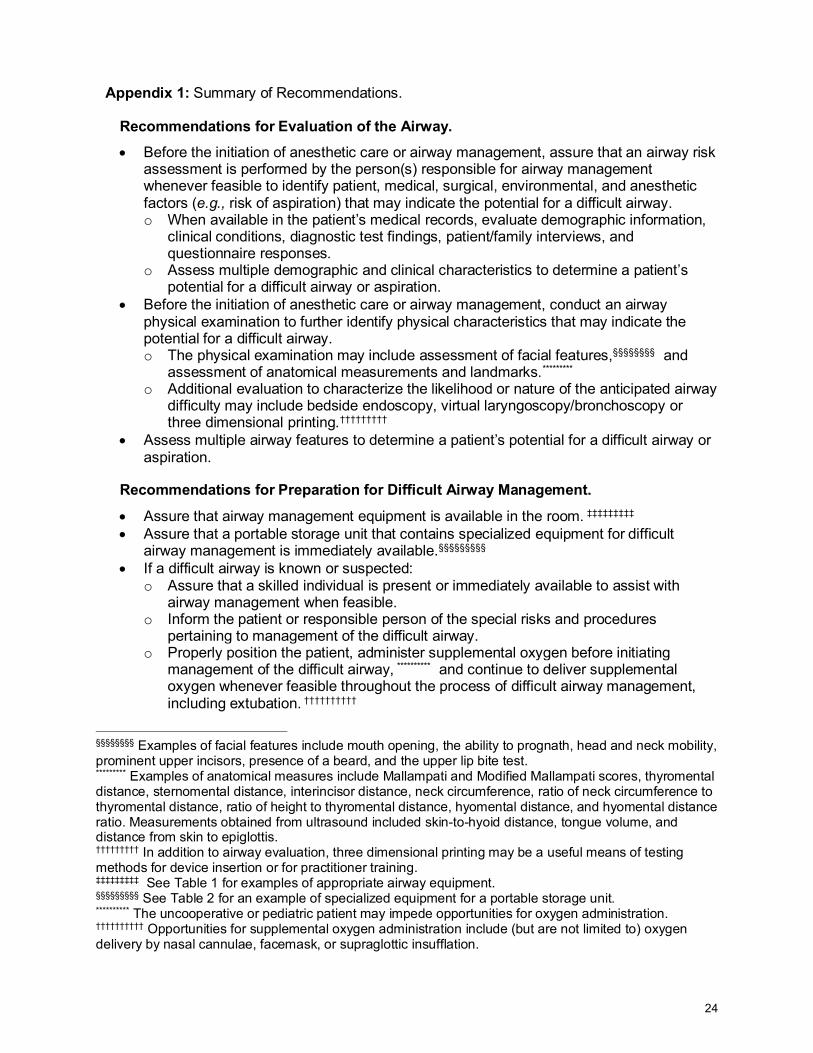
Appendix 1: Summary of Recommendations.
Recommendations for Evaluation of the Airway. • Before the initiation of anesthetic care or airway management, assure that an airway risk
assessment is performed by the person(s) responsible for airway management whenever feasible to identify patient, medical, surgical, environmental, and anesthetic factors (e.g., risk of aspiration) that may indicate the potential for a difficult airway. o When available in the patient’s medical records, evaluate demographic information,
clinical conditions, diagnostic test findings, patient/family interviews, and questionnaire responses.
o Assess multiple demographic and clinical characteristics to determine a patient’s potential for a difficult airway or aspiration.
• Before the initiation of anesthetic care or airway management, conduct an airway physical examination to further identify physical characteristics that may indicate the potential for a difficult airway. o The physical examination may include assessment of facial features,§§§§§§§§
********* and
assessment of anatomical measurements and landmarks. o Additional evaluation to characterize the likelihood or nature of the anticipated airway
difficulty may include bedside endoscopy, virtual laryngoscopy/bronchoscopy or three dimensional printing.†††††††††
• Assess multiple airway features to determine a patient’s potential for a difficult airway or aspiration.
Recommendations for Preparation for Difficult Airway Management. • Assure that airway management equipment is available in the room. ‡‡‡‡‡‡‡‡‡ • Assure that a portable storage unit that contains specialized equipment for difficult
airway management is immediately available.§§§§§§§§§ • If a difficult airway is known or suspected:
o Assure that a skilled individual is present or immediately available to assist with airway management when feasible.
o Inform the patient or responsible person of the special risks and procedures pertaining to management of the difficult airway.
o Properly position the patient, administer supplemental oxygen before initiating management of the difficult airway, **********
††††††††††
and continue to deliver supplemental oxygen whenever feasible throughout the process of difficult airway management, including extubation.
§§§§§§§§ Examples of facial features include mouth opening, the ability to prognath, head and neck mobility, prominent upper incisors, presence of a beard, and the upper lip bite test. ********* Examples of anatomical measures include Mallampati and Modified Mallampati scores, thyromental distance, sternomental distance, interincisor distance, neck circumference, ratio of neck circumference to thyromental distance, ratio of height to thyromental distance, hyomental distance, and hyomental distance ratio. Measurements obtained from ultrasound included skin-to-hyoid distance, tongue volume, and distance from skin to epiglottis. ††††††††† In addition to airway evaluation, three dimensional printing may be a useful means of testing methods for device insertion or for practitioner training. ‡‡‡‡‡‡‡‡‡ See Table 1 for examples of appropriate airway equipment. §§§§§§§§§ See Table 2 for an example of specialized equipment for a portable storage unit. ********** The uncooperative or pediatric patient may impede opportunities for oxygen administration. †††††††††† Opportunities for supplemental oxygen administration include (but are not limited to) oxygen delivery by nasal cannulae, facemask, or supraglottic insufflation.

25
• Assure that, at a minimum, monitoring according to the ASA Standards for Basic Anesthesia Monitoring are followed immediately before, during and after airway management of all patients.‡‡‡‡‡‡‡‡‡‡
Recommendations for Anticipated Difficult Airway Management. • Have a preformulated strategy for management of the anticipated difficult airway.
o This strategy will depend, in part, on the anticipated surgery, the condition of the patient, patient cooperation/consent, the age of the patient, and the skills and preferences of the anesthesiologist.
o Identify a strategy for: (1) awake intubation, (2) the patient who can be adequately ventilated but is difficult to intubate, (3) the patient who cannot be ventilated or intubated, and (4) difficulty with emergency invasive airway rescue.
o When appropriate, perform awake intubation if the patient is suspected to be a difficult intubation and one or more of the following apply: (1) difficult ventilation (face mask/supraglottic airway), (2) increased risk of aspiration, (3) the patient is likely incapable of tolerating a brief apneic episode, or (4) there is expected difficulty with emergency invasive airway rescue.§§§§§§§§§§
o The uncooperative or pediatric patient may restrict the options for difficult airway management, particularly options that involve awake intubation. Airway management in the uncooperative or pediatric patient may require an approach (e.g., intubation attempts after induction of general anesthesia) that might not be regarded as a primary approach in a cooperative patient.
o Proceed with airway management after induction of general anesthesia when the benefits are judged to outweigh the risks.
o For either awake or anesthetized intubation, airway maneuver(s) may be attempted to facilitate intubation.
o Before attempting intubation of the anticipated difficult airway, determine the benefit of a noninvasive versus invasive approach to airway management. If a noninvasive approach is selected, identify a preferred sequence of
noninvasive devices to use for airway management.*********** • If difficulty is encountered with individual techniques, combination
techniques may be performed.††††††††††† • Be aware of the passage of time, number of attempts, and oxygen
saturation. • Provide and test mask ventilation after each attempt, when feasible. • Limit the number of attempts at tracheal intubation or supraglottic airway
placement to avoid potential injury and complications.
‡‡‡‡‡‡‡‡‡‡ This recommendation does not preclude local or institutional policies that require more stringent monitoring. §§§§§§§§§§ Any one factor alone (i.e., assessed difficulty with intubation, or ventilation, increased risk of aspiration or desaturation) may be of sufficient clinical importance to warrant an awake intubation. *********** Noninvasive devices include rigid laryngoscopic blades of alternative designs and sizes (with adequate face mask ventilation after induction); adjuncts (e.g., introducers, bougies, stylets, alternative tracheal tubes, and supraglottic airways), video/video-assisted laryngoscopy; flexible intubation scopes; supraglottic airway devices; lighted or optical stylets; alternative optical laryngoscopes; and rigid bronchoscopes. ††††††††††† Combination techniques may include but are not limited to: (1) direct or video laryngoscopy combined with either optical/video stylet, flexible scope intubation, airway exchange catheter, retrograde placed guide wire or supraglottic airway placement, and (2) supraglottic airway combined with either optical/video stylet, flexible scope intubation (with or without hollow guide catheter), retrograde placed guide wire.

26
If an elective invasive approach to the airway is selected, identify a preferred intervention.‡‡‡‡‡‡‡‡‡‡‡ • Assure that an invasive airway is performed by an individual trained in
invasive airway techniques, whenever possible. • If the selected approach fails or is not feasible, identify an alternative
invasive intervention. o Initiate extracorporeal membrane oxygenation when/if appropriate and
available.
Recommendations for Unanticipated and Emergency Difficult Airway Management. • Call for help. • Optimize oxygenation§§§§§§§§§§§ • When appropriate, refer to an algorithm************ and/or cognitive aid. • Upon encountering an unanticipated difficult airway:
o Determine the benefit of waking and/or restoring spontaneous breathing. o Determine the benefit of a noninvasive versus invasive approach to airway
management. o If a noninvasive approach is selected, identify a preferred sequence of noninvasive
devices to use for airway management.†††††††††††† If difficulty is encountered with individual techniques, combination techniques
may be performed. Be aware of the passage of time, number of attempts, and oxygen saturation. Provide and test mask ventilation after each attempt, when feasible. Limit the number of attempts at tracheal intubation or supraglottic airway
placement to avoid potential injury and complications. • If an invasive approach to the airway is necessary (i.e., cannot intubate, cannot
ventilate), identify a preferred intervention.‡‡‡‡‡‡‡‡‡‡‡‡ o Assure that an invasive airway is performed by an individual trained in invasive
airway techniques, whenever possible. o Assure that an invasive airway is performed as rapidly as possible. o If the selected invasive approach fails or is not feasible, identify an alternative
invasive intervention. Initiate Extracorporeal Membrane Oxygenation (ECMO) when/if appropriate and
available.
‡‡‡‡‡‡‡‡‡‡‡ Invasive interventions may include, but are not limited to, one of the following techniques: surgical cricothyrotomy (e.g., scalpel-bougie-tube), needle cricothyrotomy with a pressure regulated device, large bore cannula cricothyrotomy or surgical tracheostomy, retrograde wire guided intubation, and percutaneous tracheostomy. §§§§§§§§§§§ Examples include low or high flow nasal oxygen during efforts securing a tube ************ See Figures 1-4 for examples of algorithms or cognitive aids. †††††††††††† Noninvasive devices include rigid laryngoscopic blades of alternative design and size (with adequate face mask ventilation after induction); adjuncts (e.g., introducers, bougies, stylets, alternative tracheal tubes, and supraglottic airways), video/video-assisted laryngoscopy; flexible intubation scopes; supraglottic airway devices; lighted optical stylets; alternative optical laryngoscopes; and rigid bronchoscopes. ‡‡‡‡‡‡‡‡‡‡‡‡ Invasive interventions may include surgical cricothyrotomy (e.g., scalpel-bougie technique), surgical tracheostomy, needle cricothyrotomy with pressure regulated ventilation (e.g., transtracheal jet ventilation or other pressure regulated techniques), and large bore cannula cricothyrotomy (including Seldinger guided techniques).

27
Recommendations for Confirmation of Tracheal Intubation. • Confirm tracheal intubation using capnography or end-tidal carbon dioxide monitoring.
o When uncertain about the location of the tracheal tube, determine whether to either remove it and attempt ventilation or use additional techniques to confirm positioning of the tracheal tube.§§§§§§§§§§§§
Recommendations for Extubation of the Difficult Airway • Have a preformulated strategy for extubation and subsequent airway management.
o This strategy will depend, in part, on the surgery/procedure, other perioperative circumstances, the condition of the patient, and the skills and preferences of the clinician.
• Assess patient readiness for extubation.
• Assure that a skilled individual is present to assist with extubation when feasible.
• Select an appropriate time and location for extubation when possible.
• Assess the relative clinical merits and feasibility of the short-term use of an airway exchange catheter and/or supraglottic airway that can serve as a guide for expedited reintubation.************* o Minimize the use of an airway exchange catheter with pediatric patients.
• Before attempting extubation, evaluate the risks and benefits of elective surgical tracheostomy.
• Evaluate the risks and benefits of awake extubation versus extubation before the return to consciousness.
• When feasible, use supplemental oxygen throughout the extubation process. • Assess the clinical factors that may produce an adverse impact on ventilation after the
patient has been extubated.
Recommendations for Follow up Care. • Use post-extubation steroids and/or racemic epinephrine when appropriate. • Inform the patient or a responsible person of the airway difficulty that was encountered
to provide the patient (or responsible person) with a role in guiding and facilitating the delivery of future care. o The information conveyed may include (but is not limited to) the presence of a
difficult airway, the apparent reasons for difficulty, how the intubation was accomplished, and the implications for future care.
• Document the presence and nature of the airway difficulty in the medical record to guide and facilitate the delivery of future care.†††††††††††††
§§§§§§§§§§§§ Additional techniques include but are not limited to visualization (any technique), flexible bronchoscopy, ultrasonography, or radiography. ************* These interventions are considered advanced techniques. ††††††††††††† Aspects of documentation include, but are not limited to: (1) a description of the airway difficulties that were encountered, distinguishing between difficulties encountered in facemask or supraglottic airway ventilation and difficulties encountered in tracheal intubation and (2) a description of the various airway management techniques that were used, indicating the extent to which each of the techniques served either a beneficial or detrimental role in management of the difficult airway.

28
• Instruct the patient to register with an emergency notification service when appropriate and feasible.

29
Appendix 2: Methods and Analyses
For these updated guidelines, a systematic search and review of peer reviewed published
literature was conducted, with scientific findings summarized and reported below and in the
document. Assessment of conceptual issues, practicality and feasibility of the guideline recommendations was also evaluated, with opinion data collected from surveys and other
sources. Both the systematic literature review and the opinion data are based on evidence
linkages, or statements regarding potential relationships between interventions and outcomes associated with difficult airway management. The evidence model below guided the search,
providing inclusion and exclusion information regarding patients, procedures, practice settings,
providers, clinical interventions, and outcomes. After review of all evidentiary information, the
task force placed each recommendation into one of three categories: (1) provide the intervention or treatment, (2) the intervention or treatment may be provided to the patient based
on circumstances of the case and the practitioner’s clinical judgment, or (3) do not provide the
intervention or treatment. The policy of the ASA Committee on Standards and Practice Parameters is to update practice guidelines every 5 years. The ASA Committee on Standards
and Practice Parameters reviews all practice guidelines at the ASA annual meeting and
determines update and revision timelines.
Evidence Model Patients
• Inclusion criteria: o Patients with or at risk of difficult mask ventilation o Patients with or at risk of difficult laryngoscopy (direct or indirect)‡‡‡‡‡‡‡‡‡‡‡‡‡ o Patients with or at risk of difficult ventilation using a supraglottic airway o Patients with or at risk or difficult/failed tracheal intubation o Patients with or at risk of difficult/failed extubation o Anticipated difficult airway patients o Unanticipated difficult airway patients o Adult patients o Pediatric patients including infants and neonates o Obstetric patients o ICU/Critically ill patients
• Exclusion criteria
o Patients where difficult airways are not encountered o Physiologically difficult airways that are not anatomically difficult§§§§§§§§§§§§§
‡‡‡‡‡‡‡‡‡‡‡‡‡ Patients “at risk” refers to difficult laryngoscopy where it is not possible to visualize any portion
of the vocal cords after multiple attempts. §§§§§§§§§§§§§ These include, but are not limited to hypoxemia, hypotension, severe metabolic acidosis, and
right ventricular failure

30
Procedures
• Inclusion criteria: o Procedures requiring general anesthesia o Procedures requiring sedation or regional anesthesia o Elective/emergency airway management without a procedure o Diagnostic procedures o Elective procedures o Emergency procedures o Invasive airway access
• Exclusion criteria:
o Airway management during cardiopulmonary resuscitation Practice Settings
• Inclusion criteria: o In-hospital
Perioperative care settings Non-operating room anesthetic setting (NORA) Emergency department setting ICU/Critical care setting
o Ambulatory surgery centers (ASC) o Office based procedure/anesthesia (OBA) locations o Out of hospital or pre-hospital (i.e., field) settings, included only if emergency
invasive airway is performed)
• Exclusion criteria: o Out of hospital or pre-hospital (i.e., field) settings, excluded except for emergency
invasive airway.
Providers
• Inclusion criteria: o Anesthesia care providers
• Exclusion criteria:
o Individuals who do not deliver anesthetic care and airway management Interventions
• Evaluation of the Airway. o Risk prediction (for difficult airway or aspiration) obtained from history/medical
records Demographic conditions (e.g., age, sex) Clinical conditions (e.g., BMI, previous difficult airway, diabetes, obesity) Diagnostic test findings (e.g., radiography, computed tomography, magnetic
resonance imaging, bedside endoscopy, bedside ultrasound) Patient interview/questionnaires (e.g., MACOCHA, STOP-BANG)
o Airway assessment/exam (bedside and advanced) when a difficult airway is known or suspected.

31
Assessment of facial features (e.g., mouth opening, nose slope, neck slope, ratio of brow to nose to chin, full beard)
Upper lip bite test Anatomical measurements and landmarks (e.g., Mallampati/modified
Mallampati, neck circumference, neck mobility (neck radiation changes), prognathism, ruler or finger measurements of thyromental, sternomental, or temporomandibular distance) • Individual measures contained in airway scoring systems (e.g., Wilson
Risk Sum scores, Simplified airway Risk Index (SARI) scores, El-Ganzouri scores)
Imaging • Ultrasound • Virtual laryngoscopy/bronchoscopy (MRI/CT reconstruction)
3D printing Bedside endoscopy
Direct laryngoscopy (e.g., Cormac-Lehane grades) Bronchoscopy Nasopharyngoscopy
• Preparation for Difficult Airway Management. o Availability of equipment for airway management (i.e., items for anesthetizing
locations, portable storage unit, cart or trolley for difficult airway management) o Availability of an assigned individual to provide assistance when a difficult airway is
encountered (from previous evidence model) o Informing the patient with a known or suspected difficult airway o Preoxygenation**************
Preoxygenation versus room air Three to 5 minutes of O2 (3 to 5 min at tidal volume, FiO2 = 1) versus 1
minute (1 min at tidal volume, FiO2 = 1) Three to 5 minutes of O2 (3 to 5 min at tidal volume, FiO2 = 1) versus 4-12
deep breaths at forced vital capacity in 1 min or the shortest time lag, FiO2 = 1)
Three minutes of preoxygenation to reach an end-tidal oxygen concentration ≥ 0.90 (EtO2 ≥ 0.9)
Preoxygenation using non-invasive ventilation (pressure support with positive end expiratory pressure)
o Patient positioning (e.g., sniffing, sitting, head/neck extension, head-elevated laryngoscopy [HELP], ramped)
o Sedative versus hypnotic administration o Local anesthesia versus no local anesthesia o Supplemental oxygen during airway management†††††††††††††† o Patient monitoring (according to ASA standards)
• Anticipated difficult airway management. o Awake tracheal intubation (any device) Awake/sedated intubation versus intubation after induction
************** Methods to deliver preoxygenation include oxygen delivery with nasal cannulae, facemask (including
humidified nasal cannula and CPAP) or supraglottic airway insufflation. †††††††††††††† Methods to deliver supplemental oxygen include oxygen delivery with nasal cannulae, facemask
(including humidified nasal cannula and CPAP), or supraglottic airway insufflation.

32
Awake/sedated versus anesthetized intubation in patients with full stomach o Anesthetized tracheal intubation Rapid sequence induction/intubation
• With versus without cricoid pressure (Sellick maneuver) • Pressure limited mask ventilation versus ablation of spontaneous
ventilation Maintenance of spontaneous ventilation versus ablation of spontaneous
ventilation Administration of neuromuscular blockade to improve mask ventilation Rocuronium with sugammadex versus suxamethonium or succinylcholine
o Both awake and anesthetized intubation Airway maneuvers (e.g., jaw thrust chin lift, external laryngeal manipulation,
backwards/upwards/rightwards pressure) o Airway management devices Rigid laryngoscopic blades of alternative design and size: with adequate face
mask ventilation after induction (alternatives to standard blades such as Macintosh, Miller)
Adjuncts – introducers, bougies, stylets, alternative tracheal tubes Video/video-assisted laryngoscopy
• Video/video-assisted laryngoscopy versus direct laryngoscopy • Video/video-assisted laryngoscopy versus fiberoptic laryngoscopy • Channel-guided versus non-channel-guided
videolaryngoscopes‡‡‡‡‡‡‡‡‡‡‡‡‡‡ • Hyperangulated versus non-angulated devices
Flexible intubation scopes • Flexible intubation scopes vs blind tracheal or nasotracheal intubation • Flexible intubation scopes vs rigid laryngoscopic intubation
Supraglottic airway • Supraglottic airway versus face mask for ventilation • Intubation with versus without a supraglottic airway • Intubating techniques with a supraglottic airway Laryngoscopic intubation with a supraglottic airway versus blind
intubation with a supraglottic airway Flexible scope intubation with a supraglottic airway versus standard
laryngoscopic intubation with a supraglottic airway Optically/image guided intubation with a supraglottic airway versus
standard laryngoscopic intubation with a supraglottic airway • Second versus first generation supraglottic airway
Lighted stylet, light wand, optical stylet • Lighted stylet, light wand or optical stylet versus blind intubation • Lighted stylet, light wand or optical stylet versus laryngoscopic intubation
Rigid bronchoscope • Intubation with versus without a supraglottic airway • Intubating techniques with a supraglottic airway • Laryngoscopic intubation with a supraglottic airway versus blind
intubation with a supraglottic airway
‡‡‡‡‡‡‡‡‡‡‡‡‡‡ Channel-guided devices include Airtraq, Kingvision, and Pentax videolaryngoscopes. Non-channel guided devices include Glidescope, C-MAC, and McGrath videolaryngoscopes.

33
• Flexible scope intubation with a supraglottic airway versus standard laryngoscopic intubation with a supraglottic airway
• Optically/image guided intubation with a supraglottic airway versus standard laryngoscopic intubation with a supraglottic airway
o Additional airway management interventions (with anticipated failure of airway management devices) Retrograde wire-guided intubation Invasive airway
• Cricothyrotomy (percutaneous) • Cricothyrotomy (surgical) • Tracheostomy/tracheotomy • Scalpel bougie technique or scalpel bougie tube technique vs needle cannula
technique • Awake/sedated cricothyrotomy/tracheostomy for invasive airway
o Combination techniques§§§§§§§§§§§§§§
• Unanticipated and emergency (i.e., cannot oxygenate or ventilate) difficult airway management. o Call for help o Maximize oxygenation Nasal oxygen during efforts securing a tube (NO DESAT) Expiratory ventilation assistance High flow nasal cannula oxygen/transnasal humidified rapid insufflation
ventilatory exchange o Use of a cognitive aid o Airway management devices Rigid bronchoscope Rigid laryngoscopic blades of alternative design and size: with adequate face
mask ventilation after induction (alternatives to standard blades such as Macintosh, Miller)
Lighted stylet, light wand, optical stylet • Lighted stylet, light wand or optical stylet versus blind intubation • Lighted stylet, light wand or optical stylet versus laryngoscopic intubation
Flexible intubation scopes) • Flexible scope intubation vs blind tracheal or nasotracheal intubation • Flexible scope intubation vs rigid laryngoscopic intubation
Video/video-assisted laryngoscopy • Video/video-assisted laryngoscopy versus direct laryngoscopy • Video/video-assisted laryngoscopy versus Flexible scope intubation • Hyperangulated versus non-angulated devices • Channel-guided versus non-channel-guided videolaryngoscopes • Alternative optical laryngoscopes
§§§§§§§§§§§§§§ Combination techniques include: (1) direct laryngoscopy with supraglottic airway, bougie, optical stylet, flexible intubation scope, airway exchange catheter or retrograde intubation, (2) videolaryngoscopes with supraglottic airway, bougie, optical stylet, flexible scope/fiberoptic scope, airway exchange catheter or retrograde intubation, (3) flexible intubation scope with supraglottic airway, airway exchange catheter, retrograde intubation or cricothyrotomy, (4) optical stylet with supraglottic airway, bougie, flexible scope intubation scope or retrograde intubation, (5) airway exchange catheter with supraglottic airway, retrograde intubation, or cricothyrotomy.

34
Adjuncts – introducers, bougies, stylets, alternative tracheal tubes Supraglottic airway
• Supraglottic airway versus face mask for ventilation • Intubation with versus without a supraglottic airway • Intubating techniques with a supraglottic airway Laryngoscopic intubation with a supraglottic airway versus blind
intubation with a supraglottic airway Flexible scope intubation with a supraglottic airway versus standard
laryngoscopic intubation with a supraglottic airway Optically/image guided intubation with a supraglottic airway versus
standard laryngoscopic intubation with a supraglottic airway • Second versus first generation supraglottic airway
o Additional airway management interventions (with anticipated failure of airway management devices) Retrograde wire-guided intubation Emergency invasive airway
• Cricothyrotomy (percutaneous) • Cricothyrotomy (surgical) • Tracheostomy/tracheotomy • Scalpel bougie technique or scalpel bougie tube technique vs needle cannula
technique • Awake/sedated cricothyrotomy/tracheostomy for emergency invasive airway
Extracorporeal membrane oxygenation (ECMO) Jet ventilation Combination techniques
• Confirmation of successful intubation
o Pulse oximetry (for oxygen saturation levels/desaturation/hypoxemia/hypoxia) o Capnography for carbon dioxide levels/hypercarbia/hypercapnia Capnography versus capnometry Capnography versus colorimetry
o Visualization (any technique) o Flexible bronchoscopy o Ultrasound o Radiography
• Extubation o Assess readiness for extubation o Presence of a skilled individual to assist o Selection of ideal time and location o Plan for possible reintubation o Elective tracheostomy o Awake extubation or supraglottic airway removal Awake tracheal tube extubation versus asleep (anesthetized) extubation Awake supraglottic airway removal versus anesthetized supraglottic airway
removal Apnea versus spontaneous ventilation during extubation
o Supplemental oxygen throughout extubation (e.g., by mask, blow-by, nasal cannula, Continuous Positive Airway Pressure, Bilevel Positive Airway Pressure, or high flow nasal cannula)

35
o Supplemental oxygen after extubation o Staged extubation
• Airway exchange catheter • Supraglottic airway exchange catheter (Bailey maneuver)
• Follow-up Care
o Post-extubation steroids o Post-extubation epinephrine o Post-extubation counseling (i.e., informing and advising the patient or responsible
patient of the occurrence and potential complications associated with a difficult airway).
o Documentation of difficult airway and management in the medical record and to the patient
o Registration with an emergency notification service
• Human Factors Excluded Interventions:
• Interventions not addressing any aspect of airway and anesthetic management • Lung separation
o Double lumen tube o Bronchial blocker
• Physiologically difficult airway • Details of awake intubation techniques • Submental intubation • Cardiopulmonary bypass • Effects of anesthetics/sedatives on ease of intubation/supraglottic airway insertion (e.g.,
propofol) • Details of ECMO
Outcomes
• Inclusion criteria: o Identification of patient characteristics at risk of difficult intubation o Identification of patient characteristics leading to awake intubation o Intubation/ventilation success/failure: Face/bag mask ventilation (success/failure, easy/difficult) supraglottic airway placement (success/failure, number of attempts) Laryngoscopy (success/failure, number of attempts) Tracheal intubation (success/failure, number of attempts) Invasive airway
• Percutaneous cricothyrotomy (success/failure) • Surgical cricothyrotomy (success/failure) • Tracheostomy (success/failure) • Scalpel bougie technique or scalpel bougie tube technique versus needle
catheter technique (success/failure) Restoration of failed oxygenation (success/failure) Esophageal intubation Barotrauma (pneumothorax, pneumomediastinum) Subcutaneous emphysema

36
Gastric rupture Tracheal rupture Delayed tracheal stenosis
o Physiological outcomes (measurement of physiological functioning) Oxygenation/desaturation CO2 levels Hemodynamic levels (e.g., MAP, CVP)
o Clinical outcomes Hypoxemia/hypoxia Hypercapnia/hypercarbia Hemodynamic instability Aspiration Airway injury/trauma Soft tissue injuries/blind spot injuries Sore throat Palatal injury Oral/dental damage Cardiac events (e.g., cardiac arrest) Neurological injury Unplanned tracheotomy/surgical airway Neurological deficit <72 hrs
o Permanent (long-term) outcomes Death Respiratory system damage
• Airway trauma • Pneumothorax • Aspiration
Nerve/brain damage • Nerve damage • Neurological/memory deficit • Permanent brain damage • Brain injury (anoxic encephalopathy)
Cardiovascular damage • Cardiopulmonary arrest
Fetal/newborn damage Functional deficit
• Awareness/fright • Loss of employment
o Nonclinical outcomes Unplanned ICU admission Unplanned hospital admission Surgery postponed/cancelled Length of hospital stay Patient satisfaction
• Exclusion criteria: o No exclusion criteria
Evidence collection
• Literature inclusion criteria: o Randomized controlled trials

37
o Prospective nonrandomized comparative studies (e.g., quasi-experimental, cohort) o Retrospective comparative studies (e.g., case-control) o Observational studies (e.g., correlational or descriptive statistics) o Case reports, case series
• Literature exclusion criteria (except to obtain new citations): o Editorials o Literature reviews o Meta-analyses conducted by others o Unpublished studies o Studies in non-peer reviewed journals o Newspaper articles
• Survey evidence: o Expert consultant survey o ASA membership survey o Membership surveys of other participating organizations o Reliability survey o Feasibility survey
State of the Literature.
For the systematic review, potentially relevant clinical studies were identified via electronic
and manual searches. Bibliographic database searches included PubMed and EMBASE. The searches covered a 9.25-yr period from January 1, 2012, through March 31, 2021. Citation
searching (backward and forward) of relevant meta-analyses and other systematic reviews was
also performed. No search for grey literature was conducted. Publications identified by task force members were also considered. Accepted studies from the previous guidelines were re-
reviewed, covering the period of January 1, 2002, through June 31, 2012. Only studies
containing original findings from peer-reviewed journals were acceptable. Editorials, letters, and other articles without data were excluded. A literature search strategy and PRISMA* flow
diagram are available as Supplemental Digital Content 2, http://links.lww.com/ALN/XXXXXXXX. In
total, 12,544 unique new citations were identified, with 1,026 full articles assessed for eligibility.
After review, 619 were excluded, with 407 new studies meeting inclusion criteria. These studies were combined with 190 pre-2012 articles from the previous guidelines, resulting in a total of
597 articles accepted as evidence for these guidelines. In this document, 559 are referenced,
with a complete bibliography of articles used to develop these guidelines, organized by section, available as Supplemental Digital Content 3, http://links.lww.com/ALN/XXXXXXXX.
Each pertinent outcome reported in a study was classified by evidence category and level
and designated as beneficial, harmful, or equivocal. Findings were then summarized for each evidence linkage and reported in the text of the updated Guidelines.
Evidence categories refer specifically to the strength and quality of the research design of
the studies. Category A evidence represents results obtained from randomized controlled trials,

38
and category B evidence represents observational results obtained from nonrandomized study
designs or s without pertinent comparison groups. When available, category A evidence is given precedence over category B evidence for any particular outcome. These evidence categories
are further divided into evidence levels. Evidence levels refer specifically to the strength and
quality of the summarized study findings (i.e., statistical findings, type of data, and the number of studies reporting/replicating the findings). In this document, only the highest level of evidence
is included in the summary report for each intervention—outcome pair, including a designation
of benefit, harm, or equivocality.
Category A: randomized controlled trials report comparative findings between clinical interventions for specified outcomes. Statistically significant (P < 0.01) outcomes are designated
as either beneficial (B) or harmful (H) for the patient; statistically nonsignificant findings are
designated as equivocal (E). Level 1: The literature contains a sufficient number of randomized controlled trials to
conduct meta-analysis,*************** and meta-analytic findings from these
aggregated studies are reported as evidence.
Level 2: The literature contains multiple randomized controlled trials, but the number of randomized controlled trials is not sufficient to conduct a viable meta-analysis for
the purpose of these Guidelines. Findings from these randomized controlled trials
are reported separately as evidence. Level 3: The literature contains a single randomized controlled trial, and findings from
this study are reported as evidence.
Category B: Observational studies or randomized controlled trials without pertinent comparison groups may permit inference of beneficial or harmful relationships among clinical
interventions and clinical outcomes. Inferred findings are given a directional designation of
beneficial (B), harmful (H), or equivocal (E). For studies that report statistical findings, the
threshold for significance is P < 0.01. Level 1: The literature contains nonrandomized comparisons (e.g., quasiexperimental,
cohort [prospective or retrospective], or case-control research designs) with
comparative statistics between clinical interventions for a specified clinical outcome.
Level 2: The literature contains noncomparative observational studies with associative
statistics (e.g., correlation, sensitivity, and specificity).
*************** A minimum of five independent randomized controlled trials (i.e., sufficient for fitting a random-effects model) is required for meta-analysis.515

39
Level 3: The literature contains noncomparative observational studies with descriptive
statistics (e.g., frequencies, percentages). Level 4: The literature contains case reports.
Insufficient Literature. The lack of sufficient scientific evidence in the literature may occur
when the evidence is either unavailable (i.e., no pertinent studies found) or inadequate. Inadequate literature cannot be used to assess relationships among clinical interventions and
outcomes because a clear interpretation of findings is not obtained due to methodological
concerns (e.g., confounding of study design or implementation) or the study does not meet the
criteria for content as defined in the “Focus” of the guidelines.
Literature addressing risk prediction reported sensitivity, specificity, positive and negative
predictive and other common values for age, sex, body mass index, weight, height, and history of snoring; values for airway assessment were reported for facial and jaw features, anatomical
landmarks and measurements.
Literature relating to videolaryngoscopes contained enough studies with well-defined experimental designs and statistical information to conduct formal meta-analyses (table 4).
Outcomes assessed were (1) laryngoscopic view, (2) intubation success, (3) first-attempt
intubation success, (4) assist maneuvers used for intubation, and (5) time to intubation. For meta-analyses of studies reporting frequency of events, event rates and odds ratios were
pooled. Time to intubation was pooled using mean differences (continuous outcomes) for clinical
relevance. Fixed-effects models were fitted using Mantel-Haenszel or inverse variance weighting as appropriate. Random-effects models were fitted with inverse variance weighting
using the DerSimonian and Laird estimate of between-study variance. Sensitivity to effect
measure was also examined. Heterogeneity was quantified with I2 and a significance level of P < 0.01 was applied for analyses. Statistics for individual studies and forest plots are available as
supplemental digital content 4, http://links.lww.com/ALN/
Interobserver agreement among task force members and two methodologists was assessed
for this update, with agreement levels using a κ statistic for two-rater agreement pairs as follows: (1) research design, κ = 0.55 to 0.61; (2) type of analysis, κ = 0.55 to 0.83; (3) evidence linkage
assignment, κ = 0.67 to 0.79; and (4) literature inclusion for database, κ = 0.08 to 0.79. Three-
rater κ values between two methodologists and task force reviewers were: (1) research design, κ = 0.61; (2) type of analysis, κ = 0.65; (3) linkage assignment, κ = 0.67; and (4) literature
database inclusion, κ = 0.15. These values represented low to moderate levels of agreement.

40
Consensus-based evidence.
Validation of the concepts addressed by these Guidelines and subsequent recommendations proposed was obtained by consensus from multiple sources, including: (1)
survey opinion from expert consultants who were selected based on their knowledge or expertise in difficult airway management; (2) survey opinions from randomly selected samples
of active members of the ASA and participating organizations and (3) internet commentary. All
opinion-based evidence relevant to each topic was considered in the development of these
guidelines. However, only findings obtained from formal surveys are reported in the document. Opinion surveys were developed by the task force to address each clinical intervention
identified in the document. Identical surveys were distributed to expert consultants and a
random sample of members of the participating organizations.
Survey responses were recorded using a 5-point scale and summarized based on median
values.††††††††††††††† Strongly Agree: Median score of 5 (at least 50% of the responses are 5) Agree: Median score of 4 (at least 50% of the responses are 4 or 4 and 5) Equivocal: Median score of 3 (at least 50% of the responses are 3, or no other
response category or combination of similar categories contain at least 50% of the responses)
Disagree: Median score of 2 (at least 50% of responses are 2 or 1 and 2) Strongly Disagree: Median score of 1 (at least 50% of responses are 1)
For consultant respondents the rate of return for the survey addressing guideline
recommendations was 82% (n = 174 of 212), and results are presented in table 5. For
membership respondents, the survey totals were as follows: American Society of
Anesthesiologists = 220; All India Difficult Airway Association (AIDAA) = 74; European Airway Management Society (EAMS) = 79; Italian Society of Anesthesiology, Analgesia, Resuscitation
and Intensive Care Society (SIAARTI) = 177; Learning, Teaching and Investigation Difficult
Airway Group (FIDIVA) = 24; Society for Ambulatory Anesthesia (SAMBA) = 47; Society for Airway Management (SAM) = 70; Society for Head and Neck Anesthesia (SHANA) = 27;
Society for Pediatric Anesthesia (SPA) = 268; Society of Critical Care Anesthesiologists
(SOCCA) = 85; Swiss Society for Anaesthesiology and Resuscitation (SGAR) = 203; Trauma
Anesthesiology Society (TAS) = 21. Survey results for each organization are presented as
supplemental digital content 5, http://links.lww.com/ALN/XXXXXX.
††††††††††††††† When an equal number of categorically distinct responses are obtained, the median value is determined by calculating the arithmetic mean of the two middle values. Ties are calculated by a predetermined formula.

41
An additional survey was sent to the consultants accompanied by a draft of the Guidelines
asking them to indicate which, if any, of the recommendations would change their clinical practices if the Guidelines were instituted. The rate of return was 31% (n = 68 of 218). The
percentage of responding consultants expecting no change associated with each linkage were
as follows: (1) evaluation of the airway = 82%, (2) availability of airway management equipment = 79%, (3) the presence of a skilled individual to assist = 82%, (4) supplemental oxygen delivery
= 76%, strategy for management of an anticipated difficult airway = 88%, awake intubation
strategy = 81%, selection of an elective invasive airway = 84%, preferred sequence of devices
for attempting intubation = 93%, strategy for management of an unanticipated difficult airway = 88%, strategy for management of an emergency difficult airway = 87%, use of an infographic,
cognitive aid or algorithm = 65%, use of capnography for confirmation of intubation = 90%,
strategy for invasive management of a difficult airway = 82%, supplemental oxygen delivery for extubation = 87%, and documentation of the encountered difficult airway = 81%. Ninety-one
percent of the respondents indicated that the Guidelines would have no effect on the amount of
time spent on a typical case, 7% indicated that there would be an increase of the amount of time
spent on a typical case and 1% indicated a decrease in time with the implementation of these Guidelines. Seventy-two percent indicated that new equipment, supplies or training would not
be needed in order to implement the guidelines, and 86% indicated that implementation of the
Guidelines would not require changes in practice that would affect costs.

42
Table 1: Airway Management Items for Anesthetizing Locations‡‡‡‡‡‡‡‡‡‡‡‡‡‡‡
Self-inflating resuscitation bag
Suction tubing, Yankauers, suction catheters, and appropriate connectors
Various sizes of face masks Various sizes of oral and nasal airways
Various sizes and types of laryngoscope blades and handles
Various sizes and types of tracheal tubes
Tracheal tube introducer (bougie) for adult patients Tracheal tube stylets (malleable and rigid)
Equipment for emergency invasive airway management
Various sizes of supraglottic airways Water soluble medical lubricant
Nasal cannula and oxygen face masks
Video laryngoscope with appropriate stylets Standard ASA monitors
Anesthetic induction, maintenance, and rescue medications
‡‡‡‡‡‡‡‡‡‡‡‡‡‡‡ The examples listed in this table represent basic minimum contents for an anesthetizing location cart or trolley. The cart may be customized to meet the specific needs, preferences, and skills of the practitioner and healthcare facility.

43
Table 2: Portable Storage Unit Items for Difficult Airway Management§§§§§§§§§§§§§§§ Category**************** ††††††††††††††††‡‡‡‡‡‡‡‡‡‡‡‡‡‡‡‡ Item Alternative/rescue ventilation equipment Oral and nasal airways of assorted sizes Supraglottic airways of assorted sizes/ cuffed pharyngeal sealer
Nasal cannula
Alternative Intubation equipment Tracheal tubes of assorted sizes (Including microlaryngeal tubes) Rigid blades of alternate design and size for intubation
Tracheal tube guides. Examples include (but are not limited to) semirigid stylets, lighted stylets, forceps designed to manipulate the distal portion of the tracheal tube Intubating supraglottic airway
Videolaryngoscope with appropriate stylet
Optical laryngoscope Intubating video stylet
Flexible intubating bronchoscope along with topical anesthetic and equipment, and airway/bite block
Aintree catheter
Emergency airway equipment Equipment for emergency invasive airway management
Jet ventilation equipment Miscellaneous Airway exchange catheters of assorted size
Multiple exhaled carbon dioxide detectors.
A laminated version of a local accepted difficult airway algorithm/cognitive aid/checklist Defogger
§§§§§§§§§§§§§§§ The examples listed in this table represent airway management equipment beyond what may be available in the anesthetizing location (see Table 1). In areas where these items are not available at the anesthetizing location, add them to this portable storage unit. **************** Equipment and supplies sizes should match the intended population to be served (e.g., neonates, pediatrics, adults). †††††††††††††††† The items listed in this table represent suggestions. The contents of the portable storage unit should be customized to meet the specific needs, preferences, and skills of the practitioner and healthcare facility. ‡‡‡‡‡‡‡‡‡‡‡‡‡‡‡‡ Choice of some items (e.g., videolaryngoscope, jet ventilation equipment) may depend on practitioner familiarity and experience with the device.
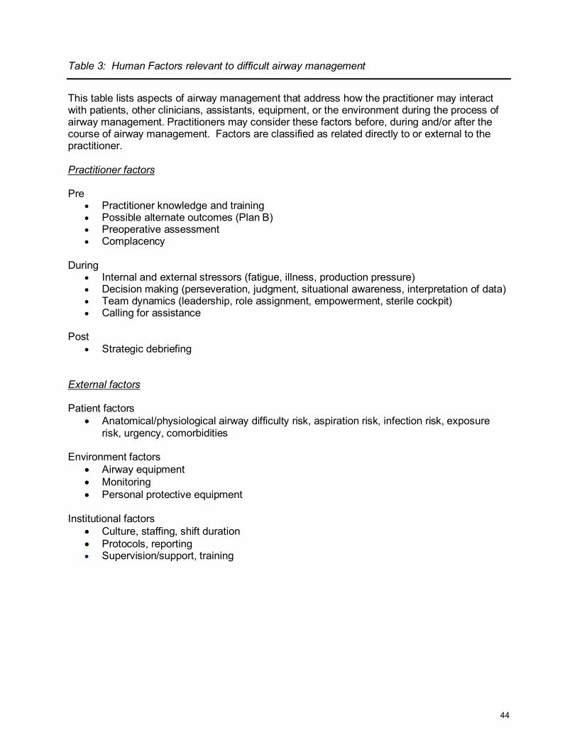
44
Table 3: Human Factors relevant to difficult airway management This table lists aspects of airway management that address how the practitioner may interact with patients, other clinicians, assistants, equipment, or the environment during the process of airway management. Practitioners may consider these factors before, during and/or after the course of airway management. Factors are classified as related directly to or external to the practitioner. Practitioner factors Pre
• Practitioner knowledge and training • Possible alternate outcomes (Plan B) • Preoperative assessment • Complacency
During
• Internal and external stressors (fatigue, illness, production pressure) • Decision making (perseveration, judgment, situational awareness, interpretation of data) • Team dynamics (leadership, role assignment, empowerment, sterile cockpit) • Calling for assistance
Post
• Strategic debriefing External factors Patient factors
• Anatomical/physiological airway difficulty risk, aspiration risk, infection risk, exposure risk, urgency, comorbidities
Environment factors
• Airway equipment • Monitoring • Personal protective equipment
Institutional factors
• Culture, staffing, shift duration • Protocols, reporting • Supervision/support, training
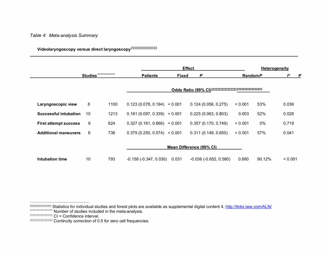
Table 4: Meta-analysis Summary
Videolaryngoscopy versus direct laryngoscopy§§§§§§§§§§§§§§§§
. . Effect . Heterogeneity. Studies***************** Patients Fixed P Random P I² P
Odds Ratio (99% CI)†††††††††††††††††‡‡‡‡‡‡‡‡‡‡‡‡‡‡‡‡‡ .
Laryngoscopic view 8 1100 0.123 (0.078, 0.194) < 0.001 0.124 (0.056, 0.275) < 0.001 53% 0.036 Successful intubation 10 1213 0.181 (0.097, 0.339) < 0.001 0.225 (0.063, 0.803) 0.003 52% 0.026 First attempt success 9 624 0.327 (0.161, 0.666) < 0.001 0.357 (0.170, 0.749) < 0.001 0% 0.719 Additional maneuvers 6 738 0.379 (0.250, 0.574) < 0.001 0.311 (0.149, 0.650) < 0.001 57% 0.041 Mean Difference (99% CI) . Intubation time 10 793 -0.158 (-0.347, 0.030) 0.031 -0.036 (-0.652, 0.580) 0.880 90.12% < 0.001
§§§§§§§§§§§§§§§§ Statistics for individual studies and forest plots are available as supplemental digital content 4, http://links.lww.com/ALN/ ***************** Number of studies included in the meta-analysis. ††††††††††††††††† CI = Confidence interval. ‡‡‡‡‡‡‡‡‡‡‡‡‡‡‡‡‡ Continuity correction of 0.5 for zero cell frequencies.

Table 5. Expert Consultant Survey Results (Response Rate = 82%) Recommendations N Strongly
Agree (%)
Agree (%)
Neutral (%)
Disagree (%)
Strongly Disagree
(%) Evaluation of the Airway 1a. Before the initiation of anesthetic care or airway management, assure that an airway risk assessment is performed by the person(s) responsible for airway management whenever feasible to identify patient, medical, surgical, environmental, and anesthetic factors (e.g., risk of aspiration) that may indicate the potential for a difficult airway.
174 92* 6 1 0 1
1b. Before the initiation of anesthetic care or airway management, conduct an airway physical examination.
174 84* 13 1 0 1
Preparation for Difficult Airway Management
2a. If a difficult airway is known or suspected, assure that a skilled individual is present or immediately available to assist with airway management.
174 94* 5 0 0 1
2b. If a difficult airway is known or suspected, inform the patient or responsible person of the special risks and procedures pertaining to management of the difficult airway.
174 74* 21 3 1 1
2c. If a difficult airway is known or suspected, administer oxygen before initiating management of the difficult airway and deliver supplemental oxygen throughout the process of difficult airway management, including extubation.
173 83* 10 6 1 1
Anticipated Difficult Airway Management
3. Identify a strategy for (1) awake intubation, (2) the patient who can be adequately ventilated but is difficult to intubate, (3) the patient who cannot be ventilated or intubated, and (4) alternative approaches to airway management failure.
164 84* 12 3 1 1
4a. When appropriate, perform awake intubation if the patient is suspected to be a difficult intubation and- Difficult ventilation (face mask/SGA) is anticipated.
165 68* 22 7 2 1
4b. When appropriate, perform awake intubation if the patient is suspected to be a difficult intubation and- Increased risk of aspiration is anticipated.
165 42 30* 15 11 2
4c. When appropriate, perform awake intubation if the patient is suspected to 166 44 34* 14 6 2

47
be a difficult intubation and-The patient is likely incapable of tolerating a brief apneic episode is anticipated. 4d. When appropriate, perform awake intubation if the patient is suspected to be a difficult intubation and- Difficulty with emergency invasive airway rescue is anticipated.
166 58* 25 11 4 1
5. If a noninvasive approach is selected, identify a preferred sequence of noninvasive devices to use for airway management.
166 63* 29 7 0 1
5a. If difficulty is encountered with individual techniques, combination techniques may be performed.
167 66* 28 5 1 1
5b. Be aware of the passage of time, number of attempts, and oxygen saturation.
166 91* 6 2 0 1
5c. Provide and test mask ventilation between attempts. 167 58* 23 13 6 1
5d. Limit the number of attempts at tracheal intubation or SGA placement to avoid potential injury and complications.
167 77* 19 2 1 1
6. If an elective invasive approach to the airway (e.g., surgical cricothyrotomy, tracheostomy, or large bore cannula cricothyrotomy) is selected, identify a preferred intervention.
165 72* 21 6 1 1
6a. Assure that an invasive airway is performed by an individual trained in invasive airway techniques, whenever possible.
166 83* 15 2 0 1
6b. If the selected invasive approach fails or is not feasible, identify an alternative invasive intervention.
166 72* 22 5 1 1
Unanticipated and Emergency Difficult Airway Management
7a. Upon encountering an unanticipated difficult airway determine the benefit of waking and/or restoring spontaneous breathing.
164 64* 23 10 2 1
7b. Upon encountering an unanticipated difficult airway determine the benefit of a noninvasive versus invasive approach to airway management.
161 62* 30 5 2 1
8. If a noninvasive approach is selected, identify a preferred sequence of noninvasive devices to use for airway management.
164 73* 24 1 1 1
8a. If difficulty is encountered with individual techniques, combination techniques may be performed.
163 66* 26 6 1 1
8b. Be aware of the passage of time, number of attempts, and oxygen saturation.
162 88* 9 2 0 1

48
8c. Provide and test mask ventilation between attempts. 159 59* 25 9 7 1
8d. Limit the number of attempts at tracheal intubation or SGA placement to avoid potential injury and complications.
163 83* 12 4 1 1
9. If an invasive approach to the airway (e.g., surgical cricothyrotomy, tracheostomy, or large bore cannula cricothyrotomy) is necessary (i.e., cannot intubate, cannot ventilate), identify a preferred intervention.
161 76* 20 2 1 1
9a. Assure that an invasive airway is performed by an individual trained in invasive airway techniques, whenever possible.
163 83* 14 2 1 1
9b. Assure that an invasive airway is performed as rapidly as possible. 163 67* 23 7 2 1
9c. If the selected invasive approach fails or is not feasible, identify an alternative invasive intervention.
163 74* 20 4 1 1
Confirmation of Tracheal Intubation 10. Confirm tracheal intubation using capnography or end-tidal carbon dioxide monitoring.
164 91* 7 0 1 1
11. When uncertain about the location of the tracheal tube, determine whether to either remove it and attempt ventilation or use additional techniques to confirm positioning of tracheal tube.
163 60* 28 7 4 1
Extubation of the Difficult Airway 12. Have a preformulated strategy for extubation and subsequent airway management.
163 91* 8 1 0 1
13. Assure that a skilled individual is present to assist with extubation. 162 72* 23 3 1 1
14. Select an appropriate time and location for extubation when possible. 163 77* 20 2 1 1 15. Assess the relative clinical merits and feasibility of the short-term use of an airway exchange catheter and/or SGA that can serve as a guide for expedited reintubation.
163 64* 29 5 1 1
16. Before attempting extubation, evaluate the risks and benefits of elective surgical tracheostomy.
163 47 33* 18 2 1
17. Evaluate the risks and benefits of awake extubation versus extubation before the return to consciousness.
163 57* 23 9 6 6
18. Assess the clinical factors that may produce an adverse impact on ventilation after the patient has been extubated.
162 75* 23 1 0 1
Follow-up Care 19. Inform the patient (or responsible person) of the airway difficulty that was 162 88* 11 1 0 1

49
encountered to provide the patient (or responsible person) with a role in guiding and facilitating the delivery of future care. 20. Document the presence and nature of the airway difficulty in the medical record to guide and facilitate the delivery of future care.
163 94* 5 1 0 1
* An asterisk beside a percentage score indicates the median.

INTUBATION ATTEMPT WITH PATIENT AWAKE
Awake Airway electively secured by Intubation3 invasive access5
SUCCESS FAIL
Consider other options4
FAIL Postpone the case
Figure 1: ASA Difficult Airway Algorithm – Adult Patients
MASK VENTILATION ADEQUATE MASK VENTILATION NOT ADEQUATE
AS CONFIRMED BY CO2 CONSIDER/ATTEMPT SUPRAGLOTTIC AIRWAY6 NON-EMERGENCY PATHWAY SUPRAGLOTTIC AIRWAY SUPRAGLOTTIC AIRWAY VENTILATION NOT ADEQUATE Ventilation adequate/Intubation unsuccessful ADEQUATE (Cannot intubate, cannot ventilate)
LIMIT ATTEMPTS AND CONSIDER AWAKENING8 THE PATIENT EMERGENCY PATHWAY LIMIT ATTEMPTS AND BE AWARE OF THE PASSAGE OF TIME Consider alternative intubation approaches,7 CALL FOR HELP / FOR INVASIVE ACCESS invasive access4 or the feasibility of other options9 Attempt alternative intubation
approaches7 as you prepare for emergency invasive airway5 SUCCESS FAIL or deteriorating ventilation FAIL SUCCESS Emergency invasive airway5
OPTIMIZE OXYGENATION THROUGHOUT2 INTUBATION ATTEMPT AFTER
INDUCTION OF GENERAL ANESTHESIA
FAIL SUCCESS
LIMIT ATTEMPTS Consider calling for help
Always evaluate for emergency invasive airway
No Suspected difficult laryngoscopy?
Significant increased risk of aspiration?
Increased risk of rapid desaturation?
Suspected difficult ventilation with face mask/supraglottic airway? Yes
Yes
Yes
No
No
No
Pre-Intubation: Before attempting intubation, choose between either an awake or post-induction airway strategy. Choice of strategy and technique should be made by the clinician managing the airway.1
Other patient factors may require an alternative strategy
Any one factor alone (assessed difficulty with intubation, ventilation, or aspiration or desaturation risk) may be clinically important enough to warrant an awake intubation
Suspected difficult emergency invasive airway
Proceed with intubation attempt Proceed with intubation attempt
Yes
Yes
No

51
Footnotes: 1. The airway manager’s choice of airway strategy and techniques should be based on their prior experience,
available resources, including equipment, availability and competency of help, and the context in which airway management will occur.
2. Low or high flow nasal cannula, head elevated position throughout procedure. Non-invasive ventilation during
preoxygenation.
3. Awake intubation techniques include flexible bronchoscope, videolaryngoscopy, direct laryngoscopy, combined techniques, and retrograde-wire aided intubation
4. Other options include, but are not limited to:
• Alternative awake technique • Awake elective invasive airway • Alternative anesthetic techniques • Induction of anesthesia (if unstable or can’t be postponed) with preparations for emergency invasive airway • Postpone the case without attempting the above options
5. Invasive airway techniques include: surgical cricothyrotomy, needle cricothyrotomy with a pressure regulated
device, large bore cannula cricothyrotomy or surgical tracheostomy. Elective invasive airway techniques include the above and retrograde wire guided intubation and percutaneous tracheostomy. Also consider rigid bronchoscopy and Extracorporeal Membrane Oxygenation.
6. Consideration of size, design, positioning, and 1st vs 2nd generation supraglottic airways may improve the ability to
ventilate.
7. Alternative difficult intubation approaches include but are not limited to: video-assisted laryngoscopy, alternative laryngoscope blades, combined techniques, intubating supraglottic airway (with or without flexible bronchoscopic guidance), flexible bronchoscopy, introducer, and lighted stylet or lightwand. Adjuncts that may be employed during intubation attempts include tracheal tube introducers, rigid stylets, intubating stylets, or tube changers and external laryngeal manipulation.
8. Includes postponing the case or postponing the intubation and returning with appropriate resources (e.g.
personnel, equipment, patient preparation, awake intubation)
9. Other options include but are not limited to: proceeding with procedure utilizing face mask or supraglottic airway ventilation. Pursuit of these options usually implies that ventilation will not be problematic.

Figure 2: Difficult Airway Algorithm – Pediatric Patients*
ADEQUATE AS CONFIRMED BY CO2 MARGINAL IMPOSSIBLE NON-EMERGENCY PATHWAY MARGINAL IMPOSSIBLE CONSIDER EMERGING THE PATIENT6 LIMIT ATTEMPTS BE AWARE OF PASSAGE OF TIME (REASSESS VENTILATION AFTER EACH ATTEMPT) Consider alternative intubation approaches,7 invasive access5 or the feasibility of other options8
SUCCESS FAIL or deteriorating ventilation Alternative intubation approaches7 as
you prepare for emergency invasive airway5
FAIL SUCCESS Emergency invasive airway5
INTUBATION ATTEMPT AFTER INDUCTION OF GENERAL ANESTHESIA
FAIL SUCCESS
LIMIT ATTEMPTS ENSURE ADEQUATE ANESTHETIC DEPTH
Consider calling for help
Yes
Suspected difficult ventilation with face mask, supraglottic airway?
CONSIDER AWAKE/SEDATED APPROACH Transfer to Tertiary Center if feasible
Awake Airway electively secured Intubation3 by invasive access5 SUCCESS FAIL Consider other options4
FAIL Postpone the case – terminate sedation if used
ASSESS OXYGENTATION/VENTILATION WITH FACE MASK/SUPRAGLOTTIC AIRWAY
EMERGENCY PATHWAY CANNOT INTUBATE, CANNOT VENTILATE
CALL FOR HELP / FOR INVASIVE ACCESS
EXCLUDE /TREAT ANATOMICAL AND FUNCTIONAL OBSTRUCTION CONSIDER CALLING FOR INVASIVE ACCESS OR ECMO
OPTIMIZE OXYGENATION THROUGHOUT2
Suspected difficult laryngoscopy?
Pre-Intubation: Before attempting intubation, choose between either an awake or post-induction airway strategy. Choice of strategy and technique should be made by the clinician managing the airway.1
No Yes

53
Footnotes: 1. The airway manager’s assessment and choice of techniques should be based on their prior experience, available
resources, including equipment, availability and competency of help, and the context in which airway management will occur.
2. Low or high flow nasal cannula, head elevated position throughout procedure. Non-invasive ventilation during
preoxygenation.
3. Awake intubation techniques include flexible bronchoscope, videolaryngoscopy, direct laryngoscopy, combined techniques, and retrograde-wire aided intubation
4. Other options include, but are not limited to:
• Alternative awake technique • Awake elective invasive airway • Alternative anesthetic techniques • Induction of anesthesia (if unstable or can’t be postponed) with preparations for emergency invasive airway • Postpone the case without attempting the above options
5. Invasive airway techniques include: surgical cricothyroidotomy, needle cricothyroidotomy if age appropriate with a
pressure regulated device, large bore cannula cricothyroidotomy or surgical tracheostomy. Elective invasive airway techniques include the above and retrograde wire guided intubation and percutaneous tracheostomy. Also consider rigid bronchoscopy and Extracorporeal Membrane Oxygenation.
6. Includes postponing the case or postponing the intubation and returning with appropriate resources (e.g.
personnel, equipment, patient preparation, awake intubation)
7. Alternative difficult intubation approaches include but are not limited to: video-assisted laryngoscopy, alternative laryngoscope blades, combined techniques, intubating supraglottic airway (with or without flexible bronchoscopic guidance), flexible bronchoscopy, introducer, and lighted stylet Adjuncts that may be employed during intubation attempts include tracheal tube introducers, rigid stylets, intubating stylets, or tube changers and external laryngeal manipulation.
8. Other options include but are not limited to: proceeding with procedure utilizing face mask or supraglottic airway
ventilation. Pursuit of these options usually implies that ventilation will not be problematic.
Title Caption: * Developed in collaboration with the Society for Pediatric Anesthesia and the Pediatric Difficult Intubation
Collaborative: John E. Fiadjoe, M.D., Thomas Engelhardt, MD, PhD, FRCA, Nicola Disma, MD, Narasimhan Jagannathan, MD, MBA, Britta S von Ungern-Sternberg, MD, PhD, DEAA, FANZCA, and Pete G. Kovatsis, MD, FAAP

54
Figure 3: Difficult Airway Management Infographic – Adult Patients Figure 3 provides three tools to aid in airway management for the patient with a planned, anticipated difficult, or unanticipated difficult airway. Part 1 is a decision tool that incorporates relevant elements of evaluation and is intended to assist in the decision to enter the Awake Airway Management pathway or the Airway Management with the Induction of Anesthesia pathway of the ASA difficult airway algorithm. Part 2 is an awake intubation algorithm. Part 3 is a strategy for managing patients with induction of anesthesia when unanticipated difficulty with ventilation (as determined by capnography) with a planned airway technique is encountered.

55

56

57
Footnotes a The airway manager’s assessment and choice of techniques should be based on their prior experience, available resources, including equipment, availability and competency of help, and the context in which airway management will occur. b Review airway strategy: consider anatomical / physiological airway difficulty risk, aspiration risk, infection risk, other exposure risk, equipment and monitoring check, role assignment, back-up and rescue plans. Awake techniques include flexible intubation scope, videolaryngoscopy, direct laryngoscopy, supraglottic airway, combined devices, retrograde-wire aided. c Adequate ventilation by any means (e.g., face mask, supraglottic airway, tracheal intubation) should be confirmed by capnography, when possible. d Follow-up care includes post-extubation care (i.e., steroids, racemic epinephrine), counseling, documentation, team debriefing, encourage patient difficult airway registry. e Postpone the case / intubation - Return with appropriate resources (e.g. personnel, equipment, patient preparation, awake intubation). f Invasive airways include: surgical cricothyroidotomy, needle cricothyroidotomy with a pressure regulated device, large bore cannula cricothyroidotomy or surgical tracheostomy. Elective invasive airways include the above and retrograde wire guided intubation, and percutaneous tracheostomy. Other options include rigid bronchoscopy and Extracorporeal Membrane Oxygenation. g Invasive airway is performed by an individual trained in invasive airway techniques, whenever possible. h In an unstable situation, or when airway management is mandatory after a failed awake intubation, a switch to the Airway Management with Induction of Anesthesia pathway may be entered with preparations for an emergency invasive airway. i Low or high flow nasal cannula, head elevated position throughout procedure. Non-invasive ventilation during pre-oxygenation. j The intent of limiting attempts at tracheal intubation and supraglottic airway insertion is to reduce the risk of bleeding, edema, and other types of trauma that may increase the difficulty of mask ventilation and/or subsequent attempts to secure a definitive airway. Persistent attempts at any airway intervention, including ineffective mask ventilation, may delay obtaining an emergency invasive airway. A reasonable approach may be to limit attempts with any technique class (i.e., face mask, supraglottic airway, tracheal tube) to 3, with 1 additional attempt by a clinician with higher skills. k Optimize: suction, relaxants, repositioning, Face mask: oral/nasal airway, two hand mask grip. Supraglottic airway: size, design, repositioning, 1st vs 2nd generation. Tracheal tube: introducer, rigid stylet, hyperangulated videolaryngoscopy, blade size, external laryngeal

58
manipulation. Consider other causes of inadequate ventilation (including but not limited to laryngospasm, bronchospasm). l First versus second generation supraglottic airway with intubation capability for initial or rescue supraglottic airway. m Videolaryngoscopy as an option for initial or rescue tracheal intubation.

59
Figure 4: Difficult Airway Management Infographic – Pediatric Patients

60
Footnotes:
A The Time Out for identification of the airway management plan. • A team based approach with identification of the following is preferred
• The primary airway manager and backup manager and role assignment • The primary equipment and the backup equipment • The person(s) available to help
• Contact ECMO Team/ENT surgeon if non-invasive airway management is likely to fail e.g., Congenital High Airway Obstruction, Airway Tumor, etc.
B Color Scheme
• The colors represent the ability to oxygenate/ventilate. • Green - Easy oxygenation/ventilation • Yellow - Difficult or Marginal oxygenation/ventilation, • Red - Impossible oxygenation/ventilation
• Reassess oxygenation/ventilation after each attempt and move to the appropriate box based on the results of the oxygenation/ventilation check
C Non-emergency Pathway (Oxygenation/Ventilation Adequate for an Intubation known or Anticipated to be challenging) • Deliver oxygen throughout airway management • Attempt airway management with the technique/device most familiar to the primary
airway manager. • Select from the following devices: supraglottic Airway (SGA), videolaryngoscopy, flexible
bronchoscopy or a combination of these devices (e.g., flexible bronchoscopic intubation through the SGA)
• Other techniques (e.g., lighted stylets, rigid stylets may be used at the discretion of the clinician)
• Optimize and alternate devices as needed • Reassess ventilation after each attempt • Limit direct laryngoscopy attempts (e.g., 1 attempt) with consideration of standard blade
videolaryngoscopy in lieu of direct laryngoscopy. • Limit total attempts (insertion of the intubating device until its removal) by the primary
airway manager (e.g., 3 attempts) and 1 additional attempt by the secondary airway manager.
• After 4 attempts consider emerging the patient and reversing anesthetic drugs if feasible. Clinicians may make further attempts if the risks and benefits to the patient favor continued attempts.
D Marginal/ Emergency Pathway (Poor or No Oxygenation/Ventilation for an Intubation
known or Anticipated to be challenging) • Treat Functional (e.g., airway reflexes with drugs) and anatomical (mechanical)
obstruction • Attempt to improve ventilation with facemask, tracheal intubation and SGA as
appropriate • If all options fail consider emerging the patient or using advanced invasive techniques
E Consider a team debrief after all difficult airway encounters
• Identify processes that worked well and opportunities for system improvement. • Provide emotional support to members of the team particularly when there is patient
morbidly or mortality

61
References
1. Apfelbaum JL, Hagberg CA, Caplan RA, Blitt CD, Connis RT, Nickinovich DG: Practice guidelines for management of the difficult airway: An updated report by the American Society of Anesthesiologists task force on management of the difficult airway. Anesthesiology 2013; 118:251-70
2. Apfelbaum JL, Connis RT: The American Society of Anesthesiologists practice parameter methodology. Anesthesiology 2019; 130:367-84
3. Connis RT ND, Caplan RA, Apfelbaum JL: Evaluation and classification of evidence for the ASA clinical practice guidelines, Miller's Anesthesia, 8th edition. Edited by Miller R. Philadelphia, Elsevier, 2015, pp 3257-70
4. Apfelbaum JL CR: American Society of Anesthesiologists evidence based practice parameters, Faust’s Anesthesiology Review, 5th edition. Edited by Trentman TL BB, Gali B, Johnson RL, Mueller JT, Rose SH, Weingarten TN. Philadelphia, Elsevier, 2020
5. Apfelbaum JL, Connis RT, Nickinovich DG: 2012 Emery A. Rovenstine Memorial Lecture: The genesis, development, and future of the American Society of Anesthesiologists evidence-based practice parameters. Anesthesiology 2013; 118:767-8
6. Ahmadi K, Ebrahimi M, Hashemian AM, Sarshar S, Rahimi-Movaghar V: GlideScope video laryngoscope for difficult intubation in emergency patients: A quasi-randomized controlled trial. Acta Med Iran 2015; 53:738-42
7. Akhlaghi M, Abedinzadeh M, Ahmadi A, Heidari Z: Predicting difficult laryngoscopy and intubation with laryngoscopic exam test: A new method. Acta Medica Iranica 2017; 55:453-8
8. Altun D, Kara H, Bozbora E, Ali A, Dinc T, Sonmez S, Buget M, Aydemir L, Basaran B, Tugrul M, Camci E: The role of indirect laryngoscopy, clinical and ultrasonographic assessment in prediction of difficult airway. Laryngoscope 2021; 131:E555-60
9. Brodsky JB, Lemmens HJ, Brock-Utne JG, Vierra M, Saidman LJ: Morbid obesity and tracheal intubation. Anesth Analg 2002; 94:732-6
10. Cattano D, Killoran PV, Cai C, Katsiampoura AD, Corso RM, Hagberg CA: Difficult mask ventilation in general surgical population: Observation of risk factors and predictors. F1000Res 2014; 3
11. Chhina AK, Jain R, Gautam PL, Garg J, Singh N, Grewal A: Formulation of a multivariate predictive model for difficult intubation: A double blinded prospective study. J Anaesthesiol Clin Pharmacol 2018; 34:62-7
12. Corso RM, Petrini F, Buccioli M, Nanni O, Carretta E, Trolio A, De Nuzzo D, Pigna A, Di Giacinto I, Agnoletti V, Gambale G: Clinical utility of preoperative screening with STOP-Bang questionnaire in elective surgery. Minerva Anestesiol 2014; 80:877-84
13. Dargin JM, Emlet LL, Guyette FX: The effect of body mass index on intubation success rates and complications during emergency airway management. Intern Emerg Med 2013; 8:75-82
14. De Cassai A, Papaccio F, Betteto G, Schiavolin C, Iacobone M, Carron M: Prediction of difficult tracheal intubations in thyroid surgery. Predictive value of neck circumference to thyromental distance ratio. PLOS ONE 2019; 14:e0212976
15. Dohrn N, Sommer T, Bisgaard J, Ronholm E, Larsen JF: Difficult tracheal intubation in obese gastric bypass patients. Obes Surg 2016; 26:2640-7
16. Domino KB, Posner KL, Caplan RA, Cheney FW: Airway injury during anesthesia: a closed claims analysis. Anesthesiology 1999; 91:1703-11
17. Ezri T, Medalion B, Weisenberg M, Szmuk P, Warters RD, Charuzi I: Increased body mass index per se is not a predictor of difficult laryngoscopy. Can J Anaesth 2003; 50:179-83
18. Ezri T, Waintrob R, Avelansky Y, Izakson A, Dayan K, Shimonov M: Pre-selection of primary intubation technique is associated with a low incidence of difficult intubation in patients with a BMI of 35 kg/m(2) or higher. Rom J Anaesth Intensive Care 2018; 25:25-30
19. Ferrari LR, Bedford RF: General anesthesia prior to treatment of anterior mediastinal masses in pediatric cancer patients. Anesthesiology 1990; 72:991-5

62
20. Frawley G, Espenell A, Howe P, Shand J, Heggie A: Anesthetic implications of infants with mandibular hypoplasia treated with mandibular distraction osteogenesis. Paediatr Anaesth 2013; 23:342-8
21. Graciano AL, Tamburro R, Thompson AE, Fiadjoe J, Nadkarni VM, Nishisaki A: Incidence and associated factors of difficult tracheal intubations in pediatric ICUs: a report from National Emergency Airway Registry for Children: NEAR4KIDS. Intensive Care Med 2014; 40:1659-69
22. Han Y, Fang J, Zhang H, Xu M, Guo X: Anterior neck soft tissue thickness for airway evaluation measured by MRI in patients with cervical spondylosis: Prospective cohort study. BMJ Open 2019; 9
23. Han YZ, Tian Y, Xu M, Ni C, Li M, Wang J, Guo XY: Neck circumference to inter-incisor gap ratio: a new predictor of difficult laryngoscopy in cervical spondylosis patients. BMC Anesthesiol 2017; 17:55
24. Heinrich S, Birkholz T, Ihmsen H, Irouschek A, Ackermann A, Schmidt J: Incidence and predictors of difficult laryngoscopy in 11,219 pediatric anesthesia procedures. Paediatr Anaesth 2012; 22:729-36
25. Heinrich S, Birkholz T, Irouschek A, Ackermann A, Schmidt J: Incidences and predictors of difficult laryngoscopy in adult patients undergoing general anesthesia: A single-center analysis of 102,305 cases. J Anesth 2013; 27:815-21
26. Iseli TA, Iseli CE, Golden JB, Jones VL, Boudreaux AM, Boyce JR, Weeks DM, Carroll WR: Outcomes of intubation in difficult airways due to head and neck pathology. Ear Nose Throat J 2012; 91:E1-5
27. Jarraya A, Choura D, Mejdoub Y, Kammoun M, Grati F, Kolsi K: New predictors of difficult intubation in obstetric patients: A prospective observational study. Trends Anaesth Crit Care 2019; 24:22-5
28. Juvin P, Lavaut E, Dupont H, Lefevre P, Demetriou M, Dumoulin JL, Desmonts JM: Difficult tracheal intubation is more common in obese than in lean patients. Anesth Analg 2003; 97:595-600
29. Karami A, Malak A, Vatankhan P, Saravi ZF: Predicting factors of difficult intubation in obese patients undergoing bariatric surgeries. Res J Med Sci 2016; 10:565-7
30. Katsiampoura AD, Killoran PV, Corso RM, Cai C, Hagberg CA, Cattano D: Laryngeal mask placement in a teaching institution: Analysis of difficult placements. F1000Res 2015; 4
31. Langeron O, Masso E, Huraux C, Guggiari M, Bianchi A, Coriat P, Riou B: Prediction of difficult mask ventilation. Anesthesiology 2000; 92:1229-36
32. Lee HC, Kim MK, Kim YH, Park HP: Radiographic predictors of difficult laryngoscopy in acromegaly patients. J Neurosurg Anesthesiol 2019; 31:50-6
33. Leoni A, Arlati S, Ghisi D, Verwej M, Lugani D, Ghisi P, Cappelleri G, Cedrati V, El Tantawi Ali Alsheraei A, Pocar M, Ceriani V, Aldegheri G: Difficult mask ventilation in obese patients: Analysis of predictive factors. Minerva Anestesiol 2014; 80:149-57
34. Loftus PA, Ow TJ, Siegel B, Tassler AB, Smith RV, Schiff BA: Risk factors for perioperative airway difficulty and evaluation of intubation approaches among patients with benign goiter. Ann Otol Rhinol Laryngol 2014; 123:279-85
35. Maddali MM, Al-Zaabi HAM, Al-Aamri ISS, Arora NR, Panchatcharam SM: Preoperative predictors of poor laryngoscope views in pediatric population undergoing cardiac catheterization. Ann Card Anaesth 2018; 21:376-81
36. Magalhaes E, Oliveira Marques F, Sousa Goveia C, Araujo Ladeira LC, Lagares J: Use of simple clinical predictors on preoperative diagnosis of difficult endotracheal intubation in obese patients. Braz J Anesthesiol 2013; 63:262-6
37. Mahmoodpoor A, Soleimanpour H, Shahsavari Nia KS, Rahimi Panahi J, Afhami M, Samad EJ, Majani K: Sensitivity of palm print, modified mallampati score and 3-3-2 rule in prediction of difficult intubation. Int J Prev Med 2013; 4:1063-9

63
38. Moon TS, Fox PE, Somasundaram A, Minhajuddin A, Gonzales MX, Pak TJ, Ogunnaike B: The influence of morbid obesity on difficult intubation and difficult mask ventilation. J Anesth 2019; 33:96-102
39. Naithani U, Gupta G, Keerti, Gupta M, Meena K, Sharma CP, Bajaj P: Predicting difficult intubation in surgical patients scheduled for general anaesthesia: A prospective study of 435 patients. J Evol Med Dent Sci 2013; 2:2270-86
40. Narkhede HH, Patel RD, Narkhede HR: A prospective observational study of predictors of difficult intubation in Indian patients. J Anaesthesiol Clin Pharmacol 2019; 35:119-23
41. Neligan PJ, Porter S, Max B, Malhotra G, Greenblatt EP, Ochroch EA: Obstructive sleep apnea is not a risk factor for difficult intubation in morbidly obese patients. Anesth Analg 2009; 109:1182-86
42. Ono K, Goto T, Nakai D, Ueki S, Takenaka S, Moriya T: Incidence and predictors of difficult nasotracheal intubation with airway scope. J Anesth 2014; 28:650-4
43. Ozdilek A, Beyoglu CA, Erbabacan SE, Ekici B, Altindas F, Vehid S, Koksal GM: Correlation of neck circumference with difficult mask ventilation and difficult laryngoscopy in morbidly obese patients: An observational study. Obes Surg 2018; 28:2860-7
44. Petrișor C, Trancă S, Szabo R, Simon R, Prie A, Bodolea C: Clinical versus ultrasound measurements of hyomental distance ratio for the prediction of difficult airway in patients with and without morbid obesity. Diagnostics 2020; 10
45. Prakash S, Kumar A, Bhandari S, Mullick P, Singh R, Gogia AR: Difficult laryngoscopy and intubation in the Indian population: An assessment of anatomical and clinical risk factors. Indian J Anaesth 2013; 57:569-75
46. Prakash S, Mullick P, Bhandari S, Kumar A, Gogia AR, Singh R: Sternomental distance and sternomental displacement as predictors of difficult laryngoscopy and intubation in adult patients. Saudi J Anaesth 2017; 11:273-8
47. Riad W, Vaez MN, Raveendran R, Tam AD, Quereshy FA, Chung F, Wong DT: Neck circumference as a predictor of difficult intubation and difficult mask ventilation in morbidly obese patients: A prospective observational study. Eur J Anaesthesiol 2016; 33:244-9
48. Saasouh W, Laffey K, Turan A, Avitsian R, Zura A, You J, Zimmerman NM, Szarpak L, Sessler DI, Ruetzler K: Degree of obesity is not associated with more than one intubation attempt: A large centre experience. Br J Anaesth 2018; 120:1110-6
49. Sanchez-Morillo J, Estruch-Perez MJ, Hernandez-Cadiz MJ, Tamarit-Conejeros JM, Gomez-Diago L, Richart-Aznar M: Indirect laryngoscopy with rigid 70-degree laryngoscope as a predictor of difficult direct laryngoscopy. Acta Otorrinolaringol Esp 2012; 63:272-9
50. Sawyer T, Foglia EE, Ades A, Moussa A, Napolitano N, Glass K, Johnston L, Jung P, Singh N, Quek BH, Barry J, Zenge J, DeMeo SD, Brei B, Krick J, Kim JH, Nadkarni V, Nishisaki A, National Emergency Airway Registry for Neonates: Incidence, impact and indicators of difficult intubations in the neonatal intensive care unit: a report from the National Emergency Airway Registry for Neonates. Arch Dis Child Fetal Neonatal Ed 2019; 104:F461-6
51. Selvi O, Kahraman T, Senturk O, Tulgar S, Serifsoy E, Ozer Z: Evaluation of the reliability of preoperative descriptive airway assessment tests in prediction of the Cormack-Lehane score: A prospective randomized clinical study. J Clin Anesth 2017; 36:21-6
52. Seo SH, Lee JG, Yu SB, Kim DS, Ryu SJ, Kim KH: Predictors of difficult intubation defined by the intubation difficulty scale (IDS): predictive value of 7 airway assessment factors. Korean J Anesthesiol 2012; 63:491-7
53. Shah PN, Sundaram V: Incidence and predictors of difficult mask ventilation and intubation. J Anaesthesiol Clin Pharmacol 2012; 28:451-5
54. Shah PJ, Dubey KP, Yadav JP: Predictive value of upper lip bite test and ratio of height to thyromental distance compared to other multivariate airway assessment tests for difficult laryngoscopy in apparently normal patients. J Anaesthesiol Clin Pharmacol 2013; 29:191-5

64
55. Shah AC, Ng WCK, Sinnott S, Cravero JP: Population analysis of predictors of difficult intubation with direct laryngoscopy in pediatric patients with and without thyroid disease. J Anesth 2018; 32:54-61
56. Sheff SR, May MC, Carlisle SE, Kallies KJ, Mathiason MA, Kothari SN: Predictors of a difficult intubation in the bariatric patient: Does preoperative body mass index matter? Surg Obes Relat Dis 2013; 9:344-9
57. Srinivasan C, Kuppuswamy B: Comparison of validity of airway assessment tests for predicting difficult intubation. Indian Anaesth Forum 2017; 18:63-8
58. Srivilaithon W, Muengtaweepongsa S, Sittichanbuncha Y, Patumanond J: Predicting difficult intubation in emergency department by intubation assessment score. J Clin Med Res 2018; 10:247-53
59. Tekeli AE, Eker E, Bartın MK, Öner MÖ: Anesthesia management in laparoscopic sleeve gastrectomy cases. East J Med 2019; 24:335-9
60. Tutuncu AC, Erbabacan E, Teksoz S, Ekici B, Koksal G, Altintas F, Kaya G, Ozcan M: The assessment of risk factors for difficult intubation in thyroid patients. World J Surg 2018; 42:1748-53
61. Uribe AA, Zvara DA, Puente EG, Otey AJ, Zhang J, Bergese SD: BMI as a predictor for potential difficult tracheal intubation in males. Front Med 2015; 2:38
62. Vest D, Lee D, Newcome K, Stamper H: A retrospective review of difficult intubations: is obstructive sleep apnea a predictor? Clin Nurse Spec 2013; 27:128-31
63. Wang B, Zheng C, Yao W, Guo L, Peng H, Yang F, Wang M, Jin X: Predictors of difficult airway in a Chinese surgical population: the gender effect. Minerva Anestesiologica 2019; 85:478-86
64. Wang CMZ, Pang KP, Guan TS, Pang KA, Pang EB, Cherilynn TYN, Chan YH, Rotenberg BW: Predictors of difficulty in intubation in patients with obstructive sleep apnoea. Med J Malaysia 2019; 74:133-7
65. Wong P, Iqbal R, Light KP, Williams E, Hayward J: Head and neck surgery in a tertiary centre: Predictors of difficult airway and anaesthetic management. Proceed Singapore Healthcare 2016; 25:19-26
66. Wong SH, Hung CT: Prevalence and prediction of difficult intubation in Chinese women. Anaesth Intensive Care 1999; 27:49-52
67. Yakushiji H, Goto T, Shirasaka W, Hagiwara Y, Watase H, Okamoto H, Hasegawa K, Japanese Emergency Medicine Network i: Associations of obesity with tracheal intubation success on first attempt and adverse events in the emergency department: An analysis of the multicenter prospective observational study in Japan. PLoS One 2018; 13:e0195938
68. Yao W, Wang B: Can tongue thickness measured by ultrasonography predict difficult tracheal intubation? Br J Anaesth 2017; 118:601-9
69. Yilmaz C, Karasu D, Dilektasli E, Taha A, Ozgunay SE, Korfali G: An evaluation of ultrasound measurements of anterior neck soft tissue and other predictors of difficult laryngoscopy in morbidly obese patients. Bariatr Surg Pract Patient Care 2018; 13:18-24
70. Yu T, Wang B, Jin XJ, Wu RR, Wu H, He JJ, Yao WD, Li YH: Predicting difficult airways: 3-3-2 rule or 3-3 rule? Ir J Med Sci 2015; 184:677-83
71. Amata AO: Difficult airway management and suspected malignant hyperthermia in a child with Cri Du Chat Syndrome. Saudi J Anaesth 2019; 13:81-3
72. Asai T, Hirose T, Shingu K: Failed tracheal intubation using a laryngoscope and intubating laryngeal mask. Can J Anaesth 2000; 47:325-8
73. Bae HM, Yoon JR, Yoo JH, Han YJ, Park YJ: A vallecular cyst in a patient with deep neck infection causing difficult airway management. Korean J Anesthesiol 2014; 67:S21-2
74. Binar M, Arslan F, Aydin U: Another cause of difficult airway in an elderly patient: Tongue-base abscess. Gerodontology 2018; 35:155-8
75. Bittar D: Respiratory obstruction associated with induction of general anesthesia in a patient with mediastinal Hodgkin's disease. Anesth Analg 1975; 54:399-403

65
76. Capistrano-Baruh E, Wenig B, Steinberg L, Stegnjajic A, Baruh S: Laryngeal web: A cause of difficult endotracheal intubation. Anesthesiology 1982; 57:123-5
77. Chaudhuri S, Duggappa AK, Mathew S, Venkatesh S: Safe intubation in Morquio-Brailsford syndrome: A challenge for the anesthesiologist. J Anaesthesiol Clin Pharmacol 2013; 29:258-61
78. Coonan TJ, Hope CE, Howes WJ, Holness RO, MacInnis EL: Ankylosis of the temporo-mandibular joint after temporal craniotomy: a cause of difficult intubation. Can Anaesth Soc J 1985; 32:158-60
79. Divekar VM, Kothari MD, Kamdar BM: Anaesthesia in Turner's syndrome. Can Anaesth Soc J 1983; 30:417-8
80. Eipe N, Fossey S, Kingwell SP: Airway management in cervical spine ankylosing spondylitis: Between a rock and a hard place. Indian J Anaesth 2013; 57:592-5
81. Gallagher DM, Hyler RL, Epker BN: Hemifacial microsomia: an anesthetic airway problem. Oral Surg Oral Med Oral Pathol 1980; 49:2-4
82. Gautam PL, Kaur M, Singh RJ, Gupta S: Large mediastinal tumor in a neonate: an anesthetic challenge. J Anesth 2012; 26:124-7
83. Ghabach MB, Rouphael MAA, Roumoulian CE, Helou MR: Airway management in a patient with Le Fort III fracture. Saudi J Anaesth 2014; 8:128-30
84. Gist RS, Miller DW, Warren T: A difficult airway in a patient with nephrogenic sclerosing fibrosis. Anesth Analg 2010; 110:555-7
85. Goni-Zaballa M, Perez-Ferrer A, Charco-Mora P: Difficult airway in a pediatric patient with Klippel-Feil syndrome and an unexpected lingual tonsil. Minerva Anestesiol 2012; 78:254-7
86. Grass B, Simma L, Reinehr M, Zimmermann U, Gysin C, Henze G, Cannizzaro V: Two case reports of unexpected tracheal agenesis in the neonate: 3 C's beyond algorithms for difficult airway management. BMC Pediatr 2017; 17:49
87. Gurumurthy T, Shailaja S, Kishan S, Stephen M: Management of an anticipated difficult airway in Hurler's syndrome. J Anaesthesiol Clin Pharmacol 2014; 30:558-61
88. Hill CM: Death following dental clearance in a patient suffering from ankylosing spondylitis - a case report with discussion on management of such problems. Br J Oral Surg 1980; 18:73-6
89. Kariya N, Nishi S, Minami W, Funao T, Mori M, Nishikawa K, Asada A: Airway problems related to laryngeal mask airway use associated with an undiagnosed epiglottic cyst. Anaesth Intensive Care 2004; 32:268-70
90. Kawai T, Shimozato K, Ochiai S: Elongated styloid process as a cause of difficult intubation. J Oral Maxillofac Surg 1990; 48:1225-8
91. Kitahata LM: Airway difficulties associated with anaesthesia in acromegaly. Three case reports. Br J Anaesth 1971; 43:1187-90
92. Kukreja V, Khurana S, Kohli G, Sadhu R: Treacher Collins syndrome: Case report of a patient with a difficult airway. J Clin Diagn Res 2012; 6:1089-91
93. Lee HC, Andree RA: Cervical spondylosis and difficult intubation. Anesth Analg 1979; 58:434-5 94. Levitt MW, Collison JM: Difficult endotracheal intubation in a patient with pseudoxanthoma
elasticum. Anaesth Intensive Care 1982; 10:62-4 95. Mackie AM, Watson CB: Anaesthesia and mediastinal masses. A case report and review of the
literature. Anaesthesia 1984; 39:899-903 96. Marcotegui Barber I, Bilbao Ares A, Azcona Salvatierra A, Carrascosa Gil A, Hualde Algarra A,
Salvador Bravo M: Lingual tonsillar hypertrophy, an unknown enemy: A case report. Colomb J Anesthesiol 2019; 47:245-8
97. McGoldrick KE, Donlon JV: Sublingual hematoma following difficult laryngoscopy. Anesth Analg 1979; 58:343-4
98. Mohan K, Rupa LM, Krishna Murthy SG, Greesham PG, Bhavana U: Anaesthesia for TMJ ankylosis with the use of TIVA, followed by endotracheal intubation. J Clin Diagn Res 2012; 6:1765-7

66
99. Nagamine Y, Kurahashi K: The use of three-dimensional computed tomography images for anticipated difficult intubation airway evaluation of a patient with Treacher Collins syndrome. Anesth Analg 2007; 105:626-8
100. Nakazawa K, Ikeda D, Ishikawa S, Makita K: A case of difficult airway due to lingual tonsillar hypertrophy in a patient with Down's syndrome. Anesth Analg 2003; 97:704-5
101. Narendra PL, Vishal NS, Jenkins B: Ludwig's angina: need for including airways and larynx in ultrasound evaluation. BMJ Case Rep 2014; 2014:09
102. Neuman GG, Weingarten AE, Abramowitz RM, Kushins LG, Abramson AL, Ladner W: The anesthetic management of the patient with an anterior mediastinal mass. Anesthesiology 1984; 60:144-7
103. Nishimori M, Matsumoto M, Nakagawa H, Ichiishi N: Unanticipated difficult airway due to undiagnosed oropharyngeal stenosis: A case report. JA Clin Rep 2016; 2:10
104. Northrip DR, Bohman BK, Tsueda K: Total airway occlusion and superior vena cava syndrome in a child with an anterior mediastinal tumor. Anesth Analg 1986; 65:1079-82
105. Ohsima N, Amaya F, Yamakita S, Nakayama Y, Kato H, Muranishi Y, Numajiri T, Sawa T: Difficult tracheal intubation and post-extubation airway stenosis in an 11-month-old patient with unrecognized subglottic stenosis: a case report. JA Clin Rep 2017; 3:10
106. Piro AJ, Weiss DR, Hellman S: Mediastinal Hodgkin's disease: A possible danger for intubation anesthesia. Intubation danger in Hodgkin's disease. Int J Radiat Oncol Biol Phys 1976; 1:415-9
107. Rajendra Kulkarni K, Namazi I, Madnaik S: Failed fiberoptic intubation and surgical tracheostomy in a case of Down's syndrome with goiter. Sri Lankan J Anaesthesiol 2013; 21:83-5
108. Ramamani M, Ponnaiah M, Bhaskar S, Rai E: An uncommon cause of unanticipated difficult airway. Paediatr Anaesth 2009; 19:643-5
109. Rasch DK, Browder F, Barr M, Greer D: Anaesthesia for Treacher Collins and Pierre Robin syndromes: A report of three cases. Can Anaesth Soc J 1986; 33:364-70
110. Reena, Kumar A, Singh SK, Agrawal V: Unsuspected subglottic web in a child managed for severe respiratory obstruction. Saudi J Anaesth 2017; 11:99-101
111. Roa NL, Moss KS: Treacher-Collins syndrome with sleep apnea: anesthetic considerations. Anesthesiology 1984; 60:71-3
112. Sabapathy S, Kulkarni P, Hanumanthappa N, Sarangi TK: Caught in the subglottic web-unanticipated difficult intubation. Biomed Res (Aligarh) 2018; 29:3174-6
113. Salhotra R, Sharma C, Tyagi A, Kumar S, Sethi A, Bhatt S: An unanticipated difficult airway in Lesch-Nyhan syndrome. J Anaesthesiol Clin Pharmacol 2012; 28:239-41
114. Samra T, Banerjee N: Anaesthesia for emergency ventriculo-peritoneal shunt in an adolescent with Noonan's Syndrome. Indian J Anaesth 2014; 58:452-5
115. Sarma J: Unexpected difficult intubation in a patient with prominent mandibular tori. Open Anesthesiol J 2015; 8
116. Sjogren P, Pedersen T: Anaesthetic problems in Hurler-Scheie syndrome. Report of two cases. Acta Anaesthesiol Scand 1986; 30:484-6
117. Smith BB, Barbara DW, Hyder JA, Smith MM: Anesthetic considerations for patients with Bardet-Biedl syndrome: a case series and review of the literature. Paediatr Anaesth 2016; 26:429-37
118. Stevic M, Bokun Z, Milojevic I, Budic I, Jovanovic B, Krstic Z, Simic D: Management of anesthesia in a child with a large neck rhabdoid tumor. Med Princ Pract 2016; 25:290-2
119. Tsuchiya M, Terai H, Mizutani K, Funai Y, Tanaka K, Yamada T, Mori T, Nishikawa K: General anesthesia management for adult mucopolysaccharidosis patients undergoing major spine surgery. Med Princ Prac 2019; 28:581-5
120. Walls RD, Timmis DP, Finucane BT: Difficult intubation associated with calcified stylohyoid ligament. Anaesth Intens Care 1990; 18:110-2
121. Weinberg S, Kravath R, Phillips L, Mendez H, Wolf GL: Episodic complete airway obstruction in children with undiagnosed obstructive sleep apnea. Anesthesiology 1984; 60:356-8

67
122. Yoshimatsu Y, Morita R, Suginaka M, Furukawa K, Nakamura N, Yamairi K, Maruyama N, Kaji M, Kamimori T, Fujiwara H: Difficult intubation due to unknown congenital tracheal stenosis in the adult: A case report and literature review. J Thorac Dis 2018; 10:E93-7
123. Badheka J, Doshi P, Vyas A, Kacha N, Parmar V: Comparison of upper lip bite test and ratio of height to thyromental distance with other airway assessment tests for predicting difficult endotracheal intubation. Indian J Crit Care Med 2016; 20:3-8
124. Butler PJ, Dhara SS: Prediction of difficult laryngoscopy: an assessment of the thyromental distance and Mallampati predictive tests. Anaesth Intensive Care 1992; 20:139-42
125. Hashim KV, Thomas M: Sensitivity of palm print sign in prediction of difficult laryngoscopy in diabetes: A comparison with other airway indices. Indian J Anaesth 2014; 58:298-302
126. Khatiwada S, Bhattarai B, Pokharel K, Acharya R: Prediction of difficult airway among patients requiring endotracheal intubation in a tertiary care hospital in eastern Nepal. J Nepal Med Assoc 2017; 56:314-8
127. Mahmoodpoor A, Soleimanpour H, Golzari SE, Nejabatian A, Pourlak T, Amani M, Hajmohammadi S, Hosseinzadeh H, Esfanjani RM: Determination of the diagnostic value of the modified mallampati score, upper lip bite test and facial angle in predicting difficult intubation: A prospective descriptive study. J Clin Anesth 2017; 37:99-102
128. Maurtua MA, Fernando M, Finnegan PS, Mehta B, Wu J, Foss J, Perilla M, Zura A, Doyle DJ: Use of the CTrach laryngeal mask airway in adult patients: A retrospective review of 126 cases. J Clin Anesth 2012; 24:370-2
129. Min JJ, Kim G, Kim E, Lee JH: The diagnostic validity of clinical airway assessments for predicting difficult laryngoscopy using a grey zone approach. J Int Med Res 2016; 44:893-904
130. Nasa VK, Kamath SS: Risk factors assessment of the difficult intubation using Intubation Difficulty Scale (IDS). J Clin Diagn Res 2014; 8:GC01-3
131. Rao KVN, Dhatchinamoorthi D, Nandhakumar A, Selvarajan N, Akula HR, Thiruvenkatarajan V: Validity of thyromental height test as a predictor of difficult laryngoscopy: A prospective evaluation comparing modified Mallampati score, interincisor gap, thyromental distance, neck circumference, and neck extension. Indian J Anaesth 2018; 62:603-8
132. Riad W, Ansari T, Shetty N: Does neck circumference help to predict difficult intubation in obstetric patients? A prospective observational study. Saudi J Anaesth 2018; 12:77-81
133. Rocke DA, Murray WB, Rout CC, Gouws E: Relative risk analysis of factors associated with difficult intubation in obstetric anesthesia. Anesthesiology 1992; 77:67-73
134. Savva D: Prediction of difficult tracheal intubation. Br J Anaesth 1994; 73:149-53 135. Tamire T, Demelash H, Admasu W: Predictive values of preoperative tests for difficult
laryngoscopy and intubation in adult patients at Tikur Anbessa specialized hospital. Anesthesiol Res Pract 2019; 2019:1790413
136. Ul Haq MI, Ullah H: Comparison of Mallampati test with lower jaw protrusion maneuver in predicting difficult laryngoscopy and intubation. J Anaesthesiol Clin Pharmacol 2013; 29:313-7
137. Xu M, Li X, Wang J, Guo X: Application of a new combined model including radiological indicators to predict difficult airway in patients undergoing surgery for cervical spondylosis. Chin Med J (Engl) 2014; 127:4043-8
138. Ali MA, Qamar-ul-Hoda M, Samad K: Comparison of upper lip bite test with Mallampati test in the prediction of difficult intubation at a tertiary care hospital of Pakistan. J Pak Med Assoc 2012; 62:1012-5
139. Andruszkiewicz P, Wojtczak J, Sobczyk D, Stach O, Kowalik I: Effectiveness and validity of sonographic upper airway evaluation to predict difficult laryngoscopy. J Ultrasound Med 2016; 35:2243-52
140. Bhaktavar J, Gupta P: Comparison of acromio-axillo-suprasternal notch index with upper lip bite test and ratio of height to thyromental distance for prediction of difficult intubation: A prospective study. Sri Lankan J Anaesthesiol 2020; 28:119-24
141. Dada OF, Faponle AF, Adenekan AT: Description and comparison of the performance of the upper lip bite test, the ratio of height to thyromental distance and other methods of preoperative

68
airway assessment in a Nigerian population – a pilot study. South Afr J Anaesth Analg 2019; 25:6-12
142. Dar S, Khan MS, Iqbal F, Nazeer T, Hussain R: Comparison of upper lip bite test (ULBT) with mallampati classification, regarding assessment of difficult intubation. Pak J Med Health Sci 2017; 11:767-9
143. Hirmanpour A, Safavi M, Honarmand A, Jabalameli M, Banisadr G: The predictive value of the ratio of neck circumference to thyromental distance in comparison with four predictive tests for difficult laryngoscopy in obstetric patients scheduled for caesarean delivery. Adv Biomed Res 2014; 3:200
144. Honarmand A, Safavi M, Ansari N: A comparison of between hyomental distance ratios, ratio of height to thyromental, modified Mallamapati classification test and upper lip bite test in predicting difficult laryngoscopy of patients undergoing general anesthesia. Adv Biomed Res 2014; 3:166
145. Honarmand A, Safavi M, Yaraghi A, Attari M, Khazaei M, Zamani M: Comparison of five methods in predicting difficult laryngoscopy: Neck circumference, neck circumference to thyromental distance ratio, the ratio of height to thyromental distance, upper lip bite test and Mallampati test. Adv Biomed Res 2015; 4:122
146. Jamuna T, Rao MI, Naveen: Comparative study between MMT classification and ULBT for predicting difficulty during endotracheal intubation. J Evol Med Dent Sci 2018; 7:1428-33
147. Kaniyil S, Anandan K, Thomas S: Ratio of height to thyromental distance as a predictor of difficult laryngoscopy: A prospective observational study. J Anaesthesiol Clin Pharmacol 2018; 34:485-9
148. Khan ZH, Arbabi S: Diagnostic value of the upper lip bite test in predicting difficulty in intubation with head and neck landmarks obtained from lateral neck X-ray. Indian J Anaesth 2013; 57:381-6
149. Kim JC, Ki Y, Kim J, Ahn SW: Ethnic considerations in the upper lip bite test: The reliability and validity of the upper lip bite test in predicting difficult laryngoscopy in Koreans. BMC Anesthesiol 2019; 19
150. Kolarkar P, Badwaik G, Watve A, Abhishek K, Bhangale N, Bhalerao A, Gupta GJ, Giri A: Upper lip bite test: a novel test of predicting difficulty in intubation. J Evol Med Dent Sci 2015; 4:4149-56
151. Konwar C, Baruah ND, Saikia P, Chakrabartty A: A prospective study of the usefullness of upper lip bite test in combination with sternomental distance, thyro-mental distance and inter-incisor distance as predictor of ease of laryngoscopy. J Evol Med Dent Sci 2015; 4:16286-9
152. Lakhe G, Poudel H, Adhikari KM: Assessment of airway parameters for predicting difficult laryngoscopy and intubation in a tertiary center in western Nepal. J Nepal Health Res Counc 2020; 17:516-20
153. Mehta T, Jayaprakash J, Shah V: Diagnostic value of different screening tests in isolation or combination for predicting difficult intubation: A prospective study. Indian J Anaesth 2014; 58:756-7
154. Rao CS, Ranganath T, Rao SPB, Sujani K: Comparison of upper lip bite test with Modified Mallampati test and thyromental distance for predicting difficulty in endotracheal intubation- A prospective study. J Evol Med Dent Sci 2017; 6:1413-6
155. Shah AA, Rafique K, Islam M: Can difficult intubation be accurately predicted using upper lip bite test? J Postgrad Med Instit 2014; 28:282-7
156. Shetty SR, V.T S: Validation of clinical versus ultrasound parameters in assessment of airway. Trends Anaesth Crit Care 2020; 35:21-7
157. Tremblay MH, Williams S, Robitaille A, Drolet P: Poor visualization during direct laryngoscopy and high upper lip bite test score are predictors of difficult intubation with the GlideScope videolaryngoscope. Anesth Analg 2008; 106:1495-500
158. Wajekar AS, Chellam S, Toal PV: Prediction of ease of laryngoscopy and intubation-role of upper lip bite test, modified mallampati classification, and thyromental distance in various combination. J Fam Med Prim Care 2015; 4:101-5
159. Yıldırım İ, İnal MT, Memiş D, Turan FN: Determining the efficiency of different preoperative difficult intubation tests on patients undergoing caesarean section. Balkan Med J 2017; 34:436-43

69
160. Abbasijahromi A, Sanie Jahromi MS, Farzaneh M, Javadpour S, Montaseri MA, Kalani N: Success in difficult airway managements with video laryngoscope after two failures in Intubation with Macintosh and McCoy Laryngoscope: A case report. Iran Red Cresc Med J 2017; 19
161. Abdel Naim HE, Mohamed SAR, Soaida SM, Eltrabily HHA: The importance of neck circumference to thyromental distance ratio (NC/TM) as a predictor of difficult intubation in obstructive sleep apnea (OSA) patients. Egypt J Anaesth 2019; 30:219-25
162. Abdelhady BS, Elrabiey MA, Abd Elrahman AH, Mohamed EE: Ultrasonography versus conventional methods (Mallampati score and thyromental distance) for prediction of difficult airway in adult patients. Egypt J Anaesth 2020; 36:83-9
163. Acar HV, Uysal HY, CeyhanKaya A, Ceyhan A, Dikmen B: Does the STOP-Bang, an obstructive sleep apnea screening tool, predict difficult intubation? Eur Rev Med Pharmacol Sci 2014; 18:1869-74
164. Aktas S, Atalay YO, Tugrul M: Predictive value of bedside tests for difficult intubations. Eur Rev Med Pharmacol Sci 2015; 19:1595-9
165. Ambesh SP, Singh N, Rao PB, Gupta D, Singh PK, Singh U: A combination of the modified Mallampati score, thyromental distance, anatomical abnormality, and cervical mobility (M-TAC) predicts difficult laryngoscopy better than Mallampati classification. Acta Anaesthesiol Taiwan 2013; 51:58-62
166. Badhe VK, Deogaonkar SG, Tambe MV, Singla A, Shidhaye RV: Clinical comparison of five different predictor tests for difficult intubation. Anaesth Pain Intensive Care 2014; 18:31-7
167. Baig MM, Khan FH: To compare the accuracy of prayer's sign and Mallampatti test in predicting difficult intubation in diabetic patients. J Pak Med Assoc 2014; 64:879-83
168. Bindra A, Prabhakar H, Bithal PK, Singh GP, Chowdhury T: Predicting difficult laryngoscopy in acromegalic patients undergoing surgery for excision of pituitary tumors: A comparison of extended Mallampati score with modified Mallampati classification. J Anaesthesiol Clin Pharmacol 2013; 29:187-90
169. Cattano D, Panicucci E, Paolicchi A, Forfori F, Giunta F, Hagberg C: Risk factors assessment of the difficult airway: an italian survey of 1956 patients. Anesth Analg 2004; 99:1774-9
170. Chan SMM, Wong WY, Lam SKT, Wong OF, Law WSS, Shiu WYY, Mak PYE: Use of ultrasound to predict difficult intubation in Chinese population by assessing the ratio of the pre-epiglottis space distance and the distance between epiglottis and vocal folds. Hong Kong J Emerg Med 2018; 25:152-9
171. Choi JW, Kim JA, Kim HK, Oh MS, Kim DK: Chest anteroposterior diameter affects difficulty of laryngoscopy for non-morbidly obese patients. J Anesth 2013; 27:563-8
172. Eiamcharoenwit J, Itthisompaiboon N, Limpawattana P, Suwanpratheep A, Siriussawakul A: The performance of neck circumference and other airway assessment tests for the prediction of difficult intubation in obese parturients undergoing cesarean delivery. Int J Obstet Anesth 2017; 31:45-50
173. El-Radaideh K, Al-Maaitah A, Hassoun M, Al-Issa A, Attaieh G: Prediction of difficult intubation depending on two different methods of airway assessment: A prospective study. Middle East J Anaesthesiol 2018; 25:181-8
174. El-Radaideh K, Dheeb E, Shbool H, Garaibeh S, Bataineh A, Khraise W, El-Radaideh B: Evaluation of different airway tests to determine difficult intubation in apparently normal adult patients: undergoing surgical procedures. Pat Safe Surg 2020; 14
175. Etezadi F, Ahangari A, Shokri H, Najafi A, Khajavi MR, Daghigh M, Moharari RS: Thyromental height: A new clinical test for prediction of difficult laryngoscopy. Anesth Analg 2013; 117:1347-51
176. Ezri T, Warters RD, Szmuk P, Saad-Eddin H, Geva D, Katz J, Hagberg C: The incidence of class "zero" airway and the impact of Mallampati score, age, sex, and body mass index on prediction of laryngoscopy grade. Anesth Analg 2001; 93:1073-5
177. Guo Y, Feng Y, Liang H, Zhang R, Cai X, Pan X: Role of flexible fiberoptic laryngoscopy in predicting difficult intubation. Minerva Anestesiol 2018; 84:337-45

70
178. Gupta S, Pareek S, Dulara SC: Comparison of two methods for predicting difficult intubation in obstetric patients. Middle East J Anaesthesiol 2003; 17:275-85
179. Han YZ, Tian Y, Zhang H, Zhao YQ, Xu M, Guo XY: Radiologic indicators for prediction of difficult laryngoscopy in patients with cervical spondylosis. Acta Anaesthesiol Scand 2018; 62:474-82
180. Harjai M, Bhaskar P, Saxena S, Rastogi S, Singh PK: Is RHTMD a new predictor for assessment of difficult intubation in non-obese patients? J Evol Med Dent Sci 2018; 7:4957-60
181. Jain N, Das S, Kanchi M: Thyromental height test for prediction of difficult laryngoscopy in patients undergoing coronary artery bypass graft surgical procedure. Ann Card Anaesth 2017; 20:207-11
182. Kanase NV, Gandhi S, Singh AK: Predicting difficult intubation through upper lip bite test and modified mallampati classification. J Cardiovasc Dis Res 2020; 11:36-9
183. Khan ZH, Eskandari S, Yekaninejad MS: A comparison of the Mallampati test in supine and upright positions with and without phonation in predicting difficult laryngoscopy and intubation: A prospective study. J Anaesthesiol Clin Pharmacol 2015; 31:207-11
184. Liaskou C, Vouzounerakis E, Moirasgenti M, Trikoupi A, Staikou C: Anatomic features of the neck as predictive markers of difficult direct laryngoscopy in men and women: A prospective study. Indian J Anaesth 2014; 58:176-82
185. Mallampati SR, Gatt SP, Gugino LD, Desai SP, Waraksa B, Freiberger D, Liu PL: A clinical sign to predict difficult tracheal intubation: a prospective study. Can Anaesth Soc J 1985; 32:429-34
186. Manabe Y, Iwamoto S, Miyawaki H, Seo K, Sugiyama K: Mallampati classification without tongue protrusion can predict difficult tracheal intubation more accurately than the traditional Mallampati classification. Oral Sci Int 2014; 11:52-5
187. Mansano AM, Modolo NS, Silva LM, Ganem EM, Braz LG, Knabe Ade C, Freitas FM: Bedside tests to predict laryngoscopic difficulty in pediatric patients. Int J Pediatr Otorhinolaryngol 2016; 83:63-8
188. McCrory CR, Moriarty DC: Laryngeal mask airway positioning is related to Mallampati grading in adults. Anesth Analg 1995; 81:1001-4
189. Meco BC, Alanoglu Z, Yilmaz AA, Basaran C, Alkis N, Demirer S, Cuhruk H: Does ultrasonographic volume of the thyroid gland correlate with difficult intubation? An observational study. Braz J Anesthesiol 2015; 65:230-4
190. Mostafa M, Saeed M, Hasanin A, Badawy S, Khaled D: Accuracy of thyromental height test for predicting difficult intubation in elderly. J Anesth 2020; 34:217-23
191. Nurullah M, Alam MS, Hossen M, Shahnawaz M: Prediction of difficult airway by thyromental height test-a comparison with modified mallampati test. Bangladesh J Med Sci 2018; 17:455-61
192. Oates JD, Macleod AD, Oates PD, Pearsall FJ, Howie JC, Murray GD: Comparison of two methods for predicting difficult intubation. Br J Anaesth 1991; 66:305-9
193. Patel B, Khandekar R, Diwan R, Shah A: Validation of modified mallampati test with addition of thyromental distance and sternomental distance to predict difficult endotracheal intubation in adults. Indian J Anaesth 2014; 58:171-5
194. Pinto J, Cordeiro L, Pereira C, Gama R, Fernandes HL, Assuncao J: Predicting difficult laryngoscopy using ultrasound measurement of distance from skin to epiglottis. J Crit Care 2016; 33:26-31
195. Rawal P, Shrestha SM: The evaluation of thyromental height test as a single, accurate predictor of difficult laryngoscopy. J Nepal Health Res Counc 2020; 18:271-6
196. Reddy PB, Punetha P, Chalam KS: Ultrasonography - A viable tool for airway assessment. Indian J Anaesth 2016; 60:807-13
197. Shivakumar PS, Sarvesh NK, Patil V, Sagar GC, Sirigeri S, Monnaiah KP, Gundappa RS: Ratio of patient's height to thyromental distance (RHTMD) compared to thyromental distance for prediction of difficult intubation. J Evol Med Dent Sci 2016; 5:4462-7
198. Tafesse D, Ataro G: Predictors of difficult tracheal intubation on adult elective patients in a teaching hospital. Open Anesthesiol J 2016; 10:34-9

71
199. Tantri AR, Firdaus R, Salomo ST: Predictors of Difficult intubation among Malay patients in Indonesia. Anesth Pain Med 2016; 6:e34848
200. Tse JC, Rimm EB, Hussain A: Predicting difficult endotracheal intubation in surgical patients scheduled for general anesthesia: A prospective blind study. Anesth Analg 1995; 81:254-8
201. Workeneh SA, Gebregzi AH, Denu ZA: Magnitude and predisposing factors of difficult airway during induction of general anaesthesia. Anesthesiol Res Pract 2017; 2017:5836397
202. Yabuki S, Iwaoka S, Murakami M, Miura H: Reliability of the thyromental height test for prediction of difficult visualisation of the larynx: A prospective external validation. Indian J Anaesth 2019; 63:270-6
203. Yadav N, Rudingwa P, Mishra S, Pannerselvam S: Ultrasound measurement of anterior neck soft tissue and tongue thickness to predict difficult laryngoscopy - An observational analytical study. Indian J Anaesth 2019; 63:629-34
204. Abraham S, Himarani J, Mary Nancy S, Shanmugasundaram S, Krishnakumar Raja VB: Ultrasound as an assessment method in predicting difficult intubation: A prospective clinical study. J Maxillofac Oral Surg 2018; 17:563-9
205. Alessandri F, Antenucci G, Piervincenzi E, Buonopane C, Bellucci R, Andreoli C, Alunni Fegatelli D, Ranieri MV, Bilotta F: Ultrasound as a new tool in the assessment of airway difficulties: An observational study. Eur J Anaesthesiol 2019; 36:509-15
206. Falcetta S, Cavallo S, Gabbanelli V, Pelaia P, Sorbello M, Zdravkovic I, Donati A: Evaluation of two neck ultrasound measurements as predictors of difficult direct laryngoscopy: A prospective observational study. Eur J Anaesthesiol 2018; 35:605-12
207. Fulkerson JS, Moore HM, Lowe RF, Anderson TS, Lucas LL, Reed JW: Airway sonography fails to detect difficult laryngoscopy in an adult Veteran surgical population. Trends Anaesth Crit Care 2019; 29:26-34
208. Koundal V, Rana S, Thakur R, Chauhan V, Ekke S, Kumar M: The usefulness of point of care ultrasound (POCUS) in preanaesthetic airway assessment. Indian J Anaesth 2019; 63:1022-8
209. Mohammadi SS, Saliminia A, Nejatifard N, Azma R: Usefulness of ultrasound view of larynx in pre-anesthetic airway assessment: A comparison with cormack-lehane classification during direct laryngoscopy. Anesth Pain Med 2016; 6:e39566
210. Ni H, Guan C, He G, Bao Y, Shi D, Zhu Y: Ultrasound measurement of laryngeal structures in the parasagittal plane for the prediction of difficult laryngoscopies in Chinese adults. BMC Anesthesiol 2020; 20
211. Parameswari A, Govind M, Vakamudi M: Correlation between preoperative ultrasonographic airway assessment and laryngoscopic view in adult patients: A prospective study. J Anaesthesiol Clin Pharmacol 2017; 33:353-8
212. Rana S, Verma V, Bhandari S, Sharma S, Koundal V, Chaudhary SK: Point-of-care ultrasound in the airway assessment: A correlation of ultrasonography-guided parameters to the Cormack-Lehane Classification. Saudi J Anaesth 2018; 12:292-6
213. Wojtczak JA: Submandibular sonography: assessment of hyomental distances and ratio, tongue size, and floor of the mouth musculature using portable sonography. J Ultrasound Med 2012; 31:523-8
214. Gemma M, Buratti L, Di Santo D, Calvi MR, Ravizza A, Bondi S, Bussi M, Beretta L: Pre-operative transnasal endoscopy as a predictor of difficult airway: A prospective cohort study. Eur J Anaesthesiol 2020; 37:98-104
215. Rosenblatt W, Ianus AI, Sukhupragarn W, Fickenscher A, Sasaki C: Preoperative endoscopic airway examination (PEAE) provides superior airway information and may reduce the use of unnecessary awake intubation. Anesth Analg 2011; 112:602-7
216. Grimes D, Macleod I, Taylor T, O'Connor M, Sidebottom A: Computed tomography as an aid to planning intubation in the difficult airway. Br J Oral Maxillofac Surg 2016; 54:80-2
217. Keenan MA, Stiles CM, Kaufman RL: Acquired laryngeal deviation associated with cervical spine disease in erosive polyarticular arthritis. Use of the fiberoptic bronchoscope in rheumatoid disease. Anesthesiology 1983; 58:441-9

72
218. Lyons G: Failed intubation. Six years' experience in a teaching maternity unit. Anaesthesia 1985; 40:759-62
219. Pollard BA, El-Beheiry H: Pott's disease with unstable cervical spine, retropharyngeal cold abscess and progressive airway obstruction. Can J Anaesth 1999; 46:772-5
220. Mathangi K, Mathews J, Mathangi CD: Assessment of perioperative difficult airway among undiagnosed obstructive sleep apnoea patients undergoing elective surgery: A prospective cohort study. Indian J Anaesth 2018; 62:538-44
221. Toshniwal G, McKelvey GM, Wang H: STOP-Bang and prediction of difficult airway in obese patients. J Clin Anesth 2014; 26:360-7
222. Lee JH, Jung HC, Shim JH, Lee C: Comparison of the rate of successful endotracheal intubation between the "sniffing" and "ramped" positions in patients with an expected difficult intubation: a prospective randomized study. Korean J Anesthesiol 2015; 68:116-21
223. Schmitt HJ, Mang H: Head and neck elevation beyond the sniffing position improves laryngeal view in cases of difficult direct laryngoscopy. J Clin Anesth 2002; 14:335-8
224. Cohn AI, Zornow MH: Awake endotracheal intubation in patients with cervical spine disease: a comparison of the Bullard laryngoscope and the fiberoptic bronchoscope. Anesth Analg 1995; 81:1283-6
225. Kaufmann J, Laschat M, Engelhardt T, Hellmich M, Wappler F: Tracheal intubation with the Bonfils fiberscope in the difficult pediatric airway: A comparison with fiberoptic intubation. Paediatr Anaesth 2015; 25:372-8
226. Larson SM, Parks DH: Managing the difficult airway in patients with burns of the head and neck. J Burn Care Rehabil 1988; 9:55-6
227. Sidhu VS, Whitehead EM, Ainsworth QP, Smith M, Calder I: A technique of awake fibreoptic intubation. Experience in patients with cervical spine disease. Anaesthesia 1993; 48:910-3
228. Dimitriou VK, Zogogiannis ID, Liotiri DG: Awake tracheal intubation using the Airtraq laryngoscope: a case series. Acta Anaesthesiol Scand 2009; 53:964-7
229. Kezo A, Patel RD, Mathkar S, Butada S: Use of a Macintosh blade in extrahepatic portal vein obstruction with difficult intubation: two case reports. J Med Case Rep 2016; 10:245
230. Redden RL, Biery KA, Campbell RL: Arterial oxygen desaturation during awake endotracheal intubation. Anesth Prog 1990; 37:201-4
231. Saxena KN, Bansal P: Endotracheal intubation under local anesthesia and sedation in an infant with difficult airway. J Anaesthesiol Clin Pharmacol 2012; 28:358-60
232. Tungaria H, Raiger LK, Paliwal R, Saxena SS, Bairwa BK: Palatonasal fistula repair - A case of unanticipated difficult intubation. J Clin Diagn Res 2016; 10:UD01-02
233. Indiveri L, Mohamed AN, Milner A: Branchio-otic syndrome: An opportunity to reassess the paediatric anaesthetists’ approach to the difficult syndromic airway. South Afr J Anaesth Analg 2019; 25:27-30
234. Beilin B, Yardeni IZ, Smolyarenko V, Zeidel A, Ram E, Mayburd E: Comparison of the Flexiblade levering laryngoscope with the English Macintosh laryngoscope in patients with a poor laryngoscopic view. Anaesthesia 2005; 60:400-5
235. Aoyama K, Nagaoka E, Takenaka I, Kadoya T: The McCoy laryngoscope expands the laryngeal aperture in patients with difficult intubation. Anesthesiology 2000; 92:1855-6
236. Asai T, Matsumoto S, Shingu K: Use of the McCoy laryngoscope or fingers to facilitate fibrescope-aided tracheal intubation. Anaesthesia 1998; 53:903-5
237. Leary JA: Mechanical failure of the McCoy laryngoscope during a difficult intubation. Anaesthesia 2001; 56:88-9
238. Usui T, Saito S, Goto F: Arytenoid dislocation while using a McCoy laryngoscope. Anesth Analg 2001; 92:1347-8
239. Sheeran P, Maguire T, Browne G: Mechanical failure of the McCoy laryngoscope during difficult intubation. Anaesthesia 2000; 55:184-5
240. Krafft P, Fitzgerald R, Pernerstorfer T, Kapral S, Weinstabl C: A new device for blind oral intubation in routine and difficult airway management. Eur J Anaesthesiol 1994; 11:207-12

73
241. Rao TL, Mathru M, Gorski DW, Salem MR: Experience with a new intubation guide for difficult tracheal intubation. Crit Care Med 1982; 10:882-3
242. Winterhalter M, Kirchhoff K, Groschel W, Lullwitz E, Heermann R, Hoy L, Heine J, Hagberg C, Piepenbrock S: The laryngeal tube for difficult airway management: a prospective investigation in patients with pharyngeal and laryngeal tumours. Eur J Anaesthesiol 2005; 22:678-82
243. Abrons RO, Vansickle RA, Ouanes JP: Seldinger technique for nasal intubation: a case series. J Clin Anesth 2016; 34:609-11
244. Kaur J, Swami AC, Kumar A, Lata S: Anesthetic management of a child with Hunter's syndrome. J Anaesthesiol Clin Pharmacol 2012; 28:255-7
245. Kovac AL: Use of the Augustine stylet anticipating difficult tracheal intubation in Treacher-Collins syndrome. J Clin Anesth 1992; 4:409-12
246. Packiasabapathy S, Chandiran R, Batra RK, Agarwala S: Difficult airway in Mowat-Wilson syndrome. J Clin Anesth 2016; 34:151-3
247. Pande A, Ramachandran R, Rewari V: Bougie-associated bronchial injury complicated by a nephropleural fistula after percutaneous nephrolithotomy: A tale of two complications. BMJ Case Rep 2018; 2018:17
248. Pius J, Ioanidis K, Noppens RR: Use of the novel C-MAC video stylet in a case of predicted difficult intubation: A case report. A A Pract 2019
249. Aziz MF, Dillman D, Fu R, Brambrink AM: Comparative effectiveness of the C-MAC video laryngoscope versus direct laryngoscopy in the setting of the predicted difficult airway. Anesthesiology 2012; 116:629-36
250. Ali QE, Amir SH, Ahmed S: A comparative evaluation of King Vision video laryngoscope (channelled blade), McCoy, and Macintosh laryngoscopes for tracheal intubation in patients with immobilized cervical spine. Sri Lankan J Anaesthesiol 2017; 25:70
251. Cordovani D, Russell T, Wee W, Suen A, Cooper RM: Measurement of forces applied using a Macintosh direct laryngoscope compared with a Glidescope video laryngoscope in patients with predictors of difficult laryngoscopy: A randomised controlled trial. Eur J Anaesthesiol 2019; 36:221-6
252. Gupta N, Rath GP, Prabhakar H: Clinical evaluation of C-MAC videolaryngoscope with or without use of stylet for endotracheal intubation in patients with cervical spine immobilization. J Anesth 2013; 27:663-70
253. Hazarika H, Saxena A, Meshram P, Kumar Bhargava A: A randomized controlled trial comparing C Mac D Blade and Macintosh laryngoscope for nasotracheal intubation in patients undergoing surgeries for head and neck cancer. Saudi J Anaesth 2018; 12:35-41
254. Jungbauer A, Schumann M, Brunkhorst V, Borgers A, Groeben H: Expected difficult tracheal intubation: a prospective comparison of direct laryngoscopy and video laryngoscopy in 200 patients. Br J Anaesth 2009; 102:546-50
255. Liu L, Yue H, Li J: Comparison of three tracheal intubation techniques in thyroid tumor patients with a difficult airway: A randomized controlled trial. Med Princ Pract 2014; 23:448-52
256. Malik MA, Subramaniam R, Maharaj CH, Harte BH, Laffey JG: Randomized controlled trial of the Pentax AWS, Glidescope, and Macintosh laryngoscopes in predicted difficult intubation. Br J Anaesth 2009; 103:761-8
257. Serocki G, Bein B, Scholz J, Dörges V: Management of the predicted difficult airway: A comparison of conventional blade laryngoscopy with video-assisted blade laryngoscopy and the GlideScope. Eur J Anaesthesiol 2010; 27:24-30
258. Serocki G, Neumann T, Scharf E, Dorges V, Cavus E: Indirect videolaryngoscopy with C-MAC D-Blade and GlideScope: a randomized, controlled comparison in patients with suspected difficult airways. Minerva Anestesiol 2013; 79:121-9
259. Zhu H, Liu J, Suo L, Zhou C, Sun Y, Jiang H: A randomized controlled comparison of non-channeled king vision, McGrath MAC video laryngoscope and Macintosh direct laryngoscope for nasotracheal intubation in patients with predicted difficult intubations. BMC Anesthesiol 2019; 19

74
260. Okumura Y, Okuda M, Sato Boku A, Tachi N, Hashimoto M, Yamada T, Yamada M: Usefulness of Airway Scope for intubation of infants with cleft lip and palate-comparison with Macintosh laryngoscope: a randomized controlled trial. BMC Anesthesiol 2019; 19
261. Pappu A, Sharma B, Jain R, Dua N, Sood J: A randomised comparative study of 'videoendoscope' with the Truview EVO2, C-MAC D blade videolaryngoscope and the Macintosh laryngoscope. Indian J Anaesth 2020; 64:S186-92
262. Abdellatif AA, Ali MA: GlideScope videolaryngoscope versus flexible fiberoptic bronchoscope for awake intubation of morbidly obese patient with predicted difficult intubation. Middle East J Anaesthesiol 2014; 22:385-92
263. Nassar M, Zanaty OM, Ibrahim M: Bonfils fiberscope vs GlideScope for awake intubation in morbidly obese patients with expected difficult airways. J Clin Anesth 2016; 32:101-5
264. Rosenstock CV, Thogersen B, Afshari A, Christensen AL, Eriksen C, Gatke MR: Awake fiberoptic or awake video laryngoscopic tracheal intubation in patients with anticipated difficult airway management: A randomized clinical trial. Anesthesiology 2012; 116:1210-6
265. Wahba SS, Tammam TF, Saeed AM: Comparative study of awake endotracheal intubation with Glidescope video laryngoscope versus flexible fiber optic bronchoscope in patients with traumatic cervical spine injury. Egypt J Anaesth 2012; 28:257-60
266. Markham TH, Nwokolo OO, Guzman-Reyes S, Medina-Rivera G, Gumbert SD, Cai C, Burnett T, Syed TA, Hagberg CA: A comparison of the King Vision® and Glidescope® video intubation systems in patients at risk for difficult intubation. Trends Anaesth Crit Care 2019; 28:27-35
267. Asai T, Liu EH, Matsumoto S, Hirabayashi Y, Seo N, Suzuki A, Toi T, Yasumoto K, Okuda Y: Use of the Pentax-AWS in 293 patients with difficult airways. Anesthesiology 2009; 110:898-904
268. Aziz MF, Healy D, Kheterpal S, Fu RF, Dillman D, Brambrink AM: Routine clinical practice effectiveness of the Glidescope in difficult airway management: an analysis of 2,004 Glidescope intubations, complications, and failures from two institutions. Anesthesiology 2011; 114:34-41
269. Burjek NE, Nishisaki A, Fiadjoe JE, Adams HD, Peeples KN, Raman VT, Olomu PN, Kovatsis PG, Jagannathan N, Hunyady A, Bosenberg A, Tham S, Low D, Hopkins P, Glover C, Olutoye O, Szmuk P, McCloskey J, Dalesio N, Koka R, Greenberg R, Watkins S, Patel V, Reynolds P, Matuszczak M, Jain R, Khalil S, Polaner D, Zieg J, Szolnoki J, Sathyamoorthy K, Taicher B, Riveros Perez NR, Bhattacharya S, Bhalla T, Stricker P, Lockman J, Galvez J, Rehman M, Von Ungern-Sternberg B, Sommerfield D, Soneru C, Chiao F, Richtsfeld M, Belani K, Sarmiento L, Mireles S, Bilen Rosas G, Park R, Peyton J, Pe DICI: Videolaryngoscopy versus fiber-optic intubation through a supraglottic airway in children with a difficult airway: An analysis from the Multicenter Pediatric Difficult Intubation Registry. Anesthesiology 2017; 127:432-40
270. Ghanem MT, Ahmed FI: GlideScope versus McCoy laryngoscope: Intubation profile for cervically unstable patients in critical care setting. Egypt J Anaesth 2017; 33:103-6
271. Lai HY, Chen IH, Chen A, Hwang FY, Lee Y: The use of the GlideScope for tracheal intubation in patients with ankylosing spondylitis. Br J Anaesth 2006; 97:419-22
272. Lange M, Frommer M, Redel A, Trautner H, Hampel J, Kranke P, Kehl F, Scholtz LU, Roewer N: Comparison of the Glidescope and Airtraq optical laryngoscopes in patients undergoing direct microlaryngoscopy. Anaesthesia 2009; 64:323-8
273. Ng I, Hill AL, Williams DL, Lee K, Segal R: Randomized controlled trial comparing the McGrath videolaryngoscope with the C-MAC videolaryngoscope in intubating adult patients with potential difficult airways. Br J Anaesth 2012; 109:439-43
274. Noppens RR, Geimer S, Eisel N, David M, Piepho T: Endotracheal intubation using the C-MAC(R) video laryngoscope or the Macintosh laryngoscope: a prospective, comparative study in the ICU. Crit Care 2012; 16:R103
275. Shravanalakshmi D, Bidkar PU, Narmadalakshmi K, Lata S, Mishra SK, Adinarayanan S: Comparison of intubation success and glottic visualization using King Vision and C-MAC videolaryngoscopes in patients with cervical spine injuries with cervical immobilization: A randomized clinical trial. Surg Neurol Int 2017; 8:19

75
276. Alain TI, Drissa BS, Flavien K, Serge I, Idriss T: Video-laryngoscopy introduction in a Sub-Saharan national teaching hospital: luxury or necessity? Pan African Med J 2015; 22:381
277. Ali QE, Amir SH, Siddiqui OA, Pal K: King vision video laryngoscope: A suitable device for severe ankylosing spondylitis. Egypt J Anaesth 2017; 33:129-31
278. Allencherril JP, Joseph L: Soft palate trauma induced during GlideScope intubation. J Clin Anesth 2016; 35:278-80
279. Chae JS, Woo JH, Kim CH, Chun EH, Baik HJ, Choi MH: Endotracheal intubation using McGrath videolaryngoscope in Klippel-Feil syndrome. EWHA Med J 2018; 41:86-9
280. Choi GS, Park SI, Lee EH, Yoon SH: Awake Glidescope® intubation in a patient with a huge and fixed supraglottic mass -A case report. Korean J Anesthesiol 2010; 59 Suppl:S26-9
281. Cooper RM: Use of a new videolaryngoscope (GlideScope) in the management of a difficult airway. Can J Anaesth 2003; 50:611-3
282. Dalal PG, Coleman M, Horst M, Rocourt D, Ladda RL, Janicki PK: Case Report: Genetic analysis and anesthetic management of a child with Niemann-Pick disease Type A. F1000Res 2015; 4
283. Gaszynski T, Gaszynska E, Szewczyk T: Dexmedetomidine for awake intubation and an opioid-free general anesthesia in a superobese patient with suspected difficult intubation. Drug Des Devel Ther 2014; 8:909-12
284. Gupta A, Gupta N: Anterior laryngeal web leading to unanticipated difficult tracheal intubation in a neonate diagnosed and managed successfully sith CMAC video laryngoscope: A case report. A A Pract 2018; 10:28-30
285. Lee SM, Lim H: McGrath® videolaryngoscopy in an awake patient with a huge dangling vocal papilloma: a case report. J Int Med Res 2019; 47:3416-20
286. Moriyama K, Mitsuda M, Kurita M, Ozaki M, Moriyama K, Yorozu T: When can we give general anesthesia to an infant with anticipated difficult airway management caused by facial vascular malformation? JA Clin Rep 2017; 3:12
287. Ozkan AS, Akbas S, Yalin MR, Ozdemir E, Koylu Z: Successful difficult airway management of a child with Coffin-siris syndrome. Clin Case Rep 2017; 5:1312-4
288. Saricicek V, Mizrak A, Gul R, Goksu S, Cesur M: GlideScope video laryngoscopy use tracheal intubation in patients with ankylosing spondylitis: a series of four cases and literature review. J Clin Monit Comput 2014; 28:169-72
289. Sethi S, Arora V: Use of glidescope and external manipulation in airway management of an unusual retropharyngeal lipoma. J Anaesthesiol Clin Pharmacol 2010; 26:557-8
290. Singh N, Rao PB, Samal RL: TruView video laryngoscope for lateral position intubation in a patient with giant presacral neurofibroma. J Emerg Med 2019; 57:380-2
291. Sinha R, Rewari V, Varma P, Kumar A: Successful use of C-Mac video laryngoscope in a child with large parapharyngeal mass. Paediatr Anaesth 2014; 24:531-3
292. Srinivasan G, Sivakumar RK, Bidkar P, Sharma D: Paediatric King Vision((R)) videolaryngoscope in a case of infantile oral mass: A useful alternative to fiberoptic bronchoscope as a first choice in paediatric difficult airway. Indian J Anaesth 2019; 63:325-7
293. Sugita T, Arisaka H: AirWay ScopeTM for difficult ventilation in a patient with epiglottic cyst. Anesth Prog 2018; 65:204-5
294. Verchere S, Khalil B, Maddukuri V, Hagberg CA: Use of the DCI video laryngoscope system in a pediatric patient with amniotic band syndrome and craniofacial abnormalities affecting the airway. J Clin Anesth 2012; 24:151-4
295. Watt S, Kalpan J, Kolli V: Case report of the use of videolaryngoscopy in thyroid goiter masses: An airway challenge. Int J Surg Case Rep 2016; 27:119-21
296. Nasreen F, Khalid A: An infant with beals-hecht syndrome: An airway challenge for the anaesthesiologist. Sri Lankan J Anaesthesiol 2020; 28:150-2
297. Zbeidy R, Torres Buendia N, Souki FG: Anaesthetic management of a parturient with spondylothoracic dysostosis. BMJ Case Rep 2020; 13:23

76
298. Vincent RD, Jr., Wimberly MP, Brockwell RC, Magnuson JS: Soft palate perforation during orotracheal intubation facilitated by the GlideScope videolaryngoscope. J Clin Anesth 2007; 19:619-21
299. Messeter KH, Pettersson KI: Endotracheal intubation with the fibre-optic bronchoscope. Anaesthesia 1980; 35:294-8
300. Alvis BD, King AB, Hester D, Hughes CG, Higgins MS: Randomized controlled pilot trial of the rigid and flexing laryngoscope versus the fiberoptic bronchoscope for intubation of potentially difficult airway. Minerva Anestesiol 2015; 81:946-50
301. Blanco G, Melman E, Cuairan V, Moyao D, Ortiz-Monasterio F: Fibreoptic nasal intubation in children with anticipated and unanticipated difficult intubation. Paediatr Anaesth 2001; 11:49-53
302. Borland LM, Casselbrant M: The Bullard laryngoscope. A new indirect oral laryngoscope (pediatric version). Anesth Analg 1990; 70:105-8
303. Fuchs G, Schwarz G, Baumgartner A, Kaltenbock F, Voit-Augustin H, Planinz W: Fiberoptic intubation in 327 neurosurgical patients with lesions of the cervical spine. J Neurosurg Anesthesiol 1999; 11:11-6
304. Abramson SI, Holmes AA, Hagberg CA: Awake insertion of the Bonfils retromolar intubation fiberscope in five patients with anticipated difficult airways. Anesth Analg 2008; 106:1215-7
305. Al Harbi M, Thomas J, Khalil Hassan N, Said Hassanin N, Wannous S, Abouras C, Al Harthi A, Dimitrou V: Anesthetic management of advanced stage ludwig's angina: A case report and review with emphasis on compromised airway management. Middle East J Anaesthesiol 2016; 23:665-73
306. Aloqab S, Chandrashekhariah M, Shah V, Adeel S: Difficult airway management: Burning no bridges. Sri Lankan J Anaesthesiol 2019; 27:169-71
307. Altun D, Demir G, Ayhan A, Türköz A: Successful anesthetic and airway management in Coffin-Siris syndrome with congenital heart disease: Case report. Egypt J Anaesth 2016; 32:593-6
308. Asghar A, Shamim F, Aman A: Fiberoptic intubation in a paediatric patient with severe temporomandibular joint (TMJ) ankylosis. J Coll Physicians Surg Pak 2012; 22:783-5
309. Berthelsen P, Prytz S, Jacobsen E: Two-stage fiberoptic nasotracheal intubation in infants: a new approach to difficult pediatric intubation. Anesthesiology 1985; 63:457-8
310. Buckland RW, Pedley J: Lingual thyroid--a threat to the airway. Anaesthesia 2000; 55:1103-5 311. Chan CS: Anaesthetic management during repair of tracheo-oesophageal fistula. Anaesthesia
1984; 39:158-60 312. Cobas MA, Martin ND, Barkin HB: Two lost airways and one unexpected problem: undiagnosed
tracheal stenosis in a morbidly obese patient. J Clin Anesth 2016; 35:225-7 313. Cohn AI, McGraw SR, King WH: Awake intubation of the adult trachea using the Bullard
laryngoscope. Can J Anaesth 1995; 42:246-8 314. Črnjar K, Kralik S, Kerovec Sorić I, Bekavac I, Barčot Z, Butković D: Difficult airway management
– an old challenge: A case report. Paediatr Croatica 2019; 63:38-41 315. Cumpston PH: Fibreoptic intubation under general anaesthesia--a simple method using an
endotracheal tube as a conduit. Anaesth Intens Care 2009; 37:296-300 316. Daum RE, Jones DJ: Fibreoptic intubation in Klippel-Feil syndrome. Anaesthesia 1988; 43:18-21 317. El Kholy J, Mohamed NN: A case report of successful awake fiberoptic intubation in a child with
severe airway burn. Egypt J Anaesth 2013; 29:171-4 318. Etemadi SH, Bahrami A, Farahmand AM, Zamani MM: Sitting nasal intubation with fiberoptic in
an elective mandible surgery under general anesthesia. Anesth Pain Med 2015; 5:e29299 319. Ghaly RF, Candido KD, Sauer R, Knezevic NN: Anesthetic management during Cesarean section
in a woman with residual Arnold-Chiari malformation Type I, cervical kyphosis, and syringomyelia. Surg Neurol Int 2012; 3:26
320. Gorback MS: Management of the challenging airway with the Bullard laryngoscope. J Clin Anesth 1991; 3:473-7
321. Greif R, Kleine-Brueggeney M, Theiler L: Awake tracheal intubation using the Sensascope in 13 patients with an anticipated difficult airway. Anaesthesia 2010; 65:525-8

77
322. Han TH, Teissler H, Han RJ, Gaines JD, Nguyen TQ: Managing difficult airway in patients with post-burn mentosternal and circumoral scar contractures. Int J Burns Trauma 2012; 2:80-5
323. Hilton G, Mihm F, Butwick A: Anesthetic management of a parturient with VACTERL association undergoing Cesarean delivery. Can J Anaesth 2013; 60:570-6
324. Hirakawa M, Nishihara T, Nakanishi K, Kitamura S, Fujii S, Ikemune K, Dote K, Takasaki Y, Yorozuya T: Perioperative management of a patient with Coffin-Lowry syndrome complicated by severe obesity: A case report and literature review. Medicine 2017; 96:e9026
325. Ikram M, Mahboob S: Anesthetic challenges in a large multinodular thyroidectomy at a peripheral hospital. Anaesth Pain Intens Care 2019; 23:311-3
326. Kim JS, Park SY, Min SK, Kim JH, Lee SY, Moon BK: Awake nasotracheal intubation using fiberoptic bronchoscope in a pediatric patient with Freeman-Sheldon syndrome. Paediatr Anaesth 2005; 15:790-2
327. Kleeman PP, Jantzen JP, Bonfils P: The ultra-thin bronchoscope in management of the difficult paediatric airway. Can J Anaesth 1987; 34:606-8
328. Kothandan H, Ho VK, Chan YM, Wong T: Difficult intubation in a patient with vallecular cyst. Singapore Med J 2013; 54:e62-5
329. Kulka PJ, Tryba M, Zenz M: Difficult airway management in a patient with severe aortic stenosis, coronary artery disease, and heart failure. J Clin Anesth 2002; 14:150-3
330. Kurnutala LN, Sandhu G, Bergese SD: Fiberoptic nasopharyngoscopy for evaluating a potentially difficult airway in a patient with elevated intracranial pressure. J Clin Anesth 2016; 34:336-8
331. Liew G, Leong XF, Wong T: Awake tracheal intubation in a patient with a supraglottic mass with the Bonfils fibrescope after failed attempts with a flexible fibrescope. Singapore Med J 2015; 56:e139-41
332. Maktabi MA, Hoffman H, Funk G, From RP: Laryngeal trauma during awake fiberoptic intubation. Anesth Analg 2002; 95:1112-4
333. Mishkel L, Wang JF, Gutierrez F, Ballard JB: Nasotracheal intubation by fiberoptic laryngoscope. South Med J 1981; 74:1407-9
334. Misquith JCR, Ribeiro KNSA: Anaesthetic management of a patient with a mobile pedunculated oropharyngeal mass. J Clin Diagn Res 2018; 12:UD03-05
335. Moon E, Jeong H, Chung J, Yi J: Central venous catheter malposition in the azygos vein and difficult endotracheal intubation in severe ankylosing spondylitis: A case report. Int J Clin Exp Med 2015; 8:21755-9
336. Ovassapian A, Doka JC, Romsa DE: Acromegaly--use of fiberoptic laryngoscopy to avoid tracheostomy. Anesthesiology 1981; 54:429-30
337. Pandey R, Garg R, Kumar A, Darlong V, Punj J, Singh SA: Case report: Airway management of a patient with popping pedunculated subglottic laryngeal polyp. Acta Anaesthesiol Belg 2009; 60:251-3
338. Pang L, Feng YH, Ma HC, Dong S: Fiberoptic bronchoscopy-assisted endotracheal intubation in a patient with a large tracheal tumor. Int Surg 2015; 100:589-92
339. Park YH, Kim SH, Lee SJ, Yang J, Kim H: Fiberoptic intubation in patient who have had unilateral radical maxillectomy – A case report. Rawal Med J 2020; 45:990-3
340. Pellis T, Leykin Y, Albano G, Zannier G, Di Capua G, Marzano B, Gullo A: Perioperative management and monitoring of a super-obese patient. Obes Surg 2004; 14:1423-7
341. Phillips M, Jagannathan N: Placement of a supraglottic airway to overcome airway obstruction when performing nasal fiberoptic intubation in infants with pierre robin sequence: A case series. A A Prac 2020
342. Rao L, Jumana H, Gururajrao M, Venkatesh KH: Successful management of difficult airway in children with the use of adult fibreoptic bronchoscope. Indian J Anaesth 2015; 59:50-1
343. Raval CB, Khan S: Airway management in submandibular abscess patient with awake fibreoptic intubation--a case report. Middle East J Anaesthesiol 2012; 21:647-51
344. Reena, Vikram A: Maxillary tumor in a child: An expected case of difficult airway. Saudi J Anaesth 2016; 10:233-5

78
345. Sardar A, Khanna P, Singh A, Sharma A: Long-standing meningomyelocele can be a predictor of difficult airway and postoperative hypoventilation: challenge to the anaesthesiologist. BMJ Case Rep 2016; 2016:16
346. Serdiuk AA, Bosek V: An adult patient with Klippel-Feil syndrome presenting for repeat operation: a cautionary tale of the GlideScope. J Clin Anesth 2012; 24:238-41
347. Sethi SK, Jain N, Khare A, Patodi V: Anaesthetic management in a case of large plunging ranula with difficult airway: A case report. Egypt J Anaesth 2017; 33:209-12
348. Shamim F, Yahya M, Ikram M: Awake fiberoptic intubation in a patient with known difficult airway due to huge thyroid goiter. Anaesth Pain Intensive Care 2017; 21:94-7
349. Shindo Y, Toda S, Kido K, Masaki E: Massive ameloblastoma: A case report of difficult fiberoptic intubation. Ann Med Surg 2018; 32:6-9
350. Shollik NA, Ibrahim SM, Ismael A, Agnoletti V, Piraccini E, Corso RM: Use of the Bonfils intubation fiberscope in patients with limited mouth opening. Case Rep Anesthesiol 2012; 2012:297306
351. Srivastava D, Dhiraaj S: Airway management of a difficult airway due to prolonged enlarged goiter using loco-sedative technique. Saudi J Anaesth 2013; 7:86-9
352. Tassonyi E, Lehmann C, Gunning K, Coquoz E, Montandon D: Fiberoptically guided intubation in children with gangrenous stomatitis (noma). Anesthesiology 1990; 73:348-9
353. Viderman D, Nurpeisov A, Balabayev O, Urunbayev Y, de Almeida G, Bilotta F: Hydatid cyst in the cervical spinal cord complicated by potentially life-threatening difficult airway: A case report. Braz J Anesthesiol 2020
354. Wolf LH, Gravenstein D: Capnography during fiberoptic bronchoscopy to verify tracheal intubation. Anesth Analg 1997; 85:701-3
355. Yoshikawa M, Shinomura T, Kishimoto K, Uga H: Anesthetic management of an adult patient with hyaline fibromatosis syndrome undergoing laparoscopic colectomy: A case report. A A Practice 2020; 14:87-9
356. Zhou ZB, Yang XY, Zhou X, Wen SH, Xiao Y, Feng X: Anesthetic manipulation in extreme airway stenosis: a case report. J Med Case Rep 2014; 8:292
357. Arevalo-Ludena J, Arcas-Bellas JJ, Alvarez-Rementeria R, Alameda LE: Fiberoptic-guided intubation after insertion of the i-gel airway device in spontaneously breathing patients with difficult airway predicted: a prospective observational study. J Clin Anesth 2016; 35:287-92
358. Asai T, Shingu K: Tracheal intubation through the intubating laryngeal mask in patients with unstable necks. Acta Anaesthesiol Scand 2001; 45:818-22
359. Barch B, Rastatter J, Jagannathan N: Difficult pediatric airway management using the intubating laryngeal airway. Int J Pediatr Otorhinolaryngol 2012; 76:1579-82
360. Ferson DZ, Rosenblatt WH, Johansen MJ, Osborn I, Ovassapian A: Use of the intubating LMA-Fastrach in 254 patients with difficult-to-manage airways. Anesthesiology 2001; 95:1175-81
361. Fukutome T, Amaha K, Nakazawa K, Kawamura T, Noguchi H: Tracheal intubation through the intubating laryngeal mask airway (LMA-Fastrach) in patients with difficult airways. Anaesth Intens Care 1998; 26:387-91
362. Joo HS, Kapoor S, Rose DK, Naik VN: The intubating laryngeal mask airway after induction of general anesthesia versus awake fiberoptic intubation in patients with difficult airways. Anesth Analg 2001; 92:1342-6
363. Shung J, Avidan MS, Ing R, Klein DC, Pott L: Awake intubation of the difficult airway with the intubating laryngeal mask airway. Anaesthesia 1998; 53:645-9
364. Theiler L, Kleine-Brueggeney M, Urwyler N, Graf T, Luyet C, Greif R: Randomized clinical trial of the i-gel and Magill tracheal tube or single-use ILMA and ILMA tracheal tube for blind intubation in anaesthetized patients with a predicted difficult airway. Br J Anaesth 2011; 107:243-50
365. Thomsen JLD, Norskov AK, Rosenstock CV: Supraglottic airway devices in difficult airway management: A retrospective cohort study of 658,104 general anaesthetics registered in the Danish anaesthesia database. Anaesthesia 2019; 74:151-7

79
366. Van Zundert T, Wong D, Marcus M, Brimacombe JR: Prospective evaluation of the LMA-SupremeTM as an airway intubation conduit in patients with a predicted difficult airway. Eur J Anaesthesiol 2012; 29:240
367. Walker RW: The laryngeal mask airway in the difficult paediatric airway: an assessment of positioning and use in fibreoptic intubation. Paediatr Anaesth 2000; 10:53-8
368. Jagannathan N, Sequera-Ramos L, Sohn L, Wallis B, Shertzer A, Schaldenbrand K: Elective use of supraglottic airway devices for primary airway management in children with difficult airways. Br J Anaesth 2014; 112:742-8
369. Ads A, Auerbach F, Ryan K, El-Ganzouri AR: Air-Q laryngeal airway for rescue and tracheal intubation. J Clin Anesth 2016; 32:108-11
370. Agro F, Brimacombe J, Brain AI, Marchionni L, Cataldo R: The intubating laryngeal mask for maxillo-facial trauma. Eur J Anaesthesiol 1999; 16:263-4
371. Ahsan B, Kamali G, Nesseri K: The laryngeal mask airway for difficult airway in temporomandibular joint ankylosis- A case report. Middle East J Anaesthesiol 2012; 21:639-42
372. Asai T, Matsumoto H, Shingu K: Awake tracheal intubation through the intubating laryngeal mask. Can J Anaesth 1999; 46:182-4
373. Asai T, Nagata A, Shingu K: Awake tracheal intubation through the laryngeal mask in neonates with upper airway obstruction. Paediatr Anaesth 2008; 18:77-80
374. Asai T, Shingu K: Tracheal intubation through the intubating laryngeal mask in a patient with a fixed flexed neck and deviated larynx. Anaesthesia 1998; 53:1199-201
375. Asai T: Intubating laryngeal mask airway after failed insertion of a Classic laryngeal mask. Anaesthesia 2006; 61:303
376. Bhat R, Mane RS, Patil MC, Suresh SN: Fiberoptic intubation through laryngeal mask airway for management of difficult airway in a child with Klippel-Feil syndrome. Saudi J Anaesth 2014; 8:412-4
377. Brimacombe J, Keller C: Awake fibreoptic-guided insertion of the ProSeal laryngeal mask airway. Anaesthesia 2002; 57:719
378. Brimacombe JR: Difficult airway management with the intubating laryngeal mask. Anesth Analg 1997; 85:1173-5
379. Char DS, Gipp M, Boltz MG, Williams GD: Case report: airway and concurrent hemodynamic management in a neonate with oculo-auriculo-vertebral (Goldenhar) syndrome, severe cervical scoliosis, interrupted aortic arch, multiple ventricular septal defects, and an unstable cervical spine. Paediatr Anaesth 2012; 22:932-4
380. Dangelser G, Dincq AS, Lawson G, Collard E: Case report: Severe laryngeal hemorrhage after withdrawal of a size 5 I-gel in elective surgery. Acta Anaesthesiol Belg 2009; 60:255-7
381. Degler SM, Dowling RD, Sucherman DR, Leighton BL: Awake intubation using an intubating laryngeal mask airway in a parturient with spina bifida. Int J Obstet Anesth 2005; 14:77-8
382. D'Mello J, Pagedar R, Butani M, Kurkal P, Pandey K: Use of the intubating laryngeal mask airway in a case of ankylosing spondylitis for coronary artery bypass grafting. Eur J Anaesthesiol 2002; 19:298-302
383. Dhanger S, Adinarayanan S, Vinayagam S, Kumar MP: I-gel assisted fiberoptic intubation in a child with Morquio's syndrome. Saudi J Anaesth 2015; 9:217-9
384. Gaitini L, Fradis M, Croitoru M, Somri M, Vaida S: The intubating laryngeal mask. Otolaryngol Head Neck Surg 2002; 126:87-8
385. Hsin ST, Chen CH, Juan CH, Tseng KW, Oh CH, Tsou MY, Tsai SK: A modified method for intubation of a patient with ankylosing spondylitis using intubating laryngeal mask airway (LMA-Fastrach)--a case report. Acta Anaesthesiol Sin 2001; 39:179-82
386. Joo H, Rose K: Fastrach--a new intubating laryngeal mask airway: successful use in patients with difficult airways. Can J Anaesth 1998; 45:253-6
387. Kidani DC, Shah NK: The use of a laryngeal mask airway after a prolonged suspension laryngoscopy to preserve a vocal cord fat graft. Anesth Analg 2007; 105:1753-4

80
388. Kim YL, Seo DM, Shim KS, Kim EJ, Lee JH, Lee SG, Ban JS: Successful tracheal intubation using fiberoptic bronchoscope via an I-gel supraglottic airway in a pediatric patient with Goldenhar syndrome -A case report. Korean J Anesthesiol 2013; 65:61-5
389. Lee JJ, Lim BG, Lee MK, Kong MH, Kim KJ, Lee JY: Fiberoptic intubation through a laryngeal mask airway as a management of difficult airwary due to the fusion of the entire cervical spine - A report of two cases. Korean J Anesthesiol 2012; 62:272-6
390. Ludena JA, Bellas JJA, Alvarez-Rementeria R, Munoz LE: Fiberoptic-guided intubation after awake insertion of the I-gel supraglottic device in a patient with predicted difficult airway. J Anaesthesiol Clin Pharmacol 2017; 33:560-1
391. Mathew S, Chaudhuri S, Arun Kumar HD, Joseph TT: Airway management in Escobar syndrome: A formidable challenge. Indian J Anaesth 2013; 57:603-5
392. McQuibban GA: LMA-FasTrach. Can J Anaesth 1998; 45:95-6 393. Michalek P, Hodgkinson P, Donaldson W: Fiberoptic intubation through an I-gel supraglottic
airway in two patients with predicted difficult airway and intellectual disability. Anesth Analg 2008; 106:1501-4
394. Nagai K, Sakuramoto C, Goto F: Unilateral hypoglossal nerve paralysis following the use of the laryngeal mask airway. Anaesthesia 1994; 49:603-4
395. Nguyen NH, Morvant EM, Mayhew JF: Anesthetic management for patients with arthrogryposis multiplex congenita and severe micrognathia: case reports. J Clin Anesth 2000; 12:227-30
396. Oe Y, Godai K, Masuda M, Kanmura Y: Difficult airway associated with bifid glottis and coexistent subglottic stenosis in a patient with Pallister-Hall syndrome: a case report. JA Clin Rep 2018; 4:20
397. Osses H, Poblete M, Asenjo F: Laryngeal mask for difficult intubation in children. Paediatr Anaesth 1999; 9:399-401
398. Patel A, Venn PJ, Barham CJ: Fibreoptic intubation through a laryngeal mask airway in an infant with Robin sequence. Eur J Anaesthesiol 1998; 15:237-9
399. Pinosky ML, Hardin CL, Bach DE, Shuman K: The reinforced laryngeal mask airway (LMA) as an alternative airway device to manage the difficult airway. Pediatr Dent 1998; 20:422-4
400. Roodneshin F: Sevoflurane as the single anesthetic agent for management of anticipated pediatric difficult airway. Tanaffos 2012; 11:69-72
401. Saini S, Hooda S, Nandini S, Sekhri C: Difficult airway management in a maxillofacial and cervical abnormality with intubating laryngeal mask airway. J Oral Maxillofac Surg 2004; 62:510-3
402. Schuschnig C, Waltl B, Erlacher W, Reddy B, Stoik W, Kapral S: Intubating laryngeal mask and rapid sequence induction in patients with cervical spine injury. Anaesthesia 1999; 54:793-7
403. Selim M, Mowafi H, Al-Ghamdi A, Adu-Gyamfi Y: Intubation via LMA in pediatric patients with difficult airways. Can J Anaesth 1999; 46:891-3
404. Sizlan A, Ozhan MO, Cekmen N, Suzer MA, Orhan ME, Comak I, Sahin S, Sahin I: Nasogastric tube aided fiberoptic intubation through laryngeal mask airway (case report). Acta Anaesthesiol Belg 2009; 60:185-8
405. Sohn L, Sawardekar A, Jagannathan N: Airway management options in a prone achondroplastic dwarf with a difficult airway after unintentional tracheal extubation during a wake-up test for spinal fusion: to flip or not to flip? Can J Anaesth 2014; 61:741-4
406. Sutton CD, Carvalho B: Supraglottic airway rescue after failed fiberoptic intubation in a patient with osteogenesis imperfecta: A case report. A A Pract 2019; 13:7-9
407. Thompson C, Moga R, Crosby ET: Failed videolaryngoscope intubation in a patient with diffuse idiopathic skeletal hyperostosis and spinal cord injury. Can J Anaesth 2010; 57:679-82
408. Thomson KD, Ordman AJ, Parkhouse N, Morgan BD: Use of the Brain laryngeal mask airway in anticipation of difficult tracheal intubation. Br J Plast Surg 1989; 42:478-80
409. Tsukamoto M, Hitosugi T, Yokoyama T: Flexible laryngeal mask airway management for dental treatment cases associated with difficult intubation. J Dent Anesth Pain Med 2017; 17:61-4
410. Wakeling HG, Ody A, Ball A: Large goitre causing difficult intubation and failure to intubate using the intubating laryngeal mask airway: lessons for next time. Br J Anaesth 1998; 81:979-81

81
411. Yamagata K, Kawamura A, Kasai S, Akazawa M, Takeda M, Tachibana K: Anesthetic management of a child with Kagami-Ogata syndrome complicated with marked tracheal deviation: a case report. JA Clin Rep 2018; 4:62
412. Zaballos M, Ginel MD, Portas M, Barrio M, López AM: Awake insertion of a laryngeal mask Airway‐Proseal™ as alternative to awake fiberoptic intubation in management of anticipated difficult airway in ambulatory surgery. Braz J Anesthesiol 2016; 66:539-42
413. Ziyaeifard M, Azarfarin R, Ferasatkish R, Dashti M: Management of difficult airway with laryngeal mask in a child with mucopolysaccharidosis and mitral regurgitation: A case report. Res Cardiovascul Med 2014; 3:e17456
414. Bhatnagar S, Mishra S, Jha RR, Singhal AK, Bhatnagar N: The LMA Fastrach facilitates fibreoptic intubation in oral cancer patients. Can J Anaesth 2005; 52:641-5
415. Hanna SF, Mikat-Stevens M, Loo J, Uppal R, Jellish WS, Adams M: Awake tracheal intubation in anticipated difficult airways: LMA Fastrach vs flexible bronchoscope: A pilot study. J Clin Anesth 2017; 37:31-7
416. Langeron O, Semjen F, Bourgain JL, Marsac A, Cros AM: Comparison of the intubating laryngeal mask airway with the fiberoptic intubation in anticipated difficult airway management. Anesthesiology 2001; 94:968-72
417. Shyam R, Chaudhary AK, Sachan P, Singh PK, Singh GP, Bhatia VK, Chandra G, Singh D: Evaluation of Fastrach laryngeal mask airway as an alternative to fiberoptic bronchoscope to manage difficult airway: a comparative study. J Clin Diagn Res 2017; 11:UC09-12
418. Michálek P, Donaldson W, McAleavey F, Abraham A, Mathers RJ, Telford C: The i-gel supraglottic airway as a conduit for fibreoptic tracheal intubation - A randomized comparison with the single-use intubating laryngeal mask airway and Ctrach laryngeal mask in patients with predicted difficult laryngoscopy. Prague Med Rep 2016; 117:164-75
419. Singh J, Yadav MK, Marahatta SB, Lal Shrestha B: Randomized crossover comparison of the laryngeal mask airway classic with i-gel laryngeal mask airway in the management of difficult airway in post burn neck contracture patients. Indian J Anaesth 2012; 56:348-52
420. Dong Y, Li G, Wu W, Su R, Shao Y: Lightwand-guided nasotracheal intubation in oromaxillofacial surgery patients with anticipated difficult airways: a comparison with blind nasal intubation. Int J Oral Maxillofac Surg 2013; 42:1049-53
421. Rhee KY, Lee JR, Kim J, Park S, Kwon WK, Han S: A comparison of lighted stylet (Surch-Lite) and direct laryngoscopic intubation in patients with high Mallampati scores. Anesth Analg 2009; 108:1215-9
422. Mahrous RSS, Ahmed AMM: The Shikani Optical Stylet as an alternative to awake fiberoptic intubation in patients at risk of secondary cervical spine injury: A randomized controlled trial. J Neurosurg Anesthesiol 2018; 30:354-8
423. Cheng T, Wang LK, Wu HY, Yang XD, Zhang X, Jiao L: Shikani optical stylet for awake nasal intubation in patients undergoing head and neck surgery. Laryngoscope 2021; 131:319-25
424. Bamgbade OA: The use of intubating lightwand in difficult airway patients with limited management options. Niger Postgrad Med J 2017; 24:187-90
425. Holzman RS, Nargozian CD, Florence FB: Lightwand intubation in children with abnormal upper airways. Anesthesiology 1988; 69:784-7
426. Hung OR, Pytka S, Morris I, Murphy M, Stewart RD: Lightwand intubation: II--Clinical trial of a new lightwand for tracheal intubation in patients with difficult airways. Can J Anaesth 1995; 42:826-30
427. Subhash, Dhama VK, Manik YK, Tiwari T, Singh G: Use of lightwand for nasotracheal intubation in adult patients with limited mouth opening undergoing elective surgery. Anaesth Pain Intensive Care 2015; 19:468-72
428. Yang D, Tong SY, Jin JH, Tang GZ, Sui JH, Wei LX, Deng XM: Shikani optical stylet-guided intubation via the intubating laryngeal airway in patients with scar contracture of the face and neck. Chin Med Sci J 2013; 28:195-200

82
429. Agrò F, Totonelli A, Gherardi S: Planned lightwand intubation in a patient with a known difficult airway. Can J Anaesth 2004; 51:1051-2
430. Gaszynska E, Wieczorek A, Gaszynski T: Awake endotracheal intubation in patients with severely restricted mouth opening- alternative devices to fiberscope: Series of cases and literature review. Cent Eur J Med 2014; 9:768-72
431. Jain M, Gupta A, Garg M, Rastogi B, Chauhan H: Innovative lighted stylet--succeeds where conventional lighted stylet fails. Middle East J Anaesthesiol 2009; 20:447-50
432. Jeong H, Chae M, Seo H, Yi JW, Kang JM, Lee BJ: Face-to-face intubation using a lightwand in a patient with severe thoracolumbar kyphosis: a case report. BMC Anesthesiol 2018; 18:92
433. Kovacs G, Law AJ, Petrie D: Awake fiberoptic intubation using an optical stylet in an anticipated difficult airway. Ann Emerg Med 2007; 49:81-3
434. Shukry M, Hanson RD, Koveleskie JR, Ramadhyani U: Management of the difficult pediatric airway with Shikani optical stylet. Paediatr Anaesth 2005; 15:342-5
435. Stone DJ, Stirt JA, Kaplan MJ, McLean WC: A complication of lightwand-guided nasotracheal intubation. Anesthesiology 1984; 61:780-1
436. Uakritdathikarn T: Lightwand-assisted nasotracheal intubation in awake ankylosing spondylitis. J Med Assoc Thai 2006; 89:1976-80
437. Xue FS, Yang QY, Liao X, He N, Liu HP: Lightwand guided intubation in paediatric patients with a known difficult airway: a report of four cases. Anaesthesia 2008; 63:520-5
438. Wu CN, Ma WH, Wei JQ, Wei HF, Cen QY, Cai QX, Cao Y: Laryngoscope and a new tracheal tube assist lightwand intubation in difficult airways due to unstable cervical spine. PLoS One 2015; 10:e0120231
439. Mazzinari G, Rovira L, Henao L, Ortega J, Casasempere A, Fernandez Y, Acosta M, Belaouchi M, Esparza-Minana JM: Effect of dynamic versus stylet-guided intubation on first-attempt success in difficult airways undergoing glidescope laryngoscopy: A randomized controlled trial. Anesth Analg 2019; 128:1264-71
440. Khan MU: Endotracheal intubation in patients with unstable cervical spine using LMA-Fastrach and gum elastic bogie. J Coll Physicians Surg Pak 2014; 24:4-7
441. Kihara S, Watanabe S, Brimacombe J, Taguchi N, Yaguchi Y, Yamasaki Y: Segmental cervical spine movement with the intubating laryngeal mask during manual in-line stabilization in patients with cervical pathology undergoing cervical spine surgery. Anesth Analg 2000; 91:195-200
442. Lenhardt R, Burkhart MT, Brock GN, Kanchi-Kandadai S, Sharma R, Akca O: Is video laryngoscope-assisted flexible tracheoscope intubation feasible for patients with predicted difficult airway? A prospective, randomized clinical trial. Anesth Analg 2014; 118:1259-65
443. Normand KC, Vargas LA, Burnett T, Sridhar S, Cai C, Zhang X, Markham TH, Guzman-Reyes S, Hagberg CA: Use of the McGRATH™ MAC: To view or not to view? Trends Anaesth Crit Care 2018; 19:25-33
444. Rogers SN, Benumof JL: New and easy techniques for fiberoptic endoscopy-aided tracheal intubation. Anesthesiology 1983; 59:569-72
445. Van Zundert TCRV, Wong DT, Van Zundert AAJ: The LMA-Supreme™ as an intubation conduit in patients with known difficult airways: A prospective evaluation study. Acta Anaesthesiol Scand 2013; 57:77-81
446. Mort TC, Braffett BH: Conventional versus video laryngoscopy for tracheal tube exchange: glottic visualization, success rates, complications, and rescue alternatives in the high-risk difficult airway patient. Anesth Analg 2015; 121:440-8
447. Choi EK, Kim JE, Soh SR, Kim CK, Park WK: Usefulness of a Cook(R) airway exchange catheter in laryngeal mask airway-guided fiberoptic intubation in a neonate with Pierre Robin syndrome -A case report. Korean J Anesthesiol 2013; 64:168-71
448. Chung MY, Park B, Seo J, Kim CJ: Successful airway management with combined use of McGrath((R)) MAC video laryngoscope and fiberoptic bronchoscope in a severe obese patient with huge goiter -a case report. Korean J Anesthesiol 2018; 71:232-6

83
449. Ciccozzi A, Angeletti C, Guetti C, Papola R, Angeletti PM, Paladini A, Varrassi G, Marinangeli F: GlideScope and Frova introducer for difficult airway management. Case Rep Anesthesiol 2013; 2013:717928
450. Correll LR, Jin C, Park MS, Webber AM: Urgent complex intraoperative reintubation in a known difficult airway after endotracheal tube damage: A case report. A A Pract 2019; 13:4-6
451. Ellard L, Brown DH, Wong DT: Extubation of a difficult airway after thyroidectomy: Use of a flexible bronchoscope via the LMA-ClassicTM. Can J Anaesth 2012; 59:53-7
452. Fitzmaurice BC, Lambert BG: Failed fiberoptic intubation in a child with epidermolysis bullosa, rescued with combined use of the Glidescope(R). Paediatr Anaesth 2016; 26:455-6
453. Furutani K, Kodera Y, Hiruma M, Ishii H, Baba H: Difficult tracheal intubation in a patient with maternal uniparental disomy 14. JA Clin Rep 2016; 2:25
454. Huang R-C, Hsu C-H, Chuang Y-S, Chan W-H, Wu Z-F, Cherng C-H, Kuo C-Y: Successful nasotracheal intubation in a patient with distorted airway anatomy by combined use of flexible fiberoptic bronchoscope and trachway. J Med Sci 2014; 34:95-7
455. Kanemaru H, Tsurumaki T, Kurata S, Tanaka Y, Yoshikawa H, Sato Y, Kodama Y, Suda A, Yamada Y, Seo K: Endotracheal intubation complicated by a palatal tooth in a patient with Treacher Collins Syndrome. Anesth Prog 2019; 66:42-3
456. Kim SM, Kim HJ: Successful advancement of endotracheal tube with combined fiberoptic bronchoscopy and videolaryngoscopy in a patient with a huge goiter. SAGE Open Med Case Rep 2020; 8
457. Kim Y, Kim JE, Jeong DH, Lee J: Combined use of a McGrath(R) MAC video laryngoscope and Frova Intubating Introducer in a patient with Pierre Robin syndrome: A case report. Korean J Anesthesiol 2014; 66:310-3
458. Liew GHC, Wong TGL, Lu A, Kothandan H: Combined use of the glidescope and flexible fibrescope as a rescue technique in a difficult airway. Proceed Singapore Health 2015; 24:117-20
459. Lim WY, Wong P: Awake supraglottic airway guided flexible bronchoscopic intubation in patients with anticipated difficult airways: A case series and narrative review. Korean J Anesthesiol 2019; 72:548-57
460. McCrirrick A, Pracilio JA: Awake intubation: A new technique. Anaesthesia 1991; 46:661-3 461. Moda N, Kumar N: Combined use of video laryngoscopy and fiberoptic for airway management in
a patient with fixed cervical spine. Asian J Pharma Clin Res 2018; 11:1-3 462. Mukaihara K, Godai K, Yamada T, Hasegawa-Moriyama M, Kanmura Y: Successful airway
management using a MultiViewScope handle with a stylet scope in a patient with Schwartz-Jampel syndrome. JA Clin Rep 2016; 2:36
463. Park CD, Lee HK, Yim JY, Kang IH: Anesthetic management for a patient with severe mento-sternal contracture: difficult airway and scarce venous access -A case report. Korean J Anesthesiol 2013; 64:61-4
464. Saruki N, Saito S, Sato J, Takahashi T, Tozawa R: Difficult airway management with the combination of a fibreoptic stylet and McCoy laryngoscope. Can J Anaesth 2001; 48:212
465. Sung JK, Kim HG, Kim JE, Jang MS, Kang JM: Endotracheal tube intubation with the aid of a laryngeal mask airway, a fiberoptic bronchoscope, and a tube exchanger in a difficult airway patient: a case report. Korean J Anesthesiol 2014; 66:237-9
466. Thompson NCP: Concurrent use of videolaryngoscope and fiberoptic bronchoscope in a child with neurofibromatosis to facilitate endotracheal intubation. J Nat Med Assoc 2020
467. Ul Haq MI, Shamim F, Lal S, Shafiq F: Airway management in a patient with severe ankylosing spondylitis causing bamboo spine: Use of Aintree intubation catheter. J Coll Physicians Surg Pak 2015; 25:900-2
468. Yadav SS, Gupta S, Choudhary B: Smaller size laryngeal mask airway and gum elastic bougie combination is a failsafe technique for tracheal intubation in a child with temporomandibular joint ankylosis. J Evol Med Dent Sci 2014; 3:12225-9

84
469. Bhola N, Jadhav A, Borle R, Khemka G, Ajani AA: Awake endotracheal retrograde intubation in restricted mouth opening: a 'J'-tipped guide wire technique--A retrospective study. Oral Maxillofac Surg 2014; 18:393-6
470. Çelik F, Tokgoz O, Dogan E, Guzel A, Ciftci T, Tufek A: Retrograde intubation in the patient with cystic tumor located at the base of tongue. Middle East J Anaesthesiol 2013; 22:333-6
471. Dey S, Ninu M, Yunus M, Syiemiong N: Fiberoptic guided retrograde intubation in an anticipated difficult airway: revival of an antiquated technique. J Clin Diagn Res 2016; 10:UD06-07
472. Krishna R, Shenoy TV, Goneppanavar U: Airway management in an infant with congenital trismus: The role of retrograde intubation. South Afr J Anaesth Analg 2012; 18:267-9
473. Takaishi K, Kawahito S, Tomioka S, Eguchi S, Kitahata H: Cuffed oropharyngeal airway for difficult airway management. Anesth Prog 2014; 61:107-10
474. Gandhe RU, Bhave CP, Kakde AS, Sathe KA: Unanticipated difficulty in an anticipated difficult airway in the neurointervention suite: A case report. J Neuroanaesth Crit Care 2018; 5:190-2
475. Morrison S, Aerts S, Van Rompaey D, Vanderveken O: Failed awake intubation for critical airway obstruction rescued with the Ventrain device and an Arndt exchange catheter: A case report. A A Pract 2019; 13:23-6
476. Wahlen BM, Al-Thani H, El-Menyar A: Ventrain: from theory to practice. Bridging until re-tracheostomy. BMJ Case Rep 2017; 2017:16
477. Driver BE, Prekker ME, Klein LR, Reardon RF, Miner JR, Fagerstrom ET, Cleghorn MR, McGill JW, Cole JB: Effect of use of a bougie vs endotracheal tube and stylet on first-attempt intubation success among patients with difficult airways undergoing emergency intubation: A randomized clinical trial. JAMA 2018; 319:2179-89
478. Gaszynski T, Gaszynska E: The Clarus Video System stylet for awake intubation in a very difficult urgent intubation. Anaesthesiol Intensive Ther 2013; 45:153-4
479. Gupta N, Rath GP, Bala R, Reddy BK, Chaturvedi A: Anesthetic management in children with Hurler's syndrome undergoing emergency ventriculoperitoneal shunt surgery. Saudi J Anaesth 2012; 6:178-80
480. Hajjar WM, Alsubaie N, Nouh TA, Al-Nassar SA: Is it safe to use frova airway intubating device during tracheal intubation in difficult airway patient with multiple and chest trauma? Saudi J Anaesth 2016; 10:477-9
481. Hansda U, Agarwal J, Patra C, Ganjoo P: Extradural hematoma surgery in a child with Hutchinson-Gilford progeria syndrome: Perioperative concerns. J Pediatr Neurosci 2013; 8:165-7
482. Sime J, Bailitz J, Moskoff J: The bougie: An inexpensive lifesaving airway device. J Emerg Med 2012; 43:e393-5
483. Strutt JR, Thompson NR, Stotesbery JL, Horvath B: Emergency endotracheal intubation with a rigid stylet of an infant with severe subglottic stenosis. J Emerg Med 2020; 58:e157-60
484. Subedi A, Tripathi M, Bhattarai B, Pokharel K, Dhital D: Successful intubation with McCoy laryngoscope in a patient with ankylosing spondylitis. J Nepal Health Res Counc 2014; 12:70-2
485. Yaman F, Arslan B, Yuvanç E, Büyükkoçak U: Unexpected difficult airway with hypogonadotropic hypogonadism. Int Med Case Rep J 2014; 7:75-7
486. Michailidou M, O'Keeffe T, Mosier JM, Friese RS, Joseph B, Rhee P, Sakles JC: A comparison of video laryngoscopy to direct laryngoscopy for the emergency intubation of trauma patients. World J Surg 2015; 39:782-8
487. Mosier JM, Stolz U, Chiu S, Sakles JC: Difficult airway management in the emergency department: GlideScope videolaryngoscopy compared to direct laryngoscopy. J Emerg Med 2012; 42:629-34
488. Aziz MF, Brambrink AM, Healy DW, Willett AW, Shanks A, Tremper T, Jameson L, Ragheb J, Biggs DA, Paganelli WC, Rao J, Epps JL, Colquhoun DA, Bakke P, Kheterpal S: Success of intubation rescue techniques after failed direct laryngoscopy in adults: A retrospective comparative analysis from the multicenter perioperative outcomes group. Anesthesiology 2016; 125:656-66

85
489. Cavus E, Neumann T, Doerges V, Moeller T, Scharf E, Wagner K, Bein B, Serocki G: First clinical evaluation of the C-MAC D-Blade videolaryngoscope during routine and difficult intubation. Anesth Analg 2011; 112:382-5
490. Kilicaslan A, Topal A, Tavlan A, Erol A, Otelcioglu S: Effectiveness of the C-MAC video laryngoscope in the management of unexpected failed intubations. Braz J Anesthesiol 2014; 64:62-5
491. Noppens RR, Mobus S, Heid F, Schmidtmann I, Werner C, Piepho T: Evaluation of the McGrath Series 5 videolaryngoscope after failed direct laryngoscopy. Anaesthesia 2010; 65:716-20
492. Asai T: Pentax-AWS videolaryngoscope for awake nasal intubation in patients with unstable necks. Br J Anaesth 2010; 104:108-11
493. El-Tahan MR, Doyle DJ, Khidr AM, Abdulshafi M, Regal MA, Othman MS: Use of the King Vision video laryngoscope to facilitate fibreoptic intubation in critical tracheal stenosis proves superior to the GlideScope(R). Can J Anaesth 2014; 61:213-4
494. González-Giraldo D, Largo-Pineda CE, Zamudio-Burbano MA: Successful rescue with videolaryngoscopy after failed fibroscopy in anticipated difficult airway: Case series. Colomb J Anesthesiol 2020; 48:96-9
495. Gupta N, Pandia MP, Prabhakar H, Chauhan M: Video laryngoscopy added fiberoptic intubation in a patient with difficult airway. J Anaesthesiol Clin Pharmacol 2013; 29:283-4
496. Hariharan U, Shah SB, Naithani BK: Difficult intubation due to outgrowth between the epiglottic fold and the vocal cords: C-MAC™ to our rescue! Sri Lankan J Anaesthesiol 2015; 23
497. Dalal RJ, Pai H, Pandya S: Difficult airway with HELLP syndrome. Int J Infertil Fetal Med 2012; 3:65-7
498. Huang L, Wang J, Chen S, Fang X: Study and reflection on anesthesia for tracheobronchopathia osteochondroplastica. J Int Med Res 2020; 48
499. Krishnan PL, Thiessen BH: Use of the Bonfils intubating fibrescope in a baby with a severely compromised airway. Paediatr Anaesth 2013; 23:670-2
500. Levin R, Kissoon N, Froese N: Fibreoptic and videoscopic indirect intubation techniques for intubation in children. Pediatr Emerg Care 2009; 25:473-9
501. Song JA, Bae HB, Choi JI, Kang J, Jeong S: Difficult intubation and anesthetic management in an adult patient with undiagnosed congenital tracheal stenosis: a case report. J Int Med Res 2020; 48
502. Takeshita S, Ueda H, Goto T, Muto D, Kakita H, Oshima K, Tainaka T, Ono T, Kazaoka Y, Yamada Y: Case report of Pierre Robin sequence with severe upper airway obstruction who was rescued by fiberoptic nasotracheal intubation. BMC Anesthesiol 2017; 17
503. Yun HJ, So E, Karm MH, Kim HJ, Seo KS: Orotracheal intubation in a patient with difficult airway by using fiberoptic nasotracheal intubation: A case report. J Dent Anesth Pain Med 2018; 18:125-8
504. Parmet JL, Colonna-Romano P, Horrow JC, Miller F, Gonzales J, Rosenberg H: The laryngeal mask airway reliably provides rescue ventilation in cases of unanticipated difficult tracheal intubation along with difficult mask ventilation. Anesth Analg 1998; 87:661-5
505. Cook TM, Brooks TS, Van der Westhuizen J, Clarke M: The Proseal LMA is a useful rescue device during failed rapid sequence intubation: two additional cases. Can J Anaesth 2005; 52:630-3
506. Fabregat-Lopez J: Successful pre-emptive emergency management of a compromised airway with a Proseal Laryngeal Mask Airway followed by tracheostomy. Minerva Anestesiol 2012; 78:619-21
507. Godley M, Reddy AR: Use of LMA for awake intubation for caesarean section. Can J Anaesth 1996; 43:299-302
508. Kalra S: Unanticipated difficult intubation in a child with Beals-Hecht syndrome presenting for emergency surgery. Sri Lankan J Anaesthesiol 2015; 23:27-8
509. Kannan S, Chestnutt N, McBride G: Intubating LMA guided awake fibreoptic intubation in severe maxillo-facial injury. Can J Anaesth 2000; 47:989-91

86
510. Lee KH, Kang ES, Jung JW, Park JH, Choi YG: Use of the i-gel supraglottic airway device in a patient with subglottic stenosis -a case report. Korean J Anesthesiol 2013; 65:254-6
511. Maxey-Jones CL, Palmerton A, Farmer JR, Bateman BT: Difficult airway management caused by local anesthetic allergy during emergent cesarean delivery: A case report. A A Case Rep 2017; 9:84-6
512. Palmer JH, Ball DR: Awake tracheal intubation with the intubating laryngeal mask in a patient with diffuse idiopathic skeletal hyperostosis. Anaesthesia 2000; 55:70-4
513. Parr MJ, Gregory M, Baskett PJ: The intubating laryngeal mask. Use in failed and difficult intubation. Anaesthesia 1998; 53:343-8
514. Pavoni V, Froio V, Nella A, Simonelli M, Gianesello L, Horton A, Malino L, Micaglio M: Tracheal intubation with Aura-i and aScope-2: How to minimize apnea time in an unpredicted difficult airway. Case Rep Anesthesiol 2015; 2015:453547
515. Portereiko JV, Perez MM, Hojman H, Frankel HL, Rabinovici R: Acute upper airway obstruction by an over-inflated Combitube esophageal obturator balloon. J Trauma 2006; 60:426-7
516. Preis C, Czerny C, Preis I, Zimpfer M: Variations in ILMA external diameters: another cause of device failure. Can J Anaesth 2000; 47:886-9
517. Preis CA, Hartmann T, Zimpfer M: Laryngeal mask airway facilitates awake fiberoptic intubation in a patient with severe oropharyngeal bleeding. Anesth Analg 1998; 87:728-9
518. Ravalia A, Goddard JM: The laryngeal mask and difficult tracheal intubation. Anaesthesia 1990; 45:168
519. Siddiqui S, Seet E, Chan WY: The use of laryngeal mask airway Supreme™ in rescue airway situation in the critical care unit. Singapore Med J 2014; 55:e205-6
520. Watson NC, Hokanson M, Maltby JR, Todesco JM: The intubating laryngeal mask airway in failed fibreoptic intubation. Can J Anaesth 1999; 46:376-8
521. Sarkar S, Jafra A, Mathew P: Emergency airway management in Pierre Robin Sequence, our nightmare experiences. Trends Anaesth Crit Care 2020
522. Jain A, Naithani M: Infant with unanticipated difficult airway - Trachlight to the rescue. J Anaesthesiol Clin Pharmacol 2012; 28:361-3
523. Ushiroda J, Inoue S, Egawa J, Kawano Y, Kawaguchi M, Furuya H: Life-threatening airway obstruction due to upper airway edema and marked neck swelling after labor and delivery. Braz J Anesthesiol 2013; 63:508-10
524. Madan K, Shrestha P, Garg R, Hadda V, Mohan A, Guleria R: Bronchoscopic management of critical central airway obstruction by thyroid cancer: Combination airway stenting using tracheal and inverted-Y carinal self-expanding metallic stents. Lung India 2017; 34:202-5
525. Dimitriou V, Voyagis GS, Brimacombe JR: Flexible lightwand-guided tracheal intubation with the intubating laryngeal mask Fastrach in adults after unpredicted failed laryngoscope-guided tracheal intubation. Anesthesiology 2002; 96:296-9
526. Brenman S, Gupta S, Tseeng S: Successful retrograde intubation after failed fiberoptic intubation and percutaneous cricothyrotomy. J Emerg Med 2017; 53:550-3
527. Choi CG, Yang KH, Jung JK, Han JU, Lee CS, Cha YD, Song JH: Endotracheal intubation using i-gel(R) and lightwand in a patient with difficult airway: a case report. Korean J Anesthesiol 2015; 68:501-4
528. Dimitriou V, Voyagis GS, Douma A: Unexpected resistance during tracheal tube insertion through the intubating laryngeal mask. Eur J Anaesthesiol 1999; 16:419-20
529. Lillie EM, Harding L, Thomas M: A new twist in the pediatric difficult airway. Paediatr Anaesth 2015; 25:428-30
530. Pai Bh P, Shariat AN: Revisiting a case of difficult airway with a rigid laryngoscope. BMJ Case Rep 2019; 12:15
531. Pradhan D, Bhattacharyya P: Difficult airway management from Emergency Department till Intensive Care Unit. Indian J Crit Care Med 2015; 19:557-9

87
532. Ramkisson A, Hodgson RE: Nasal intubation of a difficult airway following supraglottic airway rescue facilitated by video laryngoscopy and a flexible intubation scope. South Afr J Anaesth Analg 2019; 25:42-5
533. Richa F: Intubating laryngeal mask airway combined to fibreoptic intubation in subglottic stenosis. BMJ Case Rep 2013
534. Sowers N, Kovacs G: Use of a flexible intubating scope in combination with a channeled video laryngoscope for managing a difficult airway in the emergency department. J Emerg Med 2016; 50:315-9
535. Vinayagam S, Dhanger S, Tilak P, Gnanasekar R: C-MAC® video laryngoscope with D-BLADE™ and Frova introducer for awake intubation in a patient with parapharyngeal mass. Saudi J Anaesth 2016; 10:471-3
536. Wei W, Qiu HR, Wang HX, Xue FS: Anesthesia and airway managements for emergency removal of esophageal foreign body in a trisomy 21 patient with mental retardation and predicted difficult airway: A case report. Medicine 2020; 99:e23710
537. Miner JR, Rubin J, Clark J, Reardon RF: Retrograde intubation with an extraglottic device in place. J Emerg Med 2015; 49:864-7
538. Beshey BN, Helmy TA, Asaad HS, Ibrahim EEDM: Emergency percutaneous tracheotomy in failed intubation. Egypt J Chest Dis Tuberc 2014; 63:939-45
539. Ayoub E, Tohme J, Lutfallah AA, Jabbour H, Chalhoub V, Naccache N: Intractable course of a submandibular abscess following difficult endotracheal intubation a case report. J Med Libanais 2019; 67:103-6
540. Bruserud Ø, Wendelbo Ø, Vetti N, Goplen FK, Johansen S, Reikvam H: Critical upper airway obstruction as the first symptom of acute myeloid leukemia - An anesthesiologic reminder. Clin Prac 2020; 10:34-6
541. Hodgson RE, Pillay TK: Awake percutaneous tracheostomy as an alternative to open emergency tracheostomy in a threatened airway. South Afr J Anaesth Analg 2017; 23:23-8
542. Kwon YS, Lee CA, Park S, Ha SO, Sim YS, Baek MS: Incidence and outcomes of cricothyrotomy in the "cannot intubate, cannot oxygenate" situation. Medicine 2019; 98:e17713
543. McCaffer CJ, Douglas C, Wickham MH, Picozzi GL: Acute upper airway obstruction and emergency front of neck access in an achondroplastic patient. BMJ Case Rep 2015; 2015
544. Nasa P, Singh A, Juneja D, Garg N, Singh O, Javeri Y: Emergency percutaneous tracheostomy in two cancer patients with difficult airway: An alternative to cricothyroidotomy? South Asian J Canc 2012; 1:90-2
545. Bouroche G, Motamed C, de Guibert JM, Hartl D, Bourgain JL: Rescue transtracheal jet ventilation during difficult intubation in patients with upper airway cancer. Anaesth Crit Care Pain Med 2018; 37:539-44
546. Li Q, Xie P, Zha B, Wu Z, Wei H: Supraglottic jet oxygenation and ventilation saved a patient with 'cannot intubate and cannot ventilate' emergency difficult airway. J Anesth 2017; 31:144-7
547. Liang H, Hou Y, Wei H, Feng Y: Supraglottic jet oxygenation and ventilation assisted fiberoptic intubation in a paralyzed patient with morbid obesity and obstructive sleep apnea: A case report. BMC Anesthesiol 2019; 19
548. Kakizaki R, Bunya N, Uemura S, Narimatsu E: Successful difficult airway management with emergent venovenous extracorporeal membrane oxygenation in a patient with severe tracheal deformity: a case report. Acute Med Surg 2020; 7
549. Malpas G, Hung O, Gilchrist A, Wong C, Kent B, Hirsch GM, Hart RD: The use of extracorporeal membrane oxygenation in the anticipated difficult airway: A case report and systematic review. Can J Anaesth 2018; 65:685-97
550. Yunoki K, Miyawaki I, Yamazaki K, Mima H: Extracorporeal membrane oxygenation-assisted airway management for difficult airways. J Cardiothorac Vasc Anesth 2018; 32:2721-5
551. Dohi S, Inomata S, Tanaka M, Ishizawa Y, Matsumiya N: End-tidal carbon dioxide monitoring during awake blind nasotracheal intubation. J Clin Anesth 1990; 2:415-9

88
552. Williamson JA, Webb RK, Szekely S, Gillies ER, Dreosti AV: The Australian Incident Monitoring Study. Difficult intubation: An analysis of 2000 incident reports. Anaesth Intensive Care 1993; 21:602-7
553. Earl DS, Shinde S, Bullen KE, Carter JA: Novel use of capnography during an awake fibreoptic intubation. Anaesthesia 2002; 57:194-5
554. Lin YT, Lee YS, Jeng MJ, Chen WY, Tsao PC, Chan IC, Soong WJ: Flexible bronchoscopic findings and the relationship to repeated extubation failure in critical children. J Chin Med Assoc 2018; 81:804-10
555. Mort TC: Continuous airway access for the difficult extubation: The efficacy of the airway exchange catheter. Anesth Analg 2007; 105:1357-62
556. McManus S, Jones L, Anstey C, Senthuran S: An assessment of the tolerability of the Cook staged extubation wire in patients with known or suspected difficult airways extubated in intensive care. Anaesthesia 2018; 73:587-93
557. Yegian CC, Volz LM, Galgon RE: Use of an Airway exchange catheter-assisted extubation with continuous end-tidal carbon dioxide monitoring in a pediatric patient with a known difficult airway: A case report. A A Prac 2018; 11:233-5
558. Fetterman D, Dubovoy A, Reay M: Unforeseen esophageal misplacement of airway exchange catheter leading to gastric perforation. Anesthesiology 2006; 104:1111-2
559. Salzarulo HH, Taylor LA: Diabetic "stiff joint syndrome" as a cause of difficult endotracheal intubation. Anesthesiology 1986; 64:366-8