The transcription factor MEF2C mediates cardiomyocyte hypertrophy induced by IGF-1 signaling
6
The transcription factor MEF2C mediates cardiomyocyte hypertrophy induced by IGF-1 signaling Juan Pablo Muñoz a,b , Andres Collao a,b , Mario Chiong a,b , Carola Maldonado a,b , Tatiana Adasme a,b , Loreto Carrasco a,b , Paula Ocaranza a,b , Roberto Bravo a,b , Leticia Gonzalez a,b , Guillermo Díaz-Araya a,b , Cecilia Hidalgo a,c , Sergio Lavandero a,b,c, * a Centro FONDAP Estudios Moleculares de la Célula, Facultad de Medicina, Universidad de Chile, Santiago 8380492, Chile b Facultad de Ciencias Químicas y Farmacéuticas, Facultad de Medicina, Universidad de Chile, Santiago 8380492, Chile c Instituto de Ciencias Biomédicas, Facultad de Medicina, Universidad de Chile, Santiago 8380492, Chile article info Keywords: IGF-1 MEF2C Hypertrophy Heart Growth factor Cell signaling Transcription factor abstract Myocyte enhancer factor 2C (MEF2C) plays an important role in cardiovascular development and is a key transcription factor for cardiac hypertrophy. Here, we describe MEF2C regulation by insulin-like growth factor-1 (IGF-1) and its role in IGF-1-induced cardiac hypertrophy. We found that IGF-1 addition to cul- tured rat cardiomyocytes activated MEF2C, as evidenced by its increased nuclear localization and DNA binding activity. IGF-1 stimulated MEF2 dependent-gene transcription in a time-dependent manner, as indicated by increased MEF2 promoter-driven reporter gene activity; IGF-1 also induced p38-MAPK phosphorylation, while an inhibitor of p38-MAPK decreased both effects. Additionally, inhibitors of phos- phatidylinositol 3-kinase and calcineurin prevented IGF-1-induced MEF2 transcriptional activity. Via MEF2C-dependent signaling, IGF-1 also stimulated transcription of atrial natriuretic factor and skeletal a-actin but not of fos-lux reporter genes. These novel data suggest that MEF2C activation by IGF-1 medi- ates the pro-hypertrophic effects of IGF-1 on cardiac gene expression. Introduction Cardiac hypertrophy is an adaptive response of the heart to an array of different conditions, including hemodynamic overload, neurohumoral factors, ischemic diseases or intrinsic defects in car- diac genes coding for structural proteins. At the cellular level, the cardiomyocyte hypertrophic response is characterized by an in- crease in cell size and protein content, myofibrillar reorganization and changes in gene transcription, including increased immediate early gene expression (e.g. c-fos), re-expression of atrial natriuretic factor (ANF), b-myosin heavy chain (b-MHC), skeletal a-actin (SKA) and upregulation of other contractile protein genes [1]. Different intracellular signaling pathways contribute to cardiac hypertrophy, such as the mitogen activated kinase (MAPK) cas- cades, calcineurin and calmodulin-dependent protein kinase path- ways [2]. Given that multiple signaling pathways can elicit similar cellular responses, it appears likely that during hypertrophy differ- ent transduction pathways ultimately converge on common down- stream targets. The transcription factor myocyte enhancer factor 2 (MEF2), which integrates multiple Ca 2+ /calmodulin-dependent sig- naling pathways in cardiomyocytes and other cell types [3], is a major candidate for a common endpoint target. MEF2C has been implicated in cardiac and skeletal muscle differentiation [4] and activation of genes that promote cardiac hypertrophy [5]. Distinct evidences suggest that insulin-like growth factor-1 (IGF-1) participates in the initiation and development of cardiac hypertrophy [6]. We and others have shown that IGF-1 activates multiple signal transduction pathways in cardiomyocytes, includ- ing activation of phosphatidylinositol 3-kinase (PI3-K), ERK and JAK–STATs cascades, and recently, activation of a cascade compris- ing a G protein, PI3-K and phospholipase C, which enhances IP 3 generation and promotes Ca 2+ release [7,8]. The aim of this study was to determine if IGF-1 regulates MEF2C activity, and whether MEF2C participates in IGF-1-induced transcriptional activation associated to hypertrophy. We found that IGF-1 activated MEF2C and induced MEF2 depen- dent-gene transcription. We also observed that IGF-1 activated p38- MAPK, whereas inhibitors of p38-MAPK, PI3-K and calcineurin all decreased IGF-1-induced MEF2C transcriptional activation. Tran- scription of ANF and SKA, but not of fos-lux reporter genes, required IGF-1-induced MEF2C signaling. Our results show for the first time that IGF-1 stimulates cardiac MEF2C activity, which mediates the genetic program underlying IGF-1-induced hypertrophy. * Corresponding author. Address: Centro FONDAP de Estudios Moleculares de la Célula, Universidad de Chile, Olivos 1007, Santiago 8380492, Chile. Fax: +56 2 737 8920. E-mail address: [email protected] (S. Lavandero).
Transcript of The transcription factor MEF2C mediates cardiomyocyte hypertrophy induced by IGF-1 signaling

The transcription factor MEF2C mediates cardiomyocyte hypertrophy inducedby IGF-1 signaling
Juan Pablo Muñoz a,b, Andres Collao a,b, Mario Chiong a,b, Carola Maldonado a,b, Tatiana Adasme a,b,Loreto Carrasco a,b, Paula Ocaranza a,b, Roberto Bravo a,b, Leticia Gonzalez a,b, Guillermo Díaz-Araya a,b,Cecilia Hidalgo a,c, Sergio Lavandero a,b,c,*
a Centro FONDAP Estudios Moleculares de la Célula, Facultad de Medicina, Universidad de Chile, Santiago 8380492, Chileb Facultad de Ciencias Químicas y Farmacéuticas, Facultad de Medicina, Universidad de Chile, Santiago 8380492, Chilec Instituto de Ciencias Biomédicas, Facultad de Medicina, Universidad de Chile, Santiago 8380492, Chile
a r t i c l e i n f o a b s t r a c t
Keywords:IGF-1MEF2CHypertrophyHeartGrowth factorCell signalingTranscription factor
* Corresponding author. Address: Centro FONDAP dCélula, Universidad de Chile, Olivos 1007, Santiago 838920.
E-mail address: [email protected] (S. Lavandero)
Myocyte enhancer factor 2C (MEF2C) plays an important role in cardiovascular development and is a keytranscription factor for cardiac hypertrophy. Here, we describe MEF2C regulation by insulin-like growthfactor-1 (IGF-1) and its role in IGF-1-induced cardiac hypertrophy. We found that IGF-1 addition to cul-tured rat cardiomyocytes activated MEF2C, as evidenced by its increased nuclear localization and DNAbinding activity. IGF-1 stimulated MEF2 dependent-gene transcription in a time-dependent manner, asindicated by increased MEF2 promoter-driven reporter gene activity; IGF-1 also induced p38-MAPKphosphorylation, while an inhibitor of p38-MAPK decreased both effects. Additionally, inhibitors of phos-phatidylinositol 3-kinase and calcineurin prevented IGF-1-induced MEF2 transcriptional activity. ViaMEF2C-dependent signaling, IGF-1 also stimulated transcription of atrial natriuretic factor and skeletala-actin but not of fos-lux reporter genes. These novel data suggest that MEF2C activation by IGF-1 medi-ates the pro-hypertrophic effects of IGF-1 on cardiac gene expression.
Introduction
Cardiac hypertrophy is an adaptive response of the heart to anarray of different conditions, including hemodynamic overload,neurohumoral factors, ischemic diseases or intrinsic defects in car-diac genes coding for structural proteins. At the cellular level, thecardiomyocyte hypertrophic response is characterized by an in-crease in cell size and protein content, myofibrillar reorganizationand changes in gene transcription, including increased immediateearly gene expression (e.g. c-fos), re-expression of atrial natriureticfactor (ANF), b-myosin heavy chain (b-MHC), skeletal a-actin (SKA)and upregulation of other contractile protein genes [1].
Different intracellular signaling pathways contribute to cardiachypertrophy, such as the mitogen activated kinase (MAPK) cas-cades, calcineurin and calmodulin-dependent protein kinase path-ways [2]. Given that multiple signaling pathways can elicit similarcellular responses, it appears likely that during hypertrophy differ-ent transduction pathways ultimately converge on common down-stream targets. The transcription factor myocyte enhancer factor 2
e Estudios Moleculares de la80492, Chile. Fax: +56 2 737
.
(MEF2), which integrates multiple Ca2+/calmodulin-dependent sig-naling pathways in cardiomyocytes and other cell types [3], is amajor candidate for a common endpoint target. MEF2C has beenimplicated in cardiac and skeletal muscle differentiation [4] andactivation of genes that promote cardiac hypertrophy [5].
Distinct evidences suggest that insulin-like growth factor-1(IGF-1) participates in the initiation and development of cardiachypertrophy [6]. We and others have shown that IGF-1 activatesmultiple signal transduction pathways in cardiomyocytes, includ-ing activation of phosphatidylinositol 3-kinase (PI3-K), ERK andJAK–STATs cascades, and recently, activation of a cascade compris-ing a G protein, PI3-K and phospholipase C, which enhances IP3
generation and promotes Ca2+ release [7,8]. The aim of this studywas to determine if IGF-1 regulates MEF2C activity, and whetherMEF2C participates in IGF-1-induced transcriptional activationassociated to hypertrophy.
We found that IGF-1 activated MEF2C and induced MEF2 depen-dent-gene transcription. We also observed that IGF-1 activated p38-MAPK, whereas inhibitors of p38-MAPK, PI3-K and calcineurin alldecreased IGF-1-induced MEF2C transcriptional activation. Tran-scription of ANF and SKA, but not of fos-lux reporter genes, requiredIGF-1-induced MEF2C signaling. Our results show for the first timethat IGF-1 stimulates cardiac MEF2C activity, which mediates thegenetic program underlying IGF-1-induced hypertrophy.
156 J.P. Muñoz et al. /
Materials and methods
Plasmids and adenoviruses. The ANF-lux reporter constructpANF(�638) LD50 and pON249, encoding cytomegalovirus (CMV)promoter-driven b-galactosidase, were generously provided byDr. K.R. Chien (Harvard Medical School). Lux reporter constructsfor SKA and c-fos serum responsive element (SRE) were gifts fromDr. M.D. Schneider (Baylor College of Medicine). The plasmidsencoding dominant negative pcDNA MEF2C 1-105 (mtMEF2C275), constitutively active pcDNA MEF2C 1–117 fused to VP16(mtMEF2C 219) and MEF-lux constructs were kindly donated byDr. E.N. Olson (University of Texas Southwestern). AdenovirusCAIN (AdCAIN) was kindly provided by Dr. J.D. Molkentin (Univer-sity of Cincinnati, Cincinnati). Human recombinant IGF-1 was do-nated by Dr. C. George-Nascimento (Austral Biological, SanRamon, CA).
Culture of rat cardiomyocytes. All studies conform to the Guidefor the Care and Use of Laboratory Animals (US National Institutesof Health, Publication No. 85–23, revised 1996), and were ap-proved by our Institutional Ethics Review Committee. Cardiomyo-cytes were isolated from neonatal rat ventricles and cultured asdescribed [7]. Cell cultures were at least 95% pure. Serum waswithdrawn 24 h before the cells were further incubated with10 nM IGF-1. Before incubation with IGF-1, cardiomyocytes werepreincubated for 30 min with inhibitors of PI3-K (LY294005,50 lM), p38-MAPK (SB302580, 10 lM), MEK1/2 (PD98059,50 lM) or calcineurin (cyclosporine A, CsA, 250 nM). IGF-1 recep-tor tyrosine kinase inhibitor AG538 (50 lM) was added 24 h beforeincubation with IGF-1.
Immunocytochemistry. Cardiomyocytes grown on coverslipswere fixed with PBS containing 4% paraformaldehyde and perme-abilized with 0.3% Triton X-100. Nonspecific sites were blockedwith 5% BSA in PBS. Cells were subsequently incubated overnightwith MEF2C antibodies (Santa Cruz Biotechnology) 1:1000 andincubated with anti-rabbit IgG-FITC antibodies. Nuclei werestained with 5 lg/mL propidium iodide. The resulting fluorescencewas evaluated in a scanning confocal microscope (Carl ZeissAxiovert).
Transient transfection and reporter assay of cultured cardiomyo-cytes. Cardiomyocytes were transfected using lipofectamineaccording to manufacturer’s instructions. 0.5 lg of lux reporterplasmid, 0.5 lg of control pON249 and 0.5 lg of test plasmid, asindicated, were transfected for 24 h. Transfected cells were incu-bated with 10 nM IGF-1 for an additional 24 h period. Cardiomyo-cytes were washed twice with ice-cold PBS and luciferase activitywas determined with Luciferase Assay System and normalizedwith b-galactosidase activity.
Electrophoretic mobility shift assay (EMSA). Nuclear fractionsfrom cultured cardiomyocytes were prepared as described [9].Fraction purity was assessed using lactic dehydrogenase activityas cytosolic marker and TFIIB transcription factor as nuclear mar-ker. MEF2 binding activity was determined using the MEF2 con-sensus oligonucleotide 50-GATCGCTCTAAAAATAACCCTGTCG-30.Supershift assays were performed incubating 5 lg of nuclear ex-tracts with anti-MEF2C antibody for 2 h. As controls, 100-fold ex-cess of non-radioactive MEF2 consensus sequence and 1000-foldexcess of mutated (50-GATCGCTGTAAACATAACCCTGTCG-30) oligo-nucleotide were used.
p38-MAPK activity. Endogenous p38-MAPK, diluted in a solutioncontaining 50 mM Tris, pH 7.5, 150 M NaCl, 5 mM EDTA, 1% NP-40,0.5% sodium deoxycholate, 0.1% SDS, 2 lg/mL aprotinin, 2 lg/mLleupeptin, 1 lg/mL pepstatin and 100 lg/mL PMSF, was immuno-precipitated at 4 �C with a p38-MAPK antibody, followed by pro-tein A/G-Sepharose precipitation. Immunoprecipitates werewashed with kinase assay buffer (50 mM Hepes, 10 mM MgCl2,
5 mM MnCl2, 1 mM DTT, pH 7.5). Immunocomplex activity was as-sayed for 30 min at 30 �C in the presence of 1 lCi [c-32P]-ATP and10 lg of myelin basic protein (MBP) as substrate. The p38-MAPKactivity was analyzed by autoradiography after SDS–PAGE in 12%gels.
Calcineurin activity. Calcineurin activity was assessed using acommercial colorimetric assay (Calbiochem).
Expression of results and statistical methods. Data are presentedeither as means ± SEM of the number of independent experimentsindicated (n) or as examples of representative experiments per-formed on at least three separate occasions. Data were analyzedby ANOVA and comparisons between groups were performedusing Tukey’s t test. A value of p < 0.05 was set as the limit of sta-tistical significance.
Results
Effect of IGF-1 on nuclear localization and transcriptional activity ofMEF2C in cardiomyocytes
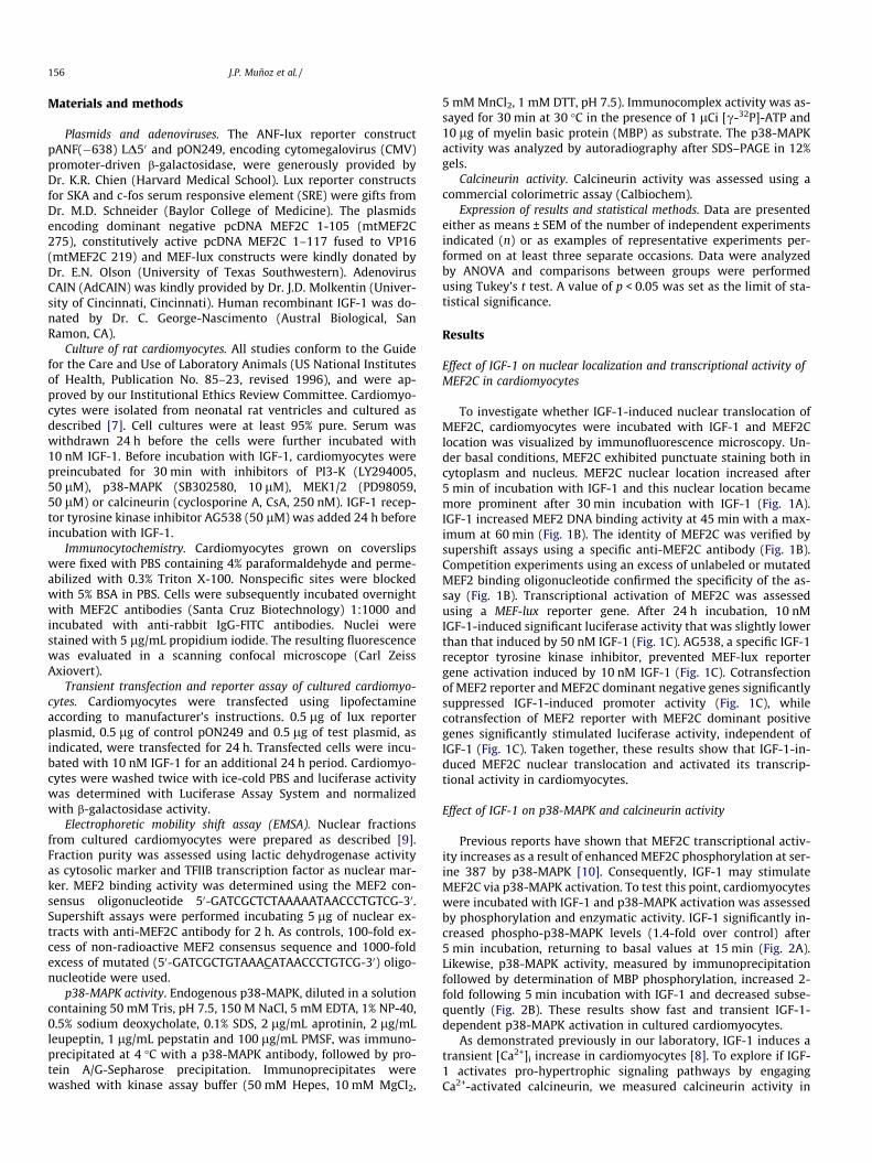
To investigate whether IGF-1-induced nuclear translocation ofMEF2C, cardiomyocytes were incubated with IGF-1 and MEF2Clocation was visualized by immunofluorescence microscopy. Un-der basal conditions, MEF2C exhibited punctuate staining both incytoplasm and nucleus. MEF2C nuclear location increased after5 min of incubation with IGF-1 and this nuclear location becamemore prominent after 30 min incubation with IGF-1 (Fig. 1A).IGF-1 increased MEF2 DNA binding activity at 45 min with a max-imum at 60 min (Fig. 1B). The identity of MEF2C was verified bysupershift assays using a specific anti-MEF2C antibody (Fig. 1B).Competition experiments using an excess of unlabeled or mutatedMEF2 binding oligonucleotide confirmed the specificity of the as-say (Fig. 1B). Transcriptional activation of MEF2C was assessedusing a MEF-lux reporter gene. After 24 h incubation, 10 nMIGF-1-induced significant luciferase activity that was slightly lowerthan that induced by 50 nM IGF-1 (Fig. 1C). AG538, a specific IGF-1receptor tyrosine kinase inhibitor, prevented MEF-lux reportergene activation induced by 10 nM IGF-1 (Fig. 1C). Cotransfectionof MEF2 reporter and MEF2C dominant negative genes significantlysuppressed IGF-1-induced promoter activity (Fig. 1C), whilecotransfection of MEF2 reporter with MEF2C dominant positivegenes significantly stimulated luciferase activity, independent ofIGF-1 (Fig. 1C). Taken together, these results show that IGF-1-in-duced MEF2C nuclear translocation and activated its transcrip-tional activity in cardiomyocytes.
Effect of IGF-1 on p38-MAPK and calcineurin activity
Previous reports have shown that MEF2C transcriptional activ-ity increases as a result of enhanced MEF2C phosphorylation at ser-ine 387 by p38-MAPK [10]. Consequently, IGF-1 may stimulateMEF2C via p38-MAPK activation. To test this point, cardiomyocyteswere incubated with IGF-1 and p38-MAPK activation was assessedby phosphorylation and enzymatic activity. IGF-1 significantly in-creased phospho-p38-MAPK levels (1.4-fold over control) after5 min incubation, returning to basal values at 15 min (Fig. 2A).Likewise, p38-MAPK activity, measured by immunoprecipitationfollowed by determination of MBP phosphorylation, increased 2-fold following 5 min incubation with IGF-1 and decreased subse-quently (Fig. 2B). These results show fast and transient IGF-1-dependent p38-MAPK activation in cultured cardiomyocytes.
As demonstrated previously in our laboratory, IGF-1 induces atransient [Ca2+]i increase in cardiomyocytes [8]. To explore if IGF-1 activates pro-hypertrophic signaling pathways by engagingCa2+-activated calcineurin, we measured calcineurin activity in

Fig. 1. IGF-1 activates MEF2C in cultured cardiomyocytes. (A) Cells were incubated with 10 nM IGF-1 for 5 or 30 min and MEF2C was detected by indirectimmunofluorescence using anti-MEF2C antibody (green). Nuclei were stained with propidium iodide. (B) Electrophoretic mobility shift assays (EMSA, upper panel) werecarried out using nuclear protein extracts prepared from cardiomyocytes incubated with 10 nM IGF-1 for up to 90 min. The arrow shows the MEF2/DNA complex. Supershiftassays (lower panel) were carried out using nuclear protein extracts prepared from cardiomyocytes incubated with 10 nM IGF-1 for 1 h, and subsequently incubated withMEF2C specific antibody. The arrow indicates the supershift complex. As control, 100-fold molar excess of the unlabeled or mutant MEF2 oligonucleotides was used. (C) Cellswere cotransfected with reporter MEF2-lux and b-galactosidase expression plasmids (pON) and incubated with IGF-1 (10 or 50 nM) for 24 h (upper panel). Cells werepreincubated with AG538 (a specific IGF-1 receptor inhibitor) for 24 h before incubation with IGF-1 (10 nM) (middle panel). Cells cotransfected with MEF2-lux and b-galactosidase plasmids, together with constructs expressing either control vector (pcDNA 3.1), dominant positive MEF2C (mtMEF2C VP16) or dominant negative MEF2C(mtMEF2C 1–105), were incubated with or without 10 nM IGF-1 for 24 h (lower panel). Lux activity was normalized by b-galactosidase activity. Values are expressed asmeans ± SEM of triplicates from 3 independent experiments. *p < 0.05 vs. control; #p < 0.05 vs. IGF-1. (For interpretation of the references to colour in this figure legend, thereader is referred to the web version of this article.)
J.P. Muñoz et al. / 157
cultured cardiomyocytes incubated with 10 nM IGF-1. After 5 or20 min of IGF-1 addition, calcineurin activity increased 1.8- and2.2-fold over control, respectively (Fig. 2C). In summary, these datashow that IGF-1 activated p38-MAPK and calcineurin signalingpathways in cardiomyocytes.
Effect of inhibition of p38-MAPK, PI3-K, ERK/MEK and calcineurin onIGF-1-stimulated MEF2C activation
To assess the influence of ERK/MEK, p38-MAPK, PI3-K and calci-neurin signaling on MEF2 transcriptional activity, cultured cardio-myocytes transfected with MEF2-lux reporter were preincubatedwith different inhibitors and were then incubated with IGF-1.The p38-MAPK inhibitor SB302580 and the PI3-K inhibitorLY294002 significantly reduced IGF-1-dependent MEF2 reportergene activation (Fig. 3A). In contrast, the MEK inhibitor PD98059
did not modify IGF-1-induced MEF2-lux activation, strongly sug-gesting that the MEK/ERK pathway is not involved in the activationof MEF2C-dependent transcription induced by IGF-1. The calcineu-rin inhibitor Ad-CAIN suppressed while cyclosporin A, another lessspecific calcineurin inhibitor, significantly reduced IGF-1-depen-dent activation of the MEF2-lux reporter gene (Fig. 3B and C).These data directly implicate p38-MAPK, PI3-K and calcineurin sig-naling pathways in the regulation of MEF2C transcriptional activityby IGF-1.
Role of MEF2C in cardiomyocyte hypertrophy induced by IGF-1
Transcriptional activation of immediately early genes, i.e. c-fos,and of genes involved in the fetal expression program—such asANF, SKA and b-MHC—is a classical hallmark of cardiac hypertro-phy [1]. IGF-1 increased 4-fold c-fos-lux, 10-fold SKA-lux and

Fig. 2. IGF-1 receptor signaling mediates MEF2C phosphorylation in cultured cardiomyocytes. (A) Phosphorylation and (B) activity of p38-MAPK in cells incubated with10 nM IGF-1 for 5–30 min. Myelin basic protein (MBP) is p38-MAPK substrate. (C) Calcineurin activity in cells incubated with 10 nM IGF-1 for 5–30 min. *p < 0.05 vs. 0 min;**p < 0.01 vs. 0 min. Values represent media ± SEM of at least 5 independent experiments.
158 J.P. Muñoz et al. /
20-fold ANF-lux reporter gene activities (Fig. 4A–C). Co-expressionof the MEF2C dominant negative mtMEF2C 275 suppressed theenhancement of SKA and ANF reporter activities induced by IGF-1 (Fig. 4B and C). The c-fos promoter activity, however, did not de-crease significantly in the presence of the MEF2C dominant nega-tive suggesting that IGF-1-mediated induction of c-fos wasindependent of MEF2C activation (Fig. 4A). These results indicatethat IGF-1 promotes MEF2C transcriptional activation in cardio-myocytes, which in turn activates the hypertrophic gene programthat involves SKA and ANF gene enhanced transcription.
Discussion
Several studies have shown that MEF2C is necessary for cardiacand muscular development and plays a part in the activation ofseveral fetal cardiac genes [11,12]; MEF2C regulates as well severalgenes that participate in the metabolic adaptations and cytoskele-ton reorganization that occur during the development of hypertro-phy [5,11]. Moreover, different signaling pathways contribute tothe pro-hypertrophic effects of IGF-1 in cardiomyocytes [6–8].Accordingly, we investigated in this work if MEF2C activationforms part of the adaptive response to cardiac injury that is basedon IGF-1-promoted hypertrophy.
Our results showed for the first time that IGF-1 activatedMEF2C-dependent transcription in cultured neonatal rat cardio-myocytes, promoting the nuclear location of MEF2C, and its DNAbinding activity. Functional assays using luciferase reporter genesrevealed that nanomolar concentrations of IGF-1 enhanced MEF2Cpromoter activity. The specific IGF-1 receptor inhibitor AG538 pre-vented this activation, whereas overexpression of dominant nega-tive MEF2C significantly decreased IGF-1-induced MEF2 reportergene expression. These results strongly suggest that IGF-1 receptorsignaling encompasses MEF2C activation in cardiomyocytes.
Previous studies have shown that IGF-1 activates several signal-ing pathways in cardiomyocytes, including ERK1/2, PI3-K, PKC,PKB, JAK/STAT, and PLC [7,8]. Here, we describe that IGF-1-induced
p38-MAPK activation mediates MEF2C enhanced phosphorylation.MEF2C phosphorylation by p38-MAPK has been reported previ-ously [3,10]. There are conflicting reports regarding the role ofthe p38-MAPK stress-activated kinase in cardiac hypertrophy.Thus, it was reported that overexpression of a heart targeted dom-inant negative a-p38-MAPK promotes hypertrophic cardiomyopa-thy through upregulation of calcineurin-NFAT signaling [13]. Theseresults, however, contradict previous observations showing thatpharmacologic inhibition of p38 kinase activity with the antago-nists SB203580 or SB202190 attenuated agonist-stimulatedhypertrophy in cultured cardiomyocytes [14,15]. Moreover, over-expression of a dominant negative b-p38-MAPK blunted thegrowth response of cultured cardiomyocytes [16], and significantlydecreased agonist-induced B-type natriuretic peptide (BNP)promoter activity in vitro [17]. Our data do not allow us to discrim-inate whether a- or b-p38-MAPK, or both, is responsible for IGF-1-dependent MEF2C activation. This point remains to be clarified.
Interestingly, an inhibitor of PI3-K abolished IGF-1-stimulatedMEF2C transcriptional activity. The PI3-K/PKB pathway has beenrecently involved in MEF2C activation in cerebral granule neurons[18]. Activation of PI3-K/PKB is associated with cell survivalthrough several pathways, including inhibition of p53 and theForkhead transcription factor, activation of the survival-promotingtranscription factors CREB and NFjB, and phosphorylation andinactivation of BAD and caspase 9 [19]. Accordingly, the regulationof MEF2C transcriptional activity by the p38-MAPK and PI3-K path-ways reported here suggests that in cardiomyocytes hypertrophicand survival signaling pathways converge at the level of MEF2C.
Calcium-dependent calcineurin activation modifies geneexpression and induces hypertrophy in cardiomyocytes [20]. Calci-neurin activation also promotes skeletal muscle hypertrophy in re-sponse to IGF-1 [21] and induces MEF2 transcription in skeletalmyocytes [22]. Calcineurin and MEF2 form a physical complex incultured C2C12 cells, where MEF2 becomes hypophosphorylatedwhen calcineurin is active and calcineurin augments the potencyof the transcriptional activation domain of MEF2 [23]. Our resultsindicate that exposure of cardiomyocytes to IGF-1 activates calci-
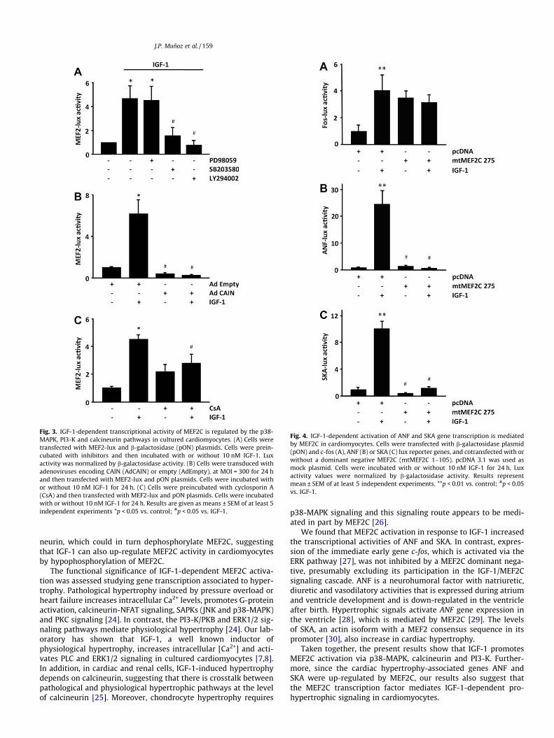
Fig. 3. IGF-1-dependent transcriptional activity of MEF2C is regulated by the p38-MAPK, PI3-K and calcineurin pathways in cultured cardiomyocytes. (A) Cells weretransfected with MEF2-lux and b-galactosidase (pON) plasmids. Cells were prein-cubated with inhibitors and then incubated with or without 10 nM IGF-1. Luxactivity was normalized by b-galactosidase activity. (B) Cells were transduced withadenoviruses encoding CAIN (AdCAIN) or empty (AdEmpty), at MOI = 300 for 24 hand then transfected with MEF2-lux and pON plasmids. Cells were incubated withor without 10 nM IGF-1 for 24 h. (C) Cells were preincubated with cyclosporin A(CsA) and then transfected with MEF2-lux and pON plasmids. Cells were incubatedwith or without 10 nM IGF-1 for 24 h. Results are given as means ± SEM of at least 5independent experiments *p < 0.05 vs. control; #p < 0.05 vs. IGF-1.
Fig. 4. IGF-1-dependent activation of ANF and SKA gene transcription is mediatedby MEF2C in cardiomyocytes. Cells were transfected with b-galactosidase plasmid(pON) and c-fos (A), ANF (B) or SKA (C) lux reporter genes, and cotransfected with orwithout a dominant negative MEF2C (mtMEF2C 1–105). pcDNA 3.1 was used asmock plasmid. Cells were incubated with or without 10 nM IGF-1 for 24 h. Luxactivity values were normalized by b-galactosidase activity. Results representmean ± SEM of at least 5 independent experiments. **p < 0.01 vs. control; #p < 0.05vs. IGF-1.
J.P. Muñoz et al. / 159
neurin, which could in turn dephosphorylate MEF2C, suggestingthat IGF-1 can also up-regulate MEF2C activity in cardiomyocytesby hypophosphorylation of MEF2C.
The functional significance of IGF-1-dependent MEF2C activa-tion was assessed studying gene transcription associated to hyper-trophy. Pathological hypertrophy induced by pressure overload orheart failure increases intracellular Ca2+ levels, promotes G-proteinactivation, calcineurin-NFAT signaling, SAPKs (JNK and p38-MAPK)and PKC signaling [24]. In contrast, the PI3-K/PKB and ERK1/2 sig-naling pathways mediate physiological hypertrophy [24]. Our lab-oratory has shown that IGF-1, a well known inductor ofphysiological hypertrophy, increases intracellular [Ca2+] and acti-vates PLC and ERK1/2 signaling in cultured cardiomyocytes [7,8].In addition, in cardiac and renal cells, IGF-1-induced hypertrophydepends on calcineurin, suggesting that there is crosstalk betweenpathological and physiological hypertrophic pathways at the levelof calcineurin [25]. Moreover, chondrocyte hypertrophy requires
p38-MAPK signaling and this signaling route appears to be medi-ated in part by MEF2C [26].
We found that MEF2C activation in response to IGF-1 increasedthe transcriptional activities of ANF and SKA. In contrast, expres-sion of the immediate early gene c-fos, which is activated via theERK pathway [27], was not inhibited by a MEF2C dominant nega-tive, presumably excluding its participation in the IGF-1/MEF2Csignaling cascade. ANF is a neurohumoral factor with natriuretic,diuretic and vasodilatory activities that is expressed during atriumand ventricle development and is down-regulated in the ventricleafter birth. Hypertrophic signals activate ANF gene expression inthe ventricle [28], which is mediated by MEF2C [29]. The levelsof SKA, an actin isoform with a MEF2 consensus sequence in itspromoter [30], also increase in cardiac hypertrophy.
Taken together, the present results show that IGF-1 promotesMEF2C activation via p38-MAPK, calcineurin and PI3-K. Further-more, since the cardiac hypertrophy-associated genes ANF andSKA were up-regulated by MEF2C, our results also suggest thatthe MEF2C transcription factor mediates IGF-1-dependent pro-hypertrophic signaling in cardiomyocytes.
160 J.P. Muñoz et al. /
Acknowledgments
This work was supported by FONDAP (Fondo de Areas Priorita-rias, Chile) Grant No. 15010006 (S.L. and C.H.) and CONICYT post-doctoral Fellowship 3060082 (J.P.M.).
References
[1] K.R. Chien, K.U. Knowlton, H. Zhu, S. Chien, Regulation of cardiac geneexpression during myocardial growth and hypertrophy: molecular studies ofan adaptive physiologic response, FASEB J. 5 (1991) 3037–3046.
[2] T.A. McKinsey, E.N. Olson, Cardiac hypertrophy: sorting out the circuitry, Curr.Opin. Genet. Dev. 9 (1999) 267–274.
[3] T.A. McKinsey, C.L. Zhang, E.N. Olson, MEF2: a calcium-dependent regulator ofcell division, differentiation and death, Trends Biochem. Sci. 27 (2002) 40–47.
[4] J.D. Molkentin, B.L. Black, J.F. Martin, E.N. Olson, Mutational analysis of theDNA binding, dimerization, and transcriptional activation domains of MEF2C,Mol. Cell. Biol. 16 (1996) 2627–2636.
[5] J. Xu, N.L. Gong, I. Bodi, B.J. Aronow, P.H. Backx, J.D. Molkentin, Myocyteenhancer factors 2A and 2C induce dilated cardiomyopathy in transgenic mice,J. Biol. Chem. 281 (2006) 9152–9162.
[6] J.R. McMullen, S. Izumo, Role of the insulin-like growth factor 1 (IGF1)/phosphoinositide-3-kinase (PI3K) pathway mediating physiological cardiachypertrophy, Novartis. Found. Symp. 274 (2006) 90–111.
[7] R. Foncea, M. Andersson, A. Ketterman, V. Blakesley, M. Sapag-Hagar, P.H.Sugden, D. LeRoith, S. Lavandero, Insulin-like growth factor-I rapidly activatesmultiple signal transduction pathways in cultured rat cardiac myocytes, J. Biol.Chem. 272 (1997) 19115–19124.
[8] C. Ibarra, M. Estrada, L. Carrasco, M. Chiong, J.L. Liberona, C. Cardenas, G. Diaz-Araya, E. Jaimovich, S. Lavandero, Insulin-like growth factor-1 induces aninositol 1,4,5-trisphosphate-dependent increase in nuclear and cytosoliccalcium in cultured rat cardiac myocytes, J. Biol. Chem. 279 (2004) 7554–7565.
[9] G. Courtois, S.T. Whiteside, C.H. Sibley, A. Israel, Characterization of a mutantcell line that does not activate NF-kappaB in response to multiple stimuli, Mol.Cell. Biol. 17 (1997) 1441–1449.
[10] J. Han, Y. Jiang, Z. Li, V.V. Kravchenko, R.J. Ulevitch, Activation of thetranscription factor MEF2C by the MAP kinase p38 in inflammation, Nature386 (1997) 296–299.
[11] S.M. Kolodziejczyk, L. Wang, K. Balazsi, Y. DeRepentigny, R. Kothary, L.A.Megeney, MEF2 is upregulated during cardiac hypertrophy and is required fornormal post-natal growth of the myocardium, Curr. Biol. 9 (1999) 1203–1206.
[12] Q. Lin, J. Schwarz, C. Bucana, E.N. Olson, Control of mouse cardiacmorphogenesis and myogenesis by transcription factor MEF2C, Science 276(1997) 1404–1407.
[13] J.C. Braz, O.F. Bueno, Q. Liang, B.J. Wilkins, Y.S. Dai, S. Parsons, J. Braunwart, B.J.Glascock, R. Klevitsky, T.F. Kimball, T.E. Hewett, J.D. Molkentin, Targetedinhibition of p38 MAPK promotes hypertrophic cardiomyopathy throughupregulation of calcineurin-NFAT signaling, J. Clin. Invest. 111 (2003) 1475–1486.
[14] S. Nemoto, Z. Sheng, A. Lin, Opposing effects of Jun kinase and p38 mitogen-activated protein kinases on cardiomyocyte hypertrophy, Mol. Cell. Biol. 18(1998) 3518–3526.
[15] D. Zechner, D.J. Thuerauf, D.S. Hanford, P.M. McDonough, C.C. Glembotski, Arole for the p38 mitogen-activated protein kinase pathway in myocardial cellgrowth, sarcomeric organization, and cardiac-specific gene expression, J. Cell.Biol. 139 (1997) 115–127.
[16] Y. Wang, S. Huang, V.P. Sah, J. Ross Jr., J.H. Brown, J. Han, K.R. Chien, Cardiacmuscle cell hypertrophy and apoptosis induced by distinct members of thep38 mitogen-activated protein kinase family, J. Biol. Chem. 273 (1998) 2161–2168.
[17] F. Liang, S. Lu, D.G. Gardner, Endothelin-dependent and -independentcomponents of strain-activated brain natriuretic peptide gene transcriptionrequire extracellular signal regulated kinase and p38 mitogen-activatedprotein kinase, Hypertension 35 (2000) 188–192.
[18] M. Wiedmann, X. Wang, X. Tang, M. Han, M. Li, Z. Mao, PI3K/Akt-dependent regulation of the transcription factor myocyte enhancer factor-2 in insulin-like growth factor-1- and membrane depolarization-mediatedsurvival of cerebellar granule neurons, J. Neurosci. Res. 81 (2005) 226–234.
[19] Z.Z. Chong, F. Li, K. Maiese, Activating Akt and the brain’s resources to drivecellular survival and prevent inflammatory injury, Histol. Histopathol. 20(2005) 299–315.
[20] B.J. Wilkins, J.D. Molkentin, Calcium-calcineurin signaling in the regulationof cardiac hypertrophy, Biochem. Biophys. Res. Commun. 322 (2004)1178–1191.
[21] A. Musaro, K.J. McCullagh, F.J. Naya, E.N. Olson, N. Rosenthal, IGF-1 inducesskeletal myocyte hypertrophy through calcineurin in association with GATA-2and NF-ATc1, Nature 400 (1999) 581–585.
[22] H. Wu, F.J. Naya, T.A. McKinsey, B. Mercer, J.M. Shelton, E.R. Chin, A.R. Simard,R.N. Michel, R. Bassel-Duby, E.N. Olson, R.S. Williams, MEF2 responds tomultiple calcium-regulated signals in the control of skeletal muscle fiber type,EMBO J. 19 (2000) 1963–1973.
[23] H. Wu, B. Rothermel, S. Kanatous, P. Rosenberg, F.J. Naya, J.M. Shelton, K.A.Hutcheson, J.M. DiMaio, E.N. Olson, R. Bassel-Duby, R.S. Williams, Activation ofMEF2 by muscle activity is mediated through a calcineurin-dependentpathway, EMBO J. 20 (2001) 6414–6423.
[24] G.W. Dorn, T. Force, Protein kinase cascades in the regulation of cardiachypertrophy, J. Clin. Invest. 115 (2005) 527–537.
[25] J.L. Gooch, Y. Tang, J.M. Ricono, H.E. Abboud, Insulin-like growth factor-Iinduces renal cell hypertrophy via a calcineurin-dependent mechanism, J. Biol.Chem. 276 (2001) 42492–42500.
[26] L.A. Stanton, S. Sabari, A.V. Sampaio, T.M. Underhill, F. Beier, P38 MAP kinasesignalling is required for hypertrophic chondrocyte differentiation, Biochem. J.378 (2004) 53–62.
[27] G.J. Babu, M.J. Lalli, M.A. Sussman, J. Sadoshima, M. Periasamy,Phosphorylation of elk-1 by MEK/ERK pathway is necessary for c-fos geneactivation during cardiac myocyte hypertrophy, J. Mol. Cell. Cardiol. 32 (2000)1447–1457.
[28] K.L. Vikstrom, T. Bohlmeyer, S.M. Factor, L.A. Leinwand, Hypertrophy,pathology, and molecular markers of cardiac pathogenesis, Circ. Res. 82(1998) 773–778.
[29] M.X. Zang, Y. Li, L.X. Xue, H.T. Jia, H. Jing, Cooperative activation of atrialnaturetic peptide promoter by dHAND and MEF2C, J. Cell. Biochem. 93 (2004)1255–1266.
[30] G.E. Muscat, S. Perry, H. Prentice, L. Kedes, The human skeletal alpha-actingene is regulated by a muscle-specific enhancer that binds three nuclearfactors, Gene Expr. 2 (1992) 111–126.