Use of Long-term Cultured Embryoid Bodies May Enhance Cardiomyocyte Differentiation by BMP2
Upload
independentCategory
view
1download
0
Garnier, Anne-Marie Lompré, Grégoire Vandecasteele and Frank Lezoualc'hEric Morel, Andrea Marcantoni, Monique Gastineau, Rikke Birkedal, Francesca Rochais, Anne
cAMP-Binding Protein Epac Induces Cardiomyocyte Hypertrophy
Print ISSN: 0009-7330. Online ISSN: 1524-4571 Copyright © 2005 American Heart Association, Inc. All rights reserved.is published by the American Heart Association, 7272 Greenville Avenue, Dallas, TX 75231Circulation Research
doi: 10.1161/01.RES.0000194325.31359.862005;97:1296-1304; originally published online November 3, 2005;Circ Res.
http://circres.ahajournals.org/content/97/12/1296World Wide Web at:
The online version of this article, along with updated information and services, is located on the
http://circres.ahajournals.org/content/suppl/2005/11/03/01.RES.0000194325.31359.86.DC1.htmlData Supplement (unedited) at:
http://circres.ahajournals.org//subscriptions/
is online at: Circulation Research Information about subscribing to Subscriptions:
http://www.lww.com/reprints Information about reprints can be found online at: Reprints:
document. Permissions and Rights Question and Answer about this process is available in the
located, click Request Permissions in the middle column of the Web page under Services. Further informationEditorial Office. Once the online version of the published article for which permission is being requested is
can be obtained via RightsLink, a service of the Copyright Clearance Center, not theCirculation Researchin Requests for permissions to reproduce figures, tables, or portions of articles originally publishedPermissions:
at Biblioteche biomediche Universita' di Torino on November 12, 2012http://circres.ahajournals.org/Downloaded from

cAMP-Binding Protein Epac InducesCardiomyocyte Hypertrophy
Eric Morel, Andrea Marcantoni, Monique Gastineau, Rikke Birkedal, Francesca Rochais, Anne Garnier,Anne-Marie Lompre, Gregoire Vandecasteele, Frank Lezoualc’h
Abstract—cAMP is one of the most important second messenger in the heart. The discovery of Epac as a guanine exchangefactor (GEF), which is directly activated by cAMP, raises the question of the role of this protein in cardiac cells. Herewe show that Epac activation leads to morphological changes and induces expression of cardiac hypertrophic markers.This process is associated with a Ca2�-dependent activation of the small GTPase, Rac. In addition, we found that Epacactivates a prohypertrophic signaling pathway, which involves the Ca2� sensitive phosphatase, calcineurin, and itsprimary downstream effector, NFAT. Rac is involved in Epac-induced NFAT dependent cardiomyocyte hypertrophy.Blockade of either calcineurin or Rac activity blunts the hypertrophic response elicited by Epac indicating thesesignaling molecules coordinately regulate cardiac gene expression and cellular growth. Our results thus open newinsights into the signaling pathways by which cAMP may mediate its biological effects and identify Epac as a newpositive regulator of cardiac growth. (Circ Res. 2005;97:1296-1304.)
Key Words:cAMP � guanine nucleotide exchange factor � small G protein � transcription factor
In the heart, cyclic adenosine 3�,5�-monophosphate (cAMP)regulates many physiological processes such as contractil-
ity and relaxation. Classically, these effects are attributed toactivation of hyperpolarization-activated cyclic nucleotide-gated channels and protein kinase A (PKA) by cAMP.1 Therecent discovery of Epac as proteins which are directlyactivated by cAMP has broken the dogma surrounding cAMPand PKA.2–4 Epac proteins are guanine nucleotide exchangefactors (GEFs) that bind cAMP with affinities similar to thatof the regulatory subunit of PKA.2,3 They have been shown tofunction as GEFs for the Ras-like small GTPases Rap1 andRap2 and are directly activated by cAMP in a PKA indepen-dent manner.4 There are two isoforms of Epac, Epac 1 andEpac 2 both consisting of a regulatory and a catalyticregion.2,3 Epac 2 has an additional cAMP binding domain thatis dispensable for cAMP-induced Rap activation.5 AftercAMP binding, Epac catalyzes the exchange of GDP for GTPon the small GTPases Rap, allowing interaction with theirtarget effectors.6 Recent studies indicate that Epac is involvedin cell adhesion,7,8 neurite extension,9 and regulates insulinsecretion and the amyloid precursor protein processing.10,11
To date the role of Epac in the heart is unknown.Among the superfamily of small G proteins, the Rho
family, which includes Rho, Rac, and Cdc42, has attractedmuch interest for they have been shown to play key roles inthe generation of cytoskeletal structures.12 Indeed, Rho is
important for the formation of stress fibers and focal adhe-sions in fibroblasts, whereas Rac and Cdc42 are involved inthe regulation of more dynamic structures such as membraneruffles, lamellipodia and filopodia.12 Several studies havepointed out the role of Rho proteins in the development ofcardiomyocyte hypertrophy.13 For instance, two potent hy-pertrophic stimuli, endothelin 1 (ET-1) and phenylephrine(PE), induce rapid activation of endogenous Rac in neonatalcardiomyocytes.14 In addition, adenoviral infection of cardio-myocytes with a constitutive active form of Rac (RacG12V)increases protein synthesis and promotes morphologicalchanges associated with myocyte hypertrophy.15 In vivoevidence for the role of Rho proteins in cardiac hypertrophycame from transgenic mice specifically expressing RacG12V inthe heart. These mice develop a dilated cardiomyopathyassociated with deregulation of cardiomyocyte focal adhe-sions.16 These data suggest that Rho proteins, especially Raccontrol hypertrophic response and are likely to be involved incardiac remodeling, and the pathogenesis of cardiomyopathycharacterized by cellular enlargement.
Recently, we have provided experimental evidence thatEpac stimulates the activity of the small GTPase, Rac, in acAMP-dependent but PKA-independent manner in neuronalcells.11 These observations combined with the high expres-sion of Epac in the heart2,3 prompted us to focus our researchon the potential role of Epac in cardiomyocyte hypertrophy.
Original received May 27, 2005; revision received October 12, 2005; accepted October 25, 2005.From the Cardiologie Cellulaire et Moleculaire, Inserm U-446, IFR-75, Faculte de Pharmacie, Universite Paris XI, 5 Rue JB Clement, 92296 Cha�tenay
Malabry, FrancePresent address for A.-M.L.: Inserm U-621, Faculte de Medecine Pitie-Salpetriere, 91, bd de l’Hopital, 75634 Paris Cedex 13, France.Correspondence to Frank Lezoualc’h, Cardiologie Cellulaire et Moleculaire, Inserm U-446, IFR-75, Faculte de Pharmacie, Universite Paris XI, 5 Rue
JB Clement, 92296 Cha�tenay Malabry, France. E-mail [email protected]© 2005 American Heart Association, Inc.
Circulation Research is available at http://circres.ahajournals.org DOI: 10.1161/01.RES.0000194325.31359.86
1296 at Biblioteche biomediche Universita' di Torino on November 12, 2012http://circres.ahajournals.org/Downloaded from

Here we found that Epac stimulates the activity of the smallGTPase, Rac, and increases the expression of hypertrophicgene markers in primary cardiac myocytes. Furthermore, weshow that Epac induces cardiomyocyte hypertrophy. Thisprocess is associated with the activation of Rac and thecalcineurin/NFAT signaling pathway, which coordinatelyregulates cell growth and gene expression. Altogether, thesefindings identify the cAMP-binding protein, Epac, as a newpositive regulator of cardiac growth.
Materials and MethodsAdenoviral InfectionBicistronic adenoviruses (Ad5) bearing either EpacWT orEpac�cAMP under the control of a cytomegalovirus promoter andgreen fluorescent protein (GFP) under internal ribosomal entry sitecontrol were constructed and amplified at the Genethon Center ofEvry (France). Adenoviruses encoding VIVIT, a selective peptideinhibitor of calcineurin-mediated NFAT activation, and Rac wereprovided by Drs S. Kraner and C. Norris (University of Kentucky)and T. Finkel (Cardiology Branch, National Heart, NIH, Bethesda,Md), respectively. One day after plating, cardiomyocytes wereincubated for 2 hours with recombinant adenoviruses. After removalof the virus suspension, cells were replaced in maintenance mediumfor 2 days and then stimulated with the different drugs. Viruses wereused at a multiplicity of infection (MOI) of 100.
Plasmid Constructs and TransfectionThe plasmid constructs were generously provided by the following:the rat ANF promoter fused to the luciferase reporter gene (ANF-Luc) by Dr K. Knowlton, Luciferase reporter genes linked topromoters for skeletal muscle �-actin (SkM-�-actin-Luc) and serumresponse element-regulated c-fos (c-fos-SRE-Luc) by Dr M. D.Schneider, Epac1 plasmid constructs by Drs J. Bos and X. Cheng.The luciferase reporter plasmid driven by four NFAT consensusbinding sites (NFAT-Luc) was obtained from Stratagene. Transienttransfection experiments were performed with Lipofectamine 2000(Invitrogen Life Technologies, France) in optimum medium in thepresence of 1 �g of the various plasmid constructs according to themanufacturers’ instructions.
Rac Activation AssayRac pull-down experiments were performed using a GST fusionprotein containing the Cdc42/Rac Interactive Binding Domain(CRIB) of p21-activated kinase (PAK) exactly as previouslydescribed.11
Statistical AnalysisResults are expressed as means�SEM. Differences between groupshave been analyzed by one-way ANOVA followed by unpairedStudent t test. Differences were considered significant at P�0.05,P�0.01, and P�0.001.
For a description of other methods, see the expanded Materialsand Methods, available online at http://circres.ahajournals.org.
ResultsEpac Activates the Small G Protein Racin CardiomyocytesActivation of endogenous Epac with a selective activator ofthis GEF, 8-pCPT-2�-O-Me-cAMP (8-CPT), which does notactivate PKA17 increased Rac activation in rat cardiac myo-cytes (Figure 1). Similarly, infection of cardiomyocytes withan adenovirus encoding Epac1WT (Ad.EpacWT) significantlyenhanced Rac GTP-loading compared with control cellsinfected with GFP (Figure 1). Rac activation was furtherincreased when cells infected with Ad.EpacWT were treated
with 8-CPT (Figure 1). An adenovirus bearing a constitutiveactivated form of Epac1 (Ad.Epac-�cAMP)2 also inducedRac activation (Figure 1). Altogether, these results demon-strate that recombinant and native Epac increase the amountof Rac-GTP in cardiomyocytes.
Epac Increases the Expression of HypertrophyGene MarkersAs Rac has been found to be involved in cardiac myocytehypertrophy,15,16 we next tested the potential involvement ofEpac in this process. Re-expression of embryonic genes andtransient activation of immediate early genes are frequentlyused indexes of myocyte hypertrophy.18 The ability of Epacto stimulate gene expression was determined using luciferase(Luc) constructs under the control of promoters for ANF,SkM �-actin, and the c-fos-SRE. Figure 2A shows a three-fold activation of the ANF-Luc reporter gene in neonatalcardiomyocytes stimulated with 8-CPT compared with con-trol cells. Transient transfection of EpacWT or Epac-�cAMPincreased the basal level of ANF-Luc activity (Figure 2A).The effect of EpacWT on ANF-Luc activity was furtherincreased by the application of 8-CPT (Figure 2A). RacG12V
mimicked the effect of Epac on ANF-Luc activity (Figure2A). Next, to analyze the effect of Epac on ANF mRNAcontent in cardiac myocytes, we used Ad.EpacWT to maxi-mize the expression of this GEF in primary cardiomyocyte.Consistent with the effect of Epac on ANF-Luc reporter gene,endogenous expression of ANF mRNA was significantlyincreased in cardiomyocytes infected with Ad.EpacWT and
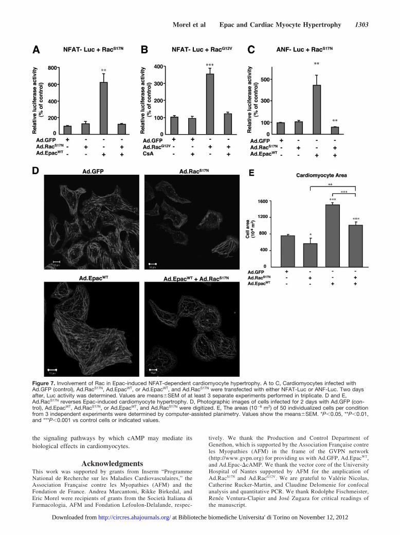
Figure 1. Epac activates the small G protein Rac in primaryventricular cardiomyocytes. Cardiomyocytes were infected witheither Ad.GFP as a control or with Ad.EpacWT or Ad.Epac-�cAMP as described in Materials and Methods. Two days afterinfection, cells were treated or not for 10 minutes with theselective activator of Epac, 8-CPT (10�6 mol/L). Amounts ofRac-GTP were determined by pull-down experiments. A controlfor total Rac expression (total lysates) is shown. The upperpanel shows a typical immunoblot. Expression of recombinantproteins was determined by Western blot using an anti-HA anti-body. The lower panel shows means�SEM of 3 independentexperiments. Results are expressed as fold activation of controlcells. *P�0.05, **P�0.01 compared with control.
Morel et al Epac and Cardiac Myocyte Hypertrophy 1297
at Biblioteche biomediche Universita' di Torino on November 12, 2012http://circres.ahajournals.org/Downloaded from

stimulated or not with 8-CPT, as compared with control cells(Figure 2B). Similar results were obtained with an adenovirusexpressing RacG12V (Ad.RacG12V) (Figure 2B). In addition,when cotransfection experiments were performed with SkM-�-actin-Luc or c-fos-SRE-Luc, 8-CPT, EpacWT, Epac-�cAMP, or RacG12V significantly increased Luc activity com-pared with control cells (Figure 2C and 2D). The effect ofEpacWT on Luc activity was further increased by the applica-tion of 8-CPT (Figure 2C and 2D).
Epac Increases Cardiomyocyte Size andSarcomeric OrganizationFurther studies were undertaken to determine the effects ofEpac on other features of the hypertrophic program such ascell size and sarcomeric organization. Cardiomyocyte treat-ment with Ad.GFP and 8-CPT as well as infection ofcardiomyocytes with Ad.EpacWT induced an apparent increaseof the F-actin meshwork and a heavily striated appearance,reflecting the organization of this F-actin cytoskeleton intosarcomeric structures, as compared with cardiomyocytesinfected with Ad.GFP alone (Figure 3A). Cells overexpress-ing Epac were hypertrophied and were not contaminated byfibroblast as shown by the �-actinin staining in supplemen-tary Figure I. In addition, the effects of Epac on sarcomericorganization were comparable to Ad.Epac-�cAMP (data notshown) and PE (Figure 3A), a well-known inducer of cardiachypertrophy.
Next, we measured the effect of Epac on cell surface area.Activation of endogenous Epac with 8-CPT produced atwo-fold increase in cell surface area when compared withcells infected with control Ad.GFP (Figure 3B). Identicalresults were obtained when cardiomyocytes were infectedwith Ad.EpacWT (Figure 3B), Ad.Epac-�cAMP (data notshown), or Ad.GFP and treated with PE (Figure 3B). Theeffect of Ad.EpacWT on cell surface area was not furtherincreased in the presence of 8-CPT suggesting that basalintracellular cAMP was sufficient to activate recombinant
Epac to induce its maximal effect on protein synthesis (Figure3B). Finally, the effect of Epac on protein synthesis wasanalyzed by measurement of [3H]-leucine incorporation intocardiac myocytes. Expression of this cAMP-GEF resulted inan increase in [3H]-leucine uptake into cardiomyocytes (Fig-ure 3C). Similarly, cell treatment with 8-CPT or the goldstandard, PE resulted in an approximately two-fold increasein protein synthesis (Figure 3C). Altogether, these resultsshow that Epac activation confers to primary cardiomyocytesall the features of the hypertrophic phenotype.
Intracellular Ca2� Is Involved in Epac-DependentRac ActivationAlterations in intracellular Ca2� handling progressively exac-erbate a hypertrophic or cardiomyopathic phenotype, in partthrough sustained activation of Ca2�-sensitive signal trans-duction pathways.19 Given the involvement of Epac in cardiachypertrophy, we examined whether its activation could affectintracellular Ca2� concentration ([Ca2�]i) in neonatal myo-cytes (Figure 4). At physiological external [Ca2�], cardiacmyocytes exhibited spontaneous Ca2� transients with a lowfrequency (0.120�0.015 Hz, n�20) (Figure 4A). Applicationof the Epac agonist 8-CPT triggered a dramatic increase in thefrequency of these Ca2� oscillations (0.51�0.04 Hz, n�7)without changing the amplitude of the spikes. This effect wasalso observed at 100 nM 8-CPT (0.40�0.05 Hz, n�13, datanot shown).
Because Epac induced Rac activation, we examined thedependence of Rac activation on Ca2� signaling. Treatment ofcardiac myocytes with the Ca2� ionophore ionomycin as wellas an inhibitor of the Ca2�-ATPase, thapsigargin increasedRac activation in a time dependent manner (Figure 4B and4C). The effect of ionomycin and thapsigargin on Racactivation was as potent as the positive control, PE (Figure 4Band 4C). Pretreatment with BAPTA-AM, an intracellularCa2� chelator, attenuated Epac-induced Rac activation (Fig-ure 4D). From these results we conclude that elevation of
Figure 2. Epac stimulates a hypertrophicpattern of gene expression. A, C, and D,Neonatal cardiomycoytes were trans-fected with ANF-Luc, SkM-�-actin-Lucor c-fos-SRE-Luc and EpacWT, Epac-�cAMP, RacG12V, or the empty vector(mock) as control and treated or not with8-CPT (10�6 mol/L). Two days aftertransfection, cells were assayed for Lucactivity. Results are expressed as per-centage activation of control. Results aremeans�SEM from 3 independent experi-ments performed in triplicates. B, Epacinduces expression of ANF mRNA. Car-diomyocytes were infected with Ad.E-pacWT, Ad.RacG12V, or Ad.GFF (control)and stimulated or not with 8-CPT (10-6
mol/L) for 2 days. ANF mRNA expressionwas determined by quantitative PCR.Values are expressed relative to theANF/GCB ratio and results were normal-ized to control for each experiment.Results are presented as themean�SEM of 3 independent experi-ments performed in duplicates. *P�0.05,**P�0.01, ***P�0.001 compared withcontrol.
1298 Circulation Research December 9/23, 2005
at Biblioteche biomediche Universita' di Torino on November 12, 2012http://circres.ahajournals.org/Downloaded from

intracellular [Ca2�]i after Epac activation is sufficient toactivate Rac.
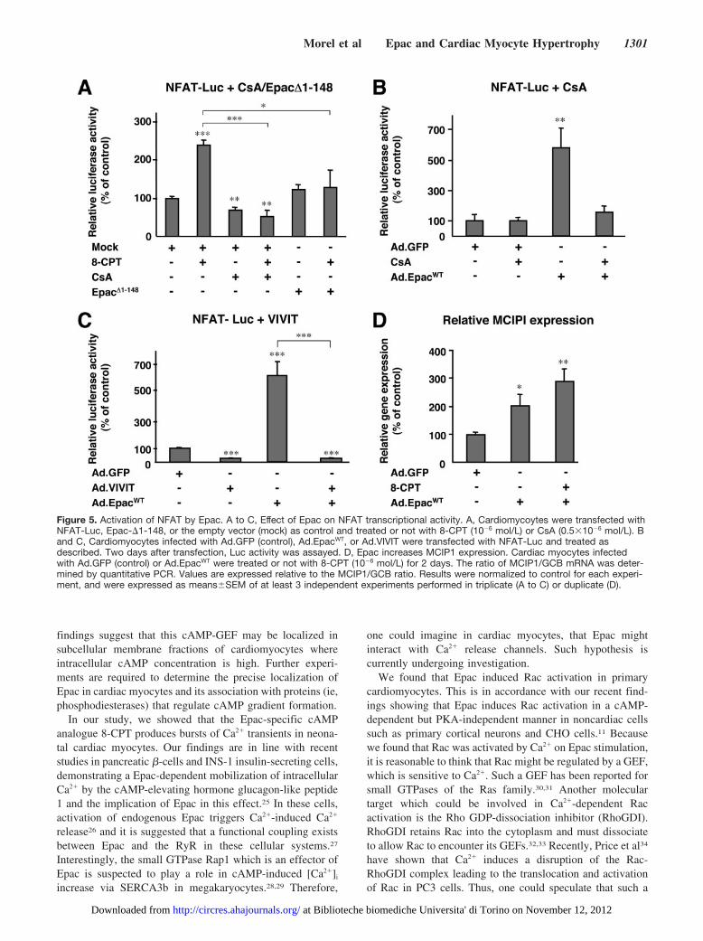
Epac Activates the HypertrophicCalcineurin/NFAT Signaling PathwaysOne prominent Ca2�-dependent pathway that plays a crucialrole in cardiomyocyte hypertrophy involves the phosphatasecalcineurin.20 Activation of calcineurin by Ca2� results in thedephosphorylation and nuclear translocation of cytoplasmicNFAT transcription factors, which then upregulate transcrip-tion of hypertrophic genes. To test whether endogenous Epacmay activate the hypertrophic calcineurin NFAT signaling
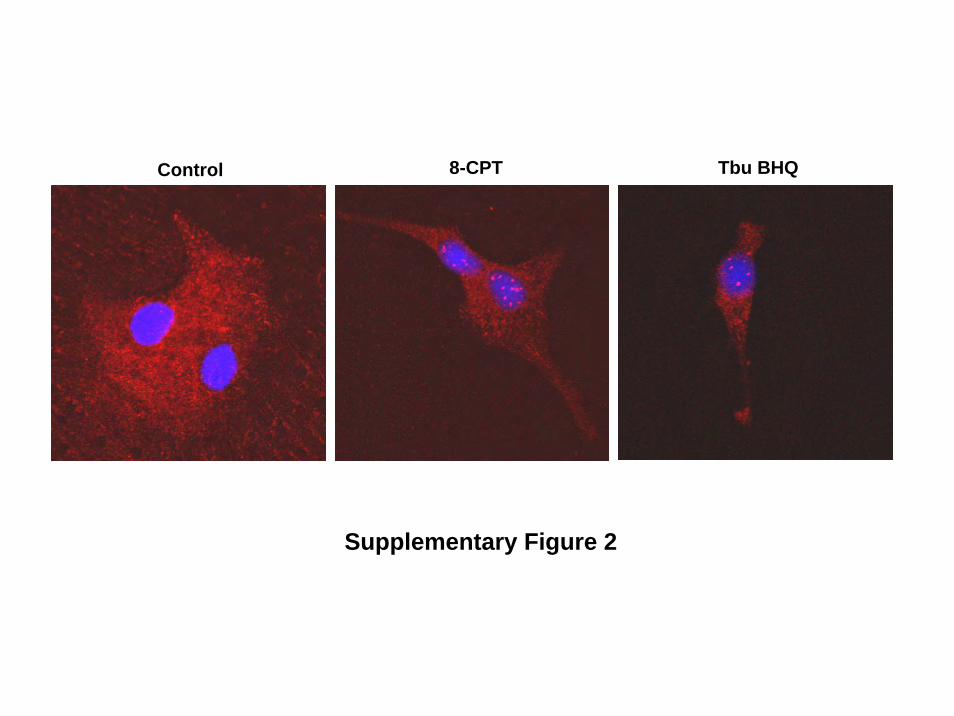
pathway, cardiomyocytes were transfected with NFAT-Lucand treated or not with 8-CPT (Figure 5A). 8-CPT signifi-cantly increased NFAT transcriptional activity as comparedwith control cells (Figure 5A). Accordingly, 8-CPT increasedNFAT nuclear translocation (supplementary Figure II). Thestimulating effect of 8-CPT on NFAT-Luc was significantlyblocked by a pharmacological inhibitor of calcineurin, cyclo-sporine A (CsA) or transfection of a dominant negative formof Epac (Epac�1-148)21 (Figure 5A).
Recombinant EpacWT also increased NFAT transcriptionalactivity which was blocked by CsA or an adenovirus bearinga selective peptide inhibitor of calcineurin named VIVIT
Figure 3. Epac induces cardiomyocyteshypertrophy. A, Fluorescent microscopicanalyses of the effects of Epac on sarco-meric organization. Morphology of repre-sentative myocytes 48 hours after infec-tion with Ad.GFP as a control, orAd.EpacWT is shown. The Epac selectiveactivator, 8-CPT was used at 10�6 mol/Lfor 2 days in cells infected with Ad.GFP.Positive control cells were infected withAd.GFP and treated with PE (10�5 mol/L)for 2 days. Actin filaments were visual-ized by using Rhodamin-conjugatedphalloidin. B, Photographic images ofcells treated as above were digitized.The areas (10�6 m2) of 30 to 50 individu-alized cells per condition from 2 to 3independent experiments were deter-mined by computer-assisted planimetry.Values show the means�SEM. C,[3H]-leucine incorporation. Cardiomyo-cytes were treated as in (A) and totalradioactivity of incorporated [3H]-leucineinto proteins was determined by scintilla-tion counting. The figure shows themean�SEM of data for 3 experimentsperformed in duplicate. *P�0.05,**P�0.01, ***P�0.001 compared withcontrol Ad.GFP.
Morel et al Epac and Cardiac Myocyte Hypertrophy 1299
at Biblioteche biomediche Universita' di Torino on November 12, 2012http://circres.ahajournals.org/Downloaded from

(Ad.VIVIT)22 (Figure 5B and 5C). Consistent with thesefindings, cardiac myocytes infected with Ad.EpacWT andtreated or not with 8-CPT (Figure 5D), or Ad.Epac-�cAMP(data not shown) had an increased content of mRNA encod-ing the modulatory calcineurin-interacting protein 1(MCIP1), a mediator of calcineurin signaling during cardiachypertrophy.23 Furthermore, coinfection with Ad.VIVIT andAd.EpacWT reduced the enhancement of sarcomeric organiza-tion and cell surface area induced by Ad.EpacWT (Figure 6Aand 6B). Altogether these data show that NFAT is a down-stream component of Epac hypertrophic signaling pathway.
Involvement of Rac in Epac-InducedNFAT-Dependent Cardiomyocyte HypertrophyBecause Rac was found to be a downstream component ofEpac signaling pathway (Figure 1), we next examined theinvolvement of Rac in Epac-induced NFAT transcriptionalactivity. Ad.RacS17N completely inhibited Epac-inducedNFAT transcriptional activity (Figure 7A) whereas thestimulating effect of RacG12V on NFAT-Luc was blocked byCsA (Figure 7B). These data clearly indicate that Rac isable to influence the calcineurin/NFAT signaling pathway.The involvement of Rac in Epac signaling pathway con-trolling cardiomyocyte hypertrophy was further supportedby the observation that RacS17N inhibited the stimulatingeffect of endogenous Epac activation or EpacWT on ANFexpression (Figure 7C and supplementary Figure III).Consistent with these findings, Ad.RacS17N inhibited Epac-
induced cytoskeletal reorganization (Figure 7D) and in-crease in cell surface area (Figure 7E). Finally, as Rac hasbeen shown to induce the production of reactive oxygenspecies (ROS),24 we analyzed the effect of EpacWT onANF-Luc in the presence of the antioxidant,N-acetylcysteine (NAC). We found that NAC inhibitedEpac-induced ANF-Luc activity suggesting that oxidativestress is increased by Epac (supplementary Figure IV).
DiscussionThe present study shows for the first time that cAMP-dependent activation of Epac induces cardiomyocyte hyper-trophy. This is based on our observation that Epac activationleads to morphological changes, cytoskeletal reorganization,increases in protein synthesis, and induces expression ofcardiac hypertrophic markers. In addition, we found that Epacactivates a prohypertrophic signaling pathway which involvescalcineurin and its primary downstream effector, NFAT.Epac-induced NFAT activation was dependent on Rac activ-ity. Interestingly, overexpression of Epac was able to influ-ence cardiomyocyte morphology without cAMP analoguetreatment indicating that the level of intracellular cAMP wassufficient to activate the recombinant EpacWT protein. Similarobservations have been reported in other cellular systemsbecause transfection of EpacWT in HEK293 and COS cells hasbeen previously shown to influence cell signaling and mor-phology at resting basal cAMP levels.21 Because Epac needsmicromolar concentration of cAMP to be activated,4 these
Figure 4. Intracellular Ca2� is involved in Epac- dependent Rac activation. A, Effect of 8-CPT (10�5 mol/L) on spontaneous spikingactivity at 1.8�10�3 mol/L external [Ca2�]. Cardiomyocytes at day 1 or 2 after plating were loaded with the Ca2� indicator Fluo3-AMand perfused with a control external Ringer solution. B to D, Effect of ionomycin, thapsigargin and BAPTA-AM on Rac activation. Ven-tricular cardiomyocytes were treated at 2 days in vitro with either ionomycin (10-6 mol/L) (B) or thapsigargin (0.2�10�6 mol/L) (C) for dif-ferent times of incubation or PE (10�5 mol/L) for 15 minutes. D, Cells were pretreated with BAPTA-AM (1.5 �10�5 mol/L) for 10 minutesand then they were stimulated with 8-CPT (10�5 mol/L) for 15 minutes. The amount of Rac-GTP was determined by pull-downexperiments.
1300 Circulation Research December 9/23, 2005
at Biblioteche biomediche Universita' di Torino on November 12, 2012http://circres.ahajournals.org/Downloaded from
findings suggest that this cAMP-GEF may be localized insubcellular membrane fractions of cardiomyocytes whereintracellular cAMP concentration is high. Further experi-ments are required to determine the precise localization ofEpac in cardiac myocytes and its association with proteins (ie,phosphodiesterases) that regulate cAMP gradient formation.
In our study, we showed that the Epac-specific cAMPanalogue 8-CPT produces bursts of Ca2� transients in neona-tal cardiac myocytes. Our findings are in line with recentstudies in pancreatic �-cells and INS-1 insulin-secreting cells,demonstrating a Epac-dependent mobilization of intracellularCa2� by the cAMP-elevating hormone glucagon-like peptide1 and the implication of Epac in this effect.25 In these cells,activation of endogenous Epac triggers Ca2�-induced Ca2�
release26 and it is suggested that a functional coupling existsbetween Epac and the RyR in these cellular systems.27
Interestingly, the small GTPase Rap1 which is an effector ofEpac is suspected to play a role in cAMP-induced [Ca2�]i
increase via SERCA3b in megakaryocytes.28,29 Therefore,
one could imagine in cardiac myocytes, that Epac mightinteract with Ca2� release channels. Such hypothesis iscurrently undergoing investigation.
We found that Epac induced Rac activation in primarycardiomyocytes. This is in accordance with our recent find-ings showing that Epac induces Rac activation in a cAMP-dependent but PKA-independent manner in noncardiac cellssuch as primary cortical neurons and CHO cells.11 Becausewe found that Rac was activated by Ca2� on Epac stimulation,it is reasonable to think that Rac might be regulated by a GEF,which is sensitive to Ca2�. Such a GEF has been reported forsmall GTPases of the Ras family.30,31 Another moleculartarget which could be involved in Ca2�-dependent Racactivation is the Rho GDP-dissociation inhibitor (RhoGDI).RhoGDI retains Rac into the cytoplasm and must dissociateto allow Rac to encounter its GEFs.32,33 Recently, Price et al34
have shown that Ca2� induces a disruption of the Rac-RhoGDI complex leading to the translocation and activationof Rac in PC3 cells. Thus, one could speculate that such a
Figure 5. Activation of NFAT by Epac. A to C, Effect of Epac on NFAT transcriptional activity. A, Cardiomycoytes were transfected withNFAT-Luc, Epac-�1-148, or the empty vector (mock) as control and treated or not with 8-CPT (10�6 mol/L) or CsA (0.5�10�6 mol/L). Band C, Cardiomyocytes infected with Ad.GFP (control), Ad.EpacWT, or Ad.VIVIT were transfected with NFAT-Luc and treated asdescribed. Two days after transfection, Luc activity was assayed. D, Epac increases MCIP1 expression. Cardiac myocytes infectedwith Ad.GFP (control) or Ad.EpacWT were treated or not with 8-CPT (10�6 mol/L) for 2 days. The ratio of MCIP1/GCB mRNA was deter-mined by quantitative PCR. Values are expressed relative to the MCIP1/GCB ratio. Results were normalized to control for each experi-ment, and were expressed as means�SEM of at least 3 independent experiments performed in triplicate (A to C) or duplicate (D).
Morel et al Epac and Cardiac Myocyte Hypertrophy 1301
at Biblioteche biomediche Universita' di Torino on November 12, 2012http://circres.ahajournals.org/Downloaded from

mechanism might occur in cardiomyocytes and contribute toEpac-induced Ca2�-dependent Rac activation
We report for the first time to our knowledge that Epac isimplicated in the activation of NFAT in cardiac myocytes.The ability of Epac to stimulate NFAT activity was signifi-cantly inhibited by treatment with CsA and VIVIT, suggest-ing that calcineurin activity is regulated by Epac. Accord-ingly, we found that Epac upregulates the expression ofMCIP1, a well known modulator of calcineurin signaling thatpossesses a series of NFAT binding sites in its gene promot-er.23,35 In addition, Ad.VIVIT partially reversed Epac-induced cardiomyocyte hypertrophy indicating that Epac is a
new regulator of the hypertrophic calcineurin/NFAT signal-ing pathway. As the Ca2�/calmodulin-dependent protein ki-nase II (CaMKII) is well-known to play a key role in cardiachypertrophy,36 it would be interesting to test the potentialinvolvement of this signaling pathway in Epac-induced car-diac hypertrophy. In addition, Epac might be an importantmediator of oxidative stress because an antioxidant blockedits effect on ANF-Luc. In accordance with this observation,Rac activation is thought to be an important mediator of ROSproduction induced by adrenoreceptor stimulation.37 Further-more, G�(12/13)-mediated ROS production is essential for an-giotensin II-induced NFAT transcriptional activation.24
An important finding of the present study is that the effectof Epac on NFAT activation was inhibited by RacS17N, anegative dominant form of Rac. Inversely, Rac under itsactivated form increased NFAT activity and this effect wasblocked by CsA. In line with these data, we found that RacS17N
inhibited Epac-induced ANF transcriptional activity and cellgrowth. Altogether these results indicate that Rac is involvedin the regulation of the hypertrophic calcineurin/NFAT sig-naling pathway initiated by Epac in cardiomyocytes. Incontrast to our findings, a previous study has shown that Rasbut not Rho GTPases regulates NFAT activity in cardiaccells.38 The reasons for these discrepancies are still unclear.However, the stimulating effect of Rac on NFAT activity issupported by previous reports showing that humoral factorsinduce Rac-dependent NFAT activation in various cellularsystems including immune and cardiac cells.24,39,40 In addi-tion, RacG12V has been shown to upregulate ANF expressionin rat primary cardiac myocytes.14
Besides Epac, sustained activation of other cAMP effectorshave been shown to be deleterious for cardiac cells. Forinstance, constitutive activation of PKA in the hearts oftransgenic mice leads to cardiomyocyte hypertrophy and aprogressive decline in cardiac function.41 In a similar manner,increasing �1-adrenergic receptor (�1-AR) signaling cascadeor G�s protein levels induces, through intracellular Ca2�
elevation, a progressive development of cardiac hypertrophyand heart failure.42,43 But although our data and the theseobservations point to a negative role of persistent activity ofcAMP/Epac/PKA, any elevation of cAMP does not automat-ically cause deleterious effects. For instance, transgenic miceoverexpressing �2-AR in the heart,44 Adenyl cyclase type 6(AC6)45 or AC846 do not show early signs of hypertrophy orheart failure. Clearly, these data show that the same secondmessenger conveys different information and cAMP com-partmentation is a key actor and determines the quality of theresponse. As a next step, it therefore will be crucial todetermine not only the spatial localization of Epac and itspossible interaction with cAMP-PDE but also the neurohor-monal factors which are involved in the regulation of itsactivity.
Thus, we propose a new cAMP signaling pathway in whichactivation of Epac leads to an increase in [Ca2�]i, which thenactivates calcineurin and Rac. The latter controls NFATactivation. This signaling cascade activates hypertrophic geneexpression and induces the morphological aspects of cardiacmyocyte hypertrophy. Our results thus open new insights into
Figure 6. Ad.VIVIT inhibits Epac-induced cardiomyocyte hyper-trophy. A, Fluorescent microscopic analyses of the effects ofEpac on sarcomeric organization. Morphology of representativemyocytes 48 hours after infection with Ad.GFP (control),Ad.VIVIT, Ad.EpacWT, or Ad.EpacWT and Ad.VIVIT is shown. B,Photographic images of cardiac myocytes infected as abovewere digitized. Areas (10�6 m2) of around 50 individualized cellsper condition from 3 independent experiments were determinedby computer-assisted planimetry. Values show themeans�SEM. *P�0.05, **P�0.01, and ***P�0.001 comparedwith control or vs indicated values.
1302 Circulation Research December 9/23, 2005
at Biblioteche biomediche Universita' di Torino on November 12, 2012http://circres.ahajournals.org/Downloaded from
the signaling pathways by which cAMP may mediate itsbiological effects in cardiomyocytes.
AcknowledgmentsThis work was supported by grants from Inserm “ProgrammeNational de Recherche sur les Maladies Cardiovasculaires,” theAssociation Francaise contre les Myopathies (AFM) and theFondation de France. Andrea Marcantoni, Rikke Birkedal, andEric Morel were recipients of grants from the Societa Italiana diFarmacologia, AFM and Fondation Lefoulon-Delalande, respec-
tively. We thank the Production and Control Department ofGenethon, which is supported by the Association Francaise contreles Myopathies (AFM) in the frame of the GVPN network(http://www.gvpn.org) for providing us with Ad.GFP, Ad.EpacWT,and Ad.Epac-�cAMP. We thank the vector core of the UniversityHospital of Nantes supported by AFM for the amplication ofAd.RacS17N and Ad.RacG12V. We are grateful to Valerie Nicolas,Catherine Rucker-Martin, and Claudine Delomenie for confocalanalysis and quantitative PCR. We thank Rodolphe Fischmeister,Renee Ventura-Clapier and Jose Zugaza for critical readings ofthe manuscript.
Figure 7. Involvement of Rac in Epac-induced NFAT-dependent cardiomyocyte hypertrophy. A to C, Cardiomyocytes infected withAd.GFP (control), Ad.RacS17N, Ad.EpacWT, or Ad.EpacWT, and Ad.RacS17N were transfected with either NFAT-Luc or ANF-Luc. Two daysafter, Luc activity was determined. Values are means�SEM of at least 3 separate experiments performed in triplicate. D and E,Ad.RacS17N reverses Epac-induced cardiomyocyte hypertrophy. D, Photographic images of cells infected for 2 days with Ad.GFP (con-trol), Ad.EpacWT, Ad.RacS17N, or Ad.EpacWT, and Ad.RacS17N were digitized. E, The areas (10�6 m2) of 50 individualized cells per conditionfrom 3 independent experiments were determined by computer-assisted planimetry. Values show the means�SEM. *P�0.05, **P�0.01,and ***P�0.001 vs control cells or indicated values.
Morel et al Epac and Cardiac Myocyte Hypertrophy 1303
at Biblioteche biomediche Universita' di Torino on November 12, 2012http://circres.ahajournals.org/Downloaded from
References1. Fimia GM, Sassone-Corsi P. Cyclic AMP signaling. J Cell Sci. 2001;
114:1971–1972.2. de Rooij J, Zwartkruis FJT, Verheijen MHG, Cool RH, Nijman SMB,
Wittinghofers A, Bos JL. Epac is a Rap1 guanine-nucleotide-exchangefactor directly activated by cyclic AMP. Nature. 1998;396:474–477.
3. Kawasaki H, Springett GM, Mochizuki N, Toki S, Nakaya M, MatsudaM, Housman DE, Graybel AM. A family of cAMP-binding that directlyactivate Rap1. Science. 1998;282:2275–2279.
4. Bos JL. Epac : a new cAMP target and new avenues in cAMP research.Nat Rev Mol Cell Biol. 2003;2:369–377.
5. de Rooij J, Rehmann H, van Triest M, Cool RH, Wittinghofer A, Bos JL.Mechanism of regulation of the Epac family of cAMP-dependentRapGEFs. J Biol Chem. 2000;275:20829–20836.
6. Rehmann H, Schwede F, Doskeland SO, Wittinghofer A, Bos JL. Ligand-mediated activation of the cAMP-responsive guanine nucleotideexchange factor Epac. J Biol Chem. 2003;278:38548–38556.
7. Rangarajan S, Enserink JM, Kuiperij HB, de Rooij J, Price LS, SchwedeF, Bos JL. Cyclic AMP induces integrin-mediated cell adhesion throughEpac and Rap1 upon stimulation of the beta 2-adrenergic receptor. J CellBiol. 2003;160:487–493.
8. Enserink JM, Price LS, Methi T, Mahic M, Sonnenberg A, Bos JL,Tasken K. The cAMP-Epac-Rap1 pathway regulates cell spreading andcell adhesion to laminin-5 through the alpha3beta1 integrin but not thealpha6beta4 integrin. J Biol Chem. 2004;279:44889–44896.
9. Christensen AE, Selheim F, de Rooij J, Dremier S, Schwede F, Dao KK,Martinez A, Maenhaut C, Bos JL, Genieser HG, Doskeland SO. cAMPanalog mapping of Epac1 and cAMP kinase. Discriminating analogsdemonstrate that Epac and cAMP kinase act synergistically to promotePC-12 cell neurite extension. J Biol Chem. 2003;278:35394–35402.
10. Ozaki N, Shibasaki T, Kashima Y, Miki T, Takahashi K, Ueno H, Sunaga Y,Yano H, Matsuura Y, Iwanaga T, Takai Y, Seino S. cAMP-GEFII is a directtarget of cAMP in regulated exocytosis. Nat Cell Biol. 2000;2:805–811.
11. Maillet M, Robert SJ, Cacquevel M, Gastineau M, Vivien D, Bertoglio J,Zugaza JL, Fishmeister R, Lezoualc’h F. Crosstalk between Rap1 andRac regulates secretion of sAPP. Nature Cell Biol. 2003;5:633–639.
12. Hall A. Rho GTPases and the actin cytoskeleton. Science. 1998;279:509–514.13. Clerk A, Sugden PH. Small guanine nucleotide-binding proteins and
myocardial hypertrophy. Circ Res. 2000;86:1019–1023.14. Clerk A, Pham FH, Fuller SJ, Sahai E, Aktories K, Marais R, Marshall C, Sugden
PH. Regulation of mitogen-activated protein kinases in cardiac myocytes throughthe small G protein Rac1. Mol Cell Biol. 2001;21:1173–1184.
15. Pracyk JB, Tanaka K, Hegland DD, Kim KS, Sethi R, Rovira R, BlazinaDR, Lee L, Bruder JT, Kovesdi I, Goldshmidt-Clermont PJ, Irani K,Finkel TA. requirement for the rac1 GTPase in the signal transductionpathway leading to cardiac myocyte hypertrophy. J Clin Invest. 1998;102:929–937.
16. Sussman MA, Welch S, Walker A, Klevitsky R, Hewett TE, Price RL,Schaefer E, Yager K. Altered focal adhesion regulation correlates withcardiomyopathy in mice expressing constitutively active Rac1. J ClinInvest. 2000;105:875–886.
17. Enserink JM, Christensen AE, de Rooij J, van Triest M, Schwede F,Genieser HG, Doskeland SO, Blank JL, Bos JL. A novel Epac-specificcAMP analogue demonstrates independent regulation of Rap1 and ERK.Nat Cell Biol. 2002;4:901–906.
18. Chien KR, Knowlton KU, Zhu H, Chien S. Regulation of cardiac geneexpression during myocardial growth and hypertrophy: molecular studiesof an adaptive physiologic response. FASEB J. 1991;5:3037–3046.
19. Balke CW, Shorofsky SR. Alterations in calcium handling in cardiachypertrophy and heart failure. Cardiovasc Res. 1998;37:290–299.
20. Molkentin JD. Calcineurin-NFAT signaling regulates the cardiac hypertrophicresponse in coordination with the MAPKs. Cardiovasc Res. 2004;63:467–475.
21. Qiao J, Mei FC, Popov VL, Vergara LA, Cheng X. Cell cycle-dependentsubcellular localization of exchange factor directly activated by cAMP.J Biol Chem. 2002;277:26581–26586.
22. Aramburu J, Yaffe MB, Lopez-Rodriguez C, Cantley LC, Hogan PG, RaoA. Affinity-driven peptide selection of an NFAT inhibitor more selectivethan cyclosporin A. Science. 1999;285:2129–2133.
23. Yang J, Rothermel B, Vega RB, Frey N, McKinsey TA, Olson EN,Bassel-Duby R, Williams RS. Independent Signals control expression ofthe calcineurin inhibitory proteins MCIP1 and MCIP2 in striated muscles.Circ Res. 2000;87:61–68.
24. Fujii T, Onohara N, Maruyama Y, Tanabe S, Kobayashi H, Fukutomi M,Nagamatsu Y, Nishihara N, Inoue R, Sumimoto H, Shibasaki F, Nagao T,Nishida M, Kurose H. G�12/13-mediated production of reactive oxygen
species is critical for angiotensin receptor-induced NFAT activation incardiac fibroblasts. J Biol Chem. 2005;280:23041–23047.
25. Tsuboi T, da Silva Xavier G, Holz GG, Jouaville LS, Thomas AP, RutterGA. Glucagon-like peptide-1 mobilizes intracellular Ca2� and stimulatesmitochondrial ATP synthesis in pancreatic MIN6 beta-cells. Biochem J.2003;369:287–299.
26. Kang G, Joseph JW, Chepurny OG, Monaco M, Wheeler MB, Bos JL, SchwedeF, Genieser HG, Holz GG. Epac-selective cAMP Analog 8-pCPT-2�-O-Me-cAMP as a stimulus for Ca2�-induced Ca2� release and exocytosis in pan-creatic beta cells. J Biol Chem. 2003;278:8279–8285.
27. Holz GG Epac: A new cAMP-binding protein in support of glucagon-likepeptide-1 receptor-mediated signal transduction in the pancreaticbeta-cell. Diabetes. 2004;53:5–13.
28. Magnier C, Bredoux R, Kovacs T, Quarck R, Papp B, Corvazier E, deGunzburg J, Enouf J. Correlated expression of the 97 kDa sarcoendo-plasmic reticulum Ca2�-ATPase and Rap1B in platelets and various celllines. Biochem J. 1994;297:343–350.
29. den Dekker E, Heemskerk JW, Gorter G, van der Vuurst H, Donath J,Kroner C, Mikoshiba K, Akkerman JW. Cyclic AMP raises intracellularCa2� in human megakaryocytes independent of protein kinase A. Arte-rioscler Thromb Vasc Bio. 2002;22:179–186.
30. Keiper M, Stope MB, Szatkowski D, Bohm A, Tysack K, Vom Dorp F, Saur O,Oude Weernink PA, Evellin S, Jakobs KH, Schmidt M. Epac- and Ca2�
-controlled activation of Ras and extracellular signal-regulated kinases byGs-coupled receptors. J Biol Chem. 2004;279:46497–46508.
31. Quilliam LA, Rebhun JF, Castro AF. A growing family of guaninenucleotide exchange factors is responsible for activation of Ras-familyGTPases. Prog Nucleic Acid Res Mol Biol. 2002;71:391–444.
32. Schmidt A, Hall A Guanine nucleotide exchange factors for RhoGTPases: turning on the switch. Genes Dev. 2002;16:1587–1609.
33. Robbe K, Otto-Bruc A, Chardin P, Antonny B. Dissociation of GDPdissociation inhibitor and membrane translocation are required forefficient activation of Rac by the Dbl homology-pleckstrin homologyregion of Tiam. J Biol Chem. 2003;278:4756–4762.
34. Price LS, Langeslag M, Klooster JPT, Hordijk PL, Jalink K, Collard JG.Calcium signaling regulates translocation and activation of Rac. J BiolChem. 2003;278:39413–39421.
35. Vega RB, Yang J, Rothermel BA, Bassel-Duby R, Williams RS. Multipledomains of MCIP1 contribute to inhibition of calcineurin activity. J BiolChem. 2002;277:30401–30407.
36. McKinsey TA, Olson EN. Cardiac histone acetylation-therapeutic oppor-tunities abound. Trends Genet. 2004;20:206–213.
37. Zhang GX, Kimura S, Nishiyama A, Shokoji T, Rahman M, Yao L, NagaiY, Fujisawa Y, Miyatake A, Abe Y. Cardiac oxidative stress in acute andchronic isoproterenol-infused rats. Cardiovasc Res. 2005;65:230–238.
38. Ichida M, Finkel T. Ras regulates NFAT3 activity in cardiac myocytes.J Biol Chem. 2001;276:3524–3530.
39. Turner H, Gomez M, MacKenzie E, Kirchem A, Lennard A, Cantrell D.Rac-1 regulates nuclear factor of activated T cells (NFAT) C1 nucleartranslocation in response to Fc� receptor type 1 stimulation of mast cells.J Exp Med. 1998;188:527–537.
40. Vigorito E, Billadeu DD, Savoy D, McAdam S, Doody G, Fort P, TurnerM. RhoG regulates gene expression and the actin cytoskeleton in lym-phocytes. Oncogene. 2003;22:330–342.
41. Antos C, Frey N, Marx S, Reiken S, Gaburjakova M, Richardson J, MarksA, Olson E. Dilated cardiomyopathy and sudden death resulting fromconstitutive activation of protein kinase A. Circ Res. 2001;89:997–1004.
42. Iwase M, Uechi M, Vatner DE, Asai K, Shannon RP, Kudej RK, WagnerTE, Wight DC, Patrick TA, Ishikawa Y, Homcy CJ, and Vatner SF.(1997) Cardiomyopathy induced by cardiac Gs� overexpression. Am JPhysiol. 1997;41:H585–H589.
43. Engelhardt S, Hein L, Wiesmann F, Lhose MJ. Progressive hypertrophyand heart failure in �1 adrenergic receptor transgenic mice. Proc NatlAcad Sci U S A. 1999;96:7059–7064.
44. Milano CA, Allen LF, Rockman HA, Dolber PC, McMinn TR, Chien KR,Johnson TD, Bond RA, Lefkowitz RJ. Enhanced myocardial function intransgenic mice overexpressing the beta 2-adrenergic receptor. Science. 1994;264:582–586.
45. Roth DM, Gao MH, Lai NC, Drumm J, Dalton N, Zhou JY, Zhu J,Entrikin D, Hammond HK. Cardiac-directed adenylyl cyclase expressionimproves heart function in murine cardiomyopathy. Circulation. 1999;99:3099–3102.
46. Lipskaia L, Defer N, Esposito G, Hajar I, Garel MC, Rockman HA,Hanoune J. Enhanced cardiac function in transgenic mice expressing aCa(2�)-stimulated adenylyl cyclase. Circ Res. 2000;86:795–801.
1304 Circulation Research December 9/23, 2005
at Biblioteche biomediche Universita' di Torino on November 12, 2012http://circres.ahajournals.org/Downloaded from

n
Supplementary information
Methods
Materials and cell culture
All media, sera and antibiotics used in the cell culture were purchased from Invitrogen (Cergy
Pontoise, France). 8-(4-chloro-phenylthio)-2’-O-methyladenosine-3’-5’cyclic monophosphate
(8-pCPT-2’-O-Me-cAMP) was from Biolog Life Science Institute (Bremen, Germany). PE
was obtained from Calbiochem (France Biochem, Meudon, France). Ionomycin, thapsigargin
and BAPTA-AM were purchased from Sigma. Neonatal rat ventricular myocytes were
isolated according to the protocol described by Wollert and colleagues.1
[3H]-leucine incorporation
The assessment of protein synthesis was achieved by adding 1 μCi/ml of [3H]-leucine
(Amersham Chemical Corp.) to each well for 24 h in the presence of adenoviruses and/or in
stimulated conditions in the maintenance medium. Thereafter, the [3H]-leucine-containing
medium was aspirated. Myocytes were washed with phosphate-buffered saline (PBS) and
incubated with 5% trichloroacetic acid for 30 min at 4°C. The cell residues were rinsed in
ethanol and solubilized in 0.33 M NaOH for 1 h. Radioactivity was measured in a liquid
scintillation counter (LS 6000 SC Beckman).
Actin staining and surface area
Cardiomyocytes were stained with rhodamine phalloidin (Sigma Aldrich, L’isle d’Abeau
Chesmes, France) and F-actin was analysed by confocal scanning laser microscope. Optical
section series were obtained with a Plan Apochromat 63X objective (NA 1.4, oil immersion).
at Biblioteche biomediche Universita' di Torino on November 12, 2012http://circres.ahajournals.org/Downloaded from
Morel et al., Supplementary information
Morphometric parameters were determined by computer-assisted planimetry (Perkin Elmer).
Thirty to fifty individualized cells were analysed for each condition.
α-actinin staining
To detect the α-actinin staining, indirect double immuno-fluorescence was performed as
previously described.2 Briefly, cardiomyocytes were cultured on Lab-Tek glass chamber
slides (Nunc), coated with gelatin (0,2%) and incubated in presence or absence of various
agents. After 48 h, cells were fixed by immersion in freshly prepared 4% paraformaldehyde
(prepared in PBS). Then, myocytes were incubated with 0.2% Triton X-100 for 5 min,
followed by 0.5 mol/L NH4Cl in PBS for 15 min. After a preincubation in 5% bovine serum
albumin in PBS for 30 min, myocytes were incubated overnight with a mouse monoclonal
antibody directed against cardiac sarcomeric α-actinin (cloneEA-53, 1/300, Sigma, France).
The secondary antibody was a goat anti-mouse IgG coupled to Alexa Fluor®594 (1/150,
MolecularProbes). The cells were rinsed with PBS, and finally mounted on glass coverslips in
mowiol antifading mounting medium (mowiol 8%, glycerol 8%, DABCO (di-aza-bicyclo-
octane)). Stained-cardiomyocytes were analysed by confocal laser microscopy (Zeiss
LSM510 confocal scanning laser microscope). Optical section series were obtained with a
Plan Apochromat 63X objective (NA 1.4, oil immersion).
NFAT immunofluorescence
The nuclear localisation of NFAT was determined by immunostaining. Briefly, cells were
grown in Lab-Tek plastic chamber slides for 48 hours. Following incubation in the presence
or absence of various agents, cells were fixed in methanol and permeabilised with 0.1% triton.
Non specific binding has been avoided by blocking with a 4% BSA/PBS solution. Cells were
then incubated in the presence of an antibody directed against NFATc1 (K18, Santa Cruz
2 at Biblioteche biomediche Universita' di Torino on November 12, 2012http://circres.ahajournals.org/Downloaded from

Morel et al., Supplementary information
Biotechnology) at 37°C for 30 min, followed by a biotinylated secondary antibody (Vector
laboratories) in the same conditions and duration. Cells were rinsed with a 1% BSA/PBS
solution and then treated with a Texas-red fluorophore-conjugated streptavidin (Invitrogen)
and mounted on glass coverslips with mowiol antifading mounting medium (France Biochem,
Meudon France). Nuclei were stained with 1 μM/ml of the Dye Hoechst (Sigma). Stained-
cardiomyocytes were analysed by confocal laser microscopy (Zeiss LSM510 confocal
scanning laser microscope). Optical section series were obtained with a Plan Apochromat 63X
objective (NA 1.4, oil immersion).
Reverse Transcription –Polymerase Chain Reaction (RT-PCR)
Quantitative RT–PCR was conducted with a LightCycler system (Roche) using a
LightCycler-FastStart DNA Master SYBR Green I kit (Roche) and specific primer pair for
ANF (5’-GGGCTCCTTCTCCATCACCAA-3’; 5’-CTTCATCGGTCTGCTCGCTCA-3’) or
MCIP1 (5’-AGCGAAAGTGAGACCAGGGC-3’; 5’-GGCAGGGGGAGAGATGAGAA-3’).
PCR reactions were performed using the following cycle conditions: denaturation for 10 sec
at 95°C, annealing for 5 sec at 60°C and extension for 11 sec at 72°C. Dissociation curves
were generated after each PCR run to ensure that a single specific product was amplified.
Glucocerebrosidase (GCB) was measured as a reference gene using the primer pair 5’-
GCACAACTTCAGCCTCCCAGA-3’ and 5’-CTTCCCATTCACCGCTCCATT-3’.
Intracellular Ca2+ measurements
Ventricular myocytes at day 1 to 2 after isolation were loaded with the Ca2+ indicator Fluo 3
AM (Molecular Probes, 10-5 mol/L, 30 min, 37°C) in serum-free medium, and then washed
for additional 30 min in external Ringer solution containing (10-3 mol/L) NaCl 121, KCl 5.4,
Hepes 10, Glucose 5, Na-pyruvate 5, NaHCO3 4, Na2HPO4 0.8, MgCl2 1.8, CaCl2 1.8. Images
3 at Biblioteche biomediche Universita' di Torino on November 12, 2012http://circres.ahajournals.org/Downloaded from

Morel et al., Supplementary information
at 535 nm were captured by a CCD camera (Sensicam QE, photoline) driven by Metafluor
software.
4 at Biblioteche biomediche Universita' di Torino on November 12, 2012http://circres.ahajournals.org/Downloaded from

Morel et al., Supplementary information
Supplementary references
1. Wollert KC, Taga T, Saito M, Narazaki M, Kishimoto T, Glembotski CC, Vernallis AB, Heath JK, Pennica D, Wood WI, Chien KR. Cardiotrophin-1 activates a distinct form of cardiac muscle cell hypertrophy. Assembly of sarcomeric units in series VIA gp130/leukemia inhibitory factor receptor-dependent pathways. J Biol Chem. 1996;271:9535-9545.
2. Rücker-Martin C, Pecker F, Godreau D, Hatem SN. Dedifferentiation of atrial myocytes
during atrial fibrillation: role of fibroblast proliferation in vitro. Cardiovasc Res. 2002;55:38-52.
5 at Biblioteche biomediche Universita' di Torino on November 12, 2012http://circres.ahajournals.org/Downloaded from

Morel et al., Supplementary information
Supplementary Figure legends
Supplementary Figure 1.
α-actinin staining of cardiac myocytes infected with either the Ad. bicistronic
adenoviruses bearing EpacWT and GFP (lower panels) or Ad. GFP (upper panels).
Morphology of representative myocytes 48 h after infection with Ad.GFP as a control, or
Ad.EpacWT/GFP is shown. α-actinin was visualized by immunocytochemistry as described in
Methods. Pictures show from left to right : GFP expression (left), α-actinin staining (middle)
and merge from the two previous pictures (right).
Supplementary Figure 2.
Activation of endogenous Epac with 8-CPT induces NFAT nuclear localisation. Pictures
of representative cardiomyocytes which were stimulated or not with 8-CPT (10-6 mol/L) for
15 min. Cells treated with Tbu BHQ (2,5-di(tertiary-butyl)-1,4-benzohydroquinone) (10-4
mol/L) for 1 h were used as a positive control for the induction of NFAT nuclear
translocation. NFAT was visualized by immunocytochemistry as described in Methods and
nuclei were visualized by labelling with the dye Hoechst. Stained-cardiomyocytes were
analysed by confocal laser microscopy.
Supplementary Figure 3.
A negative dominant form of Rac, RacS17N inhibits 8-CPT- induced ANF-Luc
transcriptional activity. Neonatal cardiomyocytes were transfected with ANF-Luc and the
empty vector as control (mock) or RacS17N and treated or not with 8-CPT (10-6 mol/L). Two
days after transfection, cells were assayed for Luc activity. Results were normalized to control
for each experiment and expressed as percentage activation of control. They are means ±
6 at Biblioteche biomediche Universita' di Torino on November 12, 2012http://circres.ahajournals.org/Downloaded from

Morel et al., Supplementary information
S.E.M. from 3 independent experiments performed in triplicates, *p<0.05 and **p<0.01 versus
control cells. Specific comparison between two groups are indicated by a bracket with the
significance indicated above.
Supplementary Figure 4.
The antioxidant, N-acetylcysteine (NAC) inhibits Epac-induced ANF transcriptional
activity. Neonatal rat cardiomycoytes were transfected with ANF-Luc and the empty vector
(mock) as control or EpacWT and treated or not with NAC (10 mM). Two days after
transfection, cells were assayed for Luc activity. Results were normalized to control for each
experiment and expressed as percentage activation of control. Results are means ± S.E.M.
from 3 independent experiments performed in triplicates, *p<0.05 and *** p<0.001 versus
control cells. Specific comparison between two groups are indicated by a bracket with the
significance indicated above.
7 at Biblioteche biomediche Universita' di Torino on November 12, 2012http://circres.ahajournals.org/Downloaded from

Ad.
Epac
WT
GFP expression α-actinin staining Merge
Ad.
GFP
GFP expression α-actinin staining Merge
Supplementary Figure 1
10 μm
10 μm10 μm10 μm
10 μm 10 μm at Biblioteche biom
ediche Universita' di T
orino on Novem
ber 12, 2012http://circres.ahajournals.org/
Dow
nloaded from
Control 8-CPT Tbu BHQ
Supplementary Figure 2
at Biblioteche biom
ediche Universita' di T
orino on Novem
ber 12, 2012http://circres.ahajournals.org/
Dow
nloaded from

0
100
200
300
**
Rel
ativ
e lu
cife
rase
activ
ity
(% o
f con
trol
)
MockRacS17N
8-CPT
+--
*
ANF-Luc + RacS17N
-+-
+-+
-++
*
Supplementary Figure 3
at Biblioteche biom
ediche Universita' di T
orino on Novem
ber 12, 2012http://circres.ahajournals.org/
Dow
nloaded from

Supplementary Figure 4
0100
300
500
700
900
Rel
ativ
e lu
cife
rase
activ
ity
(% o
f con
trol
)
*
ANF-Luc + NAC
MockNACEPACWT
+--
++-
--+
-++
***
*
at Biblioteche biom
ediche Universita' di T
orino on Novem
ber 12, 2012http://circres.ahajournals.org/
Dow
nloaded from
Copyright © 2022 FDOKUMEN