Epicardial Induction of Fetal Cardiomyocyte Proliferation via a Retinoic Acid-Inducible Trophic...
10
Epicardial Induction of Fetal Cardiomyocyte Proliferation via a Retinoic Acid-Inducible Trophic Factor Tim H.-P. Chen,* Tsai-Ching Chang,* Ji-One Kang,* Bibha Choudhary,* Takako Makita,* Chanh M. Tran,* John B. E. Burch,† Hoda Eid,‡ and Henry M. Sucov* ,1 *Institute for Genetic Medicine, University of Southern California Keck School of Medicine, 2250 Alcazar St., IGM240, Los Angeles, California 90033; †Fox Chase Cancer Center, 7701 Burholme Avenue, Philadelphia, Pennsylvania 19111; and ‡University of Ottawa, Faculty of Medicine, 451 Smyth Road, Ottawa, Ontario K1H 3M5, Canada Mouse embryos lacking the retinoic acid receptor RXR properly undergo the early steps of heart development, but then fail to initiate a proliferative expansion of cardiomyocytes that normally results in the formation of the compact zone of the ventricular chamber wall. RXR / embryos have a hypoplastic ventricular chamber and die in midgestation from cardiac insufficiency. In this study, we have investigated the underlying mechanistic basis of this phenotype. We find that interference with retinoic acid receptor function in the epicardium of transgenic embryos recapitulates the hypoplastic phenotype of RXR deficient embryos. We further show that wild type primary epicardial cells, and an established epicardial cell line (EMC cells), secrete trophic protein factors into conditioned media that stimulate thymidine incorporation in primary fetal cardiomyocytes, and thymidine incorporation, cell cycle progression, and induction of cyclin D1 and E activity in NIH3T3 cells. In contrast, primary epicardial cells derived from RXR / embryos and an EMC subline constitutively expressing a dominant negative receptor construct both fail to secrete activity into conditioned media. The production of trophic factors is induced by retinoic acid treatment and is inhibited by a retinoid receptor antagonist. Fetal atrial and ventricular myocytes both respond to epicardial-derived trophic signaling, although postnatal cardiomyocytes are nonre- sponsive. We therefore propose that the fetal epicardium, in response to retinoic acid and in a manner requiring the activity of RXR, secretes trophic factors which drive fetal cardiomyocyte proliferation and promote ventricular chamber morphogenesis. © 2002 Elsevier Science (USA) INTRODUCTION Heart development, as for most aspects of organogenesis, requires the concerted interaction between cells of different lineage origin in order to promote the elaboration of com- plex structures. At least four independent primary cell types—myocardium, endocardium and endothelium, epi- cardium, and neural crest— contribute to the developing heart and are responsible for its morphogenesis. The myo- cardial and endocardial lineages are present and distinct even before the actual formation of the heart from splanch- nic mesoderm (Cohen-Gould and Mikawa, 1996), which in mouse embryos, occurs at embryonic day 7.5 (E7.5). The neural crest cell lineage migrates into the heart around E9.5 and is restricted to populating the outflow region of the heart (Jiang et al., 2000). The epicardial lineage originates from a region adjacent to the septum transversum, and migrates over the outer surface to ultimately (by E9.5– E10.5) surround the heart (Viragh and Challice, 1981). Subsequent interactions between these primary cell layers are responsible for the derivation of additional cardiac cell types [e.g., mesenchymal cells of the endocardial cushions are derived from the endocardium (Markwald et al., 1977), and mesenchymal cells, which constitute the endothelium and smooth muscle of the coronary arteries, as well as the cardiac fibroblast lineage, are derived from the epicardium (Mikawa, 1999)]. The fetal ventricular chamber at E9.5 is a thin-walled and undivided structure; formation of a mature ventricular 1 To whom correspondence should be addressed. Fax: (323) 442- 2764. E-mail: [email protected]. Developmental Biology 250, 198 –207 (2002) doi:10.1006/dbio.2002.0796 0012-1606/02 $35.00 © 2002 Elsevier Science (USA) All rights reserved. 198
-
Upload
independent -
Category
Documents
-
view
0 -
download
0
Transcript of Epicardial Induction of Fetal Cardiomyocyte Proliferation via a Retinoic Acid-Inducible Trophic...

Developmental Biology 250, 198–207 (2002)doi:10.1006/dbio.2002.0796
Epicardial Induction of Fetal CardiomyocyteProliferation via a Retinoic Acid-InducibleTrophic Factor
Tim H.-P. Chen,* Tsai-Ching Chang,* Ji-One Kang,* Bibha Choudhary,*Takako Makita,* Chanh M. Tran,* John B. E. Burch,†Hoda Eid,‡ and Henry M. Sucov* ,1
*Institute for Genetic Medicine, University of Southern California Keck School of Medicine,2250 Alcazar St., IGM240, Los Angeles, California 90033; †Fox Chase Cancer Center,7701 Burholme Avenue, Philadelphia, Pennsylvania 19111; and ‡University of Ottawa,Faculty of Medicine, 451 Smyth Road, Ottawa, Ontario K1H 3M5, Canada
Mouse embryos lacking the retinoic acid receptor RXR� properly undergo the early steps of heart development, but then failto initiate a proliferative expansion of cardiomyocytes that normally results in the formation of the compact zone of theventricular chamber wall. RXR��/� embryos have a hypoplastic ventricular chamber and die in midgestation from cardiacinsufficiency. In this study, we have investigated the underlying mechanistic basis of this phenotype. We find thatinterference with retinoic acid receptor function in the epicardium of transgenic embryos recapitulates the hypoplasticphenotype of RXR� deficient embryos. We further show that wild type primary epicardial cells, and an established epicardialcell line (EMC cells), secrete trophic protein factors into conditioned media that stimulate thymidine incorporation inprimary fetal cardiomyocytes, and thymidine incorporation, cell cycle progression, and induction of cyclin D1 and E activityin NIH3T3 cells. In contrast, primary epicardial cells derived from RXR��/� embryos and an EMC subline constitutivelyexpressing a dominant negative receptor construct both fail to secrete activity into conditioned media. The production oftrophic factors is induced by retinoic acid treatment and is inhibited by a retinoid receptor antagonist. Fetal atrial andventricular myocytes both respond to epicardial-derived trophic signaling, although postnatal cardiomyocytes are nonre-sponsive. We therefore propose that the fetal epicardium, in response to retinoic acid and in a manner requiring the activityof RXR�, secretes trophic factors which drive fetal cardiomyocyte proliferation and promote ventricular chambermorphogenesis. © 2002 Elsevier Science (USA)
INTRODUCTION
Heart development, as for most aspects of organogenesis,requires the concerted interaction between cells of differentlineage origin in order to promote the elaboration of com-plex structures. At least four independent primary celltypes—myocardium, endocardium and endothelium, epi-cardium, and neural crest—contribute to the developingheart and are responsible for its morphogenesis. The myo-cardial and endocardial lineages are present and distincteven before the actual formation of the heart from splanch-nic mesoderm (Cohen-Gould and Mikawa, 1996), which inmouse embryos, occurs at embryonic day 7.5 (E7.5). The
1 To whom correspondence should be addressed. Fax: (323) 442-
2764. E-mail: [email protected].198
neural crest cell lineage migrates into the heart around E9.5and is restricted to populating the outflow region of theheart (Jiang et al., 2000). The epicardial lineage originatesfrom a region adjacent to the septum transversum, andmigrates over the outer surface to ultimately (by E9.5–E10.5) surround the heart (Viragh and Challice, 1981).Subsequent interactions between these primary cell layersare responsible for the derivation of additional cardiac celltypes [e.g., mesenchymal cells of the endocardial cushionsare derived from the endocardium (Markwald et al., 1977),and mesenchymal cells, which constitute the endotheliumand smooth muscle of the coronary arteries, as well as thecardiac fibroblast lineage, are derived from the epicardium(Mikawa, 1999)].
The fetal ventricular chamber at E9.5 is a thin-walled and
undivided structure; formation of a mature ventricular0012-1606/02 $35.00© 2002 Elsevier Science (USA)
All rights reserved.

chamber requires two distinct proliferative and morpho-genic processes, both of which initiate around E9.5–E10.5.Proliferation of the myocardium on the inner (endocardial)side of the ventricular chamber wall and the alignment ofthese cells into bundles forms the trabecular layer of themyocardium. Proliferation of the myocardium on the outer(epicardial) side of the chamber wall, and the alignment ofthese cells along the circumferential axis of the heart, formsthe compact zone of the ventricular chamber wall. En-hanced compact zone proliferation and ingression of cardi-omyocytes at the ventricular sulcus leads to formation ofthe interventricular septum, which divides the ventricularchamber into right and left sides. It has been suggested thatthe trabecular layer accounts for a greater percentage of thecontractile force of the early heart (prior to approx. E12.5),although thereafter, the rapidly expanding compact layeraccounts for an increasingly greater proportion of contrac-tile strength (Sedmera and Thomas, 1996). This inference isborne out by mouse mutants which selectively affect eitherprocess individually, such that defects in trabecular forma-tion result in embryo lethality around E10.5, whereasdefects in compact zone formation result in lethalityaround E14.5–E16.5 (Sucov, 1998).
The signals which mediate these morphogenic processesare of obvious interest. Retinoic acid, the bioactive form ofvitamin A, is known to be involved in compact zoneformation, as rat embryos deficient in vitamin A (Wilsonand Warkany, 1949) and mouse embryos lacking the reti-noic acid receptor gene RXR� (Sucov et al., 1994; Kastner etal., 1994) both suffer from an inability to properly form thecompact zone. This results in a thin-walled hypoplasticventricular chamber with a poorly formed interventricularseptum that leads to midgestation lethality. Direct count-ing of mitotic cells has demonstrated that this represents aproliferative defect, and there is no indication that elevatedcell death contributes to this phenotype (Kastner et al.,1997). Although this is ultimately a phenotype of themyocardium, a number of approaches have demonstratedthat the myocardium does not utilize RXR� to respond toretinoic acid. Thus, tissue-specific knockout of the RXR�gene in the myocardium results in a normally formed heart(Chen et al., 1998), individual cardiomyocytes lackingRXR� in chimeric embryos proliferate normally (Tran andSucov, 1998), and transgenic expression of RXR� specifi-cally in the myocardium does not rescue the defects ofRXR��/� hearts (Subbarayan et al., 2000). Consequently, theprocesses that are initiated by retinoic acid, and whichresult in cardiomyocyte proliferation and formation of theventricular chamber compact zone, are inferred to be indi-rect.
A reasonable model is that another tissue first respondsto retinoid signaling through RXR�, and then in a secondstep, influences myocardial proliferation. Candidate tissuesin the heart for the site of RXR� action include theendocardium, the epicardium, and the neural crest, as wellas derivatives of these primary cell types. In principle, atissue or process external to the heart might also be respon-
sible for the RXR��/� cardiac phenotype. RXR� is ubiqui-tously expressed during early and midgestation (Dolle et al.,1994; Mangelsdorf et al., 1992), providing no insight into itssite of action.
In this study, we have further investigated the underlyingbasis of the RXR��/� phenotype. We show that the epicar-dium, in response to retinoic acid and in a manner whichrequires RXR�, secretes trophic factors which stimulatecardiomyocyte proliferation and compact zone morphogen-esis.
MATERIALS AND METHODS
Conditional and Chimeric Mouse Embryos
Embryos bearing the Wnt1-cre transgene (Jiang et al., 2000) andhomozygous for the conditional RXR� allele (Chen et al., 1998)were isolated at E14.5 or later for histological assessment of cardiacmorphogenesis. To create chimeric embryos, a previously de-scribed RXR��/� ES cell line (Tran and Sucov, 1998) was introducedby aggregation into recipient blastocysts derived by crosses ofFlk1�/� parents. Embryos were isolated at E14.5, and the genotypeof the recipient blastocyst was identified retrospectively by geno-type determination of the visceral endoderm (Hogan et al., 1994).
Generation and Analysis of Transgenic Mice
The dominant negative human RAR� clone used in these studies(Saitou et al., 1995) carries a point mutation (G303E) that ishomologous to a point mutation in the thyroid hormone receptorthat causes generalized thyroid hormone resistance. The humankeratin 18 clone (Thorey et al., 1993) used for transgenic studies(GenBank Accession No. AF179904) contains 2.5 kb of 5� sequence,the entire transcribed region of the gene, and 3.5 kb of sequence 3�to the polyadenylation site. The endogenous ATG site of the K18clone was altered by mutagenesis into a EagI site, and the RAR303Esequence, containing a consensus translation initiation sequence,was cloned into that site. Transgenic embryos were generated bypronuclear injection of excised K18-RAR303E DNA and wereisolated at E14.5 for analysis. Genotyping was performed by PCRamplification using DNA extracted from yolk sac tissue.
Epicardial Cell Culture and Conditioned Media
To derive primary epicardial cells, fetal and neonatal hearts weredissected, cut coarsely into four to eight pieces, and plated ontogelatin-coated dishes in DMEM/15%FBS. Epicardial cells migrateaway and form a monolayer surrounding the remaining cardiactissue, which after 4 days, was removed using forceps. The epicar-dial cells were further cultured in DMEM/10%FBS, and trypsinizedfor further passaging. Primary cells were expanded by passaging atleast four times without alteration in epithelial morphology. Cellpurity was assessed at single cell level by using antibodies againstpan-cytokeratin (Sigma, cat #2562), sarcomeric myosin (MF20), andPECAM (M-20, Santa Cruz Biotechnology). For RT-PCR evalua-tion, RNA was extracted from coarsely cut whole heart tissue orfrom passaged primary epicardial cells. Following random-primedreverse transcription, PCR amplification of the following segmentsof GenBank entries was done by using standard conditions: 139-626of NM_009022.1 (RALDH2), 456-847 of M11686 (K18), 215-551 of
199Epicardial Induction of Cardiomyocyte Proliferation
© 2002 Elsevier Science (USA). All rights reserved.

AF047418 (epicardin), 460-996 of NM_009608 (cardiac actin), 84-545 of XM_132324 (MLC2v), 275-701 of AY056464 (MHC�), 1132-1489 of L06039 (PECAM1), and 2188-2490 of X71426.1 (Tie2).
The RAR303E construct described above was cloned into theplasmid pcDNA3 (Invitrogen), where its expression is driven by theCMV promoter. This construct, and a control lacking the insert,were stably transfected into EMC cells (Eid et al., 1992) by calciumphosphate followed by selection in 250 �g/ml G418 for 2 weeks.Stable colonies were individually picked and expanded. The domi-nant negative subline EMCdn was chosen for use on the basis ofexpression of the full-length RAR303E sequence.
To prepare conditioned media, cells were grown in the presenceof serum to near confluency, and then switched to DMEM alone for1 day. The media was then replaced with fresh DMEM, retinoids[all-trans retinoic acid at 10�6 M; the retinoid receptor antagonistAGN193109 (Agarwal et al., 1996) at 10�7 M (kindly provided by R.Chandraratna, Allergan Pharmaceuticals)] or solvent were added,and the cells were cultured 2 more days before the conditionedmedia was collected. Heat treatment of conditioned media was at65°C for 30 min.
Cardiomyocyte Proliferation Assay
Ventricular tissue was isolated from either E13.5 mouse or E7chick embryos, or at other stages as indicated in the text. Followingserial enzymatic digestion with collagenase (0.5 mg/ml for mouse;0.44 mg/ml for chick) and pancreatin (1 mg/ml for mouse; 0.44mg/ml for chick), the cell suspension was diluted with ADS buffer(1 � ADS buffer � 116 mM NaCl, 20 mM Hepes, pH 7.3, 1 mMNaH2PO4, 5.36 mM KCl, 0.83 mM MgSO4, 0.1% dextrose), andlayered on top of a Percoll step gradient. The bottom layer of thisgradient contained a 4:1 ratio of Percoll stock [90% Percoll (Amer-sham-Pharmacia) � 10% 10� ADS buffer]: 1 � ADS buffer; the toplayer contained a 9:11 ratio for E12.5–E15.5 mouse cells, a 19:21ratio for E16.5–E18.5 mouse cells, a 1:1 ratio for cells from newbornto 3-day-old mouse pups, and a 17:23 ratio for chick heart cells. Thestep gradients were spun at 3000 rpm for 30 min, and cardiomyo-cytes were collected at the interface above the bottom layer. Usingthis method, we were able to routinely obtain cardiomyocytes with�98% purity (assessed by MF20 immunostaining). After purifica-tion, cardiomyocytes were plated on gelatin-coated 48-well dishesin plating media (4:1 DMEM/M199 � 5% FBS � 10% horse serum).After 24 h, the media was replaced with serum-free DMEM, and thecells were cultured for another 24 h. Thereafter, the media wasreplaced with fresh DMEM or with serum-free conditioned media,and 0.5 �Ci [3H]thymidine was added per well. When used,genistein was added to cardiomyocyte cultures at a concentrationof 30 �M. After 48 h of incorporation, cells were washed threetimes with ice-cold PBS, fixed with ice-cold 10% TCA, and lysed in1 N NaOH. Cell lysates were pH neutralized with 1 N acetic acidand transferred into scintillation vials to count radioactivity. Abackground subtraction (generally 50–100 cpm) was calculated foreach experiment, measured by adding [3H]-thymidine to cellscultured in DMEM alone 5 min before extraction. All data pointsrepresent at least duplicate samples when mouse cardiomyocyteswere used, and at least quadruplicate samples when chick cardio-myocytes or NIH3T3 cells were used.
NIH3T3 Cell Culture
NIH3T3 cells were grown in DMEM/10% FBS, then switched toDMEM alone for 24 hr before replacement of media with fresh
DMEM or with epicardial cell-conditioned media. Thymidineincorporation was exactly as above. For assessment of cell cycle byflow cytometry, cells were isolated by trypsinization, washed, andfixed overnight in 70% ethanol. Fixed cells were washed, andincubated with 100 �g/ml RNase and 20 �g/ml propidium iodidefor 30 min at room temperature in the dark. For each sample, 104
cells were analyzed on an Epics XL/MCL flow cytometer (BeckmanCoulter).
Cyclin Immunodetection Assays
Cells were washed three times with PBS and solubilized byrocking at 4°C for 30 min in lysis buffer (20 mM Tris, pH 8.0, 1%NP40, 137 mM NaCl, 10 mM EDTA, 2 mM CaCl2, 1 mM MgCl2,10% glycerol, 1 mM DTT, 1 mM PMSF, 2 mM Na3VO4, 10 mMNaF, 10 mM Na4P2O4, 1 �g/ml leupeptin, 1 �g/ml aprotinin).Following microcentrifugation for 20 min at 15,000g, the superna-tant was assayed for protein content. For Western blotting, celllysates were resolved by 10% SDS-PAGE (Coomassie-stained gelsof parallel aliquots confirmed equal protein loading), followed byelectrophoretic transfer onto Immun-Blot PVDF membranes (Bio-Rad). The membranes were then probed with anti-cyclin D1(1:2000) or anti-cyclin E (1:2000) antibodies (Santa Cruz Biotech-nology). After secondary incubation with an HRP-conjugated anti-body, the signal was detected by using enhanced chemilumines-cence (ECL) reagents.
RESULTS
As described above, the hypoplastic ventricular chamberphenotype seen in RXR��/� embryos does not represent arole of RXR� in the myocardium. To explore the potentialinvolvement of RXR� in the endocardial and endothelialcell lineages, we employed chimera technology. We havepreviously demonstrated that ES cells lacking RXR�, whenintroduced into wild type recipient blastocysts, can contrib-ute to all cell types of the developing heart in the resultingchimeric embryos, including the endothelium (Tran andSucov, 1998). In contrast, other studies have shown thatabsence of the Flk1 gene prevents formation of the endo-thelial and endocardial lineages and causes embryo lethal-ity at E9.5. This is a cell-autonomous phenotype, since onlyFlk1-positive cells can form endothelium in chimeric em-bryos (Shalaby et al., 1997). We therefore introducedRXR��/� ES cells into blastocysts derived from crosses ofFlk1�/� parents. We examined all chimeric embryos atE14.5 and recovered two embryos in which the RXR��/� EScells had been introduced into Flk1�/� blastocysts (as deter-mined by retrospective genotyping). In such chimeric em-bryos, the endothelial and endocardial cells must be derivedfrom the RXR��/� ES cells. Both embryos were viable andnormal upon isolation, and histology did not reveal anyobvious deficiency in ventricular chamber morphogenesis(data not shown). To address the possible function of RXR�in the neural crest lineage, we crossed a conditional(“floxed”) allele of RXR� (Chen et al., 1998) against theWnt1-cre transgene, which we have previously demon-strated drives highly efficient recombination in the neural
200 Chen et al.
© 2002 Elsevier Science (USA). All rights reserved.

crest lineage beginning from the time that these cells arederived from the dorsal neural tube (Jiang et al., 2000). In allof several [Wnt1-cre, RXR�f1/f1] embryos isolated at E14.5 orlater, ventricular chamber morphogenesis was normal, andthese embryos were viable at least to full term (data notshown; see also Jiang et al., 2002). Thus, when the entireneural crest cell or endothelial cell lineage of an embryo isconstituted by cells deficient in RXR�, normal ventricularmorphogenesis still ensues.
We then turned our attention to the epicardial celllineage. The epicardium (although not epicardial-derivedmesenchymal cells) is positive for expression of EndoB (themouse form of human keratin 18) in vivo, and a humankeratin 18 (K18) promoter/enhancer construct has previ-ously been shown to be expressed in the epicardium oftransgenic mouse embryos, as well as in other K18-positivetissues outside of the heart (Thorey et al., 1993). We usedthis promoter to drive expression of a dominant negative
RAR construct (RAR303E), which has been previously usedto induce phenotypes in skin and skeletal tissue whenexpressed by appropriate promoters in transgenic embryos(Saitou et al., 1995; Yamaguchi et al., 1998). Fertilized eggswere injected with the K18-RAR303E construct, implantedinto recipient females, and the embryos then isolated atE14.5. Of 21 recovered embryos, 6 were visibly edematousupon isolation (Fig. 1), and only these 6 embryos werepositive for the transgene construct upon genotyping. Pe-ripheral edema is a typical sign of fetal cardiac insuffi-ciency, and is seen in RXR��/� embryos at the same stage(Fig. 1c). Histology of these transgenic embryos revealed aventricular chamber hypoplastic phenotype (thin compactzone, and poorly formed ventricular septum) that strikinglyreplicated what is seen in RXR��/� embryos (Fig. 1),whereas all nontransgenic littermate embryos exhibitednormal ventricular morphology. In transgenic embryos, asin conventional RXR��/� embryos, the epicardial-derived
FIG. 1. Transgenic expression of K18-RAR303E recapitulates the RXR��/� cardiac phenotype. Shown are images of embryos and sections of anontransgenic embryo (a, d, g), a K18-RAR303E transgenic (b, e, h), and a conventional RXR�-deficient embryo (c, f, i). The nontransgenic andtransgenic embryos are littermates; all embryos were isolated at E14.5. (a–c) Whole-mount views of isolated embryos. Note the peripheral edema(indicated by an asterisk) in the transgenic (b) and knockout (c) embryos, which is suggestive of cardiac insufficiency; note also the presence ofan eye defect in knockout embryos (Kastner et al., 1994), which is not replicated by K18-RAR303E transgenic embryos. (d–f) Sections through theheart, taken at a level close to the aortic (Ao) valve. Note in both transgenic and knockout embryos the hypoplastic ventricular chamber wall,poorly formed ventricular septum, and interventricular septal defect (VSD). (g–i) High magnification views of the right ventricular chamber wall(same magnification in each panel). Note the thinner compact zone (cz) of the transgenic and mutant embryos, and the normal presence offorming coronary artery blood vessels (bv). Epi, epicardium; t, trabecular myocardium.
201Epicardial Induction of Cardiomyocyte Proliferation
© 2002 Elsevier Science (USA). All rights reserved.

mesenchyme still forms and nascent coronary arteries areapparent in the hypoplastic ventricular chamber wall. Thisindicates that epicardial expression of the dominant nega-tive receptor does not interfere with formation ofepicardial-derived cells, and because epicardial-derived cellsdo not express cytokeratins (including K18; Thorey et al.,1993), this suggests that defects in the epicardial-derivedmesenchymal population are unlikely to account for thephenotype of RXR��/� hearts. We therefore conclude thatinterference in retinoid receptor function within the do-main of expression of the K18 transgenic promoter issufficient to induce the ventricular hypoplastic phenotype,and because the epicardium is one such site, we suggestthat the site of action of retinoid receptors during heartdevelopment is in the epicardium.
To further explore this model, we established an in vitroassay to explore the proliferative behavior of fetal cardio-myocytes. We isolated wild type primary fetal cardiomyo-cytes to �98% purity from either E12.5–E13.5 mouse
embryos or E7 chick embryos, plated these cells overnightin the presence of serum to promote attachment, furthercultured in serum-free media for 24 h, and then cultured foran additional 48 h in the presence of [3H]thymidine. Cardi-omyocytes cultured without any additional factors in themedia have a modest basal level of thymidine incorpora-tion, whereas treatment of these cells with serum induces asubstantial increase in incorporation of the label (Fig. 2).Treatment of cardiomyocytes with retinoic acid did nothave any proliferative effect (data not shown). We thenexposed cardiomyocyte cultures to serum-free conditionedmedia prepared from a variety of sources. As a negativecontrol, conditioned media from HeLa cells, whether un-treated or treated with retinoic acid, had no activity in thisassay (Fig. 2a).
We next isolated primary epicardial cells from individualmidgestation mouse embryos isolated from crosses ofRXR��/� parents. Such cells can be expanded for at leastseveral passages in culture and retain their epithelial mor-
FIG. 2. Retinoic acid- and retinoid receptor-dependent induction of cardiomyocyte trophic activity in epicardial cells. All panels representresults of tritiated thymidine incorporation in cultured chick cardiomyocytes exposed to serum-free DMEM media (SF), DMEM plus 10%FBS (S), or to serum-free conditioned media as indicated. All data points were done in quadruplicate, and the ordinate axis for all panelsrepresents CPM incorporated. (a) Establishment of baseline conditions for the assay. HeLa cells, whether untreated (�) or retinoic acidtreated (RA; at 10�6 M), produce no activity in conditioned media that is trophic for cardiomyocytes. (b) Positive activity from wildtype (WT)but not RXR��/� (KO) primary epicardial cells. Wild type cells express a relatively high basal level of activity in the absence of treatment,whereas RXR�-deficient epicardial cells express little or no activity even following RA treatment. (c) Positive activity from EMC cells isRA-inducible and is suppressed by exposure to the retinoid receptor antagonist AGN193109 (An; 10� M). (d) Normal expression of activityin parental epicardial cells (EMC) and in a control subline (EMCc), and suppression of basal and inducible activity in EMC cellsconstitutively expressing a dominant negative retinoic acid receptor construct (EMCdn). (e) The activity present in conditioned media fromEMC cells treated with RA is sensitive to heat inactivation (heat). (f) Genistein (Gen; 30 �M) inhibits the proliferative response ofcardiomyocytes exposed to conditioned media from EMC cells.
202 Chen et al.
© 2002 Elsevier Science (USA). All rights reserved.
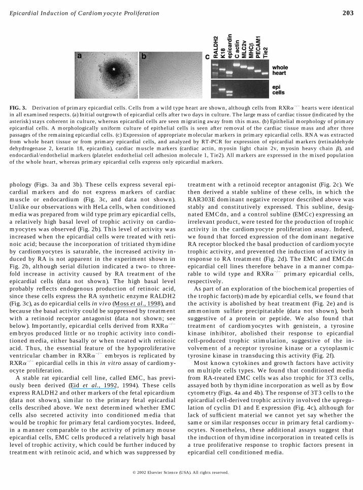
phology (Figs. 3a and 3b). These cells express several epi-cardial markers and do not express markers of cardiacmuscle or endocardium (Fig. 3c, and data not shown).Unlike our observations with HeLa cells, when conditionedmedia was prepared from wild type primary epicardial cells,a relatively high basal level of trophic activity on cardio-myocytes was observed (Fig. 2b). This level of activity wasincreased when the epicardial cells were treated with reti-noic acid; because the incorporation of tritiated thymidineby cardiomyocytes is saturable, the increased activity in-duced by RA is not apparent in the experiment shown inFig. 2b, although serial dilution indicated a two- to three-fold increase in activity caused by RA treatment of theepicardial cells (data not shown). The high basal levelprobably reflects endogenous production of retinoic acid,since these cells express the RA synthetic enzyme RALDH2(Fig. 3c), as do epicardial cells in vivo (Moss et al., 1998), andbecause the basal activity could be suppressed by treatmentwith a retinoid receptor antagonist (data not shown; seebelow). Importantly, epicardial cells derived from RXR��/�
embryos produced little or no trophic activity into condi-tioned media, either basally or when treated with retinoicacid. Thus, the essential feature of the hypoproliferativeventricular chamber in RXR��/� embryos is replicated byRXR��/� epicardial cells in this in vitro assay of cardiomy-ocyte proliferation.
A stable rat epicardial cell line, called EMC, has previ-ously been derived (Eid et al., 1992, 1994). These cellsexpress RALDH2 and other markers of the fetal epicardium(data not shown), similar to the primary fetal epicardialcells described above. We next determined whether EMCcells also secreted activity into conditioned media thatwould be trophic for primary fetal cardiomyocytes. Indeed,in a manner comparable to the activity of primary mouseepicardial cells, EMC cells produced a relatively high basallevel of trophic activity, which could be further induced bytreatment with retinoic acid, and which was suppressed by
treatment with a retinoid receptor antagonist (Fig. 2c). Wethen derived a stable subline of these cells, in which theRAR303E dominant negative receptor described above wasstably and constitutively expressed. This subline, desig-nated EMCdn, and a control subline (EMCc) expressing anirrelevant product, were tested for the production of trophicactivity in the cardiomyocyte proliferation assay. Indeed,we found that forced expression of the dominant negativeRA receptor blocked the basal production of cardiomyocytetrophic activity, and prevented the induction of activity inresponse to RA treatment (Fig. 2d). The EMC and EMCdnepicardial cell lines therefore behave in a manner compa-rable to wild type and RXR��/� primary epicardial cells,respectively.
As part of an exploration of the biochemical properties ofthe trophic factor(s) made by epicardial cells, we found thatthe activity is abolished by heat treatment (Fig. 2e) and isammonium sulfate precipitatable (data not shown), bothsuggestive of a protein or peptide. We also found thattreatment of cardiomyocytes with genistein, a tyrosinekinase inhibitor, abolished their response to epicardialcell-produced trophic stimulation, suggestive of the in-volvement of a receptor tyrosine kinase or a cytoplasmictyrosine kinase in transducing this activity (Fig. 2f).
Most known cytokines and growth factors have activityon multiple cells types. We found that conditioned mediafrom RA-treated EMC cells was also trophic for 3T3 cells,assayed both by thymidine incorporation as well as by flowcytometry (Figs. 4a and 4b). The response of 3T3 cells to theepicardial cell-derived trophic activity involved the upregu-lation of cyclin D1 and E expression (Fig. 4c), although forlack of sufficient material we cannot yet say whether thesame or similar responses occur in primary fetal cardiomy-ocytes. Nonetheless, these additional assays suggest thatthe induction of thymidine incorporation in treated cells isa true proliferative response to trophic factors present inepicardial cell conditioned media.
FIG. 3. Derivation of primary epicardial cells. Cells from a wild type heart are shown, although cells from RXR��/� hearts were identicalin all examined respects. (a) Initial outgrowth of epicardial cells after two days in culture. The large mass of cardiac tissue (indicated by theasterisk) stays coherent in culture, whereas epicardial cells are seen migrating away from this mass. (b) Epithelial morphology of primaryepicardial cells. A morphologically uniform culture of epithelial cells is seen after removal of the cardiac tissue mass and after threepassages of the remaining epicardial cells. (c) Expression of appropriate molecular markers in primary epicardial cells. RNA was extractedfrom whole heart tissue or from primary epicardial cells, and analyzed by RT-PCR for expression of epicardial markers (retinaldehydedehydrogenase 2, keratin 18, epicardin), cardiac muscle markers (cardiac actin, myosin light chain 2v, myosin heavy chain �), andendocardial/endothelial markers (platelet endothelial cell adhesion molecule 1, Tie2). All markers are expressed in the mixed populationof the whole heart, whereas primary epicardial cells express only epicardial markers.
203Epicardial Induction of Cardiomyocyte Proliferation
© 2002 Elsevier Science (USA). All rights reserved.

Finally, we addressed how the production of and responseto trophic activity changes during development. We foundthat conditioned media prepared from wild type mouseepicardial cells derived from late gestation embryos wascomparably active to that produced by epicardial cellsderived from midgestation embryos (Fig. 5a). However,epicardial cells obtained from early postnatal animals pro-duced substantially less of the activity (although there wasstill detectable activity made from these cells). Midgesta-tion fetal atrial cardiomyocytes were comparably respon-sive to stimulation as ventricular cardiomyocytes fromembryos of the same age (Fig. 5b). However, the ability ofcardiomyocytes to respond to stimulation decreases pro-gressively as development proceeds, being highest in cellsisolated at the earliest developmental stages and falling offas development proceeds. In the postnatal period, cardio-myocytes isolated from newborn pups were only marginallyresponsive to trophic stimulation, and cells isolated from3-day-old pups were refractory to proliferation.
DISCUSSION
Based on the results described in this study, we proposethat the fetal epicardium secretes one or more protein
factors that stimulate fetal cardiomyocyte proliferation.The production of this factor is induced by retinoic acid,which is probably produced in an autocrine or paracrinemanner by epicardial expression of RALDH2, and thisinduction requires the involvement of RXR�. We proposethat the pathology seen in the hearts of RXR��/� embryosreflects a failure in the production of this activity and theresulting lack of cardiomyocyte proliferation.
A prediction of this model is that ablation of the epicar-dium should result in a hypoplastic ventricular chamberwall. This appears to be the case in VCAM-1 (Gurtner et al.,1995; Kwee et al., 1995) and possibly also �4 integrin (Yanget al., 1995) mutant mice, where a physical interactionbetween these two molecules is normally responsible foradherence (and persistence) of the epicardium to the myo-cardium. Similarly, in chick embryos, formation of theepicardium can be partially prevented by the insertion of apiece of egg shell membrane in front of the proepicardialorgan (the precursor of the epicardium). This results in patchesof absent epicardium, with the myocardium underneath be-coming hypoplastic (Gittenberger-de Groot et al., 2000).
The existence of an epicardial proliferative signal ac-counts for two aspects of fetal ventricular development that
FIG. 5. Changes in production of and response to trophic activityduring development. (a) Production of trophic activity in primarywild type mouse epicardial cells isolated from embryos or pups atvarious stages as indicated. Conditioned media was prepared fromRA-treated cultured primary epicardial cells and collected andstored; all samples were assayed on chick cardiomyocytes in thesame experiment. Solvent-treated cells showed lower but propor-tional levels of activity (not shown). Data is expressed as CPM oftritiated thymidine incorporated. (b) Response of primary mousecardiomyocytes isolated from embryos or pups at the indicatedstages. All cardiomyocytes were treated with the same batch ofconditioned media from EMC cells treated with RA, althoughbecause all assays were done at different times, the data is ex-pressed in terms of fold induction caused by conditioned mediarelative to the basal level of thymidine incorporation in cardiomy-ocytes exposed only to serum-free DMEM. Cardiomyocytes wereprepared from ventricular (V) or atrial (A) tissue as indicated.
FIG. 4. Response of NIH3T3 cells to trophic stimulation byconditioned media from RA-treated epicardial cells. (a) Thymidineincorporation assay. Serum-free and serum controls are as in Fig. 2.(b) Flow cytometry assay. Cells were cultured in serum-free mediasupplemented with conditioned media from RA-treated EMC cellsfor the indicated times, then propidium iodide-stained and passedthrough a cell sorter. Cells were categorized as being in the resting(G0 or G1) or cycling (S, G2, or M) phase of the cell cycle, based onDNA content. (c) Expression of cyclin D1 and cyclin E is inducedby exposure to epicardial cell-conditioned media. Shown are West-ern blots of equal levels of loaded protein (36 �g) taken from cellsexposed to conditioned media [EMC(RA)] or 10% serum (S) for theindicated times (in h).
204 Chen et al.
© 2002 Elsevier Science (USA). All rights reserved.

have previously been obscure. First, it has been noted thatcardiomyocyte proliferation is greater on the epicardial sideof the chamber wall than on the endocardial side, which issuggestive of a proliferative signal originating from theepicardium (Rumyantsev, 1977; Tokuyasu, 1990). Second,the utilization of the epicardium as the source of a prolif-erative signal provides a simple developmental mechanismfor coordinating the timing of ventricular chamber growthand for formation of the compact layer of the ventricularchamber wall, which in mouse embryos begins aroundE10.5, immediately following formation of the epicardiallayer.
Previous studies have given rise to the suggestion that theRXR��/� cardiac phenotype may result indirectly fromplacental dysfunction (Kastner et al., 1997; Sapin et al.,1997). Our results are not incompatible with this model,and in fact, the hypoplastic ventricular phenotype mayrepresent the combined effects of placental deficiency aswell as local epicardial deficiency. We cannot say at thistime which of these effects is more important in definingthe overall phenotype.
A number of reciprocal interactions occur between adja-cent layers of the developing heart, mediated by secretedfactors. The myocardium secretes factors, including TGF�3,that act on the endocardium to induce an epithelial–mes-enchymal transformation that allows endocardial-derivedcells to populate the atrioventricular and conotruncal cush-ions (Brown et al., 1999; Runyan et al., 1992). Similarly, themyocardium produces additional factors, including FGFs,that act on the epicardium to induce an epithelial–mesenchymal transformation that creates the vascular en-dothelium of the coronary arteries, the smooth muscle cellsthat surrounds these arteries, and the cardiac fibroblastlineages (Morabito et al., 2001). In return, the endocardiumsecretes neuregulin, which acts at the inner surface of themyocardium to induce cardiomyocyte proliferation andtrabecular ingrowth (Gassmann et al., 1995; Lee et al., 1995;Meyer and Birchmeier, 1995). Based on our observations, wepropose a fourth axis of signaling, in which factors secretedby the epicardium act on the outer surface of the myocar-dium to induce cardiomyocyte proliferation and compactzone formation.
The identity of the factor (or factors) produced by theepicardium in response to retinoic acid signaling is cur-rently unknown. EMC cells are known to express endothe-lins (Eid et al., 1994), but endothelin expression is notretinoic acid regulated in these cells, and blocking antibod-ies against endothelins do not suppress cardiomyocyteproliferation induced by epicardial cell conditioned media(unpublished observations). We have also explored the pos-sibility that erythropoietin, which our prior work hasshown to be a primary target gene of retinoic acid and RAreceptor action (Makita et al., 2001), may be involved in thecardiac defect of RXR��/� embryos. However, erythropoi-etin is expressed at only trace levels by epicardial cells, andtransgenic expression of erythropoietin in the fetal heartfrom a � myosin heavy chain promoter does not rescue the
cardiac defects of RXR��/� embryos (unpublished observa-tions). We have also explored a number of additional can-didate factors, including IGF1, midkine, PDGF, and severalothers, but none of these appear to be regulated by retinoicacid in epicardial cells. It is of course possible that retinoicacid induces the expression of a processing enzyme thatacts on a constitutively expressed precursor to create anactive factor, although we currently have no evidence thatwould support this model. We have initiated biochemicaland molecular strategies to identify the factors responsiblefor the cardiomyocyte trophic response, to identify thereceptors of these factors, and to define the target genes thatare immediately retinoic acid regulated at the onset of thisprocess. These investigations may also help understandwhy the atrial compartment does not undergo the prolifera-tive expansion that occurs in the ventricular chamber invivo, even though atrial cardiomyocytes seems equally ableto respond to epicardial trophic stimulation in vitro (Fig. 4).Possibly, the production of trophic factors by the epicar-dium in vivo differs between the atrial and ventricularchambers.
It has long been known that fetal cardiomyocytes prolif-erate, whereas postnatal cardiomyocytes are mostly if notcompletely postmitotic. Our results with cardiomyocytesisolated from different developmental stages and exposed toepicardial cell conditioned media indicate that the myocar-dium itself changes over time in its ability to respond totrophic stimulation. In fact, our results are most easilyexplained by a stochastic loss of proliferative potential inthe cardiomyocyte population as development progresses.This was previously understood based on the response offetal and postnatal cardiomyocytes to serum stimulation,but our results represent a more biologically relevant con-text from which to draw this conclusion. The identificationof the receptor of the epicardial trophic signal, which asdescribed above may be a member of the receptor tyrosinekinase family, should enable the definition of the signalingpathways which are triggered by trophic stimulation infetal cardiomyocytes, and the alterations in signal transduc-tion that occur in postnatal cells that prevent a proliferativeresponse.
A number of mouse gene mutations result in a ventricu-lar hypoplastic phenotype which is comparable if not iden-tical to that of RXR� deficiency (Rossant, 1996; Sucov,1998). One of these, the N-myc gene product, acts cellautonomously in fetal cardiomyocytes to support compactzone formation, and we have previously shown that RXR�function is not epistatic to N-myc expression (i.e., mycexpression does not rescue the RXR��/� phenotype) (Tranand Sucov, 1998). A long-term goal of these studies is toassemble a pathway of genetic control of cardiomyocyteproliferation and compact zone morphogenesis. Our dem-onstration of the epicardium as a critical source of trophicstimulation, and the in vitro culture assays we have em-ployed in this study, should facilitate the experimentaldissection of the roles of these other genes in these pro-cesses.
205Epicardial Induction of Cardiomyocyte Proliferation
© 2002 Elsevier Science (USA). All rights reserved.

Ischemic heart disease is the leading cause of death in theWestern world and represents myocardial cell death causedby coronary artery obstruction and the inability of adultmyocardium to repair itself via proliferation. Recent inves-tigations have led to the conception that there may existcardiomyocyte stem cells, either within the heart or derivedfrom extracardiac sources, which to a limited extent mayinitiate proliferation within the scar of ischemic damage(Anversa and Nadal-Ginard, 2002). The factors described inthis study may be useful in stimulating such cells to furtheror more rapidly divide, in a manner which could be thera-peutically meaningful.
ACKNOWLEDGMENTS
We thank Josh Seiffert and Dr. Jean-Sebastien Silvestre forcontributions during the early phase of this project. We thank JanetRossant for Flk1-deficient mice, Ken Chien for conditional RXR�mice, Andy McMahon for Wnt1cre mice, Akira Kakizuka for theRAR303E construct, and Bob Oshima for the K18 genomic construct.
REFERENCES
Agarwal, C., Chandraratna, R. A., Johnson, A. T., Rorke, E. A., andEckert, R. L. (1996). AGN193109 is a highly effective antagonistof retinoid action in human ectocervical epithelial cells. J. Biol.Chem. 271, 12209–12212.
Anversa, P., and Nadal-Ginard, B. (2002). Myocyte renewal andventricular remodelling. Nature 415, 240–243.
Brown, C. B., Boyer, A. S., Runyan, R. B., and Barnett, J. V. (1999).Requirement of type III TGF-beta receptor for endocardial celltransformation in the heart. Science 283, 2080–2082.
Chen, J., Kubalak, S. W., and Chien, K. R. (1998). Ventricularmuscle-restricted targeting of the RXR� gene reveals a non-cell-autonomous requirement in cardiac chamber morphogenesis.Development 125, 1943–1949.
Cohen-Gould, L., and Mikawa, T. (1996). The fate diversity ofmesodermal cells within the heart field during chicken earlyembryogenesis. Dev. Biol. 177, 265–273.
Dolle, P., Fraulob, V., Kastner, P., and Chambon, P. (1994). Devel-opmental expression of murine retinoid X receptor (RXR) genes.Mech. Dev. 45, 91–104.
Eid, H., Larson, D. M., Springhorn, J. P., Attawia, M. A., Nayak,R. C., Smith, T. W., and Kelly, R. A. (1992). Role of epicardialmesothelial cells in the modification of phenotype and functionof adult rat ventricular myocytes in primary coculture. Circ. Res.71, 40–50.
Eid, H., de Bold, M. L., Chen, J. H., and de Bold, A. J. (1994).Epicardial mesothelial cells synthesize and release endothelin.J. Cardiovasc. Pharmacol. 24, 715–720.
Gassmann, M., Casagranda, F., Orioli, D., Simon, H., Lai, C., Klein,R., and Lemke, G. (1995). Aberrant neural and cardiac develop-ment in mice lacking the ErbB4 neuregulin receptor. Nature 378,390–394.
Gittenberger-de Groot, A. C., Vrancken Peeters, M. P., Bergwerff,M., Mentink, M. M., and Poelmann, R. E. (2000). Epicardialoutgrowth inhibition leads to compensatory mesothelial outflowtract collar and abnormal cardiac septation and coronary forma-tion. Circ. Res. 87, 969–971.
Gurtner, G. C., Davis, V., Li, H., McCoy, M. J., Sharpe, A., andCybulsky, M. I. (1995). Targeted disruption of the murineVCAM1 gene: Essential role of VCAM-1 in chorioallantoicfusion and placentation. Genes Dev. 9, 1–14.
Hogan, B., Beddington, R., Costantini, F., and Lacy, E. (1994).“Manipulating the Mouse Embryo: A Laboratory Manual.” ColdSpring Harbor Laboratory Press, Cold Spring Harbor, NY.
Jiang, X., Rowitch, D. H., Soriano, P., McMahon, A. P., and Sucov,H. M. (2000). Fate of the mammalian cardiac neural crest.Development 127, 1607–1616.
Jiang, X., Choudhary, B., Merki, E., Chien, K. R., Maxson, R. E., andSucov, H. M. (2002). Normal fate and altered function of thecardiac neural crest cell lineage in retinoic acid receptor mutantembryos. Mech. Dev., in press.
Kastner, P., Grondona, J. M., Mark, M., Gansmuller, A., LeMeur,M., Decimo, D., Vonesch, J. L., Dolle, P., and Chambon, P. (1994).Genetic analysis of RXR� developmental function: Convergenceof RXR and RAR signaling pathways in heart and eye morpho-genesis. Cell 78, 987–1003.
Kastner, P., Messaddeq, N., Mark, M., Wendling, O., Grondona,J. M., Ward, S., Ghyselinck, N., and Chambon, P. (1997). VitaminA deficiency and mutations of RXR�, RXR�, and RAR� leads toearly differentiation of embryonic ventricular cardiomyocytes.Development 124, 4749–4758.
Kwee, L., Baldwin, H. S., Shen, H. M., Stewart, C. L., Buck, C.,Buck, C. A., and Labow, M. A. (1995). Defective development ofthe embryonic and extraembryonic circulatory systems in vas-cular cell adhesion molecule (VCAM-1) deficient mice. Develop-ment 121, 489–503.
Lee, K. F., Simon, H., Chen, H., Bates, B., Hung, M. C., and Hauser,C. (1995). Requirement for neuregulin receptor erbB2 in neuraland cardiac development. Nature 378, 394–398.
Makita, T., Hernandez-Hoyos, G., Chen, T. H., Wu, H., Rothen-berg, E. V., and Sucov, H. M. (2001). A developmental transitionin definitive erythropoiesis: Erythropoietin expression is sequen-tially regulated by retinoic acid receptors and HNF4. Genes Dev.15, 889–901.
Mangelsdorf, D. J., Borgmeyer, U., Heyman, R. A., Zhou, J. Y., Ong,E. S., Oro, A. E., Kakizuka, A., and Evans, R. M. (1992). Charac-terization of three RXR genes that mediate the action of 9-cisretinoic acid. Genes Dev. 6, 329–344.
Markwald, R. R., Fitzharris, T. P., and Manasek, F. J. (1977).Structural development of endocardial cushions. Am. J. Anat.148, 85–120.
Meyer, D., and Birchmeier, C. (1995). Multiple essential functionsof neuregulin in development. Nature 378, 386–390.
Mikawa, T. (1999). Cardiac lineages. In “Heart Development” (R. P.Harvey and N. Rosenthal, Eds.), pp. 19–33. Academic Press, SanDiego.
Morabito, C. J., Dettman, R. W., Kattan, J., Collier, J. M., andBristow, J. (2001). Positive and negative regulation of epicardial-mesenchymal transformation during avian heart development.Dev. Biol. 234, 204–215.
Moss, J. B., Xavier-Neto, J., Shapiro, M. D., Nayeem, S. M.,McCaffery, P., Drager, U. C., and Rosenthal, N. (1998). Dynamicpatterns of retinoic acid synthesis and response in the developingmammalian heart. Dev. Biol. 199, 55–71.
Rossant, J. (1996). Mouse mutants and cardiac development: newmolecular insights into cardiogenesis. Circ. Res. 78, 349–353.
Rumyantsev, P. P. (1977). Interrelations of the proliferation anddifferentiation processes during cardiact myogenesis and regen-eration. Int. Rev. Cytol. 51, 186–273.
206 Chen et al.
© 2002 Elsevier Science (USA). All rights reserved.

Runyan, R. B., Potts, J. D., and Weeks, D. L. (1992). TGF�3-mediated tissue interaction during embryonic heart develop-ment. Mol. Reprod. Dev. 32, 152–159.
Saitou, M., Sugai, S., Tanaka, T., Shimouchi, K., Fuchs, E., Naru-miya, S., and Kakizuka, A. (1995). Inhibition of skin developmentby targeted expression of a dominant-negative retinoic acidreceptor. Nature 374, 159–162.
Sapin, V., Dolle, P., Hindelang, C., Kastner, P., and Chambon, P.(1997). Defects of the chorioallantoic placenta in mouse RXR�null fetuses. Dev. Biol. 191, 29–41.
Sedmera, D., and Thomas, P. S. (1996). Trabeculation in theembryonic heart. Bioessays 18, 607.
Shalaby, F., Ho, J., Stanford, W. L., Fischer, K. D., Schuh, A. C.,Schwartz, L., Bernstein, A., and Rossant, J. (1997). A requirementfor Flk1 in primitive and definitive hematopoiesis and vasculo-genesis. Cell 89, 981–990.
Subbarayan, V., Mark, M., Messadeq, N., Rustin, P., Chambon, P.,and Kastner, P. (2000). RXR� overexpression in cardiomyo-cytes causes dilated cardiomyopathy but fails to rescue myocar-dial hypoplasia in RXR�-null fetuses. J. Clin. Invest. 105, 387–394.
Sucov, H. M. (1998). Molecular insights into cardiac development.Annu. Rev. Physiol. 60, 287–308.
Sucov, H. M., Dyson, E., Gumeringer, C. L., Price, J., Chien, K. R.,and Evans, R. M. (1994). RXR� mutant mice establish a geneticbasis for vitamin A signaling in heart morphogenesis. Genes Dev.8, 1007–1018.
Thorey, I. S., Meneses, J. J., Neznanov, N., Kulesh, D. A., Pedersen,R. A., and Oshima, R. G. (1993). Embryonic expression of human
keratin 18 and K18-�-galactosidase fusion genes in transgenicmice. Dev. Biol. 160, 519–534.
Tokuyasu, K. T. (1990). Co-development of embryonic myocar-dium and myocardial circulation. In “Developmental Cardiol-ogy: Morphogenesis and Function” (E. B. Clark and A. Takao,Eds.), pp. 205–218. Futura Publishing, Mt. Kisco, NY.
Tran, C. M., and Sucov, H. M. (1998). The RXR� gene functions ina non-cell-autonomous manner during mouse cardiac morpho-genesis. Development 125, 1951–1956.
Viragh, S., and Challice, C. E. (1981). The origin of the epicardiumand the embryonic myocardial circulation in the mouse. Anat.Rec. 201, 157–168.
Wilson, J. G., and Warkany, J. (1949). Aortic arch and cardiacanomalies in the offspring of vitamin A deficient rats. Am. J.Anat. 85, 113–155.
Yamaguchi, M., Nakamoto, M., Honda, H., Nakagawa, T., Fujita,H., Nakamura, T., Hirai, H., Narumiya, S., and Kakizuka, A.(1998). Retardation of skeletal development and cervical abnor-malities in transgenic mice expressing a dominant-negativeretinoic acid receptor in chondrogenic cells. Proc. Natl. Acad.Sci. USA 95, 7491–7496.
Yang, J. T., Rayburn, H., and Hynes, R. O. (1995). Cell adhesionevents mediated by �4 integrins are essential in placental andcardiac development. Development 121, 549–560.
Received for publication March 28, 2002Revised July 23, 2002
Accepted July 23, 2002Published online August 27, 2002
207Epicardial Induction of Cardiomyocyte Proliferation
© 2002 Elsevier Science (USA). All rights reserved.