Identification of hypoxia-related prognostic signature for ...

J O U R N A L O F T H E AM E R I C A N C O L L E G E O F C A R D I O L O G Y V O L . 6 5 , N O . 4 , 2 0 1 5
ª 2 0 1 5 B Y T H E AM E R I C A N C O L L E G E O F C A R D I O L O G Y F O U N DA T I O N I S S N 0 7 3 5 - 1 0 9 7 / $ 3 6 . 0 0
P U B L I S H E D B Y E L S E V I E R I N C . h t t p : / / d x . d o i . o r g / 1 0 . 1 0 1 6 / j . j a c c . 2 0 1 4 . 1 0 . 0 6 5
Secretoneurin Is a Novel PrognosticCardiovascular Biomarker AssociatedWith Cardiomyocyte Calcium Handling
Anett Hellebø Ottesen, MSC,*yz William E. Louch, PHD,yz Cathrine R. Carlson, PHD,yz Ole J.B. Landsverk, PHD,xJouni Kurola, MD, PHD,k Rune Forstrøm Johansen,{ Morten K. Moe, PHD,# Jan Magnus Aronsen, MD,yzArne Didrik Høiseth, MD, PHD,*z Hilde Jarstadmarken, MSC,yz Ståle Nygård, PHD,yz Magnar Bjørås, PHD,{Ivar Sjaastad, MD, PHD,yz Ville Pettilä, MD, PHD,** Mats Stridsberg, MD, PHD,yy Torbjørn Omland, MD, PHD, MPH,*zGeir Christensen, MD, PHD, MHA,yz Helge Røsjø, MD, PHD*zABSTRACT
Fro
Os
Un
No
Un
Lø
yyDwe
an
fun
Re
fun
ge
rol
pre
BACKGROUND Secretoneurin (SN) levels are increased in patients with heart failure (HF), but whether SN provides
prognostic information and influences cardiomyocyte function is unknown.
OBJECTIVES This study sought to evaluate the merit of SN as a cardiovascular biomarker and assess effects of SN on
cardiomyocyte Ca2þ handling.
METHODS We assessed the association between circulating SN levels and mortality in 2 patient cohorts and the
functional properties of SN in experimental models.
RESULTS In 143 patients hospitalized for acute HF, SN levels were closely associated with mortality (n ¼ 66) during
follow-up (median 776 days; hazard ratio [lnSN]: 4.63; 95% confidence interval: 1.93 to 11.11; p ¼ 0.001 in multivariate
analysis). SN reclassified patients to their correct risk strata on top of other predictors of mortality. In 155 patients with
ventricular arrhythmia–induced cardiac arrest, SN levels were also associated with short-term mortality (n ¼ 51; hazard
ratio [lnSN]: 3.33; 95% confidence interval: 1.83 to 6.05; p < 0.001 in multivariate analysis). Perfusing hearts with SN
yielded markedly increased myocardial levels and SN internalized into cardiomyocytes by endocytosis. Intracellularly, SN
reduced Ca2þ/calmodulin (CaM)-dependent protein kinase II d (CaMKIId) activity via direct SN-CaM and SN-CaMKII
binding and attenuated CaMKIId-dependent phosphorylation of the ryanodine receptor. SN also reduced sarcoplasmic
reticulum Ca2þ leak, augmented sarcoplasmic reticulum Ca2þ content, increased the magnitude and kinetics of cardio-
myocyte Ca2þ transients and contractions, and attenuated Ca2þ sparks and waves in HF cardiomyocytes.
CONCLUSIONS SN provided incremental prognostic information to established risk indices in acute HF and ventricular
arrhythmia–induced cardiac arrest. (J Am Coll Cardiol 2015;65:339–51) © 2015 by the American College of Cardiology
Foundation.
m the *Division of Medicine, Akershus University Hospital, Lørenskog, Norway; yInstitute for Experimental Medical Research,
lo University Hospital, Ullevål, Oslo, Norway; zCenter for Heart Failure Research and K.G. Jebsen Cardiac Research Centre,
iversity of Oslo, Oslo, Norway; xCentre for Immune Regulation, Department of Molecular Biosciences, University of Oslo, Oslo,
rway; kDivision of Intensive Care Medicine, Kuopio University Hospital, Kuopio, Finland; {Department of Microbiology, Oslo
iversity Hospital, Rikshospitalet, Oslo, Norway; #Division for Diagnostics and Technology, Akershus University Hospital,
renskog, Norway; **Department of Surgery, Intensive Care Units, Helsinki University Hospital, Helsinki, Finland; and the
epartment of Medical Sciences, Uppsala University, Uppsala, Sweden. Generous project funding was received from the Nor-
gian National Association for Public Health to Ms. Ottesen, the Anders Jahre Trust for Promotion of Science to Dr. Christensen,
d the Norwegian Research Council to the ACE 2 Study to Dr. Omland. Drs. Omland and Røsjø have additionally received
ding relating to the current work from Akershus University Hospital, the University of Oslo, the South-Eastern Norway
gional Health Authority, and the K.G. Jebsen Family Foundation. The FINNRESUSCI Study was supported by institutional
ding (EVO) from Kuopio University Hospital and Helsinki University Hospital (EVO T102010070), the Foundation of Emer-
ncy Medicine, Finska Läkaresällskapet Foundation, and by the Päivikki and Sakari-Sohlberg Foundation. The sponsors had no
e in any of the following: design and conduct of the study, collection, management, analysis and interpretation of the data, or
paration, review, and approval of the manuscript. Drs. Stridsberg, Omland, Christensen, and Røsjø are partners in a patent

ABBR EV I A T I ON S
AND ACRONYMS
AUC = area under the receiver-
operating characteristic curve
BNP = B-type
natriuretic peptide
CaM = calmodulin
CaMKIId = Ca2D/calmodulin-
dependent protein kinase II d
CI = confidence interval
COPD = chronic obstructive
pulmonary disease
CV = cardiovascular
CVD = cardiovascular disease
HF = heart failure
HR = hazard ratio
hs-TnT = high-sensitivity
troponin T
ln = natural logarithm
NE = norepinephrine
NT-proBNP = N-terminal pro–
B-type natriuretic peptide
OHCA-VF = out-of-hospital
cardiac arrest with
ventricular fibrillation
PLB = phospholamban
RyR2 = ryanodine receptor 2
SN = secretoneurin
application
patients w
to disclose
Manuscrip
Ottesen et al. J A C C V O L . 6 5 , N O . 4 , 2 0 1 5
Secretoneurin, Biomarker, and Calcium Regulation F E B R U A R Y 3 , 2 0 1 5 : 3 3 9 – 5 1
340
C irculating biological substances (bio-markers) can identify specific patho-physiology in an individual patient
(1). Cardiovascular (CV) biomarkers currentlyin clinical use are the B-type natriuretic pep-tides (BNPs), which reflect cardiomyocytestress, and cardiac-specific troponins thatmeasure cardiomyocyte injury (2). However,established biomarkers do not assess addi-tional processes relevant to CV disease (CVD)progression such as increased neuroendocrinetone and cardiomyocyte Ca2þ homeostasis.
SEE PAGE 352
Accumulating evidence suggests that thechromogranin-secretogranin (granin) familyof proteins plays an important role in cardiacpathophysiology and may serve as CV bio-markers (3–8). One interesting member of thisfamily is the high-capacity, low-affinity Ca2þ-binding protein secretogranin II (9). Secre-togranin II is cleaved by the proteases PC1/3and PC2 to the 33-amino acid peptide secre-toneurin (SN) (SgII154-186) (9), and theseproteases’ activity is increased by 3-fold inthe failing myocardium (10).
Previous work has shown that SN is thepredominant form of secretogranin II im-
munoreactivity in plasma (11,12). By employing anSN-binding assay (epitope SgII154-165), we havedemonstrated increased circulating SN levels in pa-tients with heart failure (HF) (10). Additionally, SNhas been shown to attenuate myocardial ischemia/reperfusion injury, reduce cardiomyocyte apoptosis,induce angiogenesis, and improve left ventricularfunction after myocardial infarction in experimentalanimal models (10,13). Given these data and theestablished role of granin proteins in modulating Ca2þ
handling in noncardiac cells (14), we postulated thatSN could have a direct, protective effect on car-diomyocyte Ca2þ handling. Accordingly, in this studywe wanted to assess the potential of SN as a prog-nostic CV biomarker and explore the effects of SN oncardiomyocyte Ca2þ homeostasis.
METHODS
Details regarding methods and significant statisticaland data elements are found in the Online Appendix.
filed by the University of Oslo regarding the use of secretoneu
ith critical illness. All other authors have reported that they have n
.
t received September 2, 2014; revised manuscript received Octobe
CLINICAL COHORTS. We obtained blood samples<24 h following hospital admission in 143 patientswith acute HF and <6 h following admission in 155patients with ventricular arrhythmias and out-of-hospital cardiac arrest (153 patients with ventricularfibrillation; OHCA-VF). Blood samples were alsoavailable 24 h later in most patients (n ¼ 104 for acuteHF and n ¼ 150 for OHCA-VF) and prior to hospitaldischarge in a subset of HF patients (n ¼ 46). Weadditionally collected blood samples from 84 patientswith acute chronic obstructive pulmonary disease(COPD) and from 62 healthy control subjects. Survivalstatus was obtained from electronic hospital records.
EXPERIMENTAL MODELS. Internalization of SN(2.8 mM and 28 nM) was studied in neonatal mouseand rat cardiomyocytes and in isolated mouse hearts.For assessment of phosphorylation status, BayK(10 mM) was included in the perfusate of mouse heartsex vivo to enhance myocardial Ca2þ/calmodulin(CaM)-dependent protein kinase II d (CaMKIId) ac-tivity. Immunoblotting was performed with a primaryantibody for SN (SgII172-186) (15). We assessedSN-CaMKIId and SN-CaM interactions by severalmethods, and examined CaMKIId activity in vitro bykinase activity assays. The effects of SN treatment oncardiomyocyte Ca2þ homeostasis were examined inisolated ventricular myocytes from adult healthymice and in ventricular myocytes obtained adjacentto the infarct area (border zone) in HF rats (OnlineTable 1, Online Figure 1).
STATISTICAL ANALYSIS. Clinical data are expressedas mean � SD with the exception of biomarkers,which are reported as median (quartile 1 to 3), andexperimental data are presented as mean � SEM.Differences between groups were examined by theStudent t test or the Mann-Whitney U test, and serialmeasurements tested by the related-samples Wil-coxon signed rank test. Categorical data are presentedas absolute numbers and percents, and werecompared by the chi-square or the Fisher Exact test.We used Spearman rank correlation, and de-terminants of biomarker levels were assessed bylinear regression analyses. Prognostic utility wasassessed by Kaplan-Meier plots with SN quartilescompared by the log-rank test and by Cox propor-tional hazards regression analysis. Biomarkers weretransformed by the natural logarithm (ln) before
rin as a biomarker in cardiovascular disease and in
o relationships relevant to the contents of this paper
r 22, 2014, accepted October 28, 2014.

FIGURE 1 SN Presence in HF Patients as Assessed by Liquid Chromatography-Mass
Spectrometry
100
10080604020
00.0 1.0 2.0 3.0 4.0 5.0
75
50
25
1224.5 1225.0 1225.5 1226.0 1226.5 1227.0 1227.5 m/z
m/z
Chromatogram Secretoneurin (0.2 ng/ml) Recorded at m/z=Re
l. Ab
und.
10080604020
0
Rel.
Abun
d.
10080604020
0
Rel.
Abun
d.
10080604020
0
Rel.
Abun
d.
(min)
0.0 1.0 2.0 3.0 4.0 5.0 (min)
2.89
2.97
1224.92686-1224.93910+1225.26084-1225.27310+1225.59519-1225.60745
1224.92686-1224.93910+1225.26084-1225.27310+1225.59519-1225.60745
Extracted Mass Spectrum of Secretoneurin (tR=2.89 min)
1224.6 1225.0 1225.4 1225.8 1226.2
m/z1224.6 1225.0 1225.4 1225.8 1226.2 1226.6
Chromatogram Patient Sample Recorded at m/z=
Extracted Mass Spectrum of the Eluting at tR=2.97 min
1224
.594
89z=
3
1224
.596
65z=
3
1224
.596
3z=
3
1224
.930
6z=
3
1225
.264
9z=
3
1225
.599
2z=
3
1225
.933
4z=
3
1226
.267
6z=
3
1226
.601
8z=
3
1226
.936
0z=
3
1227
.270
2z=
3
1227
.604
5z=
3
1224
.930
33z=
3
1225
.264
44z=
3
1225
.598
39z=
3
1225
.932
80z=
3
1226
.266
90z=
3
1226
.599
43z=
3
1224
.930
19z=
3
1225
.264
41z=
3
1225
.598
05z=
3
1225
.932
98z=
3
1226
.266
90z=
3
1226
.601
89z=
3
Rela
tive
Abun
danc
e
Secretoneurin, Theoretical Mass Spectrum for [M - 3h]3-A
B
C
The mass spectrum of secretoneurin (SN) based on (A) theoretical calculation and the mass
spectrum of (B) synthetic SN and (C) SN in heart failure (HF) patient samples were nearly
identical.
J A C C V O L . 6 5 , N O . 4 , 2 0 1 5 Ottesen et al.F E B R U A R Y 3 , 2 0 1 5 : 3 3 9 – 5 1 Secretoneurin, Biomarker, and Calcium Regulation
341
regression analyses. Calibration of the basic riskmodel was examined by calculating the category-freenet reclassification index (16), and prognostic accu-racy was assessed by calculating the area underthe receiver-operating characteristic curve (AUC);p values <0.05 were considered significant.
The clinical studies were approved by the RegionalEthics Committees and the experimental studiesperformed according to National Institutes of Healthguidelines.
RESULTS
CIRCULATING SN LEVELS ON ADMISSION IN
PATIENTS WITH ACUTE HF. We first confirmed thepresence of free SN in the circulation of HF patientsby liquid chromatography-mass spectrometry. Themass spectra of SN based on theoretical calculation(Figure 1A) and synthetic SN (Figure 1B) were found tobe nearly identical to the spectrum in the plasma ofHF patients (Figure 1C).
Using radioimmunoassay (17), we measured SNlevels of 140 (122 to 170) pmol/l in acute HF patientswith a range between 84 and 677 pmol/l (Figure 2A).SN levels correlated with N-terminal pro–B-typenatriuretic peptide (NT-proBNP) (r ¼ 0.38; p <
0.001) and high-sensitivity troponin T (hs-TnT)levels (r ¼ 0.27; p ¼ 0.001), but not with left ven-tricular ejection fraction (r ¼ –0.03; p ¼ 0.77) ornorepinephrine (NE) levels (r ¼ 0.15; p ¼ 0.08).Patients classified as New York Heart Associationfunctional class IV exhibited higher SN levels thanthe other patients (Online Figure 2). Creatinineclearance, diabetes mellitus, age, inhalation therapywith short-acting b2-agonist, and NT-proBNP levelswere associated with SN levels (details in OnlineAppendix). The duration of dyspnea prior to hospi-tal admission (Online Figure 3) and the time fromhospital admission to blood sampling did not influ-ence SN levels in the acute HF patients (r ¼ –0.06;p ¼ 0.46).
ADMISSION SN LEVELS ASSOCIATED WITH
MORTALITY IN PATIENTS WITH ACUTE HF. Sixty-sixpatients (46%) died during a median follow-up of776 (246 to 983) days (patient characteristics accord-ing to mortality in Online Table 2). There was agraded increase in mortality in proportion toadmission SN levels (Central Illustration) (p < 0.001).SN was strongly associated with mortality by Coxregression analyses that adjusted for other risk var-iables, including NT-proBNP, hs-TnT, and NE levels(hazard ratio [HR] [lnSN]: 4.63; 95% confidence in-terval [CI]: 1.93 to 11.11; p ¼ 0.001) (Table 1 andOnline Table 3 with medication). Adding SN to a
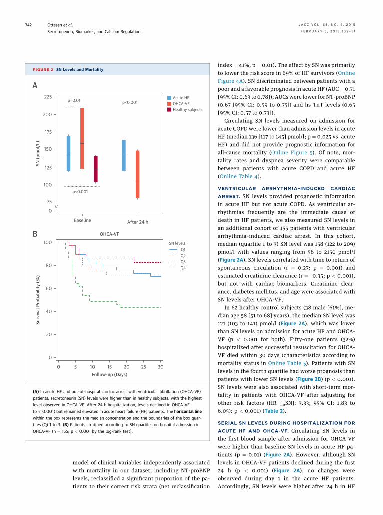
FIGURE 2 SN Levels and Mortality
225
200
175
150
125
100
75
0
100
80
60
40
20
0
0 5 10 15 20 25 30
SN (p
mol
/L)
p=0.01
p<0.001
p<0.001Acute HFOHCA-VFHealthy subjects
Baseline After 24 h
Surv
ival
Pro
babi
lity
(%)
Follow-up (Days)
OHCA-VF
SN levelsQ1Q2Q3Q4
A
B
(A) In acute HF and out-of-hospital cardiac arrest with ventricular fibrillation (OHCA-VF)
patients, secretoneurin (SN) levels were higher than in healthy subjects, with the highest
level observed in OHCA-VF. After 24 h hospitalization, levels declined in OHCA-VF
(p < 0.001) but remained elevated in acute heart failure (HF) patients. The horizontal line
within the box represents the median concentration and the boundaries of the box quar-
tiles (Q) 1 to 3. (B) Patients stratified according to SN quartiles on hospital admission in
OHCA-VF (n ¼ 155; p < 0.001 by the log-rank test).
Ottesen et al. J A C C V O L . 6 5 , N O . 4 , 2 0 1 5
Secretoneurin, Biomarker, and Calcium Regulation F E B R U A R Y 3 , 2 0 1 5 : 3 3 9 – 5 1
342
model of clinical variables independently associatedwith mortality in our dataset, including NT-proBNPlevels, reclassified a significant proportion of the pa-tients to their correct risk strata (net reclassification
index ¼ 41%; p ¼ 0.01). The effect by SN was primarilyto lower the risk score in 69% of HF survivors (OnlineFigure 4A). SN discriminated between patients with apoor and a favorable prognosis in acute HF (AUC¼ 0.71[95%CI: 0.63 to 0.78]); AUCswere lower for NT-proBNP(0.67 [95% CI: 0.59 to 0.75]) and hs-TnT levels (0.65[95% CI: 0.57 to 0.73]).
Circulating SN levels measured on admission foracute COPD were lower than admission levels in acuteHF (median 136 [117 to 145] pmol/l; p ¼ 0.025 vs. acuteHF) and did not provide prognostic information forall-cause mortality (Online Figure 5). Of note, mor-tality rates and dyspnea severity were comparablebetween patients with acute COPD and acute HF(Online Table 4).
VENTRICULAR ARRHYTHMIA–INDUCED CARDIAC
ARREST. SN levels provided prognostic informationin acute HF but not acute COPD. As ventricular ar-rhythmias frequently are the immediate cause ofdeath in HF patients, we also measured SN levels inan additional cohort of 155 patients with ventriculararrhythmia–induced cardiac arrest. In this cohort,median (quartile 1 to 3) SN level was 158 (122 to 209)pmol/l with values ranging from 58 to 2150 pmol/l(Figure 2A). SN levels correlated with time to return ofspontaneous circulation (r ¼ 0.27; p ¼ 0.001) andestimated creatinine clearance (r ¼ –0.35; p < 0.001),but not with cardiac biomarkers. Creatinine clear-ance, diabetes mellitus, and age were associated withSN levels after OHCA-VF.
In 62 healthy control subjects (38 male [61%], me-dian age 58 [51 to 68] years), the median SN level was121 (103 to 141) pmol/l (Figure 2A), which was lowerthan SN levels on admission for acute HF and OHCA-VF (p < 0.001 for both). Fifty-one patients (32%)hospitalized after successful resuscitation for OHCA-VF died within 30 days (characteristics according tomortality status in Online Table 5). Patients with SNlevels in the fourth quartile had worse prognosis thanpatients with lower SN levels (Figure 2B) (p < 0.001).SN levels were also associated with short-term mor-tality in patients with OHCA-VF after adjusting forother risk factors (HR [lnSN]: 3.33; 95% CI: 1.83 to6.05): p < 0.001) (Table 2).
SERIAL SN LEVELS DURING HOSPITALIZATION FOR
ACUTE HF AND OHCA-VF. Circulating SN levels inthe first blood sample after admission for OHCA-VFwere higher than baseline SN levels in acute HF pa-tients (p ¼ 0.01) (Figure 2A). However, although SNlevels in OHCA-VF patients declined during the first24 h (p < 0.001) (Figure 2A), no changes wereobserved during day 1 in the acute HF patients.Accordingly, SN levels were higher after 24 h in HF

CENTRAL ILLUSTRATION Secretoneurin: Calcium Regulator and Biomarker
(A) As a biomarker, secretoneurin (SN) levels stratify mortality risk at hospital admission for patients with acute heart failure (p < 0.001 by the log-rank test for SN
quartiles [Q]). (B) Experimental studies of SN in isolated cells and explanted hearts demonstrate that (1.) SN is internalized into cardiomyocytes and the intact heart by
endocytosis. (2.) SN binds directly to calmodulin (CaM) and CaM-dependent protein kinase II d (CaMKIId), and inhibits CaMKIId activity. (3.) This leads to reduced
Ser2814-ryanodine receptor 2 (RyR2) phosphorylation, and (4.) improved Ca2þ homeostasis.
J A C C V O L . 6 5 , N O . 4 , 2 0 1 5 Ottesen et al.F E B R U A R Y 3 , 2 0 1 5 : 3 3 9 – 5 1 Secretoneurin, Biomarker, and Calcium Regulation
343
patients than in patients successfully resuscitated forOHCA-VF (p < 0.001) (Figure 2A).
In the subset of HF patients with serial biomarkermeasurements, the AUC for all-cause mortality for SNlevels measured after 24 h was 0.65 (95% CI: 0.55 to0.74) compared to 0.63 (95% CI: 0.53 to 0.72) for NT-proBNP, although the AUC on discharge for SN was0.61 (95% CI: 0.45 to 0.74) and 0.56 (95% CI: 0.41 to0.71) for NT-proBNP. Compared to concentrations atbaseline, NT-proBNP levels tended to be lower after24 h (median reduction 117 pg/ml; p ¼ 0.065) andwere significantly reduced at hospital discharge(median reduction 1172 pg/ml; p < 0.001). NE levelsalso were significantly reduced prior to hospitaldischarge (median reduction 1,596 pmol/l; p ¼ 0.001).In contrast, SN levels stayed unchanged during theHF hospitalization with median 1.0 pmol/l increaseduring the first 24 h (p ¼ 0.91) and median 1.0 pmol/ldecrease on discharge (p ¼ 0.37).SN INTERNALIZED INTO CARDIOMYOCYTES AND
THE INTACT HEART. On the basis of the strong
association between SN levels and mortality in CVDand the association between granin proteins and Ca2þ
homeostasis in noncardiac cells (14), we examinedwhether SN affects cardiomyocyte Ca2þ homeostasis.Although the receptor for SN has not been identified(9), a report has suggested that other short granin-derived peptides are internalized in cells by endocy-tosis (18), so we examined whether SN may bedirectly internalized into isolated cardiomyocytesand into the intact heart. We observed increasedintracellular cardiomyocyte SN levels in proportion tothe SN concentration in the suspension (Figure 3A).The internalization of SN was validated by confocalfluorescence microscopy (Figure 3B, Online Video 1).SN colocalized with Alexa 647–labeled transferrin(Invitrogen/Molecular Probes, Carlsbad, California)(Figure 3B), which is internalized via clathrin-mediated endocytosis (19) but not with dextran-alexa647, a marker of unspecific macropinocytosis(Online Video 1). SN also colocalized with markers ofearly (EEA1) and late endosomes (LAMP1) (Figure 3C).

TABLE 1 Predictors of Mortality During Follow-Up in Patients With Acute
Heart Failure
Hazard Ratio 95% CI p Value
Univariate analysis
Age, per 1-yr increase 1.04 1.02–1.07 0.002
Male 0.53 0.32–0.86 0.010
Body mass index, per 1 unit increase 0.94 0.89–0.99 0.012
Creatinine clearance, ml/min 0.98 0.97–0.99 <0.001
Heart rate, admission 1.00 0.99–1.01 0.40
Systolic blood pressure, admission, mm Hg 0.99 0.98–0.99 0.004
Diastolic blood pressure, admission, mm Hg 0.97 0.96–0.99 0.001
NYHA functional class IV versus II/III 2.01 1.23–3.29 0.006
LVEF, continuous 1.00 0.98–1.02 0.88
History of
Heart failure 1.43 0.86–2.38 0.17
Myocardial infarction 1.00 0.62–1.63 1.00
PCI 0.69 0.37–1.29 0.24
CABG 1.17 0.66–2.05 0.59
Hypertension 0.83 0.51–1.36 0.47
Atrial fibrillation 1.07 0.66–1.74 0.78
Diabetes mellitus 1.78 1.08–2.95 0.024
Chronic obstructive pulmonary disease 1.85 1.14–3.01 0.013
lnSN, admission* 5.06 2.83–9.03 <0.001
lnNT-proBNP, admission* 1.53 1.24–1.89 <0.001
lnhs-TnT, admission* 1.37 1.10–1.71 0.005
lnNE, admission* 2.40 1.33–4.32 0.004
Multivariate analysis: final model
Age, per 1-yr increase 1.06 1.03–1.09 <0.001
Systolic blood pressure, admission, mm Hg 0.99 0.98–0.99 0.001
Diabetes mellitus 1.77 1.02–3.09 0.044
Chronic obstructive pulmonary disease 2.20 1.29–3.76 0.001
lnSN, admission* 4.63 1.93–11.11 0.001
lnNT-proBNP, admission* 1.44 1.12–1.85 0.005
lnNE, admission* 2.01 1.06–3.82 0.033
*Transformed by the natural logarithm (ln) before regression analysis.
CABG ¼ coronary artery bypass grafting; CI ¼ confidence interval; hs-TnT ¼ high-sensitivitycardiac troponin T; LVEF ¼ left ventricular ejection fraction; NE ¼ norepinephrine; NT-proBNP ¼ N-terminal pro–B-type naturietic peptide; NYHA ¼ New York Heart Association;PCI ¼ percutaneous coronary intervention; SN ¼ secretoneurin.
TABLE 2 Predictors of Short-Term Mortality in Patients With
Ventricular Arrhythmia–Induced Cardiac Arrest
Hazard Ratio 95 % CI p Value
Univariate analysis
Age, per 1-yr increase 1.04 1.01–1.06 0.005
Male 1.33 0.57–3.13 0.51
Body mass index, per 1-Uincrease
1.02 0.96–1.08 0.59
Creatinine clearance, ml/min 0.99 0.98–0.99 0.025
History of
Coronary artery disease 2.11 1.22–3.67 0.008
Diabetes mellitus 1.76 0.97–3.22 0.065
Hypertension 1.50 0.86–2.60 0.15
Heart failure 2.38 1.27–4.48 0.007
Witnessed cardiac arrest 0.32 0.15–0.69 0.004
Bystander CPR 0.90 0.51–1.60 0.72
Time to ROSC 1.05 1.03–1.07 <0.001
Therapeutic hypothermia 0.93 0.42–2.06 0.86
Coronary angiography 0.33 0.13–0.82 0.018
PCI 0.15 0.02–1.11 0.064
lnSN, admission* 1.92 1.25–2.95 0.003
lnNT-proBNP, admission* 1.39 1.12–1.73 0.003
lnhs-TnT, admission* 1.25 1.02–1.55 0.034
Multivariate analysis: final model
Age, per 1-yr increase 1.06 1.03–1.09 <0.001
Witnessed cardiac arrest 0.31 0.14–0.66 0.003
Time to ROSC 1.05 1.03–1.08 <0.001
lnSN levels, admission* 3.33 1.83–6.05 <0.001
*Transformed by the natural logarithm before regression analysis.
CPR ¼ cardiopulmonary resuscitation; ROSC ¼ return of spontaneous circula-tion; other abbreviations as in Table 1.
Ottesen et al. J A C C V O L . 6 5 , N O . 4 , 2 0 1 5
Secretoneurin, Biomarker, and Calcium Regulation F E B R U A R Y 3 , 2 0 1 5 : 3 3 9 – 5 1
344
We also observed a diffuse cytosolic SN signal thatmay reflect release of SN from endosomes. SN accu-mulated in the myocardium of isolated heartsperfused with SN (2.8 mM) (Figure 3D), indicating thatSN internalization also occurs in the intact organ.
SN INHIBITS CAMKIId VIA DIRECT SN-CALMODULIN
AND SN-CAMKIId INTERACTIONS. Bioinformaticanalyses suggested that SN binds directly to bothCaMKII and CaM (Figures 4A and 4B), which are nodalkinases that regulate intracellular Ca2þ handling incardiomyocytes (20). In line with this, SN coprecipi-tated with CaMKIId in HEK293 cells overexpressingHis-CaMKIId-T287D (Figure 4C). We also found a Ca2þ-dependent direct interaction between recombinantHis-CaMKIId-T287D and SN by immunoprecipitation(Figure 4D) as well as a direct interaction between
endogenous CaMKIId and SN in intact hearts perfusedwith SN (Online Figure 6A). A direct interaction be-tween SN and CaMKIId was further demonstrated bysurface plasmon resonance analyses, which revealeda dissociation equilibrium constant (KD) for SN-CaM-KIId of 8 � 3 � 10–8 M, an association rate constant (ka)of 3 � 1 � 103 M–1s–1, and a dissociation rate constant(kd) of (3 � 2) � 10–4 s–1 (Figure 4E). This interactionwas not present in the absence of Ca2þ. We furtherobserved SN to bind CaM in a CaM-agarose pull-downexperiment in the presence of Ca2þ. Precipitation wasmarkedly attenuated in the presence of ethyleneglycol tetraacetic acid, supporting a Ca2þ-dependenceof the SN-CaM interaction (Figure 4F). Consistent withliterature reports and validating the experimentalconditions, CaM-agarose also precipitated CaMKIIdin a Ca2þ-dependent fashion (Online Figure 6B).There was a direct SN-CaM interaction by surfaceplasmon resonance analyses with a KD of 1.4 � 0.3 �10–7 M, a ka of 1.3 � 0.1 � 103 M–1s–1, and a kd ¼ 1.8 �0.2 � 10–4 s–1 (Figure 4G).
Using an in vitro kinase assay we observed thatSN reduced CaMKIId activity in a dose-dependent

J A C C V O L . 6 5 , N O . 4 , 2 0 1 5 Ottesen et al.F E B R U A R Y 3 , 2 0 1 5 : 3 3 9 – 5 1 Secretoneurin, Biomarker, and Calcium Regulation
345
manner (Figure 4H). Although SN showed somesequence similarity to the region containingthe Thr286 autophosphorylation site in CaMKII(Online Figure 6C), no phosphorylation of SN wasobserved (Online Figure 6D). We found no chelatingeffect of SN on Ca2þ (Online Figure 6E); thus, inhibi-tion of CaMKIId activity by SN cannot be attributed todirect Ca2þ buffering.SN REDUCES CAMKIId PHOSPHORYLATION. Auto-phosphorylation of CaMKIId at Thr286 leads to acti-vation of the catalytic domain and a constitutivelyactive kinase (21). Treating isolated cardiomyocyteswith SN (2.8 mM) reduced CaMKIId autophosphor-ylation compared to control cells (Figure 5A). Simi-larly, in isolated mouse hearts perfused with BayK,inclusion of SN (2.8 mM) in the perfusate reducedmyocardial CaMKIId autophosphorylation (Figure 5B).
The ryanodine receptor 2 (RyR2) is the majorintracellular Ca2þ release channel in cardiomyocytesand its function is closely regulated by CaMKIIdphosphorylation (20). We assessed RyR2 phosphory-lation at Ser2814, an established CaMKIId regulatorysite (20), in isolated BayK-perfused mice hearts inthe presence and absence of SN. We observed aprominent BayK-induced increment in Ser2814-RyR2phosphorylation, which was reversed by adding SN tothe perfusate (Figure 5C). In contrast, Ser2808-RyR2phosphorylation (Figure 5D), which is regulated byprotein kinase A (22), was not influenced by BayK orSN treatment (Figure 5D).
We examined whether SN affects phosphorylationof phospholamban (PLB), a kinase that controls Ca2þ
reuptake into the sarcoplasmic reticulum via thesarcoplasmic reticulum Ca2þ transport ATPase (20).Similar to observations made for RyR2, SN reducedCaMKIId-dependent phosphorylation of Thr17-PLB(Figure 5E), but did not influence protein kinase A-regulated Ser16-PLB phosphorylation (Figure 5F).SN MODULATES CARDIOMYOCYTE CALCIUM HOMEOSTASIS.
As SN regulates CaMKIId-dependent phosphorylationof RyR2 and PLB, we assessed SN effects on car-diomyocyte Ca2þ handling. In line with reducedCaMKIId activity at RyR2, SN (2.8 mM) reduced Ca2þ
spark frequency in permeabilized and intact car-diomyocytes (Figure 6A). Using a 100-fold lower SNconcentration (28 nM), which is only w10-fold higherthan circulating levels in patients and within thepresumed myocardial SN concentration range aftertaking into account para- and autocrine production,SN induced a consistent and marked reduction inCa2þ spark frequency (Figure 6A). SN treatment alsoreduced incidence of both Ca2þ sparks and waves incardiomyocytes isolated from rats exhibiting HFfollowing myocardial infarction (Figure 6B). SN
treatment increased themagnitude of caffeine-elicitedCa2þ release, but did not affect rates of sarcoplasmicreticulum Ca2þ reuptake or sarcolemmal extrusion ofCa2þ (Figure 6C), thus linking the SN-induced incre-ment in sarcoplasmic reticulum Ca2þ content to areduction in RyR2 Ca2þ leak. Additionally, SN en-hanced cardiomyocyte Ca2þ transient amplitude andreduced the time to half decay of the transient(Figure 6D), increased cardiomyocyte contractionamplitude, and reduced the time to peak of thecontraction (Figure 6E).
DISCUSSION
This study’s principal results show that circulatingSN levels provide strong prognostic information inpatients with CVD and SN influences cardiomyocyteCa2þ handling via direct CaMKIId inhibition (CentralIllustration). Accordingly, SN seems to be a novelprotective mediator activated in the most severelyill HF patients and a biomarker that reflects CVpathophysiology not covered by established riskindices.
A key requirement of novel biomarkers is that theyadd information beyond what is already availablethrough established indices. In the present study, wedemonstrate that SN provides incremental informa-tion to patient history, clinical assessment, and levelsof hs-TnT, NT-proBNP, and NE in independent co-horts of patients with CVD. We chose to validate ourresults in a large cohort of OHCA-VF patients, asventricular arrhythmias are responsible for a sizableproportion of HF deaths. The high SN levels measuredwithin 6 h of OHCA-VF indicate patients with ven-tricular arrhythmias will have high circulating SNlevels; those with more severe damage will have thehighest levels. Of note, SN levels in healthy subjectswere markedly lower than those in patients hospi-talized for acute HF or after ventricular arrhythmia–induced cardiac arrest.
Accordingly, 3 SN characteristics seem to explainthe powerful effect by SN on CV risk assessment:1) circulating SN levels appear more narrowly distrib-uted than most other circulating protein biomarkers(as found in the healthy subjects); 2) SN levels arestrongly increased in the subgroup of patients withhigh risk of subsequent mortality; and 3) there are noclose correlations between SN levels and other indicesof risk, including hs-TnT, NT-proBNP, and NE levels.
The relative contribution by different organs tocirculating SN levels in CVD has not been fullyestablished. However, current data support SNrelease to the circulation from the neuroendocrinesystem and from damaged cardiomyocytes and

FIGURE 3 SN Internalized Into Cardiomyocytes and the Intact Heart
80
60
40
20
0
SN in
LV
Tiss
ueHo
mog
enat
e (n
g SN
/mg)
SN Ctr
Below LoD(<0,03 ng/mg)
***
anti-SN
SN Ctr9.04.2
2.8 μM 28 μM
A
B
C
D
(A) Intracellular secretoneurin (SN) levels in neonatal mice cardiomyocytes increased in proportion to the extracellular SN concentration. (B) Confocal fluorescence
microscopy identified Alexa-labeled SN as distributed in endosomes of neonatal rat cardiomyocytes (left image; white). Transferrin is delivered to early endosomes
(middle image; white), and transferrin (right image; red) colocalized with SN (right image; green, colocalization yellow). (C) Alexa-labeled SN (green/white) colo-
calized with early (EEA1 positive; red/white) and late (LAMP1 positive; blue/white) endosomes. (D) SN concentration was markedly increased in the myocardium of intact
adult mice hearts perfused with SN. ***p < 0.001 LoD ¼ limit of detection; LV ¼ left ventricular. (See Online Video 1.)
Ottesen et al. J A C C V O L . 6 5 , N O . 4 , 2 0 1 5
Secretoneurin, Biomarker, and Calcium Regulation F E B R U A R Y 3 , 2 0 1 5 : 3 3 9 – 5 1
346

FIGURE 4 SN: Endogenous CaMKIId Inhibitor Via Direct SN-CaM and SN-CaMKII Binding
100
50
0
No P
eptid
e
SN 5
uM
SN 2
0 uM
SN 4
0 uM
CN21
a 5
uM
CN21
a 20
uM
AIP
5 uM
CaM
KII δδ
Act
ivity
(%)
******
*** *** ***
80
60
40
20
00 1000 2000 3000 4000
Time (s)
Resp
onse
Uni
t (RU
)
80
100
120
60
40
20
0 0 1000 2000 3000 4000Time (s)
Resp
onse
Uni
t (RU
)
SNinput
SNinput
Pull-down with CaM-agarose beads + SN
anti-SN
anti-SN
8.4
3.8
8.4
3.8
7550
75
50
10
EGTA (5 mM)Ca2+ (2.5 mM)
anti-CaMKIIanti-SN
anti-CaMKII
BeadsIP-His-CaMKIIδ
Lysa
te
IP-S
N
IP-Ig
G
IP-Ig
GSN
inpu
t
IP-S
NSN, Sgll (154,186):
SN, Sgll (154,186):
CaMKIIδ (274-311):
CHR:
CaM binding motif 1-8-14 BL(X)6F(X)5L
CaM binding motif 1-8-14 BL(X)6F(X)5L
minimal CaM binding site in CaMKIIδ
A
B
C D
E F
G H
0 1000 2000 3000 40
0 1000 2000 3000 4
8
3
Secretoneurin (SN) exhibits sequence similarities to the CaM (calmodulin) binding motifs of (A) CaM-dependent protein kinase II d
(CaMKIId) and (B) chromofungin (CHR). Black boxes indicate identical or functionally similar amino acids (Lasergene, DNASTAR, Madison,
Wisconsin). (C) SN immunoprecipitation demonstrated coprecipitation with CaMKII at 50 kDa and 72 kDa (HEK293 cell lysate). (D)
Recombinant His-CaMKIId-T287D immunoprecipitation demonstrated coprecipitation with SN. Beads incubated without His-CaMKIId-
T287D were used as negative control. (E) Titration binding profile for increasing concentration of biotin-SN and His-CaMKIId target. (F)
CaM-agarose pull-down experiment demonstrated Ca2þ-dependent SN-CaM interaction (no binding with ethylene glycol tetraacetic acid
[EGTA]). (G) Titration binding profile for increasing CaM concentration and SN target. The binding response (black) is overlaid with the fit of
a 1:1 interaction model (gray). (H) Effect of SN on CaMKIId activity (CN21a and autocamtide-2-related inhibitory peptide [AIP] were used as
positive controls). ***p < 0.001.
J A C C V O L . 6 5 , N O . 4 , 2 0 1 5 Ottesen et al.F E B R U A R Y 3 , 2 0 1 5 : 3 3 9 – 5 1 Secretoneurin, Biomarker, and Calcium Regulation
347

FIGURE 5 SN Reduced CaMKIId Autophosphorylation and CaMKIId-Dependent Phosphorylation of RyR2 and PLB
120
100
80
60
40
20
0
Rela
tive
pThr
286-
CaM
KII
Leve
l (%
)
CtrSN
CtrSN
**
*
anti-pCaMKII
anti-CaMKIIδ
anti-pCaMKII
anti-CaMKIIδ
Ctr CtrSN
50
50
560 9
9
9560
560
50
50
BayK SN + BayK
Ctr
BayK
SN+B
ayK
CtrBayKSN + BayK
CtrBayKSN + BayK
anti-pSer2814-RyR2
anti-pSer2808-RyR2
anti-RyR2
ISO
Ctr SNISO
anti-pThr-PLB
anti-pSer16-PLB
anti-PLB
16014012010080604020
0Rela
tive
pThr
286-
CaM
KII
Leve
l (%
)
***225
200175
125150
1007550250
Rela
tive
pSer
2814
-RyR
2Le
vel (
%)
225200175
125150
1007550250
100
400600800
1000
50
0Rela
tive
pSer
2808
-RyR
2Le
vel (
%)
200
300
400
100
50
0
Rela
tive
pSer
16-P
LBLe
vel (
%)
Rela
tive
pThr
17-P
LBLe
vel (
%)
A
C D E F
B
(A) Secretoneurin (SN) reduced autophosphorylation of Thr286-CaMKII in isolated neonatal cardiomyocytes. (B) SN reduced myocardial Thr286-CaMKII autophos-
phorylation in BayK-perfused hearts. (C,D) SN reduced myocardial BayK-induced Ser2814-RyR2 phosphorylation, but not Ser2808-RyR2 phosphorylation. (E,F) SN
reduced CaMKII-dependent phosphorylation of Thr17-PLB in isolated neonatal cardiomyocytes, but demonstrated no effect on Ser16-PLB. ISO (100 nM) was used as a
positive control. The lower panels are representative immunoblots. *p < 0.05; **p < 0.01. CaMKII ¼ calmodulin-dependent protein kinase II d; PLB ¼ phospholamban;
RyR2 ¼ ryanodine receptor 2.
Ottesen et al. J A C C V O L . 6 5 , N O . 4 , 2 0 1 5
Secretoneurin, Biomarker, and Calcium Regulation F E B R U A R Y 3 , 2 0 1 5 : 3 3 9 – 5 1
348
myocardium on the basis of increased SN immuno-reactivity in the myocardium, but not in other organs(e.g., liver, spleen) of post-infarction HF mice (10).Neuroendocrine cells likely also contribute to circu-lating SN levels as granin proteins are foundthroughout the neuroendocrine system (9). SN levelswere high within 6 h of OHCA-VF and normalizedafter cardioversion, which might support the releaseof SN from injured cardiomyocytes in situations ofprofound systemic and myocardial stress. Further-more, as SN levels returned to normal within 24 h, SNseems to be a dynamic CV biomarker that reflectsclinical outcome.
Additional support for the theory that SN is abiomarker specific for CVD is derived from theobserved correlations between SN levels and levelsof hs-TnT and NT-proBNP on admission for acuteHF, and from the lack of prognostic value of SNlevels in acute COPD. SN appears to be regulated bydifferent mechanisms than established CV bio-markers, as elevated SN levels were observedthroughout the acute HF admission, whereas NT-proBNP and NE levels decreased during hospitaliza-tion. Thus, SN provided a consistent and strongsignal of pathophysiology of relevance for survivalthat was not modified by our HF patients’ current

FIGURE 6 SN Modulates Cardiomyocyte Calcium Homeostasis
2.0
1.6
1.2
0.8
0.4
0
Spar
k Fr
eque
ncy
(s-1
)
2.8 μM
20 μm100 ms
*
Ctr SN
1.0
0.8
0.6
0.4
0.2
0
Spar
k Fr
eque
ncy
(s-1
)
Spar
k Fr
eque
ncy
(s-1
)
28 nM
**
Ctr SN
8
6
4
2
0
Spar
k Fr
eque
ncy
(s-1
)
2.8 μM
***
Ctr SN
Intact cells Permeabilized cells
6
5
4
3
2
1
Caffe
ine
Tran
sient
s (F/
F 0)
Tran
sient
Am
plitu
de (F
/F0)***
*
*
Ctr SN
CtrSN
Ctr
SN
CtrSN
1.0
0.8
0.6
0.4
0.2
0.0Rate
Con
stan
t Ext
rusio
n (s
-1)
Ctr SN
12
10
8
6
4
2
0Rate
Con
stan
t Reu
ptak
e (s
-1)
Ctr SN
5
4
3
2
1
0
Frac
tiona
l Sho
rten
ing
(%)
Ctr SN
60
50
40
30
20
10
0
Tim
e to
Pea
k (m
s)
Ctr SN
**
*
4.0
3.5
3.0
2.5
2.0
1.5
1.0 Ctr SN
Half
Deca
y Ti
me
(ms) *
250
200
150
100
50
0 Ctr SN
0.8
0.6
0.4
0.2
0.0 Ctr SN
Wav
e Fr
eque
ncy
(s-1
)
*
0.8
0.6
0.4
0.2
0.0 Ctr SN
2 μm
50 ms
20 μm100 ms
20 μm100 ms
20 μm100 ms
0.5 F/F0
F/F0
100 ms
0.5 F/F01 s
Heart failure cells
4321
Caffeine
A B
C
E
D
(A) Secretoneurin (SN; 2.8 mM and 28 nM) reduced Ca2þ spark frequency in intact cardiomyocytes. SN also decreased reduced Ca2þ spark frequency in permeabilized
cardiomyocytes. (B) SN reduced Ca2þ spark and waves frequency in post-infarction heart failure rat cardiomyocytes. (C) Caffeine-elicited Ca2þ release demonstrated
increased sarcoplasmic reticulum Ca2þ content during SN treatment but no change in rates of sarcoplasmic reticulum Ca2þ reuptake or Ca2þ extrusion from the cell. (D)
SN increased Ca2þ transient amplitude and reduced the time to half decay. (E) SN enhanced mice cardiomyocyte contractions and reduced time to peak. Ctr ¼ control.
J A C C V O L . 6 5 , N O . 4 , 2 0 1 5 Ottesen et al.F E B R U A R Y 3 , 2 0 1 5 : 3 3 9 – 5 1 Secretoneurin, Biomarker, and Calcium Regulation
349

Ottesen et al. J A C C V O L . 6 5 , N O . 4 , 2 0 1 5
Secretoneurin, Biomarker, and Calcium Regulation F E B R U A R Y 3 , 2 0 1 5 : 3 3 9 – 5 1
350
therapy. Determination of the precise stimuli for SNproduction in HF will require additional study, butprevious data have found that hypoxia, NE, andtransforming growth factor-b stimulation increaseSN production in cardiomyocytes (10,13). In contrast,as in our work here, previous data (23) also suggestthat SN is not a surrogate marker for NE levels, nor amarker that is correlated with left ventricular ejec-tion fraction. SN levels were higher in patients withimpaired renal function, probably as a result ofreduced clearance.
Earlier work has demonstrated beneficial effects ofSN, including in vivo effects following myocardialinfarction (13), but no direct receptor for SN has beenidentified (9,13). We now propose endocytosis,intracellular vesicle transport, and the release of SNinto the cytosol as an alternative mechanism wherebySN may regulate cellular function. Endocytosis rep-resents a mechanism permitting internalization ofsubstances from interstitial fluid or plasma to distinctintracellular compartments (24). Internalization byendocytosis has also been reported for chromograninA–derived peptides (18), suggesting that endocytosiscould be an important mechanism whereby shortgranin peptides modulate cellular function.
We also present novel data that SN modifies car-diomyocyte Ca2þ handling by reducing CaMKIId ac-tivity via a direct interaction. Our data on the effectsof SN on cardiomyocyte Ca2þ homeostasis support thetheory that SN acts as a CaMKIId inhibitor, which hasbeen found to reduce Ca2þ sparks while increasingsarcoplasmic reticulum Ca2þ content and the magni-tude of Ca2þ transients and contractions (25,26).Whether SN may also reduce CaMKII activity due toincreased oxidant stress (27) will require additionalstudy. However, we demonstrate that SN reduces thefrequency of Ca2þ sparks and waves in HF car-diomyocytes obtained from the border zone of theinfarcted left ventricle, the area with the most pro-nounced oxidative stress (27).
Substances that are increased in the circulation ofHF patients in proportion to disease severity mayboth promote disease progression or represent pro-tective compensatory mechanisms. The observed ac-tions of SN on cardiomyocyte Ca2þ homeostasiswould be expected to be protective. Spontaneous RyRopenings and resulting delayed afterdepolarizationsare established mechanisms underlying triggeredarrhythmia; excessive CaMKII-dependent sensitiza-tion of RyRs to Ca2þ is believed to contribute toincreased arrhythmogenesis (20) and CaMKII inhibi-tion protects against triggered arrhythmia (25).Hence, SN may be a compensatory mechanism duringventricular arrhythmias, a theory supported by the
findings of high SN levels within 6 h of ventriculararrhythmia–induced cardiac arrest. HF is also widelyreported to be associated with reduced sarcoplasmicreticulum Ca2þ content and release (28) and the largerCa2þ transients and contractions observed during SNwould be expected to counteract such deficits.Accordingly, SN is released by neuroendocrine andmyocardial cells and reflects clinical outcome in CVD,whereas the direct effects of SN on CV pathophysi-ology may be protective by reducing diastolic Ca2þ
leak via direct CaMKIId inhibition. This is analogousto high BNP levels being associated with poor prog-nosis, whereas the physiological effects of BNP areconsidered protective in CVD (2). The associationbetween SN levels and cardiomyocyte Ca2þ homeo-stasis also may explain the potent prognostic infor-mation provided by SN in acute HF but not acuteCOPD, although we do not have access to cause ofdeath in the HF patients. Alternatively, SN releasefrom injured cardiomyocytes could have a local,negative effect via augmented CaMKIId activity andincreased spontaneous Ca2þ release.
STUDY LIMITATIONS. The lack of information on thecause of death in our HF patients is a limitation of ourstudy.
CONCLUSIONS
Circulating SN levels provide strong and comple-mentary information to established risk indices inpatients with acute HF and in patients with ventric-ular arrhythmia–induced cardiac arrest. We alsodemonstrated a direct effect by SN on cardiomyocyteCa2þ handling via inhibition of CaMKIId activity, a keypathophysiological mediator in CVD.
ACKNOWLEDGMENTS The authors acknowledgeAnnika Lorentzen, Vigdis Bakkelund, and MaritJørgensen for assistance with blood sampling, and JonBrynildsen for assistance with patient data collectionin the heart failure study. They additionally thank theDivision of Medicine, Akershus University Hospital,the FINNRESUSCI Study Group and all participatinginvestigators and study nurses, Heidi Kvaløy forassistance with Western blotting experiments, PerKristian Lunde for assistance with the luminescencespectrometry experiment, and the Section of Com-parative Medicine, Institute for Experimental MedicalResearch, Oslo University Hospital, Ullevål, for expertanimal care.
REPRINT REQUESTS AND CORRESPONDENCE: Dr.Helge Røsjø, Akershus University Hospital, Syke-husveien 25, 1478 Lørenskog, Norway. E-mail: [email protected].

PERSPECTIVES
COMPETENCY IN MEDICAL KNOWLEDGE:
Cardiovascular biomarkers such as B-type natriuretic pep-
tides, which reflect cardiomyocyte stress, and cardiac-
specific troponins, which reflect cardiomyocyte injury have
diagnostic and prognostic value and are useful in guiding
clinical management. Additional substances, such as secre-
toneurin (SN) may provide additional, complementary in-
formation proportionate to cardiovascular disease severity.
TRANSLATIONAL OUTLOOK: Additional clinical
studies are needed to validate the incremental diagnostic
value and therapeutic implications of novel biomarkers
such as SN that reflect cardiomyocyte membrane
permeability and may correlate with risk of progressive
heart failure and ventricular arrhythmias.
J A C C V O L . 6 5 , N O . 4 , 2 0 1 5 Ottesen et al.F E B R U A R Y 3 , 2 0 1 5 : 3 3 9 – 5 1 Secretoneurin, Biomarker, and Calcium Regulation
351
RE F E RENCE S
1. Maisel A. Biomonitoring and biomarker-guidedtherapy: the next step in heart failure andbiomarker research. J Am Coll Cardiol 2011;58:1890–2.
2. Braunwald E. Biomarkers in heart failure. N EnglJ Med 2008;358:2148–59.
3. Ceconi C, Ferrari R, Bachetti T, et al. Chro-mogranin A in heart failure; a novel neurohumoralfactor and a predictor for mortality. Eur Heart J2002;23:967–74.
4. Jansson AM, Røsjø H, Omland T, et al. Prog-nostic value of circulating chromogranin A levelsin acute coronary syndromes. Eur Heart J 2009;30:25–32.
5. Røsjø H, Masson S, Latini R, et al. Prognosticvalue of chromogranin A in chronic heart failure:data from the GISSI-Heart Failure trial. Eur J HeartFail 2010;12:549–56.
6. Helle KB. The chromogranin A-derived peptidesvasostatin-I and catestatin as regulatory peptidesfor cardiovascular functions. Cardiovasc Res 2010;85:9–16.
7. Røsjø H, Husberg C, Dahl MB, et al. Chromog-ranin B in heart failure: a putative cardiacbiomarker expressed in the failing myocardium.Circ Heart Fail 2010;3:503–11.
8. Heidrich FM, Zhang K, Estrada M, Huang Y,Giordano FJ, Ehrlich BE. Chromogranin B regulatescalcium signaling, nuclear factor kappaB activity,and brain natriuretic peptide production in car-diomyocytes. Circ Res 2008;102:1230–8.
9. Bartolomucci A, Possenti R, Mahata SK, Fischer-Colbrie R, Loh YP, Salton SR. The extended graninfamily: structure, function, and biomedical impli-cations. Endocr Rev 2011;32:755–97.
10. Røsjø H, Stridsberg M, Florholmen G, et al.Secretogranin II; a protein increased in themyocardium and circulation in heart failure withcardioprotective properties. PLoS One 2012;7:e37401.
11. Leitner B, Fischer-Colbrie R, Scherzer G,Winkler H. Secretogranin II: relative amounts and
processing to secretoneurin in various rat tissues.J Neurochem 1996;66:1312–7.
12. Ischia R, Gasser RW, Fischer-Colbrie R, et al.Levels and molecular properties of secretoneurin-immunoreactivity in the serum and urine of controland neuroendocrine tumor patients. J Clin Endo-crinol Metab 2000;85:355–60.
13. Albrecht-Schgoer K, Schgoer W, Holfeld J,et al. The angiogenic factor secretoneurin inducescoronary angiogenesis in a model of myocardialinfarction by stimulation of vascular endothelialgrowth factor signaling in endothelial cells. Cir-culation 2012;126:2491–501.
14. Yoo SH, Hur YS. Enrichment of the inositol 1,4,5-trisphosphate receptor/Ca2þ channels insecretory granules and essential roles of chro-mogranins. Cell Calcium 2012;51:342–50.
15. Stridsberg M, Grimelius L, Portela-Gomes GM.Immunohistochemical staining of human islet cellswith region-specific antibodies against secretog-ranins II and III. J Anat 2008;212:229–34.
16. Pencina MJ, D’Agostino RB Sr., Steyerberg EW.Extensions of net reclassification improvementcalculations to measure usefulness of new bio-markers. Stat Med 2011;30:11–21.
17. Stridsberg M, Eriksson B, Janson ET. Mea-surements of secretogranins II, III, V and pro-convertases 1/3 and 2 in plasma from patients withneuroendocrine tumours. Regul Pept 2008;148:95–8.
18. Zhang D, Shooshtarizadeh P, Laventie BJ, et al.Two chromogranin A-derived peptides inducecalcium entry in human neutrophils by calmodulin-regulated calcium independent phospholipase A2.PLoS One 2009;4:e4501.
19. Nesterov A, Carter RE, Sorkina T, Gill GN,Sorkin A. Inhibition of the receptor-binding func-tion of clathrin adaptor protein AP-2 by dominant-negative mutant mu2 subunit and its effects onendocytosis. EMBO J 1999;18:2489–99.
20. Anderson ME, Brown JH, Bers DM. CaMKII inmyocardial hypertrophy and heart failure. J MolCell Cardiol 2011;51:468–73.
21. Miller SG, Patton BL, Kennedy MB. Sequencesof autophosphorylation sites in neuronal type IICaM kinase that control Ca2(þ)-independentactivity. Neuron 1988;1:593–604.
22. Shan J, Betzenhauser MJ, Kushnir A, et al.Role of chronic ryanodine receptor phosphoryla-tion in heart failure and beta-adrenergic receptorblockade in mice. J Clin Invest 2010;120:4375–87.
23. Røsjø H, Opstad PK, Hoff JE, et al. Effect ofshort- and long-term physical activities on circu-lating granin protein levels. Regul Pept 2013;185:14–9.
24. Marsh M, Helenius A. Virus entry: opensesame. Cell 2006;124:729–40.
25. Sag CM, Wadsack DP, Khabbazzadeh S, et al.Calcium/calmodulin-dependent protein kinase IIcontributes to cardiac arrhythmogenesis in heartfailure. Circ Heart Fail 2009;2:664–75.
26. Zhang T, Guo T, Mishra S, et al. Phospho-lamban ablation rescues sarcoplasmic reticulumCa(2þ) handling but exacerbates cardiacdysfunction in CaMKIIdelta(C) transgenic mice.Circ Res 2010;106:354–62.
27. Erickson JR, Joiner ML, Guan X, et al.A dynamic pathway for calcium-independentactivation of CaMKII by methionine oxidation.Cell 2008;133:462–74.
28. Bers DM, Eisner DA, Valdivia HH. Sarcoplasmicreticulum Ca2þ and heart failure: roles of diastolicleak and Ca2þ transport. Circ Res 2003;93:487–90.
KEY WORDS biomarker, calcium cycling/excitation-contraction coupling,Ca2þ/calmodulin (CaM)-dependent proteinkinase II, ventricular arrhythmias
APPENDIX For expanded Methods,Results, and References sections, and supple-mental tables, figures, and a video, please seethe online version of this article.
Copyright © 2022 FDOKUMEN