Portomesenteric vein gas: diagnostic and prognostic value
11
1 23 La radiologia medica Official Journal of the Italian Society of Medical Radiology ISSN 0033-8362 Radiol med DOI 10.1007/s11547-014-0461-5 Imaging assessment and clinical significance of pneumatosis in adult patients Francesco Lassandro, Tullio Valente, Gaetano Rea, Giulia Lassandro, Erica Golia, Luca Brunese & Andrea Laghi
-
Upload
independent -
Category
Documents
-
view
0 -
download
0
Transcript of Portomesenteric vein gas: diagnostic and prognostic value

1 23
La radiologia medicaOfficial Journal of the Italian Society ofMedical Radiology ISSN 0033-8362 Radiol medDOI 10.1007/s11547-014-0461-5
Imaging assessment and clinicalsignificance of pneumatosis in adultpatients
Francesco Lassandro, Tullio Valente,Gaetano Rea, Giulia Lassandro, EricaGolia, Luca Brunese & Andrea Laghi
1 23
Your article is protected by copyright and all
rights are held exclusively by Italian Society
of Medical Radiology. This e-offprint is for
personal use only and shall not be self-
archived in electronic repositories. If you wish
to self-archive your article, please use the
accepted manuscript version for posting on
your own website. You may further deposit
the accepted manuscript version in any
repository, provided it is only made publicly
available 12 months after official publication
or later and provided acknowledgement is
given to the original source of publication
and a link is inserted to the published article
on Springer's website. The link must be
accompanied by the following text: "The final
publication is available at link.springer.com”.

1 3
Radiol medDOI 10.1007/s11547-014-0461-5
EMERGENCY RADIOLOGY
Imaging assessment and clinical significance of pneumatosis in adult patients
Francesco Lassandro · Tullio Valente · Gaetano Rea · Giulia Lassandro · Erica Golia · Luca Brunese · Andrea Laghi
Received: 27 August 2014 / Accepted: 15 September 2014 © Italian Society of Medical Radiology 2014
and if associated with portomesenteric vein gas, when observed in an acute abdominal setting should raise the suspicion of mesenteric infarct and prompt a careful search for other signs of intestinal involvement, so as not to miss cases of life-threatening intestinal infarct or allow them to further evolve into extensive necrosis with worse prognosis. In this review we illustrate the most rel-evant aspects of these debated but significant radiological signs.
Keywords Pneumatosis · Intestinal pneumatosis · Portal pneumatosis · Mesenteric pneumatosis · Mesenteric ischemia · CT · Portal vein gas · Infarct · Intestinal infarction
Introduction
Intestinal pneumatosis (IP) consists of gas located in the bowel wall, whatever the cause or location (Fig. 1). Por-tomesenteric pneumatosis (PMP) is the presence of gas in the portal vein and its branches (Fig. 2). PMP was first described by Wolfe and Evans on plain radiography in an infant in [1].
These findings are now more frequently detected with computed tomography (CT), which has a higher accuracy than traditional radiography (Fig. 3). Pneumatosis can occur as a primary (or idiopathic) form in 15 % of cases or as a secondary form in 85 % of cases [2]. In adults, IP and PMP have been usually considered indicative of advanced intestinal infarction [3–5]. Nevertheless, these findings have been reported in a wide range of pathological condi-tions including intra-abdominal abscess, diverticular dis-ease, pylephlebitis, inflammatory bowel disease, acute gastric or intestinal dilatation, hepatic transplant, barium
Abstract Gas detection in the bowel wall and in por-tomesenteric venous vessels in adults has long been related to intestinal infarction and poor outcome; many case reports have shown that pneumatosis may be associ-ated with a large variety of pathological situations, rang-ing from absolutely benign and asymptomatic forms to abdominal catastrophes. Several studies have been con-ducted on this topic with different conclusions, prob-ably due to differences in population so that the clinical value of these signs is still questioned. Intestinal pneu-matosis, especially if presenting with a band-like pattern
F. Lassandro (*) · T. Valente · G. Rea · G. Lassandro Department of Radiology, Monaldi Hospital, via Leonardo Bianchi, 80131 Naples, Italye-mail: [email protected]
T. Valente e-mail: [email protected]
G. Rea e-mail: [email protected]
G. Lassandro e-mail: [email protected]
E. Golia Department of Diagnostic Imaging, Pineta Grande Medical Center, Caserta, Italye-mail: [email protected]
L. Brunese Department of Health Sciences, Chair of Radiology, University of Molise, Località Tappino, 86100 Campobasso, Italye-mail: [email protected]
A. Laghi Department of Radiological Sciences, Oncology and Pathology, “Sapienza” University of Rome, ICOT Hospital, via Franco Faggiana 34, 04100 Latina, Italye-mail: [email protected]
Author's personal copy

Radiol med
1 3
enema, umbilical catheterisation, chronic pulmonary dis-ease, corticosteroid therapy, gastric volvulus, intestinal hernias, ingestion of corrosives, carcinoma of the colon, colonoscopy either optical or virtual, drugs, ulcerative dis-ease, and trauma [6–25] (Table 1). Recently, IP and PMP have been described in patients treated with molecular tar-geted therapy, probably as a result of intestinal toxicity; all patients recovered spontaneously after discontinuation of therapy [26].
The purpose of this paper is to describe the radiological appearances of IP and PMP, and to review the clinical sig-nificance and value of these findings.
Pathogenesis
Although IP and PMP are usually discussed separately in the literature, they have always been considered to be related phenomena; in fact, passage of gas into the mesen-teric and portal veins starts from the bowel wall collection of gas. In one pathogenetic model gas may pass from the bowel lumen to bowel wall through mucosal defects, lead-ing to IP [27, 28]. Another theory, which is better applied to the congenital form of IP pneumatosis cystoides intes-tinalis, suggests that increased intestinal permeability would permit penetration of anaerobic, gas-producing bac-teria into the intestinal wall, with subsequent IP [28–30]. The subsequent step after IP is migration of gas along the mesenteric veins draining the intestine to the portal vein and its branches into the liver [2, 6, 31]. This mechanism explains the fact that gas is located peripherally in the liver in PMP since it is pushed by blood circulation. This par-ticular peripheral localisation of gas also permits an easy distinction from pneumobilia in which fluid pushes the gas in the opposite direction, towards the hepatic hilum. In this pathogenetic model IP precedes PMP and we can expect that isolated IP is associated with less advanced conditions. There is another theory related to pulmonary conditions (the pulmonary theory) which hypothesise that in patients with chronic obstructive pulmonary disorders or asthma, gas may spread through alveolar rupture with interstitial dissection and spread to the intestinal serosa following a perivascular or perilymphatic route [27, 28].
Radiological appearance
Although both IP and PMP were first described on plain radiography [1], CT is far more effective in detecting pneu-matosis (Fig. 3), so that this radiological sign that was pre-viously considered very rare has been observed in many conditions with the growing use of CT [28, 29]. Proper use of window settings allows easy detection of the abnormal presence of intramural or intravascular air and its morphol-ogy (Fig. 4).
Three patterns of pneumatosis have been described: a bubble-like or cystoid pattern characterised by separate bubbles of gas with a cystic appearance (Fig. 5), a lin-ear pattern in which the gas has a curvilinear or a cres-cent shape (Fig. 6) and a circular or circumferential form in the bowel wall (Fig. 7); in some cases all these types may be seen at the same time (Fig. 8). These different pat-terns appear frequently related to different pathological conditions, with the bubble-like cystic pattern more often observed in primary (or idiopathic) pneumatosis (Fig. 9) [28]. Primitive pneumatosis has a benign course and there
Fig. 1 Intestinal pneumatosis (IP) in small bowel intestinal infarct (arrows)
Fig. 2 Gas in portal branches (white arrows) in intestinal infarct. Gastric pneumatosis can also be seen (black arrow)
Author's personal copy

Radiol med
1 3
is clinical and experimental evidence that bacteria are involved in the pathogenesis since the gas was found to be nitrogen on pathological specimens, the experimental
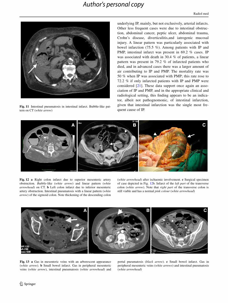
injection of bacteria in the intestinal submucosa caused pneumatosis, and in some cases pneumatosis resolved after antibiotic or hyperbaric therapy [28–32]. The secondary form of pneumatosis is much more frequent and often has a linear circumferential shape (Fig. 10), even though a bub-ble-like pattern is also observed (Fig. 11) [21, 33]. Colonic bowel wall enhancement is very difficult to analyse due to its reduced thickness; in this case IP is a capital sign in the recognition of intestinal infarct (Fig. 12). In some cases, and especially in pneumatosis cystoides intestinalis, IP may be associated with pneumoperitoneum [28] (Fig. 9).
PMP is related to presence the of a larger amount of gas in the bowel wall passing into the veins, and often indi-cates a more advanced stage of pathology (Fig. 13) [21]. Gas is more easily seen in the portal vein and its branches,
Fig. 3 a Plain abdominal X-ray. Portal pneumatosis (black arrow) and parietal gas-tric pneumatosis (white arrow-heads) in intestinal infarct. b Computed tomography (CT). Portal pneumatosis (black arrow) and parietal gastric pneumatosis (white arrow-heads) in intestinal infarct
Table 1 Causes of intestinal pneumatosis (IP) and portomesenteric pneumatosis (PMP)
(a) Increased mucosal permeability
–Pneumatosis cystoides intestinalis
–Drugs (corticosteroids, chemotherapeutic agents)
–Abdominal infections
–Neoplasms
–Solid organ and bone marrow transplantation
–Inflammatory bowel diseases
–Intra-abdominal abscess
–Infectious diseases
(b) Mucosal disruption
–Intestinal ischaemia
–Bowel obstruction
–Ulcerative disease
–Acute gastric or intestinal dilatation
–Blunt trauma
–Diverticular disease
–Ingestion of corrosives
–Intestinal carcinoma
–Iatrogenic causes
–Endoscopy
–Feeding tube (naso-gastric and naso-jejunal)
–Post-surgical anastomoses
–Drugs (corticosteroids, antineoplastic agents)
(c) Pulmonary conditions (alveolar rupture with interstitial dissection by air bubbles)
–Chronic obstructive pulmonary disease (COPD), bronchitis, emphysema
–Asthma
–Cystic fibrosis
–Positive end-expiratory pressure (PEEP)
Fig. 4 CT intestinal pneumatosis of a jejunal loop in intestinal infarct (black arrowhead). A small amount of gas located in a mesen-teric vein (white arrow) is clearly visible thanks to the proper window that allows a clear distinction of gas from fat
Author's personal copy

Radiol med
1 3
and may sometimes also be seen overimposed on the liver shadow on plain abdominal radiographs (Fig. 3). In some phases, the gas may be seen in the mesenteric veins on CT scans (Fig. 13). Intravascular gas has been rarely observed in systemic veins [34, 35]. In one case it was observed in the systemic veins (gluteal veins, haemorroidal plexus, periprostatic veins, femoral veins) associated with porto-mesenteric pneumatosis and an extensive intestinal infarct [34]. Infrequently, gas may be seen in the spleen; this has been described in an intestinal infarct [36] and in a case of traumatic rupture of the stomach associated with extensive PMP and pneumoperitoneum (Fig. 14) [23].
Clinical significance
IP and PMP have long been considered a capital sign of advanced intestinal infarction and a predictor of poor prognosis. In recent times, however, several papers have
Fig. 5 CT bubble-like intestinal pneumatosis in intestinal infarct (white arrowhead)
Fig. 6 Linear pattern of intestinal pneumatosis in intestinal infarct (white arrowhead)
Fig. 7 a Circumferential pat-tern of intestinal pneumatosis in intestinal infarct (black arrows). b Circumferential pattern of pneumatosis in the oesophagus of a patient with a massive intestinal infarct (black arrows)
Fig. 8 Bubble-like (white arrow), linear (black arrow) and circum-ferential (white arrowhead) pattern of intestinal pneumatosis simulta-neously present in intestinal infarct
Author's personal copy

Radiol med
1 3
reported these signs in many clinical conditions other than intestinal ischaemia [6–24]. Most of these papers were case reports so that the unusual findings were magnified com-pared to the more common conditions.
In 1997, Faberman and Mayo-Smith [37] analysed a series of 17 patients with portal venous gas detected by CT and they found an unexpected low mortality in these patients, passing from a previously reported overall mortal-ity of 75 % and of 85 % in non-iatrogenic cases to 29 %. The authors noted that CT has a greater capacity to recog-nise portal gas compared to plain radiographs; however, they also note the lack of correlation between mortality and the amount of portal gas. Furthermore, in their series there was no association between portal pneumatosis and intes-tinal pneumatosis. Following this study the significance
of portal pneumatosis was questioned and it appeared due to many pathological processes and no longer associated with high mortality rates. In 2001 Wiesner et al. analysed the correlation between IP or PMP, or both, the sever-ity of mural involvement, and the clinical outcome in 23 patients with bowel ischaemia concluding that these find-ings do not allow prediction of transmural bowel infarction because they may be observed with only partial ischaemic bowel wall damage; furthermore they state that both these signs are unrelated to advanced infarct also in the setting of bowel ischaemia [33].
The clinical significance of IP and portomesenteric venous gas (PVG) changed significantly over time, with the findings appearing more closely related to advanced intes-tinal infarct, and their prognostic value appearing strongly reduced, with important repercussions on clinical practice.
Wiesner’s group also noted a difference between the IP patterns, with the band-like pattern being more closely cor-related to transmural infarction than the bubble-like pattern (90 % versus 70 % of patients). They also observed that PMP was associated with transmural bowel infarction in 81 % of patients, and pronounced PMP was slightly more often associated with transmural bowel infarction (86 %) than mild PMP (78 %). Finally if both CT findings of pneu-matosis and portomesenteric venous gas were seen, their presence was associated with transmural bowel infarction in 91 % of patients, regardless of their aspect and extent [33].
To better understand the relevance of IP and PMP our group analysed a large number of patients with PMP. Out of 47 patients with PMP, 91.5 % had a surgically proven necrosis along the gastrointestinal tract, the majority had arterial infarcts, perhaps because of fluid congestion pre-venting air penetration into the bowel wall and mesenteric vessels. Other causes in our series were iatrogenic or due to trauma. Among these patients, there was a high mortality rate (68.1 %), which rose to 79.5 % if only subjects with infarct were considered [22].
In a subsequent study we examined 102 patients with IP. In this study 62 % had gastrointestinal necrosis
Fig. 9 Bubble-like pattern of intestinal pneumatosis in a patient with pneumatosis cystoides intestinalis (white arrows). Rupture of a bub-ble caused pneumoperitoneum in this patient (white arrowhead)
Fig. 10 a Intestinal pneu-matosis in intestinal infarct. Linear pattern on CT. b Intes-tinal pneumatosis in intestinal infarct. Circumferential pattern on CT (white arrows). Gas in mesenteric vein (portomesen-teric pneumatosis, PMP) (white arrowhead)
Author's personal copy
Radiol med
1 3
underlying IP, mainly, but not exclusively, arterial infarcts. Other less frequent cases were due to intestinal obstruc-tion, abdominal cancer, peptic ulcer, abdominal trauma, Crohn’s disease, diverticulitis,and iatrogenic mucosal injury. A linear pattern was particularly associated with bowel infarction (75.5 %). Among patients with IP and PMP, intestinal infarct was present in 69.2 % cases. IP was associated with death in 30.4 % of patients, a linear pattern was present in 79.2 % of infarcted patients who died, and in advanced cases there was a larger amount of air contributing to IP and PMP. The mortality rate was 50 % when IP was associated with PMP; this rate rose to 72.2 % if only infarcted patients with IP and PMP were considered [21]. These data support once again an asso-ciation of IP and PMP, and in the appropriate clinical and radiological setting, this finding appears to be an indica-tor, albeit not pathognomonic, of intestinal infarction, given that intestinal infarction was the single most fre-quent cause of IP.
Fig. 11 Intestinal pneumatosis in intestinal infarct. Bubble-like pat-tern on CT (white arrow)
Fig. 12 a Right colon infarct due to superior mesenteric artery obstruction. Bubble-like (white arrow) and linear pattern (white arrowhead) on CT. b Left colon infarct due to inferior mesenteric artery obstruction. Intestinal pneumatosis with a linear pattern (white arrow) of the sigmoid colon. Note thickening of the descending colon
(white arrowhead) after ischaemic involvement. c Surgical specimen of case depicted in Fig. 12b. Infarct of the left part of the transverse colon (white arrow). Note that right part of the transverse colon is still viable and has a normal pink colour (white arrowhead)
Fig. 13 a Gas in mesenteric veins with an arborescent appearance (white arrow). b Small bowel infarct. Gas in peripheral mesenteric veins (white arrow), intestinal pneumatosis (white arrowhead) and
portal pneumatosis (black arrow). c Small bowel infarct. Gas in peripheral mesenteric veins (white arrows) and intestinal pneumatosis (white arrowhead)
Author's personal copy
Radiol med
1 3
Pitfalls
Some conditions may mimic the radiological appearance of IP or PMP. Bubbles trapped in faeces and between mucosal intestinal folds can appear like intramural bubbles (Fig. 15). The key to distinguish true IP from trapped intraluminal air is the location of bubbles, as in IP these should not be lim-ited to the nondependent part of the bowel but also extend to the posterior side (Figs. 1, 3, 4, 5) [28]. CT examina-tion with a lung window setting helps to differentiate these
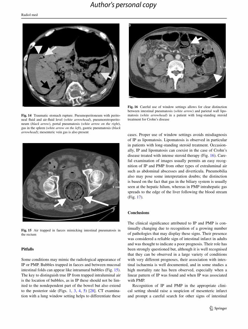
cases. Proper use of window settings avoids misdiagnosis of IP as lipomatosis. Lipomatosis is observed in particular in patients with long-standing steroid treatment. Occasion-ally, IP and lipomatosis can coexist in the case of Crohn’s disease treated with intense steroid therapy (Fig. 16). Care-ful examination of images usually permits an easy recog-nition of IP and PMP from other types of extraluminal air such as abdominal abscesses and diverticula. Pneumobilia also may pose some interpretation doubts; the distinction is based on the fact that gas in the biliary system is usually seen at the hepatic hilum, whereas in PMP intrahepatic gas spreads to the edge of the liver following the blood stream (Fig. 17).
Conclusions
The clinical significance attributed to IP and PMP is con-tinually changing due to recognition of a growing number of pathologies that may display these signs. Their presence was considered a reliable sign of intestinal infarct in adults and was thought to indicate a poor prognosis. Their role has been strongly questioned but, although it is well recognised that they can be observed in a large variety of conditions with very different prognoses, their association with intes-tinal ischaemia is well documented, and in some studies a high mortality rate has been observed, especially when a linear pattern of IP was found and when IP was associated with PMP.
Recognition of IP and PMP in the appropriate clini-cal setting should raise a suspicion of mesenteric infarct and prompt a careful search for other signs of intestinal
Fig. 14 Traumatic stomach rupture. Pneumoperitoneum with perito-neal fluid and air-fluid level (white arrowhead), pneumoretroperito-neum (black arrow), portal pneumatosis (white arrow on the right), gas in the spleen (white arrow on the left), gastric pneumatosis (black arrowhead); mesenteric vein gas is also present
Fig. 15 Air trapped in faeces mimicking intestinal pneumatosis in the rectum
Fig. 16 Careful use of window settings allows for clear distinction between intestinal pneumatosis (white arrow) and parietal wall lipo-matosis (white arrowhead) in a patient with long-standing steroid treatment for Crohn’s disease
Author's personal copy

Radiol med
1 3
involvement so as not to miss cases of intestinal infarct or allow them further evolve into wide necrosis, with worsen prognosis.
Conflict of interest The authors declare no conflict of interest.
References
1. Wolfe N, Evans WA (1995) Gas in the portal veins of the liver in infants: a roentgenographic demonstration with postmortem ana-tomical correlation. AJR Am J Roentgenol 74:486–489
2. Feczko PJ1, Mezwa DG, Farah MC, White BD (1992) Clinical significance of pneumatosis of the bowel wall. Radiographic 12:1069–1078
3. Liebman PR, Patten MT, Manny J et al (1978) Hepatic-portal venous gas in adults. Ann Surg 187:281–287
4. Taourel PG, Deneuville M, Pradel JA et al (1996) Acute mesen-teric ischemia: diagnosis with contrast enhanced CT. Radiology 199:632–636
5. Clark RA (1987) Computed tomography of bowel infarction. JCAT 11:757–762
6. Sebastià C, Quiroga S, Espin E et al (2000) Portomesenteric vein gas: pathologic mechanisms, CT findings, and prognosis. Radio-graphics 20:1213–1224
7. Baker SR (2000) Invited commentary. Radiographics 20: 1224–1226
8. Cho KC, Baker SR (1994) Extraluminal air. Diagnosis and sig-nificance. Rad Clin North Am 32:829–844
9. Kessler RM, Lentz JC, Abdnenour GC, Poole CA (1981) Mesen-teric vascular gas secondary to ischemic bowel in transmensen-teric hernia. Radiology 140:645–646
10. Brinkley AB Jr (1982) Mesenteric vascular gas secondary to ischemic bowel in transmensenteric hernia. Radiology 143:573
11. Scaglione M, Lassandro F, Pinto F et al (2001) Gastric pneuma-tosis and portal vein gas: incidental findings at helical CT after blunt abdominal trauma. Emerg Radiol 8:162–164
12. Lamberto S, Vinci S, Salamone I, Racchiusa S (2001) Intra-hepatic portal vein gas. Good prognosis with gastric dilatation. Radiol Med 101:508–510
13. Benson MD (1985) Adult survival with intrahepatic portal venous gas secondary to acute gastric dilatation, with a review of portal venous gas. Clin Radiol 36:441–443
14. Friedman D, Flancbaum L, Ritter E, Trooskin SZ (1991) Hepatic portal venous gas identified by computed tomography in a patient with blunt abdominal trauma: a case report. J Trauma 31:290–292
15. Radin DR, Rosen RS, Halls JM (1987) Acute gastric dilata-tion: a rare cause of portal venous gas. AJR Am J Roentgenol 148:279–280
16. Haswell DM, Carsky EW (1979) Hepatic portal venous gas and gastric emphysema with survival. AJR Am J Roentgenol 133:1183–1185
17. Fisher JK (1984) Computed tomography of colonic pneuma-tosis intestinalis with mesenteric and portal venous air. JCAT 8:573–574
18. Zhang D, Weltman D, Baykal A (1999) Portal vein gas and colonic pneumatosis after enema, with spontaneous resolution. AJR Am J Roentgenol 173:1140–1141
19. Kingsley DD, Albrecht RM, Vogt DM (2000) Gastric pneumato-sis and hepatoportal venous gas in blunt trauma: clinical signifi-cance in a case report. J Trauma 49:951–953
20. Torriero F, Macori F, Misiti A et al (1998) A case of gas occur-rence in the venous hepato-portal area after blunt abdominal trauma of the abdomen. Radiol Med 96:118–120
21. Lassandro F, Mangoni di Santo Stefano ML, Maria Porto A et al. Intestinal pneumatosis in adults: diagnostic and prognostic value. Emerg Radiol. 2010 Apr 15 [Epub ahead of print]
22. Lassandro F, Scaglione M, Rossi G et al (2002) Portomesen-teric vein gas: diagnostic and prognostic value. Emerg Radiol 9:96–99
23. Lassandro F, Romano S, Rossi G (2006) Gastric traumatic inju-ries: CT findings. Eur J Radiol 59:349–354
24. Makiyama H, Kataoka R, Tauchi M et al (2014) Do alpha-glu-cosidase inhibitors have the potential to induce portal venous gas? Two clinical case reports. Intern Med 53:691–694
25. Flor N, Pricolo P, Rovere A et al (2013) J Med Case Rep 7:205 26. Shinagare AB, Howard SA, Krajewski KM et al (2012) Pneuma-
tosis intestinalis and bowel perforation associated with molecular targeted therapy: an emerging problem and the role of radiolo-gists in its management. AJR Am J Roentgenol 199:1259–1265
27. Pear BL (1998) Pneumatosis intestinalis: a review. Radiology 207:13–19
28. Soyer P, Martin-Grivaud S, Boudiaf M et al (2008) Linear or bub-bly: a pictorial review of CT features of intestinal pneumatosis in adults. J Radiol 89:1907–1920
29. Moschetta M, Telegrafo M, Rella L (2014) Multi-detector CT features of acute intestinal ischemia and their prognostic correla-tions. World J Radiol 6:130–138
Fig. 17 a Ultrasonography. Intrahepatic gas due to pneumobilia (white arrow). Although the gas spreads peripherally in this case it is possible to see the associated portal branch (white arrowhead). b Plain radiograph of the abdomen. Same case as a. Intrahepatic gas
(black arrow). In this case portomesenteric pneumatosis (PMP) can-not be distinguished from pneumobilia. c CT intrahepatic gas due to pneumobilia (black arrow). Note the associated portal branch (black arrowhead)
Author's personal copy

Radiol med
1 3
30. Koss LG (1952) Abdominal gas cysts (pneumatosis cystoides intestinorum hominis): an analysis with a report of a case and a critical review of the literature. AMA Arch Pathol 53:523–549
31. Mao YC1, Lin TC, Wang JD (2009) Education and imaging. Gas-trointestinal: benign cause for pneumatosis intestinalis and por-tomesenteric venous gas. J Gastroenterol Hepatol 24:927
32. Miralbés M, Hinojosa J, Alonso J, Berenguer (1983) Oxy-gen therapy in pneumatosis coli. What is the minimum oxygen requirement? J Dis Colon Rectum 26:458–460
33. Wiesner W, Mortelé KJ, Glickman JN (2001) Pneumatosis intes-tinalis and portomesenteric venous gas in intestinal ischemia: correlation of CT findings with severity of ischemia and clinical outcome. AJR Am J Roentgenol 177:1319–1323
34. Rossi G, Pinto A, Pinto F et al (2000) Unreported CT findings in entire colon infarction: gas in the inferior mesenteric vein and in the parietal and visceral branches of the internal iliac veins. Radiol Med 99:107–109
35. Kriegshauer JS, Reading CC, King BF, Welch TJ (1990) Com-bined systemic and portal venous gas: sonographic and CT detec-tion in two cases. AJR Am J Roentgenol 154:1219–1221
36. Frola C, Cantoni S, Turtulici I, Loria F (1994) Case report: bowel infarction with splenic air embolism: computed tomography find-ings. Br J Radiol 67:1272–1274
37. Faberman RS, Mayo-Smith WW (1997) Outcome of 17 patients with portal venous gas detected by CT. AJR Am J Roentgenol 169:1535–1538
Author's personal copy