Sphingosine Forms Channels in Membranes That Differ Greatly from Those Formed by Ceramide
Upload
independentCategory
view
2download
0
Changes in mitochondrial dynamics duringceramide-induced cardiomyocyte early apoptosis
Valentina Parra1,2, Veronica Eisner1,3, Mario Chiong1,2, Alfredo Criollo1,2, Francisco Moraga1,2,Alejandra Garcia3, Steffen Hartel3, Enrique Jaimovich1,3, Antonio Zorzano4, Cecilia Hidalgo1,3, andSergio Lavandero1,2,3*
1Centro FONDAP Estudios Moleculares de la Celula, Universidad de Chile, Olivos 1007, Santiago 8380492, Chile; 2Facultad deCiencias Quımicas y Farmaceuticas, Universidad de Chile, Santiago, Chile; 3Instituto de Ciencias Biomedicas, Facultad deMedicina, Universidad de Chile, Santiago, Chile; and 4Institute for Research in Biomedicine (IRB) and Departament deBioquımica y Biologia Molecular, Facultat de Biologia, Universitat de Barcelona, Barcelona, Spain
Received 1 May 2007; revised 26 September 2007; accepted 27 September 2007; online publish-ahead-of-print 4 October 2007
Time for primary review: 19 days
Aims In cells, mitochondria are organized as a network of interconnected organelles that fluctuatebetween fission and fusion events (mitochondrial dynamics). This process is associated with celldeath. We investigated whether activation of apoptosis with ceramides affects mitochondrial dynamicsand promotes mitochondrial fission in cardiomyocytes.Methods and results Neonatal rat cardiomyocytes were incubated with C2-ceramide or the inactiveanalog dihydro-C2-ceramide for up to 6 h. Three-dimensional images of cells loaded with mitotrackergreen were obtained by confocal microscopy. Dynamin-related protein-1 (Drp-1) and mitochondrialfission protein 1 (Fis1) distribution and levels were studied by immunofluorescence and western blot.Mitochondrial membrane potential (DCm) and cytochrome c (cyt c) distribution were used as indexesof early activation of apoptosis. Cell viability and DNA fragmentation were determined by propidiumiodide staining/flow cytometry, whereas cytotoxicity was evaluated by lactic dehydrogenase activity.To decrease the levels of the mitochondrial fusion protein mitofusin 2, we used an antisense adenovirus(AsMfn2). C2-ceramide, but not dihydro-C2-ceramide, promoted rapid fragmentation of the mitochon-drial network in a concentration- and time-dependent manner. C2-ceramide also increased mitochon-drial Drp-1 and Fis1 content, Drp-1 colocalization with Fis1, and caused early activation of apoptosis.AsMfn2 accentuated the decrease in DCm and cyt c redistribution induced by C2-ceramide. Doxorubicin,which induces cardiomyopathy and apoptosis through ceramide generation, also stimulated mitochon-drial fragmentation.Conclusion Ceramides stimulate mitochondrial fission and this event is associated with early activationof cardiomyocyte apoptosis.
KEYWORDSCeramide;
Mitochondrial fission;
Mitochondrial dynamics;
Drp-1;
Fis1;
Mitofusin 2;
Doxorubicin;
Cell death;
Apoptosis;
Cardiomyocytes
1. Introduction
Mitochondria are complex interconnected organelles thatdisplay particular morphology and distribution in differentcell types.1,2 In cardiomyocytes, mitochondrial activity hasa key role in energy generation through oxidative phos-phorylation and in the regulation of calcium homeostasis,contraction, production of reactive oxygen species, andcell death.3,4
The mitochondrial network exists in a continuous balancebetween local fission and fusion events.5,6 Mitochondrialfission entails fragmentation of tubular interconnected mito-chondria into several smaller individual organelles. The outer
mitochondrial membrane protein fission 1 (Fis1) and theGTPase dynamin-related protein-1 (Drp-1) are the mainelements of the mitochondrial fission machinery.7,8 Whenthe fission process occurs, cytosolic Drp-1 is recruited intothe mitochondrial fission foci where it interacts with Fis1.9
In contrast, mitochondrial fusion promotes the assembly ofindividual mitochondria that combine their membranes.This process is also controlled by GTPases, including themito-fusins (Mfns) 1 and 2 and dynamin-related protein OPA1.2,10
Although Drp-1, Fis1, and Mfn2 are all highly expressed inwhole cardiac tissue,11–13 how and when mitochondrialfission events take place in cardiomyocytes remain unknown.Likewise, there are no reports concerning the mechanism ofmitochondrial fission in cardiomyocytes undergoing loss ofmitochondrial network integrity. Conversely, interconnectedmitochondria facilitate energy delivery from the cell periphery* Corresponding author. Tel: þ34 562 9782919.; fax: þ34 562 7378920.
E-mail address: [email protected]
Published on behalf of the European Society of Cardiology. All rights reserved. & The Author 2007.For permissions please email: [email protected]
Cardiovascular Research (2008) 77, 387–397doi:10.1093/cvr/cvm029
by guest on June 27, 2014http://cardiovascres.oxfordjournals.org/
Dow
nloaded from

to the cell core and organize protonic potential to formATP.14 Moreover, Benard et al.15 have recently proposedthat there is a bidirectional relationship between mitochon-drial network organization and bioenergetics. In skeletalmuscle cells, Mfn2 repression causes fragmentation of themitochondrial network, reduces mitochondrial metabolicresponse, and inhibits expression of oxidative phosphoryl-ation enzymes.13,16
Mitochondria have a central role in apoptotic cell deathproduced by multiple conditions.17 Activation of the mito-chondrial fission machinery and further mitochondrialfragmentation have been associated with apoptosis, albeitthis point remains contentious.6,18,19 Under some specificconditions, Drp-1 interacts with Bax in mitochondrial scis-sion loci, and mitochondrial fragmentation is a prerequisitefor mitochondrial outer membrane permeabilization (MOMP)and cytochrome c (cyt c) release.19 In contrast, mitochon-drial fragmentation in other models is independent of theoccurrence of apoptosis.18,20
In HeLa cells, ceramide induces mitochondrial networkfragmentation and calcium release from the endoplasmicreticulum.21 Moreover, through Drp-1 over expressionSzabadkai et al.22 demonstrated that mitochondrialnetwork integrity is necessary for C2-ceramide-induceddeath. Ceramides, produced either by de novo synthesis orby the action of acid or neutral sphingomyelinases, interactwith different organelles, especially with mitochondria, andboth exogenous and endogenous ceramides can inducecell arrest and death.23 C2-ceramide alters mitochondrialmembrane potential (DCm) and promotes cyt c releaseand channel formation in the mitochondrial outer membranewhereas dihydro-C2-ceramide (DH-C2-ceramide) is inac-tive.23–25 In cardiomyocytes, ceramides mediate bothischaemia–reperfusion and TNFa-induced cell death.26,27
C2-ceramide triggers apoptosis in cultured cardiomyocytes,stimulating DCm decrease through p38-MAPK and the acti-vation of caspases 8 and 3.28,29 In addition, ischaemicpreconditioning prevents ceramide accumulation.30,31 Yet,C2-ceramide also has positive ionotropic effects andenhances contraction in adult cardiomyocytes.32
Given that (a) mitochondrial dynamics are associated withmetabolism and cell death and (b) ceramides change mito-chondrial homeostasis and trigger apoptosis, and consideringthat mitochondrial dynamics in cardiomyocytes has receivedlittle attention until now, we investigated whether cera-mides alter mitochondrial dynamics and through theseeffects promote cell death in cultured rat neonatal cardio-myocytes. To this aim, we analysed the effects of ceramideson mitochondrial network, DCm, the expression of the mito-chondrial fission proteins Fis1 and Drp-1, and cell death. Inaddition, we studied whether decreasing mitochondrial con-nectivity (by a decrease of Mfn2 expression) enhanced theeffects of C2-ceramide.
We show here for the first time that C2-ceramide pro-motes fragmentation of the mitochondrial network incultured neonatal cardiomyocytes. This fragmentation cor-related with mitochondrial fission. C2-ceramide also pro-moted rapid DCm decrease and cyt c release. DecreasingMfn2 expression accentuates the effects of C2-ceramide onmitochondrial fragmentation and in the DCm decrease.Collectively, our data show that C2-ceramide stimulatesmitochondrial fission events linked to an early activationof apoptosis.
2. Methods
2.1 Materials
Antibodies against Drp-1 and cyt c were purchased from BDBiosciences. Fis1 antibody was from Alexis Biochemicalsand mt-Hsp70 antibody was from Affinity BioReagents.Anti-b-myosin heavy chain antibody was from NovocastraLabs. Tetramethylrhodamine methyl ester (TMRM) and mito-tracker green were from Molecular Probes. FBS was fromInvitrogen. TRITC conjugated anti-IgG mouse polyclonalantibody, FITC conjugated anti-IgG rabbit, C2-ceramide,DH-C2-ceramide, anti-b-actin antibody, propidium iodide(PI), carbonyl cyanide m-chlorophenylhydrazone (CCCP),Dulbecco’s modified Eagle’s medium (DMEM), M199medium, doxorubicin, and other reagents were purchasedfrom Sigma-Aldrich Corp. Protein assay reagents were fromBio-Rad. The generation and use of AsMfn2, an adenovirusexpressing Mfn2 antisense mRNA, was previously described.13
Cardiomyocytes were transduced with adenoviral vectors at amultiplicity of infection (MOI) of 1000, 48 h before ceramidetreatment. An empty adenovirus was used as control (mock).
2.2 Culture of cardiomyocytes
Cardiomyocytes were isolated from hearts of neonatalSprague–Dawley rats as described previously.33 Rats werebred in the Animal Breeding Facility of the University ofChile. All studies conform to the Guide for the Care andUse of Laboratory Animals published by the US NationalInstitutes of Health (NIH Publication No. 85-23, revised1996) and it was approved by our Institutional EthicsReview Committee. Cardiomyocytes were plated at a finaldensity of 1–8 � 103/mm2 on gelatin-coated 35-, 60- or100 mm Petri dishes. For fluorescence measurements, cellswere plated on gelatin-precoated 25 mm glass coverslips in35 mm Petri dishes. Our initial studies showed that serumdeprivation stimulated fragmentation of the mitochondrialnetwork (data not shown). Accordingly, primary cell cultureswere incubated with or without C2-ceramide or DH-C2-ceramide (0–40 mM) for 0–6 h or Doxo (1 mM for 24 h) inDMEM/M199 (4:1) medium containing 10% FBS. Cultured car-diomyocytes were identified using an anti-b-myosin heavychain antibody as previously described23 and cell cultureswere at least 95% pure.
2.3 Subcellular fractionation
Mitochondrial and cytosolic fractions were obtained bydifferential centrifugation of cardiomyocyte homogenates.Cells were scraped, pelleted, and re-suspended in ice-coldbuffer containing 250 mM sucrose, 1 mM EGTA, and 10 mMHepes, pH 7.4; and the protease inhibitors PMSF, leupeptin,pepstatin A, and aprotinin. Cells were homogenized using ahomogenizer with a tight fitting Teflon pestle. The homogen-ates were centrifuged (750 g, 10 min) to remove nucleiand unbroken cells, and supernatants were centrifuged(10 000 g, 25 min) to obtain a pellet highly enriched in mito-chondria. The protein content was determined by Bradford’smethod.34 The purity of mitochondrial fraction, assessed bymt-Hsp70 levels,35 was 85%.
V. Parra et al.388
by guest on June 27, 2014http://cardiovascres.oxfordjournals.org/
Dow
nloaded from

2.4 Western blot analysis
Equal amounts of protein were separated by SDS–PAGE (12%polyacrylamide gels) and electrotransfered to nitrocellu-lose. Membranes were blocked with 5% milk in Tris-bufferedsaline, pH 7.6, containing 0.1% (v/v) Tween 20 (TBST). Mem-branes were incubated with primary antibodies at 48C andre-blotted with horseradish peroxidase-linked secondaryantibody [1:5000 in 1% (w/v) milk in TBST]. The bandswere detected using ECL with exposure to Kodak film andquantified by scanning densitometry. Protein contentswere normalized by b-actin and mt-Hsp70 levels.
2.5 Mitochondrial dynamics analysis
Cells were preincubated with mitotracker green FM (400 nM)andmaintained in Krebs solution. Confocal image stacks werecaptured with a Zeiss LSM-5, Pascal 5 Axiovert 200 micro-scope, using LSM 5 3.2 image capture and analysis softwareand a Plan-Apochromat 63x/1.4 Oil DIC objective. Imageswere deconvolved with Image J (NIH), Z-stacks analysis ofthe thresholded images were volume-reconstituted usingthe VolumeJ plug-in, and changes in number (objects) andvolume of individual mitochondria were quantified using theImageJ-3D Object counter plug-in. Each experiment wasdone at least four times and 16–25 cells per condition werequantified. Within cells, two to three regions of interest(ROI) of equal area were defined, and mitochondria countsand volume were measured for each ROI. The ROI samplingcriteria considered only cytoplasmic mitochondria, excludingcondensed perinuclearmitochondria (Supplementarymaterialonline, Figure A). Fragmentation criteria were: mitochondriaindividual volume decrease and increase in number of mito-chondria.22,36 Percentage of cells with a fragmented patternwas also determined.36
2.6 Immunofluorescence studies for Drp-1 andFis1 and colocalization analysis
Cells grown on coverslips were fixed with PBS containing 4%paraformaldehyde and incubated in ice-cold 0.3% TritonX-100 for permeabilization. Nonspecific sites were blockedwith 5% BSA in PBS for 1 h. Cells were then incubated withDrp-1, Fis1 or mt-Hsp70 antibodies (1:1000). Nuclei werestained with 5 mg/mL Hoechst 33342 (Invitrogen). Secondaryantibodies were anti-mouse IgG-TRITC and anti-rabbitIgG-FITC (1:1000). For details in image acquisition andcolocalization analysis, Supplementary material online,Materials and methods.
2.7 Cell death assays
Cells were incubated with either C2-ceramide or DH-C2-ceramide at the concentrations indicated for up to 6 h at378C. DNA fragmentation was used as a marker of apoptosis.Cells were collected and permeabilized with methanol for24 h. Later permeabilized cells were treated with RNAasefor 2 h and2 mL PI (25 mg/mL)was addedprior to flowcytome-try analysis.37 For determination of cell viability, cells wereharvested and stained with PI (10 mg/mL).38 Samples wereanalysed by a FACS Scan and data were evaluated using CellQuest software (Becton Dickinson). Cytotoxicity was alsoquantified by lactate dehydrogenase (LDH) activity in cellsupernatant using the LDH kit from Sigma following the man-ufacturer’s instructions. To determine DCm dissipation, cells
were loaded with 100 nM TMRM for 20 min at 378C.39 Fluor-escence imaging of cells was conducted in a confocal micro-scope (excitation 543 nm, emission 560 nm). Cyt c release incardiomyocytes undergoing ceramide-induced apoptosis wasdetermined as previously described.38
2.8 Statistical analysis
Data shown are given as mean+ SEM of the number of inde-pendent experiments indicated (n) and represent exper-iments performed on at least three separate occasionswith similar outcomes. Data were analysed by ANOVA andcomparisons between groups were performed using a pro-tected Tukey’s test. Statistical significance was defined asP , 0.05.
3. Results
3.1 Ceramides stimulate mitochondrialfragmentation in cultured cardiomyocytes
As shown in Figure 1A, cardiomyocytes contain two proteins(80 and 17 kDa immunoreactive bands) corresponding toDrp-1 and Fis1, which are fully coincident with the proteinbands found in HeLa total cell extracts.8,22 Images of thesubcellular distributions of Drp-1 and Fis1 in cardiomyo-cytes, illustrated in Figure 1B, show that Drp-1 waspresent throughout the cytoplasm but presented particularpunctuate accumulations that partially overlapped withmt-Hsp70, used as a mitochondrial marker.35 In contrast,Fis1 was mostly distributed in the mitochondrial networkand presented a similar ‘spaghetti-like’ distribution as thatdescribed in HeLa cells.40 Neither Drp-1 nor Fis1 weredetected in the nucleus.
Figure 2A shows that C2-ceramide, but not the inactiveanalogue DH-C2-ceramide, gradually converted the mito-chondrial tubular shape to a spherical conformation in atime-dependent manner. Exposure of cultured cardiomyo-cytes to 20, 30 and 40 mM C2-ceramide for 6 h resulted in sig-nificant increases in the percentage of cells that displayedfragmented mitochondria, from control values (in %) of11+7 to 46+4, 65+2, and 80+2, respectively(Figure 2B). To assess mitochondrial network integrity, wedetermined after 3D reconstitution both the average indi-vidual volume of each object (mitochondria) and thenumber of mitochondria per cell.22,36 Figure 2C indicatesthat C2-ceramide gradually decreased mitochondrialvolume in a time- and concentration-dependent manner.Individual mitochondrial volume decreased 20, 40 and60% after 2, 4 and 6 h, respectively, of incubation withC2-ceramide. Figure 2D shows that the number of mitochon-dria per cell increased significantly from 150+20 (control)to 280+20 in cells incubated with 40 mM C2-ceramidefor 6 h. Incubation with 40 mM DH-C2-ceramide for 6 h,however, did not modify any of the three parametersdescribed above (Figure 2B–D). On the basis of theseresults, we used 40 mM C2-ceramide in all subsequentexperiments.
3.2 Ceramides increase the mitochondrialcontents of Drp-1 and Fis1
According to some authors,9 the initial step in mitochon-drial fission is migration of Drp-1 to Fis1-containing fission
Ceramides regulate cardiomyocyte mitochondrial dynamics 389
by guest on June 27, 2014http://cardiovascres.oxfordjournals.org/
Dow
nloaded from

points. Thus, we evaluated next whether mitochondrialfragmentation triggered by C2-ceramide was associatedwith changes in the distribution of Drp-1 and Fis1.Western blot analysis revealed that during the timeperiod studied, Drp-1 levels did not change in total cellextracts from cardiomyocytes incubated with C2-ceramide,whereas Fis1 content increased after 6 h of incubation with40 mM C2-ceramide (Figure 3A). Subsequent subcellularfractionation of C2-ceramide-incubated cardiomyocytesinto mitochondria revealed that Drp-1 and Fis1 levelsincreased transiently in the mitochondrial fraction after2 h of incubation with C2-ceramide and decreased atlonger times to control levels (Figure 3B). These effectswere specific to C2-ceramide because DH-C2-ceramide didnot modify Drp-1 and Fis1 total protein levels (Figure 3A)or their levels in the mitochondrial fraction (Figure 3B).Using immunofluorescence and confocal microscopy as acomplementary approach revealed that Drp-1 displayed apunctuated distribution pattern, which was accentuatedafter 2 h of incubation with C2-ceramide (Figure 3C). Atthe same time, an increase in Fis1 content was observedmainly in mitochondria. The extent of colocalizationof both proteins increased two-fold in response toC2-ceramide (P , 0.05) (Figure 3C). We next quantifiedFis1 fluorescence levels and changes in Drp-1 distribution.After stimulation with C2-ceramide, the Fis1 coupled inten-sities increased �1.75-fold relative to the Drp-1 coupledfluorescence (P , 0.001) (Figure 3D). C2-ceramide alsoincreased the effective colocalization of Drp-1 with Fis1(P , 0.001) but not the effective colocalization ofFis1 with Drp-1 (Figure 3E). The increased size of theFis1 related ROIs (green) 2 h after C2-ceramide additionreflects the increased fluorescence intensities of thegreen channel. Only a very small fraction (�5%) of theFis1-labelled structures colocalized with Drp-1-labelledstructures, independent on the stimuli or size of the seg-mented area. In contrast, Drp-1-labelled structures (redROIs) increased significantly their effective colocalization
with Fis1-labelled structures and independent of theincreasing size of the Fis1 related ROIs.
3.3 Down-regulation of Mfn2 levels alters themitochondrial network but does not change theeffects of ceramide
Mfns are key players in themitochondrial fusion process. Mfn2is particularly expressed in the heart.13 As previously shown,down-regulation of Mfn2 causes a reduction in mitochondrialfusion; as a result, net mitochondrial fragmentation isdetected.10 In order to determine whether alterations inmitochondrial fusionmodify the effects of ceramides onmito-chondrial fragmentation, we studied whether a decrease inMfn2 levels altered the cellular response to ceramides.13
Overexpression of AsMfn2 produced a 75% decrease in Mfn2protein levels in cultured cardiomyocytes (Figure 4A). Thisreduction in Mfn2 levels prompted mitochondrial fragmenta-tion in cardiomyocytes and decreased the mitochondrialaverage volume by 25% (P , 0.05). The cardiomyocytes trans-duced with AsMfn2 displayed a further decrease in mitochon-drial volume and an increase in the number of cells withfragmented mitochondria following the incubation withC2-ceramide (Figure 4B). The changes in mitochondrialvolume and in the number of cells with fragmentedmitochon-dria induced by AsMfn2 and C2-ceramide were not synergisticbut additive (Figure 4B). At every time studied, C2-ceramidetriggered equivalent mitochondrial volume reductions inmock and AsMfn2-transduced cardiomyocytes (Figure 4B).Yet, Drp-1 and Fis1 showed higher colocalization in AsMfn2-containing cardiomyocytes after 2 h of incubation withC2-ceramide (Figure 4C).
Taken together these results suggest that addition ofC2-ceramide to cardiomyocytes undergoing Mfn2 down-regulation induced similar mitochondrial fragmentationthan in controls, which was associated with an increase ofmitochondrial Drp-1 and Fis1. Both C2-ceramide and Mfn2down-regulation showed an additive rather than a synergic
Figure 1 Detection of Drp-1 and Fis1 in cultured cardiomyocytes and HeLa cells. (A) Protein levels were determined by western blot (n ¼ 4). (B) The subcellulardistributions of Drp-1, Fis1, and mt-Hsp70 were studied by confocal microscopy. Nuclei were stained with Hoechst (blue). Images are representative of threeindependent determinations. The scale bar is 10 mm.
V. Parra et al.390
by guest on June 27, 2014http://cardiovascres.oxfordjournals.org/
Dow
nloaded from

effect on mitochondrial fission. These results furtherstrengthen the view that ceramides largely affect mitochon-drial fission, not fusion.
3.4 Mitochondrial fusion regulates mitochondrialouter membrane permeabilization and apoptosistriggered by ceramide
Figure 5A shows that transduction of cardiomyocytes withAsMfn2 but not with Mock increased the rate of Dcm
decrease induced by C2-ceramide (Figure 5A). A similarresult in cyt c release from mitochondria was observedafter C2-ceramide incubation (Figure 5B). AsMfn2 itself trig-gered cyt c redistribution (Figure 5B). C2-ceramide (40 mMfor 6 h) or DH-C2-ceramide did not change cell viability inAsMfn2 or mock transduced cardiomyocytes (Figure 5C). Asillustrated in Figure 5D–E, DNA fragmentation and LDHactivity levels did not change with C2-ceramide. Thesedata suggest that ceramides did not stimulate cell deathby necrosis or the final execution of apoptosis until up 6 h.
Figure 2 Time–course effect of C2-ceramide on mitochondrial fragmentation in cultured cardiomyocytes. (A) Cells were incubated with C2-ceramide (40 mM) atindicated times or with dihydro-C2-ceramide (DH-C2, 40 mM) for 6 h and then loaded with mitotracker green. Multi-slice imaging reconstitutions were obtained byconfocal microscopy to show mitochondrial morphology. The scale bar is 10 mm. (B) The percentage of cells with fragmented mitochondria, (C) the individualmitochondrial volume, and (D) the number of mitochondria per cell were determined in cells incubated without (white bars) or with C2-ceramide (grey bar:20 mM for 6 h; dark grey bar: 30 mM for 6 h; black bars: 40 mM for 2–6 h) or DH-C2 (black bars: 40 mM for 6 h). Values are given as mean+ SEM (n ¼ 4),*P , 0.05, **P , 0.01 and ***P , 0.001 vs. time 0 h.
Ceramides regulate cardiomyocyte mitochondrial dynamics 391
by guest on June 27, 2014http://cardiovascres.oxfordjournals.org/
Dow
nloaded from

Conversely, these results collectively indicate that mito-chondrial fission modulates MOMP and early apoptosis trig-gered by C2-ceramide in cardiomyocytes.
3.5 Doxorubicin also stimulates mitochondrialfragmentation in cardiomyocytes
Doxo is an antineoplastic agent widely used in cancer che-motherapy in spite of the fact that it causes cardiomyopathy.
Doxo induces cell death through activation of the intrinsicapoptotic pathway.41–44 The work of Delpy et al.45 demon-strated that Doxo induces cardiomyocyte apoptosis throughceramide generation. Accordingly, given our present resultswe investigated whether Doxo stimulates mitochondrial frag-mentation in cardiomyocytes. Figure 6A shows that Doxochanged mitochondrial morphology from a tubular shape toa spherical conformation, resulting in significant increase in
Figure 3 C2-ceramide changes the subcellular distribution of Drp-1 and Fis1 in cultured cardiomyocytes. (A) Total extracts and (B) mitochondrial fraction wereprepared from cells incubated with C2-ceramide (40 mM) for the indicated times. Drp-1 and Fis1 levels were determined by western blot. Representative westernblots are shown. Protein contents were normalized using anti-b-actin and anti-mt-Hsp70 antibodies for total and mitochondrial fractions, respectively. Densito-metric analysis for Drp-1 and Fis1 normalized levels are shown in left and right panels for total and mitochondrial fractions, respectively. Data correspond tomean+ SEM, n ¼ 5, *P , 0.05 and **P , 0.01 vs. 0 h. (C) Control cells or cells incubated with C2-ceramide (40 mM) for 2 h were stained for Drp-1 (red) orFis1 (green) to determine colocalization. The mean level of Drp-1 and Fis1 colocalization was determined in 20 cells (n ¼ 3). The scale bar is 10 mm, *P ,
0.05 vs. 0 h. (D) Fis1 coupled fluorescence intensity evaluation. Fis1 and Drp-1 coupled fluorescence intensities were determined inside the segmented contoursof cultured cardiomyocytes (n ¼ 12 for control and n ¼ 7 for C2-ceramide-incubated cells; see representative segmentations on grey contours in the insert). Datarepresent the mean values +SEM of the Drp-1 normalized Fis1 coupled intensities (grey bars) (***P , 0.001). The scale bar is 15 mm. (E) Effective colocalization ofDrp-1 with Fis1. C2-ceramide also increased the effective colocalization of Drp-1 with Fis1 (black bars, ***P , 0.001) but not the effective colocalization of Fis1with Drp-1 (grey bars). M1 and M2: Manders colocalization coefficients for Fis1 and Drp-1, respectively. Mean values +SEM are plotted for n ¼ 12 (control) andn ¼ 7 (C2-ceramide). For effective colocalization definition and quantification details, Supplementary material online.
V. Parra et al.392
by guest on June 27, 2014http://cardiovascres.oxfordjournals.org/
Dow
nloaded from
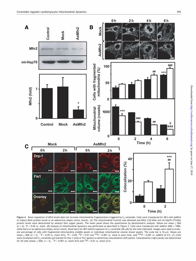
Figure 4 Down-regulation of Mfn2 levels does not increase mitochondrial fragmentation triggered by C2-ceramide. Cells were transduced for 48 h with AsMfn2to reduce Mfn2 protein levels or an adenovirus empty vector (mock). (A) The mitochondrial fraction was obtained and Mfn2 (122 kDa) and mt-Hsp70 (75 kDa)protein levels were determined by western blot (upper panel). The lower panel shows the quantitation by densitometric analysis. Values are mean+ SEM(n ¼ 4), *P , 0.05 vs. mock. (B) Analysis of mitochondrial dynamics was performed as described in Figure 2. Cells were transduced with AsMfn2 (MOI ¼ 1000,white bars) or an adenovirus empty vector (mock, black bars) for 48 h before exposure to C2-ceramide (40 mM) for the time indicated. Images were used to evalu-ate percentage of cells with fragmented mitochondria (middle panel) or individual mitochondrial volume (lower panel). The scale bar is 10 mm. Values aremean+ SEM (n ¼ 3), ***P , 0.05 vs. mock (0 h), #P , 0.05, ##P , 0.01 and ###P , 0.001 vs. mock in each time, and &&&P , 0.001 vs. AsMfn2 at 0 h. (C) Cellswere incubated with C2-ceramide and stained for Drp-1 (red) or Fis1 (green) to determine colocalization (left panel). Colocalization (right panel) was determinedfor 20 cells (mean+ SEM, n ¼ 3), ***P , 0.001 vs. mock (0 h) and ##P , 0.01 vs. mock (2 h).
Ceramides regulate cardiomyocyte mitochondrial dynamics 393
by guest on June 27, 2014http://cardiovascres.oxfordjournals.org/
Dow
nloaded from

the percentage of cells with fragmented mitochondriarespect to control (Figure 6B) and decreased individual mito-chondrial volume (Figure 6C). Both Doxo and Mfn2 down-regulation also showed an additive effect on mitochondrialfission (Supplementary material online, Figure B).
4. Discussion
In cardiomyocytes, mitochondrial dynamics is emerging as afundamental cell biological process, important not only forcontrol of the shape but also the function of mitochondria,
Figure 5 C2-ceramide stimulates early activation of apoptosis in cultured cardiomyocytes, which is accentuated by down-regulation of Mfn2 levels. Cells weretransduced for 48 h with an adenovirus empty vector (mock, black bars) or with an adenovirus antisense against mitofusin-2 (AsMfn2, MOI ¼ 1000, white bars) andincubated with C2-ceramide (40 mM) at indicated times, or DH-C2 (40 mM) for 6 h. (A) The mitochondrial membrane potential (Dcm) was determined in cells pre-loaded with TMRM and incubated for the indicated times with C2-ceramide (40 mM) or DH-C2-ceramide (40 mM), added at the first arrow. The maximum Dcm
decrease was obtained following the addition of CCCP (10 mM). (B) Cyt c redistribution was evaluated by immunofluorescence. Images obtained by confocalmicroscopy are shown in upper panel. The percentage of cells with redistributed cyt c are shown in bottom panel. (C) Cell cytotoxicity was quantified usingflow cytometry by incorporation of propidium iodide and (D) DNA fragmentation (in methanol-permeabilized cells) and (E) LDH activity in cell supernatantswere determined as described in Section 2. Values are mean+ SEM (n ¼ 3), ***P , 0.001 vs. mock 0 h and ###P , 0.001 vs. respective mock.
V. Parra et al.394
by guest on June 27, 2014http://cardiovascres.oxfordjournals.org/
Dow
nloaded from

which have a central role in determining the life or death ofcells. Our results strongly suggest that C2-ceramide regu-lates mitochondrial dynamics through the stimulation ofmitochondrial fission, among other events involved in theearly activation of cardiomyocyte apoptosis. This proposalis based on the following evidences: (a) C2-ceramide, butnot the inactive analog DH-C2-ceramide, gradually modifiedmitochondrial tubular shape to a spherical conformation in atime- and concentration-dependent manner, and decreasedindividual mitochondrial volume while increasing thenumber of mitochondria per cell. (b) The mitochondrialfission proteins Drp-1 and Fis1 were detected in cardiomyo-cytes. Total Drp-1 levels did not change following incubationwith C2-ceramide whereas total Fis1 levels only increasedafter 6 h; after 2 h, however, C2-ceramide induced Drp-1migration to mitochondria where it colocalized with Fis1.(c) C2-ceramide triggered rapid activation of apoptosis, evi-denced by DCm dissipation and redistribution of cyt c,although without DNA fragmentation (the final step of apop-tosis), necrosis (evaluated by LDH release) and cell viability.(d) Mfn2 down-regulation caused DCm decay and cyt c redis-tribution, but did not increase C2-ceramide-dependentmitochondrial fragmentation. (e) Doxo, an anticancer drugwith cardiotoxic properties linked to ceramide generationand activation of the apoptotic intrinsic pathway, alsostimulated mitochondrial fragmentation in culturedcardiomyocytes.
4.1 Ceramide-induced mitochondrial fission
Most of the current knowledge concerning mitochondrialfission comes from tumour cell lines and little informationexists in normal cells.46 In fact, there are few studies onmitochondrial fission/fusion in cardiac cells. One exception
is the work of Terman et al.47 who investigated thisprocess in senescent cardiomyocytes, reporting an accumu-lation of big and defective mitochondria with age. In thepresent work, we evaluated the induction of mitochondrialfission in cultured cardiomyocytes incubated with ceramide,a condition that stimulates cell death.26–29 Previous studiesshowed that Drp-1 and Fis1 are expressed in the wholeheart,11,12 yet their expression in specific cardiac celltypes remained unknown. We show here that both proteinsare present in cardiomyocytes with a subcellular localizationcoincident with other reports.7,8 Our data also revealed anincrease in Drp-1 and Fis1 levels in mitochondrial fractionsobtained from C2-ceramide-treated cells, which occurredprior to mitochondrial fragmentation.9 These findingsagree with a previous study reporting that C2-ceramidealso stimulated mitochondrial network fragmentation,linked to endoplasmic reticulum calcium release and HeLacell death.21 Likewise, Brady et al.48 showed that mitochon-dria undergo extensive fragmentation during simulatedischaemia in the HL-1 cardiac cell line, albeit the natureof the proteins involved in mitochondrial fragmentationwas not reported. Interestingly, our results indicate thatthe decrease in Mfn2 levels induced by AsMfn2 or incubationwith Doxo also triggered mitochondrial fission in culturedcardiomyocytes. Collectively, these results indicate thatcardiomyocyte mitochondrial dynamics can be regulatedwith different stimuli, including ceramide, Doxo and bythe manipulation of the balance between fission/fusionproteins.
Various studies have shown that ceramides alter thehomeostasis of distinct organelles, particularly mitochon-dria.23 In this regard, C2- and C16-ceramides form channelsin planar lipid bilayers that are large enough to allowrelease of cyt c,23 whereas C2-ceramide (but not
Figure 6 Doxorubicin stimulates mitochondrial fragmentation in cultured cardiomyocytes. Cells were preloaded with mitotracker green and incubated withDoxo (1 mM) for 24 h. Multi-slice imaging reconstitutions were obtained as described before and representative images are shown in (A). The percentage ofcells with fragmented mitochondria (B) and mitochondrial volume (C) were determined as described in Section 2. The scale bar is 10 mm, ***P , 0.001 vs. control.
Ceramides regulate cardiomyocyte mitochondrial dynamics 395
by guest on June 27, 2014http://cardiovascres.oxfordjournals.org/
Dow
nloaded from

C16-ceramide) decreases DCm in isolated heart mitochon-dria.49 Ceramides have been linkedwith cardiomyocyte apop-tosis.26–29 Our results agree with these reports, since wefound that C2-ceramide decreased DCm and stimulated cytc release from the mitochondria to the cytoplasm beforethe final execution of programmed cardiomyocyte death.The relationship between mitochondrial fission and apoptosishas been controversial.6,18,19 Our results show that loss ofDCm precedesmitochondrial fission, which is in turn followedby cyt c release, suggesting that ceramide-inducedmitochon-drial fission is a prerequisite to the ensuing apoptosis. Furtherstudies should clarify whether the loss of mitochondrial con-nectivity induced by C2-ceramide as a cause or a consequenceof the cell death process.
4.2 Mitochondrial fission and apoptosis
Cells deficient in Mfn1 or Mfn2 have aberrant mitochondrialmorphology, decreased mitochondrial fusion and altereddynamics.10 Cellular repression of Mfn2 in primary skeletalmyotubes decreases DCm and metabolism, indicating thatMfn2 plays a key role in mitochondrial homeostasis.13 Ourresults show that the 75% decrease in Mfn2 protein levelsobserved in AsMfn2-cardiomyocytes was accompanied byaltered mitochondrial dynamics, with increased fission. Inthese cells, an imbalance between fusion and fissionevents may have promoted mitochondrial fragmentation.Attenuation of Mfn2 levels also increased the rate of Dcm
loss in cardiomyocytes and promoted cyt c release frommitochondria, but did not prompt to the apoptosis executionduring the first 6 h. These results imply that loss of mito-chondrial connectivity predisposes cardiomyocytes to earlyphase of the apoptotic programme. These results differfrom the work of Shen et al.50 who reported that Mfn2 is amajor determinant of oxidative stress-mediated cardiomyo-cyte apoptosis. Yet, attenuation of Mfn2 levels did notmodify significantly the mitochondrial fragmentation trig-gered by C2-ceramide, suggesting that ceramide promotesapoptosis by affecting fission.
4.3 Concluding remarks
The use of cultured neonatal rat cardiomyocytes is one of themain limitations of the present study. Although cultured neo-natal rat cardiomyocytes have represented a very usefulmodel for the understanding of the cellular aspects of theelectrophysiological, contractile, morphological, metabolic,and molecular properties of the myocardium,51,52 theyexhibit important differences respect to adult cardio-myocytes, specially in metabolism and mitochondrial archi-tecture.53 Hence, further work in cultured adultcardiomyocytes will be required to investigate the role of cer-amides on mitochondrial dynamics and apoptosis in fully dif-ferentiated cardiac cells. Additionally, we analysed onlyDrp-1 and Fis1 response after C2-ceramide stimulation; yet,other known fusion proteins, such as Opa-1 and Mfn1, canalso regulate mitochondrial dynamics in cardiomyocytes.
Our present findings raise the possibility that, in additionto ceramides, other cell death-inducing stimuli may also actby altering mitochondrial dynamics. Given that pertur-bations in mitochondrial fusion/fission could result in signifi-cant loss in cardiac function, in vivo studies are necessary todetermine the role of mitochondrial network dynamics inthe apoptotic cell death observed in some cardiac diseases.
Supplementary material
Supplementary material is available at CardiovascularResearch online.
Acknowledgements
V.P., A.C., and F.M hold a PhD fellowships from CONICYT, Chile.
Conflict of interest: none declared.
Funding
Comision Nacional de Ciencia y Tecnologıa (CONICYT)-Chile(FONDAP 15010006 to S.L, C.H and E.J, FONDECYT Postdoc-toral 3070043 to V.E, FONDECYT 1060890 and 1071001 toS.H); Ministerio de Educacion y Ciencia, MEC, Spain(SAF2005-00445 to A.Z).
References1. Kuznetsov AV, Troppmair J, Sucher R, Hermann M, Saks V, Margreiter R.
Mitochondrial subpopulations and heterogeneity revealed by confocalimaging: possible physiological role? Biochim Biophys Acta 2006;1757:686–691.
2. Bossy-Wetzel E, Barsoum MJ, Godzik A, Schwarzenbacher R, Lipton SA.Mitochondrial fission in apoptosis, neurodegeneration and aging. CurrOpin Cell Biol 2003;15:706–716.
3. McFalls EO, Liem D, Schoonderwoerd K, Lamers J, Sluiter W, Duncker D.Mitochondrial function: the heart of myocardial preservation. J Lab ClinMed 2003;142:141–148.
4. Crow MT, Mani K, Nam YJ, Kitsis RN. The mitochondrial death pathwayand cardiac myocyte apoptosis. Circ Res 2004;95:957–970.
5. Chan DC. Mitochondria: dynamic organelles in disease, aging, and devel-opment. Cell 2006;125:1241–1252.
6. Lee Y, Jeong S, Karbowski M, Smith C, Youle RJ. Roles of the mammalianmitochondrial fission and fusion mediators Fis1, Drp1, and Opa1 in apop-tosis. Mol Biol Cell 2004;15:5001–5011.
7. Smirnova E, Griparic L, Shurland DL, Vanderbliek AM. Dynamin-relatedprotein Drp-1 is required for mitochondrial division in mammalian cells.Mol Biol Cell 2001;12:2245–2256.
8. James DI, Parone PA, Mattenberger Y, Martinou JC. Fis1, a novel com-ponent of the mammalian mitochondrial fission machinery. J Biol Chem2003;278:36373–36379.
9. Yoon Y, Krueger EW, Oswald BJ, McNiven MA. The mitochondrial proteinhFis1 regulates mitochondrial fission in mammalian cells through aninteraction with the dynamin-like protein DLP1. Mol Cell Biol 2003;23:5409–5420.
10. Chen H, Detmer SA, Ewald AJ, Griffin EE, Fraser SE, Chan DC. MitofusinsMfn1 and Mfn2 coordinately regulate mitochondrial fusion and are essen-tial for embryonic development. J Cell Biol 2003;160:189–200.
11. Imoto M, Tachibana I, Urrutia R. Identification and functional character-ization of a novel human protein highly related to the yeast dynamin-likeGTPase Vps1p. J Cell Sci 1998;111:1341–1349.
12. Stojanovski D, Koutsopoulos OS, Okamoto K, Ryan MT. Levels of humanFis1 at the mitochondrial outer membrane regulate mitochondrial mor-phology. J Cell Sci 2004;117:1201–1210.
13. Bach D, Pich S, Soriano FX, Vega N, Baumgartner B, Oriola J et al.Mitofusin-2 determines mitochondrial network architecture and mito-chondrial metabolism. A novel regulatory mechanism altered inobesity. J Biol Chem 2003;278:17190–17197.
14. Skulachev VP. Mitochondrial filaments and clusters as intracellular power-transmitting cables. Trends Biochem Sci 2001;26:23–29.
15. Benard G, Bellance N, James D, Parrone P, Fernandez H, Letellier Tet al.Mitochondrial bioenergetics and structural network organization. J CellSci 2007;120:838–848.
16. Pich S, Bach D, Briones P, Liesa M, Camps M, Testar X et al. TheCharcot-Marie-Tooth type 2A gene product, Mfn2, up-regulates fuel oxi-dation through expression of OXPHOS system. Hum Mol Genet 2005;14:1405–1415.
V. Parra et al.396
by guest on June 27, 2014http://cardiovascres.oxfordjournals.org/
Dow
nloaded from

17. Garrido C, Galluzzi L, Brunet M, Puig PE, Didelot C, Kroemer G. Mechan-isms of cytochrome c release from mitochondria. Cell Death Differ 2006;13:1423–1433.
18. Arnoult D. Mitochondrial fragmentation in apoptosis. Trends Cell Biol2007;17:6–12.
19. Martinou JC, Youle RJ. Which came first, the cytochrome c release or themitochondrial fission? Cell Death Differ 2006;13:1291–1295.
20. Parone PA, James DI, Da Cruz S, Mattenberger Y, Donze O, Barja F et al.Inhibiting the mitochondrial fission machinery does not prevent Bax/Bak-dependent apoptosis. Mol Cell Biol 2006;26:7397–7408.
21. Pinton P, Ferrari D, Rapizzi E, Di Virgilio F, Pozzan T, Rizzuto R. The Ca2þ
concentration of the endoplasmic reticulum is a key determinant ofceramide-induced apoptosis: significance for the molecular mechanismof Bcl-2 action. EMBO J 2001;20:2690–2701.
22. Szabadkai G, Simoni AM, Chami M, Wieckowski MR, Youle RJ, Rizzuto R.Drp-1-dependent division of the mitochondrial network blocks intraorga-nellar Ca2þ waves and protects against Ca2þ-mediated apoptosis. MolCell 2004;16:59–68.
23. Van Blitterswijk WJ, van der Luit AH, Veldman RJ, Verheij M, Borst J. Cer-amide: second messenger or modulator of membrane structure anddynamics? Biochem J 2003;369:199–211.
24. Tomassini B, Testi R. Mitochondria as sensors of sphingolipids. Biochimie2002;84:123–129.
25. Siskind LJ, Kolesnick RN, Colombini M. Ceramide forms channels in mito-chondrial outer membranes at physiologically relevant concentrations.Mitochondrion 2006;6:118–125.
26. Bielawska AE, Shapiro JP, Jiang L, Melkonyan HS, Piot C, Wolfe CL et al.Ceramide is involved in triggering of cardiomyocyte apoptosis induced byischemia and reperfusion. Am J Pathol 1997;151:1257–1263.
27. Krown KA, Page MT, Nguyen C, Zechner D, Gutierrez V, Comstock KL et al.Tumor necrosis factor alpha-induced apoptosis in cardiomyocytes. Invol-vement of the sphingolipid signaling cascade in cardiac cell death. J ClinInvest 1996;98:2854–2865.
28. Kong JY, Klassen SS, Rabkin SW. Ceramide activates a mitochondrial p38mitogen-activated protein kinase: a potential mechanism for loss ofmitochondrial transmembrane potential and apoptosis. Mol CellBiochem 2005;278:39–51.
29. Wang J, Zhen L, Klug MG, Wood D, Wu X, Mizrahi J. Involvement ofcaspase 3- and 8-like proteases in ceramide-induced apoptosis of cardio-myocytes. J Card Fail 2000;6:243–249.
30. Beresewicz A, Dobrzyn A, Gorski J. Accumulation of specific ceramides inischemic/reperfused rat heart: effect of ischemic preconditioning.J Physiol Pharmacol 2002;53:371–382.
31. Argaud L, Prigent AF, Chalabreysse L, Loufouat J, Lagarde M, Ovize M.Ceramide in the antiapoptotic effect of ischemic preconditioning. Am JPhysiol 2004;286:H246–H251.
32. Liu SJ, Kennedy RH. Positive inotropic effect of ceramide in adult ventri-cular myocytes: mechanisms dissociated from its reduction in Ca2þ influx.Am J Physiol 2003;285:H735–H744.
33. Galvez AS, Ulloa JA, Chiong M, Criollo A, Eisner V, Barros LF et al. Aldosereductase induced by hyperosmotic stress mediates cardiomyocyte apop-tosis: differential effects of sorbitol and mannitol. J Biol Chem 2003;278:38484–38494.
34. Bradford MM. A rapid and sensitive method for the quantitation of micro-gram quantities of protein utilizing the principle of protein-dye binding.Anal Biochem 1976;72:248–254.
35. Liu Q, D’Silva P, Walter W, Marszalek J, Craig EA. Regulated cycling ofmitochondrial Hsp70 at the protein import channel. Science 2003;300:139–141.
36. Yu T, Robotham JL, Yoon Y. Increased production of reactive oxygenspecies in hyperglycemic conditions requires dynamic change of mito-chondrial morphology. Proc Natl Acad Sci USA 2006;103:2653–2658.
37. Hetz CA, Hunn M, Rojas P, Torres V, Leyton L, Quest AF. Caspase-dependent initiation of apoptosis and necrosis by the Fas receptor in lym-phoid cells: onset of necrosis is associated with delayed ceramideincrease. J Cell Sci 2002;115:4671–4683.
38. Criollo A, Galluzzi L, Maiuri MC, Tasdemir E, Lavandero S, Kroemer G.Mitochondrial control of cell death induced by hyperosmotic stress.Apoptosis 2007;12:3–18.
39. Voronina SG, Barrow SL, Gerasimenko OV, Petersen OH, Tepikin AV.Effects of secretagogues and bile acids on mitochondrial membranepotential of pancreatic acinar cells: comparison of different modes ofevaluating DCm. J Biol Chem 2004;279:27327–27338.
40. Rizzuto R, Pinton P, Carrington W, Fay FS, Fogarty KE, Lifshitz LM et al.Close contacts with the endoplasmic reticulum as determinants of mito-chondrial Ca2þ responses. Science 1998;280:1763–1766.
41. Galvez A, Morales MP, Eltit JM, Ocaranza P, Carrasco L, Campos X et al. Arapid and strong apoptotic process is triggered by hyperosmotic stress incultured rat cardiac myocytes. Cell Tissue Res 2001;304:279–285.
42. Childs AC, Phaneuf SL, Dirks AJ, Phillips T, Leeuwenburgh C. Doxorubicintreatment in vivo causes cytochrome C release and cardiomyocyte apop-tosis, as well as increased mitochondrial efficiency, superoxide dismutaseactivity, and Bcl-2:Bax ratio. Cancer Res 2002;62:4592–4598.
43. Ueno M, Kakinuma Y, Yuhki K, Murakoshi N, Iemitsu M, Miyauchi T et al.Doxorubicin induces apoptosis by activation of caspase-3 in cultured car-diomyocytes in vitro and rat cardiac ventricles in vivo. J Pharmacol Sci2006;101:151–158.
44. Green PS, Leeuwenburgh C. Mitochondrial dysfunction is an early indi-cator of doxorubicin-induced apoptosis. Biochim Biophys Acta 2002;1588:94–101.
45. Delpy E, Hatem SN, Andrieu N, de Vaumas C, Henaff M, Rucker-Martin Cet al. Doxorubicin induces slow ceramide accumulation and late apopto-sis in cultured adult rat ventricular myocytes. Cardiovasc Res 1999;43:398–407.
46. Parone PA, Martinou JC. Mitochondrial fission and apoptosis: an ongoingtrial. Biochim Biophys Acta 2006;1763:522–530.
47. Terman A, Dalen H, Eaton JW, Neuzil J, Brunk UT. Aging of cardiac myo-cytes in culture: oxidative stress, lipofuscin accumulation, and mitochon-drial turnover. Ann N Y Acad Sci 2004;1019:70–77.
48. Brady NR, Hamacher-Brady A, Gottlieb RA. Proapoptotic BCL-2 familymembers and mitochondrial dysfunction during ischemia/reperfusioninjury, a study employing cardiac HL-1 cells and GFP biosensors.Biochim Biophys Acta 2006;1757:667–678.
49. Di Paola M, Cocco T, Lorusso M. Ceramide interaction with the respiratorychain of heart mitochondria. Biochemistry 2000;39:6660–6668.
50. Shen T, Zheng M, Cao C, Chen C, Tang J, Zhang W et al. Mitofusin-2 is amajor determinant of oxidative stress-mediated heart muscle cell apop-tosis. J Biol Chem 2007;282:23354–23361.
51. Long CS, Kariya K, Karns L, Simpson PC. Sympathetic modulation of thecardiac myocyte phenotype: studies with a cell-culture model of myocar-dial hypertrophy. Basic Res Cardiol 1992;87:S19–S31.
52. Athias P, Vandroux D, Tissier C, Rochette L. Development of cardiac phy-siopathological models from cultured cardiomyocytes. Ann CardiolAngeiol 2006;55:90–99.
53. Mitcheson JS, Hancox JC, Levi AJ. Cultured adult cardiac myocytes:future applications, culture methods, morphological and electrophysio-logical properties. Cardiovasc Res 1998;39:280–300.
Ceramides regulate cardiomyocyte mitochondrial dynamics 397
by guest on June 27, 2014http://cardiovascres.oxfordjournals.org/
Dow
nloaded from
Copyright © 2022 FDOKUMEN