Ceramide Levels Regulated by Carnitine Palmitoyltransferase 1C Control Dendritic Spine Maturation...
10
G. Hegardt, Mara Dierssen and Núria Casals Laura Herrero, Amparo Acker-Palmer, Fausto Gratacós, Adriana Y. Sierra, Dolors Serra, McDonald, Sara Ramírez, Jordi Jacas, Esther Patricia Carrasco, Ignasi Sahún, Jerome Spine Maturation and Cognition Palmitoyltransferase 1C Control Dendritic Ceramide Levels Regulated by Carnitine Lipids: doi: 10.1074/jbc.M111.337493 originally published online April 26, 2012 2012, 287:21224-21232. J. Biol. Chem. 10.1074/jbc.M111.337493 Access the most updated version of this article at doi: . JBC Affinity Sites Find articles, minireviews, Reflections and Classics on similar topics on the Alerts: When a correction for this article is posted • When this article is cited • to choose from all of JBC's e-mail alerts Click here http://www.jbc.org/content/287/25/21224.full.html#ref-list-1 This article cites 26 references, 9 of which can be accessed free at at Biblioteca de la Universitat de Barcelona on October 4, 2013 http://www.jbc.org/ Downloaded from at Biblioteca de la Universitat de Barcelona on October 4, 2013 http://www.jbc.org/ Downloaded from at Biblioteca de la Universitat de Barcelona on October 4, 2013 http://www.jbc.org/ Downloaded from at Biblioteca de la Universitat de Barcelona on October 4, 2013 http://www.jbc.org/ Downloaded from at Biblioteca de la Universitat de Barcelona on October 4, 2013 http://www.jbc.org/ Downloaded from at Biblioteca de la Universitat de Barcelona on October 4, 2013 http://www.jbc.org/ Downloaded from at Biblioteca de la Universitat de Barcelona on October 4, 2013 http://www.jbc.org/ Downloaded from at Biblioteca de la Universitat de Barcelona on October 4, 2013 http://www.jbc.org/ Downloaded from at Biblioteca de la Universitat de Barcelona on October 4, 2013 http://www.jbc.org/ Downloaded from at Biblioteca de la Universitat de Barcelona on October 4, 2013 http://www.jbc.org/ Downloaded from
-
Upload
independent -
Category
Documents
-
view
2 -
download
0
Transcript of Ceramide Levels Regulated by Carnitine Palmitoyltransferase 1C Control Dendritic Spine Maturation...

G. Hegardt, Mara Dierssen and Núria CasalsLaura Herrero, Amparo Acker-Palmer, Fausto Gratacós, Adriana Y. Sierra, Dolors Serra,McDonald, Sara Ramírez, Jordi Jacas, Esther Patricia Carrasco, Ignasi Sahún, Jerome Spine Maturation and CognitionPalmitoyltransferase 1C Control Dendritic Ceramide Levels Regulated by CarnitineLipids:
doi: 10.1074/jbc.M111.337493 originally published online April 26, 20122012, 287:21224-21232.J. Biol. Chem.
10.1074/jbc.M111.337493Access the most updated version of this article at doi:
.JBC Affinity SitesFind articles, minireviews, Reflections and Classics on similar topics on the
Alerts:
When a correction for this article is posted•
When this article is cited•
to choose from all of JBC's e-mail alertsClick here
http://www.jbc.org/content/287/25/21224.full.html#ref-list-1
This article cites 26 references, 9 of which can be accessed free at
at Biblioteca de la Universitat de Barcelona on October 4, 2013http://www.jbc.org/Downloaded from at Biblioteca de la Universitat de Barcelona on October 4, 2013http://www.jbc.org/Downloaded from at Biblioteca de la Universitat de Barcelona on October 4, 2013http://www.jbc.org/Downloaded from at Biblioteca de la Universitat de Barcelona on October 4, 2013http://www.jbc.org/Downloaded from at Biblioteca de la Universitat de Barcelona on October 4, 2013http://www.jbc.org/Downloaded from at Biblioteca de la Universitat de Barcelona on October 4, 2013http://www.jbc.org/Downloaded from at Biblioteca de la Universitat de Barcelona on October 4, 2013http://www.jbc.org/Downloaded from at Biblioteca de la Universitat de Barcelona on October 4, 2013http://www.jbc.org/Downloaded from at Biblioteca de la Universitat de Barcelona on October 4, 2013http://www.jbc.org/Downloaded from at Biblioteca de la Universitat de Barcelona on October 4, 2013http://www.jbc.org/Downloaded from
Ceramide Levels Regulated by Carnitine Palmitoyltransferase1C Control Dendritic Spine Maturation and Cognition*
Received for publication, December 23, 2011, and in revised form, April 25, 2012 Published, JBC Papers in Press, April 26, 2012, DOI 10.1074/jbc.M111.337493
Patricia Carrasco‡§1, Ignasi Sahun¶, Jerome McDonald¶, Sara Ramírez‡§, Jordi Jacas‡§, Esther Gratacós‡2,Adriana Y. Sierra‡1,3, Dolors Serra§�, Laura Herrero§�, Amparo Acker-Palmer**, Fausto G. Hegardt§�,Mara Dierssen¶‡‡, and Núria Casals‡§4
From the ‡Department of Basic Sciences, Facultat de Medicina i Ciències de la Salut, Universitat Internacional de Catalunya (UIC),E-08195 Sant Cugat del Valles, Spain, the §Centro de Investigación Biomédica en Red (CIBER) de Fisiopatología de la Obesidad yNutrición (CIBERobn), Instituto de Salud Carlos III, E-28029 Madrid, Spain, the ¶Genes and Disease Program, Centre for GenomicRegulation (CRG), Parc de Recerca Biomèdica de Barcelona (PRBB), E-08003 Barcelona, Spain, the �Department of Biochemistry andMolecular Biology, School of Pharmacy, Universitat de Barcelona (UB), E-08028 Barcelona, Spain, the **Institute of Cell Biology andNeuroscience and Buchmann Institute for Molecular Life Sciences (BMLS), University of Frankfurt, Max-von-Laue-Strasse 15,D-60438, Frankfurt am Main, Germany, and ‡‡CIBER de Enfermedades RARAS (CIBERER), Instituto de Salud Carlos III,E-28029 Madrid, Spain
Background: CPT1C is highly expressed in hippocampus, but its cellular and physiological function is unknown.Results: CPT1C overexpression increases ceramide levels, and CPT1C deficiency impairs dendritic spine morphology andspatial learning.Conclusion: Regulation of ceramide levels by CPT1C is necessary for proper spine maturation.Significance:We describe a new function of CPT1C in cognition.
The brain-specific isoform carnitine palmitoyltransferase 1C(CPT1C) has been implicated in the hypothalamic regulation offood intake and energy homeostasis. Nevertheless, itsmolecularfunction is not completely understood, and its role in otherbrain areas is unknown. We demonstrate that CPT1C isexpressed in pyramidal neurons of the hippocampus and islocated in the endoplasmic reticulum throughout the neuron,even inside dendritic spines. We used molecular, cellular, andbehavioral approaches to determine CPT1C function. First, weanalyzed the implication of CPT1C in ceramide metabolism.CPT1C overexpression in primary hippocampal cultured neu-rons increased ceramide levels, whereas in CPT1C-deficientneurons, ceramide levels were diminished. Correspondingly,CPT1C knock-out (KO) mice showed reduced ceramide levelsin the hippocampus. At the cellular level, CPT1C deficiencyaltered dendritic spine morphology by increasing immaturefilopodia and reducing mature mushroom and stubby spines.Total protrusion density and spine head area in mature spineswere unaffected. Treatment of cultured neurons with exoge-nous ceramide reverted the KO phenotype, as did ectopic over-expressionofCPT1C, indicating thatCPT1Cregulationof spine
maturation ismediated by ceramide. To study the repercussionsof theKOphenotype on cognition,we performed the hippocam-pus-dependent Morris water maze test on mice. Results showthat CPT1C deficiency strongly impairs spatial learning. All ofthese results demonstrate that CPT1C regulates the levels ofceramide in the endoplasmic reticulum of hippocampal neu-rons, and this is a relevant mechanism for the correct matura-tion of dendritic spines and for proper spatial learning.
Carnitine palmitoyltransferase 1 (CPT1)5 enzymes catalyzethe conversion of long-chain acyl-CoA to acyl-carnitines, thusfacilitating the transport of long-chain fatty acids across intra-cellularmembranes. There are three isoforms: the liver isoformCPT1A (1), the muscle isoform CPT1B (2), and the brain-spe-cific isoformCPT1C (3). CPT1AandCPT1Bare localized in theouter mitochondrial membrane and are rate-limiting enzymesin fatty acid �-oxidation.
The main isoform in brain, CPT1C, highly differs from thetwo other isozymes. Its C-terminal region is longer than that ofthe other CPTs (3) and is located in the endoplasmic reticulum(ER) of cells rather than in mitochondria (4). It has low CPT1activity (4), but it binds the CPT1 physiological inhibitor mal-onyl-CoA with the same affinity as CPT1A (5). Finally, CPT1Cis only present in mammals and appears to stem from a rela-tively recent cpt1a gene duplication (3). The other isozymes areexpressed in such organisms as fish, reptiles, amphibians, orinsects. This suggests a specific role for CPT1C inmore evolvedbrains.
* This work was supported by Ministerio de Educación y Ciencia, Spain,Grants SAF2007-61926, 2009SGR131, SAF2010-16427, CureFXSEU/FISPS09102673, and SAF2011-30520-C02-02 and by a grant from FundacióLa Marató de TV3 (2007), Catalunya.
1 Recipients of fellowships from Universitat Internacional de Catalunya.2 Present address: IDIBELL, Dept. of Pathology and Experimental Therapeu-
tics, Faculty of Medicine, University of Barcelona, Feixa Llarga s/n, 08907L’Hospitalet de Llobregat, Spain.
3 Present address: Ciencias de la Salud, Universidad Metropolitana, 50576 Bar-ranquilla, Colombia.
4 To whom correspondence should be addressed: Basic Sciences Dept., Fac-ultat de Medicina i Ciències de la Salut, Universitat Internacional de Cata-lunya, Josep Trueta s/n, 08195 Sant Cugat del Vallés, Spain. Tel.: 34-935-042-002; E-mail: [email protected].
5 The abbreviations used are: CPT1, carnitine palmitoyltransferase 1; ER,endoplasmic reticulum; MWM, Morris water maze; DIV, day(s) in vitro; AAV,adeno-associated virus; ANOVA, analysis of variance; EGFP, enhanced GFP.
THE JOURNAL OF BIOLOGICAL CHEMISTRY VOL. 287, NO. 25, pp. 21224 –21232, June 15, 2012© 2012 by The American Society for Biochemistry and Molecular Biology, Inc. Published in the U.S.A.
21224 JOURNAL OF BIOLOGICAL CHEMISTRY VOLUME 287 • NUMBER 25 • JUNE 15, 2012
At the physiological level, CPT1C contributes to the controlof food intake and energy homeostasis (5, 6). Two independentgroups developed a CPT1C knock-out (KO) mouse, and bothlines showed decreased food intake with respect to wild-type(WT) animals. However, when fed a high fat diet, they weremore susceptible to obesity and diabetes, presenting lower ratesof peripheral fatty acid oxidation. All of these effects wereattributed to the hypothalamic function of CPT1C becauseectopic overexpression of CPT1C in hypothalamus protectedmice from adverse weight gain caused by a high fat diet (7).Moreover, the involvement of CPT1C in energy homeostasishas also been confirmed in transgenic animals overexpressingCPT1C specifically in the brain (8). At the molecular level, incollaboration with the group of Dr. Gary Lopaschuk, weshowed thatCPT1C is involved in the anorectic action of leptin,by modulating ceramide synthesis in the arcuate nucleus of thehypothalamus (9).Interestingly, recent findings in tumor cells showed a new,
unexpected role of CPT1C in the metabolic transformationsreported in tumor cell growth (10). The authors demonstratedthat CPT1C is frequently expressed in human lung tumors andprotects cancerous cells from death induced by glucose depri-vation or hypoxia. The results suggest that CPT1C might pro-vide unidentified fatty acid-derived products that would bebeneficial for cell survival under metabolic stress.However, despite these recent findings about CPT1C, little is
known about its physiological role during brain developmentand function. The finding that CPT1C is highly expressed inhippocampus (3) prompted us to look after other brain CPT1Cfunctions beyond the control of energy homeostasis. Ourresults show that CPT1C is located in the ER of hippocampalneurons and regulates the maturation of dendritic spines byincreasing ceramide levels. At the behavioral level, we demon-strate for the first time that CPT1C is involved in spatiallearning.
EXPERIMENTAL PROCEDURES
Construction of Targeting Vector and Generation of KOMice—A construct was generated using the pPNT vector (11). Aftercorrect recombination, this vector caused a 2.9-kb genomicdeletion, including exons 12–15. The targeting construct waselectroporated into 129/SvEv embryonic stem cells (ESC) bythe Centre de Biotecnologia Animal i Teràpia Gènica at theUniversitat Autonoma de Barcelona. Two positive ESC cloneswere expanded and verified for correct recombination by PCRamplification and Southern blot analysis. CPT1C�/� cells wereinjected into C57BL/6J blastocyst. Chimeric mice displaying�50% coat color chimerism were bred with C57BL/6J femalesto generate F1 offspring. The sixth backcrossed generation wasused in all of the experiments.Animal Housing—In behavioral studies, only males at 10–14
weeks of age were tested (n � 12). All of the behavioral testingwas conducted by the same experimenters, blinded as to thegenetic status of animals, in an isolated room and at the sametime of day. All animal procedures met the guidelines of Euro-pean Community Directive 86/609/EEC (EU directive 86/609,EU decree 2001-486) and Standards for Use of Laboratory Ani-
mals A5388-01 (National Institutes of Health) and wereapproved by the local ethics committee.MorrisWaterMaze (MWM)Test—To test hippocampus-de-
pendent spatial cognition, the MWM test was used, asdescribed elsewhere (12). The water maze consisted of a circu-lar pool (diameter 1.20 m, height 0.5 m). A white escape plat-form (15-cm diameter, height 24 cm) was located 1 cm belowthe water surface in a fixed position (northeast quadrant, 22 cmaway thewall). All of the trials were recorded and tracedwith animage tracking system (SMART, Panlab, Spain) connected to avideo camera. Escape latencies, length of the swimming paths,and swimming speed for each animal and trial were monitoredand computed.Cell Cultures and Plasmid Transfection—Hippocampal cul-
tured neurons were obtained and cultured as described else-where (13). For plasmid transfection, neurons were grown for14 days in vitro (DIV), transfected using the Effecten kit (Qia-gen), and analyzed at 15 DIV. After transfection, neurons werefixed with 4% paraformaldehyde and 4% sucrose in PBS. Sam-ples were mounted using Gel/Mount anti-fading medium(Invitrogen).Virus Development and Cell Culture Infection—Two adeno-
associated virus (AAV) vectors, serotype 1, AAV1-GFP, AAV1-CPT1C were constructed to drive cell expression of GFP andCPT1C, respectively. Vector plasmids carried the transgeneexpression cassette, including the cytomegalovirus promoter,the cDNA sequence of GFP and the rat CPT1C (3), the wood-chuck posttranscriptional regulatory element (accession num-ber AY468 486) to enhance transcription (14), and the bovinegrowth hormone polyadenosine transcription termination sig-nal (bGH poly(A)) (bases 2326–2533, GenBankTM accessionnumber M57764). The expression cassette was flanked by twoinverted terminal repeats derived from AAV serotype 2. AAV1vectors were produced in insect cells using baculovirus (15).The vector preparations used in this study had titers of 1� 1012and 2.5 � 1012 genome copies/ml for AAV1-GFP and AAV1-CPT1C, respectively.AAV1-CPT1C virus infection was performed at 7 DIV in
cells cultured in 6-well plates. Medium was removed and keptapart to be reused later. 0.5 ml of neurobasal medium withoutB27 and containing 0.5mM glutamine and AAV at a concentra-tion of 100,000 viruses/cell was added to each well and left tostand for 2 h. Then 1.5 ml of the preconditioned medium keptapart was added and left to stand for a further 7 days. Then cellswere removed for analysis of CPT1C expression and ceramidelevels. Myriocin (Sigma) treatment was performed 8 h beforecell recollection.Immunodetection in Brain Sections and Cultured Cells—
Coronal sections (30 �m) from adult mouse forebrains wereincubated with primary antibodies against glial fibrillary acidicprotein (1:500; Chemicon MAB360) and CPT1C (1:100) over-night at 4 °C, washed three times in PBS (0.1 M), and incubatedfor 2 h with secondary antibodies coupled to fluorochromesAlexa 488 (for green fluorescence) and Alexa 568 (for red fluo-rescence) at a dilution of 1:500. In cultured neurons, anti-calre-ticulin polyclonal antibody (BD Biosciences) was used at a dilu-tion of 1:50 for 1 h at 37 °C, and for the red fluorescence, thesecondary antibody goat anti-mouseAlexafluor 546 (Molecular
CPT1C and Dendritic Spinogenesis
JUNE 15, 2012 • VOLUME 287 • NUMBER 25 JOURNAL OF BIOLOGICAL CHEMISTRY 21225
Probes) (1:500) was used. Sections and coverslips weremounted with Mowiol and observed using a Confocal LeicaTCS SP2 (Leica Lasertechnik GmbH, Mannheim, Germany).Image Analysis and Quantification of Dendrite Spine
Density—Imageswere acquired using a digital camera (SpotRT,Diagnostic Instruments) attached to an epifluorescence micro-scope (Zeiss) equipped with a �63 objective (Plan-Apochro-mat, Zeiss). All quantitative measurements were carried outusing MetaMorph software (Molecular Devices). Approxi-mately 100 dendrites from independent transfectionswere ran-domly selected for each construct to quantify the number ofprotrusions in proximal 50-mm sections of dendrites. Lengthsof protrusions were determined by measuring the distancebetween the tip and the base.Western Blot Analysis—Rabbit antibodies against the C-ter-
minal region of mouse CPT1C and against CPT1A were asdescribed elsewhere (4). Generally, 60 �g of protein extractswere subjected to SDS-PAGE. Dilutions of 1:500 and 1:1000 ofanti-CPT1C and anti-CPT1A primary antibodies, respectively,were used. A 1:5000 dilution of secondary antibody was used.The blots were developed with the ECL Western blotting sys-tem (Amersham Biosciences).Ceramide Quantification—Ceramides were extracted and
analyzed via an LC-electrospray ionization-MS/MS system(API 3000 PE Sciex) in positive ionization as described else-where (16). Their concentrations were measured by MRMexperiments using N-heptadecanoyl-D-erythro-sphingosine(C17-ceramide) as an internal standard (50 ng/ml). Themethod was linear over the range from 2 to 600 ng/ml.Cell Feeding with Deuterated Serine—Hippocampal neurons
at 14 DIV were treated with 4 mM DL-serine-d7 (CDN Isotopes,Cluzau Infolab) for different times. Ceramides were extracted,and two C18:0 deuterated ceramides were identified with anorbitrap mass spectrometer (Thermo Scientific). These cera-mides were subsequently quantified using LC-electrosprayionization-MS/MS (API 3000 PE Sciex). The most abundantanalyte corresponded to ceramide C18:0-d3, determined by aQ1 m/z � 569,567 and Q3 m/z � 267,287. Areas under thepeak were measured and normalized with sample proteinconcentration.Statistics—Data are expressed asmeans� S.E. Statistical sig-
nificance was determined by Student’s t test for the differencebetween two normal groups, and theMann-WhitneyU test wasused for non-normal distribution.One-wayANOVAwith Bon-ferroni test for post hoc analysis was used for more than twogroups. Performances in theMWM tests were compared usingrepeated measures ANOVA.
RESULTS
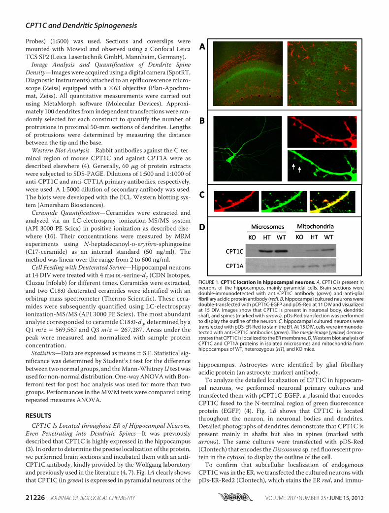
CPT1C Is Located throughout ER of Hippocampal Neurons,Even Penetrating into Dendritic Spines—It was previouslydescribed that CPT1C is highly expressed in the hippocampus(3). In order to determine the precise localization of the protein,we performed brain sections and incubated them with an anti-CPT1C antibody, kindly provided by the Wolfgang laboratoryand previously used in the literature (4, 7). Fig. 1A clearly showsthat CPT1C (in green) is expressed in pyramidal neurons of the
hippocampus. Astrocytes were identified by glial fibrillaryacidic protein (an astrocyte marker) antibody.To analyze the detailed localization of CPT1C in hippocam-
pal neurons, we performed neuronal primary cultures andtransfected them with pCPT1C-EGFP, a plasmid that encodesCPT1C fused to the N-terminal region of green fluorescenceprotein (EGFP) (4). Fig. 1B shows that CPT1C is locatedthroughout the neuron, in neuronal bodies and dendrites.Detailed photographs of dendrites demonstrate that CPT1C ispresent mainly in shafts but also in spines (marked witharrows). The same cultures were transfected with pDS-Red(Clontech) that encodes theDiscosoma sp. red fluorescent pro-tein in the cytosol to display the outline of the cell.To confirm that subcellular localization of endogenous
CPT1Cwas in the ER, we transfected the cultured neuronswithpDs-ER-Red2 (Clontech), which stains the ER red, and immu-
FIGURE 1. CPT1C location in hippocampal neurons. A, CPT1C is present inneurons of the hippocampus, mainly pyramidal cells. Brain sections weredouble-immunodetected with anti-CPT1C antibody (green) and anti-glialfibrillary acidic protein antibody (red). B, hippocampal cultured neurons weredouble-transfected with pCPT1C-EGFP and pDS-Red at 11 DIV and visualizedat 15 DIV. Images show that CPT1C is present in neuronal body, dendriticshaft, and spines (marked with arrows). pDs-Red transfection was performedto display the outline of the neuron. C, hippocampal cultured neurons weretransfected with pDS-ER-Red to stain the ER. At 15 DIV, cells were immunode-tected with anti-CPT1C antibodies (green). The merge image (yellow) demon-strates that CPT1C is localized to the ER membrane. D, Western blot analysis ofCPT1C and CPT1A proteins in isolated microsomes and mitochondria fromhippocampus of WT, heterozygous (HT), and KO mice.
CPT1C and Dendritic Spinogenesis
21226 JOURNAL OF BIOLOGICAL CHEMISTRY VOLUME 287 • NUMBER 25 • JUNE 15, 2012
nodetected endogenous CPT1C with anti-CPT1C antibody (ingreen). Fig. 1C shows that CPT1C is localized in the ER of cul-tured hippocampal neurons. Finally,Western blot experimentswere also performed with different cellular fractions frommouse brain. Anti-CPT1A antibodies were used as amarker formitochondria. Samples were retrieved from WT, CPT1C KO,and heterozygous mice developed in our laboratory (describedunder “Experimental Procedures”). Fig. 1D shows that CPT1Cis presentmainly in themicrosomal fraction and that CPT1A ispresent mainly in mitochondria, confirming that CPT1C local-izes to the ER membrane of cells.CPT1C Regulates Levels of Ceramide in Cultured Neurons—
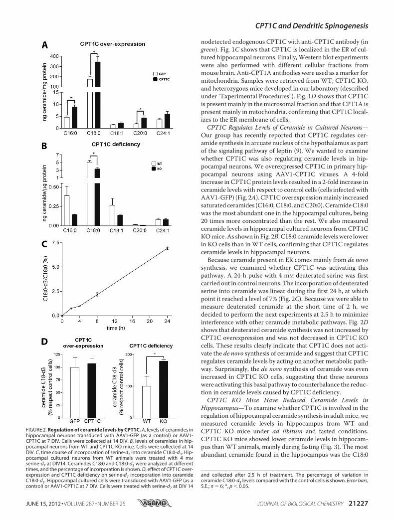
Our group has recently reported that CPT1C regulates cer-amide synthesis in arcuate nucleus of the hypothalamus as partof the signaling pathway of leptin (9). We wanted to examinewhether CPT1C was also regulating ceramide levels in hip-pocampal neurons. We overexpressed CPT1C in primary hip-pocampal neurons using AAV1-CPT1C viruses. A 4-foldincrease in CPT1C protein levels resulted in a 2-fold increase inceramide levels with respect to control cells (cells infected withAAV1-GFP) (Fig. 2A). CPT1Coverexpressionmainly increasedsaturated ceramides (C16:0, C18:0, andC20:0). CeramideC18:0was the most abundant one in the hippocampal cultures, being20 times more concentrated than the rest. We also measuredceramide levels in hippocampal cultured neurons fromCPT1CKOmice. As shown in Fig. 2B, C18:0 ceramide levelswere lowerin KO cells than inWT cells, confirming that CPT1C regulatesceramide levels in hippocampal neurons.Because ceramide present in ER comes mainly from de novo
synthesis, we examined whether CPT1C was activating thispathway. A 24-h pulse with 4 mM deuterated serine was firstcarried out in control neurons. The incorporation of deuteratedserine into ceramide was linear during the first 24 h, at whichpoint it reached a level of 7% (Fig. 2C). Because we were able tomeasure deuterated ceramide at the short time of 2 h, wedecided to perform the next experiments at 2.5 h to minimizeinterference with other ceramide metabolic pathways. Fig. 2Dshows that deuterated ceramide synthesis was not increased byCPT1C overexpression and was not decreased in CPT1C KOcells. These results clearly indicate that CPT1C does not acti-vate the de novo synthesis of ceramide and suggest that CPT1Cregulates ceramide levels by acting on another metabolic path-way. Surprisingly, the de novo synthesis of ceramide was evenincreased in CPT1C KO cells, suggesting that these neuronswere activating this basal pathway to counterbalance the reduc-tion in ceramide levels caused by CPT1C deficiency.CPT1C KO Mice Have Reduced Ceramide Levels in
Hippocampus—To examine whether CPT1C is involved in theregulation of hippocampal ceramide synthesis in adultmice, wemeasured ceramide levels in hippocampus from WT andCPT1C KO mice under ad libitum and fasted conditions.CPT1C KO mice showed lower ceramide levels in hippocam-pus than WT animals, mainly during fasting (Fig. 3). The mostabundant ceramide found in the hippocampus was the C18:0
FIGURE 2. Regulation of ceramide levels by CPT1C. A, levels of ceramides inhippocampal neurons transduced with AAV1-GFP (as a control) or AAV1-CPT1C at 7 DIV. Cells were collected at 14 DIV. B, levels of ceramides in hip-pocampal neurons from WT and CPT1C KO mice. Cells were collected at 14DIV. C, time course of incorporation of serine-d7 into ceramide C18:0-d3. Hip-pocampal cultured neurons from WT animals were treated with 4 mM
serine-d7 at DIV14. Ceramides C18:0 and C18:0-d3 were analyzed at differenttimes, and the percentage of incorporation is shown. D, effect of CPT1C over-expression and CPT1C deficiency on serine-d7 incorporation into ceramideC18:0-d3. Hippocampal cultured cells were transduced with AAV1-GFP (as acontrol) or AAV1-CPT1C at 7 DIV. Cells were treated with serine-d7 at DIV 14
and collected after 2.5 h of treatment. The percentage of variation inceramide C18:0-d3 levels compared with the control cells is shown. Error bars,S.E.; n � 6; *, p � 0.05.
CPT1C and Dendritic Spinogenesis
JUNE 15, 2012 • VOLUME 287 • NUMBER 25 JOURNAL OF BIOLOGICAL CHEMISTRY 21227
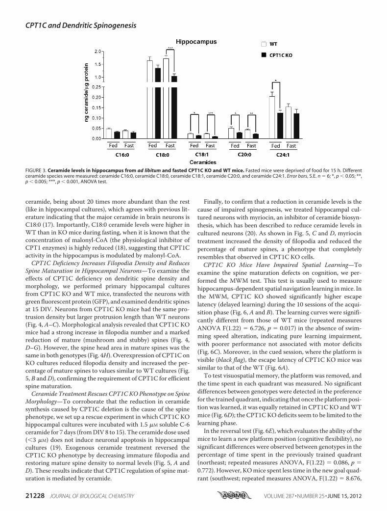
ceramide, being about 20 times more abundant than the rest(like in hippocampal cultures), which agrees with previous lit-erature indicating that the major ceramide in brain neurons isC18:0 (17). Importantly, C18:0 ceramide levels were higher inWT than in KO mice during fasting, when it is known that theconcentration of malonyl-CoA (the physiological inhibitor ofCPT1 enzymes) is highly reduced (18), suggesting that CPT1Cactivity in the hippocampus is modulated by malonyl-CoA.CPT1C Deficiency Increases Filopodia Density and Reduces
Spine Maturation in Hippocampal Neurons—To examine theeffects of CPT1C deficiency on dendritic spine density andmorphology, we performed primary hippocampal culturesfrom CPT1C KO and WT mice, transfected the neurons withgreen fluorescent protein (GFP), and examineddendritic spinesat 15 DIV. Neurons from CPT1C KO mice had the same pro-trusion density but larger protrusion length than WT neurons(Fig. 4, A–C). Morphological analysis revealed that CPT1C KOmice had a strong increase in filopodia number and a markedreduction of mature (mushroom and stubby) spines (Fig. 4,D–G). However, the spine head area in mature spines was thesame in both genotypes (Fig. 4H). Overexpression of CPT1C onKO cultures reduced filopodia density and increased the per-centage of mature spines to values similar to WT cultures (Fig.5, B andD), confirming the requirement of CPT1C for efficientspine maturation.Ceramide Treatment Rescues CPT1CKOPhenotype on Spine
Morphology—To corroborate that the reduction in ceramidesynthesis caused by CPT1C deletion is the cause of the spinephenotype, we set up a rescue experiment in which CPT1C KOhippocampal cultures were incubated with 1.5 �M soluble C-6ceramide for 7 days (fromDIV 8 to 15). The ceramide dose used(�3 �M) does not induce neuronal apoptosis in hippocampalcultures (19). Exogenous ceramide treatment reversed theCPT1C KO phenotype by decreasing immature filopodia andrestoring mature spine density to normal levels (Fig. 5, A andD). These results indicate that CPT1C regulation of spine mat-uration is mediated by ceramide.
Finally, to confirm that a reduction in ceramide levels is thecause of impaired spinogenesis, we treated hippocampal cul-tured neurons with myriocin, an inhibitor of ceramide biosyn-thesis, which has been described to reduce ceramide levels incultured neurons (20). As shown in Fig. 5, C and D, myriocintreatment increased the density of filopodia and reduced thepercentage of mature spines, a phenotype that completelyresembles that observed in CPT1C KO cells.CPT1C KO Mice Have Impaired Spatial Learning—To
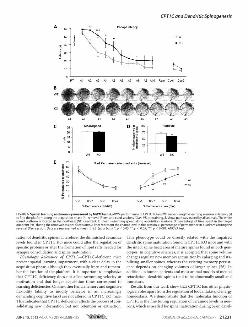
examine the spine maturation defects on cognition, we per-formed the MWM test. This test is usually used to measurehippocampus-dependent spatial navigation learning inmice. Inthe MWM, CPT1C KO showed significantly higher escapelatency (delayed learning) during the 10 sessions of the acqui-sition phase (Fig. 6, A and B). The learning curves were signifi-cantly different from those of WT mice (repeated measuresANOVA F(1.22) � 6.726, p � 0.017) in the absence of swim-ming speed alteration, indicating pure learning impairment,with poorer performance not associated with motor deficits(Fig. 6C). Moreover, in the cued session, where the platform isvisible (black flag), the escape latency of CPT1C KO mice wassimilar to that of the WT (Fig. 6A).To test visuospatial memory, the platformwas removed, and
the time spent in each quadrant was measured. No significantdifferences between genotypes were detected in the preferencefor the trained quadrant, indicating that once the platformposi-tion was learned, it was equally retained in CPT1CKO andWTmice (Fig. 6D); the CPT1CKOdeficits seem to be limited to thelearning phase.In the reversal test (Fig. 6E), which evaluates the ability of the
mice to learn a new platform position (cognitive flexibility), nosignificant differences were observed between genotypes in thepercentage of time spent in the previously trained quadrant(northeast; repeated measures ANOVA, F(1.22) � 0.086, p �0.772). However, KOmice spent less time in the new goal quad-rant (southwest; repeated measures ANOVA, F(1.22) � 8.676,
FIGURE 3. Ceramide levels in hippocampus from ad libitum and fasted CPT1C KO and WT mice. Fasted mice were deprived of food for 15 h. Differentceramide species were measured: ceramide C16:0, ceramide C18:0, ceramide C18:1, ceramide C20:0, and ceramide C24:1. Error bars, S.E. n � 6; *, p � 0.05; **,p � 0.005; ***, p � 0.001, ANOVA test.
CPT1C and Dendritic Spinogenesis
21228 JOURNAL OF BIOLOGICAL CHEMISTRY VOLUME 287 • NUMBER 25 • JUNE 15, 2012

p � 0.007), thus supporting the hypothesis of a hippocampus-dependent learning deficit in CPT1C KO mice.
DISCUSSION
Dendritic spine formation begins in the embryo and contin-ues into early postnatal life but also occurs in the adult orga-nism,where it contributes significantly to learning andmemoryformation. We demonstrate that the brain isoform CPT1C ispresent in dendritic spines and regulates the levels of ceramidein neurons, which is key to the transformation of dendriticfilopodia into mature spines. This is the first time that CPT1C
or ceramide levels have been directly involved in spinemorpho-genesis. At the physiological level, we show for the first timethat CPT1C is involved in spatial learning.CPT1C Regulates Ceramide Levels in Neurons—One of the
relevant contributions of this study is the confirmation thatCPT1C increases the levels of ceramide. We had previouslydescribed it in the arcuate nucleus of hypothalamus (9), and wenow demonstrate it in hippocampal cultured neurons. In con-sequence, it may be a general phenomenon in neurons. We donot know themolecularmechanism bywhichCPT1C increasesceramide levels, but our results clearly demonstrate that it does
FIGURE 4. Dendritic spine density and morphology from CPT1C KO and WT hippocampal neurons. Hippocampal neurons were transfected (13 DIV) withpEGFP to visualize the outline of the cell. Protrusions were analyzed 2 days after transfection. Protrusion density (A) and protrusion length (B and C) weremeasured. Mature spines (A) and filopodia (B) are indicated. Spine morphology (D–F) was assayed by analysis of types of protrusions: filopodia (without head),mushroom (with head and neck), and stubby (with only head). G, percentage of mature spines (mushroom and stubby) relative to the total number ofprotrusions was also measured. H, spine head area was measured in mushroom and stubby spines. I, a representative image of dendritic spines from WT andCPT1C KO neurons. For the quantification of protrusion density, spine length, and morphology, �100 dendrites from independent transfections were selectedrandomly. Student’s t tests were used to assess statistical significance of the differences. Error bars, S.E.; ***, p � 0.001.
CPT1C and Dendritic Spinogenesis
JUNE 15, 2012 • VOLUME 287 • NUMBER 25 JOURNAL OF BIOLOGICAL CHEMISTRY 21229

not enhance de novo synthesis, as suggested previously (9).Although the de novo synthesis of ceramide is the main sourceof ceramide in ER, it can also be produced from the sphingosinepool (salvage pathway) or from the dephosphorylation of cer-amide-1-phosphate. Therefore, CPT1C could be activatingeither of these two pathways. Another possibility is that CPT1Cincreases the levels of ceramide by inhibiting its elimination (byconversion to sphingosine, phosphorylation to ceramide-1-phosphate or incorporation into sphingomyelin). Furtherresearch is therefore required to determine the precise meta-bolic pathway in which CPT1C is involved.Because CPT1C has low catalytic activity in vitro (4, 5), we
hypothesize that CPT1C regulates the activity of this otherenzyme involved in ceramide metabolism by protein-proteininteraction. Therefore, under fasting conditions or reduction ofmalonyl-CoA levels, CPT1C might change its conformationand regulate this other enzyme, resulting in increased levels ofceramide.CPT1C in Dendritic Spine Maturation—Our results impli-
cate CPT1C in dendritic spine maturation. In absolute num-bers, in cultured hippocampal neurons from CPT1C KOmice,the increase in filopodia correspondswith the decline inmaturespine number, without altering the overall density of dendriticprotrusions, which indicates that CPT1C is not necessary forthe formation of new protrusions. However, it is necessary forthe conversion of filopodia into mature spines. In addition,results show that the effect of CPT1C on dendritic spines ismediated by ceramide. The addition of ceramide to the cul-tured medium at low concentration reversed the CPT1C KOphenotype and induced spine maturation. A recent study dem-onstrates the presence of a new long-chain acyl-CoA synthetase(ACSL4) isoenzyme that localizes specifically in the ER of neu-rons. Its deficiency increases the percentage of filopodia andreduces the percentage of mature spines (21), in accordancewith our results. This highlights the importance of fatty acidmetabolism in spinogenesis and suggests that ACSL could pro-vide the substrate necessary for ceramide synthesis in the ER ofneurons.There is only one study that correlates ceramide with the
formation of dendritic spines (20). The authors report that cou-pled inhibition of cholesterol and ceramide synthesis causesalterations in the density and morphology of dendritic spines.Ourwork sheds light on the regulation of this process and iden-tifies a role for CPT1C in the fine tuning of the modulation ofceramide synthesis, which is essential for the maturation ofdendritic spines. The mechanism by which ceramides regulatespine maturation is unknown. However, ceramide binds to andregulates the activity of enzymes and signaling proteins, such askinases, phosphatases, ormembrane receptors (22). One exam-ple is protein phosphatase 1,which is activated by ceramide (23)and has been implicated in the conversion of filopodia intomature spines (24). In addition, ceramide is the building blockof all cellular sphingolipids, which, in addition to cholesterol,are essential components of lipid rafts. These membranemicrodomains are needed for the correct trafficking, anchor-age, and activity of synaptic proteins and are preferred plat-forms for membrane-linked actin polymerization (25). All ofthese phenomena are necessary for synapse stability and matu-
FIGURE 5. Rescue of CPT1C KO phenotype on spine morphology by CPT1Cexpression or ceramide treatment. A, hippocampal neurons treated with1.5 �M C6-ceramide at 7 DIV and transfected with pEGFP (BD Biosciences) at12 DIV, fixed, and analyzed for the morphology of dendritic protrusions at 15DIV. B, hippocampal neurons were transfected with pIRES-CPT1C at 7 DIV andanalyzed for spine morphology at 15 DIV. pIRES-CPT1C vector expresses bothCPT1C and GFP proteins, which permits us to visualize in green the cells over-expressing CPT1C. C, hippocampal neurons at DIV9 were treated with 10 �M
myriocin until 15 DIV. Cells were transfected with pEGFP at 12 DIV and ana-lyzed for the morphology of dendritic protrusions at 15 DIV. D, a representa-tive image showing dendritic spines from WT mice, KO mice, KO mice treatedwith C6-ceramide, KO mice transfected with pIRES-CPT1C, and WT micetreated with myriocin. For the quantification of spine morphology, �100 den-drites from independent transfections were selected randomly. Student’s ttests and ANOVA post hoc were used to assess statistical significance of thedifferences. Error bars, S.E.; *, p � 0.05; ***, p � 0.001.
CPT1C and Dendritic Spinogenesis
21230 JOURNAL OF BIOLOGICAL CHEMISTRY VOLUME 287 • NUMBER 25 • JUNE 15, 2012
ration of dendritic spines. Therefore, the diminished ceramidelevels found in CPT1C KO mice could alter the regulation ofspecific proteins or alter the formation of lipid rafts needed forsynapse consolidation and spine maturation.Physiologic Relevance of CPT1C—CPT1C-deficient mice
present spatial learning impairment, with a clear delay in theacquisition phase, although they eventually learn and remem-ber the location of the platform. It is important to emphasizethat CPT1C deficiency does not affect swimming velocity ormotivation and that longer acquisition times correspond tolearning deficiencies.On the other hand,memory and cognitiveflexibility (ability to modify behavior in an increasinglydemanding cognitive task) are not altered in CPT1C KO mice.This indicates thatCPT1Cdeficiency affects the process of con-solidating new information but not retention or extinction.
This phenotype could be directly related with the impaireddendritic spine maturation found in CPT1C KOmice and withthe intact spine head area of mature spines found in both gen-otypes. In cognitive sciences, it is accepted that spine volumechanges regulate newmemory acquisition by enlarging and sta-bilizing smaller spines, whereas the existing memory persist-ence depends on changing volumes of larger spines (26). Inaddition, in human patients and most animal models of mentalretardation, dendritic spines tend to be abnormally small andimmature.Results from our work show that CPT1C has other physio-
logical roles apart from the regulation of food intake and energyhomeostasis. We demonstrate that the molecular function ofCPT1C is the fine tuning regulation of ceramide levels in neu-rons, which is needed for spine maturation during brain devel-
FIGURE 6. Spatial learning and memory measured by MWM test. A, MWM performance of CPT1C KO and WT mice during the learning sessions as latency (s)to find the platform along the acquisition phase (A), removal (Rem), and cued sessions (Cue). PT, pretraining. B, visual pathway traced by all animals. The whiteround platform is located in the northeast (NE) quadrant. C, mean swimming speed along acquisition sessions. D, percentage of time spent in the targetquadrant (NE) during the removal session; discontinuous lines represent the chance level in this session. E, percentage of permanence in quadrants during thereversal (Rev) session. Data are represented as mean � S.E. (error bars); *, p � 0.05; **, p � 0.05; ***, p � 0.001, ANOVA test.
CPT1C and Dendritic Spinogenesis
JUNE 15, 2012 • VOLUME 287 • NUMBER 25 JOURNAL OF BIOLOGICAL CHEMISTRY 21231

opment. At the behavioral level, we demonstrate for the firsttime the involvement of CPT1C in learning, which opens thepossibility that CPT1C mutations might be the cause of somehuman cognition disabilities of unknown etiology.
Acknowledgments—The editorial help of Robin Rycroft is gratefullyacknowledged. We thank Josep Clotet for valuable discussions andJulia Geiger for technical support with cell cultures.
REFERENCES1. Esser, V., Britton, C. H., Weis, B. C., Foster, D. W., and McGarry, J. D.
(1993) Cloning, sequencing, and expression of a cDNA encoding rat livercarnitine palmitoyltransferase I. Direct evidence that a single polypeptideis involved in inhibitor interaction and catalytic function. J. Biol. Chem.268, 5817–5822
2. Yamazaki, N., Shinohara, Y., Shima, A., and Terada, H. (1995) High ex-pression of a novel carnitine palmitoyltransferase I-like protein in ratbrown adipose tissue and heart. Isolation and characterization of itscDNA clone. FEBS Lett. 363, 41–45
3. Price, N., van der Leij, F., Jackson, V., Corstorphine, C., Thomson, R.,Sorensen, A., and Zammit, V. (2002) A novel brain-expressed proteinrelated to carnitine palmitoyltransferase I. Genomics 80, 433–442
4. Sierra, A. Y., Gratacos, E., Carrasco, P., Clotet, J., Urena, J., Serra, D., Asins,G., Hegardt, F. G., andCasals, N. (2008) CPT1c is localized in endoplasmicreticulum of neurons and has carnitine palmitoyltransferase activity.J. Biol. Chem. 283, 6878–6885
5. Wolfgang,M. J., Kurama, T., Dai, Y., Suwa, A., Asaumi,M.,Matsumoto, S.,Cha, S. H., Shimokawa, T., and Lane, M. D. (2006) The brain-specificcarnitine palmitoyltransferase-1c regulates energy homeostasis. Proc.Natl. Acad. Sci. U.S.A. 103, 7282–7287
6. Gao, X. F., Chen,W., Kong, X. P., Xu, A.M.,Wang, Z. G., Sweeney, G., andWu, D. (2009) Enhanced susceptibility of Cpt1c knockout mice to glucoseintolerance induced by a high-fat diet involves elevated hepatic gluconeo-genesis and decreased skeletal muscle glucose uptake. Diabetologia 52,912–920
7. Dai, Y., Wolfgang, M. J., Cha, S. H., and Lane, M. D. (2007) Localizationand effect of ectopic expression of CPT1c in CNS feeding centers.Biochem. Biophys. Res. Commun. 359, 469–474
8. Reamy, A. A., and Wolfgang, M. J. (2011) Carnitine palmitoyltrans-ferase-1c gain-of-function in the brain results in postnatal microenceph-aly. J. Neurochem. 118, 388–398
9. Gao, S., Zhu, G., Gao, X., Wu, D., Carrasco, P., Casals, N., Hegardt, F. G.,Moran, T. H., and Lopaschuk, G. D. (2011) Important roles of brain-specific carnitine palmitoyltransferase and ceramide metabolism in leptinhypothalamic control of feeding. Proc. Natl. Acad. Sci. U.S.A. 108,9691–9696
10. Zaugg, K., Yao, Y., Reilly, P. T., Kannan, K., Kiarash, R., Mason, J., Huang,P., Sawyer, S. K., Fuerth, B., Faubert, B., Kalliomäki, T., Elia, A., Luo, X.,Nadeem, V., Bungard, D., Yalavarthi, S., Growney, J. D., Wakeham, A.,Moolani, Y., Silvester, J., Ten, A. Y., Bakker, W., Tsuchihara, K., Berger,S. L., Hill, R. P., Jones, R. G., Tsao, M., Robinson, M. O., Thompson, C. B.,Pan, G., and Mak, T. W. (2011) Carnitine palmitoyltransferase 1C pro-motes cell survival and tumor growth under conditions of metabolic
stress. Genes Dev. 25, 1041–105111. Tybulewicz, V. L., Crawford, C. E., Jackson, P. K., Bronson, R. T., and
Mulligan, R. C. (1991) Neonatal lethality and lymphopenia in mice with ahomozygous disruption of the c-abl proto-oncogene. Cell 65, 1153–1163
12. Arque, G., Fotaki, V., Fernandez, D., Martınez de Lagran, M., Arbones,M. L., and Dierssen, M. (2008) Impaired spatial learning strategies andnovel object recognition in mice haploinsufficient for the dual specificitytyrosine-regulated kinase-1A (Dyrk1A). PLoS One 3, e2575
13. Segura, I., Essmann, C. L., Weinges, S., and Acker-Palmer, A. (2007) Grb4and GIT1 transduce ephrinB reverse signals modulating spine morpho-genesis and synapse formation. Nat. Neurosci. 10, 301–310
14. Grimm,D., Kern, A., Rittner, K., andKleinschmidt, J. A. (1998)Novel toolsfor production and purification of recombinant adenoassociated virusvectors. Hum. Gene Ther. 9, 2745–2760
15. Dentin, R., Pegorier, J. P., Benhamed, F., Foufelle, F., Ferre, P., Fauveau, V.,Magnuson, M. A., Girard, J., and Postic, C. (2004) Hepatic glucokinase isrequired for the synergistic action of ChREBP and SREBP-1c on glycolyticand lipogenic gene expression. J. Biol. Chem. 279, 20314–20326
16. Merrill, A. H., Jr., Sullards, M. C., Allegood, J. C., Kelly, S., and Wang, E.(2005) Sphingolipidomics. High throughput, structure-specific, andquantitative analysis of sphingolipids by liquid chromatography tandemmass spectrometry.Methods 36, 207–224
17. Ben-David, O., and Futerman, A. H. (2010) The role of the ceramide acylchain length in neurodegeneration. Involvement of ceramide synthases.Neuromolecular Med. 12, 341–350
18. Tokutake, Y., Onizawa, N., Katoh, H., Toyoda, A., and Chohnan, S. (2010)Coenzyme A and its thioester pools in fasted and fed rat tissues. Biochem.Biophys. Res. Commun. 402, 158–162
19. Mitoma, J., Ito, M., Furuya, S., and Hirabayashi, Y. (1998) Bipotential rolesof ceramide in the growth of hippocampal neurons. Promotion of cellsurvival and dendritic outgrowth in dose- and developmental stage-de-pendent manners. J. Neurosci. Res. 51, 712–722
20. Hering, H., Lin, C. C., and Sheng,M. (2003) Lipid rafts in themaintenanceof synapses, dendritic spines, and surface AMPA receptor stability. J. Neu-rosci. 23, 3262–3271
21. Meloni, I., Parri, V., De Filippis, R., Ariani, F., Artuso, R., Bruttini, M.,Katzaki, E., Longo, I., Mari, F., Bellan, C., Dotti, C. G., and Renieri, A.(2009) TheXLMRgeneACSL4 plays a role in dendritic spine architecture.Neuroscience 159, 657–669
22. Breslow, D. K., andWeissman, J. S. (2010) Membranes in balance. Mech-anisms of sphingolipid homeostasis.Mol. Cell 40, 267–279
23. Chalfant, C. E., Kishikawa, K., Mumby,M. C., Kamibayashi, C., Bielawska,A., andHannun, Y. A. (1999) Long chain ceramides activate protein phos-phatase-1 and protein phosphatase-2A. Activation is stereospecific andregulated by phosphatidic acid. J. Biol. Chem. 274, 20313–20317
24. Terry-Lorenzo, R. T., Roadcap, D. W., Otsuka, T., Blanpied, T. A., Zamo-rano, P. L., Garner, C. C., Shenolikar, S., and Ehlers, M. D. (2005) Neura-bin/protein phosphatase-1 complex regulates dendritic spine morpho-genesis and maturation.Mol. Biol. Cell 16, 2349–2362
25. Allen, J. A., Halverson-Tamboli, R. A., and Rasenick, M. M. (2007) Lipidraft microdomains and neurotransmitter signalling.Nat. Rev. Neurosci. 8,128–140
26. Kasai, H., Fukuda, M., Watanabe, S., Hayashi-Takagi, A., and Noguchi, J.(2010) Structural dynamics of dendritic spines in memory and cognition.Trends Neurosci. 33, 121–129
CPT1C and Dendritic Spinogenesis
21232 JOURNAL OF BIOLOGICAL CHEMISTRY VOLUME 287 • NUMBER 25 • JUNE 15, 2012