Caloric restriction augments ROS defense in S. cerevisiae , by a Sir2p independent mechanism
Upload
khangminh22Category
view
5download
0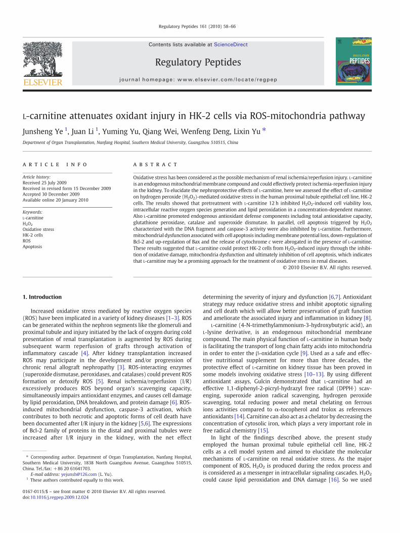
Regulatory Peptides 161 (2010) 58–66
Contents lists available at ScienceDirect
Regulatory Peptides
j ourna l homepage: www.e lsev ie r.com/ locate / regpep
L-carnitine attenuates oxidant injury in HK-2 cells via ROS-mitochondria pathway
Junsheng Ye 1, Juan Li 1, Yuming Yu, Qiang Wei, Wenfeng Deng, Lixin Yu ⁎Department of Organ Transplantation, Nanfang Hospital, Southern Medical University, Guangzhou 510515, China
⁎ Corresponding author. Department of Organ TranSouthern Medical University, 1838 North GuangzhouChina. Tel./fax: +86 20 61641703.
E-mail address: [email protected] (L. Yu).1 These authors contributed equally to this work.
0167-0115/$ – see front matter © 2010 Elsevier B.V. Adoi:10.1016/j.regpep.2009.12.024
a b s t r a c t
a r t i c l e i n f oArticle history:Received 25 July 2009Received in revised form 15 December 2009Accepted 30 December 2009Available online 20 January 2010
Keywords:L-carnitineH2O2
Oxidative stressHK-2 cellsROSApoptosis
Oxidative stress has been considered as the possiblemechanism of renal ischemia/reperfusion injury. L-carnitineis an endogenousmitochondrialmembrane compound and could effectively protect ischemia-reperfusion injuryin the kidney. To elucidate the nephroprotective effects of L-carnitine, here we assessed the effect of L-carnitineon hydrogen peroxide (H2O2)-mediated oxidative stress in the human proximal tubule epithelial cell line, HK-2cells. The results showed that pretreatment with L-carnitine 12 h inhibited H2O2-induced cell viability loss,intracellular reactive oxygen species generation and lipid peroxidation in a concentration-dependent manner.Also L-carnitine promoted endogenous antioxidant defense components including total antioxidative capacity,glutathione peroxidase, catalase and superoxide dismutase. In parallel, cell apoptosis triggered by H2O2
characterized with the DNA fragment and caspase-3 activity were also inhibited by L-carnitine. Furthermore,mitochondrial dysfunction associatedwith cell apoptosis includingmembrane potential loss, down-regulation ofBcl-2 and up-regulation of Bax and the release of cytochrome c were abrogated in the presence of L-carnitine.These results suggested that L-carnitine could protect HK-2 cells from H2O2-induced injury through the inhibi-tion of oxidative damage, mitochondria dysfunction and ultimately inhibition of cell apoptosis, which indicatesthat L-carnitine may be a promising approach for the treatment of oxidative stress in renal diseases.
splantation, Nanfang Hospital,Avenue, Guangzhou 510515,
ll rights reserved.
© 2010 Elsevier B.V. All rights reserved.
1. Introduction
Increased oxidative stress mediated by reactive oxygen species(ROS) have been implicated in a variety of kidney diseases [1–3]. ROScan be generated within the nephron segments like the glomeruli andproximal tubule and injury initiated by the lack of oxygen during coldpresentation of renal transplantation is augmented by ROS duringsubsequent warm reperfusion of grafts through activation ofinflammatory cascade [4]. After kidney transplantation increasedROS may participate in the development and/or progression ofchronic renal allograft nephropathy [3]. ROS-interacting enzymes(superoxide dismutase, peroxidases, and catalases) could prevent ROSformation or detoxify ROS [5]. Renal ischemia/reperfusion (I/R)excessively produces ROS beyond organ's scavenging capacity,simultaneously impairs antioxidant enzymes, and causes cell damageby lipid peroxidation, DNA breakdown, and protein damage [6]. ROS-induced mitochondrial dysfunction, caspase-3 activation, whichcontributes to both necrotic and apoptotic forms of cell death havebeen documented after I/R injury in the kidney [5,6]. The expressionsof Bcl-2 family of proteins in the distal and proximal tubules wereincreased after I/R injury in the kidney, with the net effect
determining the severity of injury and dysfunction [6,7]. Antioxidantstrategy may reduce oxidative stress and inhibit apoptotic signalingand cell death which will allow better preservation of graft functionand ameliorate the associated injury and inflammation in kidney [8].
L-carnitine (4-N-trimethylammonium-3-hydroxybutyric acid), anL-lysine derivative, is an endogenous mitochondrial membranecompound. The main physical function of L-carnitine in human bodyis facilitating the transport of long chain fatty acids into mitochondriain order to enter the β-oxidation cycle [9]. Used as a safe and effec-tive nutritional supplement for more than three decades, theprotective effect of L-carnitine on kidney tissue has been proved insome models involving oxidative stress [10–13]. By using differentantioxidant assays, Gulcin demonstrated that L-carnitine had aneffective 1,1-diphenyl-2-picryl-hydrazyl free radical (DPPH∙) scav-enging, superoxide anion radical scavenging, hydrogen peroxidescavenging, total reducing power and metal chelating on ferrousions activities compared to α-tocopherol and trolox as referencesantioxidants [14]. Carnitine can also act as a chelator by decreasing theconcentration of cytosolic iron, which plays a very important role infree radical chemistry [15].
In light of the findings described above, the present studyemployed the human proximal tubule epithelial cell line, HK-2cells as a cell model system and aimed to elucidate the molecularmechanisms of L-carnitine on renal oxidative stress. As the majorcomponent of ROS, H2O2 is produced during the redox process andis considered as a messenger in intracellular signaling cascades. H2O2
could cause lipid peroxidation and DNA damage [16]. So we used

59J. Ye et al. / Regulatory Peptides 161 (2010) 58–66
H2O2 as an inducer of oxidative stress for HK-2 cells and testedwhether pretreatment cells with L-carnitine resulted in the resistanceof HK-2 to H2O2 challenge. Furthermore, the effect of L-carnitine onoxidative stress conditions such as ROS production, lipid peroxidation,antioxidant defensive system, mitochondrial dysfunction and DNAdamage associated with cell apoptosis were also studied.
2. Materials and methods
2.1. Chemicals and reagents
L-carnitine, H2O2 and 3-(4,5-dimethylthiazol-2-yl)-2-5-diphenyl-tetrazolium bromide (MTT), were purchased from Sigma (St. Louis,MO, USA). ROS detection kit, acetyl-L-cysteine (NAC) and DNA Ladderwas purchased from Beyotime Company (Jiangsu, China). All otherchemicals were of analytic grade.
2.2. Cell culture and treatment
HK-2 cells (Cell Culture Center, Beijing, China), an immortalizedhuman proximal tubule epithelial cell line, were grown and passagedin 75-cm2 cell culture flasks that contained DMEM Ham's F12media (1:1, Gibco BRL) supplemented with 10% FCS and antibiotics(100 U/mL penicillin G, 100 μg/mL streptomycin). To determinethe effects of L-carnitine on H2O2-exposed HK-2, subconfluent (80%)HK-2 cells were harvested and seeded into 96 well plates or six-welltissue culture plates. The cells were allowed to adhere for 18 h inan incubator at 37 °C with 5% CO2 in 95% air. Immediately beforeexperimental treatments, the medium was replaced with freshmedium and cells were treated with indicated doses of L-carnitinefor 12 h before H2O2 exposure. Thereafter, cells were washed withPBS to make them L-carnitine free and then cells in fresh mediumwere exposed to the desired doses of H2O2. Afterwards, cells wererinsed with fresh medium (without H2O2) and incubated. Cells wereharvested for further analysis.
2.3. Cell proliferation and viability analysis
Cell viability and proliferation was determined using the MTTassay, which is a sensitive measurement of the normal metabolicstatus of cells. Briefly, cultured HK-2 cells were initially plated intriplicate at a density of 1×10⁴ cells/100 μL in 96 well plates for 24 h.The cells were pre-incubated with or without L-carnitine followingincubation with H2O2 for different time. Afterwards, cells were rinsedwith fresh medium (without H2O2) and incubated for 24 h. Thecells were then incubated with 0.5 mg/mL MTT at 37 °C for 4 h. Theformazan crystals generated by viable mitochondrial succinatedehydrogenase from MTT were extracted using an equal volumeof the solubilizing buffer (0.01 N HCl and 10% SDS). Absorbancewas measured at a wavelength of 490 nm using a MolecularDevices VERSAmax microplate reader (Molecular devices, Sunnyvale,CA, USA). All experiments were performed in triplicate.
2.4. Measurement of ROS accumulation and lipid peroxidation
Cultured HK-2 cells were initially plated in triplicate at a densityof 5×10⁵ cells/well in 6 well plates for 24 h. The cells were then pre-incubated with or without L-carnitine following incubation withH2O2. At the indicated time points, to monitor intracellular accumu-lation of ROS, the treated cells were incubated with 5 μM DCF-DA for30 min at 37 °C. Cells were subsequently washed twice with D-Hanksand collected, DCF fluorescence intensity of 100 μL cell suspensionwas quantified with a fluorometer (GENios, USA) using 485 nmexcitation and 535 nm emission filters [17]. The results are given aspercents relative to the oxidative stress of the control cells set to 100%.All experiments were performed in triplicate.
Malonyl dialdehyde (MDA), a terminal product of lipid peroxida-tion, was measured to estimate the content of lipid peroxidation inHK-2 cells [18]. MDA concentration in cell homogenates wasdetermined with commercial kits purchased from Jiancheng Bioen-gineering Institute (Nanjing, China), using the thiobarbituric acidmethod. The assay was based on the conjugation ability of MDA withthiobarbituric acid, to form a red product which has maximumabsorbance at 532 nm.
2.5. Assays for total antioxidative capacity (T-AOC) and antioxidantdefense enzymes
The cells were pre-incubated with or without L-carnitine followingincubation with H2O2. At the indicated time points, HK-2 cellswere washed 3 times with ice-cold D-Hanks and lysed in theextraction buffer [50 mmol/L Tris–HCl (pH7.4), 1 mmol/L ethylene-glycolbis (2-aminoethylether) tetraacetic acid, 150 mmol/L NaCl,1% (v/v) Triton X-100, 1 mmol/L phenylmethylsulfonyl, 10 µg/mLaprotinin, 10 mmol/L EDTA, 1 mmol/L NaF, and 1 mmol/L Na3VO4] onice for 30 min. Then the cells were scraped from the plates and thelysates were subjected to 20,000×g centrifugation at 4 °C for 10 min.The amount of proteins in the cleared lysates was quantified with abicincho-ninic acid assay (Beyotime Biotechnology, China). Afterdetermining the amount of total proteins in the supernatants, wedetected T-AOC and the endogenous antioxidant defense componentslike glutathione peroxidase (GPx), catalase (CAT) and superoxidedismutase (SOD) using biochemical methods following the instruc-tions for the reagent kits (Nanjing Institute of Jiancheng BiologicalEngineering, Nanjing, China) and absorbance were measured using amicroplate reader. T-AOC reflects the overall cellular endogenousantioxidative capability including both enzymatic and non-enzymaticantioxidants. These antioxidants can reduce Fe3+ to Fe2+, the lattercan form colored and stable chelates combing with phenanthroline,which was then chelated with porphyrin to produce a purplecomplex. GPx activities were assayed by the decrease of the GSH.CAT activity was determined by the decrease of H2O2. SOD activitywas measured using the nitroblue tetrazoliummethod which utilizeda tetrazolium salt to quantify the superoxide radicals generated byxanthine oxidase and hypoxanthine. Data were defined as theamount of protein that inhibits the rate of nitroblue tetrazoliumreduction by 50%. Values were calculated using absorbation (520 nmfor T-AOC, 412 nm for GPx, 405 nm for CAT and 550 nm for SOD)and expressed as units (U) per mg protein. The activities of T-AOC,GPx, CAT and SOD were expressed as U/mg protein, respectively.The data were calculated and represented as percentage of controlcells. All experiments were performed in triplicate.
2.6. Flow cytometric detection of apoptosis assay
Apoptotic cells were quantified by determining DNA content ofcells by propidium iodide staining by flow cytometry [19]. Briefly, thepellets were resuspended in ice-cold 70% ethanol and fixed at 4 °C for24–48 h. The cells were then centrifuged, and ethanol was removedby washing thoroughly with PBS. The cell pellets were resuspendedin 1 ml DNA staining reagent containing 50 μg/ml RNase, 0.1% tritonX-100, 0.1 mM EDTA (pH 7.4), and 50 μg/ml PI. The staining wasstable at 4 °C for 30 min. Red fluorescence (DNA) was detectedthrough a 563–607 nm bandpass filter by using a FACS 440 flowcytometer (Becton Dickinson). In flow cytometric histograms,apoptotic cells will give DNA fluorescence in the subdiploid regions,which arewell separated from the normal G1 peak. Ten thousand cellsin each sample were analyzed and the percentage of apoptotic cellaccumulation in the sub-G1 peak was calculated. Each measurementwas carried out in triplicate.

60 J. Ye et al. / Regulatory Peptides 161 (2010) 58–66
2.7. Measurement of mitochondrial membrane potential (MMP, ΔψМ)
Mitochondrial membrane was monitored using the fluorescentdye Rh123, a cell permeable cationic dye, which preferentiallypartitions into mitochondria based on the highly negative MMP aspreviously described [20]. Rh123 was added to cell cultures to attain afinal concentration of 10 μM for 30 min at 37 °C after cells weretreated as described above. The cells were collected by pipetting andwashed twice with PBS and then analyzed by flow cytometry andanalyzed using CELLQuest software within 1 h. The laser was adjustedto emit at 480 nm, and a 530 nm long-pass filter was used.
2.8. Cytochrome c release from mitochondria
The cells were harvested after the respective treatments, washedonce with ice-cold PBS. For isolation of mitochondria and cytosol toobserve the cytochrome c release from mitochondria, the cells weresonicated in buffer containing 10 mM Tris–HCl pH 7.5, 10 mM NaCl,175 mM sucrose and 12.5 mMEDTA and the cell extract centrifuged at1000 g for 10 min to pellet nuclei. The supernatant thus obtained wascentrifuged at 18,000 g for 30 min to pellet the mitochondria. Theresulting supernatant was termed as the cytosolic fraction. The purityof the fractions was confirmed by assaying the marker enzymessuccinate dehydrogenase for mitochondria, lactate dehydrogenase forthe cytosol. The protein concentration was measured using a Bradfordprotein assay kit (BioRad). Cytochrome c determination in cytosolicand mitochondrial fractions was done by Western blotting.
2.9. Western blot analysis
For the quantification of various protein expression, Western blotanalysis was used. The cells were placed in lysis buffer (1% NP40,0.5% sodium deoxycholate, 0.1% SDS in normal PBS; pH 6.8) con-taining a protease inhibitor cocktail (10 μL/mL; Sigma-Aldrich). Thehomogenate was centrifuged at 12,000×g at 4 °C for 20 min, and thesupernatant was stored at −80 °C. Total protein was determined bythe Bradford assay. Equal amounts of protein (10 μg) from bothfraction were separated on a 10% SDS-PAGE and transferredelectrophoretically to the nitrocellulose membranes. The membranewas blocked by 5% skim milk in Tris-buffered saline containing 0.1%Triton X-100 (TBS-T) for 2 h at room temperature. Immunoblots wereperformed with appropriate antibodies: primary antibodies for BCL-2(1:200, Santa Cruz), Bax (1:200, Santa Cruz), cytochrome c (1:200,Santa Cruz) and β-actin (1:1000, Santa Cruz). Immunoblot analysiswas performed with horseradish peroxidase-conjugated anti-mouseand anti-rabbit IgG using enhanced chemiluminescence Westernblotting detection reagents (Amersham Bioscience, Piscataway,NJ, USA). The bands corresponding to BCL-2, Bax and cytochrome cor β-actin, were scanned and densitometrically analyzed usingan automatic image analysis system (Alpha Innotech Corporation,San Leandro, CA, USA). These quantitative analyses were normalizedto β-actin (after stripping).
2.10. Flow cytometric detection of caspase-3 activity
The extent of caspase-3 activation in HK-2 cells treated with H2O2
for 12 h was detected by flow cytometry analysis using the anti-activecaspase-3 monoclonal antibody, which specifically recognizes theactive form of caspase-3. Briefly, cells were washed twice in PBS, fixedusing 4% polyoxymethylene for 30 min and then permeabilized using1% TritonX-100 for 10 min at room temperature, before they werewashed twice with PBS. Cells were stained with anti-active caspase-3antibody for 30 min at room temperature in the dark. Followingincubation with the antibody, cells were washed in wash buffer,resuspended in wash buffer and analyzed by flow cytometry(FACSVantage SE, BD Biosciences).
2.11. Statistical analysis
All data are expressed as means±S.E.M. Comparisons betweenthe different groups were performed by ANOVA followed byStudent's t-test. In all tests, the criterion for statistical significancewas pb0.05.
3. Results
3.1. L-carnitine protects HK-2 cells against H2O2-induced cytotoxicity
We first determined the dose and time of exposure to H2O2
to reduce cell viability by 50%. The results showed that H2O2 injuredHK-2 cells in a dose- and time-dependent manner and treatment ofHK-2 cells with 500 μM of H2O2 for 30 min resulted in moderatecellular injury. So 500 μM of H2O2 for 30 min was used in thesubsequent study (Fig. 1A and B). Studies have showed that the auto-oxidation of some antioxidants produces semiquinone radicals,superoxide anion and H2O2 and higher concentrations of L-carnitinemay be cytotoxic to the cells. To investigate the cytoprotectiveeffect of L-carnitine, we tested the effect of L-carnitine by cell viabilityassay. As shown in Fig. 1C, at concentrations from 0.1 to 1000 μM for12 h, L-carnitine slightly increased cell viability and did not inducechanges in cell morphology. To evaluate the dose-dependent effectsof L-carnitine against H2O2-induced injury, HK-2 cells were treatedwith L-carnitine at concentration of 1, 10, 50, 100, 200, 500, 1000 μMfor 12 h followed by 30 min of 0.5 mM H2O2. Conversely, pre-treatment with the concentration ranging from 10 μM to 100 μMof L-carnitine for 12 h significantly increased the viability of HK-2cells against 500 μM H2O2-induced cytotoxicity in a concentration-dependent manner, while higher concentrations (200 and 500 μM) ofL-carnitine could not show more protection than low concentrations(Fig. 1D). These results show that L-carnitine is effective for protectingcell viability of HK-2 cells against H2O2 exposure. So in all furtherexperiments, we choose the low concentrations of (10–100 μM) todefine the cytoprotective effects of L-carnitine on H2O2-inducedoxidative stress in HK-2 cells in vitro.
3.2. L-carnitine inhibits H2O2-induced ROS production and lipidperoxidation in HK-2 cells
Toevaluate the direct effect of L-carnitineonH2O2-inducedoxidativestress, we observed ROS formation and lipid peroxidation of cellmembrane of HK-2 cells. After incubated with L-carnitine (10–100 μM)for 12 h, cells were subjected to 500 μM H2O2 insult for 30 min thenincubated in fresh medium for 30 min, 1 h, 3 h, 6 h 12 h, 18 h and 24 hfor the evaluation of ROS production andMDA. Although ROS formationand MDA were detectable at 1 and 1.5 h after H2O2 treatmentrespectively, maximal changes was observed at 12 h (data notshown). At 12 h, the intracellular ROS level significantly increasedcompared with untreated cells as indicated by the increase in RFU,revealing that H2O2 enhanced ROS production in HK-2 cells. Treatmentof (10–100 μM) L-carnitine obviously attenuated an increase in ROScaused by H2O2 in a concentration-dependent manner. Meanwhile,as shown in Fig. 2B, the exposure of cells to H2O2 increased LPOby approximately four fold relative to non-H2O2-exposed controlcells. Pretreatment of cells with (10–100 μM) L-carnitine decreasedH2O2-induced LPO in PC12 cells significantly in a concentration-dependent manner. And the effect of 50 μM L-carnitine on ROS andMDA is relatively equal to 5 mM NAC, a known strong ROS scavenger.Given alone, 50 μM L-carnitine did not induce the intracellular ROSaccumulation and LPO in H2O2-untreated cells. These data suggestedthat the effect of L-carnitine on cell viability, as shown in Fig. 1B, involvesthe abilities of this antioxidant to reduce intracellular ROS and LPO.

Fig. 1. Treatment of HK-2 cells with L-carnitine inhibits H2O2-induced cell injury by MTT assay. (A) HK-2 cells were subjected to H2O2 insult (200–1000 μM ) for 30 min, and thenmaintained in the fresh growth medium for 24 h. (B) HK-2 cells were challenged with 500 μM H2O2 for 15, 30 and 60 min, and further incubated in fresh growth medium for 24 h.(C) Cells were incubated in medium containing various concentrations of L-carnitine for 24 h. (D) HK-2 cells were pretreated with different concentrations of L-carnitine for 12 hand then incubated with or without 400 μM H2O2 for 30 min, and further incubated in fresh growth medium for 24 h. Cell viability was all examined by MTT assay and the datawere shown as means±S.E.M of three independent experiments. #pb0.05 compared to the non-treated cells and *pb0.05 relative to H2O2 treated cells.
61J. Ye et al. / Regulatory Peptides 161 (2010) 58–66
3.3. L-carnitine promotes endogenous antioxidant defense in HK-2 cellsunder oxidative stress
Studies have shown that endogenous H2O2 is synthesized by SODand decomposed by catalase or glutathione peroxidase. Also,exogenous H2O2 could pass through the cell membrane into the cell[21]. We next investigated whether L-carnitine could modulate theantioxidant defense system in HK-2 cells after H2O2 exposure for24 h. The results showed that H2O2 alone decrease the levels ofSOD, CAT, Gpx and T-AOC significantly compared to untreated controlcells (Fig. 3). Pretreatment of cells with L-carnitine obviouslyincreased the levels of SOD, CAT, Gpx and T-AOC respectively in aconcentration-dependent manner and 50 μM L-carnitine alonecould increase original antioxidant defense and T-AOC in non-stressedHK-2 cells slightly. Taken together, these data strongly suggested thatpretreatment HK-2 cells with L-carnitine restored the imbalanceof antioxidant firewall induced by H2O2 challenge.
3.4. L-carnitine inhibit H2O2-induced DNA fragmentation
ROS can cause cell death via apoptosis. The concentration of ROSand the microenvironment appear to be important in determiningthe mode of cell death. Cells undergoing apoptosis exhibit shrinkageof the nucleus, blebbing of membranes, condensation or fragmen-tation of chromatin, and internucleosomal DNA degradation byendonucleases into fragments in multiples of 180–200 bp. UsingFACS analysis, we investigated the ability of L-carnitine on H2O2
induced DNA fragmentation, which are also called sub-G1 cells as anindex of apoptosis. As shown in Fig. 4, H2O2 treatment lead toincreased DNA fragment compared with untreated cells and it wasreduced by treatment with L-carnitine in a concentration-dependentmanner. This was further confirmed by nuclear staining assay usingchromatin dye Hoechst 33258 (data not shown). These observationssuggested that L-carnitine could inhibit H2O2-induced apoptosis inHK-2 cells. Also, the results showed that the ROS scavenger NAC alsoperform anti-apoptotic activity (Fig. 4), suggesting that H2O2-inducedapoptosis in HK-2 cells is mediated by ROS and the protective effectof L-carnitine on H2O2-induced apoptosis is partly contributed byits antioxidative activity.
3.5. L-carnitine restored the mitochondrial function in HK-2 cells afterH2O2 treatment
Mitochondrial dysfunction is recognized as a critical event inapoptosis. It will lead to the dissipation of the trans-membranepotential and permeability changes, which eventually release solubleinter-membrane proteins, such as cytochrome c, through the outermembrane. In the present study, as shown by the decrease of thefluorescence intensity of mitochondrial specific probe, rhodamine 123(Fig. 5A), the mitochondrial trans-membrane potential (ΔψМ) wasrapidly reduced when HK-2 cells were exposed to H2O2 for 6 h andH2O2-induced dissipation of ΔψМ was significantly blocked by thepretreatment with L-carnitine. Further, Western blotting in both thecytosolic and mitochondrial fractions demonstrated a consistent

Fig. 2. Effect of L-carnitine on intracellular ROS level and LPO in HK-2 cells followingH2O2 challenge. HK-2 cells treated with L-carnitine prior to H2O2 challenge and thenincubated for 12 h. (A) ROS levels were measured by analysis of DCF-DA -stained cells.(B) LPO was measured by analysis of MDA. The data were expressed as means±S.E.Mof the percentage of untreated control cells from three independent experiments.#pb0.05 compared to the non-treated cells and *pb0.05 compared to the H2O2-alone-treated cells by Student's t-test.
62 J. Ye et al. / Regulatory Peptides 161 (2010) 58–66
increase in cytochrome c in cytosol after treatment with H2O2.Simultaneously, there was a decrease in cytochrome c in mitochon-drial fraction, indicating that there is a time-dependent release ofcytochrome c, and suggesting the involvement of mitochondria inH2O2 mediated apoptosis (data not shown). There was significantdecrease in the mitochondrial cytochrome c level at 6 h, which wasaccompanied by a simultaneous increase in cytochrome c level in thecytosol (Fig. 5B). Treatment of HK-2 cells with L-carnitine reducedcytochrome c in the cytosol and increased the mitochondrialcytochrome c in a concentration-dependent manner at the sametime. L-carnitine alone did not change the MMP and the release ofcytochrome c significantly.
3.6. L-carnitine regulated H2O2-induced Bcl-2 and Bax expression inHK-2 cells
Studies have reported that Bcl-2 family plays a regulatory role incontrolling the membrane potential of mitochondria and couldprevent apoptotic mitochondrial changes. Bcl-2 homodimer revealsanti-apoptotic effect, whereas this effect is inhibited by Bax due to theformation of heterodimer complex of Bcl-2 and Bax protein [22]. Inthe present study, when HK-2 cells were exposed to H2O2, the
expression rate of Bcl-2 was significantly reduced and the expressionof Bax was increased in a time-dependent manner. And thesechanges were considerably blocked by pretreatment with L-carnitineat the same time (data not shown). Bcl-2 protein reached to a plateauwhile Bax increased to the maximum after 12 h of H2O2 treatmentand L-carnitine at 100 μM and 200 μM reversed the changes in a dose-dependent manner at this time point (Fig. 6A). When HK-2 cellswere treated with 50 μM L-carnitine alone for 12 h, the expressionrate of Bcl-2 was markedly increased compared with control. Theabove results showed that L-carnitine not only enhanced theexpression of Bcl-2, but also antagonized the up-regulation of Baxand down regulation of expression of Bcl-2 induced by H2O2.
3.7. L-carnitine prevents H2O2-induced caspase-3 activation inHK-2 cells
Activation of caspases by cytochrome c is a key event duringapoptosis caused by various toxic agents. To confirm whether caspasesare activated after cytochrome c release we measured the changes incaspases-3 activity in HK-2 cells after H2O2 treatment. As shown inFig. 6B, H2O2treatment induced the significant activation of caspase-3comparedwith untreated cells, whichwasmeasured by flow cytometrywith anti-active caspase-3 monoclonal antibody. When cells were pre-incubatedwithdifferent concentrationsof L-carnitine, caspase-3 activitydecreased in dose-dependent manner. These results suggested thatapoptosis in H2O2-treated HK-2 cells is mediated by caspase-3 pathwayand theprotectiveactionof L-carnitinemay, at least inpart, be attributedto inhibition of the caspase cascade.
4. Discussion
Oxygen free radicals are considered to be important mediators ofrenal I/R injury [1–3]. To maintain cellular redox homeostasis andprotect against oxidative damage, a plausible way is the removal ofexcess reactive oxygen species or suppression of their generationthrough pharmacological intake of antioxidants. Being an energyprecursor and antioxidant accumulated in renal cells, L-carnitine is aninteresting candidate to beused against renal oxidative injury [9–14]. Asrenal proximal tubular cells are much more susceptible to oxidant-reperfusion injury than those with distal tubular cells, here we utilizedH2O2-mediated oxidant injury as an in vitromodel of reperfusion injuryto first observe the antioxidant activity of L-carnitine in HK-2 cellsin vivo. As expected, pretreated cells with L-carnitine resulted inresistance to H2O2 toxicity as shown in increased cell viability.
H2O2 is formed in mitochondria as a dismutation product of thesuperoxide radical under physiological conditions. However, ischemicstress produces a profoundly increased burst of O2 and H2O2 whenoxygen is reintroduced during reperfusion. H2O2 and its far more toxicmetabolite, the hydroxyl radical, contribute significantly to renalinjury during reperfusion [3]. Cytotoxic oxidant free radicals producecellular lipid peroxidation, impaired energy metabolism, which canultimately lead to cell injury and this effect can be blocked by additionof antioxidants. Given an antioxidant function of L-carnitine, wefurther examined the effect of L-carnitine on intracellular ROSproduction and LPO after H2O2 exposure. H2O2 challenge causedan apparent increase in intracellular ROS and LPO and in vitro treat-ment of HK-2 cells with L-carnitine reduce H2O2-induced cellularROS overproduction in a dose-dependent manner. The effect of50 μM L-carnitine is relatively equal to positive ROS scavenger NAC,which verified the antioxidative characters of L-carnitine. Thereduction in oxidative stress correlates with the increase in viabilityobserved in those cells pretreated with L-carnitine and theseobservations supported the idea that L-carnitine did significantlyprotect HK-2 cells from H2O2-induced cytotoxicity. These resultsare in line with some in vivo and in vitro studies on antioxidantand antiradical activities of L-carnitine [9–14]. The mechanism of

Fig. 3. Effect of L-carnitine on endogenous antioxidant defense in HK-2 cells following H2O2 challenge. Cells were exposed to 500 μMH2O2 for 30 min with or without pretreatmentwith L-carnitine. Cells were further incubated in fresh medium for 24 h and cytosols were prepared for the analysis of SOD (A), CAT (B), Gpx (C) and T-AOC (D). The data wereexpressed as means±S.E.M of the percentage of untreated control cells from three independent experiments, #pb0.05 compared to the non-treated cells and *pb0.05 compared tothe H2O2-alone-treated cells by Student's t-test.
63J. Ye et al. / Regulatory Peptides 161 (2010) 58–66
the antioxidative action of L-carnitine may be related to its effectiveDPPHI scavenging, superoxide anion radical scavenging, hydrogenperoxide scavenging, total reducing power and metal chelating onferrous ions activities [14].
If excessive ROS remain in the biological system they may alsoinactivate certain enzymes including antioxidant enzymes. The levelsof antioxidant enzymes, such as superoxide dismutase, catalase andperoxidase, are closely linked with cellular responses to variousoxidative stresses [8]. Superoxide dismutase catalyzes the dismuta-tion of the superoxide radical to molecular oxygen and hydrogenperoxide, which in turn is metabolized to harmless water and oxygenby catalase and glutathione peroxidase [23]. Studies have shown thatL-carnitine supplementation enhances the activities of antioxidantenzymes, such as SOD, CAT and GPx, and GSH levels and decreases theMDA concentration in kidney tissues of 24-month-old rats. Moreover,L-carnitine can protect these enzymes from further peroxidativedamage and is very effective in normalizing age-associated alterations[15]. In line with these reports, our results showed that L-carnitinecould enhance the activities of superoxide dismutase, glutathioneperoxidase and catalase in HK-2 cells exposed to H2O2 and furtherincrease the total antioxidative capacity of cells. From our results andthose found in the literature, it may be explained that the modulationof the endogenous antioxidants might be a possible mechanisminvolved in the nephroprotective effect of L-carnitine in HK-2 cells.
The importance of the mitochondria, particularly in cellinjury induced by oxidative stress, should not be underestimated.Mitochondria are both the target and the source of ROS [24].Meanwhile, mitochondria are responsible for local energy production,Ca2+ homeostasis, and clearance of reactive oxygen species. Mito-chondria dysfunction will lead to the loss of mitochondrial trans-membrane potential, the release of cytochrome c and Ca2+ from themitochondria, which will cause caspase-3 activation and nuclearcondensation [25]. Consistent with these findings, our results showedthat the H2O2 treatment caused significant impairment of themitochondrial membrane potential and the release of cytochrome c,confirming the involvement of mitochondria in H2O2-mediated HK-2cells apoptosis. Our results showed that pretreatment of HK-2cells with L-carnitine could restore the mitochondrial function byblocking the dissipation of ΔψМ and releasing of cytochrome cinto the cytosol.
The loss of the mitochondrial membrane potential is believed tobe due to opening of the permeability transition (PT) pore [26,27], amitochondrial megachannel, which is strongly affected by conditionsof oxidative stress, with oxidative agents increasing the probability ofpore opening [28]. It has been shown that Bax and Bcl-2 are able toregulate the status of the PT pore complex; Bax can open it andinfluences permeability and the release of cytochrome c from theinter-membrane space into the cytosol, while Bcl-2 is able to stabilize

Fig. 4. Effect of L-carnitine treatment on H2O2-induced DNA fragment in HK-2 cells. (A) HK-2 cells were treated with different concentrations of L-carnitine and H2O2 for 24 h, DNAfragment was assessed by FACS analysis. Ten thousand cells in each sample were analyzed and the percentage of apoptotic cell accumulation in the sub-G1 peak was calculated. Datawere expressed as mean values±S.E.M of a ratio of untreated controls from three independent experiments, #pb0.05 compared to the non -treated cells and *pb0.05 compared tothe H2O2-alone-treated cells by Student's t-test.
64 J. Ye et al. / Regulatory Peptides 161 (2010) 58–66
and inhibit its opening ultimately protects against oxidative stresswhere reactive oxygen species are generated, includingmembranes ofthe mitochondria, nuclei, and endoplasmic reticulum [22,27,29]. Theratio between anti-apoptotic (Bcl-2) and proapoptotic (Bax) has beensuggested as a primary event in determining the susceptibility toapoptosis through maintaining the integrity of the mitochondria andinhibiting the activation of caspase cascade [27]. Our results showedthat H2O2 could downregulate Bcl-2 and upregulate Bax proteincompared with control, and this effect was blocked by the pre-treatment of L-carnitine. Given the key role of the ratio between Bcl-2and Bax proteins in the apoptotic cascade, it is not surprising thattreatment with L-carnitine is also associated with the inhibition ofdownstream apoptotic signaling pathways and ultimately preventsthe cell apoptosis. Based on previous reports and observations in thepresent study, we propose that L-carnitine modulates the Bcl-2 family
protein levels in response to H2O2 insult, which then regulate asuccession of mitochondria-mediated downstream molecular events.As MTT reduction assay is based on the catalytic activity of somemetabolic enzymes in intact mitochondria and L-carnitine is anendogenous mitochondrial membrane compound [30], these datasupport the idea that L-carnitine-mediated cytoprotection is due, inpart, to inhibition of the mitochondrial apoptotic pathway.
Cytochrome c release from mitochondria is a critical step in theapoptotic cascade and this activates downstream caspases such ascaspase-3, which is implicated in the pathogenesis of renal injuryand may be blocked by antioxidants [31,32]. In the present study,the results showed that H2O2 treatment induced the significantactivation of caspase-3 compared with untreated cells, whichwas inhibited by L-carnitine in dose-dependent manner (Fig. 6B).These results suggested that apoptosis in H2O2-treated HK-2 cells is

Fig. 5. Effects of L-carnitine treatment on the mitochondrial function in H2O2-treatedHK-2 cells. HK-2 cells were pretreated with different concentrations of L-carnitine for12 h and then incubated with or without 400 μM H2O2 for 30 min, and furtherincubated in fresh growth medium for 6 h, (A) the H2O2-induced reduction ofmitochondrial membrane potential was measured by flow cytometry; (B) the effects ofL-carnitine on H2O2-induced release of cytochrome c from mitochondria was measuredby western blot analysis. β-actin was detected for controls. Data were expressed asmean values±S.E.M of a ratio of untreated controls from three independentexperiments, #pb0.05 compared to the non -treated cells and *pb0.05 compared tothe H2O2-alone-treated cells by Student's t-test.
Fig. 6. Effects of L-carnitine treatment on the expression of Bcl-2 and Bax and activation ofcaspase-3 in H2O2-treated HK-2 cells. Cells were pretreated with different concentrationsof L-carnitine and 500 μMH2O2 for 12 h, (A) the protein expression of Bcl-2 and Bax wereanalyzedbywesternblot analysis.β-actinwas detected for controls; (B) theH2O2-inducedactivation of caspase-3 was measured by flow cytometry. Data were expressed as meanvalues±S.E.M of a ratio of untreated controls from three independent experiments,#pb0.05 compared to the non-treated cells and *pb0.05 compared with the H2O2-alone-treated cells (Student's t-test).
65J. Ye et al. / Regulatory Peptides 161 (2010) 58–66
mediated by caspase-3 pathway and the protective action of L-carnitinecould exert a protective role at the execution phase of apoptosis. Theinhibitory effect of L-carnitine on apoptosis was strengthened by theinhibition of the fragmentationofDNA(ahallmarkof apoptosis) inducedby H2O2 (Fig. 4). These results indicated that L-carnitine may functionas an antioxidant and protect H2O2-induced apoptosis by inhibitionof DNA injury. As caspase-3 can be activated by ROS and cytochrome c,the suppressive effect of L-carnitine on the activity of caspase-3 furtherdemonstrated that the inhibitory effect of L-carnitine on cell deathcould be also related with antioxidant property and protectingmitochondrial function.
In conclusion, we have provided evidence that the nephroprotectiveeffect of L-carnitineweremediated, at least, through scavenging oxygenfree radicals, prevention of oxidation of lipids, enforcement ofendogenous antioxidant defense, protecting mitochondrial function,inhibition of cell apoptosis and regulation apoptosis related gene ex-pression of Bcl-2 and Bax. Our results show that the antioxidant effectmay be a major mechanism for L-carnitine-mediated nephroprotection
and L-carnitine may be a candidate chemical for the treament ofoxidative stress-induced renal disease.
Acknowledgments
We are grateful to Dr. Junli YE for corrections to the manuscriptand for thoughtful discussions. We also wish to thank Dr Ling LUfor providing us with antibodies and advice.
References
[1] Verzola D, Bertolotto MB, Villaggio B, Ottonellox L, Dallegri F, Salvatore F, Berruti V,Gandolfo MT, Garibotto G, Deferrari G. Oxidative stress mediates apoptoticchanges induced by hyperglycemia in human tubular kidney cells. J Am SocNephrol 2004;15(Suppl 1):S85–7.
[2] Djamali A. Oxidative stress as a common pathway to chronic tubulointerstitialinjury in kidney allografts. Am J Physiol Renal Physiol 2007;293:445–55.
[3] Daemen MA, Van't VC, Denecker G, Heemskerk VH, Wolfs TG, Clauss M,Vandenabeele P, Buurman WA. Inhibition of apoptosis induced by ischemia-reperfusion prevents inflammation. J Clin Invest 1999;104:541–9.
[4] Kaushal GP, Singh AB, Shah SV. Identification of gene family of caspases in ratkidney and altered expression in ischemia reperfusion injury. Am J Physiol1998;274:F587–95.

66 J. Ye et al. / Regulatory Peptides 161 (2010) 58–66
[5] Chien CT, Lee PH, Chen CF, MaMC, Lai MK, Hsu SM. De novo demonstration and co-localization of free-radical production and apoptosis formation in rat kidneysubjected to ischemia/reperfusion. J Am Soc Nephrol 2001;12:973–82.
[6] Supavekin S, Zhang W, Kucherlapati R, Kaskel FJ, Moore LC, Devarajan P.Differential gene expression following early renal ischemia/reperfusion. KidneyInt 2003;63:1714–24.
[7] Yoshida T, Kurella M, Beato F, Min H, Ingelfinger JR, Stears RL, Swinford RD, GullansSR, Tang SS. Monitoring changes in gene expression in renal ischemia-reperfusionin the rat. KidneyInt 2002;61:1646–54.
[8] Manning Jr RD, Tian N, Meng S. Oxidative stress and antioxidant treatment inhypertension and the associated renal damage. Am J Nephrol 2005;25:311–7.
[9] Rebouche CJ, Seim H. Carnitine metabolism and its regulation in microorganismsand mammals. Annu Rev Nutr 1998;18:39–61.
[10] ChangB,NishikawaM, SatoE,UtsumiK, InoueM. L-Carnitine inhibits cisplatin-inducedinjury of the kidney and small intestine. Arch Biochem Biophys 2002;405:55–64.
[11] Ergun O, Ulman C, Kilicalp AS, Ulman I. Carnitine as a preventive agent inexperimental renal ischemia-reperfusion injury. Urol Res 2001;29:186–9.
[12] Kopple JD, Ding H, Letoha A, Ivanyi B, Qing DP, Dux L, et al. L-Carnitine amelioratesgentamicininduced renal injury in rats. Nephrol Dial Transplant 2002;17:2122–31.
[13] Sener G, Paskaloglu K, Satiroglu H, Alican I, Kacmaz A, Sakarcan A. L-Carnitineameliorates oxidative damage due to chronic renal failure in rats. J CardiovascPharmacol 2004;43:698–705.
[14] Gülçin I.Antioxidant andantiradical activities of L-carnitine. Life Sci 2006;78:803–11.[15] Rani PJ, Panneerselvam C. Carnitine as a free radical scavenger in aging. Exp
Gerontol 2001;36:1713–26.[16] Le CB, Ischiropoulos H, Bondy SC. Evaluation of the probe 2′,7′-dichlorofluorescin
as an indicator of reactive oxygen species formation and oxidative stress. ChemRes Toxicol 1992;5:227–31.
[17] Mihara M, Uchiama M. Determination of malonaldheyde precursor in tissues bythiobarbituric acid test. Anal Biochem 1978;86:271–8.
[18] Telford WG, King LE, Fraker PJ. Evaluation of glucocorticoid-induced DNAfragmentation in mouse thymocytes by flow cytometry. Cell Prolif 1991;24(5):447–59.
[19] Zhang Y, Zhao B. Green tea polyphenols enhance sodium nitroprusside-inducedneurotoxicity in human neuroblastoma SH-SY5Y cells. J Neurochem 2003;86(5):1189–200.
[20] Khodarev NN, Sokolova IA, Vaughan AT. Mechanisms of induction of apoptoticDNA fragmentation. Int J Radiat Biol 1998;73:455–67.
[21] Giorgio M, Trinei M, Migliaccio E, Pelicci PG. Hydrogen peroxide: a metabolic by-product or a common mediator of ageing signals? Nat Rev Mol Cell Biol 2007;8:722–8.
[22] Cory S, Adams JM. The Bcl-2 family: regulators of cellular life-or-death switch. NatRev Cancer 2002;2:647–56.
[23] Michiels C, Raes M, Toussaint O, Remacle J. Importance of Se-glutathioneperoxidase, catalase, and Cu/Zn-SOD for cell survival against oxidative stress.Free Radic Biol Med 1994;17:235–48.
[24] Terzioglu M, Larsson NG. Mitochondrial dysfunction in mammalian ageing.Novartis Found Symp 2007;287:197–208.
[25] Ott M, Gogvadze V, Orrenius S, Zhivotovsky B. Mitochondria, oxidative stress andcell death. Apoptosis 2007;12:913–22.
[26] Orrenius S, Gogvadze V, Zhivotovsky B. Mitochondrial oxidative stress: implica-tions for cell death. Annu Rev Pharmacol Toxicol 2007;47:143–83.
[27] Yang J, Liu X, Bhalla K, Kim CN, Ibrado A, Cai J. Prevention of apoptosis by Bcl-2:release of cytochrome c from mitochondria blocked. Science 1997;275:1129–32.
[28] Cropton M. Bax, bid and the permeabilization of the mitochondrial outermembrane in apoptosis. Curr Opin Cell Biol 2000;12:414–9.
[29] Kowaltowski AJ, Fiskum G. Redox mechanisms of cytoprotection by Bcl-2.Antioxid Redox Signal 2005;7:508–14.
[30] Ueda S, Masutani H, Nakamura H, Tanaka T, Ueno M, Yodoi J. Redox control of celldeath. Antioxid Redox Signal 2002;4(3):405–14.
[31] Kashihara N, Sugiyama H, Makino H. Implication of apoptosis in progression ofrenal diseases. Contrib Nephrol 2003;139:156–72.
[32] Basnakian AG, Kaushal GP, Shah SV. Apoptotic pathways of oxidative damage torenal tubular epithelial cells. Antioxid Redox Signal 2002;4(6):915–24.
Copyright © 2022 FDOKUMEN