Screening and preliminary characterization of biosurfactants produced by Ochrobactrum sp. 1C and...
8
(This is a sample cover image for this issue. The actual cover is not yet available at this time.) This article appeared in a journal published by Elsevier. The attached copy is furnished to the author for internal non-commercial research and education use, including for instruction at the authors institution and sharing with colleagues. Other uses, including reproduction and distribution, or selling or licensing copies, or posting to personal, institutional or third party websites are prohibited. In most cases authors are permitted to post their version of the article (e.g. in Word or Tex form) to their personal website or institutional repository. Authors requiring further information regarding Elsevier’s archiving and manuscript policies are encouraged to visit: http://www.elsevier.com/copyright
-
Upload
independent -
Category
Documents
-
view
0 -
download
0
Transcript of Screening and preliminary characterization of biosurfactants produced by Ochrobactrum sp. 1C and...

(This is a sample cover image for this issue. The actual cover is not yet available at this time.)
This article appeared in a journal published by Elsevier. The attachedcopy is furnished to the author for internal non-commercial researchand education use, including for instruction at the authors institution
and sharing with colleagues.
Other uses, including reproduction and distribution, or selling orlicensing copies, or posting to personal, institutional or third party
websites are prohibited.
In most cases authors are permitted to post their version of thearticle (e.g. in Word or Tex form) to their personal website orinstitutional repository. Authors requiring further information
regarding Elsevier’s archiving and manuscript policies areencouraged to visit:
http://www.elsevier.com/copyright

Author's personal copy
Screening and preliminary characterization of biosurfactants producedby Ochrobactrum sp. 1C and Brevibacterium sp. 7G isolated fromhydrocarbon-contaminated soils
Samira Ferhata,b,1, Sami Mnifb,1, Abdelmalek Badisa,b,*, Kamel Eddouaoudaa,b, Redha Alouaouic,Ahmed Boucherita, Najla Mhirib, Nadji Moulai-Mostefac, Sami Sayadib
aDépartement de Chimie Industrielle, Université Saad Dahlab de Blida, B.P. 270, 09000 Blida, Algeriab Laboratoire des Bioprocédés Environnementaux, Pole d’Excellence Régional des bioprocédés environnementaux et industriels (AUF), Centre de Biotechnologie de Sfax (CBS),B.P. 1177, Route de Sidi Mansour Km 6, 3018 Sfax, Tunisiac Laboratoire de chimie physique, Université Yahia Fares de Médéa, 26000 Médéa, Algeria
a r t i c l e i n f o
Article history:Received 27 February 2011Received in revised form21 July 2011Accepted 22 July 2011Available online xxx
Keywords:BiosurfactantHydrocarbonsCharacterizationIndustrial application
a b s t r a c t
Biosurfactant-producing bacteria were isolated from a crude-oil-contaminated soil in Hassi Messaoud(southern Algeria). Isolates were screened for biosurfactant/bioemulsifier production on different carbonsources (glucose, olive oil, and hexadecane) on the basis of their ability to reduce the surface tension. Thehighest number of positive isolates (19) was obtained on a medium containing hexadecane as carbonsource. Culture broth from all 19 isolates emulsified motor oil and 14 exhibited high emulsion-stabilizingcapacity, maintaining 50% of the original emulsion volume for 48 h. The cell-free culture broth of the twobest-performing isolates (identified as Ochrobactrum sp. 1C and Brevibacterium sp. 7G) reduced thesurface tension below 31.5 mN m�1. Biosurfactant produced by strains 1C and 7G exhibited tolerance,with slight variations for heat, pH, and salinity, and based on spectral features, they were glycolipids.Furthermore, they exhibited antimicrobial activities against Pseudomonas aeruginosa and Staphylococcusaureus.
� 2011 Elsevier Ltd. All rights reserved.
1. Introduction
Petroleum hydrocarbons (crude oils and by-products) areserious environmental pollutants because of their persistence andhigh toxicity to all biological systems. Microorganisms playa significant role in petroleum degradation (Das and Mukherjee,2007). Bioavailability is a major limiting factor controlling thebiodegradation of these chemicals because of their low watersolubility (Iwabuchi et al., 2002). In soil, hydrocarbons remainpartitioned in a separate non-aqueous liquid phase covering thesoil particles; thus their availability to microbial cells is low. Twogeneral types of microbial cell interaction with liquid alkanes havebeen postulated: specific adhesion of cells to large oil drops andpseudo-solubilization involving the cellular assimilation of emul-sified small oil droplets (Hommel, 1994). These processes are often
mediated by biosurfactants, which are amphiphilic chemicalspromoting the ability of microorganisms to use hydrophobiccompounds as growth substrates. Hydrocarbon-degrading bacteriarelease surfactants/emulsifiers in order to facilitate assimilation ofthese insoluble substrates (Lotfabad et al., 2009). Biosurfactantsserve to decrease tension at the hydrocarbonewater interface andcan result in hydrocarbon pseudo-solubilization via micelle orvesicle formation, leading to increased mobility, bioavailability, andsubsequent biodegradation (Bodour and Maier, 2002).
Synthetic surfactants that are used to increase contaminantsolubility are often toxic, representing an additional source ofcontamination (Banat et al., 2004). Biosurfactants have recentlyreceived more attention because of their potential to become anenvironment-friendly alternative to conventional syntheticsurfactants due to their biodegradability, low toxicity, productionfrom renewable resources, and functionality under extremeconditions (Desai and Banat, 1997). Moreover, biosurfactants arebiodegradable products and can be produced in situ (Bai et al.,1997).
Microbial biosurfactants are thus useful for bioremediationpurposes. For these reasons, isolation of bacteria from areas
* Corresponding author. Laboratoire de Biochimie et de Microbiologie Industri-elle, Département de Chimie Industrielle, Université Saad Dahlab de Blida, B.P. 270,09000 Blida, Algeria. Tel.: þ213 7 72 17 44 32; fax: þ213 25 4336 31.
E-mail address: [email protected] (A. Badis).1 These two authors contributed equally for the realization of this work.
Contents lists available at SciVerse ScienceDirect
International Biodeterioration & Biodegradation
journal homepage: www.elsevier .com/locate/ ibiod
0964-8305/$ e see front matter � 2011 Elsevier Ltd. All rights reserved.doi:10.1016/j.ibiod.2011.07.013
International Biodeterioration & Biodegradation 65 (2011) 1182e1188

Author's personal copy
contaminated with hydrocarbon are often rich in microorganismswith desired characteristics for both in-situ and ex-situ bioreme-diation processes (Lotfabad et al., 2009). Biosurfactants with provenpotential for remediation of contaminated sites include surfactinproduced by Bacillus subtilis PT2, rhamnolipids from Pseudomonasaeruginosa SP4, and trehalolipids, which are produced by severalspecies of Rhodococcus, Arthrobacter, and Mycobacterium (Batistaet al., 2006; Pornsunthorntawee et al., 2008). Glycolipid bio-surfactants are of great interest because of their high surfaceactivity, and are also the most promising for commercial produc-tion and use (Kitamoto et al., 2002). Recently, biosurfactants havegained numerous industrial and environmental applications thatfrequently involve exposure to extreme conditions. As a result,researchers have focused on isolating and screening microbialproducers of biosurfactants under extreme environments, espe-cially for microbial-enhanced oil recovery (MEOR) and bioremedi-ation purposes (Darvishi et al., 2011).
Biosurfactants and bioemulsifiers are not only of interest forbioremediation processes in the petroleum industry, but they arepotentially useful for reducing heavy oil viscosity, cleaning oilstorage tanks, increasing flow though pipelines, and stabilizing fuelwatereoil emulsions (Makkar and Cameotra, 2002).
The efficiency of MEOR has been proven in field studies in theU.S., Czech Republic, Romania, Hungary, Poland, and Holland, withsignificant increases in oil recovery observed in all cases (Desai andBanat, 1997; Karanth et al., 1999). The food, pharmaceutical, andcosmetic industries also represent potential markets for bio-surfactants since these natural biodegradable compounds arecharacterized by their antimicrobial and anti-carcinogenic activi-ties (Karanth et al., 1999). Biosurfactants from many microorgan-isms have demonstrated antimicrobial properties and are currentlybeing extracted and investigated (Ilori et al., 2008). The mostimportant properties that should be retained when screeningmicrobial surfactants with potential industrial applications aresurface tension reduction and emulsion forming and stabilizingcapacity. The major criterion used for selecting microbial bio-surfactants is their ability to reduce the surface tension below40 mN m�1 (Cooper and Zajic, 1980), while a criterion cited foremulsion-stabilizing capacity is the ability to maintain at least 50%of the original emulsion volume 24 h after formation (Willumsenand Karlson, 1997).
In this study we have set up a screening process to isolatebiosurfactant-producing bacteria from hydrocarbon-contaminatedsoil in Algeria. The two isolates producing the best-performingbiosurfactants were identified and their biosurfactants werepartially characterized.
2. Materials and methods
2.1. Soils and culture media
Three hydrocarbon-contaminated soil samples (crude oilcontamination) were collected from the Hassi Messaoud region(south Algeria). Samples were collected from soils at differentperiods of the year: Soil 1 was collected in January 2005 (surface),soil 2 in September 2006 (20 cm depth), and the third in March2008 (surface). Samples were transferred directly to sterile bottlesand stored in the dark at 4 �C until use.
The basal medium used for the enrichment culture contained(grams per liter of distilled water) 0.5 KH2PO4, 0.4 NH4Cl, 0.33MgCl2$6H2O, 0.05 CaCl2, 0.1 yeast extract, 10 NaCl plus 1 ml of thetrace-element solution described below. The pH was adjusted to 7and the mediumwas sterilized by autoclaving at 121 �C for 20 min.Crude oil was added to the medium at a concentration of 1% (v/v).King B agar medium was used for Pseudomonas spp. selection,
which contained (grams per liter): 20 peptone, 10 glycerol, 1.5K2HPO4$3H2O, 1.5 MgSO4$7H2O, and 15 agar. For biosurfactantproduction, the following medium was used according toAbouseoud et al. (2008). It contains (grams per liter): 2.2 Na2HPO4,0.06 MgSO4$7H2O, 0.05 NaCl, 1.4 KH2PO4, 0.01 FeSO4$7H2O, 0.02CaCl2, and 0.02 yeast extract. The pHwas adjusted to 7. The nitrogensource used was NH4NO3 at a concentration of 1 g l�1. The oligo-elements solution contained (milligrams per liter): 525ZnSO4$7H2O, 705 CuSO4$5H2O, 200 CoCl2$6H2O, 200 MnSO4$H2O,27 NiSO4$6H2O, and 15H3BO3. The LB medium contained (gramsper liter): 5 yeast extract, 10 peptone, and 10 NaCl.
2.2. Enrichment, isolation procedure, and identification of isolates
Two grams from each of the three soil samples were used toinoculate each 45 ml of the enrichment culture basal medium,which contained 1% (v/v) crude oil. The cultures were then incu-bated at 37 �C on a rotator shaker (GFL3005, Germany) at 180 rpm.Each enrichment culture was sub-cultured several times under thesame conditions prior to isolation. Non-inoculated culture mediumrun under the same conditions served as a control to monitorabiotic hydrocarbon depletion. Cell growth was monitored bymeasuring the optical density at 600 nm (UV1800, Shimadzu,Japan) (Mnif et al., 2009). Crude oil depletion was confirmed byGCeMS analysis (Agilent model 6890N). Aliquots (100 ml) of 10�1 to10�10 dilutions were plated onto crude oil (1%, v/v) agar basalmedium. The plates were incubated at the adequate temperatureunder aerobic conditions for 1e2 days, until colony formation.According to Desai and Banat (1997) colonies surrounded by a clearzone in this medium were presumed to produce surfactants andthey were transferred to the fresh basal media containing 1% (v/v)crude oil (Kiyohara et al., 1982). Each isolate was inoculated on KingB agar medium and those growing in this medium are consideredas possibly belonging to the genus Pseudomonas or to a closelyrelated genus. The isolates growing on King B were tested for bio-surfactant production in the biosurfactant-producing medium.Selected isolates were identified by the API system, and the twoisolates producing the best-performing biosurfactant were identi-fied from their 16S rRNA gene sequence according to Mnif et al.(2009).
2.3. Production and isolation of the biosurfactants
For selecting the optimal carbon source for biosurfactantproduction, four carbon sources were tested: glucose (20 g l�1),olive oil (1%, v/v), hexadecane (2%, v/v) (Merck, Darmstadt), andcrude oil (1%, v/v). All substrates were sterilized by filtrationthrough a 0.22-mm filter. The reduction in surface tension was usedas the criterion for the selection of the best carbon source for bio-surfactant production.
The surfactants produced by isolates 1C and 7G were producedin large quantity and isolated for further characterization. In thiscase, each isolate was grown in LB broth overnight. Then, 2 ml weretransferred into 50 ml of biosurfactant production medium con-taining hexadecane (2%, v/v) as the carbon source. The cultureswere grown at 37 �C and at 200 rpm for 48 h. The culture brothswere centrifuged for 15 min at 4500 rpm to remove the cells. Theclear supernatants served as the source of the crude biosurfactant.The biosurfactants were recovered from the cell-free culturesupernatants by cold acetone precipitation specific for glycolipidbiosurfactant (Desai and Banat, 1997). Three volumes of chilledacetone were added and allowed to stand for 24 h at 4 �C. Theprecipitate was collected by centrifugation and evaporated todryness to remove residual acetone, after which it was re-dissolvedin sterile water.
S. Ferhat et al. / International Biodeterioration & Biodegradation 65 (2011) 1182e1188 1183

Author's personal copy
2.4. Biosurfactant functional characterization
The pH was measured with a digital pH-meter (InoLab, WTW,Germany). A ring tensiometer (Tensiometer K6, Krüss GmbH,Hamburg, Germany) was used tomeasure the surface tension of theculture broth supernatants or biosurfactant solutions, using theduNouy ring method (Kim et al., 2000). The values reported are themean of three measurements. For emulsifying activity essays,a mixture of 4 ml supernatant and 4 ml motor oil was verticallystirred for 2 min and the height of emulsion layer was measuredafter 24 h to determine the emulsification index (Kosaric, 1992).The equation used to determine the emulsification index (E24 %)(Sarubbo, 2006) is as follows:
E24% ¼ EE0
� 100
where E ¼ the length of the emulsified layer (mm), and E0 [ thetotal length of the mixture (mm).
The emulsifying property was also evaluated with diesel oil,kerosene, or sunflower oil using the same protocol as describedabove.
The foaming ability was determined by manually shaking a testtube containing a solution of the surfactant in distilled water for30 s, and letting it rest for 5 min.
The antimicrobial activity against Staphylococcus aureus ATCC9144 or P. aeruginosa ATCC 1544 was determined according toDhiman et al. (2011). Each bacterium to be tested was inoculated(100 ml of 106 cfuml�1) on LB agarmedium and a sterile disc (6mm)immersed in a solution of the isolated biosurfactant was depositedon the surface of the agar. The plates were incubated at 37 �C for24 h. The inhibitory activity was assessed on the basis of the size ofthe clear zone around the disc.
The thermal stability was evaluated by incubating the culturebroth supernatant at different temperatures (20, 50, 70, and 100 �C)for 15 min and then measuring the surface tension and emulsifyingindex. The effect of pH was determined by varying the pH of thesupernatant from 4 to 11. Fifteen minutes after the pH change, thesurface tension and emulsifying index were measured. To deter-mine the effect of salinity, sodium chloride (NaCl) was added to theculture broth supernatant at different concentrations (5, 15, 50, and100 g l�1). After 30 min at room temperature, their surface tensionand emulsifying index were measured.
The critical micellar concentration (CMC) is defined as theconcentration of surfactants above whichmicellesform and almostall additional surfactants added to the system go to micelles. ForCMC determination, the surface tension of serially diluted bio-surfactant solutions was measured. The CMC was determined byplotting the surface tension versus concentration of biosurfactantin the solution (Yin et al., 2008) Biosurfactant concentrationbeyond which the surface tension did not change was defined asthe CMC. These experiments were conducted in triplicate and theresults presented are the average data.
2.5. Biosurfactant structural characterization
The presence of carbohydrate groups in the biosurfactants wasdetermined using the rhamnose test according the method ofDubois et al. (1956).
The infrared spectra were obtained using an infrared spectro-photometer with Fourier transform (FTIR; series 8400, Shimadzu,Japan). The surfactant precipitate (Section 2.3) was dissolved incarbon tetrachloride (CCl4), and the analysis was run at wave-lengths between 500 and 4000 cm�1 according to Lotfabad et al.(2009).
For high-performance liquid chromatography (HPLC) the bio-surfactant precipitate (Section 2.3) was dissolved in acetonitrile(ACN):water (1:1, v/v), centrifuged and filtered, and injected intoa C18 column using a Shimadzu HPLC (LC-10ATvp pump with a UVdetector SPD-10Avp, Japan). The surfactant was eluted under iso-cratic conditions at 1 ml min�1 with a mixture of ACN: (60:40, v/v)and detected at 280 nm (Chamkha et al., 2008).
Biosurfactants were also analyzed by liquid chromatographymass spectroscopy (LCeMS). The biosurfactant was dissolved inmethanol. The LCeMS data were obtained using an Agilent 1100series LC-MSD system coupled to an Agilent MSD Ion Trap XCTmassspectrometer equipped with an ESI ion source. The chromato-graphic system was equipped with an automatic injector and a C18reverse-phase column (2.1 � 150 mm). The HPLC analytical condi-tions were: Five-microliter samples were injected at a split rate of1:4. The column was eluted initially at a flow rate of 250 ml min�1
with a mixture of 25% ACN and 75% water, then by applying a lineargradient over 30 min to 50% ACN and 50% water and over 15 min to90% ACN and 10% water. The column was then washed for 1 min,and ultimately re-equilibrated to 25% ACN and 75% water for30 min. For electrospray ionization with positive ion polarity thecapillary voltage was set to 3.5 kV, the drying temperature to350 �C, the nebulizer pressure to 40 psi, and the drying gas flow rateto 10 l min�1. Themaximum accumulation timewas 50ms, the scanspeed was 26 000 m z�1 s�1 (ultra scan mode), and the fragmen-tation time was 30 ms.
3. Results and discussion
3.1. Enrichment and preliminary screening for biosurfactantproducers
Positive enrichment cultures initiated in basal medium con-taining 1% (v/v) crude oil as the carbon and energy source becameturbid and the dark layer of crude oil became clear, indicating thehydrocarbons were degraded. Enrichment cultures were dilutedand transferred to solid media for colony isolation. The preliminaryselection of strains was based on their high capacity to degrade 1%(v/v) crude oil in liquid medium. From these enrichments, theisolates surrounded by a clear zone on solid medium containingcrude oil were then picked up for further analysis. Among 40strains, 19 were able to grow on King B solid medium and thesewere screened for their ability to produce biosurfactant whengrown on hexadecane.
Fourteen of those produced biosurfactants. On the basis of Gramcoloration, catalase and oxydase test, growth on MacConkeymedium, and API identification tests (API 20E, API 50 CH) allisolates were identified as belonging to the genus Pseudomonas orto one of the closely related genera that are well known hydro-carbon degraders and biosurfactant producers (Prieto et al., 2008).
Our screening program used was based on evaluating the abilityof soil isolates to produce biosurfactants (Cirigliano and Carman,1985). The best-performing biosurfactant producers among the14 isolates were further screened based on the reduction in surfacetension during growth on hexadecane. The highest reduction ofsurface tension was achieved by two isolates, which were desig-nated 7G (31.2 mN m�1) and 1C (31 mN m�1).
3.2. 16S rRNA identification of isolates 7G and 1C
The 16S rRNA gene sequences of isolates 7G and 1C have beendeposited with the GenBank database under accession numbers JF293081 and JF 293080. Based on the 16S rRNA gene sequences andusing the Genbank BLAST tool, isolate 1C was found to be closelyrelated to Ochrobactrum intermedium, with a percentage of
S. Ferhat et al. / International Biodeterioration & Biodegradation 65 (2011) 1182e11881184

Author's personal copy
similarity of 99.2%, and isolate 7G was related to Brevibacteriumlutescens, with a percentage of similarity of 99.8%.
3.3. Effects of carbon source on biosurfactant production
The influence of the carbon source (hexadecane, olive oil,glucose, crude oil) on biosurfactant production was evaluated bymeasuring the surface tension of the culture broth. For both isolates7G and 1C, hexadecane was the best carbon source where values of33.6 mNm�1 and 31.2 mNm�1 were recorded for isolate 7G and 1C,respectively. This is consistent with the fact that these strains wereisolated from hydrocarbon-contaminated soils and thus hex-adecane was a likely source of carbon under natural conditions(Mnif et al., 2009). A similar observation was made by Snehal et al.(2008) with Rhodococcus spp., where their ability to grow on hex-adecane correlated with biosurfactant production.
3.4. Biosurfactant isolation and characterization
The precipitate obtained from the culture broth of both isolateswas a white crystalline powder, soluble in distilled water and/ormethanol. The production yields using the biosurfactant produc-tion protocol described in the Materials and methods Section were2.5 g l�1 for strain 1C and 2.0 g l�1 for strain 7G on the basis of dryweight. These results are in accordance with those of Haba et al.(2000). Consistent with the data reported by Lotfabad et al.(2009) both surfactants exhibited a high foaming power.
Surface and interfacial tensions, and stabilization of an oil andwater emulsion, are commonly used as surface activity indicators(Ron and Rosenberg, 2002). As shown in Fig. 1, kerosene and dieseloil were not emulsified by the biosurfactants, but E24 were recordedfor both biosurfactants when motor oil and sunflower oil wereused. A glycolipid biosurfactant produced by Brevibacterium caseiMSA19 showed a higher emulsification index than syntheticsurfactants such as SDS, Tween 20, and Tween 80 (Seghal-Kiranet al., 2010a). Moreover, an EPS emulsifier produced by Ochrobac-trum anthropi showed significant emulsification activities againsthydrocarbons such as octane, toluene, and crude oil (Calvo et al.,2008).
The biosurfactants produced from Brevibacterium 7G andOchrobactrum 1C cultures showed antimicrobial activity against thetwo pathogenic bacteria S. aureus ATCC 9144 and P. aeruginosa ATCC1544. The zones of inhibition obtained for 7G biosurfactant were379.9 mm2 and 234.2 mm2 with P. aeruginosa and S. aureus,respectively, and for 1C biosurfactant they were 254.3 and125.3 mm2. The broad antimicrobial range of these surfactants isconsistent with previous research in which Escherichia coli and
Lactococcus lactis were both inhibited by the same surfactant(Saravanakumari and Mani, 2009). A lipopeptide biosurfactantproduced by Brevibacterium aureum strain MSA13 was previouslycharacterized and showed a broad spectrum of activity againstpathogenic microbes including Candida albicans, P. aeruginosa, andStaphylococcus epidermis (Seghal-Kiran et al., 2010b).
The surface tension decreases with successive dilutions of bio-surfactants, reaching a minimal value near 31e32 mN m�1 fora biosurfactant concentration greater than or equal to 1.5 g l�1 for1C biosurfactant and 2 g l�1 for 7G biosurfactant. These valuesdefine the CMCs for the respective biosurfactants. The CMCs forbiosurfactants are lower than for chemical surfactants such as SDS(sodium dodecyl sulfate) (Suk et al., 1999).
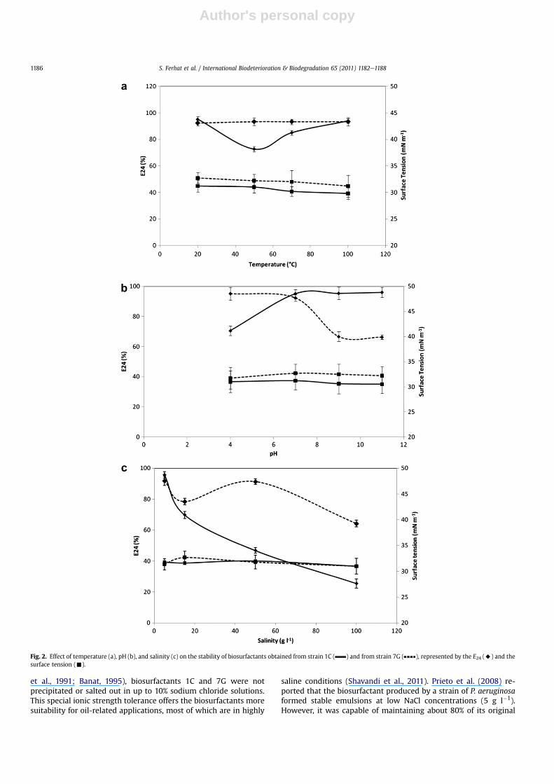
Fig. 2a shows that the surface tension of biosurfactant 1C wasnot influenced significantly by temperature. Its surface tensionremains similar over a range of temperatures between 20 and100 �C (average of 31 mN m�1). However, its E24 values decreasedwith temperatures between 20 �C (E24 ¼ 95.23%) and 50 �C(E24 ¼ 72.72%), and this value increased at temperatures above70 �C (E24 ¼ 85.10%) to reach a value of E24 ¼ 94% at 100 �C. Simi-larly, the surface tension of biosurfactant 7G was thermostable(average of 32 mN m�1 between 20 �C and 100 �C) but, unlikebiosurfactant 1C, its E24 values remain stable (average of 95%)(Fig. 2a).
These results show that the thermostability of biosurfactants 1Cand 7G is comparable to that of other biosurfactants (Makkar andCameotra, 2002). The emulsification index of these biosurfactantswas quite stable compared to synthetic surfactants such as SDS,where a drop of more than 70% was recorded for emulsifying indexvalues in the range of 20e100 �C (Ivshina et al., 1998).
The surface tension of biosurfactant 1C was stable from pH 4(g ¼ 31 mN m�1) to pH 11 (g ¼ 30.5 mN m�1) and from pH 4(g ¼ 31.7 mNm�1) to pH 11 (g ¼ 32.2 mNm�1) for biosurfactant 7G(Fig. 2b). The effect of pH on surface tension has been reported forbiosurfactants for different microorganisms (Abu-Ruwaida et al.,1991). Similarly, the surface tension of liposan produced byCandida lipolytica remained stable between pH 2.0 and 5.0(Cirigliano and Carman, 1985), and that of emulsan produced byAcinetobacter calcoaceticusRAG-1was stable betweenpH5.0 and6.0.
With respect to E24, values for biosurfactant 1C were optimalfrom pH 7 to 11 (E24 average of 95), but they decreased at acid pH(pH 4: E24 ¼ 70.58%). On the other hand, for biosurfactant 7G, theE24 values were optimal from pH 4 to pH 7 (E24 average of 95%) andthey decreased at basic pH (pH 11: E24 ¼ 32.2%).
These results are similar to thoseobtainedbyPornsunthorntaweeet al. (2008). Emulsification capacity was very sensitive to the pHchanges (Shavandi et al., 2011). The biosurfactant produced by strain1C shows increased emulsification efficiency with increasing pHbetween 4 and 7. Similar results were obtained for biosurfactantproduced by P. aeruginosa MR01 (Lotfabad et al., 2009). In compar-ison, the synthetic surfactant SDSmaintains its emulsifying capacityfor pH values ranging between 4.0 and 9.5.
The surface tension of biosurfactant 1C or 7G remained stable insolution with increasing concentrations of NaCl from 5 g l�1 to100 g l�1 (average g ¼ 31 mN m�1). However, the E24 valuesdecreased when the salt concentration was increased from 5 to100 g l�1 (Fig. 2c).
There are reports that the presence of salts results in disruptionof emulsions of oil and water, thus affecting the surface tension andemulsifying ability of surfactants (Jung et al., 1995). As evidencedfrom data shown in Fig. 2c, the surface tension of biosurfactantremained stable up to 100 g l�1 of NaCl. This also indicates thatbiosurfactants produced by isolates 1C and 7G are effective in thepresence of monovalent ions (Naþ). While 20e30 g l�1 of salt isoften sufficient to deactivate chemical surfactants (Abu-Ruwaida
Fig. 1. Emulsification activity (E24) of biosurfactants against different hydrocarbonsand oils: in dark gray, for 1C biosurfactant and in light gray, for 7G biosurfactant.
S. Ferhat et al. / International Biodeterioration & Biodegradation 65 (2011) 1182e1188 1185
Author's personal copy
et al., 1991; Banat, 1995), biosurfactants 1C and 7G were notprecipitated or salted out in up to 10% sodium chloride solutions.This special ionic strength tolerance offers the biosurfactants moresuitability for oil-related applications, most of which are in highly
saline conditions (Shavandi et al., 2011). Prieto et al. (2008) re-ported that the biosurfactant produced by a strain of P. aeruginosaformed stable emulsions at low NaCl concentrations (5 g l�1).However, it was capable of maintaining about 80% of its original
Fig. 2. Effect of temperature (a), pH (b), and salinity (c) on the stability of biosurfactants obtained from strain 1C ( ) and from strain 7G ( ), represented by the E24 (A) and thesurface tension (-).
S. Ferhat et al. / International Biodeterioration & Biodegradation 65 (2011) 1182e11881186

Author's personal copy
activity up to a salinity level 10%. There are few reports in theliterature indicating that in saline soil, as compared to non-salinesoil, degradation of parathion and total crude oil was lowered(Reddy and Sethunathan, 1985; Minai-Tehrani et al., 2009). Thus,there is a need for extensive studies in order to evaluate theeffectiveness of the bioremediation processes for polluted siteshaving different levels of salinity.
3.5. Structural determination
The carbohydrate test based on the Dubois assay was positive,which indicates that the isolated biosurfactants could be glyco-lipids. A glycolipid biosurfactant was also described from themarine B. casei MSA19 (Seghal-Kiran et al., 2010c), whereas a lip-opeptide biosurfactant produced by B. aureum strain MSA13 waspreviously characterized (Seghal-Kiran et al., 2010b). However, fewreports have described and characterized the production of bio-surfactant from Ochrobactrum. For example, an EPS emulsifier wasproduced by an O. anthropi strain (Calvo et al., 2008).
Analysis using FTIR showed relatively similar profiles for bothbiosurfactants. The deformation vibrations at 1377 cm�1 (Fig. 3a)and 1461 (Fig. 3b) confirm the presence of alkyl groups. Thecarbonyl stretching band was found at 1700 cm�1, which is char-acteristic for ester compounds. The ester carbonyl group was alsoshown from the band at 1310 cm�1, which corresponds to CeOdeformation vibrations (Fig. 3a and b).
The adsorption bands located at 2723 and 1461 (Fig. 3b) with1377 cm�1 (Fig. 3a) indicate that all of them have chemical
structures identical to those of glycolipids. The adsorption peakspresent at 2723 cm�1 (Fig. 3a and b) are expected to be the CeHstretching vibrations of the hydrocarbon chain positions. Thecharacteristic peak displayed at 1770 cm�1 (Fig. 3a) and 1774(Fig. 3b) relates to the C]O stretching vibrations of the carbonylgroups, while the CeO stretching bands at 1458 cm�1 (Fig. 3a),1461 cm�1 (Fig. 3b) and 1377 cm�1 (Fig. 3a and b) confirm thepresence of the bonds formed between carbon atoms and hydroxylgroups in the chemical structures of the glycoside part.
The type of glycolipids produced depends on the bacterial strain,the carbon source used and the process strategy (Déziel et al., 1999).The LCeMS analysis of biosurfactant 1C shows two peaks exhibitingthe same molecular mass of 328 and fragmentation pattern at m/zof 328.9, 312.8, 250.8, and 207.8. The LCeMS spectra of bio-surfactant 7G shows two peaks exhibiting fragmentation patternsof m/z of 144.8, 127.9, 99.9, 85.8, 72, and 57.1 for peak 1 and 336.5,318.4, and 300.5 for peak 2.Mass spectroscopy analysis should beconfirmed by NMR analysis in order to determine the exact struc-tures of the biosurfactants.
4. Conclusion
Screening of biosurfactant-producing bacteria from soilscontaminated by hydrocarbons constitutes a powerful tool for theselection of strains with high potential production of emulsifyingagent. Biosurfactants are of interest due to their capacity to emul-sify hydrophobic products and antimicrobial agents. In this study,the crude biosurfactants produced from Ochrobactrum sp. strain 1Cand Brevibacterium sp. strain 7G were chemically characterized asa mixture of glycolipid species. Crude biosurfactants showedcomparable physicochemical properties in terms of the surfaceactivities. Moreover, the antibacterial activities of these two bio-surfactants were demonstrated. Crude biosurfactants were able toreduce the surface tension of pure water to 31e32 mN m�1 andcritical micellar concentrations (CMC) were found to be 1.5 g l�1
and 2 g l�1 for 1C and 7G biosurfactants, respectively. Crude bio-surfactants also exhibited a robust tolerance with slight differencesfor heat, pH, and salinity, and were able to form stable water-in-oilemulsions with various types of vegetable or hydrocarbon oils. It issuggested that the bacterial isolates are suitable candidates forpractical field application for effective in-situ bioremediation ofhydrocarbon-contaminated sites.
Acknowledgments
This work was supported by a grant provided by the AUF(Agence Universitaire de la Francophonie, project contractn�6313PS652). Many thanks go to Mr. Adel Gargoubi and MissLobna Jlail for their help in structural analyses. Thanks also toMr. Brahim Djelassi for help in bacterial strain identifications.
References
Abouseoud, M., Maachi, R., Amrane, A., Boudergua, S., Nabi, I., 2008. Evaluation ofdifferent carbon and nitrogen sources in production of biosurfactant by Pseu-domonas fluorescens. Desalination 223, 143e151.
Abu-Ruwaida, A.S., Banat, S., Haditirto, I.M., Salem, A., Kadri, M., 1991. Isolation ofbiosurfactant-producing bacteria product characterization, and evaluation. ActaBiotechnologica 11, 315e324.
Bai, G., Brusseau, M.L., Miller, R.M., 1997. Biosurfactant-enhanced removal ofresidual hydrocarbon from soil. Journal of Contaminant Hydrology 25, 157e170.
Banat, I.M., 1995. Biosurfactants production and possible uses in microbialenhanced oil recovery and oil pollution remediation: a review. BioresourceTechnology 51, 1e12.
Banat, I.M., Marchant, R., Rahman, T.J., 2004. Geobacillus debilis sp. nov., a novelobligately thermophilic bacterium isolated from a cool soil environment, andreassignment of Bacillus pallidus to Geobacillus pallidus comb. nov. InternationalJournal of Systematic and Evolutionary in Microbiology 54, 2197e2201.
Fig. 3. FTIR analysis of biosurfactants (A: obtained from strain 1C, and B: obtainedfrom strain 7G).
S. Ferhat et al. / International Biodeterioration & Biodegradation 65 (2011) 1182e1188 1187

Author's personal copy
Batista, S.B., Mounteer, A.H., Amorim, F.R., Totola, M.R., 2006. Isolation and char-acterization of biosurfactant/bioemulsifier from petroleum-contaminated sites.Bioresource Technology 97, 868e875.
Bodour, A.A., Maier, R.M., 2002. Biosurfactants: types, screening methods, andapplications. In: Bitton, G. (Ed.), Encyclopedia of environmental microbiology.John Wiley and Sons Inc, Hoboken, New Jersey, pp. 750e770.
Calvo, C., Silva-Castro, G.A., Uad, I., García-Fandiño, C., Laguna, J., González-López, J.,2008. Eficiency of the EPS emulsifier produced by Ochrobactrum anthropi indifferent hydrocarbon bioremediation assays. Journal of Industrial Microbiologyand Biotechnology 35, 1493e1501.
Chamkha, M., Mnif, S., Sayadi, S., 2008. Isolation of a thermophilic and halophilictyrosol-degrading Geobacillus from a Tunisian high temperature oil field. FEMSMicrobiology Letters 283, 23e29.
Cirigliano, M.C., Carman, G.M., 1985. Purification and characterization of liposana bioemulsifier from Candida lipolytica. Applied and Environmental Microbi-ology 54, 747e750.
Cooper, D.G., Zajic, J.E., 1980. Surface-active compounds from microorganisms.Advances in Applied Microbiology 26, 229e253.
Darvishi, P., Ayatollahi, S., Mowla, D., Niazi, A., 2011. Biosurfactant production underextreme environmental conditions by an efficient microbial consortium,ERCPPI-2. Colloids and Surfaces B-Biointerfaces 84, 292e300.
Das, K., Mukherjee, A.K., 2007. Crude petroleum-oil bio-degradation efficiency ofBacillus subtilis and Pseudomonas aeruginosa strains isolated from a petroleum-oil contaminated soil from North-East India. Bioresource Technology 98,1339e1345.
Desai, J.D., Banat, I.M., 1997. Microbial production of surfactants and theircommercial potential. Microbiology and Molecular Biology Reviews 61, 47e64.
Déziel, E., Lépine, F., Dennie, D., Boismenu, D., Mamer, O.A., Villemur, R., 1999. Liquidchromatography/mass spectrometry analysis of mixtures of rhamnolipidsproduced by Pseudomonas aeruginosa strain 57RP grown on mannitol ornaphthalene. Biochimica et Biophysica Acta (BBA) e Molecular and Cell Biologyof Lipids 2-3, 244e252.
Dhiman, A., Nanda, A., Ahmad, S., Narasimhan, B., 2011. In vitro antimicrobialactivity of methanolic leaf extract of Psidium guajava L. Journal of Pharmacy andBioallied Sciences 3, 226e229.
Dubois, M., Gilles, K.A., Hamilton, J.K., Rebers, P.A., Smith, F., 1956. Colorimetricmethods for determination of sugars and related substances. Analytic Chem-istry 28, 350e356.
Haba, E., Espuny, M.J., Busquets, M., Manresa, A., 2000. Screening and production ofrhamnolipids by Pseudomonas aeroginusa 47T2 NCIB 40044 from waste fryingoils. Journal of Applied Microbiology 88, 379e387.
Hommel, R.K., 1994. In: Ratledge, C. (Ed.), Formation and function of biosurfactantsfor degradation of water-insoluble substrates. KluwerAcademic Publishers,Dordrecht, pp. 63e87.
Ilori, M.O., Adebusoye, S.A., Ojo, A.C., 2008. Isolation and characterization ofhydrocarbon-degrading and biosurfactant-producing yeast strains obtainedfrom a polluted lagoon water. World Journal of Microbiology & Biotechnology24, 2539e2545.
Ivshina, I.B., Kuyukina, M.S., Philp, J.C., Christofi, N., 1998. Oil desorption frommineral and organic materials using biosurfactant complexes produced byRhodococcus species. World Journal of Microbiology and Biotechnology 14,711e717.
Iwabuchi, N., Sunairi, M., Urai, M., Itoh, C., Anzai, H., Naka-jima, M., Harayama, S.,2002. Extracellular polysaccharides of Rhodococcus rhodochrous S-2 stimulatethe degradation of aromatic components in crude oil by indigenous marinebacteria. Applied and Environmental Microbiology 68, 2337e2343.
Jung, H.K., Lee, J.B., Yim, G.B., Kim, E.K., 1995. Properties of microbial surfactant S-acid. Korea Journal of Biotechnology and Bioengineering 10, 71e77.
Karanth, N.G.K., Deo, P.G., Veenanadig, N.K., 1999. Microbial production of bio-surfactant and their importance. Current Science 77, 116e126.
Kim, S.H., Lim, E.J., Lee, S.O., Lee, J.D., Lee, T.H., 2000. Purification and character-ization of biosurfactants from Nocardia sp. L417. Biotechnology Applied ofBiochemistry 31, 249e253.
Kitamoto, D., Isoda, H., Nakahara, T., 2002. Functions and potential applications ofglycolipid biosurfactants: from energy saving materials to gene deliverycarriers. Journal of Bioscience and Bioengineering 94, 187e201.
Kiyohara, H., Nagao, K., Yana, K., 1982. Rapid screen for bacteria degrading water-insoluble, solid hydrocarbons on agar plates. Applied and EnvironmentalMicrobiology 43, 454e457.
Kosaric, N., 1992. Biosurfactants in industry. Pure Applied Chemistry 64, 1731e1737.Lotfabad, T.B., Shourianc, M., Roostaazada, R., Najafabadib, A.R., Adelzadeha, M.R.,
Noghabic, K.A., 2009. An efficient biosurfactant-producing bacterium Pseudo-monas aeruginosa MR01, isolated from oil excavation areas in south of Iran.Colloids and Surfaces B: Biointerfaces 69, 183e193.
Makkar, R.S., Cameotra, S.S., 2002. An update on the use of unconventionalsubstrates for biosurfactant production and their new applications. AppliedMicrobiology and Biotechnology 58, 428e434.
Minai-Tehrani, D., Minoui, S., Herfatmanesh, A., 2009. Effect of salinity on biodeg-radation of polycyclic aromatic hydrocarbons (PAHs) of heavy crude oil in soil.Contaminant Toxicology 82, 179e184.
Mnif, S., Chamkha, M., Sayadi, S., 2009. Isolation and characterization of a Hal-omonas sp. a hydrocarbon degrading bacterium under hypersaline condition.Journal of Applied Microbiology 107, 785e794.
Pornsunthorntawee, O., Arttaweeporn, N., Paisanjit, S., Somboonthanate, P., Abe, M.,Rujiravanit, R., Chavadej, S., 2008. Isolation and comparison of biosurfactantsproduced by Bacillus subtilis PT2 and Pseudomonas aeruginosa SP4 for microbialsurfactant-enhanced oil recovery. Biochemical Engineering Journal 42, 172e179.
Prieto, L.M., Michelon, M., Burkert, J.F.M., Kalil, S.J., Burkert, C.A.V., 2008. Theproduction of rhamnolipid by a Pseudomonas aeruginosa strain isolated froma southern coastal zone in Brazil. Chemosphere 71, 1781e1785.
Reddy, B.R., Sethunathan, N., 1985. Salinity and the persistence of parathion inflooded soil. Soil Biology and Biochemistry 17, 235e239.
Ron, E.Z., Rosenberg, E., 2002. Biosurfactants and oil remediation. Current Opinionin Biotechnology 3, 249e252.
Saravanakumari, P., Mani, K., 2009. Structural characterization of a novel xylolipidbiosurfactant from Lactococcus lactis and analysis of antibacterial activityagainst multi-drug resistant pathogens. Bioresource Technology 101,8851e8854.
Sarubbo, L.A., 2006. Production and stability studies of the bioemulsifier obtainedfrom a strain of Candida glabrata UCP 1002. Journal of Biotechnology 9,400e406.
Seghal-Kiran, G., Sabu, A., Selvin, J., 2010a. Synthesis of silver nanoparticles byglycolipid biosurfactant produced from marine Brevibacterium casei MSA19.Journal of Biotechnology 148, 221e225.
Seghal-Kiran, G., Anto-Thomas, T., Joseph, s., Sabarathnam, B., Lipton, A.P., 2010b.Optimization and characterization of a new lipopeptide biosurfactant producedby marine Brevibacterium aureum MSA13 in solid state culture. BioresourceTechnology 101, 2389e2396.
Seghal-Kiran, S., Sabarathnam, B., Selvin, J., 2010c. Biofilm disruption potential ofa glycolipid biosurfactant from marine Brevibacterium casei. FEMS Immunologyand Medical Microbiology 3, 432e438.
Shavandi, M., Mohebali, G., Haddadi, A., Shakarami, H., Nuhi, A., 2011. Emulsificationpotential of a newly isolated biosurfactant-producing bacterium, Rhodococcussp. strain TA6. Colloids and Surfaces B. Biointerfaces 82, 477e482.
Snehal, R., Mutalik-Bhalchandra, K., Renuka, M.J., Desai, M., Sanjay, N., 2008. Use ofresponse surface optimization for the production of biosurfactant from Rho-dococcus spp. MTCC 2574. Bioresource Technology 99, 7875e7880.
Suk, W.S., Lim, H.J., Lee, S.J., 1999. Compositional analysis and some properties ofbiosurfactant from Pseudomonas sp. SW1. Korea Journal of Applied Microbi-ology and Biotechnology 27, 41e45.
Willumsen, P.A., Karlson, U., 1997. Screening of bacteria isolated from PAH-contaminated soils, for production of biosurfactants and bioemulsifiers.Biodegradation 7, 415e423.
Yin, H., Qiang, J., Jia, Y., Ye, J., Peng, H., Qin, H., Zhang, N., He, B., 2008. Characteristicsof biosurfactant produced by Pseudomonas aeruginosa S6 isolated from oil-containing wastewater. Process Biochemistry 44, 302e308.
S. Ferhat et al. / International Biodeterioration & Biodegradation 65 (2011) 1182e11881188