Amorphous iron-chromium oxide nanoparticles prepared by sonochemistry

Chromium resistance strategies and toxicity: what makesOchrobactrum tritici 5bvl1 a strain highly resistant
Paula Vasconcelos Morais • Rita Branco •
Romeu Francisco
Received: 30 November 2010 / Accepted: 24 March 2011 / Published online: 7 April 2011
� Springer Science+Business Media, LLC. 2011
Abstract Large-scale industrial use of chromium
(Cr) resulted in widespread environmental contami-
nation with hexavalent chromium (Cr(VI)). The
ability of microorganisms to survive in these envi-
ronments and detoxify chromate requires the pres-
ence of specific resistance systems. Several Cr(VI)
resistant species, belonging to a variety of genera,
have been isolated in recent years. Ochrobactrum
tritici strain 5bvl1 is a model for a highly Cr(VI)-
resistant and reducing microorganism, with different
strategies to cope with chromium. The strain contains
the transposon-located (TnOtChr) chromate resis-
tance genes chrB, chrA, chrC, chrF. The chrB and
chrA genes were found to be essential for the
establishment of high resistance but not chrC or
chrF genes. Other mechanisms involved in chromium
resistance in this strain were related to strategies such
as specific or unspecific Cr(VI) reduction, free-
radical detoxifying activities, and repairing DNA
damage. Expression of the chrB, chrC or chrF genes
was related to increased resistance to superoxide-
generating agents. Genetic analyses also showed that,
the ruvB gene is related to chromium resistance in
O. tritici 5bvl1. The RuvABC complex probably does
not form when ruvB gene is interrupted, and the
repair of DNA damage induced by chromium is
prevented. Aerobic or anaerobic chromate reductase
activity and other unspecific mechanisms for chro-
mium reduction have been identified in different
bacteria. In the strain O. tritici 5bvl1, several
unspecific mechanisms were found. Dichromate and
chromate have different effects on the physiology of
the chromium resistant strains and dichromate seems
to be more toxic. Toxicity of Cr(VI) was evaluated by
following growth, reduction, respiration, glucose
uptake assays and by comparing cell morphology.
Keywords Chromium � Chromate � Dichromate �Toxicity � Resistant bacteria
Introduction
Of the various toxic heavy metals discharged into the
environment through various industrial wastewaters
that constitute one of the major causes of environ-
mental pollution, chromium is one of the most toxic,
and has become a serious health concern. Extensive
use of chromium, e.g., in electroplating, tanning,
textile dyeing and as a biocide in power plant cooling
water, results in discharges of chromium-containing
effluents. The effluents from these industries contain
Cr(VI) and trivalent chromium (Cr(III)) in high
P. V. Morais (&)
Department of Life Sciences, FCTUC, University of
Coimbra, 3001-401 Coimbra, Portugal
e-mail: [email protected]
P. V. Morais � R. Branco � R. Francisco
IMAR-CMA, 3004-517 Coimbra, Portugal
123
Biometals (2011) 24:401–410
DOI 10.1007/s10534-011-9446-1

concentrations. Cr(VI) can be present in solution either
as chromate (CrO42-), bichromate (HCrO4
-), or
dichromate (Cr2O72-) anions, in an equilibrium
dependent on pH and ionic strength (Ramsey et al.
2001). While Cr(VI) is known to be toxic to both plants
and animals, a strong oxidizing agent and a potential
carcinogen, Cr(III) is generally only toxic to plants at
very high concentrations and is either less toxic or non-
toxic to animals. Chromate exerts diverse toxic effects
on bacteria, including competitive inhibition of sul-
phate transport as well as DNA and protein damage,
that happens after Cr(VI) intracellular reduction to
the Cr(III) species, a process that generates reactive
oxygen species (ROS) (Branco et al. 2008; Costa
2003). Mechanisms for bacterial resistance to chro-
mate are varied and may be conferred by genes located
on chromosomes or on plasmids.
Several Cr(VI) resistant species belonging to a
variety of genera have been isolated in recent years.
Genes conferring resistance to chromate, encoded
either on chromosomal genes or on plasmids, have
been found in Pseudomonas sp. (Mondaca et al.
1998; Bopp and Erlich 1988; Cervantes and Ohtake
1988), Bacillus cereus SJ1 (He et al. 2010), Shewa-
nella sp. (Aguilar-Barajas et al. 2008); Arthrobacter
sp. (Henne et al. 2009), Lysinibacillus fusiformis ZC1
(He et al. 2011) and Cupriavidus metallidurans (Nies
et al. 1989). Usually, the genes encode membrane
transporters, which directly mediate efflux of chro-
mate ions across the cytoplasmic membrane. Other
resistance systems are generally related to strategies
such as specific or unspecific Cr(VI) reduction, free-
radical detoxifying activities, repairing of DNA
damage, and processes associated with sulfur or iron
homeostasis.
The main resistance mechanisms are related with
the membrane-potential dependent efflux of chromate
through the membrane transporter ChrA (Branco et al.
2008; Nies et al. 2006), or with the presence of
chromate reductase activity, converting Cr(VI) com-
pounds to Cr(III), that is less toxic. There is evidence
for both aerobic (Kwak et al. 2003; McLean and
Beveridge 2001) and anaerobic (Chardin et al. 2003;
Daulton et al. 2001) reduction pathways with different
microbes. Anaerobic reduction is associated to dis-
similatory reduction of Cr(VI) by the respiratory chain
(Chardin et al. 2003; Daulton et al. 2001; Wang et al.
1990). The aerobic strategies described until now
were mostly related to soluble enzymes dependent
of NAD(P)H (Elangovan et al. 2006; Camargo et al.
2003), which are able to transfer electrons to Cr(VI),
thereby reducing it.
The Ochrobactrum tritici strain 5bvl1, a highly
Cr(VI) resistant strain able to reduce Cr(VI) in
mineral medium supplemented with glucose (Branco
et al. 2004) was isolated from activated sludge of a
water treatment plant which received effluents from
tanneries (Francisco et al. 2002). Its ability to resist to
high concentrations of chromate is conferred by the
presence of a transposon TnOtChr, carrying several
genes including the Cr(VI) pump ChrA (Branco et al.
2008). Therefore, this resistant and reducing strain is
here described as a model of the integrative strategies
of bacteria to cope with chromium.
Comparative organization of the chr operons
transposon
The O. tritici 5bvl1 strain was isolated from a
consortium of bacteria adapted to live in an environ-
ment with high concentrations of chromate (Francisco
et al. 2002) and contains genes that confer high
tolerance against Cr(VI), which are located on the
TnOtChr transposon (Fig. 1). The structure of this
transposon, in which the tnpR (resolvase) and tnpA
(transposase) genes flank the chr genes, indicates its
ability to distribute the chromium resistance genes.
The tnpR and tnpA genes at both ends enclose a region
that contains four chromate resistance-related genes.
Others transposons carrying chromate resistance
genes have already been found in different microor-
ganisms. In the case of pB4-based transposon Tn5719,
from an uncultivated bacterium (Tauch et al. 2003),
the tnpR and tnpA genes are physically adjacent to
each other as in the majority of the transposons of the
Tn21 subfamily. The plasmid pCNB1 from Coma-
monas sp. strain CNB-1 also contains a putative
chromate transporter and a regulator located on a
transposon (Ma et al. 2007). In the genome of
B. cereus SJ1, the chrA1 encoding the ChrA protein
is located downstream of a potential transcriptional
regulator gene, chrI. The region of chrA1 and chrI
also contained several putative coding sequences
encoding homologs of Tn7-like transposition proteins
and a resolvase that could potentially have been
involved in horizontal gene transfer events (He et al.
2010).
402 Biometals (2011) 24:401–410
123

Ochrobactrum tritici 5bvl1 is able to survive in
chromium at high millimolar concentrations, unlike
other strains, where the presence of chr genes
provided protection only in the submillimolar range.
Moreover, the chromate-inducible systems are some-
times inducible by sulfate and other molecules but
not in O. tritici 5bvl1 (Peitzsch et al. 1998). This fact
could explain the limited protective ability of some
characterized chr systems and may be related to the
high resistance profile of strain 5bvl1, since a strong
activation of the efflux pumps could lead to the co-
extrusion of sulfate and the resulting metabolic
deficiency in sulfur donors.
Genes for chromium resistant phenotype
in O. tritici strain 5bvl1
chrA
In O. tritici 5bvl1, the main chromate resistance
mechanism seems to be the chromium efflux from
cytoplasm through ChrA pump. This evidence was
confirmed by chrA mutagenesis and expression of
chrA gene in a chromate-sensitive bacterium (Branco
et al. 2008). Therefore, interruption of the chrA gene
by Tn5 transposon (mutant E117) abolished chro-
mate resistance. The complementation of type strain
O. triticiT with chrA gene increased its ability to grow
in the presence of high chromate concentrations.
This chromate resistance system operates by main-
taining low cellular chromium levels even in the
presence of millimolar extracellular chromate con-
centrations. Reduced accumulation of chromate has
been associated with chromate resistance in other
microorganisms (Pimentel et al. 2002; Alvarez et al.
1999; Nies et al. 1989). ChrA proteins from C.
metallidurans (Nies et al. 1989),Shewanella sp.
ANA-3 (Aguilar-Barajas et al. 2008) and Pseudo-
monas aeruginosa (Cervantes et al. 2001) have been
functionally characterized as chromate efflux pumps.
It was shown that the chromate transporter ChrA
works as a chemiosmotic pump that extrudes
chromate using the proton-motive force (Alvarez
et al. 1999).
ChrA proteins belong to the CHR superfamily,
which includes several putative homologs from all
three domains of life (Ramırez-Dıaz et al. 2008).
ChrA homologs can exist in two sizes: small proteins,
or SCHR (about 200 aa), having only one domain, and
large proteins, or LCHR (about 400 aa, except
eukaryotic proteins of 500–600 aa), with two homol-
ogous domains. Analysis of chr operons, which
contain two adjacent genes encoding the amino- and
the carboxy-terminal parts of the full-length ChrA
protein, suggest that LCHR probably originated by gene
duplication followed by gene fusion (Ramırez-Dıaz
et al. 2008).
tnpA chrF chrC chrA chrB tnpR
TnOtChr
IR IR
orf44 orf46 chrC chrA chrB tnpR
IR IR
tnpA
Tn5719
tnpA tnpR chrApadR
TnCNB1
IR IR
tnpR chrAchrI
B. cereus ATCC10987
Fig. 1 Comparison of genetic determinants of chromate
resistance as identified in other transposons versus TnOtChr.
TnOtChr of strain O. tritici 5bvl1 (Branco et al. 2008); Tn5719identified on plasmid pB4 (Tauch et al. 2003); TnCNB1 of
strain Comamonas CNB1 (Ma et al. 2007) and Bacillus cereus
ATCC10987 (He et al. 2010). Inverted repeats are shown as
vertical black bars. Black arrows are arsenic resistance genes,
dashed arrows are Tn7 like transposition protein genes and
grey arrows are other genes
Biometals (2011) 24:401–410 403
123

chrB
The chrB gene is a common feature of the genetic
organization of the chr operons and has been proposed
to work as an activator of the chromate resistance
determinants in C. metallidurans CH34 (Juhnke et al.
2002). Unlike ChrB proteins from C. metallidurans
CH34 that activated the chromate resistance system in
response to both chromate and sulfate (Juhnke et al.
2002; Peitzsch et al. 1998), chrB from strain 5bvl1 was
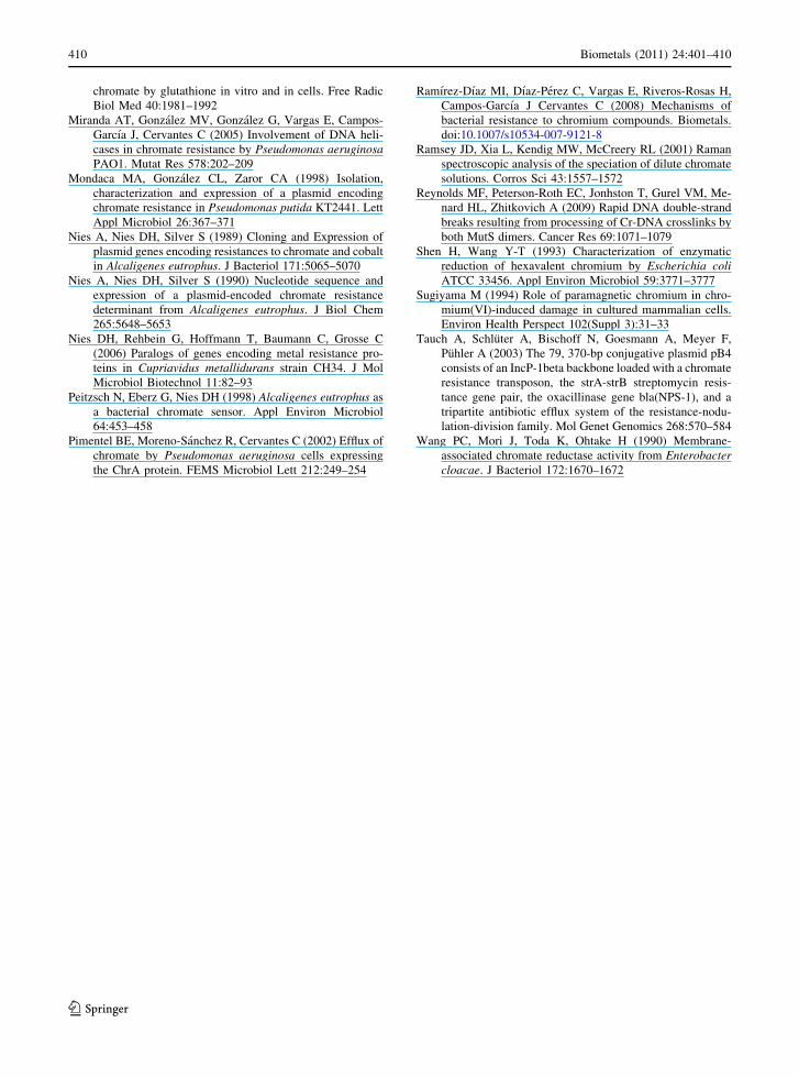
induced exclusively by Cr(VI) (Branco et al. 2008).
Chromate induction experiments with chrB-gfp fusion
show a direct relation between reporter expression and
Cr(VI) concentration and simultaneously revealed that
GFP expression is dependent on chromate time
induction (Fig. 2). Although the ChrB role in O. tritici
strain 5bvl1 is not completely clarified at this point, the
O. tritici 5bvl1 ChrB protein seems to act as the
chromate-sensitive regulator of the chr operon. This
finding is supported by a bioinformatic analysis using
protein function prediction software (Dodd and Egan
1990) that suggests a probable helix-turn-helix DNA-
binding motif in ChrB C-terminus sequence, although
other possible roles cannot be completely ignored. In
Shewanella strain ANA-3, ChrB contains a rhodanese-
like domain (Aguilar-Barajas et al. 2008) also found in
the arsenate reductase Acr2p of Saccharomyces cere-
visiae as part of its active site. Therefore, one
suggested role of ChrB in Shewanella strain ANA-3
was the reduction of Cr(VI) before extrusion by ChrA,
in analogy to arsenic resistance operons. In Arthro-
bacter sp. strain FB24, two open reading frames
encode the complete sequence for a full-length chrB
gene. The chrB-Nterm and the chrB-Cterm2 are both
maximally transcribed in the presence of different
chromate concentrations (Henne et al. 2009).
The importance of the chrB gene on chromium
resistance in strain 5bvl1 was also tested through
measurement of the respiration rate (Francisco et al.
2006; Konopka and Zakharova 1999). Clear differ-
ences were noticed when comparing strain 5bvl1 with
Cr-sensitive O. triticiT type strain and ChrA mutant
strain E117 (Tn5 inactivated ChrA). Differences in
the inhibition of respiration were observed between
the three strains, that could not only be justified by
the absence of the ChrA pump. Inhibition of the
respiration by chromium was less severe in strain
5bvl1, without functional ChrA, ChrC and ChrF
proteins, suggesting once more that ChrB has a
secondary activity, likely responsible for the remain-
ing protection (Francisco et al. 2010; Branco et al.
2008).
Additional chr genes: chrC and chrF
There is considerable dissimilarity in the genomic
context, surrounding ChrA orthologs (Ramırez-Dıaz
et al. 2008). Although its function is known, it
0
500
1000
1500
2000
2500
3000
3500
4000
4500
5000
1 10 100 1000
Chromate concentration, μM
RF
U
0
2
4
6
8
10
12
14
0 1 2 3 4 5
Chromate exposure time, h
Ind
uct
ion
rat
io
Fig. 2 a Effect of chromate on GFP expression of pchrB-gfpbiosensor after different chromate exposure periods: 0 h (filleddiamond); 1 h (open square); 2 h (filled circle); 3 h (opentriangle) and 4 h (open circle). Fluorescence (in RFU)
measured with a fluorometer is defined as culture fluorescence
divided by culture OD 600 nm. b Induction ratio of GFP
expression at different chromate exposure times, for the several
chromate concentrations: 1 lM (filled diamond); 10 lM (opensquare); 50 lM (filled triangle); 100 lM (open circle) and
500 lM (filled square)
404 Biometals (2011) 24:401–410
123

appears that the presence of a chrA gene alone cannot
explain the difference in the resistance levels found in
different microorganisms. In the case of Ochrobac-
trum, Cr(VI)-sensitive strains transformed with a
plasmid carrying the chrA and chrB genes from
TnOtChr, showed similar growth with chromate as
the wild-type strain O. tritici 5bvl1. No additional
growth advantage was provided by the presence of
the other additional genes, chrC and chrF (Branco
et al. 2008). On the other hand, in C. metallidurans
CH34, deletion of chrC resulted in a slight decrease
in chromate resistance, compared to the wild-type
strain (0.3 mM chromate minimal inhibitory concen-
tration (MIC) versus 0.35 mM, respectively). In the
same study, deletion of chrF2 did not affect chromate
resistance levels (Juhnke et al. 2002). Introduction of
chrA alone into Cr(VI) sensitive strain Arthrobacter
produced lower resistance levels than those found in
strains whose cells carried the entire operon. Similar
results were obtained when expression of chrA from
Shewanella resulted only in a slight increase of
chromium resistance of Escherichia coli compared to
strains bearing the entire ANA-3 chrBAC operon.
The ANA-3 chrA gene conferred chromate resistance
in P. aeruginosa. This phenotype was enhanced by
the presence of the host chrR regulatory gene
(Aguilar-Barajas et al. 2008), thus emphasizing the
importance of accessory genes in achieving higher
levels of chromate resistance. In conclusion, the exact
contributions made by chrC and chrF are not so
apparent and may vary depending on the host strain.
Other mechanisms of chromate resistance
SOD activity
Chromate exposure of cells has been associated with
activation of several protective systems. For instance,
when E. coli was submitted to chromium stress, the
levels of superoxide dismutase (SOD) and catalase
enzymes increased significantly but the levels of
glutathione and other thiols decreased significantly.
On the other hand, cells of Caulobacter crescentus
responded to chromium exposure with the upregula-
tion of genes responsible for dealing with metal
toxicity, including SOD and glutathione S-transferase
(Hu et al. 2005).
Plasmid pMOL28 from C. metallidurans CH34
also encodes ChrC and ChrE proteins (Juhnke et al.
2002). ChrC shows homology to iron-containing
SOD enzymes able to detoxify superoxide radicals;
however, the authors have not assigned a clear
function to ChrC (Juhnke et al. 2002).
The chrF and chrC genes from O. tritici strain
5bvl1 apparently do not play a major role in
resistance. Rescue of the growth defect in sodAsodB
E. coli double mutant in aerobic minimal medium is
commonly used as a genetic test for the presence of
SOD activity in a protein of interest. We found that
the expression of chrF, chrC or chrB genes restored
the ability of SOD-null E. coli cells to grow in media
without both aromatic and sulfur-containing amino
acids. In bacteria with abnormally high production of
toxic superoxide, in response to chromate (Ackerley
et al. 2006), the expression of ChrC, ChrF and ChrB
proteins should provide an important second line of
defense against this toxic metal.
DNA repair
Components of the recombinational DNA repair
system, like DNA helicases RecG and RuvB, were
shown to participate in the response to DNA damage
caused by chromate in P. aeruginosa (Miranda et al.
2005). Pseudomonas corrugata strain 28, a highly
chromate-sensitive mutant, was defective in the recG
gene encoding a RecG DNA helicase that functions in
resolution of the replication forks established during
DNA replication process (Decorosi et al. 2009).
Other studies with Shewanella oneidensis MR-1
(Chourey et al. 2008) and C. crescentus (Hu et al.
2005) have also supported the contribution of these
helicases as well as other proteins involved in DNA
repair damage in the chromate resistance phenotype.
In an attempt to identify chromate resistance genes
in strain O. tritici 5bvl1, this bacterium was subjected
to Tn5 transposon mutagenesis, obtaining mutant
Q152 (Branco and Morais 2006). Mutant Q152
showed an intermediate chromate-sensitive pheno-
type when compared with the chrA mutant (mutant
E117) (Fig. 3). Genetic analyses established that the
transposon was inserted into the ruvB gene encoding
putative helicase RuvB. In mutant Q152, with the
ruvB gene interrupted, the RuvABC complex prob-
ably does not form, thus blocking the migration of the
Holliday junctions and preventing the repair of DNA
Biometals (2011) 24:401–410 405
123

damage. The ROS species generated during Cr(VI)
reduction to Cr(III) have been already associated with
DNA damage and under these conditions bacteria
need a repair system (Ackerley et al. 2004). As
Cr(VI) has been known to induce the E. coli SOS
repair system (Llagostera et al. 1986), it seems that
RuvB helicase belongs to this repair mechanism and
should participate in the process of DNA repair of
damage caused by the exposure of strain 5bvl1 to
chromate and, most probably, also to other
oxyanions.
chr genes expression in E. coli
Chromate resistance determinants from C. metallidu-
rans CH34 and P. aeruginosa were found to be only
functional in their respective hosts but not in E. coli
(Cervantes et al. 1990; Nies et al. 1990). Therefore,
for a long time it was assumed that E. coli cells were
not able to express chr genes. However, in more
recent works, the expression of chr genes in E. coli
resulted in a chromate resistant phenotype. Aguilar-
Barajas et al. (2007) were able to confer Cr(VI)
resistance to an E. coli strain by expressing the chr
operon from Shewanella sp. strain ANA-3 on a
low-copy plasmid. ChrA from Bacillus subtilis could
also be functionally expressed in E. coli since trans-
formants expressing paired chr3N (chrA N-terminal)
and chr3C (chrA C-terminal) genes showed enhanced
chromate resistance compared to the control strain.
These experiments clearly demonstrate that paired
SCHR proteins confer resistance to chromate. Other
results showed that paired chr1N and chr1C genes,
encoding SCHR proteins from a gram-negative
Burkholderia strain, could also confer chromate
resistance in E. coli (Dıaz-Magana et al. 2009).
Ochrobactrum tritici 5bvl1 chromate resistance
determinants could also be functionally expressed in
E. coli. Since the chrBA genes conferred a higher
resistance level than chrA alone, chrB was required
for maximum chromate resistance (Fig. 4). These
data confirm results presented in previous chapters.
Unspecific mechanisms for chromate reduction
inside the cells
Several strains can reduce Cr(VI) to the less toxic form
Cr(III), though chromate reduction is not typically
considered a resistance mechanism (Ramırez-Dıaz
0 1 2 3 40
1
2
3
4
5
Chromate concentration (mM)
Ab
sorb
ance
(60
0nm
)
Fig. 3 Comparative chromate resistance phenotype of wild-
type O. tritici 5bvl1 (filled square) and Tn5 mutated strains:
chrA mutant (filled triangle); ruvB mutant (open circle).
Absorbance was measured after overnight growth in LB
medium
0
0.1
0.2
0.3
0.4
0.5
0.6
0.7
0.8
0.9
1
0 0.2 0.4 0.6 0.8
Chromate concentration (mM)
Ab
sorb
ance
(60
0nm
)Fig. 4 Chromate resistance of E. coli cells carrying the empty
pProbe-NT vector (filled diamond), or the vector containing
chromate resistance genes, pProbe_chrA (open square) and
pProbe_chrBA (open triangle). Absorbance was measured after
overnight growth in minimal medium (Branco et al. 2008)
406 Biometals (2011) 24:401–410
123

et al. 2008). The mechanisms described can be
grouped in those occurring anaerobically and those
occurring aerobically, with Cr(VI) reduction capacity
varying greatly between the different strains reported
as chromate reducers.
Under anaerobiosis, some bacteria such as Pseu-
domonas fluorescens LB300 (Bopp and Erlich 1988),
E. coli ATCC 33456 or anaerobic sulfate reducers
(Chardin et al. 2003; Shen and Wang 1993), can use
Cr(VI) as an electron acceptor in the electron
transport chain. In O. tritici 5bvl1 we managed to
rule out the possibility of Cr(VI) being reduced by the
electron transport chain as an oxygen substitute by
comparing the inhibition of cell respiration rates with
the inhibition of Cr(VI) reduction under the same
experimental condition (Fig. 5). Moreover, it has
been shown that strain 5bvl1 was not able to grow
under anaerobic conditions in presence of Cr(VI)
(Francisco et al. 2002). Under aerobic conditions,
chromate reduction has been commonly associated
with soluble chromate reductases that use NADH or
NADPH as cofactors. The different chromate reduc-
tases described until now have been revised recently
by Ramırez-Dıaz et al. (2008). The ability of cell
soluble extracts of strain 5bvl1 to reduce Cr(VI)
showed NADH dependence as demonstrated by
spectroscopic wavelength scan analysis and kinetic
experiments. Spectroscopic wavelength scan analysis
showed the complete disappearance of Cr(VI) in
simultaneity with the oxidation of NADH to NAD?
(Fig. 6). Similar aerobic Cr(VI) reduction strategies,
linked to soluble enzymes dependent on NAD(P)H
have in fact been commonly found in several
microorganisms (Cervantes and Campos-Garcia
2007; Ramırez-Dıaz et al. 2008), although in strain
5bvl1 no enzyme could be isolated.
Reduction of Cr(VI) may also be carried out by
chemical reactions associated with compounds such
as amino acids, nucleotides, sugars, vitamins, organic
acids or glutathione (Sugiyama 1994).
Different reduction mechanisms may occur in
strain 5bvl1, since NADH, NADPH, GSH and c-type
cytochromes were found to become oxidized when
exposing cells and cell extracts to Cr(VI). The
depletion of GSH has in fact been reported to be
connected to membrane damage (Francisco et al.
2010) and is usually used by Gram-negative bacteria
as a protection against oxidative stress, as in mito-
chondria (Hanukoglu and Rapoport 1995). The
disappearance of reduced glutathione in glucose-
starved cells exposed to Cr(VI) is a clear indication of
the oxidative stress resulting from the entry of Cr(VI)
into the cytosol as also observed in E. coli K12 when
exposed to chromate (Ackerley et al. 2006) and in
eukaryotic cells (Messer et al. 2006). This is also an
indication of a malfunction of the chromate pump
ChrA on starved cells, as it relies on the membrane-
potential.
The c-type cytochromes are frequently involved in
metal oxidation and reduction although they are not
usually referred as associated to chromium reduction
(Mehta et al. 2005; Branco et al. 2009). Nevertheless,
the reductase from chromate resistant Enterobacter
cloacae HO1 (Wang et al. 1990) is a membrane
2.0 4.0 6.0 8.01.0×10-10
3.0×10⁻¹⁰
5.0×10⁻¹⁰
7.0×10⁻¹⁰
9.0×10⁻¹⁰
1.0×10⁻¹²
7.0×10⁻¹²
1.3×10⁻¹¹
1.9×10⁻¹¹
2.5×10⁻¹¹
3.1×10⁻¹¹
3.7×10⁻¹¹
Cr (mM)
O2
(nm
ol O
2/m
in/c
ell)
Cr
(VI)
(n
mo
l Cr/
min
/cel
l)
Fig. 5 Comparison between 5bvl1 cell respiration (greysquare) and 5bvl1 cell Cr(VI) reduction (black square) in
buffered media with different Cr(VI) concentrations (added as
dichromate)
-0.2
0
0.2
0.4
0.6
0.8
1
300 350 400 450 500 550 600
Ab
sorb
ance
NADH-S2
Cr(VI)-S2
T0
T1
T3
T4
T5
T6
T7
T8
T9
λ (nm)
Fig. 6 Spectroscopic wavelength analysis of NADH (0.2 mM)
and Cr(VI) (100 lM) and sequential spectroscopic wavelength
analysis of a mixture of NADH and Cr(VI) in presence of cells
soluble extract in citrate buffer. The sample was scanned every
5 min (T0-T9) for a total of 45 min. Controls: NADH ? cells
soluble extract (NADH-S2); Cr(VI) ? cells soluble extract
(Cr(VI)-S2)
Biometals (2011) 24:401–410 407
123

associated enzyme that transfers electrons to Cr(VI)
by NADH-dependent cytochromes (Wang et al.
1990). Using cell crude extracts of O. tritici 5bvl1,
cytochrome c oxidation occurred, of completely
reduced cytochromes, (flushed with N2 for oxygen
removal and treated with sodium dithionite and
potassium cyanide) when in presence of Cr(VI).
Although some c-type cytochrome oxidation is
visible after addition of Cr(VI), it is not clear, in
this strain, if the oxidation was made by direct
interaction with the cytochrome, as certain c-type
cytochromes are known to become oxidized when
cells are under oxidative stress (Kagan et al. 2000).
Are chromate and dichromate inducing
the same toxicity on cells?
Different environmental conditions can promote the
presence of different Cr(VI) species. Cr(VI) usually
associates with oxygen to form the oxyanions chro-
mate (CrO42-) and dichromate (Cr2O7
2-). Chromate
and dichromate are in equilibrium which is sensitive
to pH changes, where lower pH pushes the equilib-
rium towards the dichromate ion (Ramsey et al.
2001). Cr(VI) compounds are highly soluble, whereas
derivatives of Cr(III) in the forms of hydroxides,
oxides and sulphates, are water insoluble. Studies of
Cr(VI) toxicity are generally performed using chro-
mate salts in solution, both when studying the effects
on prokaryotes and eukaryotes (Cervantes and
Campos-Garcia 2007; Reynolds et al. 2009), leaving
a lack of information concerning the effects caused
by dichromate salts.
Comparative studies in the literature are not found,
so the differences in toxicity of these two chromate
species are mostly based on the differences found for
O. tritici 5bvl1. Every parameter tested showed that
chromate and dichromate solutions interacted differ-
ently with 5bvl1 cells, and showed very distinct
toxicities, with dichromate causing the most damage.
The ability to grow in presence of Cr(VI), Cr(VI)-
reduction capacity, respiration, glucose uptake and
cell morphology were more severely affected by
dichromate anion (Francisco et al. 2010).
The addition of sodium dichromate caused lower
growth rates, the MIC for dichromate being of 4 mM
both in solid and liquid medium, while in presence of
chromate the strain 5bvl1 could grow above 20 mM
(Branco et al. 2008). Chromium stress induced cell
elongation and deformation and these effects were
particularly visible in cells of strain 5bvl1 (Francisco
et al. 2010) and were also visible in cells of
Ochrobactrum anthropi, S. oneidensis MR-1 and
E. coli K-12 (Ackerley et al. 2006; Chourey et al.
2008; Li et al. 2008) in the presence of dichromate.
Chromium deposition on cell surface was observed in
O. anthropi (Li et al. 2008) but not in strain 5bvl1 due
to its active ChrA chromium pump.
The higher genotoxicity of dichromate compared
to chromate on strain 5bvl1 cells suspensions was also
visible by analysis of DNA degradation (Francisco
et al. 2010). These results were in accordance with
previous studies showing DNA degradation caused
by Cr(VI) (Reynolds et al. 2009). Further compara-
tive studies are needed to confirm the higher toxicity
of dichromate compared to chromate.
Acknowledgments This research was founded by Fundacao
para a Ciencia e Tecnologia (FCT), Portugal (PTDC/MAR/
109057/2008). Rita Branco was supported by PostDoc grant
from FCT (SFRH/BPD/48330/2008).
References
Ackerley DF, Gonzalez CF, Park CH, Blake R, Keyhan M,
Matin A (2004) Mechanism of chromate reduction by the
Escherichia coli protein, NfsA, and the role of different
chromate reductases in minimizing oxidative stress during
chromate reduction. Environ Microbiol 6:851–860
Ackerley DF, Barak Y, Lynch SV, Curtin J, Matin A (2006)
Effect of chromate stress on Escherichia coli K-12.
J Bacteriol 188:3371–3381
Aguilar-Barajas E, Paluscio E, Cervantes C, Rensing C (2008)
Expression of chromate resistance genes from Shewanellasp. strain ANA-3 in Escherichia coli. FEMS Microbiol
Lett 285:97–100
Alvarez AH, Moreno-Sanchez R, Cervantes C (1999) Chro-
mate efflux by means of the ChrA chromate resistance
protein from Pseudomonas aeruginosa. J Bacteriol
181:7398–7400
Bopp LH, Erlich HL (1988) Chromate resistance and reduction
in Pseudomonas fluorescens strain LB300. Arch Micro-
biol 150:426–431
Branco R, Morais PV (2006) Identification of genes involved
in oxyanions resistance in Ochrobactrum tritici 5bvl1 by
transposon mutagenesis. Met Ions Biol Med 9:205–209
Branco R, Alpoim MC, Morais PV (2004) Ochrobactrum tri-tici strain 5bvl1: characterization of a Cr(VI)-resistant and
Cr(VI)-reducing strain. Can J Microbiol 50:697–703
Branco R, Chung AP, Johnston T, Gurel V, Morais PV, Zhit-
kovich A (2008) The chromate-inducible chrBACFoperon from the transposable element TnOtChr confers
408 Biometals (2011) 24:401–410
123

resistance to chromium(VI) and superoxide. J Bacteriol
190:6996–7003
Branco R, Francisco R, Chung AP, Morais PV (2009) Identi-
fication of an aox system that requires cytochrome c in the
highly arsenic-resistant bacterium Ochrobactrum triticiSCII24. Appl Environ Microbiol 75:5141–5147
Camargo FAO, Okeke BC, Bento FM, Frankenberger WT
(2003) In vitro reduction of hexavalent chromium by a
cell-free extract of Bacillus sp. ES 29 stimulated by Cu2?.
Appl Microbiol Biotechnol 62:569–573
Cervantes C, Campos-Garcia J (2007) Reduction and efflux of
chromate by bacteria. In: Nies DH, Silver S (eds)
Molecular microbiology of heavy metals. Microbiol
monographs, vol 6. Springer-Verlag, Berlin Heidelberg,
pp 407–419
Cervantes C, Ohtake H (1988) Plasmid-determined resistance
to chromate in Pseudomonas aeruginosa. FEMS Micro-
biol Lett 56:173–176
Cervantes C, Ohtake H, Chu L, Misra TK, Silver S (1990)
Cloning, nucleotide sequence, and expression of the
chromate resistance determinant of Pseudomonas aeru-ginosa plasmid pUM505. J Bacteriol 172:287–291
Cervantes C, Campos-Garcia J, Devars S, Gutierrez-Corona F,
Loza-Tavera H, Torres-Guzman JC, Moreno-Sanchez R
(2001) Interactions of chromium with microorganisms
and plants. FEMS Microbiol Rev 25:335–347
Chardin B, Giudici-Orticoni M-T, De Luca G, Guigliarelli B,
Bruschi M (2003) Hydrogenases in sulfate-reducing bac-
teria function as chromium reductase. Appl Microbiol
Biotechnol 63:315–321
Chourey K, Wei W, Wan X-F, Thompson DK (2008) Tran-
scriptome analysis reveals response regulator SO2426-
mediated gene expression in Shewanella oneidensis MR-1
under chromate challenge. BMC Genomics 9:395
Costa M (2003) Potential hazards of hexavalent chromium in
our drinking water. Toxicol Appl Pharmacol 188:1–5
Daulton TL, Little BJ, Lowe K, Jones-Meehan J (2001) In situ
environmental cell-transmission electron microscopy
study of microbial reduction of chromium(VI) using
electron energy loss spectroscopy. Microsc Microanal
7:470–485
Decorosi F, Tatti E, Mini A, Giovannetti L, Viti C (2009)
Characterization of two genes involved in chromate
resistance in a Cr(VI)-hyper-resistant bacterium. Ex-
tremophiles 13:917–923
Dıaz-Magana A, Aguilar-Barajas E, Moreno-Sanchez R,
Ramırez-Dıaz MI, Riveros-Rosas H, Vargas E, Cervantes
C (2009) Short-chain chromate ion transporter proteins
from Bacillus subtilis confer chromate resistance in
Escherichia coli. J Bacteriol 191(17):5441–5445
Dodd IB, Egan JB (1990) Helix-turn-helix DNA-binding
motifs prediction. Improved detection of helix-turn-helix
DNA-binding motifs in protein sequences. Nucleic Acids
Res 18:5019–5026
Elangovan R, Abhipsa S, Rohit B, Ligy P, Chandraraj K (2006)
Reduction of Cr(VI) by a Bacillus sp. Biotechnol Lett
28:247–252
Francisco R, Alpoim MC, Morais PV (2002) Diversity of
chromium-resistant and reducing bacteria in a chromium-
contaminated activated sludge. J Appl Microbiol 92:
837–843
Francisco R, Moreno A, Alpoim MC, Morais PV (2006) Cr(VI)
effect on Ochrobactrum tritici strain 5bvl1 respiration
capacity. Met Ions Biol Med 9:256–259
Francisco R, Moreno A, Morais PV (2010) Different physio-
logical responses to chromate and dichromate in the
chromium resistant and reducing strain Ochrobactrumtritici 5bvl1. Biometals 23:713–725
Hanukoglu I, Rapoport R (1995) Routes and regulation of
NADPH production in steroidogenic mitochondria.
Endocr Res 21:231–241
He M, Li X, Guo L, Miller SJ, Rensing C, Wang G (2010)
Characterization and genomic analysis of chromate
resistant and reducing Bacillus cereus strain SJ1. BMC
Microbiol 10:221
He M, Li X, Liu H, Miller SJ, Wang G, Rensing C (2011)
Characterization and genomic analysis of a highly chro-
mate resistant and reducing bacterial strain Lysinibacillusfusiformis ZC1. J Hazard Mater 185:682–688
Henne KL, Nakatsu CH, Thompson DK, Konopka AE (2009)
High-level chromate resistance in Arthrobacter sp. strain
FB24 requires previously uncharacterized accessory
genes. BMC Microbiol 9:199
Hu P, Brodie EL, Suuki Y, McAdams HH, Andersen GL
(2005) Whole-genome transcriptional analysis of heavy
metal stresses in Caulobacter crescentus. J Bacteriol
187:8437–8449
Juhnke S, Peitzsch N, Hubener N, Grosse C, Nies DH (2002)
New genes involved in chromate resistance in Ralstoniametallidurans strain CH34. Arch Microbiol 179:15–25
Kagan VE, Fabisiak JP, Shvedova AA, Tyurina YY, Tyurin
VA, Schor NF, Kawai K (2000) Oxidative signalling
pathway for externalization of plasma membrane phos-
phatidylserine during apoptosis. FEBS Lett 477:1–7
Konopka A, Zakharova T (1999) Quantification of bacterial
lead resistance via activity assays. J Microbiol Methods
37:17–22
Kwak YH, Lee DS, Kim HB (2003) Vibrio harveyi nitrore-
ductase is also a chromate reductase. Appl Environ
Microbiol 69:4390–4395
Li B, Pan D, Zheng J, Cheng Y, Ma X, Huang F, Lin Z (2008)
Microscopic investigations of the Cr(VI) uptake mecha-
nism of living Ochrobactrum anthropi. Langmuir 24:
9630–9635
Llagostera M, Garrido S, Guerrero R, Barbe J (1986) Induction
of SOS genes of Escherichia coli by chromium com-
pounds. Environ Mutagen 8:571–577
Ma Y-F, Wu J-F, Wang S-Y, Jiang C-Y, Zhang Y, Qi S-W, Liu
L, Zhao G-P, Liu S-J (2007) Nucleotide sequence of
plasmid pCNB1 from Comamonas strain CNB-1 reveals
novel genetic organization and evolution for 4-chloroni-
trobenzene degradation. Appl Environ Microbiol 77:
4477–4483
McLean J, Beveridge TJ (2001) Chromate reduction by pseu-
domonad isolated from a site contaminated with chromated
copper arsenate. Appl Environ Microbiol 67:1076–1084
Mehta T, Coppi MV, Childers SE, Lovley DR (2005) Outer
membrane c-type cytochromes required for Fe(III) and
Mn(IV) oxide reduction in Geobacter sulfurreducens.
Appl Environ Microbiol 71:8634–8641
Messer J, Reynolds M, Stoddard L, Zhitkovich A (2006)
Causes of DNA single-strand breaks during reduction of
Biometals (2011) 24:401–410 409
123
chromate by glutathione in vitro and in cells. Free Radic
Biol Med 40:1981–1992
Miranda AT, Gonzalez MV, Gonzalez G, Vargas E, Campos-
Garcıa J, Cervantes C (2005) Involvement of DNA heli-
cases in chromate resistance by Pseudomonas aeruginosaPAO1. Mutat Res 578:202–209
Mondaca MA, Gonzalez CL, Zaror CA (1998) Isolation,
characterization and expression of a plasmid encoding
chromate resistance in Pseudomonas putida KT2441. Lett
Appl Microbiol 26:367–371
Nies A, Nies DH, Silver S (1989) Cloning and Expression of
plasmid genes encoding resistances to chromate and cobalt
in Alcaligenes eutrophus. J Bacteriol 171:5065–5070
Nies A, Nies DH, Silver S (1990) Nucleotide sequence and
expression of a plasmid-encoded chromate resistance
determinant from Alcaligenes eutrophus. J Biol Chem
265:5648–5653
Nies DH, Rehbein G, Hoffmann T, Baumann C, Grosse C
(2006) Paralogs of genes encoding metal resistance pro-
teins in Cupriavidus metallidurans strain CH34. J Mol
Microbiol Biotechnol 11:82–93
Peitzsch N, Eberz G, Nies DH (1998) Alcaligenes eutrophus as
a bacterial chromate sensor. Appl Environ Microbiol
64:453–458
Pimentel BE, Moreno-Sanchez R, Cervantes C (2002) Efflux of
chromate by Pseudomonas aeruginosa cells expressing
the ChrA protein. FEMS Microbiol Lett 212:249–254
Ramırez-Dıaz MI, Dıaz-Perez C, Vargas E, Riveros-Rosas H,
Campos-Garcıa J Cervantes C (2008) Mechanisms of
bacterial resistance to chromium compounds. Biometals.
doi:10.1007/s10534-007-9121-8
Ramsey JD, Xia L, Kendig MW, McCreery RL (2001) Raman
spectroscopic analysis of the speciation of dilute chromate
solutions. Corros Sci 43:1557–1572
Reynolds MF, Peterson-Roth EC, Jonhston T, Gurel VM, Me-
nard HL, Zhitkovich A (2009) Rapid DNA double-strand
breaks resulting from processing of Cr-DNA crosslinks by
both MutS dimers. Cancer Res 69:1071–1079
Shen H, Wang Y-T (1993) Characterization of enzymatic
reduction of hexavalent chromium by Escherichia coliATCC 33456. Appl Environ Microbiol 59:3771–3777
Sugiyama M (1994) Role of paramagnetic chromium in chro-
mium(VI)-induced damage in cultured mammalian cells.
Environ Health Perspect 102(Suppl 3):31–33
Tauch A, Schluter A, Bischoff N, Goesmann A, Meyer F,
Puhler A (2003) The 79, 370-bp conjugative plasmid pB4
consists of an IncP-1beta backbone loaded with a chromate
resistance transposon, the strA-strB streptomycin resis-
tance gene pair, the oxacillinase gene bla(NPS-1), and a
tripartite antibiotic efflux system of the resistance-nodu-
lation-division family. Mol Genet Genomics 268:570–584
Wang PC, Mori J, Toda K, Ohtake H (1990) Membrane-
associated chromate reductase activity from Enterobactercloacae. J Bacteriol 172:1670–1672
410 Biometals (2011) 24:401–410
123
Copyright © 2022 FDOKUMEN