Biodegradation of Allura Red AC (ARAC) by Ochrobactrum anthropi HAR08, isolated from textile dye...
10
ISSN 2320-5407 International Journal of Advanced Research (2014), Volume 2, Issue 9, 432-441 432 Journal homepage: http://www.journalijar.com INTERNATIONAL JOURNAL OF ADVANCED RESEARCH RESEARCH ARTICLE Biodegradation of Allura Red AC (ARAC) by Ochrobactrum anthropi HAR08, isolated from textile dye contaminated soil. Kale, R.V. and P.R. Thorat P.G. Department of Microbiology and Research Center, Shri Shivaji Mahavidyalaya, Barshi – 413411, Dist. - Solapur, MS, India. Manuscript Info Abstract Manuscript History: Received: 18 July 2014 Final Accepted: 29 August 2014 Published Online: September 2014 Key words: Degradation, Decolorization, COD, Phytotoxicity, Phylogeny, Allura Red AC (ARAC) *Corresponding Author Kale, R.V A dye degrading bacterial strain HAR08 was isolated from soil contaminated with textile dyes. This isolate was identified as Ochrobactrum anthropi on the basis of biochemical characteristics and phylogenetic analysis based on 16s rRNA gene sequencing. The strain HAR08 was able to decolorize the azo dye Allura Red AC (ARAC) up to 95% in the nutrient medium within 24 h with the dye concentrations of 10 g l -1 and was confirmed by spectrophotometry. A remarkable reduction in COD (95.7% after 24 h) of the dye ARAC was observed after the action of the isolated strain OchrobactrumanthropiHAR08. Slight decrease in decolorizing ability (91%) of the strain was observed when the nutrient medium was half diluted, but in the presence of external co-substrate, the strain showed enhancement in the ability to decolorize the dye (99%). The cell-free extract showed the remarkable decolorization of the dye (92.1%) proving the involvement of intracellular enzymes for decolorizing the dye. The degradation of the ARAC was confirmed by FTIR and GCMS techniques and was found to be completely mineralized. Phytotoxicity analysis was carried out on seeds of Sorghum bicolor plant and the dye degradation product was found to be non- phytotoxic. The isolate HAR08 was able to tolerate, decolorize and degrade the azo dye ARAC at high concentrations (10 g L -1 ) within 24 h. This biodegradation and detoxification potential of the bacterial strain HAR08 makes it better candidate in treatment of dye effluents. Copy Right, IJAR, 2014,. All rights reserved. Introduction Azo dyes represent a major group of dyes causing environmental concern because of their color, bio- recalcitrance and potential toxicity to animal and human (Levine, 1991). Natural and artificial azo dyes are extensively used as coloring agents for textile, foodstuffs, drugs and cosmetics. With the development of storage and manufacturing methods, processed foods constitute 60% of total foods and are increasing annually along with food additives (Sasaki, et al., 2002). The largest group of dyes (sulfonated azo dyes) has great structural differences due to which they offer the range of different of colors (Bhoosreddy, 2014). Food dyes are used for coloring cosmetics, pills as well as foods. Erythrosine is used as a staining dye for dead Schizosaccharomyces pombe (Mutoh, et al., 2005) and to investigate dead bacteria in human dental caries. During re-evaluation of the safety of these additives, some materials have disappeared. For example, permission to use butter yellow, an azo-dye, was withdrawn due to carcinogenicity within a year after it was granted. Twelve chemical food dyes are permitted by the Japanese Government, while European Food Safety Authority is currently undertaking a series of re-evaluations on the safety of food additives, including colors (Poul, et al., 2009). The majority of azo dyes (food and textile) have LD50 values between 250-2,000 mg/kg body weight, which shows that, for a lethal dose many grams of azo dyes have to be consumed in a single dose. As azo dyes are very strong colors and become hyperactive at a level to cause allergies.
-
Upload
sudigitaluniversity -
Category
Documents
-
view
2 -
download
0
Transcript of Biodegradation of Allura Red AC (ARAC) by Ochrobactrum anthropi HAR08, isolated from textile dye...
ISSN 2320-5407 International Journal of Advanced Research (2014), Volume 2, Issue 9, 432-441
432
Journal homepage: http://www.journalijar.com INTERNATIONAL JOURNAL
OF ADVANCED RESEARCH
RESEARCH ARTICLE
Biodegradation of Allura Red AC (ARAC) by Ochrobactrum anthropi HAR08, isolated
from textile dye contaminated soil.
Kale, R.V. and P.R. Thorat
P.G. Department of Microbiology and Research Center, Shri Shivaji Mahavidyalaya, Barshi – 413411, Dist. -
Solapur, MS, India.
Manuscript Info Abstract
Manuscript History:
Received: 18 July 2014
Final Accepted: 29 August 2014
Published Online: September 2014
Key words:
Degradation, Decolorization, COD,
Phytotoxicity, Phylogeny, Allura
Red AC (ARAC)
*Corresponding Author
Kale, R.V
A dye degrading bacterial strain HAR08 was isolated from soil
contaminated with textile dyes. This isolate was identified as Ochrobactrum
anthropi on the basis of biochemical characteristics and phylogenetic
analysis based on 16s rRNA gene sequencing. The strain HAR08 was able to
decolorize the azo dye Allura Red AC (ARAC) up to 95% in the nutrient
medium within 24 h with the dye concentrations of 10 g l-1
and was
confirmed by spectrophotometry. A remarkable reduction in COD (95.7%
after 24 h) of the dye ARAC was observed after the action of the isolated
strain OchrobactrumanthropiHAR08. Slight decrease in decolorizing ability
(91%) of the strain was observed when the nutrient medium was half diluted,
but in the presence of external co-substrate, the strain showed enhancement
in the ability to decolorize the dye (99%). The cell-free extract showed the
remarkable decolorization of the dye (92.1%) proving the involvement of
intracellular enzymes for decolorizing the dye. The degradation of the ARAC
was confirmed by FTIR and GCMS techniques and was found to be
completely mineralized. Phytotoxicity analysis was carried out on seeds of
Sorghum bicolor plant and the dye degradation product was found to be non-
phytotoxic. The isolate HAR08 was able to tolerate, decolorize and degrade
the azo dye ARAC at high concentrations (10 g L-1
) within 24 h. This
biodegradation and detoxification potential of the bacterial strain HAR08
makes it better candidate in treatment of dye effluents.
Copy Right, IJAR, 2014,. All rights reserved.
Introduction Azo dyes represent a major group of dyes causing environmental concern because of their color, bio-
recalcitrance and potential toxicity to animal and human (Levine, 1991). Natural and artificial azo dyes are
extensively used as coloring agents for textile, foodstuffs, drugs and cosmetics. With the development of storage and
manufacturing methods, processed foods constitute 60% of total foods and are increasing annually along with food
additives (Sasaki, et al., 2002). The largest group of dyes (sulfonated azo dyes) has great structural differences due
to which they offer the range of different of colors (Bhoosreddy, 2014). Food dyes are used for coloring cosmetics,
pills as well as foods. Erythrosine is used as a staining dye for dead Schizosaccharomyces pombe (Mutoh, et al.,
2005) and to investigate dead bacteria in human dental caries. During re-evaluation of the safety of these additives,
some materials have disappeared. For example, permission to use butter yellow, an azo-dye, was withdrawn due to
carcinogenicity within a year after it was granted. Twelve chemical food dyes are permitted by the Japanese
Government, while European Food Safety Authority is currently undertaking a series of re-evaluations on the safety
of food additives, including colors (Poul, et al., 2009). The majority of azo dyes (food and textile) have LD50 values
between 250-2,000 mg/kg body weight, which shows that, for a lethal dose many grams of azo dyes have to be
consumed in a single dose. As azo dyes are very strong colors and become hyperactive at a level to cause allergies.
ISSN 2320-5407 International Journal of Advanced Research (2014), Volume 2, Issue 9, 432-441
433
ARAC is one of the most widely used synthetic diazo colorants. Joint FAO/WHO Expert Committee on
Food Additives and European Union’s Scientific Committee for Food assessed the toxicity of ARAC, and it showed
a very low acute toxicity as measured in different species of animals (LD50: 10,000 mg kg-1
body weight for rats
and rabbits and 5000 mg kg-1
body weight for dogs) (Fallico, et al., 2011).
Currently the wastewater generated by the textile, food and other industry is a severe problem. The removal
of color from the industrial wastewater is currently an issue of discussion and regulation all over the world (Kolekar
and Kodam, 2012). Many physico-chemical methods are proposed for treatment of dyes containing textile effluents.
These methods include, adsorption on different materials, oxidation and precipitation by Fenton’s reagent, bleaching
with chloride or ozone photo degradation or membrane filtration (Robinson, et al., 2001). Also many studies are
focused on the potential viability of oxidation of various organic pollutants by using physical methods like
coagulation/flocculation, membrane filtration and ion exchange. Disadvantage of all these methods is the transfer of
waste components from one phase to another, causing secondary loading of environment (Salem, et al., 2009).
The decolorization of the dyes is a challenging process in front of dye industry, where we can harness the
great potential of natural microbial flora and fauna as an effective tool to help decolorization of the dyes
(Sahastrabudhe, M. and Pathade, G., 2013). Biological treatment of dyes is environment friendly, cost competitive
and reproducible. The effectiveness of microbial treatment depends on the survival, adaptability and activity of the
selected target organism (Cripps, et al., 1990; PastiGrigsby, et al., 1992) and oxidation potential of the azo dyes. In
present study, the degradation of ARAC by isolated strain HAR08 was investigated at very high concentration of
dye compared to previous reports. The optimization of different physicochemical and media conditions supports its
versatility and adaptability in surviving at high concentration of dye.
Material and Methods Microorganism and culture medium
The bacterial strain HAR08 was isolated from textile dye contaminated soil, Solapur, Maharashtra, India.
The strain was isolated based on its decolorization potential. The strain HAR08 was grown in nutrient medium
containing (g l-1
): NaCl, 5.0; beef extract, 3.0 and peptone, 10.0 (pH 7.0) at 370C. This composition of the media
ensured optimum growth of strain HAR08. The culture was routinely maintained on nutrient agar and the glycerol
stocks (20%) were also prepared and maintained at 40C.
Chemicals
The dye Allura Red AC used for the experiments was obtained from Sigma-Aldrich (St. Louis, USA). All
the chemicals used were of highest purity available and of analytical grade.
Identification of bacterial isolate
The biochemical tests were performed by API20NE system (Bio Mérieux Inc., USA). The PCR
amplification and DNA sequencing of the 16s rRNA gene was carried out as described earlier (Bachate, et al.,
2012).
Decolorization studies
The strain HAR08 was cultivated in an Erlenmeyer flask containing 100 ml nutrient broth and amended
with the dye ARAC (10 g L−1
). After different time intervals 2 ml aliquot of the culture media was withdrawn and
centrifuged at 10,000 x g for 15 min in a refrigerated centrifuge (Biolabs, BL165R) to separate the bacterial cell
mass. The supernatant was measured at various time intervals at the maximum absorption wavelength (λmax – 552
nm) using UV-visible spectrophotometer (Systronics Model-106). The percentage of decolorization was calculated
from the difference between initial and final absorbance values.
Optimization of conditions
To study the effect of static anoxic and shaking (100 rpm) conditions on decolorization of ARAC, strain
HAR08 was cultivated in nutrient medium containing 10 g L−1
ARAC dye. To determine the effect of pH on
decolorization the isolate HAR08 was cultivated for 24 h in conical flasks containing 100 ml nutrient broth of
varying pH (5.0–11.0) containing 10 g L−1
ARAC dye. Similarly, effect of temperature on dye decolorization was
determined by measuring the decolorization of dye at different temperatures (viz., 25, 30, 37 and 450C). After
different time intervals, cell free supernatant was used for analysis of decolorization and all the experiments were
performed in triplicates.
Effect of media composition on decolorization of ARAC dye
To study the effect of media composition on decolorization, the nutrient media was half diluted and dye
decolorization was studied. To study the effect of co-substrate on decolorization, additional 1% glucose was added
in nutrient media and the strain HAR08 was inoculated in presence of ARAC dye and the decolorization was
observed.

ISSN 2320-5407 International Journal of Advanced Research (2014), Volume 2, Issue 9, 432-441
434
Chemical oxygen demand
To study the reduction of organic compounds in the samples after dye decolorization, COD was measured.
After complete decolorization, the COD of the supernatant was measured as per standard APHA (1998) protocol.
Analysis of biodegraded products
After complete dye decolorization, the supernatant was extracted with dichloromethane and dried over
anhydrous sodium sulfate. The solvent was evaporated and the samples were analyzed by Gas chromatography and
Mass spectroscopy (Shimadzu 2010 MS) using an integrated gas chromatograph with HP1 column (60 m long, 0.25
mm id, non-polar). Helium was used as carrier gas at a flow rate of 1 ml min-1
. The injector temperature was
maintained at 280 ºC with oven conditions as: 80ºC kept constant for 2 min and increased up to 200ºC with 10ºC
min-1 and raised up to 280ºC with 20ºC min-1
. The same sample was used to analyze the infrared spectrum on FT-IR
(Shimadzu FT-IR spectrophotometer 8400).
Phytotoxicity
The phytotoxicity of ARAC dye and its degradation products were tested on the seeds of plant Sorghum
bicolor (Jowar). The seeds (n=30) were sowed into a plastic sand pot. The sand pot was prepared by adding 50 g of
washed sand into plastic pot. The dye, ARAC and its degradation metabolites were dissolved separately in distilled
water and the final concentration made was of 10,000 ppm. Toxicity study was done by watering (5 ml) the seeds of
each plant with ARAC dye sample and extracted metabolites. The control was run by watering the seeds with
distilled water. The watering was done two times per day. Germination (%), root and shoot length were recorded
after 10 days. The experiments were carried out at 25°C.
Statistical analysis
The data were expressed as mean ± SEM and analyzed by one-way analysis of variance (ANOVA) with
Tukey-Kramer multiple comparisons test. Readings were considered significant when P was ≤0.05.
Results and discussion Isolation and identification of the isolated strain
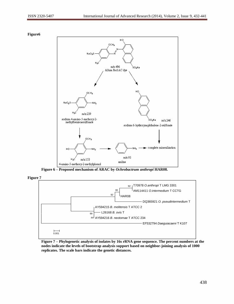
The isolate HAR08 was identified as Ochrobactrum anthropi on the basis of its 16S rDNA sequence. The
16s rDNA sequence of HAR08 was a continuous stretch of 694 bp. The 16S rDNA sequence of the strain HAR08 is
available under the EMBL GenBank accession number FR873648. The phylogenetic tree was constructed by using
MEGA 5.0 (Figure 7). The sequence analysis of 16S rDNA showed that strain HAR08 had highest similarity with
the species Ochrobactrum anthropi (99%) which has been proved to have dye decolorizing ability against the
moniliformin or structurally related to mycotoxins (Duvick and Rood, 2000). Ochrobactrum anthropi was also
reported for removal of chromium, cadmium, copper, toxic metals (Ozdemir, et al., 2003) and phenol degradation
(El-Sayed, et al., 2003).
Optimization of physicochemical parameters
Optimization of physicochemical parameters using strain HAR08 was tested for decolorization at static
anoxic and shaking conditions. The strain HAR08 was able to decolorize 95% of ARAC dye (10 g L-1
) within 24 h
at static anoxic conditions, whereas the decolorization was significantly decreased up to 56% at shaking conditions
(Figure1). The results suggests that the static anoxic condition was needed for bacterial dye decolorization which is
in good agreement with earlier reports on pure bacterial strains such as Proteus mirabilis (Chen, et al., 1999),
Pseudomonas luteola (Chang, et al., 2001) and Serratia marcescens (Verma and Madamwar, 2003). Therefore,
static conditions were adopted to investigate bacterial dye decolorization in further experiments.
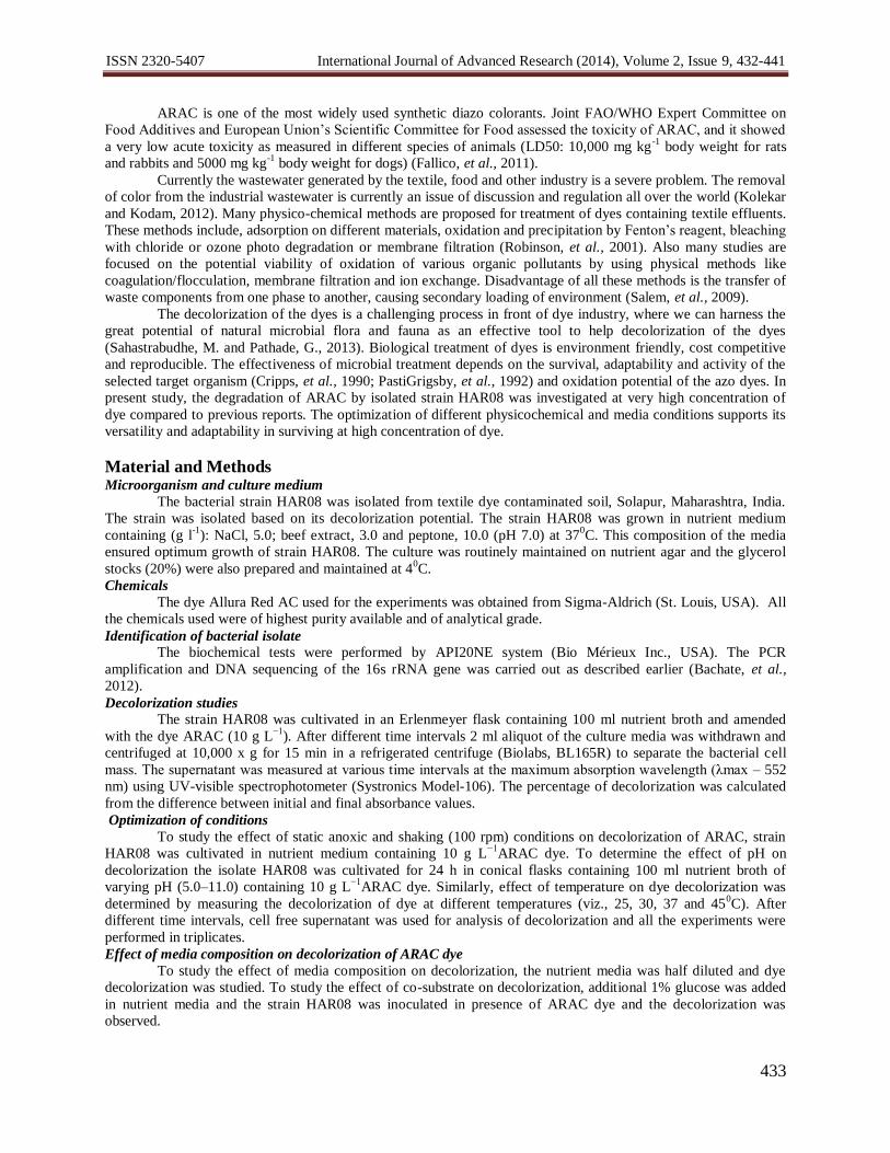
The effect of different temperature and pH studies was carried out on ARAC (10 g L-1
) decolorization. The
strain HAR08 showed the decolorization above 44% at all pH range (pH 5.0-11.0), 90.00% decolorization was
observed at pH 8.0 (Figure3 and 4). The strain HAR08 showed 75% decolorization at pH 7.0. The decolorization of
ARAC dye by strain HAR08 was found to be efficient at all pH values tested. In previous reports it has been
suggested that the pH has a major effect on the efficiency of dye decolorization, and the optimal pH for color
removal was often between 6.0 and 9.0 (Guo, et al., 2007; Kilic, et al., 2007; Wang, et al., 2009). Similarly
bacterial mixed cultures Bacillus sp. V1DMK, Lysinibacillus sp. V3DMK, Bacillus sp. V5DMK, Bacillus sp.
V7DMK, Ochrobacterium sp. V10DMK, Bacillus sp. V12DMK exhibited more than 85% decolorization over a
broad range of pH 5.0-8.5 and maximum decolorization was obtained at neutral pH 7.0 (Jain, et al., 2012).
The strain HAR08 showed decolorization ability in the temperature range of 25 ºC to 450C (Figure 4).
Although, the decolorization at 250C was 65%, it gradually increased with increase in temperature from 79% at 30
0C
and optimum decolorization was 97% at 370C. The increase in temperature up to 45
0C decreased the decolorization
to 60%, this is may be due to the loss of cell viability or deactivation of the enzymes responsible for decolorization
(Panswad and Luangdilok, 2000; Cetin and Donmez, 2006). Similar observation were obtained with Citrobacter sp.
ISSN 2320-5407 International Journal of Advanced Research (2014), Volume 2, Issue 9, 432-441
435
CK3 (Wang, et al., 2009), where strong decolorizing activity was observed between 27 to 37ºC, whereas it
significantly decreased at 42ºC. The important finding of the strain HAR08 was the ability to utilize 10 g L-1
of
ARAC dye within 24 h, which has not been reported earlier as per our knowledge.
Effect of nutrient media and co-substrate on dye decolorization
The effect of concentration of nutrient media on the decolorization of ARAC dye was studied. The half
diluted nutrient medium showed 91.25% decolorization within 24 h with similar dye concentration(Figure 2). The
results suggest that the strain HAR08 can grow and degrade the dye in presence of low nutrient indicating the ability
of the strain in the bioremediation of dye effluent.
The addition of co-substrate (1% glucose) in the nutrient medium showed 99% of dye decolorization in 24
h (Figure 2). The observed decolorization was slightly increased with addition of co-substrate. The decolorization of
reactive red 180 by Citrobacter sp. CK3 was dependent on glucose supplementation, where the presence of glucose
significantly increased the decolorization ability of the strain (Wang,et al., 2009). It has been reported earlier that the
bacteria obtain energy from glucose instead of dyes and glucose can enhance the decolorizing performance of
biological systems (Sarioglu and Bisgin, 2007). The progressive increase in glucose concentration increased rate of
dye decolorization (Jain,et al., 2012).
COD reduction
The COD removal was observed to be 92.1% after decolorization of ARAC dye (10 g L-1
) within 24 h at
static anoxic conditions (Figure 2). The decrease in COD to a certain noticeable level within the short span of time
compared to the biodegradation suggested that the degradation by strain HAR08 may be a better candidate for
bioremediation. Similarly, Bacillus sp. showed maximum COD removal of 93-97% after 24-27 h for Congo red
where the concentration was only 50 mg L-1
(Gopinath, et al., 2009). It was recently reported that Alishewanella sp.
KMK6 was able to reduce 28% COD immediately after decolorization at static anoxic conditions. Further
incubation of decolorized media up to 24 h reduced the COD by 66% at static anoxic, whereas it reduced by 90%
under shaking (aerobic) conditions (Kolekar, et al., 2012). In the view of the potential of the strain HAR08 in COD
reduction, it is important for the treatment of dye effluent under static conditions.
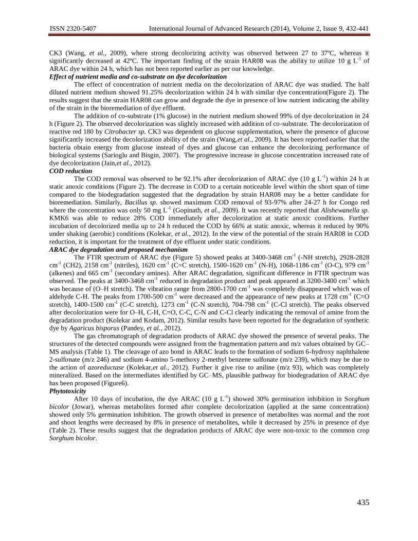
ARAC dye degradation and proposed mechanism
The FTIR spectrum of ARAC dye (Figure 5) showed peaks at 3400-3468 cm-1
(-NH stretch), 2928-2828
cm-1
(CH2), 2158 cm-1
(nitriles), 1620 cm-1
(C=C stretch), 1500-1620 cm-1
(N-H), 1068-1186 cm-1
(O-C), 979 cm-1
(alkenes) and 665 cm-1
(secondary amines). After ARAC degradation, significant difference in FTIR spectrum was
observed. The peaks at 3400-3468 cm-1
reduced in degradation product and peak appeared at 3200-3400 cm-1
which
was because of (O–H stretch). The vibration range from 2800-1700 cm-1
was completely disappeared which was of
aldehyde C-H. The peaks from 1700-500 cm-1
were decreased and the appearance of new peaks at 1728 cm-1
(C=O
stretch), 1400-1500 cm-1
(C-C stretch), 1273 cm-1
(C-N stretch), 704-798 cm-1
(C-Cl stretch). The peaks observed
after decolorization were for O–H, C-H, C=O, C-C, C-N and C-Cl clearly indicating the removal of amine from the
degradation product (Kolekar and Kodam, 2012). Similar results have been reported for the degradation of synthetic
dye by Agaricus bisporus (Pandey, et al., 2012).
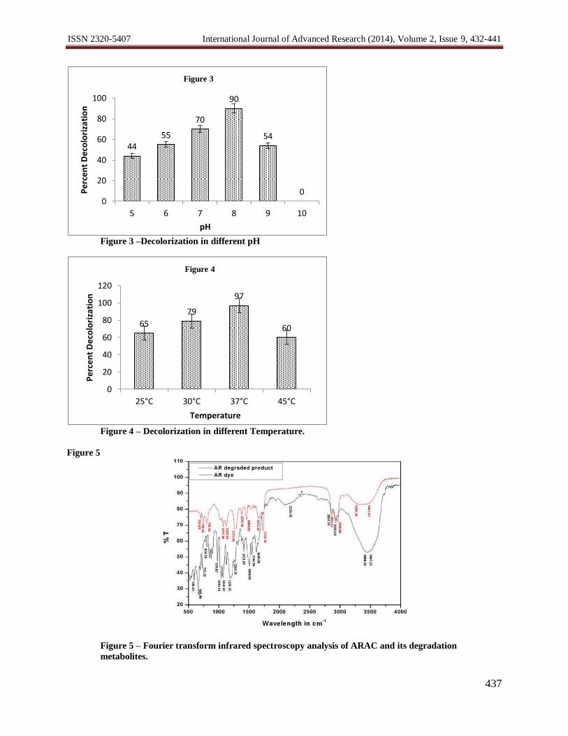
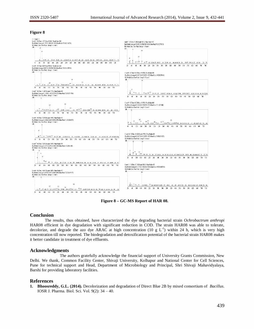
The gas chromatograph of degradation products of ARAC dye showed the presence of several peaks. The
structures of the detected compounds were assigned from the fragmentation pattern and m/z values obtained by GC–
MS analysis (Table 1). The cleavage of azo bond in ARAC leads to the formation of sodium 6-hydroxy naphthalene
2-sulfonate (m/z 246) and sodium 4-amino 5-methoxy 2-methyl benzene sulfonate (m/z 239), which may be due to
the action of azoreductase (Kolekar,et al., 2012). Further it give rise to aniline (m/z 93), which was completely
mineralized. Based on the intermediates identified by GC–MS, plausible pathway for biodegradation of ARAC dye
has been proposed (Figure6).
Phytotoxicity
After 10 days of incubation, the dye ARAC (10 g L-1
) showed 30% germination inhibition in Sorghum
bicolor (Jowar), whereas metabolites formed after complete decolorization (applied at the same concentration)
showed only 5% germination inhibition. The growth observed in presence of metabolites was normal and the root
and shoot lengths were decreased by 8% in presence of metabolites, while it decreased by 25% in presence of dye
(Table 2). These results suggest that the degradation products of ARAC dye were non-toxic to the common crop
Sorghum bicolor.

ISSN 2320-5407 International Journal of Advanced Research (2014), Volume 2, Issue 9, 432-441
436
Table 1 - Mass spectral data, retention times, and proposed identities of metabolites formed after degradation of
Allura Red AC by OchrobactrumanthropiHAR08.
Proposed intermediates Abbreviationa R.T. in min
ESI-MS
peaks
Sodium 4-amino-5-methoxy-2-methylbenzenesulfonate A 12.88 239
Sodium 6-hydroxy naphthalene-2-sulfonate B 12.45 246
4-Amino-5-methoxy-2-methylphenol C 16.62 153
Aniline D 4.9 93 aStructures of series A-D are in Figure 6
Table 2 - Phytotoxicity study of ARAC (10,000 ppm) and its degradation product extracted after 24 h (10,000 ppm)
for Sorghum bicolor.
Dye concentration (ppm) Germination
inhibition (%) Root length (cm)
Shoot length
(cm)
Distilled water 0.00 17.8 ± 1.0 14.6 ± 0.9
Dye (10,000 ppm) 40.00** 13.6 ± 0.5** 11.3 ± 0.4**
Metabolites (10,000 ppm) 0.00 16.3 ± 0.7* 13.3 ± 0.5*
Values are mean of germinated seeds of three experiments ± SEM.
Significantly different from the control at * P < 0.05, ** P < 0.001 by one-way ANOVA with Tukey-Kramer
multiple comparison test.
Figure 1 – Decolorization in static and shaking.
Figure 2– Decolorization in different conditions.
95
56
0
20
40
60
80
100
120
Static Agitated
Nutrient Broth
Pe
rce
nt
De
colo
riza
tio
n
Figure 1
91.25 99
92.1 92.1
5060708090
100110
½ StrengthNutrient
Broth
(1% Glu) Cell-FreeExtract (%)
Percent CODReduction
Pe
rce
nt
De
colo
riza
tio
n
Figure 2
ISSN 2320-5407 International Journal of Advanced Research (2014), Volume 2, Issue 9, 432-441
437
Figure 3 –Decolorization in different pH
Figure 4 – Decolorization in different Temperature.
Figure 5
Figure 5 – Fourier transform infrared spectroscopy analysis of ARAC and its degradation
metabolites.
44 55
70
90
54
0 0
20
40
60
80
100
5 6 7 8 9 10
Per
cen
t D
eco
lori
zati
on
pH
Figure 3
65
79
97
60
0
20
40
60
80
100
120
25°C 30°C 37°C 45°C
Per
cen
t D
eco
lori
zati
on
Temperature
Figure 4
ISSN 2320-5407 International Journal of Advanced Research (2014), Volume 2, Issue 9, 432-441
438
Figure6
Figure 6 – Proposed mechanism of ARAC by Ochrobactrum anthropi HAR08.
Figure 7
Figure 7 – Phylogenetic analysis of isolates by 16s rRNA gene sequence. The percent numbers at the
nodes indicate the levels of bootstrap analysis support based on neighbor-joining analysis of 1000
replicates. The scale bars indicate the genetic distances.
T70978 O.anthropi T LMG 3301
AM114411 O.intermedium T CCTG
HAR08
DQ365921 O. pseudintermedium T
AY594215 B. melitensis T ATCC 2
L26168 B. ovis T
AY594216 B. neotomae T ATCC 234
EF532794 Daeguiacaeni T K107
44 99
92
92 95
0.001
ISSN 2320-5407 International Journal of Advanced Research (2014), Volume 2, Issue 9, 432-441
439
Figure 8
Figure 8 – GC-MS Report of HAR 08.
Conclusion The results, thus obtained, have characterized the dye degrading bacterial strain Ochrobactrum anthropi
HAR08 efficient in dye degradation with significant reduction in COD. The strain HAR08 was able to tolerate,
decolorize, and degrade the azo dye ARAC at high concentration (10 g L-1
) within 24 h, which is very high
concentration till now reported. The biodegradation and detoxification potential of the bacterial strain HAR08 makes
it better candidate in treatment of dye effluents.
Acknowledgments
The authors gratefully acknowledge the financial support of University Grants Commission, New
Delhi. We thank, Common Facility Center, Shivaji University, Kolhapur and National Center for Cell Sciences,
Pune for technical support and Head, Department of Microbiology and Principal, Shri Shivaji Mahavidyalaya,
Barshi for providing laboratory facilities.
References 1. Bhoosreddy, G.L. (2014). Decolorization and degradation of Direct Blue 2B by mixed consortium of Bacillus.
IOSR J. Pharma. Biol. Sci. Vol. 9(2): 34 – 40.

ISSN 2320-5407 International Journal of Advanced Research (2014), Volume 2, Issue 9, 432-441
440
2. Bachate, S.P., Khapare, R.M. and Kodam, K.M. (2012). Oxidation of arsenite by two β-proteobacteria
isolated from soil. Appl. Microbiol. Biotechnol.93:2135-2145.
3. Chang, J.S., Chou, C., Lin, Y.C., Lin, P.J., Ho, J.Y. and Hu, T.L. (2001). Kinetics characteristics of
bacterial azo dye decolorization by Pseudomonas luteola. Water Res. 35:2841-2850.
4. Cetin, D. and Donmez, G. (2006). Decolorization of reactive dyes by mixed cultures isolated from textile
effluent under anaerobic conditions. Enz. Microbial Technol.38:926-930.
5. Chen, K.C., Huang, W.T., Wu, J.Y. and Houng, J.Y. (1999). Microbial decolorization of azo dyes by
Proteus mirabilis. J. Ind. Microbiol. Biotechnol. 23:686-690.
6. Cripps, C., Bumps, J.S. and Aust, S.D. (1990). Biodegradation of azo and heterocyclic dyes by
Phanerochaete chrysosporium. Appl. Enviro. Microbiol. 56:1114–1118.
7. Duvick, J. and Rood, T.A. (2000). Ochrobactrum anthropi bacteria which degrade monoliformin. US Patent
6083736.
8. El-Sayed, W., Ibrahim, M.K., Abu-Shady, M., El-Beih, F., Ohmura, N., Saiki, H. and Ando, A. (2003). Isolation and identification of a novel strain of the genus Ochrobactrum with phenol-degrading activity. J.
Biosci. Bioengg 96:310-312.
9. Fallico, B., Chiappara, E., Arena, E. andBallistreri, G. (2011). Assessment of the exposure to Allura Red
colour from the consumption of red juice-based and red soft drinks in Italy. Food Additives and Contaminants
Part A 28:1501-1515.
10. Gopinath, K.P., Meerasahib, H.A., Muthukumar, K. andVelan, M. (2009). Improved biodegradation of
Congo red by using Bacillus sp. Biores. Technol. 100:670-675.
11. Guo, J.B., Zhou, J.T., Wang, D., Tian, C.P., Wang, P., Uddin, M.S. and Yu, H. (2007). Bio-catalyst effects
of immobilized anthraquinone on the anaerobic reduction of azo dyes by the salt-tolerant bacteria. Water Res.
41:426-432.
12. Jain, K., Shah, V., Chapla, D. and Madamwar, D. (2012). Decolorization and degradation of azo dye-
Reactive Violet 5R by an acclimatized indigenous bacterial mixed cultures-SB4 isolated from anthropogenic
dye contaminated soil. J. Haz. Mat. 213-214:378-386.
13. Kilic, N.K., Nielsen, J.L., Yuce, M. and Donmez, G. (2007). Characterization of a simple bacterial consortium
for effective treatment of wastewaters with reactive dyes and Cr(VI). Chemosphere. 67:826-831.
14. Kolekar, Y.M. and Kodam, K.M. (2012). Decolorization of textile dyes by Alishewanella sp. KMK6. Appl.
Microbiol. Biotechnol. 95:521-529.
15. Kolekar, Y.M., Konde, P.D., Markad, V.L., Kulkarni, S.V., Chaudhari, A.U. and Kodam, K.M.
(2012). Effective bioremoval and detoxification of textile dye mixture by Alishewanella sp. KMK6.
Appl.Microbiol.Biotechnol.97(2):881-9.DOI 10.1007/s00253-012-3983-6
16. Levine, W.G. (1991). Metabolism of azo dyes: Implication for detoxication and activation. Drug Metabol.
Reviews 23:253-309.
17. Mutoh, N., Kawabata, M., Nakagawa, C.W. and Kitajima, S. (2005). Pro-oxidant action of phloxine B on
fission yeast Schizosaccharomyces pombe. Yeast. 22:91-97.
18. Ozdemir, G., Ozturk, T., Ceyhan, N., Isler, R. andCosar, T. (2003). Heavy metal biosorption by biomass of
Ochrobactrum anthropi producing exopolysaccharide in activated sludge. Biores. Technol.90:71-74.
19. Pandey, P., Singh, R.P., Singh, K.N. andManisankar, P. (2012). Evaluation of the individuality of white rot
macro fungus for the decolorization of synthetic dye. Environ. Sci. Poll. Res.20(1):238-49.DOI:
10.1007/s11356-012-0875-3
20. Panswad, T. and Luangdilok, W. (2000). Decolorization of reactive dyes with different molecular structures
under different environmental conditions. Water Res. 34:4177-4184.
21. PastiGrigsby, M.B., Paszcczynski, A., Goszczynski, S., Crawford, R.L. and Crawford, D.L., (1992).
Influence of aromatic substitution patterns on azo dye degradability by Streptomyces sp. and Phanerochaete
chrysosporium. Appl. Environ. Microbiol. 58:3605-3613.
22. Poul, M., Jarry, G., Elhkim, M.O. and Poul, J.M. (2009). Lack of genotoxic effect of food dyes amaranth,
sunset yellow and tartrazine and their metabolites in the gut micronucleus assay in mice. Food Chemi. Toxicol.
47:443-448.
23. Robinson, T., McMullan, G., Marchant, R., Nigam, P. (2001). Remediation of dyesin textile effluent: a
critical review on current treatment technologies with a proposed alternative. Biores. Technol. 77: 247-255.
24. Sahastrabudhe, M. and Pathade, G. (2013). Biodegradation of azo dye C.I. Reactive Orange 16 by an
actinobacterium Georgenia sp. CC-NMPT-T3. Int. J.Adv.Res.Vol.1 (9):91-99.
ISSN 2320-5407 International Journal of Advanced Research (2014), Volume 2, Issue 9, 432-441
441
25. Salem, M.A., Abdel-Halim, S.T., El-Sawy, A.E.H.M. andZaki, A.B. (2009). Kinetics of degradation of allura
red, ponceau 4R and carmosine dyes with potassium ferrioxalate complex in the presence of H2O2.
Chemosphere. 76:1088-1093
26. Sarioglu, M. and Bisgin, T. (2007). Removal of Maxilon Yellow GL in a mixed methano-genic anaerobic
culture. Dyes Pig.75:544-549.
27. Sasaki, Y.F., Kawaguchi, S., Kamaya, A., Ohshita, M., Kabasawa, K., Iwama, K., Taniguchi, K. and
Tsuda, S. (2002). The comet assay with 8 mouse organs: results with 39 currently used food additives. Mut.
Res. 519:103-119.
28. Verma, P. and Madamwar, D. (2003). Decolourization of synthetic dyes by a newly isolated strain of Serratia
marcescens. World J. Microbiol. Biotechnol. 19:615-618.
29. Wang, H., Su, J.Q., Zheng, X.W., Tian, Y., Xiong, X.J. and Zheng, T.L. (2009). Bacterial decolorization
and degradation of the reactive dye Reactive Red 180 by Citrobacter sp. CK3. Int. Biodeter. Biodegr. 63:395-
399.