Identification of the Domains of UreR, an AraC-Like Transcriptional Regulator of the Urease Gene...
11
10.1128/JB.183.15.4526-4535.2001. 2001, 183(15):4526. DOI: J. Bacteriol. and Harry L. T. Mobley Carrie A. Poore, Christopher Coker, Jonathan D. Dattelbaum Proteus mirabilis Urease Gene Cluster in AraC-Like Transcriptional Regulator of the Identification of the Domains of UreR, an http://jb.asm.org/content/183/15/4526 Updated information and services can be found at: These include: REFERENCES http://jb.asm.org/content/183/15/4526#ref-list-1 at: This article cites 22 articles, 13 of which can be accessed free CONTENT ALERTS more» articles cite this article), Receive: RSS Feeds, eTOCs, free email alerts (when new http://journals.asm.org/site/misc/reprints.xhtml Information about commercial reprint orders: http://journals.asm.org/site/subscriptions/ To subscribe to to another ASM Journal go to: on February 24, 2014 by guest http://jb.asm.org/ Downloaded from on February 24, 2014 by guest http://jb.asm.org/ Downloaded from
Transcript of Identification of the Domains of UreR, an AraC-Like Transcriptional Regulator of the Urease Gene...

10.1128/JB.183.15.4526-4535.2001.
2001, 183(15):4526. DOI:J. Bacteriol. and Harry L. T. MobleyCarrie A. Poore, Christopher Coker, Jonathan D. Dattelbaum
Proteus mirabilisUrease Gene Cluster inAraC-Like Transcriptional Regulator of the Identification of the Domains of UreR, an
http://jb.asm.org/content/183/15/4526Updated information and services can be found at:
These include:
REFERENCEShttp://jb.asm.org/content/183/15/4526#ref-list-1at:
This article cites 22 articles, 13 of which can be accessed free
CONTENT ALERTS more»articles cite this article),
Receive: RSS Feeds, eTOCs, free email alerts (when new
http://journals.asm.org/site/misc/reprints.xhtmlInformation about commercial reprint orders: http://journals.asm.org/site/subscriptions/To subscribe to to another ASM Journal go to:
on February 24, 2014 by guest
http://jb.asm.org/
Dow
nloaded from
on February 24, 2014 by guest
http://jb.asm.org/
Dow
nloaded from

JOURNAL OF BACTERIOLOGY,0021-9193/01/$04.0010 DOI: 10.1128/JB.183.15.4526–4535.2001
Aug. 2001, p. 4526–4535 Vol. 183, No. 15
Copyright © 2001, American Society for Microbiology. All Rights Reserved.
Identification of the Domains of UreR, an AraC-Like TranscriptionalRegulator of the Urease Gene Cluster in Proteus mirabilis
CARRIE A. POORE, CHRISTOPHER COKER, JONATHAN D. DATTELBAUM,AND HARRY L. T. MOBLEY*
Department of Microbiology and Immunology, University of Maryland, Baltimore,School of Medicine, Baltimore, Maryland 21201
Received 20 February 2001/Accepted 18 May 2001
Proteus mirabilis urease catalyzes the hydrolysis of urea to CO2 and NH3, resulting in urinary stone forma-tion in individuals with complicated urinary tract infections. UreR, a member of the AraC family, activatestranscription of the genes encoding urease enzyme subunits and accessory proteins, ureDABCEFG, as well asits own transcription in the presence of urea. Based on sequence homology with AraC, we hypothesized thatUreR contains both a dimerization domain and a DNA-binding domain. A translational fusion of the leucinezipper dimerization domain (amino acids 302 to 350) of C/EBP and the C-terminal half of UreR (amino acids164 to 293) activated transcription from the ureD promoter (pureD) and bound to a 60-bp fragment containingpureD, as analyzed by gel shift. These results were consistent with the DNA-binding specificity residing in theC-terminal half of UreR and dimerization being required for activity. To localize the dimerization domain ofUreR, a translational fusion of the DNA-binding domain of the LexA repressor (amino acids 1 to 87) and theN-terminal half of UreR (amino acids 1 to 182) was constructed and found to repress transcription frompsulA-lacZ (sulA is repressed by LexA) and bind to the sulA operator site, as analyzed by gel shift. Since LexAbinds this site only as a dimer, the UreR1–182-LexA1–87 fusion also must dimerize to bind psulA. Indeed, purifiedUreR-Myc-His eluted from a gel filtration column as a dimer. Therefore, we conclude that the dimerizationdomain of UreR is located within the N-terminal half of UreR. UreR contains three leucines that mimic theleucines that contribute to dimerization of AraC. Mutagenesis of Leu147, Leu148, or L158 alone did not sig-nificantly affect UreR function. In contrast, mutagenesis of both Leu147 and Leu148 or all three Leu residuesresulted in a 85 or 94% decrease, respectively, in UreR function in the presence of urea (P < 0.001). On thecontrary, His102 and His175 mutations of UreR resulted in constitutive induction in the absence of urea. Weconclude that a dimerization domain resides in the N-terminal half of the polypeptide, that Leu residues maycontribute to this function, and that sequences within the C-terminal half of UreR are responsible for DNAbinding to the urease promoter regions. Selected His residues also contribute significantly to UreR function.
Proteus mirabilis infects the urinary tract of humans and ismost commonly responsible for causing disease in individualswith structural abnormalities of the urinary tract or in patientswho undergo long-term catheterization (16). Cystitis, acutepyelonephritis, and urinary stone formation are all possibleconsequences of P. mirabilis infection (17).
P. mirabilis produces a urea-inducible urease, a high-molec-ular-weight, multimeric, cytoplasmic nickel metalloenzyme.Urease catalyzes the hydrolysis of urea to ammonia and carbondioxide (18). During the course of infection, the production ofammonia by urea hydrolysis raises the pH in the local environ-ment, subsequently precipitating polyvalent ions that are nor-mally soluble in urine. The result is the formation of urinarystones. The elevated pH also creates an environment that ismore favorable for growth of this species (4). Increased am-monia production can also lead to acute inflammation withpossible tissue necrosis (18).
The P. mirabilis urease gene cluster is found in single copyon the chromosome and consists of eight contiguous genes,ureRDABCEFG (12, 19, 24). The ureA (UreA, 11 kDa), ureB
(UreB, 12 kDa), and ureC (UreC, 61 kDa) genes encode thestructural polypeptides required for the assembly of a catalyt-ically inactive urease apoenzyme (18). The accessory genes,ureD (UreD, 31 kDa), ureE (UreE, 18 kDa), ureF (UreF, 23kDa), and ureG (UreG, 22 kDa), encode proteins required forinsertion of nickel ions into the metalloenzyme resulting incatalytically active urease (18). The urease gene cluster is reg-ulated by the gene product of ureR (UreR, 33 kDa).
P. mirabilis UreR and the plasmid-encoded UreR found inEscherichia coli are positive transcriptional activators of theurease genes. The two proteins share 70% amino acid identity(6) and are functionally interchangeable in the activation oftranscription from the ureR (pureR) and ureD (pureD) promotersin both the P. mirabilis and plasmid-encoded urease gene clus-ters (6). The UreR binding sites of both promoters have theconsensus sequence T(A/G)(T/C)(A/T)(T/G)(C/T)T(A/T)(T/A)ATTG (25). Both UreR proteins have been shown to activatetranscription from pureD in the presence of urea (11, 6). In ad-dition, UreR regulates its own transcription in the presence ofurea from pureR in the direction opposite the rest of the genecluster (6). In the absence of urea induction, H-NS repressesureR expression (3). Because UreR activates transcription in aurea-inducible manner, it is hypothesized that UreR bindsurea; however, this has not been directly demonstrated.
UreR is a member of the AraC family of transcriptional
* Corresponding author. Mailing address: 655 W. Baltimore St.,BRB 13-009, Department of Microbiology and Immunology, Balti-more, MD 21201. Phone: (410) 706-0466. Fax: (410) 706-6751. E-mail:[email protected].
4526
on February 24, 2014 by guest
http://jb.asm.org/
Dow
nloaded from

regulators and contains a putative helix-turn-helix in additionto an AraC signature sequence (5, 19). The AraC signaturesequence, found within all AraC family members, is a secondhelix-turn-helix that is hypothesized to also bind DNA (7).Moreover, UreR also contains three conserved leucine resi-dues (Leu147, Leu148, and Leu158) in the same relative loca-tion with the same spatial distance relative to each other as inAraC (Leu150, Leu151, and Leu161). These leucine residuesare critical for AraC dimerization (23), and we therefore alsohypothesize that UreR dimerizes via this mechanism. In thepresence of arabinose, AraC uses these three critical leucinesfor dimerization via an antiparallel coiled-coil in a “knobs-into-holes” manner, as elucidated by X-ray crystallographic studies(23). This coiled-coil is also the primary dimerization face inthe absence of arabinose, shown by both size exclusion chro-matography and sedimentation velocity analytical ultracentrif-ugation of an AraC mutant with mutations in Leu150, Leu151,Asn154, and Leu161 (15). A secondary dimerization face in theb barrel of AraC is evident; however, it does not appear torepresent the primary means of dimer interaction (15).
AraC contains two separate and independent domains, eachwith a distinct function, namely, dimerization and DNA bind-ing; UreR is predicted to have similar domains with similarfunctions. Previously, chimeric proteins containing the two do-mains of AraC to characterize each of the domain’s functionswere synthesized (2). The predicted AraC DNA-binding do-main was fused to C/EBP, a known eukaryotic transcriptionalactivator that dimerizes via a leucine zipper. The C/EBP-AraCfusion was found to bind to pBAD and activate transcription (2).The hypothesized AraC dimerization domain was fused to theLexA DNA-binding domain. This fusion was predicted tomimic full-length LexA and demonstrated the need for dimer-ization in order to repress transcription of genes normallyturned off by LexA. A chromosomal transcriptional fusion ofpsulA to lacZ was repressed in the presence of the AraC-LexAfusion protein. This strategy was used to identify both domainsof AraC (2).
In this study, we constructed fusion proteins to identify pu-tative domains of UreR and assign dimerization and DNA-binding functions to each of the domains as well as identifyingkey amino acid residues involved in dimerization and ureainduction.
MATERIALS AND METHODS
Chemicals and enzymes. All enzymes were purchased from Life Technologies(Rockville, Md.) or New England Biolabs (Beverly, Mass.). All chemicals werepurchased from Sigma-Aldrich (St. Louis, Mo.) unless otherwise noted. The DIG(digoxigenin) gel shift kit was obtained from Amersham Pharmacia Biotech(Piscataway, N.J.).
Bacterial strains and growth conditions. Bacterial strains used in this study arelisted in Table 1. Bacteria were grown either in Luria-Bertani (LB) broth at 37°Cwith aeration in a shaking incubator (200 rpm) or on LB plates containing 1.5%agar at 37°C. Plates and media were supplemented with the antibiotic chloram-phenicol (10 mg/ml), tetracycline (7.5 mg/ml), ampicillin (100 mg/ml), or kana-mycin (50 mg/ml).
PCR amplification of DNA used to make fusion proteins. PCR primers arelisted in Table 2. The amplification protocol for PCRs was as follows: denatur-ation, 94°C, 3 min; annealing, 50 to 55°C, 45 s; elongation, 72°C, 1 min; for 30cycles.
Cloning of PCR products. PCR products were ligated into pCR-BluntII-TOPO (Invitrogen, Carlsbad, Calif.). Inserts were excised using the appropriaterestriction enzymes (either NcoI-BamHI, BamHI-XhoI, NcoI-XhoI, or NcoI-HindIII) and separated by agarose gel electrophoresis. PCR amplification prod-
ucts for the inserts encoding C/EBP302–350-Myc-His and LexA1–87-Myc-His weredigested with restriction enzymes; Table 2 lists inserts and restriction enzymesused. All constructs use an NcoI site to ligate into the vector at the gene sequenceencoding the start codon. A BamHI site is at the junction of the gene sequencesencoding the domains in both C/EBP302–350-UreR164–293 and UreR1–182-LexA1–87. All constructs except the gene sequences encoding UreR-Myc-His,UreR164–293-Myc-His, C/EBP302–350-Myc-His, LexA1–87-Myc-His, L147A-L148-Myc-His, and L147A-L148A-L158A-Myc-His, which use a 39 HindIII site, con-tain a XhoI site at the 39 end for cloning into the expression vector. Inserts werepurified using a Qiaquick gel extraction kit (Qiagen) and ligated into eitherpBAD/MHA or pSE380 (Invitrogen). Plasmids were introduced into the corre-sponding laboratory strain by CaCl2 transformation (21). The LexA1–87-Myc-Hisgene sequence was ligated into pBAD/MHA to take advantage of the Myc-Hisepitopes. After transformation, the pBAD vector containing the gene sequenceencoding LexA1–87-Myc-His was purified using a Qiagen miniprep kit. The plas-mid was then used in a PCR with primers to amplify the gene sequence forLexA1–87-Myc-His. The PCR product was then cut with NcoI and XhoI andligated into the NcoI and XhoI sites in pSE380.
Construction of pureD-lacZ reporter plasmid. A low-copy-number ureD-lacZfusion reporter plasmid compatible with pBAD-Myc-His was constructed. Prim-ers MOB906 and MOB915 were used to PCR amplify an approximately 4.3 kbDNA fragment from plasmid pDR10 ureD-lacZ (11) under the following condi-tions: 95°C denaturation, 54°C annealing, and 72°C elongation for 30 cycles,using Vent DNA polymerase in the presence of 1 mM MgSO4. The PCR productwas gel purified and ligated to PCR-Blunt (Invitrogen), forming pCC026.pCC026 was digested with EcoRI, and the 4.3-kb DNA fragment encodingureD-lacZ was ligated to an EcoRI-digested derivative of pACYC184 that hadpreviously been cut with PvuII and religated (thus, it does not encode a func-tional chloramphenicol acetyltransferase). The resulting recombinant plasmid,pCC042, carries a tetracycline resistance marker and encodes a ureD-lacZ tran-scriptional fusion that is activated in the presence of UreR and urea.
TABLE 1. E. coli strains and plasmids used in this study
Strain/plasmid Description Referenceor source
E. coli strainsDH5a F2 f80dlacZDM15 D(lacZYA-argF)
U169 deoR recA 1 endA 1 hsdR17(rk
2 mk1) phoA supE44 l 2thi-1
gyrA96 relA1Top10 F2 mcrAD(mrr-hsdRMS-mcrBC)
f80lacZDM15 DlacX74 deoR recA1araD139 D(ara-leu)7697 galU galKrpsL(Strr) endA1 nupG
JL1436 F9 laclq lacZDM15::Tn9/lexA71::Tn5recA1 sulA211 (lsulA::lacZ cl ind2)
2
PlasmidspSE380 InvitrogenpBAD/Myc-His A InvitrogenpCC042 pureD-lacZ reporter construct This workpSE380-lexA 2pSE380-lexA-C/EBP 2pCP015 C/EBP302–350-UreR164–293-pBAD This workpCP016 Full UreR-pBAD This workpCP018 Full UreR-pSE380 This workpCP019 UreR1–182-LexA1–87-pSE380 This workpCP025 LexA1–87-pSE380 This workpCP026 C/EBP302–350-pBAD This workpCP029 L147A-L148A UreR This workpCP030 L147A UreR This workpCP031 L148A UreR This workpCP051 UreR164–293-pBAD This workpCP056 UreRHis175Ala This workpCP059 UreR-Myc-His-pBAD This workpCP063 UreRHis102Ala This workpCP071 UreR164–293-Myc-His-pBAD This workpCP072 C/EBP302–350-Myc-His-pBAD This workpCP073 LexA1–87-Myc-His-pSE380 This workpCP079 L147A-L148A UreR-Myc-His This workpCP086 L158A UreR This workpCP088 L147A-L148A-L158A UreR This workpCP089 L147A-L148A-L158A UreR-Myc-His This work
VOL. 183, 2001 IDENTIFICATION OF THE DOMAINS OF UreR 4527
on February 24, 2014 by guest
http://jb.asm.org/
Dow
nloaded from

TABLE 2. PCR primers used in this study
Primerno. Primer sequence Gene Enzyme
cleaved Construct
906 59 CGAAATACGGGCAGACATGG39 pureD-lacZ reporter (ureR-ureD intergenic)915 59 AGGCAAGTTCAAAATGAACATGG39 pureD-lacZ reporter (ureR-ureD intergenic)1126 59 CCATGGAATACAAACACATACTTTCTTCTAAC 39 ureR 59 NcoI UreR
UreR-Myc-HisUreR1–182-LexA1–87L147A-L148AL147AL148AL147A-L148A-Myc-HisUreRHis175AlaUreRHis102AlaL158AL147A-L148A-L158AL147A-L148A-L158A-Myc-His
1128 59 GGATCCATGAAAGCGTTAACGGCCAGGCAACAA39 lexA 59 BamHI UreR1–182-LexA1–871129 59 CTCGAGCTATGGTTCACCGGCAGCCACACGACCTAC 39 lexA 39 Xhol UreR1–182-LexA1–87
LexA1–871132 59 CCATGGAGAAAGCCAAACAGCGCAACGTGGAGACG 39 C/EBP 59 Ncol C/EBP302–350-UreR164–293
C/EBP302–350C/EBP302–350-Myc-His
1133 59 GGATCCCAAGGAGCTCTCAGGCAGCTGGCGGAA 39 C/EBP 39 BamHI C/EBP302–350-UreR164–293C/EBP302–350
1594 59 GGATCCTTGCGGATCTTGTGTTATTAGATGAGT 39 ureR 39 BamHI UreR1–182-LexA1–871595 59 GGATCCAATTATGATGAGCCAAAAAATCAGGCG 39 ureR 59 BamHI C/EBP302–350-UreR164–2931596 59 CTCGAGTTAAAATACTTTTTTTATTGATTCGTC 39 ureR 39 Xhol C/EBP302–350-UreR164–293
UreRL147A and L148AL147AL148AL158AL147A-L148A-L158AUreRHis175AlaUreRHis102AlaUreR164–293
1634 59 AGCCTCTTTTTTATTGCGGCGGCGGTTTATCACGAA 39 ureR L147A-L148AL147A-L148A-Myc-HisL147A-L148A-L158AL147A-L148A-L158A-Myc-His
1635 59 TTCGTGATAAACCGCCGCCGCAATAAAAAAGAGGCT 39 ureR L147A-L148AL147A-L148A-Myc-HisL147A-L148A-L158AL147A-L148A-L158A-Myc-His
1636 59 AGCCTCTTTTTTATTGCGCTGGCGGTTTATCAC 39 ureR L147A1637 59 GTGATAAACCGCCAGCGCAATAAAAAAGAGGCT 39 ureR L147A1638 59 CTCTTTTTTATTTTGGCGGCGGTTTATCACGAA 39 ureR L148A1639 59 TTCGTGATAAACCGCCGCCAAAATAAAAAAGAG 39 ureR L148A1646 59 CCATGGCAAAAGCGTTAACGGCCAGGCAACAA 39 lexA 59 NcoI LexA1–871647 59 CTCGAGCAAGGAGCTCTCAGGCAGCTGGCGGAA 39 C/EBP 39 Xhol C/EBP302–3501663 59 AATCAGGCGATCACTGCTCTAATAACACAAGAT 39 ureR UreRHis175Ala1664 59 ATCTTGTGTTATTAGAGCAGTGATCGCCTGATT 39 ureR UreRHis175Ala1712 59 CCATGGCAAATTATGATGAGCCAAAAAATCAGGCG 39 ureR 59 NcoI UreR164–293
UreR164–293-Myc-His1729 59AAGCTTAAATACTTTTTTTATTGATTCGTC 39 ureR 39 HindIII UreR-Myc-His
L147A-L148A-Myc-HisL147A-L148A-L158A-Myc-His
1739 59 ATTTTACACCGAGTTTCAAAAAATGCGTTTTAAATAAACAGCAAT 39 pureD oligo1740 59 ATTTTTTCTAAACAAATTGCTGTTTATTTAAAACGCATTTTTTGA39 pureD oligo1749 59 AATCAATCCAGCCCCTGTGAGTTACTGTATGGATGTACAGTACATCCAGTG 39 psulA oligo1750 59 TGATCTTTGTTGTCACTGGATGTACTGTACATCCATACAGTAACTCACAGG 39 psulA oligo1797 59 GCGCCGATTACTCGTGCTCTTCCAGATTATCAT 39 ureR UreRHis102Ala1798 59 ATGATAATCTGGAAGAGCACGAGTAATCGGCGC 39 ureR UreRHis102Ala1833 59 TTTTTTAAGCTTAAATACTTTTTTTATTGATTCGTC 39 ureR 39 HindIII UreR164–293-Myc-His1834 59 TTTTTTAAGCTTTGGTTCACCGGCAGCCACACGACCTAC 39 lexA 39 HindIII LexA1–87-Myc-His1848 59 AAAAAACCATGGAGAAAGCCAAACA 39 C/EBP 59 NcoI C/EBP302–350-Myc-His1851 59 AAAAAACCATGGCAAAAGCGTTAACGGCCAGGCAACAA 39 lexA 59 NcoI LexA1–87-Myc-His1852 59 TTTTTTCTCGAGATGATGATGATGATGATGGTC 39 lexA 39 XhoI LexA1–87-Myc-His1855 59 AAAAAAAAGCTTCAAGGAGCTCTCAGGCAGCTGGCGGAA 39 C/EBP 39 HindIII C/EBP302–350-Myc-His1883 59 GGGGTCGATATTGCTAATATTTTTCGT 39 ureR L158A
L147A-L148A-L158AL147A-L148A-L158A-Myc-His
1884 59 ACGAAAAATATTAGCAATATCGACCCC 39 ureR L158AL147A-L148A-L158AL147A-L148A-L158A-Myc-His
4528 POORE ET AL. J. BACTERIOL.
on February 24, 2014 by guest
http://jb.asm.org/
Dow
nloaded from

Sequencing. Both strands of plasmid constructs were sequenced across eachjunction and throughout the insert. Sequencing was done by the BiopolymerLaboratory at the University of Maryland, Baltimore.
b-Galactosidase expression assays. Fresh medium was inoculated with a sin-gle colony from LB-agar plates containing the appropriate antibiotics and cul-tured at 37°C overnight. Overnight cultures were used to inoculate fresh me-dium. Cultures were monitored until they reached an optical density at 600 nm(OD600) of ;0.4 to 0.6, at which time inducer (isopropyl-b-D-thiogalactopyrano-side [IPTG], arabinose, or urea) was added, and cultures were incubated for anadditional hour. Cultures were placed on ice, and the OD600 was measured.Chloroform (100 ml) and 0.1% sodium dodecyl sulfate (SDS; 50 ml) were added,and cultures were vortexed. The suspension of permeabilized cells (10 ml) wasadded to 990 ml of Z buffer and 200 ml of o-nitrophenyl-b-D-galactopyranoside(4 mg/ml in H2O). Timed reactions were stopped with 500 ml of 1 M Na2CO3.OD420 and OD550 measurements were recorded, and Miller units were calcu-lated (20). All constructs were assayed in three or more independent experi-ments.
Western blot analysis. Overnight cultures were used to inoculate 4 ml of freshLB medium containing the appropriate antibiotics and allowed to grow to mid-exponential phase. Cultures were induced with the appropriate inducer andincubated for the indicated time. Cultures were placed on ice. OD600 was de-termined, and all cultures were adjusted to the same reading. Bacteria wereharvested from 1 ml of culture by centrifugation and resuspended in equivalentamounts of Laemmli sample buffer. Samples were boiled for 5 min and placed onice. The sample volume listed for each experiment was loaded onto a 3.75%stacking and either a 12.5 or 15% SDS-polyacrylamide gel by the method ofLaemmli (13). SDS running buffer was used for electrophoresis. Gels weretransferred onto Immobilon P membranes in a transfer chamber containingtransfer buffer. Transfer occurred overnight at ;12 V at 4°C. Membranes wereblocked in 5% dry milk in 0.1% TTBS (20 mM Tris-HCl [pH 7.5], 150 mM NaCl,0.1% Tween 20) for 1 h. Primary antibodies were added to 0.1% TTBS (anti-LexA, anti-Myc and anti-His, diluted 1:5,000), and membranes were exposed toantibodies for 2 h at room temperature. Membranes were washed in 0.1% TTBSthree times for 15 min each. Secondary antibodies (anti-mouse immunoglobulinG coupled to alkaline phosphatase detected anti-Myc and anti-His antibodies;anti-rabbit immunoglobulin G coupled to alkaline phosphatase detected anti-LexA antibodies) were placed in 0.1% TTBS (dilution 1:2,000), and membraneswere incubated in the secondary antibodies for 1 h. Membranes were washedthree times for 15 min each in 0.1% TTBS and developed with nitroblue tetra-zolium–5-bromo-4-chloro-3-indolylphosphate dissolved in H2O (according to theWestern blot protocol as described in reference 1).
Gel shift analysis. Gel shift experiments used either a 60-bp (DNA-bindingdomain study) or a 64-bp (dimerization study) double-stranded oligonucleotidethat was synthesized from smaller overlapping oligonucleotide fragments thatwere allowed to anneal and extend using Vent polymerase. One microliter ofeach reaction was run on a gel for quantification purposes. Both double-strandedoligonucleotides (3.85 pmol of each) were labeled by the DIG gel shift (Amer-sham Pharmacia) protocol. Each double-stranded oligonucleotide was diluted to30 fmol for the binding study.
Overnight cultures were used to inoculate fresh LB medium containing ap-propriate antibiotics. These cultures were grown to mid-exponential phase. Theappropriate inducer molecule was added and incubated further. The cultureswere placed on ice, and OD600 measurements were taken. OD600 for all cultureswas adjusted to the same reading by using LB medium. Equivalent volumes ofeach culture were centrifuged, and the pellet was resuspended in 1 ml of TENbuffer (10 mM Tris, 1 mM EDTA, 0.1 M NaCl [pH 8.0]). Bacterial suspensionswere disrupted by passage through a French press (18,000 lb/in2). Lysates werecentrifuged (12,000 3 g, 10 min, 4°C). Supernatants were collected and used asthe extract for the binding assay. The binding assay was done as instructed by theDIG gel shift kit manufacturer (Amersham Pharmacia), using binding buffer,1 mg of poly(dI-dC), 1 mg of poly-L-lysine, specific extract volume, and 2 ml of thelabeled DNA. Binding reactions for pBAD, UreR, C/EBP302–350-UreR164–293,and UreR-Myc-His also included 100 mM urea (final concentration). The bind-ing reactions were run on a preelectrophoresed 6% native polyacrylamide gel inTAE buffer (0.04 M Tris acetate, 0.001 M EDTA) at 90 V for 1.5 h. The gel wasplaced on a Hybond N1 membrane and transferred for 30 min at 400 mA in 13TAE buffer. The membrane was developed according to the instructions of theDIG gel shift kit manufacturer (Amersham Pharmacia).
Purification of UreR derivatives. E. coli Top10 transformed with pCP016 orpCP088 was grown in Luria broth at 37°C. Expression of UreR-Myc-His6 wasinduced with 0.2% arabinose when an OD600 of ;0.6 was reached. Following 3 hof induction, cells were collected by centrifugation and lysed by two passagesthrough a French pressure cell (18,000 lb/in2). Single-step purification of UreR-
Myc-His6 was performed by nickel-chelating nitrilotriacetic acid affinity chroma-tography. A single polypeptide of approximately 33 kDa was eluted from thecolumn with 250 mM imidazole as seen on a Coomassie blue-stained 12% SDS-polyacrylamide gel (data not shown). In a similar experiment, purified proteinwas electrophoresed, transferred to a nitrocellulose membrane, and reacted withrabbit antiserum specific for the Myc epitope. Western blot anlysis showed thatarabinose-induced E. coli Top10 transformed with either pCP016 or pCP088produced a single species of 33 kDa, consistent with the predicted size for UreR-Myc-His6 (see Fig. 3). The band was absent from the vector control under iden-tical conditions.
Gel filtration chromatography. Gel filtration chromatography was performedat room temperature on a Sephadex G-75 column (1 by 35 cm) equilibrated withrunning buffer (50 mM phosphate [pH 7.5], 150 mM NaCl) at a flow rate of 0.5ml/min. Molecular weight standards bovine serum albumin (68 kDa) and car-bonic anhydrase (34.5 kDa) were used to calibrate the column. Purified wild-typeand mutant UreR-Myc-His6 proteins (100 ml of 0.1 mg/ml) were injected ontothe column, and 0.5-ml fractions were collected. The protein elution profile wasmonitored by absorbance at 280 nm. An aliquot (100 ml) of each fraction wastransferred to an Immobilon P membrane, and immunoblot analysis performedwith anti-Myc antibodies.
RESULTS
Localization of the UreR DNA-binding domain. To localizethe UreR DNA-binding domain, a protein chimera was con-structed by fusing the gene sequence encoding the C-terminalhalf of UreR to the gene sequence encoding the leucine zipperdimerization domain of C/EBP (Fig. 1; primers are listed inTable 2). All translational fusion constructs were cloned intothe E. coli arabinose-inducible expression vector pBAD/Myc-His A (Invitrogen) (Table 1). A pureD-lacZ plasmid reporterwas constructed in pACYC184 to assay specific induction ofthe urease gene cluster, using b-galactosidase activity as thereadout. Both reporter plasmid and pBAD constructs with andwithout insert were transformed into E. coli Top10.
The C/EBP302–350-UreR164–293 fusion chimera was testedfor its ability to bind to pureD and activate transcription. TheC/EBP302–350-UreR164–293 fusion activated transcription from
FIG. 1. Schematic of fusion constructs. The domain structure ofeach of the chimeric proteins is shown; the amino acid residue numberis labeled at each boundary. A BamHI restriction site (coding for aGly-Ser amino acid linker (GS)) was inserted between gene sequencesencoding each domain. Key leucine residues and helix-turn-helix mo-tifs (H-T-H) are indicated. Sig., signature.
VOL. 183, 2001 IDENTIFICATION OF THE DOMAINS OF UreR 4529
on February 24, 2014 by guest
http://jb.asm.org/
Dow
nloaded from
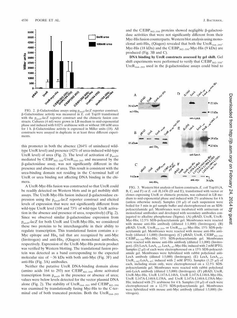
this promoter in both the absence (264% of uninduced wild-type UreR level) and presence (42% of urea-induced wild-typeUreR level) of urea (Fig. 2). The level of activation of pureD,mediated by C/EBP302–350-UreR164–293 and measured by theb-galactosidase assay, was not significantly different in thepresence and absence of urea. This result is consistent with theurea-binding domain not residing in the C-terminal half ofUreR or urea binding not affecting DNA binding in the chi-mera.
A UreR-Myc-His fusion was constructed so that UreR couldbe readily detected on Western blots and in gel mobility shiftassays. The UreR-Myc-His fusion induced b-galactosidase ex-pression using the pureD-lacZ reporter construct and elicitedlevels of expression that were not significantly different fromwild-type UreR level (104 and 73% of wild-type UreR activa-tion in the absence and presence of urea, respectively) (Fig. 2).Since we observed similar b-galactosidase expression frompureD-lacZ for both UreR and UreR-Myc-His, we consideredthese two proteins to be interchangeable in their ability toregulate transcription. This translational fusion contains a c-Myc epitope and His6 tail that are recognized by anti-Myc(Invitrogen) and anti-His5 (Qiagen) monoclonal antibodies,respectively. Expression of the UreR-Myc-His protein productwas verified by Western blotting. The translational fusion pro-tein was detected as a band corresponding to the expectedmolecular size of ;36 kDa with both anti-Myc (Fig. 3F) andanti-His (Fig. 3A) antibodies.
Neither the putative UreR DNA-binding domain alone(amino acids 164 to 293) nor C/EBP302–350 alone activatedtranscription from pureD in the presence or absence of urea;values were below levels detected for the vector plasmid DNAalone (Fig. 2). The stability of UreR164–293 and C/EBP302–350
was examined by translationally fusing Myc-His to the C-ter-minal end of both truncated proteins. Both the UreR164–293
and the C/EBP302–350 proteins showed negligible b-galactosi-dase activities that were not significantly different from theirMyc-His fusion counterparts. Western blot analysis using mono-clonal anti-His5 (Qiagen) revealed that both the UreR164–293-Myc-His (18 kDa) and the C/EBP302–350-Myc-His (9 kDa) areproduced (Fig. 3B and C).
DNA binding by UreR constructs assessed by gel shift. Gelshift experiments were performed to verify that C/EBP302–350-UreR164–293 used in the b-galactosidase assays could bind to
FIG. 2. b-Galactosidase assays using pureD-lacZ reporter construct.b-Galactosidase activity was measured in E. coli Top10 transformedwith the pureD-lacZ reporter construct and the chimeric fusion con-structs. Cultures (4 ml) were grown in LB medium to mid-exponentialphase and induced with 0.02% arabinose with or without 100 mM ureafor 1 h. b-Galactosidase activity is expressed in Miller units (18). Allconstructs were assayed in duplicate in at least three different experi-ments.
FIG. 3. Western blot analysis of fusion constructs. E. coli Top10 (A,B, C, and F) or E. coli JL1436 (D and E), transformed with vector orclones expressing UreR or chimeric proteins, was cultured in LB me-dium to mid-exponential phase and induced with 2% arabinose for 4 h(unless otherwise noted). Samples (10 ml) of each suspension wereboiled for 5 min in gel sample buffer and electrophoresed on an SDS-polyacrylamide gel. Membranes were incubated with antiserum ormonoclonal antibodies and developed with secondary antibodies con-jugated to alkaline phosphotase (Sigma). (A) pBAD, UreR, UreR-Myc-His; 12.5% SDS-polyacrylamide gel. Membranes were reactedwith mouse anti-His antibody (diluted 1:1,000) (Invitrogen). (B)pBAD, UreR, UreR164–293, or UreR164–293-Myc-His; 15% SDS-poly-acrylamide gel. Membranes were reacted with mouse anti-His anti-body (diluted 1:1,000) (Invitrogen). (C) pBAD, UreR, C/EBP302–350,C/EBP302–350-Myc-His; 15% SDS-polyacrylamide gel. Membraneswere reacted with mouse anti-His antibody (diluted 1:1,000) (Invitro-gen). (D) LexA, LexA1–87, LexA1–87-Myc-His; induced with 2 mM IPTG.Samples (2 ml) of each were electrophoresed on a 15% SDS-polyacryl-amide gel. Membranes were hybridized with rabbit polyclonal anti-LexA antibody (diluted 1:5,000) (Invitrogen). (E) LexA, LexA1–87,UreR1–182-LexA1–87; induced with 2 mM IPTG. Samples (1.25 ml) ofeach, including strain only, were electrophoresed on a 12.5% SDS-polyacrylamide gel. Membranes were reacted with rabbit polyclonalanti-LexA antibody (diluted 1:5,000) (Invitrogen). (F) pBAD, UreR,UreR-Myc-His, UreR L147A-L148A, UreR L147A-L148A-Myc-His,UreR L147A-L148A-L158A, and UreR L147A-L148A-L158A-Myc-His; induced with 2% arabinose for 4 h. Samples (10 ml) of each wereelectrophoresed on a 12.5% SDS-polyacrylamide gel. Membraneswere hybridized with mouse anti-Myc antibody (diluted 1:5,000) (In-vitrogen).
4530 POORE ET AL. J. BACTERIOL.
on February 24, 2014 by guest
http://jb.asm.org/
Dow
nloaded from

pureD. A 60-bp double-stranded oligonucleotide that containedthe DNA sequence for pureD (266 to 26 upstream of thetranscriptional start of ureD) was synthesized and labeled withDIG-11-ddUTP by terminal transferase.
Binding assays were performed with the labeled double-stranded oligonucleotide, whole-cell extracts containing eachof the overexpressed protein products, and 100 mM urea. Acell extract containing full-length UreR retarded the migrationof the double-stranded DNA (dsDNA) fragment, indicatingthat UreR protein bound to DNA (Fig. 4). The fusion protein,C/EBP302–350-UreR164–293, also bound the labeled DNA, re-flected by a somewhat less intense shifted band. The UreR-Myc-His fusion protein bound and retarded the labeled DNAas well as full-length UreR (data not shown). The pBAD vec-tor control lane showed a faint band at the highest concentra-tion of lysate; however, there are no other bands evident in thepBAD lanes containing lower concentrations of protein. Thelimited nonspecific binding seen in the vector control lane canlikely be explained by the use of whole-cell extracts incubatedwith the target DNA (14). These results demonstrate thatfull-length UreR, C/EBP302–350-UreR164–293, and UreR-Myc-His bind to the pureD DNA double-stranded oligonucleotideand are likely responsible for the activation from pureD.
Localization of the UreR dimerization domain. Protein chi-meras were constructed to localize the UreR dimerization do-main. The gene sequence encoding the N-terminal half (aminoacids 1 to 182) of UreR was fused to the gene sequence en-coding the LexA DNA-binding domain (amino acids 1 to 87),forming the UreR1–182-LexA1–87 fusion protein (Fig. 1; prim-ers are listed in Table 2). All fusion constructs used in theseexperiments were cloned into pSE380 (Invitrogen) (Table 1)under the control of an IPTG-inducible promoter. The re-porter strain, JL1436 (Table 1), originally described by Bustosand Schleif (2), consisted of a chromosomal transcriptionalfusion of psulA with lacZ placed downstream; the sulA gene isrepressed by dimerized LexA (2).
The fusion proteins were assayed for the ability to represstranscription of lacZ by binding to psulA. As expected, bothuntransformed E. coli JL1436 and JL1436 transformed withfull-length ureR showed no repression of b-galactosidase ex-pression (Fig. 5). The UreR1–182-LexA1–87 fusion repressedtranscription from psulA by factors of 70 and 56 in the absenceand presence of urea, respectively, comparable to the resultsfor full-length LexA (the repression factor is defined as theMiller units of the strain only divided by the Miller units of thestrain carrying the expression vector). LexA has repressionfactors of 92 and 127 in the absence and presence of urea,respectively. The LexA DNA-binding domain alone (LexA1–87)did not repress, indicated by repression factors of only 8 in theabsence of urea and 4 in the presence of urea. As expected, therepression factors seen for both UreR1–182-LexA1–87 and full-length LexA are not significantly different in the absence ofurea, showing that they are similar in the ability to represstranscription from psulA. However, both LexA andUreR1–182-LexA1–87 had repression factors that were signifi-cantly different (P # 0.001) from those of the LexA1–87 controlin the presence and absence of urea, suggesting that theLexA1–87 lacks a dimerization domain that was provided byUreR1–182 to repress transcription. UreR1–182-LexA1–87 didnot repress transcription in a urea-inducible manner.
We noted that LexA1–87 was not stable on Western blots.Thus, to examine whether lack of repression at the sulA re-porter was due to the lack of stability, we constructed anothercontrol. LexA1–87 protein was translationally fused to Myc-Hisat its C-terminal end. LexA1–87-Myc-His protein also did notrepress transcription from psulA, as evidenced by b-galactosi-dase assays. LexA1–87, either tagged or not with the Myc-His
FIG. 4. Gel mobility shift assay for interaction of UreR with theP. mirabilis pureD. A 60-bp double-stranded oligonucleotide was syn-thesized based on the sequence of pureD. Whole-cell extracts of E. coliTop10 transformed with either pBAD or vector expressing C/EBP302–350-UreR164–293 were obtained by inducing a 4-ml culture in mid-exponen-tial phase with 2% arabinose for 4 h. Whole-cell extracts of E. coliTop10 expressing UreR and UreR-Myc-His were obtained by inducinga 4-ml culture in mid-exponential phase with 2% arabinose and 100mM urea for 4 h. Bacterial suspensions were adjusted to the same OD,and 3 ml of each culture was harvested by centrifugation (10,000 3 g,5 min, 4°C). Bacteria were resuspended in 1 ml of TEN buffer andlysed in a French press (18,000 1b/in2). Binding reactions with the 60-bp pureD dsDNA fragment were carried out in the presence of 100 mMurea with various amounts of the extracts.
FIG. 5. b-Galactosidase reporter activity of the psulA-lacZ chromo-somal fusion reporter construct in strain JL1436. E. coli JL1436 (car-ries a single copy of a psulA-lacZ chromosomal fusion) transformedwith the chimeric fusion constructs was cultured in LB medium (4 ml)to mid-exponential phase and induced with 2 mM IPTG with or with-out 100 mM urea for 1 h. b-Galactosidase levels measured in Millerunits (18) as an index of expression from psulA-lacZ. The Repressionfactor was calculated by dividing the Miller units for strain only(JL1436) by Miller units for each construct in JL1436. All constructswere assayed in duplicate for at least three independent experiments.
VOL. 183, 2001 IDENTIFICATION OF THE DOMAINS OF UreR 4531
on February 24, 2014 by guest
http://jb.asm.org/
Dow
nloaded from

epitope, showed negligible repression and values were not sig-nificantly different.
Expression of both full-length LexA and the UreR1–182-LexA1–87 used in the b-galactosidase assay was also verified byWestern blotting using an anti-LexA polyclonal antibody (In-vitrogen) (Fig. 3E). Strong signals were observed in lanes con-taining LexA and UreR1–182-LexA1–87. Western blot analysisusing anti-LexA showed a 13-kDa band corresponding toLexA1-87-Myc-His (Fig. 3D). Therefore, the repression of b-galactosidase expression observed in the UreR1–182-LexA1–87
b-galactosidase assay was likely due to the expression of thefusion protein.
Binding of the UreR-LexA fusions to target DNA assayed bygel shift. Gel shift experiments were performed to show thatthe fusion proteins as well as the LexA control bound to psulA,providing an explanation for the inhibition of transcriptioninitiation from psulA-lacZ. A 64-bp double-stranded oligonu-cleotide comprising the LexA binding site upstream of sulA(126 to 238 relative to the transcriptional start of sulA) wassynthesized and labeled with DIG-11-ddUTP by terminal trans-ferase.
Lanes containing LexA revealed a strong signal representinga shifted band (Fig. 6). The UreR1–182-LexA1–87 lanes con-tained a retarded band that was not as strong in intensity. Cellextracts from JL1436 carrying no plasmid (Fig. 6) or JL1436transformed with the LexA1–87-Myc-His construct alone (datanot shown) were unable to retard the mobility of the dsDNAfragment.
PCR site-directed mutagenesis of leucines in the putativeUreR dimerization domain. AraC uses three critical leucines(Leu150, Leu151, and Leu161) for dimerization via an antipa-rallel coiled-coil in the absence and presence of arabinose (15).Due to the conservation of these three leucines in both overalllocation and spatial orientation within UreR, we hypothesizedthat these leucine residues are important for dimerization ofUreR. To directly test biologically whether these Leu residuesare required for dimerization of UreR monomers, PCR site-directed mutagenesis (9) was used to create leucine to alaninemutants of UreR in Leu147 (L147A) alone, Leu148 (L148A)alone, Leu158 (L158A) alone, Leu147 and Leu148 (L147A-
L148A), and Leu147, Leu148, and Leu158 (L147A-L148A-L158A). b-Galactosidase expression from the pureD-lacZ re-porter plasmid was measured for each of the UreR leucinemutants in the presence and absence of urea (Fig. 7). In thepresence of urea, the L147A, L148A, and L158A single mu-tants had levels of expression that were 71, 89, and 137%,respectively, of the wild-type UreR level. Thus, dimerization,required for activity, was not dramatically altered. The L147A-L148A double mutant, however, had an expression level thatwas only 15% of the wild-type UreR level in the presence ofurea, a significant drop in activity (P , 0.001). The expressionlevel observed for the L147A-L148A-L158A triple mutant,6% of the wild-type UreR level (P , 0.001) in the presence ofurea, tended to be even lower than that of the double mutant(P 5 0.052).
To verify that both the double and triple mutant proteinswere expressed and stable, L147A-L148A and L147A-L148A-L158A were translationally fused to Myc-His. The L147A-L148-Myc-His protein activated from pureD to the same degree(15 and 8% of the wild-type UreR level in the absence andpresence of urea, respectively) as seen for L147A-L148A pro-tein in the b-galactosidase assay. The L147A-L148A-L158A-Myc-His protein activated from pureD to the same degree (11and 4% of the wild-type UreR level in the absence and pres-ence of urea, respectively) as seen for L147A-L148A-L158Aprotein in the b-galactosidase assays. Western blot analysisusing monoclonal anti-Myc antibodies (Invitrogen) confirmedthe stable expression of two 36-kDa bands corresponding toL147A-L148A-Myc-His and L147A-L148A-L158A-Myc-His(Fig. 3F).
Dimer formation in UreR derivatives. To determine wheth-er the low activity of the triple Leu mutant of UreR-Myc-Hiswas due a loss of dimerization, we compared the elution profile
FIG. 6. Gel mobility shift assay for chimeric protein and the LexADNA-binding site upstream of psulA. A 64-bp double-stranded oligo-nucleotide was synthesized based on the nucleotide sequence of psulA.Whole-cell extracts were obtained by inducing a 100-ml culture, inmid-exponential phase, with 2 mM IPTG for 3 h. Culture (50 ml) wascentrifuged, and bacteria were resuspended in 1 ml of TEN buffer andlysed in a French press (18,000 lb/in2). All lanes contained the labeled64-bp dsDNA fragment. The lane containing no extract containedlabeled DNA only. JL1436, LexA, and UreR1–182-LexA1–87 whole-cellextracts were added at volumes of 10, 5, and 1 ml.
FIG. 7. b-Galactosidase assays using the pureD-lacZ reporter con-struct to measure activation by the UreR leucine mutants. E. coli trans-formed with the Leu mutant constructs was cultured to mid-log phasein LB medium (4 ml) and induced with 0.02% arabinose with or with-out 100 mM urea for 1 h. b-Galactosidase activity represented expres-sion from pureD-lacZ and is expressed in Miller units. All constructswere assayed in duplicate for at least three independent experiments.
4532 POORE ET AL. J. BACTERIOL.
on February 24, 2014 by guest
http://jb.asm.org/
Dow
nloaded from

of this protein to that of UreR-Myc-His on a Sephadex G-75gel filtration column. The two proteins, purified from inducedcell lysates on a Ni-nitrilotriacetic acid column, were applied tothe column. Both UreR-Myc-His and the L147A-L148A-L158A-Myc-His derivative of UreR eluted at fractions correspondingto the dimerized protein (Fig. 8). Thus, loss of activity in thesite-directed mutant was not due to the inability to dimerize.
Constitutive induction of urease genes by His mutants ofUreR. We did not observe urea inducibility using the chimericfusion proteins. We had reasoned earlier, however, that histi-dine residues were likely involved in either urea binding ortransmission of a structural alteration induced by urea activa-tion to the DNA-binding domain. Indeed, the active site ofurease coordinates urea by interaction with four His, one Cys,one Asp, and one Lys residue (18). Thus, urea could be coor-dinated by similarly configured His residues within UreR. Us-ing site-directed mutagenesis, eight His residues were changedto Ala. Six mutations (H5A, H73A, H107A, H186A, H129A,and H152A) did not significantly alter urea inducibility ofthe mutated UreR (data not shown). Two of eight His-to-Alamutants tested, however, H102A and H175A, constitutivelyinduced urease genes (i.e., in the absence of urea), as assayedusing the ureD-lacZ translational fusion to levels that were notsignificantly different from the wild-type UreR level in thepresence of urea (Table 3). The His102 residue resides in theN-terminal domain, the region predicted to bind urea. Inter-estingly, the His175 residue resides in the linker region thatjoins the dimerization domain and DNA-binding domain.
DISCUSSION
UreR chimeric proteins were constructed to identify andlocalize functional domains of the AraC-like transcriptional
activator of the P. mirabilis urease gene cluster. Our studies ledus to conclude that the N-terminal half of UreR contains thedimerization domain and the C-terminal half of UreR serves asthe DNA-binding domain. Leucine residues in the putativedimerization domain of UreR, conserved with respect to AraCand other UreR homologues, were required to fully activatetranscription. Site-directed mutagenesis studies were consis-tent with their involvement in dimerization. While it is pro-posed that urea binding by UreR is required for activation,construction of the chimeric proteins did not allow us to elu-cidate this role. Two His mutants of UreR, however, displayedconstitutive induction of urease genes.
Our experimental results support the hypothesis that thedimerization domain of UreR localizes to the N-terminal halfof UreR. Repression of psulA-lacZ by UreR1–182-LexA1–87 isconsistent with the presence of a dimerization domain suppliedby UreR. LexA1–87 alone was unable to repress transcriptionfrom psulA-lacZ (Fig. 5). Only when the N terminus of UreRwas fused to LexA1–87 was repression evident, indicating therequirement for dimerization. Other studies have demonstrat-ed that the LexA1–87 as well as the l repressor requires adimerization domain for full function (8, 10, 22). That dimer-ization occurs in the fusion protein is further supported by theobservation that UreR1–182-LexA1–87 is capable of retardingthe mobility of a double-stranded oligonucleotide containingthe sulA promoter in a gel shift assay (Fig. 6). LexA1–87-Myc-His, a stably expressed protein, does not retard the mobility ofthe target DNA. Thus, the addition of a dimerization domain,provided by the UreR N-terminal amino acid sequences, toLexA1–87 is necessary and sufficient to restore the DNA-bind-ing capability of LexA1–87 for its target, psulA.
A number of observations also led us to conclude thatthe DNA-binding domain resides in the C-terminal half ofUreR. We demonstrated that a functional chimeric protein,C/EBP302-350-UreR164–293, binds to a double-stranded oligonu-cleotide containing the mapped UreR-binding site (25) withinpureD (Fig. 4). The UreR-binding site, mapped by Thomas andCollins, is 257 to 234 upstream of the transcriptional start ofureD in the P. mirabilis ureR-ureD intergenic region (25). Inte-restingly, C/EBP302–350-UreR164–293 activated transcription frompureD to the same degree in both the absence and presence ofurea (Fig. 2). This finding is consistent with the hypothesis thatthe putative urea-binding site resides in the nonhomologousN-terminal domain. The C/EBP302–350-UreR164–293 fusion hasslightly less than optimal activation in comparison to UreR.This may have resulted from constraints imparted on theUreR164–293 by the heterologous C/EBP dimerization domain.
FIG. 8. Gel filtration chromatography of UreR-Myc-His and itstriple Leu mutant. Purified UreR-Myc-His and UreR-L147A-L148A-L158A-Myc-His (3 X Leu mutant) (approximately 10 mg of protein)were applied to a Sephadex G-75 column. Protein elution was moni-tored at 280 nm. Fractions (0.5 ml) were collected, and aliquots of the3 X Leu mutant (0.15 ml) were assayed by immunoblotting usinganti-Myc antibodies (positioned above peak fractions). The elutionvolumes of bovine serum albumin (BSA) and carbonic anhydrase (CA)are indicated by arrows. The inset shows Western blot of lysates usedfor purification, run on a denaturing gel, and developed using anti-Mycantibodies. The apparent molecular size is noted.
TABLE 3. Induction of Urease Genes by His mutants of UreR
E. coli Top10 (pureD-lacZ)cotransformed with:
b-Galactosidase activity(Miller units 6 SD)
0 mM urea 100 mM urea
pBAD (vector control) 53 6 15 51 6 17pCP016 (UreR) 627 6 204 3,654 6 1442pCP063 (UreRHis102Ala) 2,414 6 522a,b 4,229 6 296c
pCP056 (UreRHis175Ala) 2,871 6 43a,c 4,770 6 262c
a P , 0.001 compared to uninduced (0 mM urea) UreR.b P 5 0.16, not significantly different from induced (100 mM urea) UreR.c P . 0.2, not significantly different from induced (100 mM urea) UreR.
VOL. 183, 2001 IDENTIFICATION OF THE DOMAINS OF UreR 4533
on February 24, 2014 by guest
http://jb.asm.org/
Dow
nloaded from
The ability of the C/EBP302–350-UreR164–293 to bind and acti-vate transcription from pureD is consistent with DNA-bindingspecificity residing in the putative UreR DNA-binding domain.This assertion is further supported by the observation that inthe absence of a dimerizing mechanism, the UreR DNA-bind-ing domain alone was unable to activate transcription frompureD-lacZ even though UreR164–293 could recognize its targetsequence (Fig. 2) when dimerized. Furthermore, these resultssuggest that a dimerization domain, provided by C/EBP in thiscase, is necessary and sufficient to allow the binding of theC-terminal portion of UreR to the ureD promoter. Taken to-gether, these results suggest that, as in other AraC family mem-bers, dimerization of UreR is required for activation of ureasepromoters and that the DNA-binding domain, rather than thedimerization domain, resides in the C-terminal portion ofUreR.
UreR activates transcription from both pureD and pureR ina urea-inducible manner (6), leading to the hypothesis thatUreR binds urea. Likely, this mechanism involves a structuralchange induced by urea binding. We also hypothesized thatbecause the AraC family of transcriptional regulators containslittle or no homology in the N-terminal portion of the proteins,binding specificity for an inducer molecule would likely residein the N-terminal portion of the protein (7). Although UreR1–182-LexA1–87 represses transcription from psulA-lacZ, the hypoth-esized urea requirement for UreR function was not observedin the b-galactosidase assays (Fig. 5). The tertiary structure ofthe UreR1–182-LexA1–87 fusion appears to allow for UreRdimerization to occur but clearly eliminates the urea-induciblemechanism. If UreR dimerizes via two different conformationsdepending on whether urea is present or not, then possiblyonly one of the dimerization conformations is attainable whenfused to LexA1–87, allowing for binding of the UreR1–182-LexA1–87 fusion to psulA-lacZ. This may account for the unre-sponsiveness of UreR1–182-LexA1–87 to urea.
Nevertheless, some clues as to the mechanism of urea in-ducibility by UreR were uncovered by site-directed mutagen-esis. Interestingly, among eight His-to-Ala mutants of UreRtested for urea inducibility, two resulted in constitutive tran-scriptional activation from the ureD promoter in the absence ofurea. His102 resides in the putative urea-binding (N-terminal)domain. His175 resides in the amino acid sequence that com-prises a linker region between the N-terminal and C-terminaldomains. It could be speculated that this latter residue trans-mits the structural alteration that follows urea binding to theC-terminal DNA-binding domain, resulting in binding to spe-cific DNA sequences within the ureR-ureD intergenic region.
AraC dimerizes via an antiparallel coiled-coil containingthree leucines that fits into a knobs-into-holes conformation atboth ends of the coil (23, 15). UreR retains these three con-served leucines in the same spatial orientation and relativelocation. PCR site-directed mutagenesis of Leu147, Leu148,and Leu158 in UreR demonstrates the requirement of theseresidues for transcriptional activation of pureD-lacZ (Fig. 7).The levels of transcriptional activation for each of the singleleucine mutants of UreR are not significantly different fromthe wild-type UreR level in the presence of urea. The doubleand triple leucine mutants, however, show a dramatic decreasein activation in comparison to both wild-type UreR and thesingle leucine mutants in both the presence and absence of
urea (as little as 6% of native UreR activity). These resultsindicate that alteration of each one of the leucines alone is notsufficient to disrupt the protein structure to any degree in thepresence of urea. However, because both Leu147 and Leu148from one monomer may flank Leu158 from the other mono-mer and act as a dimerization anchor at each end of the coiled-coil helix, the mutation of both Leu147 and Leu148 would beexpected to perturb the native dimerization state. A mutationin one of the leucines still may allow for the interaction be-tween the nonmutated leucine and Leu158 from the othermonomer. However, mutation of both Leu147 and Leu148may eliminate the pocket into which Leu158, from the othermonomer, is anchored. Surprisingly, while the native dimeriza-tion state may have been altered, the triple Leu mutant re-mained a dimer. The purified Myc-His derivative of the triplemutant eluted at the identical fraction as purified UreR-Myc-His on a gel filtration column (Fig. 8), indicating that underphysiologic conditions the two monomers remained associated.More drastic amino acid substitutions of the homologous res-idues (Leu to Lys in combination with Leu to Ser) in a con-struct expressing the N-terminal half of AraC did disrupt thedimeric state to yield the monomers (15). Likely, this would bethe case for UreR as well. In this study, the Leu residues wereconservatively changed to only Ala. While this study is the firstto show the biological relevance of these critical leucine resi-dues in UreR, clearly additional studies are required to sub-stantiate their precise role.
The UreR-Myc-His fusion was constructed to detect andfollow a protein that mimics wild-type UreR in vivo and invitro. Although UreR-Myc-His contains the c-Myc and His6
epitopes at the C terminus of UreR, b-galactosidase reporteractivity demonstrated that UreR-Myc-His activated transcrip-tion from pureD-lacZ as well as wild-type UreR (Fig. 2). Over-expression of UreR-Myc-His and detection by anti-Myc andanti-His antibodies on a Western blot show that the fusionprotein was produced (Fig. 3). Furthermore, gel mobility shiftassays demonstrated that UreR-Myc-His was able to bind topureD, which resulted in a strong shift of labeled target DNAcomparable to the shift seen with wild-type UreR (data notshown).
Using protein fusion technology, we have provided evidencethat the AraC-like transcriptional activator UreR consists ofdimerization and DNA-binding domains. We have also eluci-dated a possible dimerization mechanism based on the knobs-into-holes conformation facilitated by three leucine residuesthat are conserved within both UreR and AraC. Our chimericproteins were unresponsive to urea activation, and thus a urea-binding domain could not be assigned although we identifiedtwo key His residues that were involved in urea inducibility.
ACKNOWLEDGMENTS
This work was supported in part by National Institutes of HealthPublic Health Service grant AI23328.
We thank Robert Schleif for the gifts of strains and plasmids. Wethank Magdeline Spence for skillful technical assistance. We thank XinLi for helpful discussions, technical advice, and critical review. We alsothank David Rasko, Susan Heimer, Janette Harro, and Angela Jansenfor critical review of the manuscript.
REFERENCES
1. Ausubel, F. M., R. Brent, R. E. Kingston, D. D. Moore, J. G. Seidman, J. A.Smith, and K. Struhl (ed.). 1995. Current protocols in molecular biology, p.
4534 POORE ET AL. J. BACTERIOL.
on February 24, 2014 by guest
http://jb.asm.org/
Dow
nloaded from

10.8.1–10.8.17. John Wiley & Sons, Inc., New York, N.Y.2. Bustos, S. A., and R. F. Schleif. 1993. Functional domains of the AraC
protein. Proc. Natl. Acad. Sci. USA 90:5638–5642.3. Coker, C., O. O. Bakare, and H. L. T. Mobley. 2000. H-NS is a repressor of
the Proteus mirabilis urease transcriptional activator gene ureR. J. Bacteriol.182:2649–2653.
4. Collins, C. M., and S. E. F. D’Orazio. 1993. Bacterial ureases: structure,regulation of expression and role in pathogenesis. Mol. Microbiol. 9:907–913.
5. D’Orazio, S. E. F., and C. M. Collins. 1993. The plasmid-encoded ureasegene cluster of the family Enterobacteriaceae is positively regulated by UreR,a member of the AraC family of transcriptional activators. J. Bacteriol.175:3459–3467.
6. D’Orazio, S. E. F., V. Thomas, and C. M. Collins. 1996. Activation oftranscription at divergent urea-dependent promoters by the urease generegulator UreR. Mol. Microbiol. 21:643–655.
7. Gallegos, M., R. Schleif, A. Bairoch, K. Hofmann, and J. L. Ramos. 1997.AraC/XylS family of transcriptional regulators. Microbiol. Mol. Biol. Rev.61:393–410.
8. Granger-Schnarr, M., E. Benusiglio, M. Schnarr, and P. Sassone-Corsi.1992. Transformation and transactivation suppressor activity of the c-Junleucine zipper fused to a bacterial repressor. Proc. Natl. Acad. Sci. USA89:4236–4239.
9. Ho, S. N., H. D. Hunt, R. M. Horton, J. K. Pullen, and L. R. Pease. 1989.Site-directed mutagenesis by overlap extension using the polymerase chainreaction. Gene 77:51–59.
10. Hu, J. C., E. K. O’Shea, P. S. Kim, and R. T. Sauer. 1990. Sequencerequirements for coiled-coils: analysis with lambda repressor-GCN4 leucinezipper fusions. Science 250:1400–1403.
11. Island, M. D., and H. L. T. Mobley. 1995. Proteus mirabilis urease: operonfusion and linker insertion analysis of ure gene organization, regulation, andfunction. J. Bacteriol. 177:5653–5660.
12. Jones, B. D., and H. L. T. Mobley. 1989. Proteus mirabilis urease: nucleotidesequence determination and comparison with jack bean urease. J. Bacteriol.171:6414–6422.
13. Laemmli, U. K. 1970. Cleavage of structural proteins during the assembly ofthe head of bacteriophage T4. Nature (London) 227:680–685.
14. Lane, D., P. Prentki, and M. Chandler. 1992. Use of gel retardation toanalyze protein-nucleic acid interactions. Microbiol. Rev. 56:509–528.
15. LaRonde-LeBlanc, N., and C. Wolberger. 2000. Characterization of the oli-gomeric states of wild type and mutant AraC. Biochemistry 39:11593–11601.
16. Mobley, H. L. T., and J. W. Warren. 1987. Urease-positive bacteriuria andobstruction of long-term urinary catheters. J. Clin. Microbiol. 25:2216–2217.
17. Mobley, H. L. T., and R. Belas. 1995. Swarming and pathogenicity of Proteusmirabilis in the urinary tract. Trends Microbiol. 3:280–284.
18. Mobley, H. L. T., M. D. Island, and R. P. Hausinger. 1995. Molecular biologyof microbial ureases. Microbiol. Rev. 59:451–480.
19. Nicholson, E. B., E. A. Concaugh, P. A. Foxall, M. D. Island, and H. L. T.Mobley. 1993. Proteus mirabilis urease: transcriptional regulation by UreR.J. Bacteriol. 175:465–473.
20. Platt, T., B. Meuler-Hill, and J. Miller. 1972. Assays of b-galactosidaseactivity, p. 352–355. In J. Miller (ed.), Experiments in molecular genetics.Cold Spring Harbor Press, Cold Spring Harbor, N.Y.
21. Sambrook, J., E. F. Fritsch, and T. Maniatis. 1989. Molecular cloning: alaboratory manual, 2nd ed. Cold Spring Harbor Press, Cold Spring Harbor,N.Y.
22. Schmidt-Dorr, T., P. Oertel-Buchheit, C. Pernelle, L. Bracco, M. Schnarr,and M. Granger-Schnarr. 1991. Construction, purification, and character-ization of a hybrid protein comprising DNA binding domain of the LexArepressor and the Jun leucine zipper: a circular dichroism and mutagenesisstudy. Biochemistry 30:9657–9664.
23. Soisson, S. M., B. MacDougall-Shackleton, R. Schleif, and C. Wolberger.1997. Structural basis for ligand-regulated oligomerization of AraC. Science276:421–425.
24. Sriwanthana, B., M. D. Island, and H. L. T. Mobley. 1993. Sequence of theProteus mirabilis urease accessory gene ureG. Gene 129:103–106.
25. Thomas, V., and C. M. Collins. 1999. Identification of UreR binding sites inthe Enterobacteriaceae plasmid-encoded and Proteus mirabilis urease geneoperons. Mol. Microbiol. 31:1417–1428.
VOL. 183, 2001 IDENTIFICATION OF THE DOMAINS OF UreR 4535
on February 24, 2014 by guest
http://jb.asm.org/
Dow
nloaded from