Metronidazole Mitigates the Virulence of Proteus mirabilis ...
16
applied sciences Article Not Only Antimicrobial: Metronidazole Mitigates the Virulence of Proteus mirabilis Isolated from Macerated Diabetic Foot Ulcer Ahdab N. Khayyat 1 , Hisham A. Abbas 2 , Mamdouh F. A. Mohamed 3 , Hani Z. Asfour 4 , Maan T. Khayat 1 , Tarek S. Ibrahim 1,5 , Mahmoud Youns 6 , El-Sayed Khafagy 7,8 , Amr S. Abu Lila 9,10 , Martin K. Safo 11 and Wael A. H. Hegazy 2, * Citation: Khayyat, A.N.; Abbas, H.A.; Mohamed, M.F.A.; Asfour, H.Z.; Khayat, M.T.; Ibrahim, T.S.; Youns, M.; Khafagy, E.-S.; Abu Lila, A.S.; Safo, M.K.; et al. Not Only Antimicrobial: Metronidazole Mitigates the Virulence of Proteus mirabilis Isolated from Macerated Diabetic Foot Ulcer. Appl. Sci. 2021, 11, 6847. https://doi.org/10.3390/ app11156847 Academic Editor: Zimi Sawacha Received: 28 June 2021 Accepted: 21 July 2021 Published: 26 July 2021 Publisher’s Note: MDPI stays neutral with regard to jurisdictional claims in published maps and institutional affil- iations. Copyright: © 2021 by the authors. Licensee MDPI, Basel, Switzerland. This article is an open access article distributed under the terms and conditions of the Creative Commons Attribution (CC BY) license (https:// creativecommons.org/licenses/by/ 4.0/). 1 Department of Pharmaceutical Chemistry, Faculty of Pharmacy, King Abdulaziz University, Jeddah 21589, Saudi Arabia; [email protected] (A.N.K.); [email protected] (M.T.K.); [email protected] (T.S.I.) 2 Department of Microbiology and Immunology, Faculty of Pharmacy, Zagazig University, Zagazig 44519, Egypt; [email protected] 3 Department of Pharmaceutical Chemistry, Faculty of Pharmacy, Sohag University, Sohag 82524, Egypt; [email protected] 4 Department of Medical Microbiology and Parasitology, Faculty of Medicine, King Abdulaziz University, Jeddah 21589, Saudi Arabia; [email protected] 5 Department of Pharmaceutical Organic Chemistry, Faculty of Pharmacy, Zagazig University, Zagazig 44519, Egypt 6 Department of Biochemistry and Molecular Biology, Faculty of Pharmacy, Helwan University, Cairo 11795, Egypt; [email protected] 7 Department of Pharmaceutics, College of Pharmacy, Prince Sattam Bin Abdulaziz University, Al-kharj 11942, Saudi Arabia; [email protected] 8 Department of Pharmaceutics and Industrial Pharmacy, Faculty of Pharmacy, Suez Canal University, Ismailia 41552, Egypt 9 Department of Pharmaceutics and Industrial Pharmacy, Faculty of Pharmacy, Zagazig University, Zagazig 44519, Egypt; [email protected] 10 Department of Pharmaceutics, College of Pharmacy, University of Hail, Hail 81442, Saudi Arabia 11 Department of Medicinal Chemistry, School of Pharmacy, Virginia Commonwealth University, Richmond, VA 23219, USA; [email protected] * Correspondence: [email protected]; Tel.: +20-110-118-8800 Abstract: Diabetic foot ulcers are recognized to be a severe complication of diabetes, increasing the risk of amputation and death. The bacterial infection of Diabetic foot ulcers with virulent and resistant bacteria as Proteus mirabilis greatly worsens the wound and may not be treated with conventional therapeutics. Developing new approaches to target bacterial virulence can be helpful to conquer such infections. In the current work, we evaluated the anti-virulence activities of the widely used antibacterial metronidazole. The minimum inhibitory concentrations (MIC) and minimum biofilm eradication concentrations (MEBC) were determined for selected antibiotics which P. mirabilis was resistant to them in the presence and absence of metronidazole in sub-MIC. The effect of metronidazole in sub-MIC on P. mirabilis virulence factors as production of exoenzymes, motilities, adhesion and biofilm formation, were evaluated. Furthermore, molecular docking of metronidazole into P. mirabilis adhesion and essential quorum sensing (QS) proteins, was performed. The results revealed a significant ability of metronidazole to in-vitro inhibit P. mirabilis virulence factors and antagonize its essential proteins. Moreover, metronidazole markedly decreased the MICs and MBECs of tested antibiotics. Conclusively, metronidazole in sub-MIC is a plausible anti-virulence and anti-QS agent that can be combined to other antibiotics as anti-virulence adjuvant to defeat aggressive infections. Keywords: Proteus mirabilis; metronidazole; anti-virulence; anti-quorum sensing; diabetic foot ulcers Appl. Sci. 2021, 11, 6847. https://doi.org/10.3390/app11156847 https://www.mdpi.com/journal/applsci
-
Upload
khangminh22 -
Category
Documents
-
view
2 -
download
0
Transcript of Metronidazole Mitigates the Virulence of Proteus mirabilis ...

applied sciences
Article
Not Only Antimicrobial: Metronidazole Mitigates theVirulence of Proteus mirabilis Isolated from MaceratedDiabetic Foot Ulcer
Ahdab N. Khayyat 1, Hisham A. Abbas 2 , Mamdouh F. A. Mohamed 3 , Hani Z. Asfour 4, Maan T. Khayat 1,Tarek S. Ibrahim 1,5 , Mahmoud Youns 6 , El-Sayed Khafagy 7,8 , Amr S. Abu Lila 9,10 , Martin K. Safo 11
and Wael A. H. Hegazy 2,*
�����������������
Citation: Khayyat, A.N.; Abbas,
H.A.; Mohamed, M.F.A.; Asfour, H.Z.;
Khayat, M.T.; Ibrahim, T.S.; Youns,
M.; Khafagy, E.-S.; Abu Lila, A.S.;
Safo, M.K.; et al. Not Only
Antimicrobial: Metronidazole
Mitigates the Virulence of Proteus
mirabilis Isolated from Macerated
Diabetic Foot Ulcer. Appl. Sci. 2021,
11, 6847. https://doi.org/10.3390/
app11156847
Academic Editor: Zimi Sawacha
Received: 28 June 2021
Accepted: 21 July 2021
Published: 26 July 2021
Publisher’s Note: MDPI stays neutral
with regard to jurisdictional claims in
published maps and institutional affil-
iations.
Copyright: © 2021 by the authors.
Licensee MDPI, Basel, Switzerland.
This article is an open access article
distributed under the terms and
conditions of the Creative Commons
Attribution (CC BY) license (https://
creativecommons.org/licenses/by/
4.0/).
1 Department of Pharmaceutical Chemistry, Faculty of Pharmacy, King Abdulaziz University,Jeddah 21589, Saudi Arabia; [email protected] (A.N.K.); [email protected] (M.T.K.);[email protected] (T.S.I.)
2 Department of Microbiology and Immunology, Faculty of Pharmacy, Zagazig University,Zagazig 44519, Egypt; [email protected]
3 Department of Pharmaceutical Chemistry, Faculty of Pharmacy, Sohag University, Sohag 82524, Egypt;[email protected]
4 Department of Medical Microbiology and Parasitology, Faculty of Medicine, King Abdulaziz University,Jeddah 21589, Saudi Arabia; [email protected]
5 Department of Pharmaceutical Organic Chemistry, Faculty of Pharmacy, Zagazig University,Zagazig 44519, Egypt
6 Department of Biochemistry and Molecular Biology, Faculty of Pharmacy, Helwan University,Cairo 11795, Egypt; [email protected]
7 Department of Pharmaceutics, College of Pharmacy, Prince Sattam Bin Abdulaziz University,Al-kharj 11942, Saudi Arabia; [email protected]
8 Department of Pharmaceutics and Industrial Pharmacy, Faculty of Pharmacy, Suez Canal University,Ismailia 41552, Egypt
9 Department of Pharmaceutics and Industrial Pharmacy, Faculty of Pharmacy, Zagazig University,Zagazig 44519, Egypt; [email protected]
10 Department of Pharmaceutics, College of Pharmacy, University of Hail, Hail 81442, Saudi Arabia11 Department of Medicinal Chemistry, School of Pharmacy, Virginia Commonwealth University,
Richmond, VA 23219, USA; [email protected]* Correspondence: [email protected]; Tel.: +20-110-118-8800
Abstract: Diabetic foot ulcers are recognized to be a severe complication of diabetes, increasingthe risk of amputation and death. The bacterial infection of Diabetic foot ulcers with virulentand resistant bacteria as Proteus mirabilis greatly worsens the wound and may not be treated withconventional therapeutics. Developing new approaches to target bacterial virulence can be helpful toconquer such infections. In the current work, we evaluated the anti-virulence activities of the widelyused antibacterial metronidazole. The minimum inhibitory concentrations (MIC) and minimumbiofilm eradication concentrations (MEBC) were determined for selected antibiotics which P. mirabiliswas resistant to them in the presence and absence of metronidazole in sub-MIC. The effect ofmetronidazole in sub-MIC on P. mirabilis virulence factors as production of exoenzymes, motilities,adhesion and biofilm formation, were evaluated. Furthermore, molecular docking of metronidazoleinto P. mirabilis adhesion and essential quorum sensing (QS) proteins, was performed. The resultsrevealed a significant ability of metronidazole to in-vitro inhibit P. mirabilis virulence factors andantagonize its essential proteins. Moreover, metronidazole markedly decreased the MICs andMBECs of tested antibiotics. Conclusively, metronidazole in sub-MIC is a plausible anti-virulenceand anti-QS agent that can be combined to other antibiotics as anti-virulence adjuvant to defeataggressive infections.
Keywords: Proteus mirabilis; metronidazole; anti-virulence; anti-quorum sensing; diabetic foot ulcers
Appl. Sci. 2021, 11, 6847. https://doi.org/10.3390/app11156847 https://www.mdpi.com/journal/applsci

Appl. Sci. 2021, 11, 6847 2 of 16
1. Introduction
Diabetic foot ulcers are the most serious complication of diabetes mellitus and themost common lower extremity injuries that are ended by amputation. The non-traumaticamputations are mainly observed among diabetics. For instance, the rate of amputationsin diabetics is 30 to 40 times higher than in non-diabetics. The diabetic foot ulcerationrisk is considerably high among diabetics and may reach 25%; the majority will sufferfrom amputation within few years after first diagnosis [1]. Unfortunately, mortality ratewithin five years after amputation is predicted to be 40% to 70% [2]. There are several riskfactors account for the increase in the incidence of diabetic foot. Neuropathy and peripheralarterial occlusive disease are considered among the major causes of diabetic foot accordingto epidemiological studies [3]. The management of ulcerations in diabetic foots requires aprofessional management emphasizing the interdisciplinary cooperation between variousmedical health care providers [2]. The worst could happen when minor trauma or anybreakdown in the skin integrity that leads to chronic diabetic foot wounds. Neuropathy,low vascularity and compromised immunity increase the chance to the flourishment ofmicrobial infection especially bacterial infections [4,5]. The diabetic foot bacterial infectionsare very parlous and if not controlled may spread deeper in underlying tissues and bones.The necessity to an efficient anti-bacterial therapy is mandatory to control infections in earlystages and to minimize the necessity to surgical interventions in later stages. Frequently,the diabetic foot ulcers can be infected by diverse Gram-positive or -negative bacteria thatare characterized by high resistance to antibiotics [2,4]. Infection with Proteus mirabilis isrecognized among the most serious diabetic foot ulcers infections.
Proteus mirabilis is a motile and non-lactose fermenter member of Gram-negative Enter-obacteriaceae family. To describe Proteus species shapeshifting, Hauser (1885) named it afterthe sea-God Proteus in Homer’s Odyssey. P. mirabilis is characterized by its dimorphic shortrods and elongated swarmer cells that express a lot of flagella [6]. P. mirabilis causes a broadrange of pathogenesis, it causes more than 3% of all nosocomial infections and up to 44% ofcatheter-associated urinary tract infections [7]. P. mirabilis is one of the most serious diabeticfoot ulcers infectious agents that isolated in percentage about 18% [8–10]. The P. mirabilisvirulence factors are encoded on chromosomally integrated or even extra-chromosomallyimported genes [11]. Theses virulence factors are widely assorted from constitutionalorganelles as flagella swarming motility and adhesive fimbria to production of variousextracellular enzymes and toxins as protease, urease and hemolysins [6,7]. Quorum sensing(QS) system is a signaling system to regulate the bacterial pathogenesis, QS plays crucialroles in organization of P. mirabilis infection, colonization and biofilm formation [12]. Fur-thermore, P. mirabilis incessantly develops a resistance to various antibiotics [13,14]. Theinfectious ability of P. mirabilis is mostly related to the biofilm formation, that constitutes anadditional difficulty in treatment by conventional antibiotics [7,12,14]. Critically, resistancedevelopment beside biofilm formation worsens the P. mirabilis infections to diabetic footulcers and impede the therapeutic treatment towards favoring surgical interventions [9,13].
In such aggressive bacterial infections as those in diabetic foot ulcers, it is essentialto invent new strategies to guarantee an effective treatment by antibiotics. One of thesestrategies is crippling bacterial virulence and targeting its QS [15–18]. Without affecting thebacterial growth, this strategy decreases the possibility of the bacterial resistance develop-ment [19–21]. In this direction, several groups investigated the anti-virulence and anti-QSactivities of safe naturally products or FDA approved drugs [16,17,21–27]. Metronidazole isvery widely used antimicrobial mainly against most anaerobic Gram-negative and -positivebacteria and protozoans [28]. It is considered gold standard antimicrobial which all otheranaerobic active antibiotics should be compared [29]. In this work, we aimed to investigatethe anti-virulence activities of metronidazole on highly resistant P. mirabilis isolated frommacerated diabetic foot ulcer.

Appl. Sci. 2021, 11, 6847 3 of 16
2. Materials and Methods2.1. Bacterial Strain and Materials
Clinical P. mirabilis specimen was isolated from macerated incurable diabetic footulcers (grade 3: deep with osteitis) of an admitted female patient (62 years old) in Hospitalsof Zagazig University; it was Gram stained and biochemically identified [20,30]. Luria-Bertani (LB) broth and agar, Tryptone soya broth (TSB) and Mueller Hinton (MH) broth andagar were purchased from Oxoid (Hampshire, UK). The used chemicals and solvents inthis study were of pharmaceutical grade. Metronidazole was ordered from Sigma-Aldrich(St. Louis, MO, USA).
The pus in the deep wound was aspirated, Gram stained and cultured on LB andMH agar and TSB broth. The isolated pure colonies were identified biochemically. Theisolates were Gram-negative rods, produced yellowish lactose non-fermenting colonieson MacConkey’s agar and showed significant P. mirabilis swarming on nutrient agar.Furthermore, the pure isolates did not ferment indole and showed black due to hydrogensulphide production in triple sugar iron (TSI) agar.
2.2. Minimum Inhibitory Concentration (MIC) Determination
The MICs of antibiotics or metronidazole to P. mirabilis were detected by the mi-crodilution in broth according to Clinical Laboratory and Standards Institute Guidelines(CLSI, 2012) [19,25]. In this assay we used antibiotics: ciprofloxacin, cefoperazone, amoxi-cillin/clavulinic acid, imipenem, gentamycin, tetracycline, chloramphenicol and metron-idazole in concentrations ranged from 0.5 µg/mL to 20 mg/mL. Escherichia coli ATCC 25922was used as bacterial control. Briefly, P. mirabilis inoculum was cultivated overnight inTSB and then standardized to have a turbidity equivalent 0.5 McFarland with MH broth.The bacterial suspensions were diluted with PBS to approximately 106 CFU/mL. Equalvolumes of serially diluted concentrations of metronidazole or antibiotics and bacterialsuspension aliquots were added to wells of 96-microtite plate and incubated at 37 ◦Cfor 24 h. The test was conducted in triplicate and the MIC was observed as the lowestconcentration that did not show turbidity in wells.
2.3. Effect of Metronidazole in Sub-MIC on P. mirabilis Growth
The metronidazole in sub-MIC effect on the tested P. mirabilis growth was evaluated toexclude any effect of metronidazole on the bacterial growth [16,24,31]. P. mirabilis overnightcultures were prepared in LB broth and adjusted to standard 0.5 McFarland. Fresh LB brothtubes with or without metronidazole ( 1
2 MIC or 14 MIC) were inoculated with P. mirabilis
(1 × 10⁸ CFU/mL) and were overnight incubated at 37 ◦C. The experiment was conductedin triplicate and the broth cultures’ optical densities were measured at 600 nm.
2.4. Protease Assay
The production of protease was assayed in presence and absence of metronidazoleusing casein substrate as described previously [20,32]. Briefly, P. mirabilis overnight cultureswere grown in LB broth in presence or absence of metronidazole in 1
2 MIC or 14 MIC at
37 ◦C for 24 h and then the supernatants were collected by centrifugation. Supernatants(1 mL) were mixed with 1 mL of 2% casein in phosphate buffer (0.05 M) and NaOH(0.1 M) at pH 7.0. After incubation for 10 min at 37 ◦C; the reaction was stopped at roomtemperature by adding 0.4 M Trichloroacetic acid (2 mL) for 30 min. The precipitates wereremoved by centrifugation and the optical densities were detected at 660 nm. The assayswere performed triplicate and the obtained optical densities were compared in presenceof metronidazole to untreated bacteria (positive control) and negative control (PBS). theprotease inhibition percentage was calculated by employing the following equation:
Metronidazole (sub−MIC) treated or untreated P. mirabilis− Negative controlPositive control − Negative control
× 100

Appl. Sci. 2021, 11, 6847 4 of 16
2.5. Hemolysis Assay
P. mirabilis isolate was overnight grown in LB broth containing or not metronidazole12 MIC or 1
4 MIC at 37 ◦C. The supernatants were collected by centrifugation of bacterialsuspensions for hemolysin assay [16,23,25]. Briefly, half mL of supernatants was mixed witherythrocyte suspensions (2%) in sterile saline (0.8 mL), kept at 37 ◦C for 2 h and centrifuged.The optical densities of released hemoglobin was assayed at 540 nm. Negative control ofun-hemolyzed erythrocytes and positive control of completely hemolyzed erythrocytes byadding Sodium Dodecyl Sulfate (0.1%) were prepared in the same conditions. The assaywas done in triplicate and the hemolysis inhibition percentage was evaluated by employingthe following equation:
Metronidazole (sub−MIC) treated or untreated P. mirabilis− Negative controlPositive control − Negative control
× 100
2.6. Urease Assay
In order to evaluate the inhibitory effects of metronidazole on production of ureasewas performed as described earlier [20]. Five µL from P. mirabilis overnight cultureswere impeded on the center of the Christensen’s urea agar plates supplemented withmetronidazole in 1
2 MIC or 14 MIC and incubated at 37 ◦C for 24 h. The color change of
the pH indicator from yellow to pink indicates the urease activity. Control plates wereprepared in the same manner; pink zones were measured in mm. The assay was repeatedtriplicate and the percentages of urease inhibition was calculated:
Pink zone diameter of control − Pink zone diameter in presence of metronidazole in sub−MICPink zone diameter of control
× 100
2.7. Assay of P. mirabilis Motilities
The inhibitory effects of metronidazole on swarming and swimming were evalu-ated [15,23,25]. Overnight P. mirabilis cultures were prepared and 5 µL from these cultureswere inoculated on the center of the dried surfaces of either LB swarming (1.5% agar) orswimming (0.4% agar) plates with or without different sub-MIC of metronidazole ( 1
2 MICor 1
4 MIC). After overnight incubation at 37 ◦C; the motility zones were measured in mm.The test was performed triplicate and control plates without metronidazole were preparedin the same conditions. The percent of motility inhibition was calculated as:
Motility diameter of control − Motility diameter in presence of metronidazole in sub−MICMotility diameter of control
× 100
2.8. Adhesion Assay
In order to evaluate the effect of metronidazole on bacterial adhesion, P. mirabilisovernight cultures were prepared, diluted with fresh TSB and adjusted to a cell density of1 × 106 CFU/mL for adhesion assay [33].
2.8.1. Adhesion to Epithelial Cells
Epithelial cells were obtained from urine of pregnant woman, washed and resus-pended in phosphate buffer saline. Epithelial cells were counted, eventually distributedin microtiter-plate and co-cultured with P. mirabilis in total volume 200 µL in presence orabsence of metronidazole in concentrations 1
2 MIC or 14 MIC. After incubation for 1 h at
37 ◦C, cells were washed with PBS 3 times, fixed for 25 min at 60 ◦C, simple stained withequal volume of safranin for 45 min and excess day was washed out. The bacterial cellsadhered to epithelial cells were counted on at least 20 epithelial cells.

Appl. Sci. 2021, 11, 6847 5 of 16
2.8.2. Adhesion to Abiotic Surfaces
P. mirabilis was grown with metronidazole in sub-MIC concentrations ( 12 MIC or
14 MIC) in microtiter-plate, incubated at 37 ◦C for 1 h, washed, fixed at 20 min for 60 ◦C,stained with crystal violet in equal volume for 20 min and the excess dye was washedout. Ethanol was added to extract the dye of adhered bacterial cells and optical densitieswere detected at 590 nm. The experiment was performed triplicate and the percentage ofadhesion inhibition was calculated:
OD590 of control − OD590 in presence of metronidazole in sub−MICOD590 of control
× 100
2.9. Biofilm Formation Assay2.9.1. Assessment of Biofilm Formation
Overnight cultures of P. mirabilis were prepared, diluted with fresh TSB and the celldensity was adjusted to 1 × 10⁶ CFU/mL for evaluation of biofilm production [15,25,34].Aliquots of the bacterial suspension (200 µL) were transferred to sterile microtiter platesand incubated overnight at 37 ◦C. The non-adherent bacterial cells were washed out andmethanol (99%) was used to fix the adherent cells for 25 min. The fixed cells were stainedwith crystal violet for 25 min, the excess dye was washed out, the plates were dried and thebounded dye was extracted with ethanol (95%). The experiment was conducted triplicateand the optical densities were detected at 590 nm. The cut off OD (ODc) was calculatedas 3 times standard deviation above the negative control mean OD. The isolate can beclassified into one of 4 groups; strong biofilm-forming (OD > 4× ODc), moderate biofilm-forming (OD > 2× ODc, but ≤4× ODc), weak biofilm-forming (OD > ODc, but ≤2× ODc),or not biofilm-forming (OD ≤ ODc) [34].
2.9.2. Biofilm Production Assay
For assessment of metronidazole inhibitory effects on biofilm formation; similar stepsused to assess the biofilm formation were performed. Aliquots of P. mirabilis suspensions(100 µL) were transferred to 96-microtitre plates containing 100 µL of metronidazole ( 1
2 MICor 1
4 MIC). The optical densities of the stain extracted form biofilms were detected at awavelength of 590 nm in the presence or absence of metronidazole. The assay was repeatedtriplicate and the inhibition of biofilm formation was calculated:
OD590 of control − OD590 in presence of metronidazole in sub−MICOD590 of control
× 100
2.9.3. Minimum Biofilm Eradication Concentration (MBEC) Determination
The MBECs of the metronidazole or antibiotics were determined by broth dilutionmethod [35]. Adjusted overnight cultures with TSB equivalent to 0.5 McFarland standard,were transferred to the wells of microtiter plates (100 µL). After incubation overnightat 37 ◦C, the wells were washed out and air dried. One hundred µL equivalent to 2-fold dilution of metronidazole or the respective antibiotic in MH broth were mixed withestablished biofilms. The plates were incubated at 37 ◦C for 20 h, the experiment wasrepeated triplicate and MBEC was considered the lowest concentration that showed noturbidity in the wells.
2.10. Combination of Metronidazole with Antibiotics
To evaluate the outcome of combining metronidazole in sub-MIC with antibiotics,the MICs of these antibiotics were detected in the presence of metronidazole ( 1
4 MIC). The96-well plates were provided with 50 µL of 4-fold the final concentrations of each metronida-zole and antibiotics and then cultured with adjusted bacterial inoculum (5 × 10⁶ CFU/mL)and overnight incubated at 37 ◦C. The MICs were observed and Fractional inhibitory

Appl. Sci. 2021, 11, 6847 6 of 16
concentration (FIC) of antibiotic was calculated [36]. The result of the combination: FIC > 4(antagonistic), FIC > 0.5 to 4 (indifferent), or FIC ≤ 0.5 (synergistic).
FIC of drug A =MIC drug A in combination
MIC drug A alone
2.11. Molecular Docking of Metronidazole onto P. mirabilis Proteins
Docking analysis was performed using the Discovery Studio 2.5 software (Accel-rys Inc., San Diego, CA, USA). Totally automatic docking tool using “Dock ligands(CDOCKER)” was used. The docked compounds were assembled by a software Chem 3Dultra 12.0 [Chemical Structure Drawing Standard; Cambridge Soft corporation, USA (2010)]and then the Discovery Studio 2.5 software. Procedure of automatic protein formulationwas conducted through the MMFF94 forcefield with the binding site sphere recognized bythe software. The receptors were defined as “input receptor molecule” in the CDOCKERprotocol. Force fields were used on the tested compounds to achieve the minimum lowestenergy structures. These poses were ranked and studied thoroughly, presenting the bestligand-interactions from the calculations and 2D and 3D examinations [37–39].
2.12. Statistical Analysis
The experiments were conducted triplicates and the results are expressed as themean ± standard error. The statistically significant difference between the control andmetronidazole was analyzed by one-way ANOVA test (Graphpad Prism 8 software). Theresults were significant statistically in case of p values < 0.05.
3. Results3.1. Determination of MIC and MBEC
The MICs of antibiotics or metronidazole were estimated by the broth microdilutionmethod and the results were summarized in Table 1.
Table 1. Metronidazole and Antibiotics MIC and MBEC to P. mirabilis isolated from diabetic foot.
Tested Agent MIC MBEC Ratio MBEC/MICMetronidazole 10 mg/mL 40 mg/mL 4Ciprofloxacin 2 µg/mL 128 µg/mL 64Cefoperazone 64 µg/mL 1024 µg/mL 16
Amoxicillin/Clavulinic acid 256 µg/mL 2048 µg/mL 8Imipenem 4 µg/mL 8 µg/mL 2
Gentamycin 16 µg/mL 512 µg/mL 32Tetracycline 64 µg/mL 2048 µg/mL 32
Chloramphenicol 64 µg/mL 2048 µg/mL 32
3.2. Effect of Metronidazole in Sub-MIC on Bacterial Growth
To confirm the absence of any effect of metronidazole in sub-MIC on the bacterialgrowth, the optical densities of overnight bacterial growth in precence and absence ofmetronidazole ( 1
2 MIC or 14 MIC) were measured at 600 nm. No significant difference was
observed between the growth of the bacterial suspensions with or without metronidazole,that means metronidaole (sub-MIC) did not affect the bacterial growth (Figure 1).

Appl. Sci. 2021, 11, 6847 7 of 16Appl. Sci. 2021, 11, x FOR PEER REVIEW 7 of 16
Untraete
d Bac
teria
Metronidaz
ole 1/2
MIC
Metronidaz
ole 1/4
MIC
0.0
0.5
1.0
(ns) (ns)
OD
at 6
00 n
m
Figure 1. Effect of metronidazole in sub-MIC on P. mirabilis growth. LB broth with or without met-ronidazole (½ MIC or ¼ MIC) was inoculated with a fresh inoculum of adjusted P. mirabilis to 0.5 McFarland Standard equivalent and the optical densities were measured after overnight culturing at 37 °C. One-Way ANOVA test was applied to compare between bacterial growth in presence and absence of metronidazole; statistical significance was assumed when p values < 0.05. There was no statistical significance between metronidazole treated and untreated cultures (p > 0.05).
3.3. Effect of Metronidazole in Sub-MIC on Production of Bacterial Virulence Enzymes To evaluate the anti-virulence activities of metronidazole in sub-MIC on P. mirabilis
virulence. The inhibition effect of metronidazole on production of some virulent exoen-zymes were assessed. Significantly, metronidazole in sub-MIC reduced the production of protease, hemolysins and urease (Figure 2).
Untreate
d P. mira
bilis
Metronidaz
ole 1/2
MIC
Metronidaz
ole 1/4
MIC
0
20
40
60
80
100
***
***
A)
% P
rote
ase
Prod
uctio
n
Untreate
d P. mira
bilis
Metronidaz
ole 1/2
MIC
Metronidaz
ole 1/4
MIC
0
20
40
60
80
100
***
***
B)
% H
emol
ysis
Untreate
d P. mira
bilis
Metronidaz
ole 1/2
MIC
Metronidaz
ole 1/4
MIC
0
20
40
60
80
100D)
***
***
% U
reas
e Pr
oduc
tionC)
Figure 2. Effect of metronidazole in sub-MIC on P. mirabilis virulence enzymes. One-way ANOVA test was employed to compare between the metronidazole in ½ MIC or ¼ MIC treated and un-treated bacterial cultures. The data were presented as mean ± standard error of percentage change from untreated cultures; and were considered statistically significant when p values < 0.05. (A) Protease production: significantly, metronidazole reduced the protease production in comparison
Figure 1. Effect of metronidazole in sub-MIC on P. mirabilis growth. LB broth with or withoutmetronidazole ( 1
2 MIC or 14 MIC) was inoculated with a fresh inoculum of adjusted P. mirabilis to 0.5
McFarland Standard equivalent and the optical densities were measured after overnight culturingat 37 ◦C. One-Way ANOVA test was applied to compare between bacterial growth in presence andabsence of metronidazole; statistical significance was assumed when p values < 0.05. There was nostatistical significance between metronidazole treated and untreated cultures (p > 0.05).
It is worthy to confirm that we tested the effect of DMSO in the used concentration(1.5%) on the bacterial growth and virulence and used it as control in each experiment.We did not observe any significant difference between DMSO treated cultures and un-treated cultures.
3.3. Effect of Metronidazole in Sub-MIC on Production of Bacterial Virulence Enzymes
To evaluate the anti-virulence activities of metronidazole in sub-MIC on P. mirabilisvirulence. The inhibition effect of metronidazole on production of some virulent exoen-zymes were assessed. Significantly, metronidazole in sub-MIC reduced the production ofprotease, hemolysins and urease (Figure 2).
3.4. Effect of Metronidazole in sub-MIC on Bacterial Motilities
Bacterial motility is important for adhesion and biofilm formation and diminishingeffect of metronidazole of bacterial motility indicate its anti-QS and anti-virulence activities.The diameters of P. mirabilis swimming and swarming were measured on LB agar (0.4% or1.5%, respectively) plates with or without metronidazole ( 1
2 MIC or 14 MIC). Significantly,
metronidazole diminished the bacterial motility (Figure 3). Moreover, light microscopeimages were taken for the P. mirabilis cells at the center and edge of swarming. Clearly,cells at the edge which represent the swarmer cells were larger and longer than those inthe center. In the presence of metronidazole ( 1
2 MIC), the cells at the sizes of swarmer cellswere smaller than swarmer cells in absence of metronidazole (Figure 3A).
3.5. Effect of Metronidazole in Sub-MIC on Bacterial Adhesion
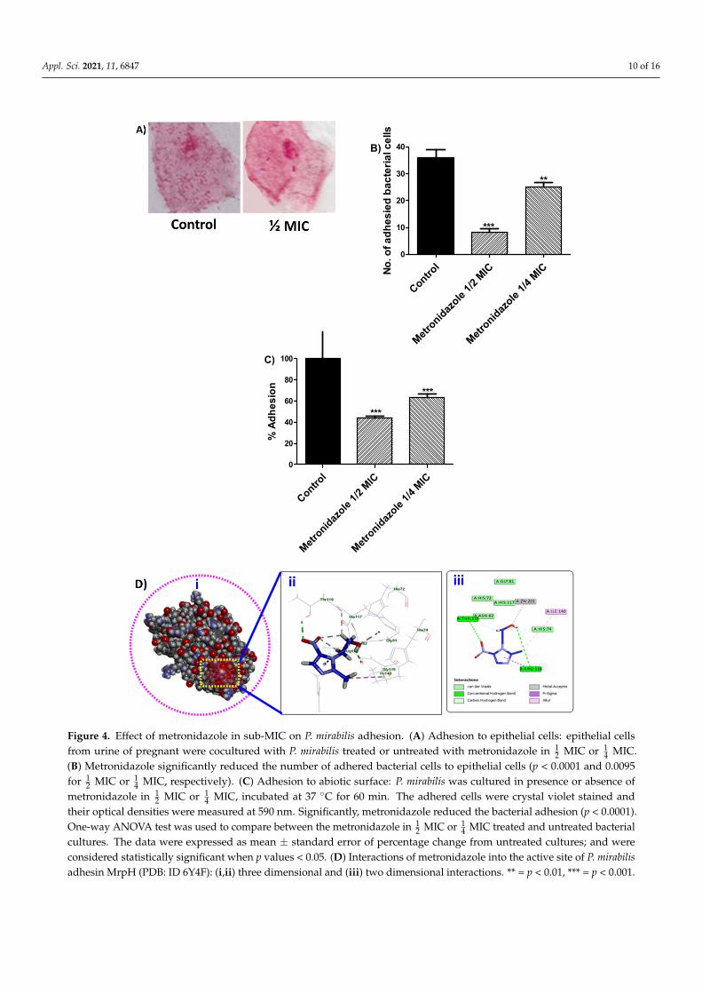
Biofilm formation and bacteria invasion are basicaly depend on the capability ofbacterial cells to adhere either to living cells or inanimate objects. In order to evalute theeffect of metronidazole on bacterial adhesion to cells or abiotic surfaces; P. mirabilis treatedor untreated with metronidazole were cocultured with epithelial cells or in microtitreplates, repectively. Markedly, the numbers of adhered bacterial cells to epithelial cellswere reduced in precence of metronidazole (Figure 4A,B). Furthermore, P. mirabilis treatedor untreated with metronidazole ( 1
2 MIC or 14 MIC) was incubated in microtitre plate for
60 min and the optical denisties of atained adhered cells were measured. As metronidazolereduced the adherence of bacterial cells to epithelial cells, metronidazole significantlyreduced the adhesion to abiotic surfaces (Figure 4C).

Appl. Sci. 2021, 11, 6847 8 of 16
Furthermore, the structural interaction between metronidazole and the Zn-dependentreceptor-binding domain of P. mirabilis MR/P fimbrial adhesin MrpH, was in-silico evalu-ated. The results indicated that metronidazole fit nicely into the binding site of P. mirabilisadhesin MrpH (PDB ID: 6Y4F) with the formation of two hydrogen bonds with Thr116 viathe oxygen atom of NO2 group and Arg118 through the proton of OH group, in additionto many hydrophobic interactions with Thr116, Arg118 and Ile140. Notably, metronidazoleengaged in the formation of one metal acceptor bond with ZN:201 with the oxygen atomof OH group (Figure 4D).
3.6. Effect of Metronidazole in Sub-MIC on Biofilm Formation
To evaluate the biofilm production, P. mirabilis was considerd according [34] as strongbiofim forming when OD > 4× ODc (OD = 0.34 and OD = 0.064). The MBECs weredetermined and presented for the selected antibiotics and metronidazole in Table 1.
Appl. Sci. 2021, 11, x FOR PEER REVIEW 7 of 16
Untraete
d Bac
teria
Metronidaz
ole 1/2
MIC
Metronidaz
ole 1/4
MIC
0.0
0.5
1.0
(ns) (ns)
OD
at 6
00 n
m
Figure 1. Effect of metronidazole in sub-MIC on P. mirabilis growth. LB broth with or without met-ronidazole (½ MIC or ¼ MIC) was inoculated with a fresh inoculum of adjusted P. mirabilis to 0.5 McFarland Standard equivalent and the optical densities were measured after overnight culturing at 37 °C. One-Way ANOVA test was applied to compare between bacterial growth in presence and absence of metronidazole; statistical significance was assumed when p values < 0.05. There was no statistical significance between metronidazole treated and untreated cultures (p > 0.05).
3.3. Effect of Metronidazole in Sub-MIC on Production of Bacterial Virulence Enzymes To evaluate the anti-virulence activities of metronidazole in sub-MIC on P. mirabilis
virulence. The inhibition effect of metronidazole on production of some virulent exoen-zymes were assessed. Significantly, metronidazole in sub-MIC reduced the production of protease, hemolysins and urease (Figure 2).
Untreate
d P. mira
bilis
Metronidaz
ole 1/2
MIC
Metronidaz
ole 1/4
MIC
0
20
40
60
80
100
***
***
A)
% P
rote
ase
Prod
uctio
n
Untreate
d P. mira
bilis
Metronidaz
ole 1/2
MIC
Metronidaz
ole 1/4
MIC
0
20
40
60
80
100
***
***
B)
% H
emol
ysis
Untreate
d P. mira
bilis
Metronidaz
ole 1/2
MIC
Metronidaz
ole 1/4
MIC
0
20
40
60
80
100D)
***
***
% U
reas
e Pr
oduc
tionC)
Figure 2. Effect of metronidazole in sub-MIC on P. mirabilis virulence enzymes. One-way ANOVA test was employed to compare between the metronidazole in ½ MIC or ¼ MIC treated and un-treated bacterial cultures. The data were presented as mean ± standard error of percentage change from untreated cultures; and were considered statistically significant when p values < 0.05. (A) Protease production: significantly, metronidazole reduced the protease production in comparison
Figure 2. Effect of metronidazole in sub-MIC on P. mirabilis virulence enzymes. One-way ANOVAtest was employed to compare between the metronidazole in 1
2 MIC or 14 MIC treated and untreated
bacterial cultures. The data were presented as mean ± standard error of percentage change fromuntreated cultures; and were considered statistically significant when p values < 0.05. (A) Proteaseproduction: significantly, metronidazole reduced the protease production in comparison to untreatedcultures (p < 0.0001). (B) Hemolytic activity: metronidazole in sub-MIC significantly decreased thehemolytic activity (p < 0.0001). (C,D) Urease inhibition: Christensen’s urea agar plates containingdifferent sub-MIC of metronidazole were used. The change in the color of the pH indicator fromyellow to pink indicates the urease activity and pink zones were measured in mm. Metronidazolesignificantly reduced the production of ureases (p < 0.0001), *** = p < 0.001.

Appl. Sci. 2021, 11, 6847 9 of 16
Appl. Sci. 2021, 11, x FOR PEER REVIEW 8 of 16
to untreated cultures (p < 0.0001). (B) Hemolytic activity: metronidazole in sub-MIC significantly decreased the hemolytic activity (p < 0.0001). (C,D) Urease inhibition: Christensen’s urea agar plates containing different sub-MIC of metronidazole were used. The change in the color of the pH indicator from yellow to pink indicates the urease activity and pink zones were measured in mm. Metronidazole significantly reduced the production of ureases (p < 0.0001), *** = p < 0.001.
3.4. Effect of Metronidazole in sub-MIC on Bacterial Motilities Bacterial motility is important for adhesion and biofilm formation and diminishing
effect of metronidazole of bacterial motility indicate its anti-QS and anti-virulence activi-ties. The diameters of P. mirabilis swimming and swarming were measured on LB agar (0.4% or 1.5%, respectively) plates with or without metronidazole (½ MIC or ¼ MIC). Sig-nificantly, metronidazole diminished the bacterial motility (Figure 3). Moreover, light mi-croscope images were taken for the P. mirabilis cells at the center and edge of swarming. Clearly, cells at the edge which represent the swarmer cells were larger and longer than those in the center. In the presence of metronidazole (½ MIC), the cells at the sizes of swarmer cells were smaller than swarmer cells in absence of metronidazole (Figure 3A).
Control
Metronidaz
ole 1/2
MIC
Metronidaz
ole 1/4
MIC
0
20
40
60
80
100
***
C)
% o
f Sw
arm
ing
Mot
ility
Control
Metronidaz
ole 1/2
MIC
Metronidaz
ole 1/4
MIC
0
20
40
60
80
100
% o
f Sw
imm
ing
Mot
ility
***
***
E)
Figure 3. Effect of metronidazole in sub-MIC on P. mirabilis motility. One-way ANOVA test was used to compare between the metronidazole in ½ MIC or ¼ MIC treated and untreated bacterial cultures. The data were shown as mean ± standard error of percentage change from untreated cultures; and were considered statistically significant when p values < 0.05. (A) Light microscope image of simple stained P. mirabilis at the center (non-swarmer cells), the edge of swarming (elongated larger swarmer cells) and swarmer cells in precence of metronidazole. Clearly, the swarmer cells in precence of metroni-dazole is smaller than those in absence of metronidazole. (B,C) Metronidazole in sub-MIC significantly reduced the swarming motility (p < 0.0001). (D,E) The diameters of swimming motiliy were significantly decreased in precence of meetonidazole in sub-MIC (p < 0.0001). *** = p < 0.001.
Figure 3. Effect of metronidazole in sub-MIC on P. mirabilis motility. One-way ANOVA test was used to compare between themetronidazole in 1
2 MIC or 14 MIC treated and untreated bacterial cultures. The data were shown as mean ± standard error
of percentage change from untreated cultures; and were considered statistically significant when p values < 0.05. (A) Lightmicroscope image of simple stained P. mirabilis at the center (non-swarmer cells), the edge of swarming (elongated largerswarmer cells) and swarmer cells in precence of metronidazole. Clearly, the swarmer cells in precence of metronidazoleis smaller than those in absence of metronidazole. (B,C) Metronidazole in sub-MIC significantly reduced the swarmingmotility (p < 0.0001). (D,E) The diameters of swimming motiliy were significantly decreased in precence of meetonidazolein sub-MIC (p < 0.0001). *** = p < 0.001.
In order to evaluate the metronidazole inhbitory effect on biofilm production; P. mirabilistreated or untreated with metronidazole ( 1
2 MIC or 14 MIC) was cultured in microtite plate
overnight at 37 ◦C. In addition, the biofilm forming bacterial cells were crystal violet stainedand optical densities were measured. Significantly, metronidazole prevented the biofilmformation (Figure 5).
3.7. In-Silico Docking of Metronidazole into P. mirabilis QS Eseential Proteins
To give a hint into the capability of metronidazole to antagonize the QS essentialproteins; the interaction between metronidazole and QS proteins was evaluated. P. mirabilisQS protein was retrieved from UniProtKB (PMI1345) and molecular docking was carriedout. The results showenin Figure 6, displayed that thereceptor well accommodatingmetronidazole inside the binding cavity and establishing appropriate interactions involvedthe formation of three hydrogen bonds; the proton of OH group engaged in one hydrogenbond with Glu57. The oxygen atom of NO2 involved in the formation of two hydrogenbonds with Gly129 and Thr130 amino acid residues. Additionally, the oxygen of OHgroup and the oxygen of NO2 group of metronidazole engaged in the formation of twoimportant metal acceptor bonds with ZN:1. Moreover, metronidazole incorporated inmany hydrophobic interactions with His54, Glu57, Cys128 and ZN:1. These results mayexplain the possible reasons for enhanced anti-QS activity of metronidazole.
Appl. Sci. 2021, 11, 6847 10 of 16
Appl. Sci. 2021, 11, x FOR PEER REVIEW 9 of 16
3.5. Effect of Metronidazole in Sub-MIC on Bacterial Adhesion Biofilm formation and bacteria invasion are basicaly depend on the capability of bac-
terial cells to adhere either to living cells or inanimate objects. In order to evalute the effect of metronidazole on bacterial adhesion to cells or abiotic surfaces; P. mirabilis treated or untreated with metronidazole were cocultured with epithelial cells or in microtitre plates, repectively. Markedly, the numbers of adhered bacterial cells to epithelial cells were re-duced in precence of metronidazole (Figure 4A,B). Furthermore, P. mirabilis treated or un-treated with metronidazole (½ MIC or ¼ MIC) was incubated in microtitre plate for 60 min and the optical denisties of atained adhered cells were measured. As metronidazole reduced the adherence of bacterial cells to epithelial cells, metronidazole significantly re-duced the adhesion to abiotic surfaces (Figure 4C).
Furthermore, the structural interaction between metronidazole and the Zn-depend-ent receptor-binding domain of P. mirabilis MR/P fimbrial adhesin MrpH, was in-silico evaluated. The results indicated that metronidazole fit nicely into the binding site of P. mirabilis adhesin MrpH (PDB ID: 6Y4F) with the formation of two hydrogen bonds with Thr116 via the oxygen atom of NO2 group and Arg118 through the proton of OH group, in addition to many hydrophobic interactions with Thr116, Arg118 and Ile140. Notably, metronidazole engaged in the formation of one metal acceptor bond with ZN:201 with the oxygen atom of OH group (Figure 4D).
Control
Metronidaz
ole 1/2
MIC
Metronidaz
ole 1/4
MIC
0
10
20
30
40
***
**
B)
No. o
f adh
esie
d ba
cter
ial c
ells
Control
Metronidaz
ole 1/2
MIC
Metronidaz
ole 1/4
MIC
0
20
40
60
80
100
***
***
C)
% A
dhes
ion
Appl. Sci. 2021, 11, x FOR PEER REVIEW 10 of 16
Figure 4. Effect of metronidazole in sub-MIC on P. mirabilis adhesion. (A) Adhesion to epithelial cells: epithelial cells from urine of pregnant were cocultured with P. mirabilis treated or untreated with metro-nidazole in ½ MIC or ¼ MIC. (B) Metronidazole significantly reduced the number of adhered bacterial cells to epithelial cells (p < 0.0001 and 0.0095 for ½ MIC or ¼ MIC, respectively). (C) Adhesion to abiotic surface: P. mirabilis was cultured in presence or absence of metronidazole in ½ MIC or ¼ MIC, incu-bated at 37 °C for 60 min. The adhered cells were crystal violet stained and their optical densities were measured at 590 nm. Significantly, metronidazole reduced the bacterial adhesion (p < 0.0001). One-way ANOVA test was used to compare between the metronidazole in ½ MIC or ¼ MIC treated and un-treated bacterial cultures. The data were expressed as mean ± standard error of percentage change from untreated cultures; and were considered statistically significant when p values < 0.05. (D) Interactions of metronidazole into the active site of P. mirabilis adhesin MrpH (PDB: ID 6Y4F): (i,ii) three dimensional and (iii) two dimensional interactions. ** = p < 0.01, *** = p < 0.001.
3.6. Effect of Metronidazole in Sub-MIC on Biofilm Formation To evaluate the biofilm production, P. mirabilis was considerd according [34] as
strong biofim forming when OD > 4× ODc (OD = 0.34 and OD = 0.064). The MBECs were determined and presented for the selected antibiotics and metronidazole in Table 1.
In order to evaluate the metronidazole inhbitory effect on biofilm production; P. mi-rabilis treated or untreated with metronidazole (½ MIC or ¼ MIC) was cultured in micro-tite plate overnight at 37 °C. In addition, the biofilm forming bacterial cells were crystal violet stained and optical densities were measured. Significantly, metronidazole pre-vented the biofilm formation (Figure 5).
Control
Metronidaz
ole 1/2
MIC
Metronidaz
ole 1/4
MIC
0
20
40
60
80
100
***
***
% B
iofil
m F
orm
atio
n
Figure 5. Effect of metronidazole in sub-MIC on biofilm formation. P. mirabilis cultures were grown in presence or absence of metronidazole (½ MIC or ¼ MIC) and incubated overnight at 37 °C. The optical densities of stained adhered cells were measured at 590 nm. Significantly, metroni-dazole diminished the biofilm formation (p < 0.0001). One-way ANOVA test was used to compare between the metronidazole in ½ MIC or ¼ MIC treated and untreated bacterial cultures. The data were presented as mean ± standard error of percentage change from untreated cultures; and and statistical significance was considered when p values < 0.05, *** = p < 0.001.
Figure 4. Effect of metronidazole in sub-MIC on P. mirabilis adhesion. (A) Adhesion to epithelial cells: epithelial cellsfrom urine of pregnant were cocultured with P. mirabilis treated or untreated with metronidazole in 1
2 MIC or 14 MIC.
(B) Metronidazole significantly reduced the number of adhered bacterial cells to epithelial cells (p < 0.0001 and 0.0095for 1
2 MIC or 14 MIC, respectively). (C) Adhesion to abiotic surface: P. mirabilis was cultured in presence or absence of
metronidazole in 12 MIC or 1
4 MIC, incubated at 37 ◦C for 60 min. The adhered cells were crystal violet stained andtheir optical densities were measured at 590 nm. Significantly, metronidazole reduced the bacterial adhesion (p < 0.0001).One-way ANOVA test was used to compare between the metronidazole in 1
2 MIC or 14 MIC treated and untreated bacterial
cultures. The data were expressed as mean ± standard error of percentage change from untreated cultures; and wereconsidered statistically significant when p values < 0.05. (D) Interactions of metronidazole into the active site of P. mirabilisadhesin MrpH (PDB: ID 6Y4F): (i,ii) three dimensional and (iii) two dimensional interactions. ** = p < 0.01, *** = p < 0.001.

Appl. Sci. 2021, 11, 6847 11 of 16
Appl. Sci. 2021, 11, x FOR PEER REVIEW 10 of 16
Figure 4. Effect of metronidazole in sub-MIC on P. mirabilis adhesion. (A) Adhesion to epithelial cells: epithelial cells from urine of pregnant were cocultured with P. mirabilis treated or untreated with metro-nidazole in ½ MIC or ¼ MIC. (B) Metronidazole significantly reduced the number of adhered bacterial cells to epithelial cells (p < 0.0001 and 0.0095 for ½ MIC or ¼ MIC, respectively). (C) Adhesion to abiotic surface: P. mirabilis was cultured in presence or absence of metronidazole in ½ MIC or ¼ MIC, incu-bated at 37 °C for 60 min. The adhered cells were crystal violet stained and their optical densities were measured at 590 nm. Significantly, metronidazole reduced the bacterial adhesion (p < 0.0001). One-way ANOVA test was used to compare between the metronidazole in ½ MIC or ¼ MIC treated and un-treated bacterial cultures. The data were expressed as mean ± standard error of percentage change from untreated cultures; and were considered statistically significant when p values < 0.05. (D) Interactions of metronidazole into the active site of P. mirabilis adhesin MrpH (PDB: ID 6Y4F): (i,ii) three dimensional and (iii) two dimensional interactions. ** = p < 0.01, *** = p < 0.001.
3.6. Effect of Metronidazole in Sub-MIC on Biofilm Formation To evaluate the biofilm production, P. mirabilis was considerd according [34] as
strong biofim forming when OD > 4× ODc (OD = 0.34 and OD = 0.064). The MBECs were determined and presented for the selected antibiotics and metronidazole in Table 1.
In order to evaluate the metronidazole inhbitory effect on biofilm production; P. mi-rabilis treated or untreated with metronidazole (½ MIC or ¼ MIC) was cultured in micro-tite plate overnight at 37 °C. In addition, the biofilm forming bacterial cells were crystal violet stained and optical densities were measured. Significantly, metronidazole pre-vented the biofilm formation (Figure 5).
Control
Metronidaz
ole 1/2
MIC
Metronidaz
ole 1/4
MIC
0
20
40
60
80
100
***
***
% B
iofil
m F
orm
atio
n
Figure 5. Effect of metronidazole in sub-MIC on biofilm formation. P. mirabilis cultures were grown in presence or absence of metronidazole (½ MIC or ¼ MIC) and incubated overnight at 37 °C. The optical densities of stained adhered cells were measured at 590 nm. Significantly, metroni-dazole diminished the biofilm formation (p < 0.0001). One-way ANOVA test was used to compare between the metronidazole in ½ MIC or ¼ MIC treated and untreated bacterial cultures. The data were presented as mean ± standard error of percentage change from untreated cultures; and and statistical significance was considered when p values < 0.05, *** = p < 0.001.
Figure 5. Effect of metronidazole in sub-MIC on biofilm formation. P. mirabilis cultures were grownin presence or absence of metronidazole ( 1
2 MIC or 14 MIC) and incubated overnight at 37 ◦C. The
optical densities of stained adhered cells were measured at 590 nm. Significantly, metronidazolediminished the biofilm formation (p < 0.0001). One-way ANOVA test was used to compare betweenthe metronidazole in 1
2 MIC or 14 MIC treated and untreated bacterial cultures. The data were
presented as mean ± standard error of percentage change from untreated cultures; and and statisticalsignificance was considered when p values < 0.05, *** = p < 0.001.
Appl. Sci. 2021, 11, x FOR PEER REVIEW 11 of 16
3.7. In-Silico Docking of Metronidazole into P. mirabilis QS Eseential Proteins
To give a hint into the capability of metronidazole to antagonize the QS essential proteins; the interaction between metronidazole and QS proteins was evaluated. P. mira-bilis QS protein was retrieved from UniProtKB (PMI1345) and molecular docking was car-ried out. The results showenin Figure 6, displayed that thereceptor well accommodating metronidazole inside the binding cavity and establishing appropriate interactions in-volved the formation of three hydrogen bonds; the proton of OH group engaged in one hydrogen bond with Glu57. The oxygen atom of NO2 involved in the formation of two hydrogen bonds with Gly129 and Thr130 amino acid residues. Additionally, the oxygen of OH group and the oxygen of NO2 group of metronidazole engaged in the formation of two important metal acceptor bonds with ZN:1. Moreover, metronidazole incorporated in many hydrophobic interactions with His54, Glu57, Cys128 and ZN:1. These results may explain the possible reasons for enhanced anti-QS activity of metronidazole.
Figure 6. Interactions of metronidazole into the active site of P. mirabilis QS protein (PMI1345). (A,B) three dimensional and (C) two dimensional.
3.8. MICs and MBECs of Tested Antibiotics in Precence of Metronidazole in Sub-MIC To evalutae the effect of metronidazole in sub-MIC as anti-virulence agent on de-
creasing of MICs and MIBCs of selected antibiotics; the MICs and MIBCs of comined met-ronidazole and selected antibiotics were determined. The outcomes of the combined effect of netronidazole (¼ MIC) and selected antibiotics were calculated according [36]. The re-sults were summarized in Table 2. Obviously, metronidazole decreased the MICs and MIBCs of antibiotics and showed considerable synergism.
Table 2. The susceptibility of isolated P. mirabilis to antibiotics in presence of Metronidazole (¼ MIC).
Antibiotic MIC MICmet FIC MBEC MBECmet FIC Ciprofloxacin 2 µg/mL 1 µg/mL 0.5 128 µg/mL 32 µg/mL 0.25 Cefoperazone 64 µg/mL 16 µg/mL 0.25 1024 µg/mL 256 µg/mL 0.25
Amoxicillin/Clavulinic acid 256 µg/mL 32 µg/mL 0.125 2048 µg/mL 512 µg/mL 0.25 Imipenem 4 µg/mL 2 µg/mL 0.5 8 µg/mL 4 µg/mL 0.5
Gentamycin 16 µg/mL 16 µg/mL 1 512 µg/mL 256 µg/mL 0.5 Tetracycline 64 µg/mL 64 µg/mL 1 2048 µg/mL 1024 µg/mL 0.5
Chloramphenicol 64 µg/mL 16 µg/mL 0.25 2048 µg/mL 256 µg/mL 0.125 MIC and MBEC: Minimum eradication concentration and minimum biofilm inhibitory concentration of tested antibiotics. MICmet and MBECmet: Minimum inhibitory concentration and minimum biofilm eradication concentration of tested anti-biotics in presence of metronidazole in concentration of ¼ MIC. FIC: Fractional inhibitory concentration = MIC drug in combination/MIC drug alone. The result of the combination may be antagonistic (FIC > 4), indifferent (FIC > 0.5 to 4), or synergistic (FIC ≤ 0.5).
Figure 6. Interactions of metronidazole into the active site of P. mirabilis QS protein (PMI1345). (A,B) three dimensional and(C) two dimensional.
3.8. MICs and MBECs of Tested Antibiotics in Precence of Metronidazole in Sub-MIC
To evalutae the effect of metronidazole in sub-MIC as anti-virulence agent on de-creasing of MICs and MIBCs of selected antibiotics; the MICs and MIBCs of cominedmetronidazole and selected antibiotics were determined. The outcomes of the combinedeffect of netronidazole ( 1
4 MIC) and selected antibiotics were calculated according [36]. Theresults were summarized in Table 2. Obviously, metronidazole decreased the MICs andMIBCs of antibiotics and showed considerable synergism.

Appl. Sci. 2021, 11, 6847 12 of 16
Table 2. The susceptibility of isolated P. mirabilis to antibiotics in presence of Metronidazole ( 14 MIC).
Antibiotic MIC MICmet FIC MBEC MBECmet FICCiprofloxacin 2 µg/mL 1 µg/mL 0.5 128 µg/mL 32 µg/mL 0.25Cefoperazone 64 µg/mL 16 µg/mL 0.25 1024 µg/mL 256 µg/mL 0.25
Amoxicillin/Clavulinicacid 256 µg/mL 32 µg/mL 0.125 2048 µg/mL 512 µg/mL 0.25
Imipenem 4 µg/mL 2 µg/mL 0.5 8 µg/mL 4 µg/mL 0.5Gentamycin 16 µg/mL 16 µg/mL 1 512 µg/mL 256 µg/mL 0.5Tetracycline 64 µg/mL 64 µg/mL 1 2048 µg/mL 1024 µg/mL 0.5
Chloramphenicol 64 µg/mL 16 µg/mL 0.25 2048 µg/mL 256 µg/mL 0.125
MIC and MBEC: Minimum eradication concentration and minimum biofilm inhibitory concentration of tested antibiotics. MICmetand MBECmet: Minimum inhibitory concentration and minimum biofilm eradication concentration of tested antibiotics in presence ofmetronidazole in concentration of 1
4 MIC. FIC: Fractional inhibitory concentration = MIC drug in combination/MIC drug alone. The resultof the combination may be antagonistic (FIC > 4), indifferent (FIC > 0.5 to 4), or synergistic (FIC ≤ 0.5).
4. Discussion
P. mirabilis causes a wide range of infections and its spread is owed to an inherenttranslocation capability using peritrichous flagellar. Moreover, P. mirabilis harbors a con-sidered arsenal of exoenzymes as urease, protease and hemolysins and strong biofilmformation capability [6,40]. In the current study, we aimed to evaluate the effects of metron-idazole on bacterial pathogenesis by challenging the virulence of highly resistant P. mirabilisisolated from diabetic foot ulcers. Metronidazole is the most globally used nitroimidazoleantimicrobial agent, it is usually prescribed to anaerobic infection and is considered asgold standard which all other anaerobic acting antibiotics should be referenced [28,29].Metronidazole interacts with bacterial DNA and consequently inhibits the bacterial proteinsynthesis. Metronidazole has activity on wide range of bacteria and its use possess severaladvantages as good tissue penetration, rapid bacterial killing and low cost [29].
In order to evaluate the effect of metronidazole on P. mirabilis virulence, we usedmetronidazole in sub-MICs to avoid any effect on bacterial growth. In a previous work,metronidazole and secnidazole which harbor similar chemical moiety were efficientlyable to curtail the Pseudomonas aeruginosa pathogenesis [41,42]. Obviously, metronidazolehas no influence at all on the bacterial growth. The proteolytic enzyme protease cleavesvarious essential proteins as casein, secretory components, serum albumin and gelatin.Furthermore, protease is able to breakdown 2 classes of immunoglobulins A and G whichfacilitate the spread of microbial infection and lowering the defense of the host [43,44].Urease enzyme is a significant virulence factor that is used by pathogens of urinary andgastrointestinal tracts as P. mirabilis. While the production of urease is constitutive in moststrains of P. mirabilis, it is inducible in some strains by pH changes [6,44,45]. Its capabilityto recycle the nitrogenous compounds enhances the resistance to several biocides [43,45].Importantly, the degraded urea due to urease provides an alkaline optimal condition forprotease action and in turn increase the microbial spread and increase the production ofboth urease and protease in repeated cycles [6,46]. The cytotoxicity of hemolysin thatis encoded by 2-component secretion system hpmBA genes greatly enhance P. mirabilispathogenesis [6,47]. These three enzymes, protease, hemolysins and urease play crucialroles in the spread of bacterial infection especially in lack of efficient immunity as in diabeticfoot ulcers [10,20]. Significantly, metronidazole in sub-MIC reduced the production of P.mirabilis exoenzymes protease, urease and hemolysins.
The P. mirabilis invading ability to epithelial cells is fundamentally owed to swarmercells but not vegetative cells [6,44]. Flagellum dependent P. mirabilis motilities (swarmingand swimming) facilitate the spread of infection and formation of biofilms [7,48]. Moreover,the formation of hyperflagellated swarmer cells is linked to significant increases in theproduction of other virulence factors as protease and urease [45,49]. It was suggested aclose correlation between biofilm formation and bacterial motility; as the formation ofbiofilms is decreased in the non-motile bacteria [8]. As a consequence, the bacterial biofilmformation capability beside its motility result in spreading of infection and enhancing of

Appl. Sci. 2021, 11, 6847 13 of 16
antibiotic resistance of the persistent microbes in diabetic foot ulcers [10]. Our findingsrevealed a significant diminishing effect of metronidazole on P. mirabilis motilities.
Biofilm formation is characteristic feature of the life style of pathogenic bacteria; It iscomposed of embedded microbial cells in dynamic communities that accumulate eitheron abiotic or living surfaces [50,51]. In sequential manner and after bacterial adhesion tosurfaces, free floating vegetative cells start to produce extracellular polymeric substanceswhich leads to irreversible attachment and entrapping of bacteria in biofilms [52]. Theflagella-driven motilities are needed for P. mirabilis adhesion, which followed by its colo-nization and biofilm formation [49,53,54]. In compliance with our previous findings thatshowed the inhibition effect of metronidazole on P. mirabilis motilities; metronidazole issignificantly reduced the adhesion of P. mirabilis to both abiotic surfaces and epithelialcells and also significantly decreased the biofilm formation. Moreover, MICs and MBECsfor the tested antibiotics were markedly lowered when combined with metronidazole( 1
4 MIC). The synergistic effect of metronidazole at sub-MIC when combined with testedantibiotics indicate that these antibiotics can be used to treat diabetic foot ulcers in lowerconcentration, which may decrease the side effects, the resistance development and costs.
Molecular docking is one of the most widely applied approaches for the study ofprotein-ligand interactions and for drug discovery and development. Therefore, moleculardocking studies were carried out to get structural insights into the interaction betweenmetronidazole and P. mirabilis QS proteins and the Zn-dependent receptor-binding domainof P. mirabilis MR/P fimbrial adhesin MrpH. Mannose Resistant Proteus like fimbriae (MRP)that are encoded by mrpABCDEFGHJ operon, are the most widely studied P. mirabilisfimbriae. MRP fimbriae are greatly essential for P. mirabilis aggregation, biofilm formationand colonization in kidney and bladder [55,56]. MrpH is one of the main virulence involvedfimbrial proteins in P. mirabilis [56]. In this study, we showed that metronidazole hasa considerable affinity to the binding site of P. mirabilis adhesin MrpH. The moleculardocking results emphasize our earlier finding in which we observed the reduction ofadhesion in presence of metronidazole. QS is signaling system which organize the bacterialpathogenesis and QS targeting is one of the efficient approaches to diminish microbialvirulence [16,17,23]. We hypothesized that hindering of metronidazole to QS system andbacterial adhesion leads to diminishing of the P. mirabilis virulence factors armory and helpgreatly in treating aggressive infections as diabetic foot ulcers. In this context, we examinedthe capability of metronidazole to antagonize the P. mirabilis QS essential proteins. One ofthe most crucial role players in P. mirabilis QS, is protein PMI1345 as it catalyzes the transferof the phosphoribosyl group of 5-phosphorylribose-1-pyrophosphate to anthranilate toyield N-(5′-phosphoribosyl)-anthranilate [57]. While LuxS and MtnN proteins are involvedin the biosynthesis of autoiducers-2; PMI1345 and MnmC proteins are involved in thetranslocation machinery in the QS system [58]. Essentially, Pawar et al., analysis revealedPMI1345 to be the prominent Eigenvector centrality protein in the 139th core of the P.mirabilis genome [57]. Our molecular docking study showed the ability of metronidazoleto antagonize the essential QS protein PMI1345. Although more detailed molecular andin-vivo investigations are needs, these findings give a preliminary expectation about themetronidazole ability to hinder the QS system. It needs further pharmacological andpharmaceutical studies to validate the application of metronidazole as anti-virulence agentbeside its antimicrobial activities.
5. Conclusions
Diabetic foot ulcers infections are among the most wretched clinical conditions, es-pecially if the infection was with tenacious microbe as P. mirabilis. In the current study,we evaluated the anti-virulence activities of one of the most globally used antibacterialmetronidazole. Our results revealed the inhibitory effectiveness of metronidazole in sub-MIC concentrations to various P. mirabilis virulence factors. Here, we introducing a newprimary insight about the use of metronidazole as an anti-virulence and anti-QS agent thatcould be attractive for researchers and clinicians to further in-vivo investigations.
Appl. Sci. 2021, 11, 6847 14 of 16
Author Contributions: Conceptualization, W.A.H.H. and H.A.A.; methodology, H.A.A., W.A.H.H.and M.F.A.M.; software, M.F.A.M. and T.S.I.; validation, A.N.K., M.T.K. and H.Z.A.; formal analysis,A.S.A.L. and E.-S.K.; investigation, A.N.K.; resources, A.N.K., M.T.K. and H.Z.A.; data curation,A.N.K., M.T.K. and H.Z.A.; writing—original draft preparation, W.A.H.H. and H.A.A.; writing—review and editing, W.A.H.H. and M.F.A.M.; visualization, M.Y.; M.K.S. and T.S.I.; supervision,A.N.K. and W.A.H.H.; project administration, W.A.H.H.; funding acquisition, M.T.K., H.Z.A., M.K.S.and A.N.K. All authors have read and agreed to the published version of the manuscript.
Funding: The Deanship of Scientific Research (DSR) at King Abdulaziz University, Jeddah, SaudiArabia has funded this project, under grant No. (RG-16-166-42).
Informed Consent Statement: This study does not include any studies with human participants.
Conflicts of Interest: The authors declare no conflict of interest.
References1. Van Battum, P.; Schaper, N.; Prompers, L.; Apelqvist, J.; Jude, E.; Piaggesi, A.; Bakker, K.; Edmonds, M.; Holstein, P.; Jirkovska, A.;
et al. Differences in minor amputation rate in diabetic foot disease throughout Europe are in part explained by differences indisease severity at presentation. Diabet. Med. 2011, 28, 199–205. [CrossRef] [PubMed]
2. Volmer-Thole, M.; Lobmann, R. Neuropathy and Diabetic Foot Syndrome. Int. J. Mol. Sci. 2016, 17, 917. [CrossRef] [PubMed]3. Boulton, A.J. The pathway to foot ulceration in diabetes. Med. Clin. N. Am. 2013, 97, 775–790. [CrossRef] [PubMed]4. Hartemann-Heurtier, A.; Senneville, E. Diabetic foot osteomyelitis. Diabetes Metab. 2008, 34, 87–95. [CrossRef]5. Hegazy, W.A.H. Hepatitis C virus pathogenesis: Serum IL-33 level indicates liver damage. Afr. J. Microbiol. Res. 2015, 9, 1386–1393.
[CrossRef]6. Armbruster, C.E.; Mobley, H.L. Merging mythology and morphology: The multifaceted lifestyle of Proteus mirabilis. Nat. Rev.
Microbiol. 2012, 10, 743–754. [CrossRef]7. Jacobsen, S.M.; Stickler, D.J.; Mobley, H.L.; Shirtliff, M.E. Complicated catheter-associated urinary tract infections due to
Escherichia coli and Proteus mirabilis. Clin. Microbiol. Rev. 2008, 21, 26–59. [CrossRef]8. Sekhar, S.; Vyas, N.; Unnikrishnan, M.; Rodrigues, G.; Mukhopadhyay, C. Antimicrobial susceptibility pattern in diabetic foot
ulcer: A pilot study. Ann. Med. Health Sci. Res. 2014, 4, 742–745. [CrossRef]9. Perim, M.C.; Borges Jda, C.; Celeste, S.R.; Orsolin Ede, F.; Mendes, R.R.; Mendes, G.O.; Ferreira, R.L.; Carreiro, S.C.; Pranchevicius,
M.C. Aerobic bacterial profile and antibiotic resistance in patients with diabetic foot infections. Rev. Soc. Bras. Med. Trop. 2015, 48,546–554. [CrossRef]
10. El-Tahawy, A.T. Bacteriology of diabetic foot. Saudi Med. J. 2000, 21, 344–347.11. Flannery, E.L.; Mody, L.; Mobley, H.L. Identification of a modular pathogenicity island that is widespread among urease-
producing uropathogens and shares features with a diverse group of mobile elements. Infect. Immun. 2009, 77, 4887–4894.[CrossRef]
12. Wasfi, R.; Hamed, S.M.; Amer, M.A.; Fahmy, L.I. Proteus mirabilis Biofilm: Development and Therapeutic Strategies. Front. CellInfect. Microbiol. 2020, 10, 414. [CrossRef]
13. Tansarli, G.S.; Athanasiou, S.; Falagas, M.E. Evaluation of antimicrobial susceptibility of Enterobacteriaceae causing urinary tractinfections in Africa. Antimicrob. Agents Chemother. 2013, 57, 3628–3639. [CrossRef]
14. Zhao, W.H.; Hu, Z.Q. Epidemiology and genetics of CTX-M extended-spectrum beta-lactamases in Gram-negative bacteria. Crit.Rev. Microbiol. 2013, 39, 79–101. [CrossRef]
15. Abbas, H.A.; Hegazy, W.A.H. Targeting the virulence factors of Serratia marcescens by ambroxol. Roum. Arch. Microbiol. Immunol.2017, 76, 27–32.
16. Abbas, H.A.; Hegazy, W.A.H. Repurposing anti-diabetic drug “Sitagliptin” as a novel virulence attenuating agent in Serratiamarcescens. PLoS ONE 2020, 15, e0231625. [CrossRef] [PubMed]
17. El-Hamid, A.; Marwa, I.; Y El-Naenaeey, E.S.; Hegazy, W.A.; Mosbah, R.A.; Nassar, M.S.; Bakhrebah, M.A.; Abdulaal, W.H.;Alhakamy, N.A.; Bendary, M.M. Promising Antibiofilm Agents: Recent Breakthrough against Biofilm Producing Methicillin-Resistant Staphylococcus aureus. Antibiotics 2020, 9, 667. [CrossRef] [PubMed]
18. Al Saqr, A.; Khafagy, E.S.; Alalaiwe, A.; Aldawsari, M.F.; Alshahrani, S.M.; Anwer, M.K.; Khan, S.; Lila, A.S.A.; Arab, H.H.;Hegazy, W.A.H. Synthesis of Gold Nanoparticles by Using Green Machinery: Characterization and In Vitro Toxicity. Nanomaterials2021, 11, 808. [CrossRef] [PubMed]
19. Askoura, M.; Hegazy, W.A.H. Ciprofloxacin interferes with Salmonella Typhimurium intracellular survival and host virulencethrough repression of Salmonella pathogenicity island-2 (SPI-2) genes expression. Pathog. Dis. 2020, 78. [CrossRef]
20. Hegazy, W.A.H. Diclofenac inhibits virulence of Proteus mirabilis isolated from diabetic foot ulcer. Afr. J. Microbiol. Res. 2016, 10,733–743. [CrossRef]
21. Hegazy, W.A.H.; Al Mamari, R.; Almazroui, K.; Al Habsi, A.; Kamona, A.; AlHarthi, H.; Al Lawati, A.I.; AlHusaini, A.H.Retrospective Study of Bone-TB in Oman: 2002-2019. J. Epidemiol. Glob. Health 2021, 11, 238–245. [CrossRef]

Appl. Sci. 2021, 11, 6847 15 of 16
22. Bendary, M.M.; Ibrahim, D.; Mosbah, R.A.; Mosallam, F.; Hegazy, W.A.H.; Awad, N.F.S.; Alshareef, W.A.; Alomar, S.Y.; Zaitone,S.A.; Abd El-Hamid, M.I. Thymol Nanoemulsion: A New Therapeutic Option for Extensively Drug Resistant FoodbornePathogens. Antibiotics 2020, 10, 25. [CrossRef]
23. Hegazy, W.A.H.; Khayat, M.T.; Ibrahim, T.S.; Nassar, M.S.; Bakhrebah, M.A.; Abdulaal, W.H.; Alhakamy, N.A.; Bendary,M.M. Repurposing Anti-diabetic Drugs to Cripple Quorum Sensing in Pseudomonas aeruginosa. Microorganisms 2020, 8, 1285.[CrossRef]
24. Hegazy, W.A.H.; Khayat, M.T.; Ibrahim, T.S.; Youns, M.; Mosbah, R.; Soliman, W.E. Repurposing of antidiabetics as Serratiamarcescens virulence inhibitors. Braz. J. Microbiol. 2021, 52, 627–638. [CrossRef] [PubMed]
25. Khayyat, A.N.; Hegazy, W.A.H.; Shaldam, M.A.; Mosbah, R.; Almalki, A.J.; Ibrahim, T.S.; Khayat, M.T.; Khafagy, E.S.;Soliman, W.E.; Abbas, H.A. Xylitol Inhibits Growth and Blocks Virulence in Serratia marcescens. Microorganisms 2021, 9, 1083.[CrossRef] [PubMed]
26. Vishwa, B.; Moin, A.; Gowda, D.V.; Rizvi, S.M.D.; Hegazy, W.A.H.; Abu Lila, A.S.; Khafagy, E.S.; Allam, A.N. Pulmonary Targetingof Inhalable Moxifloxacin Microspheres for Effective Management of Tuberculosis. Pharmaceutics 2021, 13, 79. [CrossRef]
27. Aldawsari, M.F.; Alalaiwe, A.; Khafagy, E.S.; Al Saqr, A.; Alshahrani, S.M.; Alsulays, B.B.; Alshehri, S.; Abu Lila, A.S.;Danish Rizvi, S.M.; Hegazy, W.A.H. Efficacy of SPG-ODN 1826 Nanovehicles in Inducing M1 Phenotype through TLR-9Activation in Murine Alveolar J774A.1 Cells: Plausible Nano-Immunotherapy for Lung Carcinoma. Int. J. Mol. Sci. 2021, 22, 6833.[CrossRef]
28. Freeman, C.D.; Klutman, N.E.; Lamp, K.C. Metronidazole. A therapeutic review and update. Drugs 1997, 54, 679–708. [CrossRef][PubMed]
29. Dingsdag, S.A.; Hunter, N. Metronidazole: An update on metabolism, structure-cytotoxicity and resistance mechanisms. J.Antimicrob. Chemother. 2018, 73, 265–279. [CrossRef]
30. Pearson, M.M. Culture Methods for Proteus mirabilis. Methods Mol. Biol. 2019, 2021, 5–13. [CrossRef]31. Nalca, Y.; Jansch, L.; Bredenbruch, F.; Geffers, R.; Buer, J.; Haussler, S. Quorum-sensing antagonistic activities of azithromycin in
Pseudomonas aeruginosa PAO1: A global approach. Antimicrob. Agents Chemother. 2006, 50, 1680–1688. [CrossRef] [PubMed]32. Zhang, X.; Shuai, Y.; Tao, H.; Li, C.; He, L. Novel Method for the Quantitative Analysis of Protease Activity: The Casein Plate
Method and Its Applications. ACS Omega 2021, 6, 3675–3680. [CrossRef] [PubMed]33. Vesterlund, S.; Paltta, J.; Karp, M.; Ouwehand, A.C. Measurement of bacterial adhesion-in vitro evaluation of different methods.
J. Microbiol. Methods 2005, 60, 225–233. [CrossRef] [PubMed]34. Stepanovic, S.; Vukovic, D.; Dakic, I.; Savic, B.; Svabic-Vlahovic, M. A modified microtiter-plate test for quantification of
staphylococcal biofilm formation. J. Microbiol. Methods 2000, 40, 175–179. [CrossRef]35. Cernohorska, L.; Votava, M. Antibiotic synergy against biofilm-forming Pseudomonas aeruginosa. Folia Microbiol. 2008, 53, 57–60.
[CrossRef]36. Mackay, M.L.; Milne, K.; Gould, I.M. Comparison of methods for assessing synergic antibiotic interactions. Int. J. Antimicrob.
Agents 2000, 15, 125–129. [CrossRef]37. Abou-Zied, H.A.; Youssif, B.G.M.; Mohamed, M.F.A.; Hayallah, A.M.; Abdel-Aziz, M. EGFR inhibitors and apoptotic inducers:
Design, synthesis, anticancer activity and docking studies of novel xanthine derivatives carrying chalcone moiety as hybridmolecules. Bioorg. Chem. 2019, 89, 102997. [CrossRef]
38. Al-Sanea, M.M.; Gotina, L.; Mohamed, M.F.; Grace Thomas Parambi, D.; Gomaa, H.A.M.; Mathew, B.; Youssif, B.G.M.; Alharbi,K.S.; Elsayed, Z.M.; Abdelgawad, M.A.; et al. Design, Synthesis and Biological Evaluation of New HDAC1 and HDAC2 InhibitorsEndowed with Ligustrazine as a Novel Cap Moiety. Drug Des. Dev. 2020, 14, 497–508. [CrossRef] [PubMed]
39. Youssif, B.G.M.; Mohamed, M.F.A.; Al-Sanea, M.M.; Moustafa, A.H.; Abdelhamid, A.A.; Gomaa, H.A.M. Novel aryl carboximi-damide and 3-aryl-1,2,4-oxadiazole analogues of naproxen as dual selective COX-2/15-LOX inhibitors: Design, synthesis anddocking studies. Bioorg. Chem. 2019, 85, 577–584. [CrossRef]
40. Morgenstein, R.M.; Szostek, B.; Rather, P.N. Regulation of gene expression during swarmer cell differentiation in Proteus mirabilis.FEMS Microbiol. Rev. 2010, 34, 753–763. [CrossRef] [PubMed]
41. Abbas, H.A. Inhibition of Virulence of Pseudomonas aeruginosa: A Novel Role of Metronidazole Against Aerobic Bacteria. Res. J.Pharm. Technol. 2015, 8, 1640–1644. [CrossRef]
42. Saleh, M.M.; Abbas, H.A.; Askoura, M.M. Repositioning secnidazole as a novel virulence factors attenuating agent in Pseu-domonas aeruginosa. Microb. Pathog. 2019, 127, 31–38. [CrossRef]
43. Armbruster, C.E.; Mobley, H.L.T.; Pearson, M.M. Pathogenesis of Proteus mirabilis Infection. EcoSal Plus 2018, 8. [CrossRef][PubMed]
44. Rozalski, A.; Sidorczyk, Z.; Kotelko, K. Potential virulence factors of Proteus bacilli. Microbiol. Mol. Biol. Rev. 1997, 61, 65–89.[CrossRef] [PubMed]
45. Mobley, H.L.; Island, M.D.; Hausinger, R.P. Molecular biology of microbial ureases. Microbiol. Rev. 1995, 59, 451–480. [CrossRef][PubMed]
46. Senior, B.W. The production of HlyA toxin by Proteus penneri strains. J. Med. Microbiol. 1993, 39, 282–289. [CrossRef]47. Pearson, M.M.; Sebaihia, M.; Churcher, C.; Quail, M.A.; Seshasayee, A.S.; Luscombe, N.M.; Abdellah, Z.; Arrosmith, C.; Atkin, B.;
Chillingworth, T.; et al. Complete genome sequence of uropathogenic Proteus mirabilis, a master of both adherence and motility.J. Bacteriol. 2008, 190, 4027–4037. [CrossRef]

Appl. Sci. 2021, 11, 6847 16 of 16
48. Rather, P.N. Swarmer cell differentiation in Proteus mirabilis. Environ. Microbiol. 2005, 7, 1065–1073. [CrossRef]49. Allison, C.; Emody, L.; Coleman, N.; Hughes, C. The role of swarm cell differentiation and multicellular migration in the
uropathogenicity of Proteus mirabilis. J. Infect. Dis. 1994, 169, 1155–1158. [CrossRef]50. De Kievit, T.R.; Parkins, M.D.; Gillis, R.J.; Srikumar, R.; Ceri, H.; Poole, K.; Iglewski, B.H.; Storey, D.G. Multidrug efflux pumps:
Expression patterns and contribution to antibiotic resistance in Pseudomonas aeruginosa biofilms. Antimicrob. Agents Chemother.2001, 45, 1761–1770. [CrossRef]
51. Askoura, M.; Youns, M.; Halim Hegazy, W.A. Investigating the influence of iron on Campylobacter jejuni transcriptome inresponse to acid stress. Microb. Pathog. 2020, 138, 103777. [CrossRef] [PubMed]
52. Gomez-Suarez, C.; Pasma, J.; van der Borden, A.J.; Wingender, J.; Flemming, H.C.; Busscher, H.J.; van der Mei, H.C. Influence ofextracellular polymeric substances on deposition and redeposition of Pseudomonas aeruginosa to surfaces. Microbiology 2002,148, 1161–1169. [CrossRef]
53. Belas, R.; Suvanasuthi, R. The ability of Proteus mirabilis to sense surfaces and regulate virulence gene expression involves FliL,a flagellar basal body protein. J. Bacteriol. 2005, 187, 6789–6803. [CrossRef] [PubMed]
54. Hegazy, W.A.H.; Abbas, H.A. Evaluation of the role of SsaV ‘Salmonella pathogenicity island-2 dependent type III secretionsystem components on the virulence behavior of Salmonella enterica serovar Typhimurium. Afr. J. Biotechnol. 2017, 16, 718–726.[CrossRef]
55. Schaffer, J.N.; Norsworthy, A.N.; Sun, T.T.; Pearson, M.M. Proteus mirabilis fimbriae- and urease-dependent clusters assemble inan extracellular niche to initiate bladder stone formation. Proc. Natl. Acad. Sci. USA 2016, 113, 4494–4499. [CrossRef]
56. Jiang, W.; Ubhayasekera, W.; Breed, M.C.; Norsworthy, A.N.; Serr, N.; Mobley, H.L.T.; Pearson, M.M.; Knight, S.D. MrpH, a newclass of metal-binding adhesin, requires zinc to mediate biofilm formation. PLoS Pathog. 2020, 16, e1008707. [CrossRef] [PubMed]
57. Pawar, S.; Ashraf, M.I.; Mujawar, S.; Mishra, R.; Lahiri, C. In silico Identification of the Indispensable Quorum Sensing Proteins ofMultidrug Resistant Proteus mirabilis. Front. Cell Infect. Microbiol. 2018, 8, 269. [CrossRef] [PubMed]
58. Wang, M.C.; Chien, H.F.; Tsai, Y.L.; Liu, M.C.; Liaw, S.J. The RNA chaperone Hfq is involved in stress tolerance and virulence inuropathogenic Proteus mirabilis. PLoS ONE 2014, 9, e85626. [CrossRef] [PubMed]