In vitro biodegradation of designed tubular scaffolds of electrospun protein/polyglyconate blend...
13
In Vitro Biodegradation of Designed Tubular Scaffolds of Electrospun Protein/Polyglyconate Blend Fibers Xing Zhang, 1y Vinoy Thomas, 2y Yogesh K. Vohra 2 1 Department of Biomedical Engineering, University of Alabama at Birmingham (UAB), Birmingham, Alabama 35294 2 Center for Nanoscale Materials and Bio-integration (CNMB), Department of Physics, University of Alabama at Birmingham (UAB), Birmingham, Alabama 35294 Received 16 January 2008; revised 30 April 2008; accepted 3 June 2008 Published online 8 September 2008 in Wiley InterScience (www.interscience.wiley.com). DOI: 10.1002/jbm.b.31196 Abstract: Electrospun polyglyconate (Maxon 1 ) and its blends with proteins such as gelatin and elastin, with a spatially designed layer structure, were prepared as potential scaffolds for vascular tissue engineering. In vitro biodegradation of the electrospun tubular protein/Maxon scaffolds in phosphate buffered saline (pH 5 7.3) was studied for the first time. The biodegradation is manifested by uptake of the PBS medium by the hydrophilic proteins and also by the mass loss due to the removal of degraded fragments and uncrosslinked proteins from the matrices. The effect of degradation on the structure–property relations was evaluated by IR, XRD, and DSC analyses of the aged scaffolds. The degradation of amorphous phase of Maxon in the early stages of aging has resulted in an increase in the crystallinity of the polymer. SEM analysis indicated a significant change in nanofiber morphology and fiber- breaking. The mass loss and fiber breaking have negatively impacted the mechanical properties and the effect was maximum at 15–20 days of aging. The scaffold containing low molecular weight buffer soluble elastin revealed relatively better degradation properties compared to that containing high molecular weight buffer insoluble elastin. ' 2008 Wiley Periodicals, Inc. J Biomed Mater Res Part B: Appl Biomater 89B: 135–147, 2009 Keywords: biodegradation; electrospun scaffolds; nanofibers; vascular grafts; tissue engineering INTRODUCTION In the past two decades, several research groups have attempted to engineer arterial substitutes with biological and mechanical properties as close as possible to native blood vessels. 1–5 Owing to the formation of ultrafine poly- meric fibers closely resembling the nanofeatures of native extracellular matrices, 6–10 electrospinning technique has gained much attention as a fabrication-method for scaffolds in tissue engineering. Many biodegradable synthetic poly- mers such as polyglycolide (PGA), poly(caprolactone) (PCL), poly(lactide-co-glycolide) (PLGA), and poly(L-lactic acid) (PLA) have been electrospun into fibrous meshes. 6–9 Moreover, nanofibrous tubular grafts based on native pro- teins and/or synthetic polymers for vascular tissue regener- ation can be easily constructed by electrospinning. 11–14 For biomedical applications such as drug delivery and tissue engineering, a suitable degradation rate is one of the impor- tant requirements for the matrix-material used. For exam- ple, biodegradation of polycaprolactone (PCL) is quite slow in the form of films but, it is very fast in the form of nano or microparticles. 15–17 Because electrospun nanofibers pos- sess very high surface-to-volume ratio than films or micro- particles, a high rate of biodegradation is expected for electrospun meshes. However, the chemical nature and composition, hydrophilicity or hydrophobicity, and crystal- linity of the fibers are also key factors for biodegradation. Zone et al. 18 have reported the degradation behavior of electrospun poly(glycolide-co-lactide) ultrafine fibers in phosphate buffered saline (PBS) and explained their struc- tural and morphological changes because of in vitro hydro- lytic degradation. You et al. 19,20 have reported the degradation behavior of electrospun PGA, PLA, and PLGA by soaking them in PBS (pH 5 7.3) at 378C for various periods. The degradation results demonstrated that PGA fibers possessed the highest degradation rate with only 40% residual mass left after 20 days, whereas the PLA fibers remained relatively without mass change throughout the pe- riod. Again, the initial rate of degradation of PLGA fibers was low but became fast after 25 days of aging. The elec- trospun fibers of PLA blended with PGA exhibited a y Both authors contributed equally to this work. Correspondence to: V. Thomas (e-mail: [email protected]) or Y. K. Vohra ([email protected]) Contract grant sponsor: National Science Foundation (NSF-NIRT Program); Contract grant number: DMR-0402891 ' 2008 Wiley Periodicals, Inc. 135
-
Upload
ua-birmingham -
Category
Documents
-
view
4 -
download
0
Transcript of In vitro biodegradation of designed tubular scaffolds of electrospun protein/polyglyconate blend...

In Vitro Biodegradation of Designed Tubular Scaffoldsof Electrospun Protein/Polyglyconate Blend Fibers
Xing Zhang,1y Vinoy Thomas,2y Yogesh K. Vohra2
1 Department of Biomedical Engineering, University of Alabama at Birmingham (UAB), Birmingham, Alabama 35294
2 Center for Nanoscale Materials and Bio-integration (CNMB), Department of Physics, University of Alabamaat Birmingham (UAB), Birmingham, Alabama 35294
Received 16 January 2008; revised 30 April 2008; accepted 3 June 2008Published online 8 September 2008 in Wiley InterScience (www.interscience.wiley.com). DOI: 10.1002/jbm.b.31196
Abstract: Electrospun polyglyconate (Maxon1) and its blends with proteins such as gelatin
and elastin, with a spatially designed layer structure, were prepared as potential scaffolds for
vascular tissue engineering. In vitro biodegradation of the electrospun tubular protein/Maxon
scaffolds in phosphate buffered saline (pH 5 7.3) was studied for the first time. The
biodegradation is manifested by uptake of the PBS medium by the hydrophilic proteins and
also by the mass loss due to the removal of degraded fragments and uncrosslinked proteins
from the matrices. The effect of degradation on the structure–property relations was evaluated
by IR, XRD, and DSC analyses of the aged scaffolds. The degradation of amorphous phase of
Maxon in the early stages of aging has resulted in an increase in the crystallinity of the
polymer. SEM analysis indicated a significant change in nanofiber morphology and fiber-
breaking. The mass loss and fiber breaking have negatively impacted the mechanical
properties and the effect was maximum at 15–20 days of aging. The scaffold containing low
molecular weight buffer soluble elastin revealed relatively better degradation properties
compared to that containing high molecular weight buffer insoluble elastin. ' 2008 Wiley
Periodicals, Inc. J Biomed Mater Res Part B: Appl Biomater 89B: 135–147, 2009
Keywords: biodegradation; electrospun scaffolds; nanofibers; vascular grafts; tissue
engineering
INTRODUCTION
In the past two decades, several research groups have
attempted to engineer arterial substitutes with biological
and mechanical properties as close as possible to native
blood vessels.1–5 Owing to the formation of ultrafine poly-
meric fibers closely resembling the nanofeatures of native
extracellular matrices,6–10 electrospinning technique has
gained much attention as a fabrication-method for scaffolds
in tissue engineering. Many biodegradable synthetic poly-
mers such as polyglycolide (PGA), poly(caprolactone)
(PCL), poly(lactide-co-glycolide) (PLGA), and poly(L-lactic
acid) (PLA) have been electrospun into fibrous meshes.6–9
Moreover, nanofibrous tubular grafts based on native pro-
teins and/or synthetic polymers for vascular tissue regener-
ation can be easily constructed by electrospinning.11–14 For
biomedical applications such as drug delivery and tissue
engineering, a suitable degradation rate is one of the impor-
tant requirements for the matrix-material used. For exam-
ple, biodegradation of polycaprolactone (PCL) is quite slow
in the form of films but, it is very fast in the form of nano
or microparticles.15–17 Because electrospun nanofibers pos-
sess very high surface-to-volume ratio than films or micro-
particles, a high rate of biodegradation is expected for
electrospun meshes. However, the chemical nature and
composition, hydrophilicity or hydrophobicity, and crystal-
linity of the fibers are also key factors for biodegradation.
Zone et al.18 have reported the degradation behavior of
electrospun poly(glycolide-co-lactide) ultrafine fibers in
phosphate buffered saline (PBS) and explained their struc-
tural and morphological changes because of in vitro hydro-
lytic degradation. You et al.19,20 have reported the
degradation behavior of electrospun PGA, PLA, and PLGA
by soaking them in PBS (pH 5 7.3) at 378C for various
periods. The degradation results demonstrated that PGA
fibers possessed the highest degradation rate with only 40%
residual mass left after 20 days, whereas the PLA fibers
remained relatively without mass change throughout the pe-
riod. Again, the initial rate of degradation of PLGA fibers
was low but became fast after 25 days of aging. The elec-
trospun fibers of PLA blended with PGA exhibited a
yBoth authors contributed equally to this work.Correspondence to: V. Thomas (e-mail: [email protected]) or Y. K. Vohra
([email protected])Contract grant sponsor: National Science Foundation (NSF-NIRT Program);
Contract grant number: DMR-0402891
' 2008 Wiley Periodicals, Inc.
135

decreased rate of degradation with the increase of PLA
content.20
Because tubular geometry is one of the easiest 3D
structures that can be fabricated by electrospinning, many
researchers have reported the fabrication of electrospun
natural proteins and/or synthetic polymers by depositing
the fibers onto a rotating mandrel to produce tubular
grafts.11–14,21,22 Matthews et al.23 and Boland et al.12 have
electrospun type I collagen and collagen/elastin nanofibers,
respectively, for vascular applications. Tubular scaffolds (6
mm diameter) of electrospun polydioxanone/elastin blends
having different blending ratios were prepared by Sell
et al.13 to match tensile properties to those of femoral artery.
Polydioxanone was chosen to provide the mechanical integ-
rity to the prosthetic, whereas elastin provides elasticity and
bioactivity. Stitzel et al.14 have used a rotating rod (500 rpm)
to collect the electrospin PLGA-type I collagen-elastin blend
fibers in the form of small diameter tubular scaffold (4.75
mm) for vascular graft. Incorporation of collagen in synthetic
polymers such as poly(ester urethane) urea (PEUU) has sig-
nificantly enhanced the smooth muscle cell adhesion onto
electrospun PEUU/collagen scaffolds.22 In addition to blend-
ing, multilayering of electrospun bilayered PLA (outer)/PCL
(inner) based tubular scaffolds has also been reported to
design artificial grafts.24 In this context, Thomas et al.25
have successfully constructed a small diameter (4 mm inner
diameter) tubular graft with a spatially designed layer struc-
ture via sequential electrospinning to mimic the structure of
native blood vessels. The scaffold possessed a trilayer struc-
ture: gelatin and elastin blend fibers as lumen layer; gelatin,
elastin, and a polyglyconate (Maxon) ternary-blend fibers as
middle layer; and gelatin and Maxon blend fibers as outer
layer.25 Maxon is a copolymer of 67.5 wt % glycolide and
32.5 wt % trimethylene carbonate (TMC) and is clinically
used as an absorbable surgical suture. The preliminary
mechano-morphological studies of the trilayer graft have
shown promising mechanical and structural properties invitro.25 The scaffolds (hydrated) under a cyclic pressure
range (80–120 mmHg) exhibited a mean compliance value
of 2% and a burst pressure of 1700 mmHg, which is compa-
rable to the burst pressure of saphenous vein (1680 6 307
mmHg).26 Although the degradation behavior of pure
Maxon sutures in macrofiber form under in vitro27–29 and
in vivo30,31 conditions has been extensively studied earlier,
the biodegradation behavior of electrospun nano/microfi-
brous Maxon or protein-blended-Maxon fibrous meshes has
not been reported yet. This article reports the biodegradation
of the sequentially spun trilayered protein/Maxon scaffolds
in PBS medium and the effect of in vitro degradation on the
structural, morphological, and mechanical properties.
MATERIALS AND METHODS
Materials
Poly(glycolide-co-trimethylene carbonate) was purchased in
the form of surgical sutures under the trade name Maxon1
from Advanced Inventory Management, Mokena, IL. The
cosmetic green dye distributed throughout the suture was
removed by soaking in methylene dichloride for 1 day and
then dried under vacuum. Porcine gelatin A (300 bloom
number) and 1,1,1,3,3,3-hexafluoro-2-propanol (HFP) were
purchased from Sigma-Aldrich, St Louis, MO. Two types
of elastins, sourced from bovine neck ligament, (ES60 and
ES12 from Elastin Products, Owensville, MO) were used to
make the multilayered vascular grafts.
Fabrication of Tubular Scaffolds
Two types of electrospun trilayer tubular scaffolds of 4
mm inner diameter were fabricated such that one of which
contained ‘‘buffer insoluble elastin’’ and the other contained
‘‘buffer soluble elastin.’’ HFP was used as the common sol-
vent for both proteins and Maxon. A multilayering sequen-
tial electrospinning process was employed to fabricate
protein/Maxon tubular scaffolds having a total composition
of 58 wt % Maxon, 28 wt % gelatin, and 14 wt % elastin.
The sequential spinning order was as follows: first gelatin/
elastin (4:1) as the lumen layer, then gelatin/elastin/Maxon
(1:2:8) as the middle layer, and then gelatin/Maxon (1:4) as
the outer layer. The solution for each layer was loaded into
a syringe with a 27-gauge needle and a high voltage of
30 kV (M826, Gamma High-Voltage Research, Ormond
Beach, FL) was applied. When the electric charge on poly-
mer solution at the needle tip overcomes the force due to
surface tension, a whipping polymer jet is ejected from the
needle. The jet within the electric field is directed toward
the grounded rotating collector, during which time the sol-
vent evaporates and randomly oriented fibers are deposited
at the collector. A grounded 303 stainless steel mandrel (4
mm diameter) rotating at 400 rpm was set at 30 cm apart
from the needle tip for collecting the fibers. A feeding rate
of 1–3 mL/h was used for spinning. The wall thickness of
the graft was controlled by the volume of spinning solu-
tion. Typically, the spinning of 1 mL of gelatin-elastin so-
lution (inner layer), 2 mL of gelatin-elastin-Maxon solution
(middle layer), and 1 mL of gelatin-Maxon solution (outer
layer) yielded a thickness of �0.5 mm for a 20 cm long
graft. To ensure the uniform distribution of the fibers
throughout the scaffold length, the collector was traversed
at a speed of 15 mm/min.24,25 The scaffolds were recovered
from the mandrel by pushing gently out without affecting
the tubular structure and fiber morphology. Optical and
scanning electron microscopic techniques were used to
visualize the tubular structure and fiber morphology.
In Vitro Biodegradation Studies
Electrospun tubular grafts were cut into specimens with the
length of 25 mm and grouped into a number of sets (n 53) in separate vials. The specimens were soaked in 15 mL
phosphate buffered saline (PBS) (pH 5 7.3) in closed vials
and incubated at 378C for up to 30 days. At the end of
each degradation period, three specimens were withdrawn
136 ZHANG, THOMAS, AND VOHRA
Journal of Biomedical Materials Research Part B: Applied Biomaterials

from the aging medium and blotted with Kim wipes. The
weight of specimen before aging (Mb) and immediately af-
ter aging under wet condition after blotting with Kim wipes
(Ma) were used to calculate the PBS uptake (%).
PBS uptake %ð Þ ¼ Ma �Mb
Mb
� �3 100 ð1Þ
The specimens were dried under vacuum at room tem-
perature until a constant weight was obtained and the
weight after complete dehydration (Md) was noted to calcu-
late the mass loss (%).
Mass loss %ð Þ ¼ Mb �Md
Mb
� �3 100 ð2Þ
The aged and dried specimens were characterized for
structural, morphological, and mechanical properties to
evaluate the biodegradation behavior of the scaffolds.
Morphological and Structural Characterization
The scanning electron micrographs (SEM) were captured
using an SEM instrument (Philips SEM 510) at an acceler-
ating voltage of 30 kV. The aged specimens were sputter
coated with gold to make them charge-conductive.
The Fourier-Transform infrared (FTIR) spectra were
recorded with 32 scans per sample ranging from 4000 to
400 cm21 in the attenuated total reflection (ATR) mode
using an IR spectrophotometer (Thermo Fisher Co.).
X-ray diffraction (XRD) scans were performed by
mounting specimens (1 cm 3 1 cm) onto the X-ray diffrac-
tometer (Philips Xpert MPD) using Cu-Ka radiation (k 50.1541nm). The XRD curves of all specimens were gener-
ated using a current of 40 mA, an accelerating voltage of
45kV, and a scanning speed of 0.018/s from 2y 5 108–508.The curves were analyzed by Philips software (ProFit
V1.0).
Differential scanning calorimetric (DSC) experiments
were carried out to determine the change in crystallinity of
polymer because of degradation using a DSC instrument
(DSC Q 100, TA Instruments) in nitrogen atmosphere. The
aged scaffold samples (�10 mg) were ramped at a rate of
108C/min in the temperature range from 2258C to 2308C.
Mechanical Properties Evaluation
The tubular specimens were cut open and trimmed into rec-
tangular strips (20 mm 3 5 mm). Uniaxial tensile test was
performed using a universal tensile testing machine (INS-
TRON 5565, MA). A 100 N load cell was employed with
a loading rate of 0.1 N/min. The data were recorded using
a companion software (Bluehill 2) and the stress–strain
curves were generated for each specimen to determine the
modulus of elasticity from the slope of the initial linear
segment within 10% strain.
Statistical Analysis
The statistical analysis was performed based on ANOVA
and Tukey-Kramer tests (a 5 0.05) to determine difference
between any two groups in each pool of PBS uptake (%),
weight loss (%), and mechanical properties (JMP17, 2007
SAS Institute).
RESULTS
Two types of trilayered tubular protein/Maxon scaffolds
were prepared by multilayering sequential electrospinning
process. The scaffold containing the ‘‘buffer insoluble
elastin’’ (E) was coded as GE-GEM-GM and the other
containing the ‘‘buffer soluble elastin’’ (e) was coded as
Ge-GeM-GM. According to the manufacturer, buffer insol-
uble elastin was prepared by a neutral extraction method
developed by Partridge et al.32 and the buffer soluble elas-
tin was prepared by treating bovine neck ligament elastin
in hot oxalic acid solution followed by dialysis and lyophi-
lization.32 The processing methods can affect the physico-
chemical properties. From the thermogravimetric analysis,
the degradation onset temperature was found to be �2808Cfor insoluble elastin and �2008C for soluble elastin (data
not shown). Because there is dependence between molecu-
lar weight and degradation temperature as reported by
Heitz et al.,33 soluble elastin has relatively a low molecular
weight compared to insoluble elastin. Hence, the low mo-
lecular weight elastin is easily soluble in aqueous buffer
and the high molecular weight elastin is not. However, it
dissolves in polar organic solvent such as HFP and forms a
suspension-like solution at smaller concentrations and
therefore, it is spinnable with other polymers. At a low col-
lector rotation speed (400 rpm), the co-electrospun protein/
Maxon meshes have a randomly oriented fibrous struc-
ture13,25,34 with fibers in nano/micro diameters. The present
tubular scaffolds have a spatially designed trilayer structure
to mimic the trilayer of native blood vessels.25 The inner
lumen layer is designed purely of fibers of hydrogel pro-
teins (gelatin and elastin); the middle layer is made up of
blended fibers of gelatin, elastin, and polyglyconate
(Maxon); and the outer layer is made up of gelatin and
Maxon, that is, from inner to outer layers, the protein con-
tent in the chemical composition of fibers decreases. The
protein rich inner layer is designed to support the luminal
endothelial cell growth, whereas the polyglyconate rich
outer layers are to provide the mechanical and structural in-
tegrity to the grafts. The scaffolds have high porosity with
interconnected pores for the nutrient transport and high sur-
face area for the increased cell adhesion. Porosity calcula-
tions25,35 have shown that both scaffolds have relatively
high inherent porosities (82–84%).
Figure 1 demonstrates the PBS uptake by the scaffolds,
GE-GEM-GM and Ge-GeM-GM, during aging up to 30
days. The high porosity with interconnected pores of the
scaffolds and the increased hydrophilicity due to the pres-
137BIODEGRADATION OF PROTEIN/POLYGLYCONATE BLEND FIBERS
Journal of Biomedical Materials Research Part B: Applied Biomaterials

ence of proteins explain the higher values of PBS uptake
(%) by protein/Maxon blends compared to pure Maxon.25
PBS uptake (%) reached a maximum of 345% for GE-
GEM-GM graft and 260% for Ge-GeM-GM after 5 days
soaking. Afterwards, a decrease in PBS uptake percentage
was observed in the case of both grafts due to mass loss.
This is because of the fact that at some points the mass
loss due to the removal of fragmented products exceeds the
mass gain due to PBS uptake. The PBS uptake by GE-
GEM-GM graft remained more or less at the same level
(260%) up to 30 days whereas Ge-GeM-GM exhibited a
reduction to 208% after 20 days. This can be explained by
the increased mass loss from the sample due to degradation
after 20 days as seen in Figure 2.
Figure 1. PBS uptake (%) of the protein/polyglyconate tubular scaf-folds as a function of aging time. Figure 2. Mass loss (%) of the protein/polyglyconate tubular scaf-
folds as a function of aging time.
Figure 3. SEM images showing the effect of degradation on the fiber morphology of outer layer ofGE-GEM-GM graft at various aging period; (a) 5 days, (b) 10 days, (c) 20 days, and (d) 30 days.
Arrow points out cracks and fiber breaking.
138 ZHANG, THOMAS, AND VOHRA
Journal of Biomedical Materials Research Part B: Applied Biomaterials
The biodegradation is manifested by the materials loss
from both grafts because of aging in PBS medium (Figure
2). The GE-GEM-GM graft exhibited an increase in mass
loss (%) with time up to 15 days and then, attained a pla-
teau after 15 days. The maximum mass loss (%) was found
to be a mean value of 36.25%. However, Ge-GeM-GM
exhibited a gradual increase in mass loss up to 34.59%
with 20 days and finally escalated to 52% by 30 days. The
increased mass loss has a negative effect on PBS uptake
percentage as mentioned earlier. The significant decrease in
PBS uptake % at 10 days in the case of GE-GEM-GM
scaffold is due to the increased removal of degraded frag-
ments from scaffold to the medium. In the case of Ge-
GeM-GM scaffolds, this effect was found to be pronounced
only after 20 days.
Figures 3–5 illustrate the effect of aging in hydrolytic
medium on the fibrous mophology of the scaffolds. SEM
images in Figure 3(a–d) show the degradation of GM layer,
the outer layer for both GE-GEM-GM and Ge-GeM-GM
scaffolds. The individual fibers started to swell and fuse
together after 5 days of aging and to breakdown after 10
days. The biodegradation caused a significant number of
fiber-breaking by 20 days [Figure 3(c)] and the fibrous
morphology was destroyed significantly by 30 days of
aging [Figure 3(d)]. SEM images in Figure 4(a–d) were
captured on the lumen surface of GE-GEM-GM scaffold. It
clearly reveals that the inner GE layer of the scaffold has
lost its fibrous morphology by 5 days [Figure 4(a)] and was
fully dissolved or delaminated by 10 days of aging. How-
ever, the middle layer (GEM) has retained the fibrous mor-
phology throughout the aging period but, exhibited
macroscopic distortions as seen in Figure 4(d). When com-
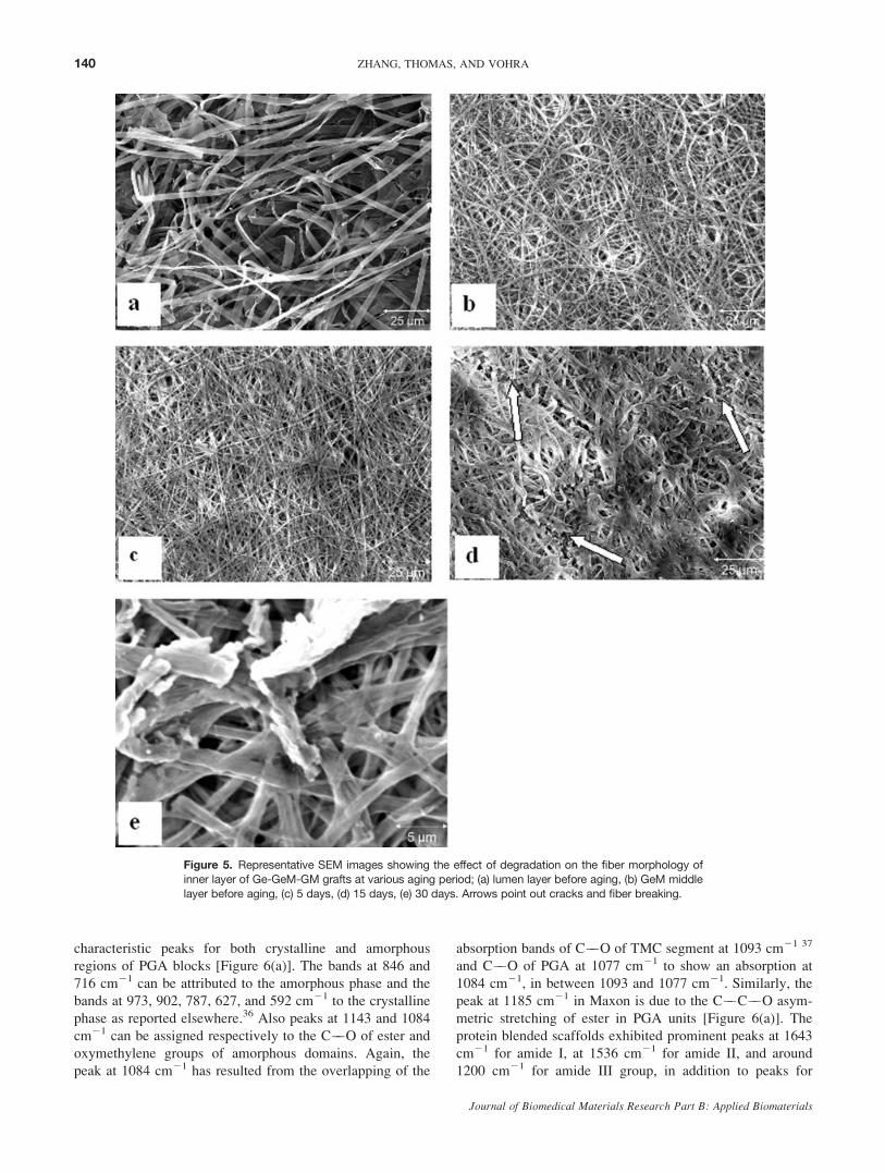
paring image (c) against (a) and (b) in Figure 5, it is
obvious that the fiber morphology in (c) is almost identical
to that in (b). This means that the inner layer (Ge) of the
Ge-GeM-GM scaffold might have completely dissolved or
removed during the initial 5 days and exposing the middle
GeM layer. Although, the middle layer (GeM) of Ge-GeM-
GM graft has maintained its fibrous morphology without
much surface distortions, individual breaking and fiber
fusion can be seen in Figure 5(d,e) due to aging in PBS.
The effect of PBS aging of the scaffolds on the structural
and chemical characteristics was analyzed by IR, XRD, and
DSC. ATR-FTIR spectra of electrospun Maxon and Maxon-
protein scaffolds before and after aging are given in Figures
6–8. A typical spectrum of virgin electrospun Maxon shows
Figure 4. SEM images of lumen surface of GE-GEM-GM graft after aging for (a) 5 days, (b) 10
days, (c) 20 days, and (d) 30 days. Arrow points out cracks or individual fiber breaking.
139BIODEGRADATION OF PROTEIN/POLYGLYCONATE BLEND FIBERS
Journal of Biomedical Materials Research Part B: Applied Biomaterials
characteristic peaks for both crystalline and amorphous
regions of PGA blocks [Figure 6(a)]. The bands at 846 and
716 cm21 can be attributed to the amorphous phase and the
bands at 973, 902, 787, 627, and 592 cm21 to the crystalline
phase as reported elsewhere.36 Also peaks at 1143 and 1084
cm21 can be assigned respectively to the C��O of ester and
oxymethylene groups of amorphous domains. Again, the
peak at 1084 cm21 has resulted from the overlapping of the
absorption bands of C��O of TMC segment at 1093 cm21 37
and C��O of PGA at 1077 cm21 to show an absorption at
1084 cm21, in between 1093 and 1077 cm21. Similarly, the
peak at 1185 cm21 in Maxon is due to the C��C��O asym-
metric stretching of ester in PGA units [Figure 6(a)]. The
protein blended scaffolds exhibited prominent peaks at 1643
cm21 for amide I, at 1536 cm21 for amide II, and around
1200 cm21 for amide III group, in addition to peaks for
Figure 5. Representative SEM images showing the effect of degradation on the fiber morphology ofinner layer of Ge-GeM-GM grafts at various aging period; (a) lumen layer before aging, (b) GeM middle
layer before aging, (c) 5 days, (d) 15 days, (e) 30 days. Arrows point out cracks and fiber breaking.
140 ZHANG, THOMAS, AND VOHRA
Journal of Biomedical Materials Research Part B: Applied Biomaterials

Maxon as shown in Figure 6(b,c). The amide I band for
C¼¼O stretching with characteristic frequencies in the range
from 1600 to 1700 cm21 is mainly associated with the
stretching vibrations on carbonyl groups along the polypep-
tide backbone and is a sensitive marker of polypeptide sec-
ondary structure.38,35 There are C��N stretching vibrations
and N��H deformations as minor vibration modes of amide
II and amide III bands as seen in Figure 6(c).
The aliphatic ester carbonyl peak (usually at 1743
cm21) and carbonate carbonyl peak (usually at 1725 cm21)
were appeared as a single band at 1738 cm21 in Maxon
and its protein blended scaffolds [Figure 6(a)]. The charac-
teristic bands for amorphous regions of PGA segments in
Maxon at 1143, 846, and 716 cm21 were disappeared com-
pletely in scaffolds hydrolyzed for 30 days [Figure 6(d)].
However, the peaks for the crystalline regions found to be
intact up to 20 days and decreased the intensity by 30
days. The ester carbonyl peak at 1738 cm21 exhibited a
significant reduction in intensity and appeared as a multip-
let by 30 days of aging [Figures 6(d) and 7(d)]. It should
be noted that the protein peaks for amide I (1643 cm21)
and amide II (1536 cm21) were also found to disappear or
exhibit reduction intensities drastically in samples aged for
30 days. After 20 days of aging, the peak at 1084 cm21
was shifted slightly to 1093 cm21 (C��O of TMC segment
appears at 1093 cm21),37 indicating the degradation of
Figure 6. ATR-FTIR spectra of (a) electrospun polyglyconate, (b) GE-GEM-GM outer layer, (c) GE-GEM-GM lumen layer, (d) GE-GEM-GM outer layer after 30 days aging. Peaks at 1143, 846, and
716 cm21 are represented by asterisks.
Figure 7. ATR-FTIR spectra of aged GE-GEM-GM scaffold; (a) GE-GEM-GM lumen layer 5 days,(b) 10 days, (c) 20 days, and (d) 30 days.
141BIODEGRADATION OF PROTEIN/POLYGLYCONATE BLEND FIBERS
Journal of Biomedical Materials Research Part B: Applied Biomaterials

PGA segments in Maxon [Figures 6(a,d) and 8(d)]. The
spectra for the inner layer of scaffolds showed a drastic
decrease in intensity of ester carbonyl peak and peaks for
��COO and ��CH groups at 1416 cm21 in addition to the
disappearance of amorphous PGA peaks (1143, 846, and
716 cm21) (Figure 7). The peak for crystalline regions
appeared with a decreased intensity by 30 days of aging
suggesting the degradation of defective crystalline PGA
blocks as stated earlier. The degradation of ester groups
produce ��COOH ends groups and accelerates the ester hy-
drolysis. It is noteworthy that the amide I and II peaks
decreased intensity significantly by 10 days. This is either
due to the dissolution/disintegration of uncrosslinked pro-
teins or due to the peel-off of the protein based lumen
layer. This inference is supported by the observation of
maximum mass loss by 10 days. The IR spectra for the
outer layer GM layer (common for both scaffolds) of aged
scaffolds are given in Figure 8. The ester carbonyl exhib-
ited no significant reduction in intensity for carbonyl peak
up to 20 days; however, the peak intensity for ester car-
bonyl and crystalline PGA segments experienced a drastic
decrease by 30 days of aging. The amide I and II peaks
also showed a drastic decrease in intensity. The characteris-
tic peaks for amorphous PGA segments at 1143, 846, and
716 cm21 were disappeared by 20 days of aging (Figure
8). The gradual shifting of the C��O peak at 1084 cm21
towards higher wave number region (1093 cm21 for C��O
of TMC) can be clearly seen in Figure 8.
The XRD spectra of GE-GEM-GM and Ge-GeM-GM
scaffolds are given in Figures 9 and 10 respectively. The
Figure 8. ATR-FTIR spectra of aged Ge-GeM-GM scaffold; (a) Ge-GeM-GM outer layer 5 days,
(b) 10 days, (c) 15 days, (d) 20 days, and (e) 30 days aging.
Figure 9. XRD patterns of aged GE-GEM-GM scaffold; (a) virginscaffold; (b) in 5 days; (c) in 15 days; (d) in 30 days.
Figure 10. XRD patterns of aged Ge-GeM-GM scaffold; (a) virginscaffold; (b) in 10 days; (c) in 15 days; (d) in 30 days.
142 ZHANG, THOMAS, AND VOHRA
Journal of Biomedical Materials Research Part B: Applied Biomaterials

(110) and (020) planes of crystalline PGA blocks in the
PGA-TMC copolymer molecular chain are confirmed by
the reflection peaks respectively at 228 and 28.58.25 Aged
specimens showed relatively narrow peaks compared to vir-
gin samples (before aging). By examining the peak at 228,a trend of initial decrease in intensity by 5–10 days and
then a trend of increase in intensity can be observed due to
the removal of proteins and amorphous phase of Maxon.
The molecular chain reorganization and plasticizing effect
of medium may be responsible for the initial decrease in
crystallinity. The turning point of the trend for the GE-
GEM-GM scaffold was 10 days whereas that for Ge-GeM-
GM scaffolds was 15 days. The peak at 228 showed maxi-
mum intensity in the spectrum of specimens aged for 15
days for both grafts.
DSC scans (first heating) of the aged electrospun scaf-
folds are shown in Figures 11 and 12. It was observed that
the melting point of electrospun Maxon in Ge-GeM-GM
virgin scaffold decreased from 209 to 2018C because of 30
days of aging. DSC scan (second heating) showed an amor-
phous a soft segment Tg around 58C (Figure 12) and an
exothermic crystallization peak around 858C. The crystal-
linity (%) of aged samples can be determined from the ra-
tio of enthalpy of fusion to standard enthalpy of fusion
(DHof ) by assuming the heat of fusion of an infinite PGA
crystal as 139.1 J/g.39 From the DSC data (first heating)
GE-GEM-GM scaffolds, the apparent crystallinity (%) was
obtained as 33.11% for 5 days, 52.83% for 10 days, 37.05
% for 15 days, 31. 56 for 20 days, and 25. 77 % for 30
days (Figure 12). It is clear that crystallinity of the fibers
increased initially due to degradation of amorphous phase
and then decreased finally due to degradation of crystalline
phase. A similar trend was observed in the DSC data of
aged Ge-GeM-GM scaffolds also (Figure 11). The apparent
percentage of crystallinity was found to be 40.03% for 5
days, 45.13 % for 10 days, 43.25% for 15 days, 53.20%
for 20 days, and 35.58% for 30 days. The crystallinity of
virgin Ge-GeM-GM sample calculated from DSC experi-
ment was 22%. This shows a significant increase in crystal-
linity with aging and dissolution of the proteins as well as
the amorphous phase of Maxon. However, by 30 days, the
crystalline PGA segments also started to degrade and this
resulted in the decrease in percentage crystallinity. The en-
thalpy of fusion decreased drastically for samples aged for
30 days indicating the degradation of defective crystalline
regions in Maxon.
The biodegradation has a deteriorative effect on me-
chanical properties of the scaffolds. Tables I and II give
the tensile test results of GE-GEM-GM scaffold and Ge-
GeM-GM scaffold, respectively. Though the weight-ratio of
Maxon to protein is identical in both scaffolds, Ge-GeM-
GM exhibited higher tensile properties (tensile strength of
2.11 6 0.53 MPa and tensile modulus of 27.38 6 7.03)
Figure 11. DSC scans of aged Ge-GeM-GM scaffold. The melting
point decreases with increasing aging period. [Color figure can be
viewed in the online issue, which is available at www.interscience.wiley.com].
Figure 12. DSC scans of aged GE-GEM-GM scaffold. Prolonged
aging up to 45 days decreased the crystallinity and melting point.
[Color figure can be viewed in the online issue, which is available atwww.interscience.wiley.com].
TABLE I. Mechanical Properties of Aged GE-GEM-GM Scaffold
Aging
Period
(day)
Tensile
Strength (MPa)
Failure
Strain (%)
Tensile
Modulus
(MPa)
0 0.76 6 0.07 45 6 4.0 3.72 6 0.27
5 0.26 6 0.11 28 6 7.0 1.32 6 0.92
10 0.27 6 0.07 16 6 0.5 2.12 6 0.36
20 0.02 6 0.01 1.0 6 0.2 –
30 Physically cracked – –
TABLE II. Mechanical Properties of Aged Ge-GeM-GM Scaffold
Aging
Period
(day)
Tensile
Strength (MPa)
Failure
Strain (%)
Tensile
Modulus
(MPa)
0 2.11 6 0.53 71 6 20 27.38 6 7.03
5 1.68 6 0.31 83 6 22 14.33 6 3.34
10 1.02 6 1.00 24 6 7.6 15.01 6 2.61
20 1.06 6 0.28 1.2 6 0.18 –
30 Physically cracked – –
143BIODEGRADATION OF PROTEIN/POLYGLYCONATE BLEND FIBERS
Journal of Biomedical Materials Research Part B: Applied Biomaterials

than GE-GEM-GM (tensile strength of 0.76 6 0.07 MPa
and tensile modulus of 3.72 6 0.27). This could be attrib-
uted to the presence of soluble elastin in the latter. The
soluble elastin with relatively lower molecular weight
chains may form a phase-mixed blend system in protein/
Maxon fibers in Ge-GeM-GM scaffolds. The high molecu-
lar weight insoluble elastin can cause some immiscibility
of the proteins with synthetic polymers in the blended-
fiber-structure and the resulted phase-separated blend struc-
ture may be responsible for the inferior mechanical proper-
ties. In vitro biodegradation impacted negatively to the
mechanical properties of both scaffolds. The tensile
strength and failure strain (%) of the aged tubular scaffolds
decreased with time. The decrease in mechanical properties
was more pronounced in the case of GE-GEM-GM scaffold
than Ge-GeM-GM scaffold. The tensile strength decreased
by more than 90% by 20 days for GE-GEM-GM scaffold
whereas that decreased only by 50% for Ge-GeM-GM scaf-
fold for the same period, that is, Ge-GeM-GM scaffold
retained 50% or more of its tensile strength up to 20 days
of PBS aging. The decrease in the failure strain (%) in the
case of both scaffolds is due to the disintegration and re-
moval of elastic proteins from the blend fibers.
DISCUSSION
For a tissue engineering scaffold, it is supposed that the
rate of degradation of the scaffold should balance the rate
of formation of new tissues. However, for engineering of
functionally dynamic tissues such as vascular and cardiac
tissues, the scaffolds have to undergo the intended cyclic
loading. Because biodegradation has a negative effect on
the mechanical and structural integrity of the scaffolds, it is
essential to study a correlation between materials degrada-
tion in vitro and mechano-morphological characteristics
before implantation in animals. It is known that most of the
biodegradable polyesters undergo degradation by hydrolysis
mechanism initialed by different hydrolyzing agent such as
water, ions, and enzymes. At first, water penetrates into the
materials and then the hydrolytic agents and pH of the me-
dium accelerate the bulk degradation of swollen materials.
Electrospun fibers have high surface area to volume ratio
and electrospun scaffolds have high porosity with well-
interconnected pores for water absorption. The protein-
blending and multilayering have further decreased the
nanofiber diameters and increased the surface area and po-
rosity in comparison with electrospun pure Maxon.25 This
explains the increased percentage of PBS medium uptake
by the trilayer scaffolds. Moreover, the hydrophilicity of
the proteins and hygroscopic nature of glycolide segments
in Maxon also contribute towards the increased PBS uptake
by the scaffolds.
It is known that the hydrolytic degradation of polyglyco-
nate occurs through a bulk degradation process, if the gly-
colide content is higher than 20 mol %.29 Again, the
kinetics of ester hydrolysis follows a first order dependence
on carboxylic end groups,28 whose concentration increases
in aging medium and thereby induces an auto catalytic
effect. Because of the large surface area and high hydrophi-
licity of protein blended Maxon, the rate of degradation is
expected to be higher compared to that for the degradation
of suture macrofilaments. Polyglyconate (Mw 5 86,000) in
the form of sutures of 0.553 mm diameter and 90 cm
length have exhibited a weight loss of 20% after 18 days
of incubation in PBS (pH 7.4) medium.28 The higher per-
centage of mass loss in the present scaffolds can be attrib-
uted to the high surface area and porosity of electrospun
scaffolds The interconnectivity of pores helps to diffuse the
fiber-fragments out from the rest of the scaffolds. Because
of the PBS uptake the individual fibers swell and fuse to-
gether in the initial stages and finally lead to fiber-breaking.
The analysis of SEM images leads to conclusion that by
10–15 days of aging fibrous morphology of the scaffolds
changed drastically and the inner protein layer dissolved or
delaminated from the tube. This can be correlated to the
initial high increase in the rate of mass loss up to 10 days
in the case of both scaffolds.
Maxon is an ABA type block copolymer with a seg-
mented nature having amorphous and crystalline blocks of
different chemical constituents. The middle soft segment is
constituted of a randomly distributed glycolide and tri-
methylene carbonate units and the hard segment is basi-
cally composed of glycolide units as the two end blocks of
the copolymer. Usually, amorphous regions are more sus-
ceptible to hydrolysis as water can penetrate easily there
than in highly packed and densely ordered crystalline
regions. In the case of Maxon, a competitive effect may
exist as a result of its chemical nature having highly hygro-
scopic polyglycolide crystalline segments.28 Therefore, the
initial increase in the crystallinity observed in the DSC
scans can be attributed due to the degradation of amor-
phous phase and further decrease in the crystallinity due to
the degradation of crystalline phase with prolonged time
(�1 month). The samples aged for 45 days further contin-
ued the decrease in crystallinity to 22.46%. In a recent
study of in vitro hydrolysis of Maxon monofilaments, Zur-
ita et al.28 have shown that the weight-average molecular
weight of Maxon clearly decreased to a value lower than
20,000 within 28 days of aging in PBS. This type of chain-
scission and fragmentation can be envisaged in the present
elctrospun Maxon scaffolds through decrease in weight and
melting point (from 208 to 2018C).In vitro biodegradation can be substantiated by IR func-
tional group analysis of aged scaffolds. The disappearance
or the loss of intensity of the peaks at 1143 and 1084 cm21
and the bands for amorphous regions (846 and 716 cm21)
clearly explains the early stages of hydrolysis of PGA and
its copolymers (Figures 6 and 7). Similar results have been
reported for the degradation of injection molded disks of
PGA, the major constituent of Maxon (67.5%). Chu et al.36
have shown that bands for amorphous region around 850,
753, 713, and 560 cm21 exhibited a drastic decrease in the
144 ZHANG, THOMAS, AND VOHRA
Journal of Biomedical Materials Research Part B: Applied Biomaterials

intensity than the bands for crystalline region around 972,
901, 806, 627, and 590 cm21 during the degradation of
PGA. The absorption band at 1244 cm-1 can be assigned to
the O��C��O asymmetric stretching of carbonate in TMC
units.40 And the intensity of which is not decreased signifi-
cantly in the present scaffolds, suggesting the relative sta-
bility of TMC units in the copolymer. The observed peak-
shift from 1084 to 1093 cm21 at 20 days of aging (Figure
6) could be due to the domination of TMC unit after the
disappearance of amorphous PGA segments.37 Previous
studies41–43 have shown the TMC based polymers degrade
slowly and the rate of hydrolysis of TMC based copoly-
mers is mainly depends on the hydrolysis of ester content
in them as the ester linkages are more susceptible to hydro-
lysis than carbonate linkages.
The vascular scaffolds for use in vivo need to provide a
high degree of graft-arterial compliance in addition to the
structural integrity. Therefore, biodegradation and its
effects on elastic and fatigue properties of prosthetic grafts
must be evaluated in vitro for a substantial period of time
to maximize the likelihood of clinical success.44 The struc-
tural and mechanical integrity of the graft should be main-
tained until mature vascular tissue is formed. As the
degradation of scaffolds proceeds, the extracellular matrices
secreted by the cells will be integrated into the cell/scaffold
construct. However, we have studied the degradation of
protein blended Maxon scaffolds in an acellular medium
(PBS) to evaluate the in vitro degradation behavior on scaf-
fold properties. The studies show that the tensile properties
of scaffolds decreased due to degradation. However, the
tensile strength of Ge-GeM-GM scaffold after 20 days
incubation in PBS are comparable to that for native femo-
ral artery (tensile strength of 1–2 MPa).13 The mechanical
disintegrations developed in the present scaffolds are in ac-
cordance to the gradual loss of fibrous morphology due to
fiber-breaking and weight loss due to the removal of frag-
mented products. For the GE-GEM-GM scaffold, SEM
images confirmed an escalated degradation after 10 days
and this resulted in decrease in mechanical properties (Ta-
ble I). The Ge-GeM-GM scaffolds have shown the fibrous
morphology up to 20 days in both the middle and outer
layers and sustained its stress, even though strain continu-
ously decreased because of fiber-breaking and dissolution
of uncrosslinked proteins (Table II). The increase in the
crystallinity during aging also contributed to the brittleness
of the scaffolds (tested under dry conditions). Under physi-
ological conditions, hydration effect may contribute posi-
tively to the elasticity. The overall results show that protein
blended polyglyconate tubular scaffolds show a deteriora-
tion in mechanical and structural properties after 15–20
days in vitro incubation in PBS. The dissolution of uncros-
slinked proteins (42 wt %) increased the rate of mechanical
and structural damage in the present scaffolds. Under invivo situation, deposition of native extracellular matrix pro-
teins by the cells and the de novo tissue formation may
compensate to retain the mechanical properties. However,
chemical crosslinking of proteins could be an alternative to
improve the mechanical and structural properties as well as
the degradation behavior of protein blended scaffolds.45,46
For vascular grafts, other mechanical properties such as
compliance matching, burst pressure, and suture retention
strength are equally important to tolerate suturing and
changing arterial pressures. Therefore, further studies on
the effect of in vitro aging on the biomechanical properties
will be undertaken in future using chemically crosslinked
protein blended scaffolds.
CONCLUSION
Studies on the in vitro aging behavior of two types of pro-
tein/polyglyconate (Maxon) fibrous tubular scaffolds having
a designed trilayer structure are reported for the first time.
Under hydrolytic aging conditions (PBS), both scaffolds
exhibited maximum PBS uptake within 5 days and mass
loss within 10–15 days. XRD spectra and DSC scans
revealed an increase in the crystallinity of Maxon after 10–
15 days of aging indicating the degradation of amorphous
phase in synthetic polymer and dissolution of uncrosslinked
proteins. The IR spectral analysis of aged scaffolds showed
that the characteristic peaks for amorphous PGA segments
at 1143, 846, and 716 cm21 decreased intensity after 15
days and disappeared completely by 20 days. The peak for
crystalline regions remained with a decreased intensity by
30 days of aging, suggesting the degradation of defective
crystalline PGA blocks. The degradation in hydrolytic
media affected the mechanical properties of scaffold con-
taining insoluble elastin than that containing soluble elastin.
Ge-GeM-GM scaffold retained 50% or more of its tensile
strength up to 20 days of PBS aging. The tensile strength
(1.2 MPa) exhibited by Ge-GeM-GM scaffold after 20 days
incubation in PBS is comparable to that for native femoral
artery. However, detailed evaluation on the effect of degra-
dation on compliance, burst pressure, and suture retention
is required for the potential use of these protein blended
Maxon grafts.
Thanks are due to Dr. D. Cakir of Department of Prosthodon-tics, Drs. D.R. Dean and R. Griffin of Department of MaterialsScience and Engineering and Dr. S.A. Catledge of Department ofPhysics for the technical support and instrumental training. Theauthors also acknowledge valuable discussions with Dr. A. Eber-hardt of Department of Biomedical Engineering.
REFERENCES
1. Niklason LE, Gao J, Abbott WM, Hirschi KK, Houser S,Marini R, Langer R. Functional arteries grown in vitro.Science 1999;284:489–493.
2. Hoenig MR, Campbell GR, Rolfe BE, Campbell JH. Tissueengineered blood vessels: Alternative to autologous grafts?Arterioscler Thromb Vasc Biol 2005;25:1128–1134.
3. Tiwari A, Salacinski HJ, Punshon G, Hamilton G, SeifalianAM. Development of a hybrid cardiovascular graft using atissue engineering approach. FASEB J 2002;16:791–796.
145BIODEGRADATION OF PROTEIN/POLYGLYCONATE BLEND FIBERS
Journal of Biomedical Materials Research Part B: Applied Biomaterials

4. Jeong SI, Kim SY, Cho SK, Chong MS, Kim KS, Kim H,Lee SB, Lee YM. Tissue-engineered vascular grafts composedof marine collagen and PLGA fibers using pulsatile perfusionbioreactors. Biomaterials 2007;28:1115–1122.
5. Buttafoco L, Engbers-Buijtenhuijs P, Poot AA, Dijkstra PJ,Vermes L, Feijen J. Physical characterization of vasculargrafts cultured in a bioreactor. Biomaterials 2006;27:2380–2389.
6. Barnes CP, Sell SA, Boland ED, Simpson DG, Bowlin GL.Nanofiber technology: Designing next generation of tissue en-gineering scaffolds. Adv Drug Deliv Rev 2007;59:1413–1433.
7. Liao S, Li B, Ma Z, We H, Chan C, Ramakrishna S. Biomi-metic electrospun nanofibers for tissue regeneration. BiomedMater 2006;1:45–53.
8. Pham QP, Sharma U, Mikos AG. Electrospinning of poly-meric nanofibers for tissue engineering applications: Areview. Tissue Eng 2006;12:1197–1211.
9. Thomas V, Dean D, Vohra YK. Nanostructured biomaterialsfor regenerative medicine. Curr Nanosci 2006;2:155–177.
10. Li M, Mondrinos MJ, Gandhi MR, Ko FK, Weiss AS, LelkesPL. Electrospun protein fibers as matrices for tissue engineer-ing. Biomaterials 2005;26:5999–6008.
11. Teo WE, Kotaki M, Mo XM, Ramakrishna S. Porous tubularstructures with controlled fiber orientation using a modifiedelectrospinning method. Nanotechnology 2005;16:918–924.
12. Boland ED, Mathews JA, Pawlowski KJ, Simpson DG, WnekGE, Bowlin GL. Electrospinning collagen and elastin: Prelim-inary vascular tissue engineering. Front Biosci 2004;9:1422–1432.
13. Sell SA, McClure MJ, Barnes CP, Knapp DC, Walpoth BH,Simpson DG, Bowlin GL. Electrospun polydioxanone-elastinblends: Potential for bioresorbable vascular grafts. BiomedMater 2006;1:72–80.
14. Stitzel J, Liu J, Lee SJ, Komura M, Berry J, Soker S, Lim G,Dyke MK, Czerw R, Yoo JJ, Atala A. Controlled fabricationof a biological vascular substitute. Biomaterials 2006;27:1088–1094.
15. Gan ZH, Fung JT, Jing XB, Wu C, Kuliche WK. A novellaser light-scattering study of enzymatic biodegradation ofpoly(e-caprolactone) nanoparticles. Polymer 1999;40:1961–1967.
16. Wu C, Gan Z. A novel method of studying polymer biodegra-dation. Polymer 1998;39:4429–4431.
17. Chen DR, Bei JZ, Wang SG. Polycaprolactone microparticlesand their biodegradation. Polym Degrad Stab 2000;67:455–459.
18. Zong XZ, Ran SF, Kim KS, Fang DF, Hsiao BS, Chu B.Structure and morphology changes during in vitro degradationof electrospun poly(glycolide-co-lactide) nanofiber membrane.Biomacromolecules 2003;4:416–423.
19. You Y, Min BM, Lee SJ, Lee TS, Park WH. In vitro degrada-tion behavior of electrospun polyglycolide, polylactide, andpoly(lactide-co-glycolide). J Appl Polym Sci 2005;95:193–200.
20. You Y, Lee SW, Youk JH, Min BM, Lee SJ, Park WH. Invitro degradation behaviour of non-porous ultra-fine poly(gly-colic acid)/poly(L-lactic acid) fibres and porous ultra-fine poly(glycolic acid) fibres. Polym Degrad Stab 2005;90:441–448.
21. Buttafoco L, Kolkman NG, Poot AA, Dijkstra PJ, Vermes I,Feijen J. Electrospinning collagen and elastin for tissue engi-neering small diameter blood vessels. J Control Release2005;101:322–324.
22. Stankus JJ, Guan J, Wagner WR. Fabrication of biodegradableelastomeric scaffolds with sub-micron morphologies. J BiomedMater Res A 2004;70:603–614.
23. Matthews JA, Simpson GD, Wnek GE, Bowlin GL. Electro-spinning of collagen nanofibers. Biomacromolecules 2002;3:232–238.
24. Vaz CM, Tuijl SV, Bouten CVC, Baaijens FPT. Design ofscaffolds for blood vessel tissue engineering using a multi-layering electrospinning technique. Acta Biomater 2005;1:575–582.
25. Thomas V, Zhang X, Catledge SA, Vohra YK. Functionallygraded electrospun scaffolds with tunable mechanical proper-ties for vascular tissue regeneration. Biomed Mater 2007;2:224–232.
26. Veith FJ, Gupta SK, Ascer E, White-Flores S, Samson RH,Scher LA, Towne JB, Bernhand VM, Bonier P, Flinn WR,Astelford P, Yao JST, Bergan JJ. Six years prospective multi-center randomized comparison of autologous saphenous veinand expanded polytetrafluoroethylene grafts in infrainguinalarterial reconstructions. J Vasc Surg 1986;3:104–114.
27. Tomihata K, Suzuki M, Ikada Y. The pH dependence ofmonofilament sutures on hydrolytic degradation. J BiomedMater Res Appl Biomater 2001;58:511–518.
28. Zurita R, Franco L, Puiggali J, Rodriguez-Galan A. Thehydrolytic degradation of a segmented glycolide-trimethylenecarbonate copolymer (MaxonTM). Polym Degrad Stab 2007;92:975–985.
29. Jie C, Zhu KJ, Shilin Y. Preparation, characterization and bio-degradable characteristics of poly(1,3-trimethylene carbonate-co-glycolide). Polym Int 1996;41:369–375.
30. Metz SA, Chegini N, Masterson BJ. In vivo and in vitro deg-radation of monofilament absorbable sutures PDS and Maxon.Biomaterials 1990;11:41–45.
31. Kangas J, Paasimaa S, Makela P, Leppilahti J, Tormala P,Waris T, Ashammakhi N. Comparison of strength properties ofpoly-L/D-lactide (PLDLA) 96/4 and polyglyconate (Maxon1)sutures: In vitro, in the subcutis, and in the achilles tendon ofrabbits. J Biomed Mater Res 2001;28:121–126.
32. Partridge SM, Davis FF, Adair ST. The chemistry of connec-tive tissues 2: Soluble proteins derived from partial hydrolysisof elastin. Biochem J 1955;61:11–21.
33. Heitz M, Carrasco F, Overend RP, Chornet E. Correlationbetween polystyrene molecular weights and a characteristictemperature derived from the thermogravimetric weight losscurves. Thermochim Acta 1989;142:83–88.
34. Thomas V, Jose MV, Chowdhury S, Sullivan JF, Dean DR,Vohra YK. Mechano-morphological studies of aligned nanofi-brous scaffolds of polycaprolactone fabricated by electrospin-ning. J Biomater Sci Polym Ed 2006;17:969–984.
35. Thomas V, Dean D, Jose MV, Mathew B, Chowdhury S,Vohra YK. Nanostructured biocomposite scaffolds based oncollagen coelectrospun with nanohydroxyapatite. Biomacro-molecules 2007;8:631–637.
36. Chu CC, Zhang L, Coyne LD. Effect of gamma-irradiationand irradiation temperature on hydrolytic degradation of syn-thetic absorbable sutures. J Appl Polym Sci 1995;56:1275–1294.
37. Wang H, Dong JH, Qiu KY. Synthesis and characterization ofABA-type block copolymer of poly(trimethylene carbonate)with poly(ethylene glycol): Bioerodible copolymer. J PolymSci Part A: Polym Chem 1998;36:695–702.
38. Payne KJ, Veis A. Fourier transform IR spectroscopy of colla-gen and gelatin solutions: Deconvolution of the amide I bandfor conformational studies. Biopolymers 1988;27:1749–1760.
39. Cohn D, Younes H, Marom G. Amorphous and crystallinemorphologies in glycolic acid and lactic acid polymers. Poly-mer 1987;28:2018–2022.
40. Wang H, Dong JH, Qiu KY, Gu ZW. Synthesis of poly(1,4-dioxan-2-one-co-trimethylene carbonate) for application indrug delivery systems. J Polym Sci Part A: Polym Chem1998;36:1301–1307.
41. Zhu KJ, Hendren RW, Jensen K, Pitt CG. Synthesis, proper-ties, and biodegradation of poly(1,3-trimethylene carbonate).Macromolecules 1991;24:1736–1740.
146 ZHANG, THOMAS, AND VOHRA
Journal of Biomedical Materials Research Part B: Applied Biomaterials

42. Albertsson AC, Eklund M. Influence of molecular structureon the degradation mechanism of degradable polymers: Invitro degradation of poly(trimethylene carbonate), poly(tri-methylene carbonate-co-caprolactone), and poly(adipic anhy-dride). J Appl Polym Sci 1995;57:87–103.
43. Pego AP, Poot AA, Grijpma DW, Feijen J. In vitro degrada-tion of trimethylene carbonate based copolymers MacromolBiosci 2002;2:411–419.
44. L’Heureux N, Dusserre N, Marini A, Garrido S, de la FuenteL, McAllister T. Technology insight: The evolution of tissue-
engineered vascular grafts- From research to clinical practice.Nat Clin Pract Cardiovasc Med 2007;4:389–395.
45. Bigi A, Cojazzi G, Panzavolta S, Rubini K, Roveri N. Me-chanical and thermal properties of gelatin at different degreesof glutaraldehyde crosslinking. Biomaterials 2001;22:763–768.
46. Sung HW, Chang Y, Chiu CT, Chen CN, Liang HC. Cross-linking characteristics and mechanical properties of a bovinepericardium fixed with a naturally occurring crosslinkingagent. J Biomed Mater Res 1999;47:116–126.
147BIODEGRADATION OF PROTEIN/POLYGLYCONATE BLEND FIBERS
Journal of Biomedical Materials Research Part B: Applied Biomaterials