Control Strategies of Plastic Biodegradation through Adjusting ...
Upload
khangminh22Category
view
3download
0
_____________________________________________________________________________________________________ *Corresponding author: E-mail: [email protected];
British Microbiology Research Journal 9(5): 1-10, 2015, Article no.BMRJ.18601
ISSN: 2231-0886
SCIENCEDOMAIN international www.sciencedomain.org
Biodegradation of Palm Oil Mill Effluent (POME) and Lipase Activity by Pseudomonas aeruginosa,
Bacillus subtilis and Candida albicans
O. L. Okwute1*, E. Stephen2 and P. I. Anyanwu1
1Department of Microbiology, University of Abuja, P.M.B. 117, Gwagwalada- Abuja, Nigeria.
2Department of Microbiology, Kogi State University, P.M.B. 1008, Anyigba-Kogi State, Nigeria.
Authors’ contributions
This work was carried out in collaboration with all authors. Author OLO designed the study, wrote the protocol and the first draft of the manuscript. Authors ES and PIA managed the analyses of the study
performed the statistical analysis and managed literature searches. All authors read and approved the final manuscript.
Article Information
DOI: 10.9734/BMRJ/2015/18601
Editor(s): (1) Raúl Rodríguez-Herrera, Autonomous University of Coahuila, México.
Reviewers: (1) Yordanka Tasheva, Industrial Technology and Management, University “Prof. Dr. A. Zlatarov - Burgas”,
Bulgaria. (2) Maulin P Shah, India.
(3) Anonymous, Universiti Putra Malaysia, Malaysia. Complete Peer review History: http://sciencedomain.org/review-history/10071
Received 30th April 2015 Accepted 22
nd June 2015
Published 6th July 2015
ABSTRACT Aims: To evaluate the biodegradation of palm oil mill effluent and lipase activity by Pseudomonas aeruginosa, Bacillus subtilis and Candida albicans treatment. Place and Duration of Study: Department of Microbiology, University of Abuja, Abuja and Microbiology laboratory of the National Institute of Pharmaceutical Research Development (NIPRD), Idu, FCT-Abuja, Nigeria, between July 2014 and September 2014. Methodology: Some physicochemical properties of the POME were analysed. Samples of POME were inoculated with Pseudomonas aeruginosa, Bacillus subtilis and Candida albicans. Microbial growth was measured by determining the total viable counts in a medium containing POME. The isolates were screened for lipase activity and degradation products were determined by gas chromatography-mass spectrometry (GC-MS) analysis. Results: The highest lipase activity was recorded on the 8th day by C. albicans as (29.83±1.40 U/ml) followed by P. aeruginosa (29.02±1.02 U/ml) while the least lipase activity was produced by B.
Original Research Article
Okwute et al.; BMRJ, 9(5): 1-10, 2015; Article no.BMRJ.18601
2
subtilis (19.08±0.40 U/ml). The total viable counts of the microbial isolates during biodegradation of POME revealed that the highest colony counts of P. aeruginosa (8.30×10
9 cfu/ml ) and B. subtilis
(7.30×10⁹ cfu/ml) occurred on the 12th day while the highest colony count of C. albicans was observed on the 16
th day (6.35×10
9 cfu/ml). The degradative enzyme (lipase) produced by the
microbial isolates was capable of breaking down complex substrates in nature, and thus may be responsible for the biodegradation of POME polluted habitats. However, the GC-MS showed that oleic acid (omega-9 fatty acid), hexadecanoic acid (palmitic acid) and octadecanoic acid (stearic acid) which were the main compounds present in the POME were not degraded by the three organisms at the end of the growth period. Conclusion: The present study revealed that P. aeruginosa, B. subtilis and C. albicans are good degraders of palm oil mill effluent and the enzyme (lipase) produced was capable of catalysing the breakdown of complex substrates in POME.
Keywords: Biodegradation; POME; lipase activity; Pseudomonas aeruginosa; Bacillus subtilis;
Candida albicans. 1. INTRODUCTION Oil palm (Elaeis guineensis) is a monoecious plant that bears both male and female flowers on the same tree. It belongs to the palmae family and is the most productive oil producing plant in the world [1]. The production of palm oil, however, results in the generation of large quantities of polluted wastewater commonly referred as palm oil mill effluent (POME). In the process of palm oil milling, POME is generated through sterilization of fresh oil palm fruit bunches, clarification of palm oil and effluent from hydrocyclone operations [2]. The separated wastewater sludge commonly referred to as POME is a brown slurry, which is composed of 4-5% solids (mainly organic), 0.5-1.0% residual oil and about 95% water and high concentration of organic nitrogen [3]. The raw POME has an extremely high content of degradable organic matter [4]. It has been identified as a major source of aquatic pollution by depleting dissolved oxygen when discharged into water bodies [5]. Studies on the effect of POME on soil showed that raw POME is acidic and alters microbiological and physicochemical properties of soil negatively, ultimately affecting soil fertility [6]. The soil however, gradually (over a period of 6 months – 1 year) becomes alkaline as biodegradation takes place [7]. A variety of microorganisms have been reported to degrade oil [8]. These organisms are widely distributed in both aquatic and terrestrial ecosystems. According to previous study [9], palm fruit is a major source of oil for lipase producing microorganisms. Studies have shown that for organisms to degrade palm oil, they must be able to produce lipase [10]. The lipolytic activity of fungi on the triglycerides of the POME may cause rancidity, acidity, bitterness, soapiness
and other off flavors. Microbial isolates with lipase activities include bacteria, fungi and yeast, and they are often associated with specific substrates [11]. Pseudomonas aeruginosa, Bacillus subtilis and Candida albicans with other organisms such as Micrococcus spp and the mould Aspergillus niger have been implicated in POME biodegradation [12]. Literature on the biodegradation of POME is scarce in literature [4], hence, the aim of this study was to compare its biodegradation by P. aeruginosa, B. subtilis and C. albicans and the production of lipase.
2. MATERIALS AND METHODS 2.1 Sample Collection The POME sample was collected from a local palm oil mill processing plant in Ankpa Local Government Area of Kogi State, Nigeria using sterile containers with sterile spoons and sterile hand gloves and then transported to the Microbiology laboratory of the University of Abuja, Abuja, Nigeria in ice boxes. They were stored at 4°C when not in use.
2.2 Collection and Identification of Pure Isolates
Pure isolates of P. aeruginosa, B. subtilis and C. albicans were collected from the culture bank of the Microbiology laboratory of the National Institute of Pharmaceutical Research Development (NIPRD), Idu, FCT-Abuja, Nigeria and were confirmed by routine identification procedures.

Okwute et al.; BMRJ, 9(5): 1-10, 2015; Article no.BMRJ.18601
3
2.3 Determination of Physicochemical Characteristics of POME
Growth of the organisms were determined by measuring the turbidity using turbidity meter (WGZ-113 Shanghai, China) at 520 nm [13]. pH was determined at ambient temperature using glass electrode pH and conductivity meter (Hannia, Italy). Phosphorus was determined using the method described by [14]. Nitrate was determined using the modification adapted by [15].
2.4 Inoculation of POME with Bacterial Isolates
A modified mineral salts medium [16] consisting of palm oil mill effluent and various inorganic salts in 1000 ml of distilled water (Mineral Salt Medium containing 2.0 g of Na2HPO4, 0.17 g of K2SO4, 4.0g of NH4NO3, 0.53 g of KH2PO4, 0.10 g of MgSO4.7H2O) was prepared in 1000 ml of distilled water. 10 ml of Mineral Salt Medium was dispensed into ten test tubes each. 2 ml of POME was added into each test tube and the solution sterilized by autoclaving. Two millilitre (2 ml) of 3 hours broth culture (peptone broth) P. aeruginosa and B. subtilis were seeded into five test tubes while another five test tubes containing the medium without the bacteria served as the control and then incubated at 37±2°C for 16 days. All the experiments were carried out in duplicates.
2.5 Inoculation of POME with Yeast Isolate
Biodegradation of POME by C. albicans was done by using medium [17] which consisted of 2ml of palm oil mill effluent and various inorganic salts (22.0 g of KH2PO4, 32.0 g of (NH4)2SO4, 10.0 g of Na2HPO4.H2O, 4.0 g of Mg SO4.7H20, 0.05 g of FeSO4.7H2O and 0.05 g of CaCl) dissolved in 1000 ml of distilled water and autoclaved at 15 labs pressure for 10 minutes. Two millilitre (2 ml) of overnight broth culture (peptone broth) of yeast isolate was added to five test tubes while another five test tubes containing the medium without the yeast served as the control and then incubated at 25±2°C for 16 days. All the experiments were carried out in duplicates.
2.6 Determination of Total Viable Count Total viable count of the inoculated organisms was carried out by spread plating 0.1 ml ten-fold
serially diluted samples onto nutrient agar (for bacteria) and Sabouraud Glucose Agar (SGA) at 4 days intervals in duplicates. The bacteria plates were incubated at 37°C for 18-24 hours while the fungal plates were incubated at 25°C for 3-5 days. After incubation, the total viable counts were determined.
2.7 Screening of Yeast and Bacterial Isolates for Lipase Activity
Primary screening was done on tributyrin agar plate. The yeast and bacteria isolates were inoculated separately on the tributyrin agar plates [18]. The plates containing the bacterial isolates were incubated at 37±2°C for 48 hours while that of the yeast was incubated at 25±2°C for 3 days and clear zones around the colonies (lipolysis) were observed. The plates which showed maximum hydrolysis halo on the medium were selected for POME biodegradation investigations.
2.8 Assay of Lipase Produced by Each
Isolate for Biodegradation Lipase produced by each isolate was measured [19]. Lipase products in the supernatant were determined by reading the absorbance at 280 nm against basal medium using UV-Spectrophotometer (JENWAY 6405). The quantity of lipase produced was used as the degradability ability of the isolates [20].
2.9 Gas Chromatography-mass Spectrometry (GC-MS)
The GC-MS analysis was carried out at day 0 (control), day 7 and day 14 for POME inoculated separately with C. albicans, P. aeruginosa and B. subtilis.
2.10 Statistical Analysis The degradation ability was tabulated and the statistical analysis was determined using Analysis of Variance (ANOVA) with SPSS Software version 19.0 [21].
3. RESULTS AND DISCUSSION Fig. 1 shows the turbidity during biodegradation of POME. There was a gradual increase in the turbidity after four days in all samples. The highest turbidity was observed in P. aeruginosa sample followed by B. subtilis and C. albicans.

Okwute et al.; BMRJ, 9(5): 1-10, 2015; Article no.BMRJ.18601
4
The turbidity increased with the increase in days. Turbidity was higher in the samples inoculated with P. aeruginosa, B. subtilis and C. albicans than their various controls. This indicates that the inoculated sample were able to grow and utilize the POME as carbon source. The reason for the increase in turbidity after 4 days till the end of the study may be due to the presence of nitrogen and phosphorus in the mineral salt media which are necessary for biodegradative activity [22] and also play a role in overcoming nutrient limitation during the biodegradative process. Turbidity was highest in C. albicans followed by P. aeuginosa and least in B. subtilis. There were no significant differences (<0.05) in the turbidity produced by the three organisms. Fig. 2 shows the level of phosphate utilized during biodegradation of POME. The phosphate
concentration declined with time. The highest phosphate utilization was observed in C. albicans followed by P. aeruginosa and B. subtilis. The phosphate also decreased from day 0 to day 16. The phosphate concentration was higher in the samples inoculated with the microbial isolates than their respective controls. Phosphate content was lowest in C. albicans followed by P. aeruginosa and the least being B. subtilis. This is in agreement with the observation of [1]. The phosphate concentration of 0.91 – 0.725 was within the Federal Environmental Protection Agency, FEPA effluent limitation guideline of 5 mg/l [23]. The phosphate concentration in the control remained higher than the medium seeded with the organisms. There were significant differences (<0.05) in the phosphate concentration produced by the three organisms.
Fig.1. Turbidity of POME during biodegradation
Fig. 2. Change in Phosphate utilization during biodegradation of POME
0
0.2
0.4
0.6
0.8
1
1.2
1.4
0 4 8 12 16
Days
Tur
bid
ity (N
TU)
P.aeruginosa
B.subtilis
C.albicans
Control
0
0.1
0.2
0.3
0.4
0.5
0.6
0.7
0.8
0.9
0 4 8 12 16
Days
Ph
osp
hat
e (
mg
/l)
P.aeruginosa
B.subtilis
C.albicans
Control

Okwute et al.; BMRJ, 9(5): 1-10, 2015; Article no.BMRJ.18601
5
Fig. 3 shows the changes in nitrate concentration during biodegradation of POME by the three organisms. The highest level of nitrate utilization was observed in C. albicans followed by P. aeruginosa and B. subtilis. Nitrate content decreased during biodegradation from day 0 to day 16 in all samples. The decline in the concentration of nitrate between day 0 to 16 could be attributed to high metabolic activity and utilization of nitrate during the biodegradation process by the microbial isolates [24]. Nitrate content was lowest in Candida albicans followed by P. aeruginosa and B. subtilis. The nitrate concentration of 0.086-2.356 mg/l at the end of the study was within the Federal Environmental Protection Agency, FEPA effluent limitation guideline of 20 mg/l [23]. There were significant differences (<0.05) in the nitrate concentration produced by the three organisms.
Fig. 4 is the lipase activity of the three organisms. The highest lipase activity was observed in C. albicans (29.83±1.40 U/ml) at day 8. The lipase activity declined in all organisms after eight days. The lowest lipase activity was observed in all organisms at day 16. The lipase activity increased from day 4 to 8, and then decreased as the days progressed. The highest lipase activity was recorded at pH 6.0 respectively on the 8th day by C. albicans (29.83±1.40 U/ml) followed by P. aeruginosa (29.02±1.02 U/ml) while the least lipase activity was produced in the 16th day by B. subtilis and C. albicans (16.52±0.20 U/ml each). This finding agrees with previous reports [10,25]. They reported that optimum pH for maximum lipase activity was 6.0. There were no significant differences (<0.05) in the lipase activity produced by the three organisms.
Fig. 3. Nitrate utilization during biodegradation of POME
Fig. 4. Lipase activity of the microbial isolates
0
0.5
1
1.5
2
2.5
3
0 4 8 12 16
Days
Nit
rate
co
ncen
trat
ion
(m
g/l
)
P.aeruginosa
B.subtilis
C.albicans
Control
0
5
10
15
20
25
30
35
1 2 3 4
Days
Lipa
se a
ctiv
ity (U
/ml)
P.aeruginosa
B.subtilis
C.albicans
Okwute et al.; BMRJ, 9(5): 1-10, 2015; Article no.BMRJ.18601
6
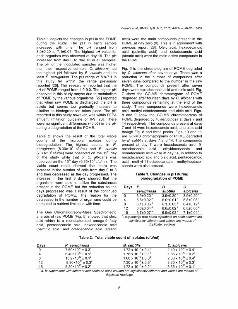
Table 1 depicts the changes in pH in the POME during the study. The pH in each sample increased with time. The pH ranged from 3.9±0.20 to 7.1±0.04. The highest pH value for each organism was observed at day 16. The pH increased from day 0 to day 16 in all samples. The pH of the inoculated samples was higher than their respective controls. C. albicans had the highest pH followed by B. subtilis and the least P. aeruginosa .The pH range of 5.8-7.1 in this study fell within the range previously reported [26]. This researcher reported that the pH of POME ranged from 4.0-9.0. The higher pH observed in this study maybe due to metabolism of POME by the various organisms. [27] reported that when raw POME is discharged, the pH is acidic but seems too gradually increase to alkaline as biodegradation takes place. The pH recorded in this study however, was within FEPA effluent limitation guideline of 6-9 [23]. There were no significant differences (<0.05) in the pH during biodegradation of the POME. Table 2 shows the result of the total viable counts of the microbial isolates during biodegradation. The highest counts in P. aeruginosa (8.30x10⁹ cfu/ml) and B. subtilis (7.30x10
9 cfu/ml) were observed on the 12
th day
of the study while that of C. albicans was observed on the 16
th day (6.35x10
9 cfu/ml). The
viable count result showed that there was increase in the number of cells from day 0 to 8 and then decreased as the day progressed. The increase in the first 8 days showed that the organisms were able to utilize the substances present in the POME but the reduction as the days progressed was a result of the continued degradation of POME. The reason for the decreased in the number of organisms could be attributed to nutrient limitation with time. The Gas Chromatography-Mass Spectrometric analysis of raw POME (Fig. 5) showed that oleic acid which is a monosaturated omega-9 fatty acid, pentadecanoic acid, hexadecanoic acid (palmitic acid) and octadecanoic acid (stearic
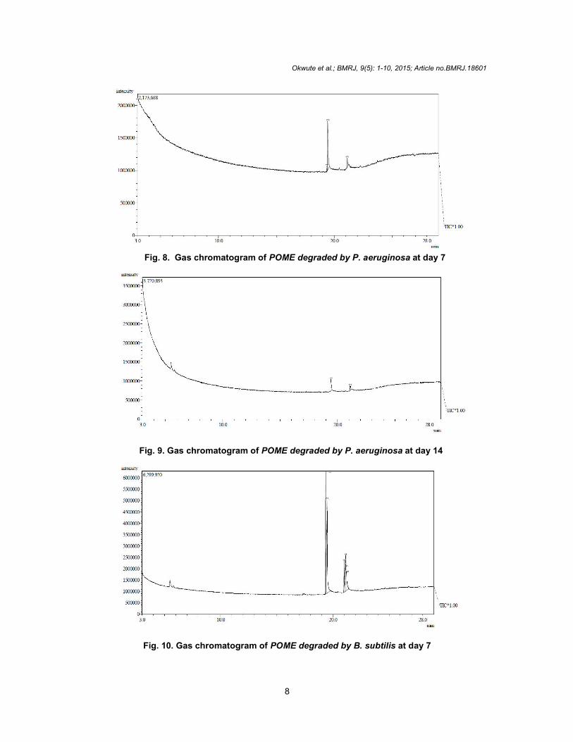
acid) were the main compounds present in the POME at day zero (0). This is in agreement with previous report [28]. Oleic acid, hexadecanoic acid (palmitic acid) and octadecanoic acid (stearic acid) were the main active compounds in the POME. Fig. 6 is the chromatogram of POME degraded by C. albicans after seven days. There was a reduction in the number of compounds after seven days compared to the number in the raw POME. The compounds present after seven days were hexadecanoic acid and oleic acid. Fig. 7 show the GC-MS chromatogram of POME degraded after fourteen days by C. albicans with three compounds remaining at the end of the study. These compounds were hexadecanoic acid, methyl octadecanoate and oleic acid. Figs. 8 and 9 show the GC-MS chromatograms of POME degraded by P. aeruginosa at days 7 and 14 respectively. The compounds present at days 7 and 14 were hexadecanoic acids and oleic acid though Fig. 8 had three peaks. Figs. 10 and 11 are GC-MS chromatograms of POME degraded by B. subtilis at days 7 and 14. The compounds present at day 7 were hexadecanoic acid, 9-octadecanoic acid, ethyldocosonate and nonadecanoic acid while at day 14, in addition to hexadecanoic acid and oleic acid, pentadecanoic acid, methyl-11-octadecanoate, methylheptaco-sonate were also present.
Table 1. Changes in pH during biodegradation of POME
Days P.
aeruginosa B. subtilis
C. albicans
0 3.9±0.20 a 3.9±0.20 a 3.9±0.20 a 4 5.8±0.02
a 6.0±0.01
a 5.8±0.05
a
8 6.1±0.06 a 6.1±0.05 a 6.4±0.12 a 12 6.6±0.04
a 6.6±0.02
a 6.8±0.00
a
16 6.7±0.07 a 6.9±0.03 a 7.1±0.04 a a: superscript with same alphabets on each column are
significantly different and values are means of duplicate readings
Table 2. Total viable count of isolates (cfu/ml)
Days P. aeruginosa B. subtilis C. albicans 0 7.60×10
10 ± 0.3
a 1.72 x 10
10 ± 0.4
b 1.40 x 10
10 ± 0.4
b
4 6.40×10¹⁰ ± 0.1a 1.76 x 10¹⁰ ± 0.1b 1.85 x 10¹⁰ ± 0.2b 8 13.2×10
10 ± 0.1
a 1.60 x 10
10 ± 0.3
b 2.80 x 10
10 ± 0.4
b
12 8.30×10¹⁰ ± 0.3a 7.30 x 10¹⁰ ± 0.3b 3.30 x 10¹⁰ ± 0.3b 16 5.20×10
10 ± 0.2
a 1.72 x 10
10 ± 0.2
b 6.35 x 10
10 ± 0.1
b
a, b: superscript with different alphabets on each column are significantly different and values are means of duplicate readings

Okwute et al.; BMRJ, 9(5): 1-10, 2015; Article no.BMRJ.18601
7
Gas chromatography-mass spectrometric analysis of POME degraded by C. albicans, P. aeruginosa and B. subtilis at days 7 and 14 showed that hexadecenoic acid (palmitic acid) and oleic acid were not degraded by the three organisms.
Their inability to degrade these compounds may be due to the absence of suitable enzymes for the mineralization of oleic acid, hexadecanoic
acid (palmitic acid) and octadecanoic acid [12].
Fig. 5. Gas chromatogram of POME at day 0 (control)
Fig. 6. Gas chromatogram of POME degraded by C. albicans at day 7
Fig. 7. Gas chromatogram of POME degraded by C. albicans at day 14
Okwute et al.; BMRJ, 9(5): 1-10, 2015; Article no.BMRJ.18601
8
Fig. 8. Gas chromatogram of POME degraded by P. aeruginosa at day 7
Fig. 9. Gas chromatogram of POME degraded by P. aeruginosa at day 14
Fig. 10. Gas chromatogram of POME degraded by B. subtilis at day 7

Okwute et al.; BMRJ, 9(5): 1-10, 2015; Article no.BMRJ.18601
9
Fig. 11. Gas chromatogram POME degraded by B.subtilis at day 14
4. CONCLUSION The present study revealed that P. aeruginosa, B. subtilis and C. albicans were able to degrade POME. However, some organic compounds were not completely degraded as they persisted throughout the period of analysis. The degradative enzyme (lipase) produced by P. aeruginosa, B. subtilis and C. albicans was capable of catalysing the breakdown of lipids in POME and since the potential industrial applications of the enzyme especially in waste management has been identified, it may be responsible for the biodegradation of POME in polluted habitats and may be applicable to other oil polluted sites where the same microorganisms are employed. The possible role of other enzymes produced by these microorganisms in the biodegradation of POME should be investigated.
ACKNOWLEDGEMENT The authors wish to thank the authorities of the National Institute of Pharmaceutical Research Development, Idu-Abuja for the use of some of their facilities and stock cultures.
COMPETING INTERESTS Authors have declared that no competing interests exist.
REFERENCES 1. Panapanaan V, Helin T, Kujanpää M,
Soukka R, Heinimö JJ, Linnanen L. Sustainability of palm oil production and
opportunities for finnish technology and know-how transfer. Springer Science. 2009;13-61.
2. Borja R, Banks CJ, Sánchez E. Anaerobic treatment of palm oil mill effluent in a two-stage up-flow anaerobic sludge blanket (UASB) reactor. Journal of Biotechnology. 1996; 45:125-135.
3. Ohimain EI, Daukoru-Olukole C, Izah SC, Eke RA, Okonkwo AC. Microbiology of palm oil mill effluents. Journal of Microbiology and Biotechnology. 2012;2 (6):852-857.
4. Ahmad A, Ismail S, Bhatia S. Water recycling from palm oil mill effluent (POME) using membrane technology. Desalination. 2003;157:87-95.
5. Igwe JC, Onyegbado CC. A review of palm oil mill effluent (POME) water treatment. Global Journal of Environmental Research. 2007;1(2):54-62.
6. Nwaugo VO, Chinyere GC, Inyang CU. Effects of palm oil mill effluent (POME) on soil bacterial flora and enzyme activities in Egbama. Plant Products Research Journal. 2008;12:10-13.
7. Okwute LO, Ijah UJJ. Changes in microbial population of palm oil mill effluent polluted soil amended with chicken droppings and cow dung. British Biotechnology Journal. 2014;4(3):279-288.
8. Leslie-Grady Jr CP, Daigger GT, Lim HC. Biological waste water treatment, 2
nd
Edition. Marcel Dekker Incorporation, New York. 1999;949-970.
9. Jerabkova H, Blanka K, Nahlik J. Biofilm of pseudomonas C12B on glass support as catalytic agent for continuous SDS removal. International Journal of

Okwute et al.; BMRJ, 9(5): 1-10, 2015; Article no.BMRJ.18601
10
Biodeterioration and Biodegradation. 1999; 44:233-241.
10. Hosseini F, Malekzadeh F, Amirmozafari N, Ghaemi N. Biodegradation of anionic surfactants by isolated bacteria from activated sludge. International Journal of Environmental Science and Technology. 2007;4(1):127-132.
11. Sakthi SS, Kanchana DP, Saranraj P, Usharani G. Evaluation of amylase activity of the amylolytic fungi Aspergillus niger using cassava as substrate. International Journal of Applied Microbiology Science. 2012;1:24-34.
12. Okwute OL, Ijah UJJ. Bioremediation of palm oil mill polluted soil using microorganisms found in organic wastes. International Journal of Biotechnology. 2014;(3)3:32-46.
13. Palin AT. Photometric determination of the colour and turbidity of water. Water and Water Engineering. 1955;59:341-345.
14. Lasslo SJ, Moldestad EA, Ross M. Phosphate concentration determination of the Watab river shed. Research project, Department of Chemistry, Saint John’s University. 2001.
15. American Public Health Association. Standard methods for the examination of water and wastewater, 20
th Edition,
American Public Health Association, Washington, DC. 1999;731.
16. Zajic C, Suplisson B. Emulsification and degradation of bunker C fuel oil by microorganisms. Biotechnology and Bioengineering. 1972;14:331-343.
17. Bushnell LD, Haas HF. The utilization of hydrocarbons by microorganisms. Journal of Bacteriology. 1941;41:653-673.
18. Gaur D, Jain PK, Sisodia YS, Bajapai V. Estimation of extracellular lipolytic enzyme activity by thermophilic Bacillus sp. isolated from arid and semi-arid region of Rajasthan, India. Journal of Microbiology, Biotechnology and Food Sciences. 2012;2 (2):619-633.
19. Rammani P, Gupta R. Optimization of medium composition for keratinase production on feather by Bacillus
licheniformis using statistical methods involving response surface methodology. Biotechnology and Applied Biochemistry. 2004;40:191-196.
20. Ugoh SC, Ijigbade B. Production and characterisation of amylase by fungi isolated from soil samples at Gwagwalada. Reports and Opinions. 2013;5(7):44-53.
21. Statistical Package for Social Sciences (SPSS). Computer Package for Windows, Version 19.0 2010. Available: http:www.spss.com
22. Adesodun JK, Mbagwu JSC. Biodegradation of waste lubricating petroleum oil in a tropical alfisol as mediated by animal droppings. Bioresource Technology. 2008;99:5659-5665.
23. Federal Environmental Protection Agency (FEPA). Guidelines and standards for environmental pollution control in Nigeria. Federal Environmental Protection Agency, Lagos-Nigeria. 1991.
24. Sanyaolu AA, Sanyaolu VT, Kolawole JO, Jawndo SS. Biodeterioration of premium motors spirit (PMS) by fungal species. International Journal of Science and Nature. 2012;3(2):276-285.
25. Prazeres JN, Cruz JAB, Pastore GM. Characterization of alkaline lipase from Fusarium oxysporum and the effect of different surfactants and detergents on the enzyme activity. Brazilian Journal of Microbiology. 2006;37:505-509.
26. Mukesh KDJ, Rejitha R, Devika S, Balakumaran MDA, Immaculate NR, Kalaichelvan PT. Production, optimization and purification of lipase from Bacillus sp. MPTK 912 isolated from oil mill effluent. Advances in Applied Science Research. 2012;3(2):930-938.
27. Ma AN. Environmental management for the oil palm industry. Palm Oil Development. 2000;30:1-10.
28. Stephen E, Emmanuel OS, Okwute LO. Comparative study of in vitro biodegradation of spent lubricating oil by Aspergillus niger and Bacillus subtilis. Researcher. 2014;6(7):50-57.
_________________________________________________________________________________ © 2015 Okwute et al.; This is an Open Access article distributed under the terms of the Creative Commons Attribution License (http://creativecommons.org/licenses/by/4.0), which permits unrestricted use, distribution, and reproduction in any medium, provided the original work is properly cited.
Peer-review history: The peer review history for this paper can be accessed here:
http://sciencedomain.org/review-history/10071
Copyright © 2022 FDOKUMEN