Microbial biodegradation of polyaromatic hydrocarbons
29
REVIEW ARTICLE Microbial biodegradation of polyaromatic hydrocarbons Ri-He Peng, Ai-Sheng Xiong, Yong Xue, Xiao-Yan Fu, Feng Gao, Wei Zhao, Yong-Sheng Tian & Quan-Hong Yao Shanghai Key Laboratory of Agricultural Genetics and Breeding, Agro-Biotechnology Research Institute, Shanghai Academy of Agricultural Sciences, Shanghai, China Correspondence: Quan-Hong Yao, Shanghai Key Laboratory of Agricultural Genetics and Breeding, Agro-Biotechnology Research Center, Shanghai Academy of Agricultural Sciences, 2901 Beidi Rd, Shanghai, China. Tel./fax: 1086 021 62203180; e-mail: [email protected] Received 8 March 2008; revised 14 April 2008; accepted 10 June 2008. First published online 25 July 2008. DOI:10.1111/j.1574-6976.2008.00127.x Editor: David Gutnick Keywords polycyclic aromatic hydrocarbons; biodegradation; microorganisms; pathway; genetic regulation. Abstract Polycyclic aromatic hydrocarbons (PAHs) are widespread in various ecosystems and are pollutants of great concern due to their potential toxicity, mutagenicity and carcinogenicity. Because of their hydrophobic nature, most PAHs bind to particulates in soil and sediments, rendering them less available for biological uptake. Microbial degradation represents the major mechanism responsible for the ecological recovery of PAH-contaminated sites. The goal of this review is to provide an outline of the current knowledge of microbial PAH catabolism. In the past decade, the genetic regulation of the pathway involved in naphthalene degradation by different gram-negative and gram-positive bacteria was studied in great detail. Based on both genomic and proteomic data, a deeper understanding of some high-molecular-weight PAH degradation pathways in bacteria was provided. The ability of nonligninolytic and ligninolytic fungi to transform or metabolize PAH pollutants has received considerable attention, and the biochem- ical principles underlying the degradation of PAHs were examined. In addition, this review summarizes the information known about the biochemical processes that determine the fate of the individual components of PAH mixtures in polluted ecosystems. A deeper understanding of the microorganism-mediated mechanisms of catalysis of PAHs will facilitate the development of new methods to enhance the bioremediation of PAH-contaminated sites. Introduction Polycyclic aromatic hydrocarbons (PAHs) are an all-pervad- ing group of hydrophobic organic compounds consisting of two or more combined benzene rings in linear, angular or cluster arrangements. PAHs are the most important compo- nents of crude oil, creosote, asphalt and coal tar. They contaminate the soil via many routes, including the burning of fossil fuels, the manufacture of gas and coal tar, wood processing, escaped automobile gasoline and the incinera- tion of waste (Harvey, 1991). There are more than 100 diverse PAH compounds, and most of them persist in the ecosystem for many years owing to their low aqueous solubility and their absorption to solid particles (Volkering et al., 1992, 1993; Bosma et al., 1997). The presence of PAHs in contaminated soil and sediment poses a significant risk to the soil, because many PAH compounds are known or suspected to be toxic, mutagenic and, in some cases, carcinogenic (Cerniglia & Heitkamp, 1989; Patnaik, 1992). On the basis of their abundance and toxicity, 16 PAH compounds have been included in the United States Soilal Protection Agency’s list of priority pollutants (Keith & Telliard, 1979). The elimination of PAHs from contaminated soil is essential during its remediation according to relevant clean- up standards. The biological treatment of soil contaminated with PAHs should be a more efficient, financially affordable and adaptable choice than physicochemical treatment be- cause it presents potential advantages such as the complete degradation of the pollutants, lower treatment cost, greater safety and less soil disturbance (Habe & Omori, 2003). Many bacteria have been discovered that degrade PAHs via either metabolism or cometabolism. The ordinary biochemical pathways of the bacterial metabolism of PAHs have been scrutinized. Low-molecular-weight (MW) PAHs, such as naphthalene, phenanthrene and anthracene, are usually FEMS Microbiol Rev 32 (2008) 927–955 c 2008 Federation of European Microbiological Societies Published by Blackwell Publishing Ltd. All rights reserved Downloaded from https://academic.oup.com/femsre/article/32/6/927/2683263 by guest on 11 July 2022
-
Upload
khangminh22 -
Category
Documents
-
view
0 -
download
0
Transcript of Microbial biodegradation of polyaromatic hydrocarbons
R E V I E W A R T I C L E
Microbial biodegradationof polyaromatic hydrocarbonsRi-He Peng, Ai-Sheng Xiong, Yong Xue, Xiao-Yan Fu, Feng Gao, Wei Zhao, Yong-Sheng Tian &Quan-Hong Yao
Shanghai Key Laboratory of Agricultural Genetics and Breeding, Agro-Biotechnology Research Institute, Shanghai Academy of Agricultural Sciences,
Shanghai, China
Correspondence: Quan-Hong Yao,
Shanghai Key Laboratory of Agricultural
Genetics and Breeding, Agro-Biotechnology
Research Center, Shanghai Academy of
Agricultural Sciences, 2901 Beidi Rd,
Shanghai, China. Tel./fax: 1086 021
62203180; e-mail:
Received 8 March 2008; revised 14 April 2008;
accepted 10 June 2008.
First published online 25 July 2008.
DOI:10.1111/j.1574-6976.2008.00127.x
Editor: David Gutnick
Keywords
polycyclic aromatic hydrocarbons;
biodegradation; microorganisms; pathway;
genetic regulation.
Abstract
Polycyclic aromatic hydrocarbons (PAHs) are widespread in various ecosystems
and are pollutants of great concern due to their potential toxicity, mutagenicity
and carcinogenicity. Because of their hydrophobic nature, most PAHs bind to
particulates in soil and sediments, rendering them less available for biological
uptake. Microbial degradation represents the major mechanism responsible for the
ecological recovery of PAH-contaminated sites. The goal of this review is to
provide an outline of the current knowledge of microbial PAH catabolism. In the
past decade, the genetic regulation of the pathway involved in naphthalene
degradation by different gram-negative and gram-positive bacteria was studied in
great detail. Based on both genomic and proteomic data, a deeper understanding
of some high-molecular-weight PAH degradation pathways in bacteria was
provided. The ability of nonligninolytic and ligninolytic fungi to transform or
metabolize PAH pollutants has received considerable attention, and the biochem-
ical principles underlying the degradation of PAHs were examined. In addition,
this review summarizes the information known about the biochemical processes
that determine the fate of the individual components of PAH mixtures in polluted
ecosystems. A deeper understanding of the microorganism-mediated mechanisms
of catalysis of PAHs will facilitate the development of new methods to enhance the
bioremediation of PAH-contaminated sites.
Introduction
Polycyclic aromatic hydrocarbons (PAHs) are an all-pervad-
ing group of hydrophobic organic compounds consisting of
two or more combined benzene rings in linear, angular or
cluster arrangements. PAHs are the most important compo-
nents of crude oil, creosote, asphalt and coal tar. They
contaminate the soil via many routes, including the burning
of fossil fuels, the manufacture of gas and coal tar, wood
processing, escaped automobile gasoline and the incinera-
tion of waste (Harvey, 1991). There are more than 100
diverse PAH compounds, and most of them persist in the
ecosystem for many years owing to their low aqueous
solubility and their absorption to solid particles (Volkering
et al., 1992, 1993; Bosma et al., 1997). The presence of PAHs
in contaminated soil and sediment poses a significant risk to
the soil, because many PAH compounds are known or
suspected to be toxic, mutagenic and, in some cases,
carcinogenic (Cerniglia & Heitkamp, 1989; Patnaik, 1992).
On the basis of their abundance and toxicity, 16 PAH
compounds have been included in the United States Soilal
Protection Agency’s list of priority pollutants (Keith &
Telliard, 1979).
The elimination of PAHs from contaminated soil is
essential during its remediation according to relevant clean-
up standards. The biological treatment of soil contaminated
with PAHs should be a more efficient, financially affordable
and adaptable choice than physicochemical treatment be-
cause it presents potential advantages such as the complete
degradation of the pollutants, lower treatment cost, greater
safety and less soil disturbance (Habe & Omori, 2003).
Many bacteria have been discovered that degrade PAHs via
either metabolism or cometabolism. The ordinary biochemical
pathways of the bacterial metabolism of PAHs have been
scrutinized. Low-molecular-weight (MW) PAHs, such as
naphthalene, phenanthrene and anthracene, are usually
FEMS Microbiol Rev 32 (2008) 927–955 c� 2008 Federation of European Microbiological SocietiesPublished by Blackwell Publishing Ltd. All rights reserved
Dow
nloaded from https://academ
ic.oup.com/fem
sre/article/32/6/927/2683263 by guest on 11 July 2022
readily degraded by bacteria in soil and under laboratory
conditions (Cerniglia, 1984, 1992; Sutherland et al., 1995).
Since the initial studies by Heitkamp et al. (1988a, b) on the
bacterial degradation of pyrene, there have been numerous
reports describing the microbial oxidation of four-ring
PAH. Recently, a Sphingomonad endowed with the remark-
able ability to grow on four-ring PAH chrysene as its sole
carbon and energy source was isolated (Willison, 2004).
However, less is known about the bacteria capable of
utilizing PAHs containing five or more rings as a carbon
and energy source, such as benzo[a]pyrene and benz[a]an-
thracene. The mechanisms involved in the degradation of
PAHs with more than five rings are unclear.
Diverse fungi capable of utilizing PAHs have been in-
vestigated as well. Some filamentous fungi, basidiomycetes,
white-rot fungi and deuteromycetes have been shown to
remove PAHs more competently than bacteria. PAHs sus-
ceptible to fungal biodegradation include naphthalene,
phenanthrene, anthracene, pyrene, benzo[a]pyrene, fluorene,
dibenzothiophene, catechol, benzo[a]anthracene, benzo[-
ghi]-perylene, chrysene, benzo[b]fluoranthene and ben-
zo[k]-fluoranthene (Cerniglia, 1997; Zheng & Obbard,
2002, 2003). At least two mechanisms are involved in PAH
biodegradation: one utilizes the cytochrome P-450 system
(Yadav et al., 2006) and the other uses the soluble extra-
cellular enzymes of lignin catabolism, including lignin
peroxidase, manganese peroxidase (Steffen et al., 2003) and
laccase (Gianfreda et al., 1999).
In the past decade, more and more microorganisms
capable of mineralizing PAH compounds have been isolated,
and knowledge about many PAH-catabolic genes from
bacteria and fungi is accumulating. However, until now,
there have been no reviews summarizing this progress. In
this review, we will summarize the recent advances in
understanding the natural diversity and capabilities of
bacteria and fungi for degrading PAH compounds.
Bacterial catabolism of polycyclicaromatic hydrocarbons
Numerous bacteria have been found that degrade PAHs, and
some can utilize low-MW PAHs as their sole carbon source.
The common biochemical pathways for the bacterial degra-
dation of PAHs such as naphthalene (Resnick et al., 1996;
Annweiler et al., 2000), phenanthrene (Menn et al., 1993;
Kiyohara et al., 1994; Pinyakong et al., 2003a, b), anthracene
and acenaphthene (Dean-Ross et al., 2001; Pinyakong et al.,
2004) have been well investigated. Biodegradation mechan-
isms require the presence of molecular oxygen to initiate the
enzymatic attack of PAH rings. In the initial step, dioxygenase-
catalyzed oxidation of arenes generally takes place in
aerobic bacterial systems to yield vicinal cis-dihydrodiols as
the early bioproducts by a multicomponent enzyme system.
These dihydroxylated intermediates may then be cleaved by
intradiol or extradiol ring-cleaving dioxygenases through
either an ortho-cleavage pathway or a meta-cleavage path-
way, leading to central intermediates such as protocatechu-
ates and catechols that are further converted to tricarboxylic
acid (TCA) cycle intermediates (Cerniglia, 1992; Eaton &
Chapman, 1992; Gibson & Parales, 2000).
Dioxygenases responsible for the formation of cis-
dihydrodiols from arenes of PAH substrates appear to be the
most ubiquitous in bacteria. They have usually been found
to contain multicomponent enzyme systems involving sev-
eral proteins, nonheme iron, and require NADH (Gibson &
Subramanian, 1984; Gibson et al., 1990; Resnick et al.,
1994). To date, dioxygenase-containing organisms have
been grown on a range of carbon sources, and these enzymes
have been classified accordingly, e.g. toluene (TDO),
naphthalene (NDO) and biphenyl (BPDO). TDO is con-
firmed to be an extremely wide-ranging substrate enzyme.
The TDO system is most suitable for cis-dihydroxylation of
substituted benzene substrates and bicyclic arenes. However,
there is a size limitation on substrate acceptability for TDO;
bulky and bendy molecules fit poorly into the active site of
this enzyme. For larger PAHs, both NDO and BPDO are
capable of catalyzing dihydroxylation, with only the latter
enzyme capable of metabolizing tetracyclic or larger exam-
ples. By contrast, the NDO enzyme appears to be generally
incapable of catalyzing the cis-dihydroxylation of monocyc-
lic aromatic rings (Boyd & Sheldrake, 1998).
Low-MW PAH dioxygenase system
The bacterial naphthalene dioxygenase system is particularly
useful for oxidizing bi- and tri-cyclic PAH substrates, such as
naphthalene, phenanthrene and anthracene. The naphtha-
lene dioxygenase system is a multicomponent enzyme,
generally including an NADH oxidoreductase, a ferredoxin
and an oxygenase component that contains the active site.
This catalytic portion of the naphthalene dioxygenase sys-
tem, termed NDO, is composed of large and small subunits
– a and b, respectively – that are in an a3b3 configuration.
NDO is a member of a large family of oxygenases whose asubunits contain a Rieske [2Fe–2S] center and mononuclear
nonheme iron, and determine the substrate specificity of
NDO (Butler & Mason, 1997; Parales et al., 1998). Generally,
two electrons from the reduced pyridine nucleotide are
transferred via the reductase, the ferredoxin, and the Rieske
center to the Fe (II) ion at the active site during a catalytic
cycle. The reducing equivalents allow the activation of
molecular oxygen, which is a prerequisite to dihydroxylation
of the substrate (Carredano et al., 2000; Ferraro et al., 2005,
2006). Interestingly, the AaAb genes, which encode the
ferredoxin and reductase components of the multicompo-
nent PAH initial dioxygenase, are not present on the phn
FEMS Microbiol Rev 32 (2008) 927–955c� 2008 Federation of European Microbiological SocietiesPublished by Blackwell Publishing Ltd. All rights reserved
928 R.-H. Peng et al.
Dow
nloaded from https://academ
ic.oup.com/fem
sre/article/32/6/927/2683263 by guest on 11 July 2022

locus of the Burkholderia sp. strain RP007 (Laurie & Lloynd-
Jones, 1999). Currently, we are trying to improve the
efficiency of the electron transport functions of the nahAcAd
gene products by DNA shuffling.
Naphthalene-degrading bacteria are broadly distributed
in nature. So far, a few naphthalene dioxygenases have been
purified and extensively characterized from different strains
of bacteria, and genetic control of the pathways involved in
naphthalene degradation has been studied in detail. The
naphthalene catabolic genes (nah) of NAH7 are organized
into two operons: the nal operon encoding the upper path-
way enzymes involved in conversion of naphthalene to
salicylate, and the sal operon encoding the lower pathway
enzymes involved in the conversion of salicylate to pyruvate
and acetyl coenzyme A. The two operons are closely
genetically linked to each other and to their common
regulatory gene, nahR. The nahR gene encodes a trans-
acting positive regulator belonging to the LysR family of
transcriptional regulators that are widely distributed in
bacteria. NahR is required for the induction of the nah genes
by salicylate and the high-level expression of those genes in
bacteria (Yen & Gunsalus, 1982, 1985; Grund & Gunsalus,
1983).
The nucleotide sequences of genes encoding the upper
pathway enzymes from several Pseudomonas strains have
been reported: ndo genes from Pseudomonas putida NCIB
9816 (Yang et al., 1994), nah genes from P. putida G7 (Menn
et al., 1993), NCIB9816-4 (Simon et al., 1993), ND6 (Li
et al., 2004), BS202 (Kosheleva et al., 1986; Balashova et al.,
2001), and P. putida sp. (Ono et al., 2007), dox genes from
Pseudomonas sp.C18 (Denome et al., 1993), pah genes from
P. putida OUS82 (Kiyohara et al., 1994; Takizawa et al.,
1994) and Pseudomonas aeruginosa Pak1 (Takizawa et al.,
1999), and nah genes from Pseudomonas stutzeri AN10
(Bosch et al., 1999a, b, 2000). All the nah structural and
regulatory genes on the former plasmid are located on a 39-
kb class II transposon, Tn4655 (Tsuda & Iino, 1990; Sota
et al., 2006). The nucleotide sequences of the upper pathway
genes are more than 90% identical. All these genes are
arranged in a similar order: nahAa (ferredoxin reductase)-
nahAb (NDO ferredoxin)- nahAc (the a-subunit
of NDO)- nahAd (the b-subunit of NDO)- nahB (naphtha-
lene-cis-dihydrodiol dehydrogenase)- nahF (salicylal-
dehyde dehydrogenase)- nahC (1,2-dihydroxynaphthalene
dioxygenase)- nahQ (unknown gene)- nahE (trans-o-hydro-
benzylidenepyruvate hydratase aldolase)- nahD (2-hydroxy-
chromene-2-carboxylate isomerase) (Habe et al., 2003)
(Fig. 1). The genetic regulation of this pathway was also
studied in detail for different bacterial strains (Fig. 2). In
Ralstonia sp. U2, the naphthalene dioxygenase genes (nag
gene) contained all of the genes corresponding to the
classical nah gene of Pseudomonas strains in the same order,
with the exception of two extra genes inserted between the
ferredoxin reductase gene and ferredoxin gene. The two
additional genes, named nagG and nagH, are structural
subunits of salicylate-5-hydroxylase and can help the host
convert naphthalene to gentisate (Fuenmayor et al., 1998;
Zhou et al., 2001). In the Comamonas testeroni strain GZ42,
the upper pathway genes are similar to those of Ralstonia sp.
U2 (Goyal & Zylstra, 1997). However, in C. testeroni strain
GZ39, the pathway genes are quite different from those of
the typical nah genes. In this strain, several genes (such as
extradiol dioxygenase) are not within the cluster, but a
glutathione-S-transferase gene was inserted into the cluster
(Goyal & Zylstra, 1996; Zylstra et al., 1997).
It was determined from the sequences of P. putida G7,
NCIB9816-4, ND6, and P. stutzeri AN10 strains that the
lower pathway genes consist of 11 genes organized in the
order nahGTHINLOMKJY. nahY is not a catabolic gene, but
a naphthalene chemotaxis gene (Grimm & Harwood, 1999;
Boronin, 2001) (Fig. 2). In the strains AN10 and ND6, there
is another salicylate hydroxylase gene (nahW) outside but
close to the sal operon. It was suggested that isofunctional
enzyme might be advantageous for the host to adjust its
metabolism to extreme conditions (Bosch et al., 1999a, b; Li
et al., 2004).
A number of PAH-degrading phenotypes cannot be
explained genotypically by comparison with nah-like se-
quences in contaminated soils. The nah-like probe can only
provide a monitor for 45% of strains isolated with a
naphthalene-degrading phenotype, and yet the same probe
was only useful for 15% of strains isolated with a phenan-
threne-degrading phenotype (Lloyd-Jones et al., 1999,
2000). The enzymes involved in the conversion of naphtha-
lene to salicylate can also degrade phenanthrene to naphtha-
lene-1,2-diol. However, it was found that the sequence
similarity and gene organization of the naphthalene cata-
bolic genes were very different between the Burkholderia sp.
RP007 strain and some classical naphthalene degradation
strains. The strain RP007’s naphthalene catabolic genes (phn
gene) encode iron–sulfur protein a and b subunits of a PAH
initial dioxygenase, but lack both ferredoxin and reductase
components. The ferredoxin and reductase genes for elec-
tron transport functions of the phn gene may be located
elsewhere in the RP007 genome or may be supplied by
cellular housekeeping genes (Laurie & Lloyd-Jones, 1999,
2000).
Different routes of naphthalene degradation have also
been illustrated. The Alcaligenes faecalis AFK2 strain makes
use of phenanthrene as its sole carbon source through the
o-phthalate pathway (Kiyohara et al., 1982). Some unique
genes, such as 3,4-dihydroxyphenanthrene dioxygenase
(phnC), 2-carboxybenzaldehyde dehydrogenase (phnI),
and trans-2-carboxybenzal-pyruvate hydratase aldolase
(phnH), appear in this pathway, which may make the strain
prefer phenanthrene to naphthalene (Nagao et al., 1988;
FEMS Microbiol Rev 32 (2008) 927–955 c� 2008 Federation of European Microbiological SocietiesPublished by Blackwell Publishing Ltd. All rights reserved
929Microbial biodegradation of polyaromatic hydrocarbons
Dow
nloaded from https://academ
ic.oup.com/fem
sre/article/32/6/927/2683263 by guest on 11 July 2022
Kiyohara et al., 1990). The Nocardioides sp. KP7 strain
degrades phenanthrene via the phthalate pathway as well
(Iwabuchi & Harayama, 1997, 1998a, b). The phd genes of
this strain belong to a new class of PAH-catabolic genes due
to variation in gene organization and sequence similarity.
The PhdA and PhdB genes, which encode the a- and b-
subunit of phenanthrene dioxygenase, are essential for
enzyme activity. Although the basic sequence features of
each protein family are conserved, each of the subunits of
the dioxygenase shows a modest (o 60%) sequence identity
to all of the known dioxygenase subunits. The ferredoxin
component of the dioxygenase PhdC belongs to the
[3Fe–4S] or [4Fe–4S] type of ferredoxin, but not to the
[2Fe–2S] type found in most components of PAH dioxy-
genase (Iwabuchi & Harayama, 1998a, b; Saito et al., 1999).
The dispensability of the ferredoxin and ferredoxin reduc-
tase components of Escherichia coli suggests the relatively
low specificity of the electron transport systems toward the
oxygenase components (Saito et al., 2000).
It has been observed that a high proportion of the PAH-
degrading isolates in soil belong to the sphingomonads.
Unlike other gram-negative bacteria strains, members of the
genus Sphingomonas are able to degrade a wide range of
natural and xenobiotic compounds, such as biphenyl, (sub-
stituted) naphthalene(s), fluorene, (substituted) phenan-
threne(s), pyrene, (chlorinated) diphenylether(s),
(chlorinated) furan(s), (chlorinated) dibenzo-p-dioxin(s),
carbazole, estradiol, polyethylenglycols, chlorinated phenols
and different herbicides and pesticides (Basta et al., 2005). It
seems possible that PAH-degrading sphingomonads are
adapted to the oligotrophic environment by having
high-affinity uptake systems and an ability to simulta-
neously take up mixed substrates rather than being
particular specialists for the degradation of hydrophobic
aromatic compounds (Eguchi et al., 1996; Schut et al., 1997;
Fredrickson et al., 1999; Pinhassi & Hagstrom, 2000). Many
sphingomonads degrade naphthalene, phenanthalene,
and anthracene via common pathways found in other
gram-negative bacteria (Pinyakong et al., 2003a, b). It was
demonstrated that large plasmids present in xenobiotic-
degrading Sphingomonas strains are responsible for the
degradative capabilities (Kim et al., 1996; Feng et al., 1997;
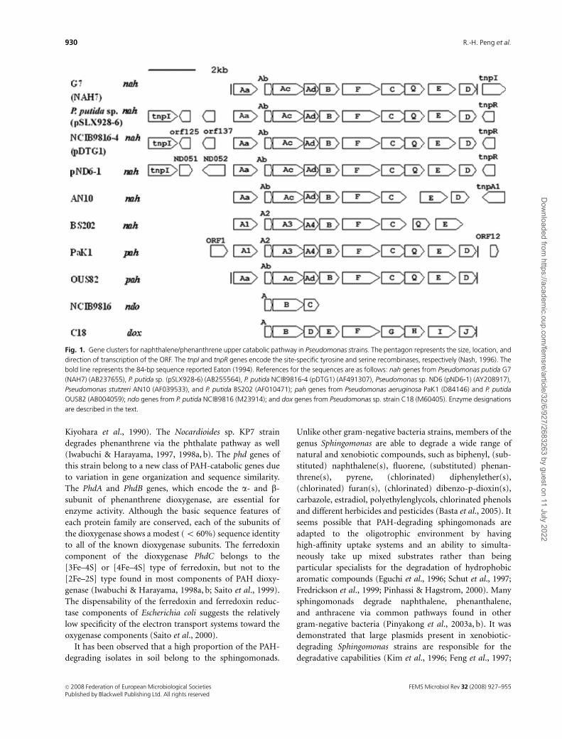
Fig. 1. Gene clusters for naphthalene/phenanthrene upper catabolic pathway in Pseudomonas strains. The pentagon represents the size, location, and
direction of transcription of the ORF. The tnpI and tnpR genes encode the site-specific tyrosine and serine recombinases, respectively (Nash, 1996). The
bold line represents the 84-bp sequence reported Eaton (1994). References for the sequences are as follows: nah genes from Pseudomonas putida G7
(NAH7) (AB237655), P. putida sp. (pSLX928-6) (AB255564), P. putida NCIB9816-4 (pDTG1) (AF491307), Pseudomonas sp. ND6 (pND6-1) (AY208917),
Pseudomonas stutzeri AN10 (AF039533), and P. putida BS202 (AF010471); pah genes from Pseudomonas aeruginosa PaK1 (D84146) and P. putida
OUS82 (AB004059); ndo genes from P. putida NCIB9816 (M23914); and dox genes from Pseudomonas sp. strain C18 (M60405). Enzyme designations
are described in the text.
FEMS Microbiol Rev 32 (2008) 927–955c� 2008 Federation of European Microbiological SocietiesPublished by Blackwell Publishing Ltd. All rights reserved
930 R.-H. Peng et al.
Dow
nloaded from https://academ
ic.oup.com/fem
sre/article/32/6/927/2683263 by guest on 11 July 2022
Romine et al., 1999; Ogram et al., 2000; Basta et al., 2004).
For example, a 40-kb DNA region was found to be related to
aromatic catabolism in Sphingobium yanoikuyae strain B1,
in which two dioxygenase genes are predicted to be required
for conversion of PAHs and biphenyl to simple aromatic
acid, and meta-cleavage genes are required for conversion of
aromatic acids to the tricarboxylic acid cycle (TAC) inter-
mediate (Yen & Serdar, 1988; Assinder & Williams, 1990;
Zylstra & Kim, 1997; Kim & Zylstra, 1999). However, these
aromatic-degradative genes for catabolic pathways are often
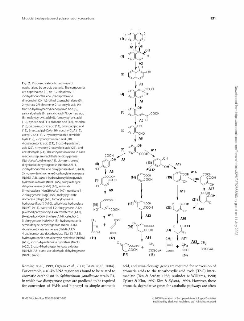
Fig. 2. Proposed catabolic pathways of
naphthalene by aerobic bacteria. The compounds
are naphthalene (1), cis-1,2-dihydroxy-1,
2-dihydronaphthalene (cis-naphthalene
dihydrodiol) (2), 1,2-dihydroxynaphthalene (3),
2-hydroxy-2H-chromene-2-carboxylic acid (4),
trans-o-hydroxybenzylidenepyruvic acid (5),
salicylaldehyde (6), salicylic acid (7), gentisic acid
(8), maleylpyruvic acid (9), fumarylpyruvic acid
(10), pyruvic acid (11), fumaric acid (12), catechol
(13), cis,cis-muconic acid (14), b-ketoadipic acid
(15), b-ketoadipyl-CoA (16), succiny-CoA (17),
acetyl-CoA (18), 2-hydroxymuconic-semialde-
hyde (19), 2-hydroxymuconic acid (20),
4-oxalocrotonic acid (21), 2-oxo-4-pentenoic
acid (22), 4-hydroxy-2-oxovaleric acid (23), and
acetaldehyde (24). The enzymes involved in each
reaction step are naphthalene dioxygenase
(NahAaAbAcAd) (step A1), cis-naphthalene
dihydrodiol dehydrogenase (NahB) (A2), 1,
2-dihydronaphthalene dioxygenase (NahC) (A3),
2-hydroxy-2H-chromene-2-carboxylate isomerase
(NahD) (A4), trans-o-hydroxybenzylidenepyruvic
hydratase-aldolase (NahE) (A5), salicylaldehyde
dehydrogenase (NahF) (A6), salicylate
5-hydroxylase (NagGHAaAb) (A7), gentisate 1,
2-dioxygenase (NagI) (A8), maleylpyruvate
isomerase (NagL) (A9), fumarylpyruvate
hydrolase (NagK) (A10), salicylylate hydroxylase
(NahG) (A11), catechol 1,2-dioxygenase (A12),
b-ketoadipate:succinyl-CoA transferase (A13),
b-ketoadipyl-CoA thiolase (A14), catechol 2,
3-dioxygenase (NahH) (A15), hydroxymuconic-
semialdehyde dehydrogenase (NahI) (A16),
4-oxalocrotonate isomerase (NahJ) (A17),
4-oxalocrotonate decarboxylase (NahK) (A18),
hydroxymuconic-semialdehyde hydrolase (NahN)
(A19), 2-oxo-4-pentenoate hydratase (NahL)
(A20), 2-oxo-4-hydroxypentenoate aldolase
(NahM) (A21), and acetaldehyde dehydrogenase
(NahO) (A22).
FEMS Microbiol Rev 32 (2008) 927–955 c� 2008 Federation of European Microbiological SocietiesPublished by Blackwell Publishing Ltd. All rights reserved
931Microbial biodegradation of polyaromatic hydrocarbons
Dow
nloaded from https://academ
ic.oup.com/fem
sre/article/32/6/927/2683263 by guest on 11 July 2022
localized in the plasmid separately from each other, or, at
least, are not organized in coordinately regulated operons.
There is no evident biochemical function of the individual
gene clusters. In plasmid pNL1 of strain F199, there are at
least 13 gene clusters predicted to encode enzymes asso-
ciated with the degradation of aromatic compounds (Ro-
mine et al., 1999). Several parts of the DNA sequence in
pNL1 regions encoding aromatic catabolic genes are similar
to those in other Sphingomonas strains (Habe et al., 2003).
The ‘flexible’ gene organization (e.g. different combinations
of conserved gene clusters) could be one of the mechanisms
that allows sphingomonads to adapt quickly and efficiently
to novel compounds in the environment (Basta et al., 2005).
In contrast to Pseudomonas and other gram-negative
bacteria whose structural genes required for naphthalene
utilization are usually clustered, the gram-positive bacteria
Rhodococcus strains NCIMB12038, P200 and P400 display
only three structural genes required for naphthalene utiliza-
tion (narAa, narAb and narB) in the region (Larkin et al.,
1999; Kulakov et al., 2005). The narAa and narAb genes
encode the a- and b-subunits of the naphthalene dioxygen-
ase (NDO) catalytic component of iron–sulfur protein
(ISPNAR). Both subunits of the NCIMB12038 NDO
showed only 30% amino acid identity to the corresponding
P. putida NDO subunits. The narB gene has 39% amino acid
identity to NahB from the P. putida G7 strain. No genes were
found in the Rhodococcus strains that corresponded to the
genes encoding the electron transport components reduc-
tase and ferredoxin of NDO in Pseudomonas. It was found
that the nar region is not organized into a single operon, but
there are several transcription units that differ in the
Rhodococcus strains. The narA and narB genes were found
to be transcribed as a single unit through different start sites,
and their transcription was induced by naphthalene. Puta-
tive regulatory genes (narR1 and narR2) are transcribed as a
single mRNA in naphthalene-induced cells (Kulakov et al.,
2000; Larkin et al., 2005).
As described above, many bacteria containing the en-
zymes involved in the conversion of naphthalene to salicy-
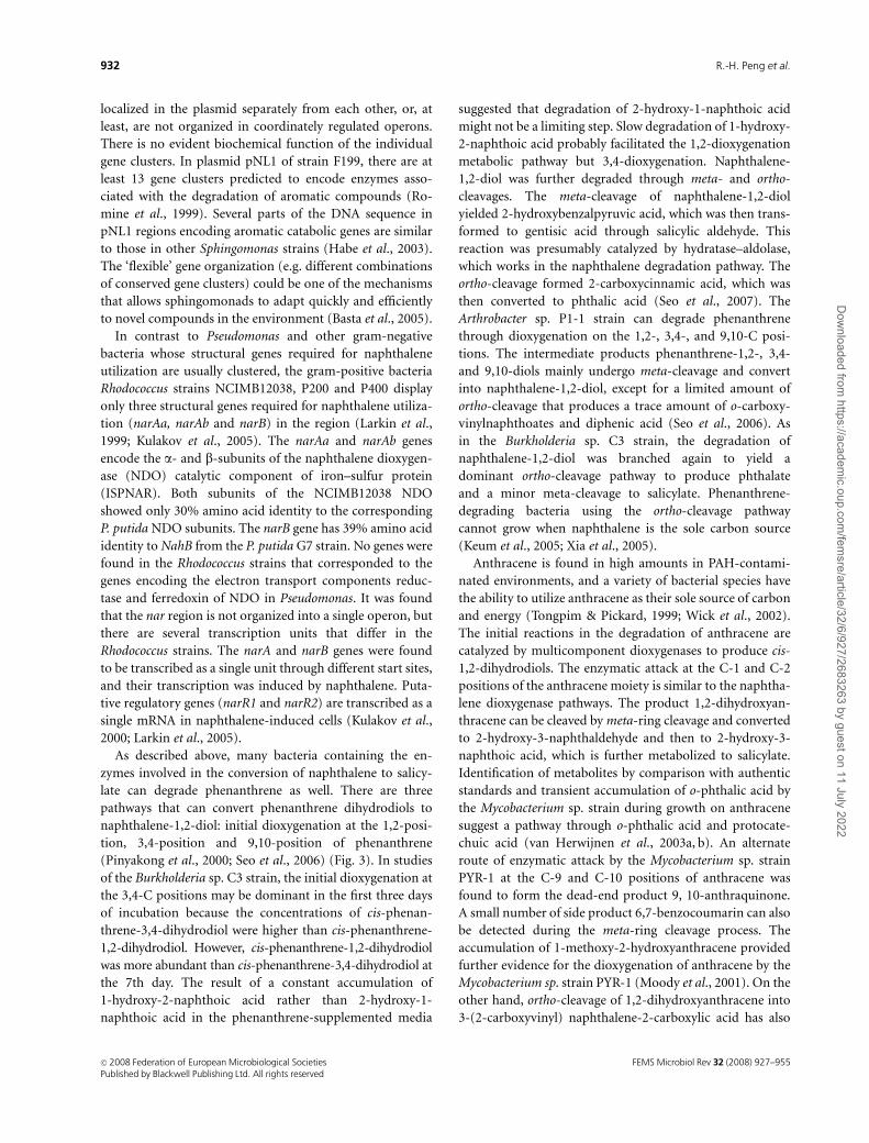
late can degrade phenanthrene as well. There are three
pathways that can convert phenanthrene dihydrodiols to
naphthalene-1,2-diol: initial dioxygenation at the 1,2-posi-
tion, 3,4-position and 9,10-position of phenanthrene
(Pinyakong et al., 2000; Seo et al., 2006) (Fig. 3). In studies
of the Burkholderia sp. C3 strain, the initial dioxygenation at
the 3,4-C positions may be dominant in the first three days
of incubation because the concentrations of cis-phenan-
threne-3,4-dihydrodiol were higher than cis-phenanthrene-
1,2-dihydrodiol. However, cis-phenanthrene-1,2-dihydrodiol
was more abundant than cis-phenanthrene-3,4-dihydrodiol at
the 7th day. The result of a constant accumulation of
1-hydroxy-2-naphthoic acid rather than 2-hydroxy-1-
naphthoic acid in the phenanthrene-supplemented media
suggested that degradation of 2-hydroxy-1-naphthoic acid
might not be a limiting step. Slow degradation of 1-hydroxy-
2-naphthoic acid probably facilitated the 1,2-dioxygenation
metabolic pathway but 3,4-dioxygenation. Naphthalene-
1,2-diol was further degraded through meta- and ortho-
cleavages. The meta-cleavage of naphthalene-1,2-diol
yielded 2-hydroxybenzalpyruvic acid, which was then trans-
formed to gentisic acid through salicylic aldehyde. This
reaction was presumably catalyzed by hydratase–aldolase,
which works in the naphthalene degradation pathway. The
ortho-cleavage formed 2-carboxycinnamic acid, which was
then converted to phthalic acid (Seo et al., 2007). The
Arthrobacter sp. P1-1 strain can degrade phenanthrene
through dioxygenation on the 1,2-, 3,4-, and 9,10-C posi-
tions. The intermediate products phenanthrene-1,2-, 3,4-
and 9,10-diols mainly undergo meta-cleavage and convert
into naphthalene-1,2-diol, except for a limited amount of
ortho-cleavage that produces a trace amount of o-carboxy-
vinylnaphthoates and diphenic acid (Seo et al., 2006). As
in the Burkholderia sp. C3 strain, the degradation of
naphthalene-1,2-diol was branched again to yield a
dominant ortho-cleavage pathway to produce phthalate
and a minor meta-cleavage to salicylate. Phenanthrene-
degrading bacteria using the ortho-cleavage pathway
cannot grow when naphthalene is the sole carbon source
(Keum et al., 2005; Xia et al., 2005).
Anthracene is found in high amounts in PAH-contami-
nated environments, and a variety of bacterial species have
the ability to utilize anthracene as their sole source of carbon
and energy (Tongpim & Pickard, 1999; Wick et al., 2002).
The initial reactions in the degradation of anthracene are
catalyzed by multicomponent dioxygenases to produce cis-
1,2-dihydrodiols. The enzymatic attack at the C-1 and C-2
positions of the anthracene moiety is similar to the naphtha-
lene dioxygenase pathways. The product 1,2-dihydroxyan-
thracene can be cleaved by meta-ring cleavage and converted
to 2-hydroxy-3-naphthaldehyde and then to 2-hydroxy-3-
naphthoic acid, which is further metabolized to salicylate.
Identification of metabolites by comparison with authentic
standards and transient accumulation of o-phthalic acid by
the Mycobacterium sp. strain during growth on anthracene
suggest a pathway through o-phthalic acid and protocate-
chuic acid (van Herwijnen et al., 2003a, b). An alternate
route of enzymatic attack by the Mycobacterium sp. strain
PYR-1 at the C-9 and C-10 positions of anthracene was
found to form the dead-end product 9, 10-anthraquinone.
A small number of side product 6,7-benzocoumarin can also
be detected during the meta-ring cleavage process. The
accumulation of 1-methoxy-2-hydroxyanthracene provided
further evidence for the dioxygenation of anthracene by the
Mycobacterium sp. strain PYR-1 (Moody et al., 2001). On the
other hand, ortho-cleavage of 1,2-dihydroxyanthracene into
3-(2-carboxyvinyl) naphthalene-2-carboxylic acid has also
FEMS Microbiol Rev 32 (2008) 927–955c� 2008 Federation of European Microbiological SocietiesPublished by Blackwell Publishing Ltd. All rights reserved
932 R.-H. Peng et al.
Dow
nloaded from https://academ
ic.oup.com/fem
sre/article/32/6/927/2683263 by guest on 11 July 2022
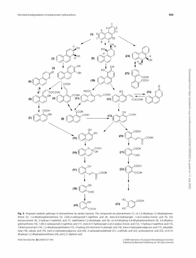
Fig. 3. Proposed catabolic pathways of phenanthrene by aerobic bacteria. The compounds are phenanthrene (1), cis-1,2-dihydroxy-1,2-dihydrophenan-
threne (2), 1,2-dihydroxyphenanthrene (3), 2-[(E)-2-carboxyvinyl]-1-naphthoic acid (4), trans-4-(2-hydroxynaph -1-yl)-2-oxobut-3-enoic acid (5), 5,6-
benzocoumarin (6), 2-hydroxy-1-naphthoic acid (7), naphthalene-1,2-dicarboxylic acid (8), cis-3,4-dihydroxy-3,4-dihydrophenanthrene (9), 3,4-dihydrox-
yphenanthrene (10), 1-[(E)-2-carboxyvinyl]-2-naphthoic acid (11), trans-4-(1-hydroxynaph-2-yl)-2-oxobut-3-enoic acid (12), 1-hydroxy-2-naphthoic acid (13),
7,8-benzocoumarin (14), 1,2-dihydroxynaphthalene (15), 2-hydroxy-2H-chromene-2-carboxylic acid (16), trans-o-hydroxybenzalpyruvic acid (17), salicylalde-
hyde (18), salicylic acid (19), trans-2-carboxybenzalpyruvic acid (20), 2-carboxybenzaldehyde (21), o-phthalic acid (22), protocatechuic acid (23), cis-9,10-
dihydroxy-1,2-dihydrophenanthrene (24), and 2,20-diphenic acid.
FEMS Microbiol Rev 32 (2008) 927–955 c� 2008 Federation of European Microbiological SocietiesPublished by Blackwell Publishing Ltd. All rights reserved
933Microbial biodegradation of polyaromatic hydrocarbons
Dow
nloaded from https://academ
ic.oup.com/fem
sre/article/32/6/927/2683263 by guest on 11 July 2022
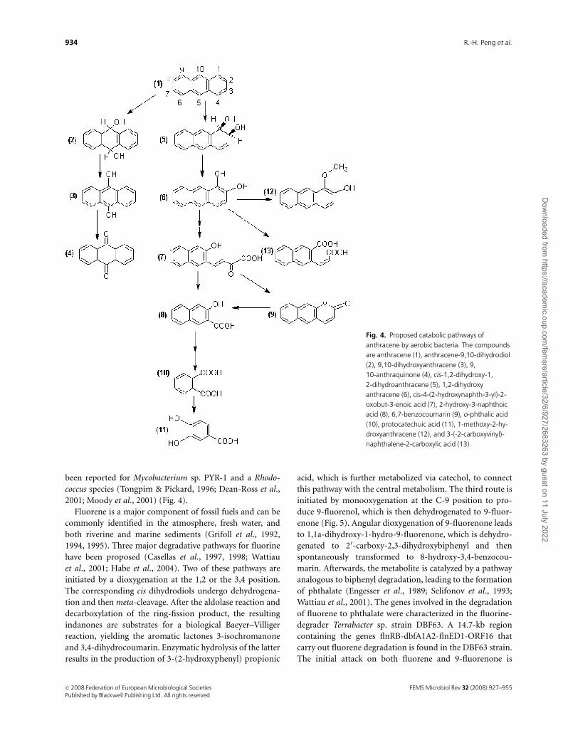
been reported for Mycobacterium sp. PYR-1 and a Rhodo-
coccus species (Tongpim & Pickard, 1996; Dean-Ross et al.,
2001; Moody et al., 2001) (Fig. 4).
Fluorene is a major component of fossil fuels and can be
commonly identified in the atmosphere, fresh water, and
both riverine and marine sediments (Grifoll et al., 1992,
1994, 1995). Three major degradative pathways for fluorine
have been proposed (Casellas et al., 1997, 1998; Wattiau
et al., 2001; Habe et al., 2004). Two of these pathways are
initiated by a dioxygenation at the 1,2 or the 3,4 position.
The corresponding cis dihydrodiols undergo dehydrogena-
tion and then meta-cleavage. After the aldolase reaction and
decarboxylation of the ring-fission product, the resulting
indanones are substrates for a biological Baeyer–Villiger
reaction, yielding the aromatic lactones 3-isochromanone
and 3,4-dihydrocoumarin. Enzymatic hydrolysis of the latter
results in the production of 3-(2-hydroxyphenyl) propionic
acid, which is further metabolized via catechol, to connect
this pathway with the central metabolism. The third route is
initiated by monooxygenation at the C-9 position to pro-
duce 9-fluorenol, which is then dehydrogenated to 9-fluor-
enone (Fig. 5). Angular dioxygenation of 9-fluorenone leads
to 1,1a-dihydroxy-1-hydro-9-fluorenone, which is dehydro-
genated to 20-carboxy-2,3-dihydroxybiphenyl and then
spontaneously transformed to 8-hydroxy-3,4-benzocou-
marin. Afterwards, the metabolite is catalyzed by a pathway
analogous to biphenyl degradation, leading to the formation
of phthalate (Engesser et al., 1989; Selifonov et al., 1993;
Wattiau et al., 2001). The genes involved in the degradation
of fluorene to phthalate were characterized in the fluorine-
degrader Terrabacter sp. strain DBF63. A 14.7-kb region
containing the genes flnRB-dbfA1A2-flnED1-ORF16 that
carry out fluorene degradation is found in the DBF63 strain.
The initial attack on both fluorene and 9-fluorenone is
Fig. 4. Proposed catabolic pathways of
anthracene by aerobic bacteria. The compounds
are anthracene (1), anthracene-9,10-dihydrodiol
(2), 9,10-dihydroxyanthracene (3), 9,
10-anthraquinone (4), cis-1,2-dihydroxy-1,
2-dihydroanthracene (5), 1,2-dihydroxy
anthracene (6), cis-4-(2-hydroxynaphth-3-yl)-2-
oxobut-3-enoic acid (7), 2-hydroxy-3-naphthoic
acid (8), 6,7-benzocoumarin (9), o-phthalic acid
(10), protocatechuic acid (11), 1-methoxy-2-hy-
droxyanthracene (12), and 3-(-2-carboxyvinyl)-
naphthalene-2-carboxylic acid (13).
FEMS Microbiol Rev 32 (2008) 927–955c� 2008 Federation of European Microbiological SocietiesPublished by Blackwell Publishing Ltd. All rights reserved
934 R.-H. Peng et al.
Dow
nloaded from https://academ
ic.oup.com/fem
sre/article/32/6/927/2683263 by guest on 11 July 2022
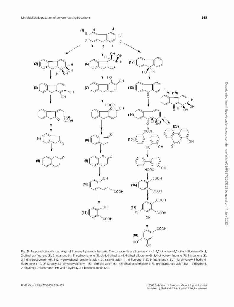
Fig. 5. Proposed catabolic pathways of fluorene by aerobic bacteria. The compounds are fluorene (1), cis-1,2-dihydroxy-1,2-dihydrofluorene (2), 1,
2-dihydroxy fluorene (3), 2-indanone (4), 3-isochromanone (5), cis-3,4-dihydroxy-3,4-dihydrofluorene (6), 3,4-dihydroxy fluorene (7), 1-indanone (8),
3,4-dihydrocoumarin (9), 3-(2-hydroxyphenyl) propionic acid (10), salicylic acid (11), 9-fluorenol (12), 9-fluorenone (13), 1,1a-dihydroxy-1-hydro-9-
fluorenone (14), 20-carboxy-2,3-dihydroxybiphenyl (15), phthalic acid (16), 4,5-dihydroxyphthalate (17), protocatechuic acid (18) 1,2-dihydro-1,
2-dihydroxy-9-fluorenone (19), and 8-hydroxy-3,4-benzocoumarin (20).
FEMS Microbiol Rev 32 (2008) 927–955 c� 2008 Federation of European Microbiological SocietiesPublished by Blackwell Publishing Ltd. All rights reserved
935Microbial biodegradation of polyaromatic hydrocarbons
Dow
nloaded from https://academ
ic.oup.com/fem
sre/article/32/6/927/2683263 by guest on 11 July 2022
catalyzed by the angular dioxygenase (DbfA1 and DbfA2) to
yield 9-fluorenol and 1,1a-dihydroxy-1-hydro-9-fluore-
none, respectively. The FlnB protein exhibited activities
against both 9-fluorenol and 1,1a-dihydroxy-1-hydro-9-
fluorenone to produce 9-fluorenone and 20-carboxy-2,3-
dihydroxybiphenyl, respectively. FlnD1 is a heteromeric
protein encoded by flnD1 and ORF16, and is a member of
the class III two-subunit extradiol dioxygenases. FlnE was
identified as a serine hydrolase for the metacleavage pro-
ducts that yield phthalate (Kasuga et al., 2001; Habe et al.,
2003, 2004).
High-MW PAH dioxygenase system
The presence of high-MW PAHs in contaminated soils
continues to pose significant problems because the persis-
tence and genotoxicity of PAHs increase with increasing
molecule size (Cerniglia, 1992). The relationship between
PAH environmental persistence and increasing numbers of
benzene rings is in agreement with the knowledge of
environmental biodegradation rates and molecule size
(Bossert & Bartha, 1986; Heitkamp & Cerniglia, 1987). For
example, half-lives of the three-ring molecule phenanthrene
in soil and sediment may range from 16 to 126 days, while
half-lives of the five-ring molecule benzo[a]pyrene (BaP)
may range from 229 to 1400 days (Shuttleworth & Cerniglia,
1995). Electrochemical stability and hydrophobicity are two
crucial factors for the accretion of high-MW PAHs in the
environment (Volkering et al., 1992, 1993; Bosma et al.,
1997; Harms & Bosma, 1997).
The first report of the biodegradation of high-MW PAHs
was presented in 1975. Gibson et al. identified two products
of BaP metabolism – cis-9,10-dihydroxy-9,10-dihydro-ben-
zo[a]pyrene and cis-7,8-dihydroxy-7,8-dihydrobenzo[a]-
pyrene – from the culture solution of a mutant Beijerinckia
sp. strain when it grew with succinate and biphenyl (Gibson
et al., 1975). However, in 1988 the bacterium that could
degrade PAHs with four aromatic rings was isolated from
sediment below an oil field by Heitkamp and Cerniglia for
the first time (Heitkamp & Cerniglia, 1988). Also in 1988,
Mahaffey et al. (1988) investigated the further catabolism of
benz[a]anthracene by the Beijerinckia sp. strain B1 (reclas-
sified as S. yanoikuyae) after induction with biphenyl,
m-xylene, or salicylate. Nuclear magnetic resonance (NMR)
and mass spectral analyses led to the identification of one
major metabolite – 1-hydroxy-2-anthranoic acid – and two
minor metabolites – 2-hydroxy-3-phenanthroic acid and 3-
hydroxy-2-phenanthroic acid. Later, Mueller et al. (1989,
1990) showed that a bacterial community isolated from a
creosote waste site was capable of utilizing fluoranthene as
its sole source of carbon and energy for bacterial growth.
Pyrene has often been used as a model compound for
high-MW PAH biodegradation because it is structurally
similar to several carcinogenic PAHs. Although a number
of bacterial isolates have been reported to grow on or
mineralize pyrene, the majority of these isolates are nocar-
dioform actinomycetes, such as members of the genus
Mycobacterium and Rhodococcus (Heitkamp & Cerniglia,
1988; Walter et al., 1991; Rehmann et al., 1998).
Multiple pathways of pyrene degradation have been
offered for the Mycobacterium vanbaalenii PYR-1 strain,
including typical dioxygenation and monooxygenation
(Heitkamp & Cerniglia, 1988; Brezna et al., 2005). Although
Mycobacteria strains can oxidize pyrene via initial dioxy-
genation at the 1,2-positions, the metabolite will form
o-methylated derivatives of pyrene-1,2-diol or the dead end
product 4-hydroxy-perinaphthenone. The chief pathway of
pyrene degradation is dioxygenation at the 4,5-positions to
produce both cis- and trans-4,5-pyrenedihydrodiol by diox-
ygenase and monooxygense, respectively. Rearomatization
of the dihydrodiol and subsequent ortho-cleavage lead to the
formation of 4,5-dicarboxyphenanthrene, which is further
decarboxylated to 4-phenanthroate. Following another di-
oxygenation reaction, 4-phenanthroate forms cis-3,4-dihy-
droxyphenanthrene-4-carboxylate. Rearomatization of the
metabolite yields 3,4-dihydroxyphenanthrene, which is
further metabolized to 1-hydroxy-2-naphthoate. The subse-
quent enzymatic reactions, including intradiol ring cleavage
dioxygenation, result in the production of o-phthalate. Then
phthalate is further transformed to TCA cycle via the
b-ketoadipate pathway (Wang et al., 2000; Kim et al., 2003)
(Fig. 6).
Based on both genomic and proteomic data, Kim et al.
have identified 27 enzymes necessary for constructing a
complete pathway for pyrene degradation. Proteomic ana-
lysis also reveals that 18 enzymes in the pathway are
upregulated more than twofold, as indicated by peptide
counting when the organism was grown with pyrene. There
are four ring-hydroxylating steps catalyzed by the corre-
sponding dioxygenase NidAB2, MvanDraft_0817/0818,
PhtAaAb, and NidA3B3. The electron transfer components
for these four steps have been found: ferredoxin (PhtAc) and
ferredoxin reductase (PhtAd). This gene arrangement of the
b- and a-subunits of dioxygenase NidAB2 has rarely been
found in other bacteria. NidA and NidB proteins are 40-
56% similar to the corresponding isofunctional enzymes in
the Nocardioides sp. strain KP7 and Pseudomonas sp. strain
NCIMB12038. Three copies of the terminal subunits of ring-
hydroxylating oxygenase (NidAB2, MvanDraft_0817/0818,
and PhtAaAb), dihydrodiol dehydrogenase (Mvan-
Draft_0815), and ring cleavage dioxygenase (Mvan-
Draft_3242) were induced in pyrene-grown cells. However,
no ORFs for enzymes such as 1-hydroxy-2-naphthoate
hydroxylase, which is necessary for 1-hydroxy-2-naphthoate
degradation through salicylate, were detected. ORFs for
phthalate 4,5-dioxygenase and protocatechuate-4,5-
FEMS Microbiol Rev 32 (2008) 927–955c� 2008 Federation of European Microbiological SocietiesPublished by Blackwell Publishing Ltd. All rights reserved
936 R.-H. Peng et al.
Dow
nloaded from https://academ
ic.oup.com/fem
sre/article/32/6/927/2683263 by guest on 11 July 2022
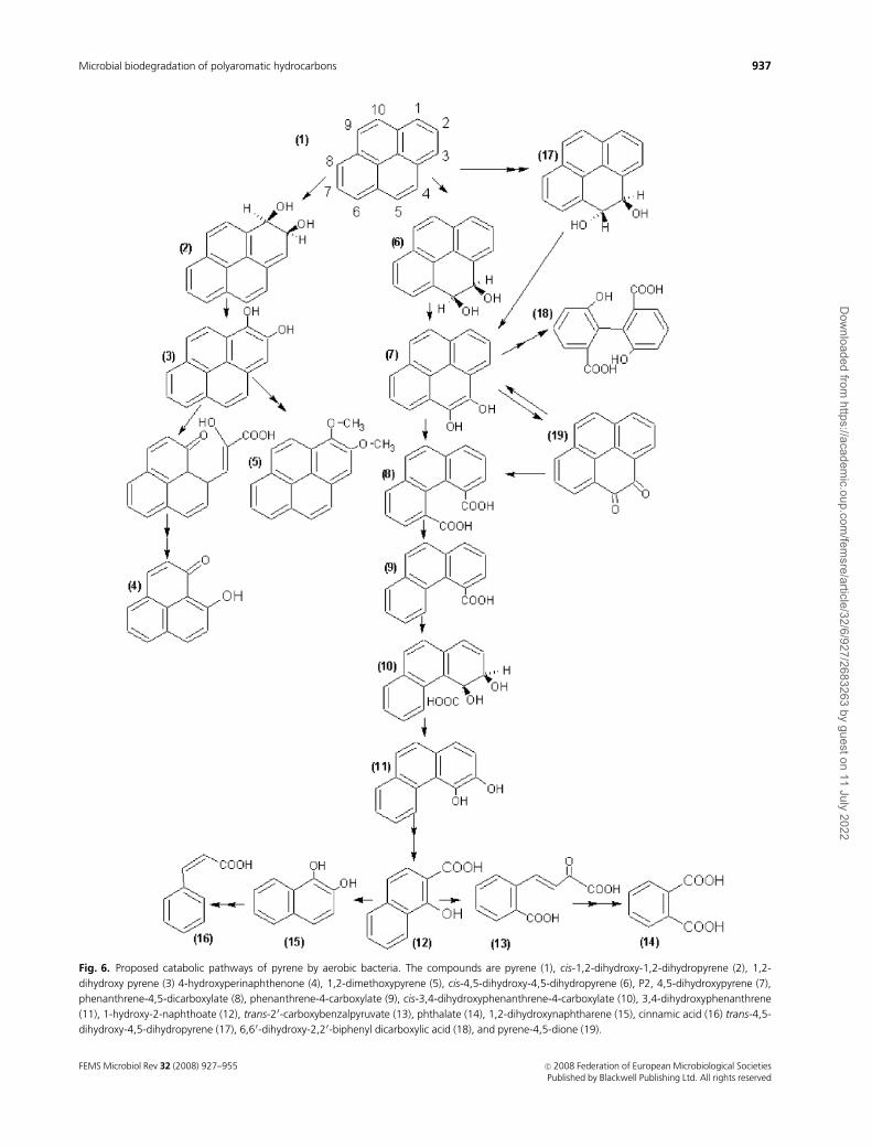
Fig. 6. Proposed catabolic pathways of pyrene by aerobic bacteria. The compounds are pyrene (1), cis-1,2-dihydroxy-1,2-dihydropyrene (2), 1,2-
dihydroxy pyrene (3) 4-hydroxyperinaphthenone (4), 1,2-dimethoxypyrene (5), cis-4,5-dihydroxy-4,5-dihydropyrene (6), P2, 4,5-dihydroxypyrene (7),
phenanthrene-4,5-dicarboxylate (8), phenanthrene-4-carboxylate (9), cis-3,4-dihydroxyphenanthrene-4-carboxylate (10), 3,4-dihydroxyphenanthrene
(11), 1-hydroxy-2-naphthoate (12), trans-20-carboxybenzalpyruvate (13), phthalate (14), 1,2-dihydroxynaphtharene (15), cinnamic acid (16) trans-4,5-
dihydroxy-4,5-dihydropyrene (17), 6,60-dihydroxy-2,20-biphenyl dicarboxylic acid (18), and pyrene-4,5-dione (19).
FEMS Microbiol Rev 32 (2008) 927–955 c� 2008 Federation of European Microbiological SocietiesPublished by Blackwell Publishing Ltd. All rights reserved
937Microbial biodegradation of polyaromatic hydrocarbons
Dow
nloaded from https://academ
ic.oup.com/fem
sre/article/32/6/927/2683263 by guest on 11 July 2022

dioxygenase, which are found in the optional routes for
oxidation of phthalate and the meta cleavage of protoca-
techuate, respectively, were also not detected in the genome
sequence (Kim et al., 2004a, 2007).
Besides the M. vanbaalenii PYR-1 strain, many other
Mycobacterium sp. strains were identified and found to
mineralize pyrene. Most of the PAH-degrading mycobacter-
ia described are fast-growing species within the genus. The
mycolic acid-rich cell walls of these soil bacteria may be an
important factor in their utilization of hydrophobic sub-
strates such as PAHs.
The Mycobacterium sp. strain RJGII-135 is capable of
degrading a wide range of PAHs. Mineralization studies
using [14C]pyrene added to contaminated soils, with and
without the addition of this strain, indicated that this strain
was able to grow on pyrene as its sole source of carbon and
energy. During the mineralization of pyrene, three steady
intermediates were formed within 4–8 h, which included
two identified metabolites in strain PYR-1 – 4-phenanthre-
necarboxylic acid and 4,5-pyrenedihydrodiol – and one
special product only in the strain RJGII-135 – 4,5-phenan-
threnedicarboxylic acid (Schneider et al., 1996).
The Mycobacterium sp. strain KMS was isolated from
vadosezone soil at the Champion International Superfund
site (Libby, MT), and is able to degrade pyrene and other
PAHs. It was determined from 2-dimensional gel electro-
phoresis that the first aromatic ring hydroxylating dioxy-
genase that oxidizes pyrene to cis-4,5-pyrene-dihydrodiol
includes the a and b subunits, 4Fe–4S ferredoxin, and the
Rieske (2Fe–2S) region, which were all induced with the
addition of pyrene to the cultures. Other proteins that
further pyrene degradation, such as dihydrodiol dehydro-
genase, oxidoreductase, and epoxide hydrolase, were also
found to be notably induced with the addition of pyrene.
There were five different b-subunits and two different Rieske
(2Fe–2S) regions of the a-subunit of aromatic ring hydro-
xylating dioxygenase, indicating that there are multiple
copies of dioxygenase expressed during pyrene degradation.
Pyrene-4,5-dione was identified to be a pyrene degradation
metabolite of the Mycobacterium sp. strain KMS. Pyrene-
4,5-dione can then be degraded into two major intermedi-
ates – phenanthrene-4,5-dicarboxylic acid and 4-phenan-
throic acid – by a flavoprotein-like, FAD-dependent
oxidoreductase. Pyrene-4,5-dione can also be reduced back
to 4,5-dihydroxypyrene by quinone reductase, as reported
for M. vanbaalenii PYR-1 (Liang et al., 2006).
The Mycobacterium sp. strain AP1 was isolated from a
pyrene enrichment culture and can grow with pyrene as its
sole source of carbon and energy. However, the strain cannot
remove the substrate completely, and its growth yield
suggests an assimilation of about 10% of the pyrene carbon.
A novel metabolite identified as 6,6-dihydroxy-2,2-biphenyl
dicarboxylic acid reveals a new branch in the pathway that
involves dioxygenation on 4,5 positions and 9,10 positions
and cleavage of both central rings of the pyrene (Vila et al.,
2001).
The Mycobacterium sp. strain KR2 was isolated from
PAH-contaminated soil originating from the area of a
former gaswork plant. This isolate metabolized up to 60%
of the pyrene added (0.5 mg mL�1) within 8 days at 20 1C.
Similar to pyrene degradation by strain PYR-1, pyrene
metabolism in the KR2 strain seems to be controlled by
metabolite induction. With the exception of 2-carboxyben-
zaldehyde and cis-3,4-phenanthrene dihydrodiol-4-car-
boxylic acid, all pyrene metabolites produced by strain KR2
have been detected in cultures of other pyrene-degrading
bacteria. The formation of 4-phenanthrol from cis-3,4-
phenanthrene dihydrodiol-4-carboxylic acid should be steri-
cally favored under the assumption that the bulky carboxy
group is extruded from the bay region, thus occupying a
pseudoaxial position necessary for elimination (Rehmann
et al., 1998).
The Mycobacterium sp. strain BB1 was isolated from a
past coal gasification site. Exponential growth on pyrene was
observed over a 35-fold increase of CO2 evolution in the
fermentor. In addition, some nonsubstrate and harmless
nonionic surfactants were found to enhance the degradation
of pyrene (Boldrin et al., 1993; Fritzsche, 1994; Tiehm,
1994).
The Mycobacterium sp. strain CH1 was isolated from
PAH-contaminated freshwater sediments and could utilize
pyrene or a wide range of branched alkanes and n-alkanes as
its sole carbon and energy sources. A lag phase of at least 3
days was detected when strain CH1 was grown in pyrene;
however, this lag phase was decreased to o 1 day when
phenanthrene or fluoranthene was supplied in the culture
media. Because there was no detectable hybridization with
the nahAc gene, the enzyme system involved in pyrene
degradation is unrelated to the naphthalene dioxygenase
pathway. Furthermore, weak hybridization of the P. oleovorans
alkB gene probe to strain CH1 DNA suggests slight
homology between the genes involved in alkane oxidation.
These observations suggest that the occurrence of both
aromatic and aliphatic hydrocarbon-degradative capacities
within a single strain may be more universal (Churchill
et al., 1999).
The pathway describing the biodegradation of the four-
ringed PAH fluoranthene by M. vanbaalenii PYR-1 has been
discovered recently (Kweon et al., 2007) (Fig. 7). Thirty-
seven fluoranthene metabolites including potential isomers
were isolated from culture medium and analyzed using
HPLC, GC-MS, and UV-visible absorption. Fifty-three en-
zymes were determined to likely be involved in fluoranthene
degradation. Four proposed pathways have been proposed
for the degradation of fluoranthene initiated by mono- and
dioxygenation reactions. The dioxygenase attack occurs at
FEMS Microbiol Rev 32 (2008) 927–955c� 2008 Federation of European Microbiological SocietiesPublished by Blackwell Publishing Ltd. All rights reserved
938 R.-H. Peng et al.
Dow
nloaded from https://academ
ic.oup.com/fem
sre/article/32/6/927/2683263 by guest on 11 July 2022
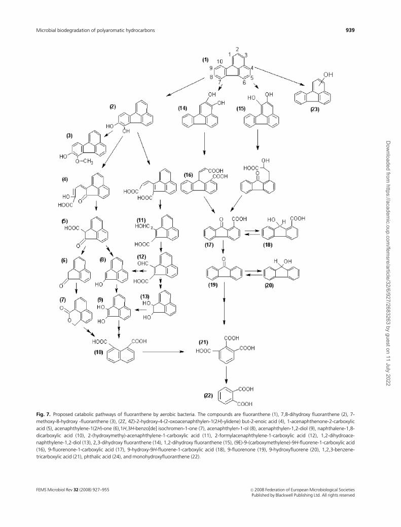
Fig. 7. Proposed catabolic pathways of fluoranthene by aerobic bacteria. The compounds are fluoranthene (1), 7,8-dihydroxy fluoranthene (2), 7-
methoxy-8-hydroxy -fluoranthene (3), (2Z, 4Z)-2-hydroxy-4-(2-oxoacenaphthylen-1(2H)-ylidene) but-2-enoic acid (4), 1-acenaphthenone-2-carboxylic
acid (5), acenaphthylene-1(2H)-one (6),1H,3H-benzo[de] isochromen-1-one (7), acenaphthylen-1-ol (8), acenaphthylen-1,2-diol (9), naphthalene-1,8-
dicarboxylic acid (10), 2-(hydroxymethy)-acenaphthylene-1-carboxylic acid (11), 2-formylacenaphthylene-1-carboxylic acid (12), 1,2-dihydroace-
naphthylene-1,2-diol (13), 2,3-dihydroxy fluoranthene (14), 1,2-dihydroxy fluoranthene (15), (9E)-9-(carboxymethylene)-9H-fluorene-1-carboxylic acid
(16), 9-fluorenone-1-carboxylic acid (17), 9-hydroxy-9H-fluorene-1-carboxylic acid (18), 9-fluorenone (19), 9-hydroxyfluorene (20), 1,2,3-benzene-
tricarboxylic acid (21), phthalic acid (24), and monohydroxyfluoranthene (22).
FEMS Microbiol Rev 32 (2008) 927–955 c� 2008 Federation of European Microbiological SocietiesPublished by Blackwell Publishing Ltd. All rights reserved
939Microbial biodegradation of polyaromatic hydrocarbons
Dow
nloaded from https://academ
ic.oup.com/fem
sre/article/32/6/927/2683263 by guest on 11 July 2022

the C-1,2, C-2,3, and C-7,8 positions of the fluoranthene.
The C-1,2 and C-2,3 dioxygenation routes degrade fluor-
anthene via fluorene-type metabolites, whereas the C-7,8
route oxidizes fluoranthene via acenaphthylene-type meta-
bolites by extra- and intradiol ring cleavages. The major site
of dioxygenation is the C-2,3 dioxygenation route, which
consists of 18 enzymatic steps via 9-fluorenone-1-carboxylic
acid and phthalate with the initial ring-hydroxylating oxy-
genase, NidA3B3, oxidizing fluoranthene to fluoranthene
cis-2,3-dihydrodiol. Six cytochrome P450 genes, including
CYP51, are responsible for monooxygenation of fluor-
anthene. Then the monooxygenation product is trans-
formed to monohydroxyfluoranthenes in the catalysis of
ring-hydroxylating oxygenases and chemical dehydration of
cis- or trans-dihydrodiols (Kweon et al., 2007).
There are only limited studies that document extensive
mineralization of PAHs that have more than four rings
(Kanaly & Harayama, 2000). Most research is focused on
the five-ring benzo[a]pyrene (BaP) because it is one of the
most important environmental contaminants, and because
its mutagenesis and carcinogenesis that cause ubiquitous
xenobiotics are well recognized (Kalf & Crommentuijn,
1997; NTP, 2002). All reported BaP oxidization by bacteria
has occurred when the bacteria were grown on other
substrates, which could induce microorganisms to produce
some enzymes that can degrade BaP. An early observation of
BaP biodegradation was its oxidation to dihydrodiols by
S. yanoikuyae B8/36 (formerly Beijerinckia sp. strain B8/36),
a strain that can be induced with biphenyl, m-xylene, or
salicylate. However, no ring cleavage products were detected
(Gibson et al., 1975; Mahaffey et al., 1988; Gibson, 1999).
Schneider et al. (1996) identified cis-7,8-dihydrodiol from
growing cultures of the Mycobacterium sp. RJGII-135 strain
where pyrene was used to maintain PAH degradation. It was
suggested that the Mycobacterium sp. RJGII-135 strain was
capable of transforming BaP to initial ring oxidation and
ring cleavage products by enzymatically attacking at the
C-4,5, C-7,8, and/or C-9,10 position (Schneider et al.,
1996). When grown in a mixture of yeast extract, peptone,
and soluble starch, M. vanbaalenii PYR-1 can biotransform
0.5 mg BaP L�1 to 24.7% aqueous and organic-extractable
metabolites. This strain was also shown to slightly degrade
BaP in a six-component PAH mixture. Recently, Moody
used resting M. vanbaalenii PYR-1 cultures induced with
phenanthrene to produce several dihydrodiols and one ring-
cleavage product, 10-oxabenzo[def]chrysene-9-one, from
benzo[a] pyrene (Moody et al., 2003, 2004).
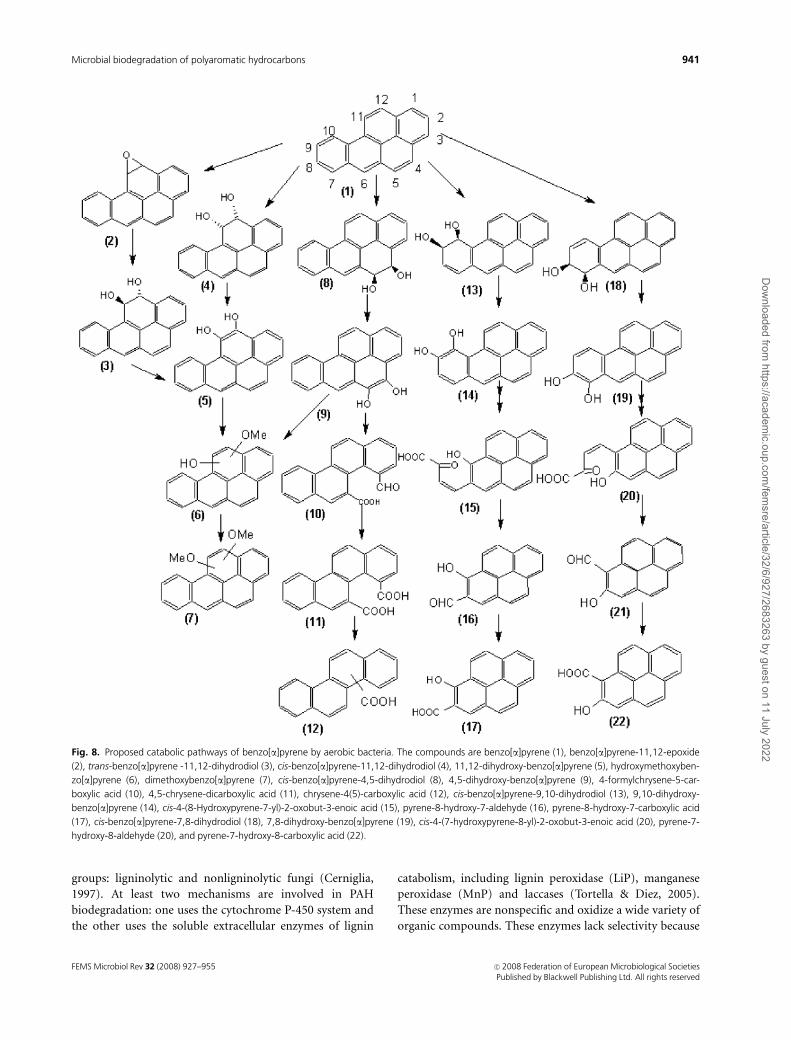
The M. vanbaalenii strain PYR-1 initially oxidized BaP
with dioxygenases and monooxygenases at C-4,5, C-9,10,
and C-11,12 (Fig. 8). It was determined from the metabo-
lites separated by reversed-phase HPLC and characterized by
UV-visible, mass, NMR, and circular dichroism spectral
analyses that the major intermediates of benzo[a]pyrene
metabolism were benzo[a]pyrene cis-4,5-dihydrodiol, ben-
zo[a]pyrene cis-11,12-dihydrodiol, benzo[a]pyrene trans-
11,12-dihydrodiol, 10-oxabenzo-[def] chrysen-9-one, and
hydroxymethoxy and dimethoxy derivatives of BaP. The
reaction of dioxygenation in the K region (4,5 positions) to
form the cis-dihydrodiol and ortho-cleavage to form chry-
sene 4,5-dicarboxylic acid are consistent with phenanthrene
and pyrene degradation. The ortho-ring fission products 4-
formylchrysene-5-carboxylic acid and 4,5-chrysene-dicar-
boxylic acid and a monocarboxylated chrysene product were
formed when replacement culture experiments were con-
ducted with benzo[a]pyrene cis-4,5-dihydrodiol. Chiral sta-
tionary-phase HPLC analysis of the dihydrodiols indicated
that benzo[a]pyrene cis-4,5-dihydrodiol had 30% 4S,5R and
70% 4R,5S absolute stereochemistry. The initial dioxygena-
tion of BaP at the C-9 and C-10 positions yields benzo[a]-
pyrene cis-9,10-dihydrodiol. Dehydration of the dihydrodiol
to the dihydroxy intermediate with subsequent meta-clea-
vage and aromatic-ring closure may lead to the formation of
10-oxabenzo-[def] chrysen-9-one. The PYR-1 strain also
oxidizes BaP to cis- and trans-11,12-dihydro-11,12-dihy-
droxybenzo[a]pyrene, which then form cis- and trans-
dihydrodiols. Benzo[a]pyrene cis-11,12-dihydrodiol
adopted an 11S,12R conformation with 100% optical purity.
The enantiomeric composition of benzo[a]pyrene trans-
11,12-dihydrodiol was an equal mixture of 11S,12S and
11R,12R molecules. The identification of the cis- and trans-
dihydrodiols is an indication that dioxygenation and mono-
oxygenation reactions occurred in naphthalene, anthracene,
phenanthrene, and pyrene degradation. The formation of
the benzo[a]pyrene trans-11,12-dihydrodiol was probably
due to the oxidation by cytochrome P450 to form ben-
zo[a]pyrene-11,12-epoxide and the successive hydrolysis of
the latter by epoxide hydrolase (Moody et al., 2004).
Additional experiments with [14C]7-benzo[a]pyrene de-
monstrated that 3.8% mineralization of BaP will take place
by the S. yanoikuyae JAR02 strain grown in the presence of
both succinate (2.0 mM) and salicylate (2.5 mM) over 7
days. Benzo[a]pyrene-cis-4,5-dihydrodiol and benzo[a]pyr-
ene-cis-7,8-dihydrodiol were observed using HPLC/MS.
Further characterization of the radio-labeled metabolite
using HPLC/MS and HPLC/MS/MS identified radio-labeled
pyrene-8-hydroxy-7-carboxylic acid and unlabeled pyrene-
7-hydroxy-8-carboxylic acid as novel ring-cleavage metabo-
lites. The similar chirality of these metabolites suggested that
the enzymatic attack position will be at C-7,8 and C-9,10
(Rentz et al., 2008).
Fungi catabolism of polycyclic aromatichydrocarbons
Several studies have shown that diverse fungi are capable of
PAH mineralization. These fungi can be classed into two
FEMS Microbiol Rev 32 (2008) 927–955c� 2008 Federation of European Microbiological SocietiesPublished by Blackwell Publishing Ltd. All rights reserved
940 R.-H. Peng et al.
Dow
nloaded from https://academ
ic.oup.com/fem
sre/article/32/6/927/2683263 by guest on 11 July 2022
groups: ligninolytic and nonligninolytic fungi (Cerniglia,
1997). At least two mechanisms are involved in PAH
biodegradation: one uses the cytochrome P-450 system and
the other uses the soluble extracellular enzymes of lignin
catabolism, including lignin peroxidase (LiP), manganese
peroxidase (MnP) and laccases (Tortella & Diez, 2005).
These enzymes are nonspecific and oxidize a wide variety of
organic compounds. These enzymes lack selectivity because
Fig. 8. Proposed catabolic pathways of benzo[a]pyrene by aerobic bacteria. The compounds are benzo[a]pyrene (1), benzo[a]pyrene-11,12-epoxide
(2), trans-benzo[a]pyrene -11,12-dihydrodiol (3), cis-benzo[a]pyrene-11,12-dihydrodiol (4), 11,12-dihydroxy-benzo[a]pyrene (5), hydroxymethoxyben-
zo[a]pyrene (6), dimethoxybenzo[a]pyrene (7), cis-benzo[a]pyrene-4,5-dihydrodiol (8), 4,5-dihydroxy-benzo[a]pyrene (9), 4-formylchrysene-5-car-
boxylic acid (10), 4,5-chrysene-dicarboxylic acid (11), chrysene-4(5)-carboxylic acid (12), cis-benzo[a]pyrene-9,10-dihydrodiol (13), 9,10-dihydroxy-
benzo[a]pyrene (14), cis-4-(8-Hydroxypyrene-7-yl)-2-oxobut-3-enoic acid (15), pyrene-8-hydroxy-7-aldehyde (16), pyrene-8-hydroxy-7-carboxylic acid
(17), cis-benzo[a]pyrene-7,8-dihydrodiol (18), 7,8-dihydroxy-benzo[a]pyrene (19), cis-4-(7-hydroxypyrene-8-yl)-2-oxobut-3-enoic acid (20), pyrene-7-
hydroxy-8-aldehyde (20), and pyrene-7-hydroxy-8-carboxylic acid (22).
FEMS Microbiol Rev 32 (2008) 927–955 c� 2008 Federation of European Microbiological SocietiesPublished by Blackwell Publishing Ltd. All rights reserved
941Microbial biodegradation of polyaromatic hydrocarbons
Dow
nloaded from https://academ
ic.oup.com/fem
sre/article/32/6/927/2683263 by guest on 11 July 2022

lignin contains a variety of aromatic structures formed in
plant cell walls by oxygen-radical coupling reactions of 4-
hydroxy cinnamyl, 3-methoxy-4-hydroxy cinnamyl and 3,5-
dimethoxo-4-hydroxy cinnamyl, resulting in a variety of
intermonomer linkages (Hammel, 1992). In nonligninolytic
fungi, cytochrome P450 monooxygenase enzymes catalyze
the oxidation of PAHs to arene oxides, which are initial
products of PAH metabolism.
The extracellular enzymes of lignin catabolism inligninolytic fungi
Because of their advantage of being able to diffuse to the
immobile PAHs, the fungal extracellular enzymes of lignin
catabolism appear to be more likely than bacterial intracel-
lular enzymes to make the initial attack on PAHs in soil.
White-rot fungi are ubiquitous in nature in their ability to
degrade and mineralize recalcitrant plant polymer lignin
(Otjen & Blanchette, 1986; Martınez et al., 2005). Several
reports have shown that white-rot fungi are capable of
degrading PAHs and have potential in the bioremediation
of PAH-polluted soils and sediments. Large quantities of
mycelia of several species of white-rot fungi are used to
increase the extent of PAH bioremediation in soil. The best-
characterized strain – Phanerochaete chrysosporium – can
oxidize pyrene, anthracene, fluorine and benzo[a]pyrene to
the corresponding quinines by lignin peroxidase and man-
ganese peroxidase (Bogan et al., 1996a, b). In contrast to
P. chrysosporium, the metabolism of PAHs with Phanerochaete
laevis is faster and more extensive (Bogan & Lamar, 1996).
Lignin peroxidase and manganese peroxidase can catalyze
the oxidation of the recalcitrant nonphenolic lignin to form
a high redox potential oxo-ferryl intermediate during the
reaction of the heme cofactor with hydrogen peroxide
(H2O2) (Jensen et al., 1996; Gold et al., 2000; Perez et al.,
2002) (Fig. 9a). Two features in the molecular structure
confer ligninolytic peroxidases with distinctive catalytic
properties: (1) a heme group, conferring high redox poten-
tial to the oxo-ferryl complex and (2) the existence of
specific-binding sites for oxidation of their characteristic
substrates, including nonphenolic aromatics in the cases of
LiP, manganous iron in the case of MnP (Reddy & D’souza,
1994; Reddy et al., 2003; Martınez et al., 2005).The heme
group is sandwiched between an N-terminal and a
C-terminal helix and rests at the bottom of the space formed
by the surfaces of both structures. The iron coordination
and the residues involved in the active site are conserved
among most peroxidases. The iron is pentacoordinated to
the four pyrrole nitrogens of the heme and to the nitrogen in
the imidazole group of the proximal histidine. Another
distal histidine, assisted by an asparagine residue, takes part
in the transfer of the oxidizing equivalents from H2O2 to the
heme. It is suggested that the length of the Fe–imidazolic
nitrogen (Fe–Ne2) bond is related to the redox potential of
the different peroxidases (Piontek et al., 1993; Poulos et al.,
1993; Sundaramoorthy et al., 1997). Laccases catalyze a one-
electron oxidation concomitantly with the four-electron
reduction of molecular oxygen to water using a range of
phenolic compounds as hydrogen donors (Fig. 9a). The
catalysis is carried out by the presence of different copper
centers, which were arranged in a trinuclear cluster with one
type-1 (T1), one type-2 (T2) and two type-3 (T3) copper
ions. The presence of the four cupric ions, each co-ordinated
to a single polypeptide chain, is an absolute requirement for
optimal activity (Messerschmidt, 1997; Solomon et al., 2001;
Baldrian, 2006).
Various researches have revealed that the mechanism of
oxidation of PAHs by fungi ligninolytic enzymes is similar to
the degradation of nonphenolic lignin. A correlation has
been found between the ionization potential (IP) of PAHs
and the specific activity of MnP and LiP. Aromatic substrates
with a lower IP were oxidized by the two ligninolytic
enzymes and the lower the IP, the faster the oxidation rate.
A threshold value of IP was found for each enzyme: LiP
oxidizes PAHs with an IP � 7.55 eV (Vazquez-Duhalt et al.,
1994), while MnP oxidizes PAHs with an IP up to 8.2 eV. For
Fig. 9. Catalysis reaction of polycyclic aromatic hydrocarbons by the
fungal lignin peroxidase, manganese peroxidase, and laccase. (a) The
mechanism of reaction is described in equation. AH2 is the reducing
substrate. AH is the reducing substrate after one electron oxidation.
(b) Oxidation of polycyclic aromatic hydrocarbons by ligninolytic fungi.
Lignin peroxidase, manganese peroxiase, and laccase are capable of
oxidizing PAHs to the corresponding quinines.
FEMS Microbiol Rev 32 (2008) 927–955c� 2008 Federation of European Microbiological SocietiesPublished by Blackwell Publishing Ltd. All rights reserved
942 R.-H. Peng et al.
Dow
nloaded from https://academ
ic.oup.com/fem
sre/article/32/6/927/2683263 by guest on 11 July 2022
example, MnP isolated from Irpex lacteus was able to
efficiently degrade three- and four-ringed PAHs with an IP
higher than 7.8 eV (Bogan & Lamar, 1995; Bogan et al.,
1996a, b). Despite the possibility of direct electron transfer
from the aromatic substrate to the heme environment, long-
range electron transfer has also been detected in LiP from
P. chrysosporium (Schoemaker, 1994). Thus, extracellular
LiPs can directly oxidize condensed PAHs to produce
unstable aryl cation radicals, whereas MnPs catalyze the
oxidation of PAHs in a reaction that requires Mn21, oxygen,
and unsaturated lipids (Baborova et al., 2006; Eibes et al.,
2006). A novel manganese–lignin peroxidase (MnLiP) hy-
brid enzyme produced by Bjerkandera adusta can efficiently
oxidize Mn(II) to Mn(III) and can also carry out Mn(II)-
independent activity on aromatic substrates (Wang et al.,
2003). The role of laccase in the oxidation of nonphenolic
lignin structures is similar to LiP. All laccases can be divided
into three groups according to their redox potential (E1) at
the T1 copper site (Cambria et al., 2008). It has been
observed that the different activities of various laccases
toward substrates can mainly be attributed to differences in
their redox potential. PAHs show E1 higher than that of
laccase (500–800 mV), and so the PAH degradation by
laccase needs to be aided by small mediators (Johannes
et al., 1996; Majcherczyk et al., 1998). The most common
mediators used are 2,20-azino-bis-(3-ethylbenzothiazoline-
6-sulfonic acid) (ABTS) (Bourbonnais et al., 1995) and
–N–OH-type mediators such as 1-hydroxybenzotriazole
(HBT) and violuric acid (VA). The E1 of ABTS, HBT, and
VA are 1.09, 1.12, and 0.91 V, respectively (Xu et al., 2000,
2001). A greater DE1 could create a more favorable transi-
tional energy state for the interaction between molecular
orbitals of the mediator and the enzyme, resulting in faster
electron transfer. Laccase efficiently oxidized [14C]phenan-
threne in the presence of HBT and Tween 80. 73% of initially
added phenanthrene was degraded within 182 h to yield
phenanthrene-9,10-quinone and 2,20-diphenic acid as the
major products (Fig. 9b). The addition of the redox med-
iator ABTS to the reaction mixture increased oxidation of
phenanthrene by c. 40% (Han et al., 2004). However, the use
of these synthetic mediators implies high added costs and
the possible generation of toxic oxidized species. Some of
the natural compounds, such as r-coumaric acid, strongly
promoted the removal of PAHs by laccase. This compound
was a better laccase mediator than ABTS and was similar to
HBT, attaining 95% removal of anthracene and benzo[a]-
pyrene and around 50% removal of pyrene within 24 h
(Canas et al., 2007).
The cytochrome P450 system
The biological functions of cytochrome P450 monooxy-
genases, such as detoxification of xenobiotics and steroido-
genesis, are based on its ability to catalyze the insertion of
oxygen into a wide variety of compounds. It was well known
that mammalian P450 plays key roles not only in the
detoxification of PAHs but also in the activation of PAHs
(Shimada, 2006). The pathways leading to the formation of
PAH diol-epoxides have been extensively studied in rat liver
P450s and epoxide hydrolase using BaP as a prototype (Fig.
10a). Traditionally, a K-region oxide, B[a]P-4,5-oxide, was
thought to be an oxidation product of BaP by P450s. The
metabolite was found to be readily hydrolyzed to an inactive
B[a]P-4,5-diol by epoxide hydrolase (Conney, 1982). Sub-
sequent examination of several BaP metabolites revealed
that CYP1A1 and CYP1B1 enzymes can activate PAHs to
toxic and carcinogenic metabolites in collaboration with
epoxide hydrolase by the formation of B[a]P-7,8-diol,
which were again oxidized to the highly reactive bay region
oxides, B[a]P-7,8-diol-9,10-epoxide (Levin et al., 1976;
Pelkonen & Nebert, 1982; Shimada & Fujii-Kuriyama,
2004).
Fungi metabolize PAH compounds to metabolites similar
to those formed by mammalian enzymes. The predominant
pathway in the initial oxidation of PAHs is a cytochrome
P450 monooxygenase/epoxide hydrolase that catalyzes the
reaction that forms trans-dihydrodiols (Mueller et al., 1995;
Cerniglia, 1997) (Fig. 10b). These metabolic steps are found
in nonligninolytic fungi, such as Cunninghamella elegans,
and ligninolytic fungi, such as Pleurotus ostreatus. For
example, C. elegans metabolized fluoranthene into fluor-
anthene trans-2,3-dihydrodiol, 8- and 9-hydroxyfluor-
anthene trans-2,3-dihydrodiol (Tortella & Diez, 2005),
whereas P. ostreatus metabolized pyrene into pyrene trans-
4,5-dihydrodiol and anthracene into anthracene trans-1,2-
dihydrodiol and 9,10-anthraquinone (Bazalel et al.,
1996a, b). The cytochrome P450 can also oxidize numerous
PAHs to phenols that are subsequently conjugated with
sulfate, glucoronic acid, or glucose (Cerniglia et al., 1982,
1986; Casillas et al., 1996; Pothuluri et al., 1996). Cunning-
hamella elegans metabolizes benzo[e]pyrene to 10-hydroxy-
3-benzo[e]pyrenylsulfate (Cerniglia et al., 1989; Pothuluri
et al., 1996) and the fungus A. niger SK9317 metabolizes
pyrene to 1-pyrenylsulfate and 1-hydroxy-8-pyrenylsulfate
(Wunder et al., 1994; Capotorti et al., 2005).
The fungus cytochrome P450 monooxygenases are a
superfamily of heme-thiolate proteins that are associated
with membranes in the endoplasmic reticulum (Degtyar-
enko, 1995; Yadav et al., 2003). Whole-genome sequencing
of the white-rot basidiomycete P. chrysosporium has revealed
the presence of the largest P450 monooxygenases in fungi
comprising c. 150 P450 genes, which could be classified into
12 families and 23 subfamilies and under 11 fungal P450
clans. The amino acid sequence of these P450 monooxygen-
ase proteins is extremely diverse, with levels of identity as
low as 16% in some cases, but their structural fold has
FEMS Microbiol Rev 32 (2008) 927–955 c� 2008 Federation of European Microbiological SocietiesPublished by Blackwell Publishing Ltd. All rights reserved
943Microbial biodegradation of polyaromatic hydrocarbons
Dow
nloaded from https://academ
ic.oup.com/fem
sre/article/32/6/927/2683263 by guest on 11 July 2022
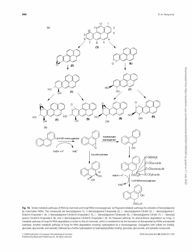
Fig. 10. Similar metabolic pathways of PAHs by mammals and fungi P450s monooxygenase. (a) Proposed metabolic pathways for activation of benzo[a]pyrene
by mammalian P450s. The compounds are benzo[a]pyrene (1), (1)benzo[a]pyrene-7,8-epoxide (2), (� )benzo[a]pyrene-7,8-diol (3), (� )benzo[a]pyrene-7,
8-diol-9,10-epoxide-1 (4), (1)benzo[a]pyrene-7,8-diol-9,10-epoxide-2 (5), (� )benzo[a]pyrene-7,8-epoxide (6), (1)benzo[a]pyrene-7,8-diol (7), (� )benzo[a]
pyrene-7,8-diol-9,10-epoxide-2 (8), and (1)benzo[a]pyrene-7,8-diol-9,10-epoxide-1 (9). (b) Proposed pathway for phenanthrene degradation by fungi. A
metabolic pathway of fungi for PAHs degradation is similar to that of mammals, which is considered to be the formation of diol-epoxides by P450s and epoxide
hydrolase. Another metabolic pathway of fungi for PAHs degradation involving: hydroxylation by a monooxygenase, conjugation with sulfate ion (methyl,
glucoside, glucuronide, and xyloside), followed by a further hydroxylation to hydroxylarylsulfates (methyl, glucoside, glucuronide, and xyloside) compounds.
FEMS Microbiol Rev 32 (2008) 927–955c� 2008 Federation of European Microbiological SocietiesPublished by Blackwell Publishing Ltd. All rights reserved
944 R.-H. Peng et al.
Dow
nloaded from https://academ
ic.oup.com/fem
sre/article/32/6/927/2683263 by guest on 11 July 2022
remained the same throughout evolution (Rabinovich et al.,
2004; Doddapaneni et al., 2005a, b). Under two distinct
nutrient conditions, nutrient-limited (ligninolytic) and nu-
trient-sufficient (nonligninolytic), P450 monooxygenases
proved to be a functional divergence in transcriptional
profiling. P450 genes in this basidiomycetous fungus
showed varying degrees of structural similarities to the
P450 genes from different ascomycetous fungi such as
Aspergillus and Fusarium, suggesting that P. chrysosporium
has acquired these P450 families as a part of a vertical
descent from a common ancestor. For instance, the CYP63
family of proteins of P. chrysosporium, which consists of
seven members (pc-1 through pc-7), shows a structural
resemblance to the CYP52 family of ascomycetous yeasts
and has, therefore, been classified under the CYP52 clan
(Doddapaneni & Yadav, 2004; Doddapaneni et al., 2005a, b;
Yadav et al., 2006). All seven genes of the CYP63 family of
proteins showed differential transcriptional induction with
alkanes and their derivatives (Anzenbacher & Anzenbacher-
ova, 2001). Moreover, their substrate specificity seems to
extend beyond alkanes/substituted alkanes as they also showed
induction in the presence of various aromatics, including
polycyclic aromatic compounds like PAHs. For example, the
gene of pc-1 was induced by mono-aromatics (m-hydroxy
benzoic acid, nitrophenol, and phenoxy acetic acid), polycyclic
aromatic compounds (naphthalene, phenanthrene, pyrene,
3-methylcholanthrene, p-p0-biphenol and polychlorinated bi-
phenyl), and alkyl-substituted aromatics (dodecylbenzene
sulfonate, 1-phenyl dodecane and limonene). On the other
hand, pc-2 showed no induction in response to mono-
aromatic compounds, but could be induced by polycyclic
aromatic compounds. pc-4 showed induction specifically in
response to polycyclic aromatic compounds and alkyl-sub-
stituted aromatics. pc-6 showed high levels of induction in
response to some polycyclic aromatic compounds (1-
naphthol and 1,2-benzanthracene) and several alkyl-substi-
tuted aromatic compounds (Yadav et al., 2006).
Under optimized conditions, the ligninolytic enzymes
LiP, MnP, and laccase are capable of oxidizing PAHs with
an IP o 7.6. However, for PAHs with a higher IP, a
cytochrome P450 monoxygenase activity is likely required
(Bazalel et al., 1996a, b). For example, the amount of CO2
released from BaP oxidation decreased by 66% in 10 days of
culture in the presence of the inhibitor of cytochrome P450,
1-aminobenzotriazole, suggesting that such an enzyme is
involved in BaP degradation by Fusarium solani (Rafin et al.,
2000). In some fungi that degrade PAHs, extracellular
laccase and peroxidase activities were not induced by the
presence of PAHs. Moreover, the percentages of degradation
did not seem to correlate with the ligninolytic enzyme
activities (Gramss et al., 1999; Verdin et al., 2004). In certain
fungi, it seems that a wide variety of PAH can be degradated
by cytochrome P450 monoxygenase.
Although cytochrome P450-type monooxygenases might
be involved in the oxidation of PAHs by several fungi, they
oxidize PAHs via a pathway similar to the mammalian
enzyme systems to form potent carcinogens – epoxides and
dihydrodiols (Mueller et al., 1995; Cerniglia, 1997). On the
contrary, peroxidase-mediated extracellular oxidation of
PAHs in cultures of white rot fungi results initially in
quinines (Bumpus, 1989; Vyas et al., 1994). Because qui-
nones are less toxic than their respective PAH and dihy-
droxylated metabolites generated by cytochrome P450
monooxygenases, enzymatic oxidation of PAHs by lignino-
lytic oxidoreductases could be a more useful strategy in
detoxification and bioremediation processes.
Degradation of environmental PAHmixtures
Several bacteria have the ability to degrade low- and high-
MW PAHs. For example, the Mycobacterium sp. AP1 strain,
for which pyrene and fluoranthene degradation pathways
have been proposed, can also utilize phenanthrene as its sole
source of carbon and energy (Vila et al., 2001; Lopez et al.,
2006). The kinetics of the disappearance of individual PAHs
was in agreement with differences in their respective aqu-
eous solubilities (Volkering et al., 1992; Harms & Bosma,
1997). Mycobacterium sp. AP1 showed a significant deple-
tion of naphthalene, phenanthrene, and anthracene when
compared with uninoculated controls after 30 days. The
decrease in the concentrations of fluoranthene, pyrene and
benzo[a]pyrene with respect to the controls was not statis-
tically significant, although typical ring-cleavage products
from those compounds could be detected (Vila et al., 2001;
Lopez et al., 2007).
It was suggested that the simultaneous bacteria-mediated
biodegradation of PAHs dissolving from a PAH mixture was
strongly influenced by their different bioavailabilities. Bioa-
vailability is considered to be a dynamic process that is
determined by the rate of substrate mass transfer to micro-
bial cells relative to their intrinsic catabolic activity. At high
mass transfer rates, the overall biodegradation rate is con-
trolled by the metabolic activity of the bacteria (Koch, 1990;
Bosma et al., 1997). The observed disappearance rates and
extents of the PAH compounds are in agreement with their
aqueous solubilities. When the growth-linked compounds
phenanthrene, fluoranthene, and pyrene are all added
together to a Mycobacterium sp. AP1 strain culture, they will
reduce simultaneously and their reduction rates will reach
maximum values during the first 168 h. However, the
biodegradation rates of fluoranthene and pyrene are sig-
nificantly lower than that of phenanthrene (Lopez et al.,
2007).
Degradation of PAHs in contaminated soil will be also
influenced by the diffusional release of PAHs from the
FEMS Microbiol Rev 32 (2008) 927–955 c� 2008 Federation of European Microbiological SocietiesPublished by Blackwell Publishing Ltd. All rights reserved
945Microbial biodegradation of polyaromatic hydrocarbons
Dow
nloaded from https://academ
ic.oup.com/fem
sre/article/32/6/927/2683263 by guest on 11 July 2022
nonaqueous phase liquids (NAPLs) phase. The specific
surface area of the NAPLs may negatively influence biode-
gradation because of kinetic restrictions to the dissolution
process (Ortega-Calvo & Alexander, 1994). For bacteria
growing on NAPL-dissolved PAHs in aqueous systems,
production of bioemulsifiers would generate a larger surface
area of the substrates and subsequently a higher substrate
flux (Reddy et al., 1982). sphingomonads and Mycobacteria
growing on NAPL-dissolved PAHs can adhere strongly to
the NAPL phase. Consequently, it appears that attachment
to and biofilm formation on PAH sources is a widespread
mechanism among bacteria to overcome mass-transfer
limitations when growing on weakly soluble and strongly
sorbed PAHs (Johnsena et al., 2005).
Environmentally derived bacterial isolates often degrade
only a narrow range of PAHs, and patterns of simultaneous
degradation of PAH mixtures are complex. The observed
pattern of utilization of PAHs is not only the result of
bioavailability but also of metabolic interactions related to
competition and cometabolism effects (Bouchez et al.,
1995). Cometabolism is an important feature of the degra-
dation of PAHs. Cometabolism of one kind of PAH could
have a synergistic effect increasing the degradation of
another. Therefore, cometabolism can widen the range of
PAH attacked by a defined strain, especially for the degrada-
tion of high-MW PAHs (van Herwijnen et al., 2003a, b).
During the incubation, a number of acid metabolites carried
out by high-MW PAH-degrading Mycobacteria accumulated
in the medium. Most of these metabolites were dicarboxylic
aromatic acids formed as a result of the utilization of growth
substrates (phenanthrene, pyrene, or fluoranthene) by mul-
tibranched pathways including meta and ortho-ring-cleavage
reactions: phthalic acid, naphthalene-1,8-dicarboxylic acid,
phenanthrene-4,5-dicarboxylic acid, diphenic acid, Z-9-car-
boxymethylenefluorene-1-carboxylic acid and 6,60-dihy-
droxy-2,20-biphenyl dicarboxylic acid. These partially
oxidized compounds produced by one microbial strain
could be degraded by others (Lopez et al., 2007).
A number of species of white-rot fungi can cometabolize
PAHs with bacteria by improving the compound bioavail-
ability. Fungi cannot utilize any high-MW PAH as their sole
carbon and energy source, but can partially degrade these if
cultured in a nutrient broth (Sutherland, 1992). Some fungi
can oxidize PAHs rapidly to a higher aqueous solubility with
their extracellular ligninolytic enzyme systems, resulting in
higher rates of mineralization of these metabolites by
bacteria. A study carried out with anthracene has confirmed
that all detected oxidation products of this compound
breakdown by white-rot fungi can be mineralized by in-
digenous bacteria (e.g. activated sludge) more rapidly than
anthracene itself is mineralized (Meulenberg et al., 1997).
Addition of indigenous microbial communities to the
white-rot fungus Bjerkandera sp. strain BOS55 resulted in
an initially rapid increase in the level of CO2 recovery from
[14C]BaP and the level of water-soluble label decreased to
16% of initial levels. This demonstrated that some polar
metabolites of fungal benzo[a]pyrene oxidation were readily
degraded by indigenous microorganisms (Kotterman et al.,
1998). Some fungi can use cytochrome P450 to transform
PAHs and activate PAHs for further degradation by bacteria.
When the nonligninolytic fungus Penicillium janthinelum
VUO 10 201 and either its bacterial consortium VUN 10 009
or S. maltophilia VUN 10 010 were combined in one culture,
25% of the BaP was mineralized to CO2 by these fungal–
bacterial cocultures over 49 days, accompanied by the
transient accumulation and disappearance of intermediates.
Inoculation of fungal–bacterial cocultures into PAH-
contaminated soil resulted in significantly improved degrada-
tion of some high-MW PAHs, such as chrysene, benzo-
[a]anthracene and dibenzo[a,h]anthracene (Boonchan
et al., 2000). Thus, it was suggested that PAH degradation
in nature is a consequence of sequential breakdown by fungi
and bacteria, with the fungi performing the initial oxidation
step (Meulenberg et al., 1997; Sack et al., 1997; Kotterman
et al., 1998).
With mixtures of two individually degradable PAHs,
metabolic competition took place whether cometabolism
occurred or not (Bouchez et al., 1995). Competition at the
active site of enzymes, in particular the initial oxygenase, has
been observed most commonly. For example, the presence
of low-MW PAHs, such as phenanthrene and fluorene, can
inhibit the degradation of fluoranthene and pyrene
(Bouchez et al., 2000; Dean-Ross et al., 2002; Lotfabat &
Gray, 2002). A second possible cause of inhibition is the
dead-end products that result from cometabolic oxidation
of nongrowth substrates, such as o-xylene and p-xylene, in a
Pseudomonas stutzeri strain (Barbieri et al., 1993; String-
fellow & Aitken, 1995).
The moisture content, pH value, aeration condition, and
availability of nutrients were determined to be the key
factors in the bioremediation of contaminated soils
(Roling-Wilfred et al., 2002; Kim et al., 2005). At early stages
(0–45 days) of the bioremediation process, the addition of
KNO3 and K2HPO4 caused significant increases in both the
heterotrophic and the PAH-degrading microbial popula-
tions, while soil stimulated only by aeration and optimum
humidity exhibited smaller increases in the sizes of both
populations and a marked delay in the increase in microbial
PAH degraders. The proportion of the PAH-degrading
microbial population compared with the heterotrophic
population not treated with nutrients increased noticeably
at 21 and 45 days, and reached 100% at 135 days, while
lower proportions were observed for the populations treated
with nutrients. Denaturing gradient gel electrophoresis
analysis and principal component analysis confirmed that
there was a remarkable shift in the composition of the
FEMS Microbiol Rev 32 (2008) 927–955c� 2008 Federation of European Microbiological SocietiesPublished by Blackwell Publishing Ltd. All rights reserved
946 R.-H. Peng et al.
Dow
nloaded from https://academ
ic.oup.com/fem
sre/article/32/6/927/2683263 by guest on 11 July 2022

bacterial community due to both the biodegradation pro-
cess and the addition of nutrients. At early stages of
biodegradation, the Alphaproteobacteria group (genera
Sphingomonas and Azospirillum) was the dominant group
in all treatments. At later stages, the Gammaproteobacteria
group (genus Xanthomonas), the Alphaproteobacteria group
(genus Sphingomonas), and the Cytophaga–Flexibacter–
Bacteroides group (Bacteroidetes) were the dominant groups
that did not receive nutrients, while the Gammaproteobacteria
group (genus Xathomonas), the Betaproteobacteria group
(genera Alcaligenes and Achromobacter), and the Alphapro-
teobacteria group (genus Sphingomonas) were the dominant
groups that received nutrients (Vinas et al., 2005). Monitor-
ing the microbial community by terminal restriction frag-
ment length polymorphism (TRFLP) analysis suggests that
the aeration change stimulated by soil excavation would
cause the change in the microbial community composition.
The fragments show several ribotypes considerably increas-
ing in aerobic organisms with a corresponding decrease in
anaerobic organisms such as Clostridia sp. (Grant et al.,
2007).
Conclusion
During the past decade, a variety of microorganisms have
been isolated and characterized for their ability to degrade
different PAHs. Furthermore, many metabolic enzymes for
the degradation of different PAHs have been isolated from
microorganisms and several novel pathways have been
elucidated based on the identification of initial ring oxida-
tion and ring cleavage products. The genes responsible for
PAH catabolic pathways are always localized as gene clusters,
and some gene clusters have been cloned and sequenced. An
analysis of the gene organization and transcription patterns
in the clusters of different species and an analysis of the
homology of related loci led to the conclusion that genetic
exchange and rearrangements played a major part in the
evolution of the regions that play a role in PAH degradation.
Genes that catalyze PAH degradation are believed to have
adaptively evolved in nature by various genetic events. In
bacteria, some aromatic ring hydroxylating dioxygenases
share high homologies in structure and exhibit very relaxed
substrate specificities, whereas some dioxygenases with
small differences in their amino acid sequences display
major differences in their enzymatic properties, such as
substrate range and regiospecificity. In fungi, more than
one isoenzyme of extracellular enzymes and P450 monoox-
ygenases was found. These genes offer an insight into the
evolution of the catalytic versatility of fungus in the detox-
ification and biodegradation of a broad range of xenobiotic
chemicals.
In the natural environment, PAHs tend to interact with
non-aqueous phases and soil organic matter and, as a
consequence, become potentially unavailable for microbial
degradation. Microorganisms have evolved different strate-
gies to adapt to the PAH compounds present in the
contaminated situation. Many microorganisms produce
biosurfactants to increase the bioavailability of the poorly
available substrates. Many microorganisms show chemo-
taxis towards PAH compounds, which could lead to im-
proved degradation. Environmentally derived bacterial
isolates often degrade only a narrow range of PAHs, and
degradation of a PAH mixture becomes visible as a coopera-
tive process involving a consortium of strains with comple-
mentary capacities. The ubiquitous coexistence of bacteria
and fungi in soil and their catabolic cooperation suggests
that fungal–bacterial interactions may be of importance for
PAH degradation in the natural environment.
Combing genetic engineering tools such as gene conver-
sion, gene duplication and transposition, we can produce
novel strains with desirable properties for bioremediation
applications according to the knowledge of PAH degrada-
tion by microorganisms. For example, the rates of PAH
mineralization can be enhanced when NAH plasmids from
different bacteria are hybridized using colony hybridization.
Overexpression of the dioxygenase fusion system from the
Mycobacterium sp. strain PYR-1 in other bacteria might
improve its ability to degrade high-MW PAHs. Other
technology designed according to the principles of microbial
PAH-degradation in soil may also be useful for PAH
bioremediation. For example, sequential treatment by bac-
terial consortia and fungi (e.g. white-rot fungi) or defined
fungal–bacterial cocultures are proposed to improve the
degradation of PAHs. Salicylate, acting as an inducer of
genetic operons, can be used to enhance the degradation of
PAH compounds. Addition of small amounts of biosurfac-
tants to the PAH-containing soil may increase the com-
pound’s dispersion and bioavailability, whereas it appears to
be a good strategy to improve emulsification of NAPL-
dissolved PAH in aqueous systems with turbulent mixing.
Acknowledgements
This research was supported by 863 Program
(2006AA06Z358; 2006AA10Z117; 2008AA10Z401); Shanghai
Subject Chief Scientist (06XD14017); Shanghai Key labora-
tory and basic research project (06DZ19103-07dz22011);
National and Shanghai Natural Science Foundation
(06ZR14073-30670179); and Shanghai municipal commit-
tee of agriculture (No. 2006-2-1).
References
Annweiler E, Richnow HH, Antranikian G, Hebenbrock S, Garms
C, Franke S, Francke W & Michaelis W (2000) Naphthalene
degradation and incorporation of naphthalene-derived carbon
FEMS Microbiol Rev 32 (2008) 927–955 c� 2008 Federation of European Microbiological SocietiesPublished by Blackwell Publishing Ltd. All rights reserved
947Microbial biodegradation of polyaromatic hydrocarbons
Dow
nloaded from https://academ
ic.oup.com/fem
sre/article/32/6/927/2683263 by guest on 11 July 2022
into biomass by the thermophile Bacillus thermoleovorans.
Appl Environ Microbiol 66: 518–523.
Anzenbacher P & Anzenbacherova E (2001) Cytochrome P450
and metabolism of xenobiotics. Cell Mol Life Sci 58: 737–747.
Assinder SJ & Williams PA (1990) The TOL plasmids:
determinants of the catabolism of toluene and the xylenes. Adv
Microb Physiol 31: 1–69.
Baborova P, Moder M, Baldrian P, Cajthamlova K & Cajtham T
(2006) Purification of a new manganese peroxidase of the
white-rot fungus Irpex lacteus, and degradation of polycyclic
aromatic hydrocarbons by the enzyme. Res Microbiol 157:
248–253.
Balashova NV, Stolz A, Knackmuss HJ, Kosheleva IA, Naumov AV
& Boronin AM (2001) Purification and characterization of a
salicylate hydroxylase involved in 1-hydroxy-2-naphthoic acid
hydroxylation from the naphthalene and phenanthrene-
degrading bacterial strain Pseudomonas putida BS202-P1.
Biodegradation 12: 179–188.
Baldrian P (2006) Fungal laccases-occurrenceand properties.
FEMS Microbiol Rev 30: 215–242.
Barbieri P, Palladino L, Di-Gennaro P & Gatli E (1993)
Alternative pathways for o-xylene or m-xylene and p-xylene
degradation in a Pseudomonas stutzeri strain. Biodegradation 4:
71–80.
Basta T, Keck A, Klein J & Stolz A (2004) Detection and
characterization of conjugative degradative plasmids in
xenobiotics degrading Sphingomonas strains. J Bacteriol 186:
3862–3872.
Basta T, Buerger S & Stolz A (2005) Structural and replicative
diversity of large plasmids from sphingomonads that degrade
polycyclic aromatic compounds and xenobiotics. Microbiology
151: 2025–2037.
Bazalel L, Hadar Y, Fu PP, Freeman JP & Cerniglia CE (1996a)
Initial oxydation products in the metabolism of pyrene,
anthracene, fluorene, and dibenzothiophene by the white rot
fungus Pleurotus ostreatus. Appl Environ Microbiol 62:
2554–2559.
Bazalel L, Hadar Y, Fu PP, Freeman JP & Cerniglia CE (1996b)
Metabolism of phenanthrene by the white rot fungus Pleurotus
ostreatus. Appl Environ Microbiol 62: 2547–2553.
Bogan BW & Lamar RT (1995) One-electron oxidation in the
degradation of creosote polycyclic aromatic hydrocarbons by
Phanerochaete chrysosporium. Appl Environ Microbiol 61:
2631–2635.
Bogan BW & Lamar RT (1996) Polycyclic aromatic hydrocarbons
degrading capability of Phanerochaete laevis HHB-1625 and its
extracellular ligninolytic enzymes. Appl Environ Microbiol 62:
1597–1603.
Bogan BW, Lamar RT & Hammel KE (1996a) Fluorene oxidation
in vivo by Phanerochaete chrysosporium and in vitro during
manganese peroxidase dependent lipid peroxidation. Appl
Environ Microbiol 62: 1788–1792.
Bogan BW, Schoenike B, Lamar RT & Cullen D (1996b)
Expression of lip genes during growth in soil and oxidation of
anthracene by Phanerochaete chrysosporium. Appl Environ
Microbiol 62: 3697–3703.
Boldrin B, Tiehm A & Fritzsche C (1993) Degradation of
phenanthrene, fluorene, fluoranthene, and pyrene by a
Mycobacterium sp. Appl Environ Microbiol 59: 1927–1930.
Boonchan S, Britz ML & Stanley GA (2000) Degradation and
mineralization of high-molecular-weight polycyclic aromatic
hydrocarbons by defined fungal–bacterial cocultures. Appl
Environ Microbiol 66: 1007–1019.
Boronin AM (2001) Purification and characterization of a
salicylate hydroxylase involved in 1-hydroxy-2-naphthoic acid
hydroxylation from the naphthalene and phenanthrene-
degrading bacterial strain Pseudomonas putida BS202-P1.
Biodegradation 12: 179–188.
Bosch R, Garcia-Valdes E & Moore ERB (1999a) Genetic
characterization and evolutionary implications of a
chromosomally encoded naphthalene-degradation upper
pathway from Pseudomonas stutzeri AN10. Gene 236: 149–157.
Bosch R, Moore ER, Garcia-Valdes E & Pieper DH (1999b)
NahW, a novel, inducible salicylate hydroxylase involved in
mineralization of naphthalene by Pseudomonas stutzeri AN10.
J Bacteriol 181: 2315–2322.
Bosch R, Garcia-Valdes E & Moore ERB (2000) Complete
nucleotide sequence and evolutionary significance of a
chromosomally encoded naphthalene-degradation lower
pathway from Pseudomonas stutzeri AN10. Gene 245: 67–74.
Bosma TNP, Middeldorp PJM, Schraa G & Zender AJB (1997)
Mass transfer limitation of biotransformation: quantifying
bioavailability. Environ Sci Technol 31: 248–252.
Bossert ID & Bartha R (1986) Structure biodegradability
relationships of polycyclic aromatic hydrocarbons in soil. Bull
Environ Contam Toxicol 37: 490–495.
Bouchez M, Blanchet D & Vandecasteele JP (1995) Degradation
of polycyclic aromatic hydrocarbons by pure strains and by
defined strain associations: inhibition phenomena and
cometabolism. Appl Microbiol Biotechnol 43: 156–164.
Bouchez M, Blanchet D, Bardin V, Haeseler F & Vandecasteele JP
(2000) Efficiency of defined strains and of soil consortia in the
biodegradation of polycyclic aromatic hydrocarbon (PAH)
mixtures. Biodegradation 10: 429–435.
Bourbonnais R, Pace MG, Reid ID, Lanthier P & Yaguchi M
(1995) Lignin oxidation by laccase isozymes from Trametes
versicolor and role of the mediator 2,20-azinobis (3-
ethylbenzthiazoline- 6-sulfonate) in kraft lignin
depolymerization. Appl Environ Microbiol 61: 1876–1880.
Boyd DR & Sheldrake GN (1998) The dioxygenase-catalysed
formation of vicinal cis-diols. Nat Prod Rep 15: 309–324.
Brezna B, Kweon O, Stingley RL, Freeman JP, Khan AA, Polek B,
Jones RC & Cerniglia CE (2005) Molecular characterization of
cytochrome P450 genes in the polycyclic aromatic
hydrocarbon degrading Mycobacterium vanbaalenii PYR-1.
Appl Microbiol Biotechnol 11: 1–11.
Bumpus JA (1989) Biodegradation of polycyclic aromatic
hydrocarbons by Phanerochaete chrysosporium. Appl Environ
Microbiol 55: 154–158.
FEMS Microbiol Rev 32 (2008) 927–955c� 2008 Federation of European Microbiological SocietiesPublished by Blackwell Publishing Ltd. All rights reserved
948 R.-H. Peng et al.
Dow
nloaded from https://academ
ic.oup.com/fem
sre/article/32/6/927/2683263 by guest on 11 July 2022
Butler CS & Mason JR (1997) Structure-function analysis of the
bacterial aromatic ring-hydroxylating dioxygenases. Adv
Microb Physiol 38: 47–84.
Cambria MT, Minniti Z, Librando V & Cambria A (2008)
Degradation of polycyclic aromatic hydrocarbons by
Rigidoporus lignosus and its laccase in the presence of redox
mediators. Appl Biochem Biotechnol 149: 1–8.
Canas A, Alcalde M, Plou F, Martınez MJ, Martınez AT &
Camarero S (2007) Transformation of polycyclic aromatic
hydrocarbons by laccase is strongly enhanced by phenolic
compounds present in soil. Environ Sci Technol 41: 2964–2971.
Capotorti G, Cesti P, Lombardi A & Guglielmetti G (2005)
Formation of sulfate conjugates metabolites in the degradation
of phenanthrene, anthracene, pyrene and benzo[a]pyrene by
the ascomycete Aspergillus terreus. Polycycl Aromat Comp 25:
197–213.
Carredano E, Karlsson A, Kauppi B, Choudhury D, Parales RE,
Parales JV, Lee K, Gibson DT, Eklund H & Ramaswamy S
(2000) Substrate binding site of naphthalene 1,2-dioxygenase:
functional implications of indole binding. J Mol Biol 296:
701–712.
Casellas M, Grifoll M, Bayona JM & Solanas AM (1997) New
metabolites in the degradation of fluorene by Arthrobacter sp.
strain F101. Appl Environ Microbiol 63: 816–826.
Casellas M, Grifoll M, Sabate J & Solanas AM (1998) Isolation
and characterization of a 9-fluorenone-degrading bacterial
strain and its role in synergistic degradation of fluorene by a
consortium. Can J Microbiol 44: 734–742.
Casillas RP, Crow SA Jr, Heinze TM, Deck J & Cerniglia CE
(1996) Initial oxidative and subsequent conjugative
metabolites produced during the metabolism of phenanthrene
by fungi. J Ind Microbiol 16: 205–215.
Cerniglia CE (1984) Microbial metabolism of polycyclic aromatic
hydrocarbons. Adv Appl Microbiol 30: 31–71.
Cerniglia CE (1992) Biodegradation of polycyclic aromatic
hydrocarbons. Biodegradation 3: 351–368.
Cerniglia CE (1997) Fungal metabolism of polycyclic aromatic
hydrocarbons: past, present and future applications in
bioremediation. J Ind Microbiol Biotechnol 19: 324–333.
Cerniglia CE & Heitkamp MA (1989) Microbial degradation of
polycyclic aromatic hydrocarbons (PAH) in the aquatic
environment. Metabolism of Polycyclic Aromatic Hydrocarbons
in the Aquatic Environment (Varanasi U, ed), pp. 41–68. CRC
Press, Boca Raton, FL.
Cerniglia CE, Freeman JP & Mitchum RK (1982) Glucuronide
and sulfate conjugation in the fungal metabolism of aromatic
hydrocarbons. Appl Environ Microbiol 43: 1070–1075.
Cerniglia CE, Kelly DW, Freeman JP & Miller DW (1986)
Microbiol metabolism of pyrene. Chemi Biol Interact 57:
203–216.
Cerniglia CE, Campbell WL, Freeman JP & Evans FE (1989)
Identification of a novel metabolite in phenanthrene
metabolism by the fungus Cunninghamella elegans. Appl
Environ Microbiol 55: 2275–2279.
Churchill SA, Harper JP & Churchill PF (1999) Isolation and
characterization of a Mycobacterium species capable of
degrading threeand four-ring aromatic and aliphatic
hydrocarbons. Appl Environ Microbiol 65: 549–552.
Conney AH (1982) Induction of microsomal enzymes by foreign
chemicals and carcinogenesis by polycyclic aromatic
hydrocarbons: GHA clowes; memorial lecture. Cancer Res 42:
4875–4917.
Dean-Ross D, Moody JD, Freeman JP, Doerge DR & Cerniglia CE
(2001) Metabolism of anthracene by a Rhodococcus species.
FEMS Microbiol Lett 204: 205–211.
Dean-Ross D, Moody J & Cerniglia CE (2002) Utilization of
mixtures of polycyclic aromatic hydrocarbons by bacteria
isolated from contaminated sediment. FEMS Microbiol Ecol
41: 1–7.
Degtyarenko KN (1995) Structural domains of P450-containing
monooxygenases systems. Protein Eng 8: 737–747.
Denome SA, Stanley DC, Olson ES & Young KD (1993)
Metabolism of dibenzothiophene and naphthalene in
Pseudomonas sp. trains: complete DNA sequence of an upper
naphthalene catabolic pathway. J Bacteriol 175: 6890–6901.
Doddapaneni H & Yadav JS (2004) Differential regulation and
xenobiotic induction of tandem P450 monooxygenase genes
pc-1 (CYP63A1) and pc-2 (CYP63A2) in the white rot fungus
Phanerochaete chrysosporium. Appl Microbiol Biotechnol 65:
559–565.
Doddapaneni H, Chakraborty R & Yadav JS (2005a) Genome-
wide structural and evolutionary analysis of the P450
monooxygenase genes (P450ome) in the white-rot fungus
Phanerochaete chrysosporium: evidence for gene duplications
and extensive gene clustering. BMC Genom 6: 1–24.
Doddapaneni H, Subramanian V & Yadav JS (2005b)
Physiological regulation, xenobiotic induction and
heterologous expression of P450 monooxygenase gene pc-3, a
new member of the CYP63 gene cluster in the white rot fungus
Phanerochaete chrysosporium. Curr Microbiol 50: 292–298.
Eaton RW (1994) Organization and evolution of naphthalene
catabolic pathways: sequence of the DNA encoding 2-
hydroxychromene-2-carboxylate isomerase and trans-o-
hydroxybenzylidenepyruvate hydratase-aldolase from the
NAH7 plasmid. J Bacteriol 176: 7757–7762.
Eaton RW & Chapman PJ (1992) Bacterial metabolism of
naphthalene: construction and use of recombinant bacteria to
study ring cleavage of 1,2-dihydroxynaphthalene and
subsequent reactions. J Bacteriol 174: 7542–7554.
Eguchi M, Nishikawa T, Macdonald K, Cavicchioli R, Gottschal
JC & Kjelleberg S (1996) Response to stress and nutrient
availability by the marine ultramicrobacterium Sphingomonas
sp. strain RB2256. Appl Environ Microbiol 62: 1287–1294.
Eibes G, Cajthaml T, Moreira MT, Feijoo G & Lema JM (2006)
Enzymatic degradation of anthracene, dibenzothiophene and
pyrene by manganese peroxidase in media containing acetone.
Chemosphere 64: 408–414.
Engesser KH, Strubel V, Christoglou K, Fischer P & Rast HG
(1989) Dioxygenolytic cleavage of aryl ether bonds: 1,10-
FEMS Microbiol Rev 32 (2008) 927–955 c� 2008 Federation of European Microbiological SocietiesPublished by Blackwell Publishing Ltd. All rights reserved
949Microbial biodegradation of polyaromatic hydrocarbons
Dow
nloaded from https://academ
ic.oup.com/fem
sre/article/32/6/927/2683263 by guest on 11 July 2022
dihydro-1,10-dihydroxyfluoren-9-one, a novel arene
dihydrodiol as evidence for angular dioxygenation of
dibenzofuran. FEMS Microbiol Lett 65: 205–210.
Feng X, Ou LT & Ogram A (1997) Plasmid-mediated
mineralization of carbofuran by Sphingomonas sp. CF-06. Appl
Environ Microbiol 63: 1332–1337.
Ferraro DJ, Gakhar L & Ramaswamy S (2005) Rieske business:
structure-function of Rieske non-heme oxygenases. Biochem
Biophy Res Co 338: 175–190.
Ferraro DJ, Okerlund AL, Mowers JC & Ramaswamy S (2006)
Structural basis for regioselectivity and stereoselectivity of
product formation by naphthalene-1,2-dioxygenase.
J Bacteriol 188: 6986–6994.
Fredrickson JK, Balkwill DL, Romine MF & Shi T (1999) Ecology,
physiology, and phylogeny of deep surface Sphingomonas
strains. J Ind Microbiol Biotechnol 23: 273–283.
Fritzsche C (1994) Degradation of pyrene at low defined oxygen
concentrations by a Mycobacterium sp. Appl Environ Microbiol
60: 1687–1689.
Fuenmayor SL, Wild M, Boyes AL & Williams PA (1998) A gene
cluster encoding steps in conversion of naphthalene to
gentisate in Pseudomonas sp. strain U2. J Bacteriol 180:
2522–2530.
Gianfreda L, Xu F & Bollag JM (1999) Laccases: a useful group of
oxidoreductases enzymes. Bioreme J 3: 1–25.
Gibson DT (1999) Beijerinckia sp strain B1: a strain by any other
name. J Ind Microbiol Biotechnol 23: 284–293.
Gibson DT & Parales RE (2000) Aromatic hydrocarbon
dioxygenases in environmental biotechnology. Curr Opin
Biotechnol 11: 236–243.
Gibson DT & Subramanian V (1984) Microbial degradation of
aromatic hydrocarbons. Microbial Degradation of Organic
Compounds (Gibson DT, ed), p. 181. Marcel Dekker Inc., New
York.
Gibson DT, Venkatanayarana M, Jerina DM, Yagi H & Yeh H
(1975) Oxidation of the carcinogens benzo[a]pyrene and
benzo[a]anthracene to dihydrodiols by a bacterium. Science
189: 295–297.
Gibson DT, Zylstra GJ & Chauhan S (1990) Biotransformations
catalyzed by toluene dioxygenase from Pseudomonas putida F1.
Pseudomonas: Biotransformations, Pathogenesis, and Evolving
Biotechnology (Silver S, Chakrabarty AM, Iglewski B & Kaplan
S, eds), pp. 121–132. American Society for Microbiology,
Washington, DC.
Gold MH, Youngs HL & Gelpke MD (2000) Manganese
peroxidase. Met Ions Biol Syst 37: 559–586.
Goyal AK & Zylstra GJ (1996) Molecular cloning of novel genes
for polycyclic aromatic hydrocarbon degradation from
Comamonas testosteroni GE39. Appl Environ Microbiol 62:
230–236.
Goyal AK & Zylstra GJ (1997) Genetics of naphthalene and
phenanthrene degradation by Comamonas testosteroni. J Indl
Microbiol Biotechnol 19: 401–407.
Gramss G, Kirsche B, Voight KD, Gunther T & Fritsche W (1999)
Conversion rates of five polycyclic aromatic hydrocarbons in
liquid cultures of fifty-eight fungi and the concomitant
production of oxidative enzymes. Mycol Res 103: 1009–1018.
Grant RJ, Muckian LM, Clipson NJW & Doyle EM (2007)
Microbial community changes during the bioremediation of
creosote-contaminated soil. Lett Appl Microbiol 44: 293–300.
Grifoll M, Casellas M, Bayona JM & Solanas AM (1992) Isolation
and characterization of a fluorene-degrading bacterium:
identification of ring oxidation and ring fission products. Appl
Environ Microbiol 58: 2910–2917.
Grifoll M, Selifonov SA & Chapman PJ (1994) Evidence for a
novel pathway in the degradation of fluorene by Pseudomonas
sp. strain F274. Appl Environ Microbiol 60: 2438–2449.
Grifoll M, Selifonov SA & Chapman PJ (1995) Transformation of
substituted fluorenes and fluorene analogs by Pseudomonas sp.
strain F274. Appl Environ Microbiol 61: 3711–3723.
Grimm AC & Harwood CS (1999) NahY, a catabolic plasmid-
encoded receptor required for chemotaxis of Pseudomonas
putida to the aromatic hydrocarbon naphthalene. J Bacteriol
181: 3310–3316.
Grund AD & Gunsalus IC (1983) Cloning of genes for
naphthalene metabolism in Pseudomonas putida. J Bacteriol
156: 89–94.
Habe H & Omori T (2003) Genetic of polycyclic aromatic
hydrocarbon metabolism in diverse aerobic bacteria. Biosci
Biotechnol Biochem 67: 225–243.
Habe H, Miyakoshi M, Chung JS, Kasuga K, Yoshida T, Nojiri H
& Omori T (2003) Phthalate catabolic gene cluster is linked to
the angular dioxygenase gene in Terrabacter sp. strain DBF63.
Appl Microbiol Biotechnol 61: 44–54.
Habe H, Chung JS, Kato H et al. (2004) Characterization of the
upper pathway genes for fluorene metabolism in Terrabacter
sp. strain DBF63. J Bacteriol 186: 5938–5944.
Hammel KE (1992) Oxidation of aromatic pollutants by
lignindegrading fungi and their extracellular peroxidases. Met
Ions Biol Sys 28: 41–60.
Han MJ, Choi HT & Song HG (2004) Degradation of
phenanthrene by Trametes versicolor and its laccase. J Microbiol
42: 94–98.
Harms H & Bosma TNP (1997) Mass transfer limitation of
microbial growth and pollutant degradation. J Ind Microbiol
18: 97–105.
Harvey RG (1991) Polycyclic Aromatic Hydrocarbons: Chemistry
and Carcinogenicity. Cambridge University Press, Cambridge.
Heitkamp MA & Cerniglia CE (1987) The effects of chemical
structure and exposure on the microbial degradation of
polycyclic aromatic hydrocarbons in freshwater and estuarine
ecosystems. Environ Toxicol Chem 6: 535–546.
Heitkamp MA & Cerniglia CE (1988) Mineralization of
polycyclic aromatic hydrocarbons by a bacterium isolated
from sediment below an oil field. Appl Environ Microbiol 54:
1612–1614.
Heitkamp MA, Franklin W & Cerniglia CE (1988a) Microbial
metabolism of polycyclic aromatic hydrocarbons: isolation
and characterization of a pyrene-degrading bacterium. Appl
Environ Microbiol 54: 2549–2555.
FEMS Microbiol Rev 32 (2008) 927–955c� 2008 Federation of European Microbiological SocietiesPublished by Blackwell Publishing Ltd. All rights reserved
950 R.-H. Peng et al.
Dow
nloaded from https://academ
ic.oup.com/fem
sre/article/32/6/927/2683263 by guest on 11 July 2022
Heitkamp MA, Freeman JP, Miller DW & Cerniglia CE (1988b)
Pyrene degradation by a Mycobacterium sp.: identification of
ring oxidation and ring fission products. Appl Environ
Microbiol 54: 2556–2565.
Iwabuchi T & Harayama S (1997) Biochemical and genetic
characterization of 2-carboxybenzaldehyde dehydrogenase, an
enzyme involved in phenanthrene degradation by Nocardioides
sp. strain KP7. J Bacteriol 179: 6488–6494.
Iwabuchi T & Harayama S (1998a) Biochemical and genetic
characterization of trans-20-carboxybenzalpyruvate hydratase-
aldolase from a phenanthrene-degrading Nocardioides strain.
J Bacteriol 180: 945–949.
Iwabuchi T & Harayama S (1998b) Biochemical and molecular
characterization of 1-hydroxy-2-naphthoate dioxygenase from
Nocardioides sp. KP7. J Biol Chem 273: 8332–8336.
Jensen KAJ, Bao W, Kawai S, Srebotnik E & Hammel KE (1996)
Manganese-dependent cleavage of nonphenolic lignin
structures by Ceriporiopsis subvermispora in the absence of
lignin peroxidase. Appl Environ Microbiol 62: 3679–3686.
Johannes C, Majcherczyk A & Huttermann A (1996) Degradation
of anthracene by laccase of Trametes versicolor in the presence
of different mediator compounds. Appl Microbiol Biotechnol
46: 313–317.
Johnsena AR, Wickb LY & Harmsb H (2005) Principles of
microbial PAH-degradation in soil. Environ Pollut 133: 71–84.
Kalf DF & Crommentuijn T (1997) Environment quality
objectives for 10 polycyclic aromatic hydrocarbons. Ecotox
Environ Safe 36: 89–97.
Kanaly RA & Harayama S (2000) Biodegradation of high
molecular-weight polycyclic aromatic hydrocarbons by
bacteria. J Bacteriol 182: 2059–2067.
Kasuga K, Habe H, Chung JS, Yoshida T, Nojiri H, Yamane H &
Omori T (2001) Isolation and characterization of the genes
encoding a novel oxygenase component of angular
dioxygenase from the gram-positive dibenzofuran-degrader
Terrabacter sp. strain DBF63. Biochem Biophy Res Co 283:
195–204.
Keith LH & Telliard WA (1979) Priority pollutants. I. A
perspective view. Environ Sci Technol 13: 416–423.
Keum YS, Seo JS, Hu Y & Li QX (2005) Degradation pathways of
phenanthrene by Sinorhizobium sp. C4. Appl Microbiol
Biotechnol 71: 935–941.
Kim E & Zylstra GJ (1999) Functional analysis of genes involved
in biphenyl, naphthalene, phenanthrene, and m-xylene
degradation by Sphingomonas yanoikuyae B1. J Ind Microbiol
Biotechnol 23: 294–302.
Kim E, Aversano PJ, Romine MF, Schneider RP & Zylstra GJ
(1996) Homology between genes for aromatic hydrocarbon
degradation in surface and deep-subsurface Sphingomonas
strains. Appl Microbiol Biotechnol 62: 1467–1470.
Kim SJ, Jones RC, Cha CJ, Kweon O, Edmondson RD & Cerniglia
CE (2004a) Identification of proteins induced by polycyclic
aromatic hydrocarbon in Mycobacterium vanbaalenii PYR-1
using two-dimensional polyacrylamide gel electrophoresis and
de novo sequencing methods. Proteomics 4: 3899–3908.
Kim SJ, Kweon O, Jones RC, Freeman JP, Edmondson RD &
Cerniglia CE (2007) Complete and integrated pyrene
degradation pathway in Mycobacterium vanbaalenii PYR-1
based on systems biology. J Bacteriol 189: 464–472.
Kim YH, Engesser KH & Cerniglia CE (2003) Two polycyclic
aromatic hydrocarbon o-quinone reductases from a pyrene-
degrading Mycobacterium. Arch Biochem Biophys 416:
209–217.
Kim YH, Moody JD, Freeman JP, Brezna B, Engesser KH &
Cerniglia CE (2004b) Evidence for the existence of PAH-
quinone reductase and catechol-O-methyltransferase in
Mycobacterium vanbaalenii PYR-1. J Ind Microbiol Biotechnol
31: 507–516.
Kim YH, Freeman JP, Moody JD, Engesser KH & Cerniglia CE
(2005) Effects of pH on the degradation of phenanthrene and
pyrene by Mycobacterium vanbaalenii PYR-1. Appl Environ
Microbiol 67: 275–285.
Kiyohara H, Nagao K, Kouno K & Yano K (1982) Phenanthrene-
degrading phenotype of Alcaligenes faecalis AFK2. Appl
Environ Microbiol 43: 458–461.
Kiyohara H, Takizawa N, Date H, Torigoe S & Yano K (1990)
Characterization of a phenanthrene degradation plasmid from
Alcaligenes faecalis AFK2. J Ferm Bioeng 69: 54–56.
Kiyohara H, Torigoe S, Kaida N, Asaki T, Iida T, Hayashi H &
Takizawa N (1994) Cloning and characterization of a
chromosomal gene cluster, pah, that encodes the upper
pathway for phenanthrene and naphthalene utilization by
Pseudomonas putida OUS82. J Bacteriol 176: 2439–2443.
Koch AL (1990) Diffusion the crucial process in many aspects of
the biology of bacteria. Adv Microb Ecol 11: 37–69.
Kosheleva IA, Tsoi TV, Kulakova AN & Boronin AM (1986)
Srtuctiral analysis of plasmid NPL-1 controlling napthhlane
oxidation in Pseudomonas putida and its derivatives. Genetika
22: 2389–2397.
Kotterman MJJ, Vis EH & Field JA (1998) Successive
mineralization and detoxification of benzo[a]pyrene by the
white rot fungus Bjerkandera sp. strain BOS55 and indigenous
microflora. Appl Environ Microbiol 64: 2853–2858.
Kulakov LA, Allen CCR, Lipscomb DA & Larkin MJ (2000)
Cloning and characterization of a novel cis-naphthalene
dihydrodiol dehydrogenase gene (narB) from Rhodococcus sp.
NCIMB 12038. FEMS Microbiol Lett 182: 327–331.
Kulakov LA, Chen SC, Allen CCR & Larkin MJ (2005) Web-type
evolution of Rhodococcus gene clusters associated with
utilization of naphthalene. Appl Environ Microbiol 71:
1754–1764.
Kweon O, Kim SJ, Jones RC, Freeman JP, Adjei MD, Edmondson
RD & Cerniglia CE (2007) A polyomic approach to elucidate
the fluoranthene-degradative pathway in Mycobacterium
vanbaalenii PYR-1. J Bacteriol 189: 4635–4647.
Larkin MJ, Allen CCR, Kulakov LA & Lipscomb DA (1999)
Purification and characterization of a novel naphthalene
dioxygenase from Rhodococcus sp. strain NCIMB 12038.
J Bacteriol 181: 6200–6204.
FEMS Microbiol Rev 32 (2008) 927–955 c� 2008 Federation of European Microbiological SocietiesPublished by Blackwell Publishing Ltd. All rights reserved
951Microbial biodegradation of polyaromatic hydrocarbons
Dow
nloaded from https://academ
ic.oup.com/fem
sre/article/32/6/927/2683263 by guest on 11 July 2022
Larkin MJ, Kulakov LK & Allen CCR (2005) Biodegradation and
Rhodococcus masters of catabolic versatility. Curr Opin
Biotechnol 12: 564–573.
Laurie AD & Lloyd-Jones G (1999) The phn genes of Burkholderia
sp. strain RP007 constitute a divergent gene cluster for
polycyclic aromatic hydrocarbon catabolism. J Bacteriol 181:
531–540.
Laurie AD & Lloyd-Jones G (2000) Quantification of phnAc and
nahAc in contaminated New Zealand soils by competitive
PCR. Appl Environ Microbiol 66: 1814–1817.
Laurie AD & Lloyd-Jones G (1999) Conserved and hybrid meta-
cleavage operons from PAH-degrading Burkholderia RP007.
Biochem Biophys Res Co 262: 308–314.
Levin W, Wood AW, Yagi H, Dansette PM, Jerina DM & Conney
AH (1976) Carcinogenicity of benzo[a]pyrene 4,5,7,8,9,10
oxides on mouse skin. Proc Natl Acad Sci USA 73: 3867–3871.
Li W, Shi J, Wang X, Han Y, Tong W, Ma L, Liu B & Cai B (2004)
Complete nucleotide sequence and organization of
thenaphthalene catabolic plasmid pND6-1 from Pseudomonas
sp. strain ND6. Gene 336: 231–240.
Liang Y, Gardner DR, Miller CD, Chen D, Anderson AJ, Weimer
BC & Sims RC (2006) Study of biochemical pathways and
enzymes involved in pyrene degradation by Mycobacterium sp.
strain KMS. Appl Environ Microbiol 72: 7821–7828.
Lloyd-Jones G, Laurie AD, Hunter EWF & Fraser R (1999)
Analysis of catabolic genes for naphthalene and phenanthrene
degradation in contaminated New Zealand soils. FEMS
Microbiol Ecol 29: 69–79.
Lopez Z, Vila J, Minguillon C & Grifoll M (2006) Metabolism of
fluoranthene by Mycobacterium sp. strain AP1. Appl Microbiol
Biotechnol 70: 747–756.
Lopez Z, Vila J, Ortega-Calvo JJ & Grifo M (2007) Simultaneous
biodegradation of creosote-polycyclic aromatic hydrocarbons
by a pyrene-degrading Mycobacterium. Appl Microbiol
Biotechnol 78: 165–172.
Lotfabat SK & Gray MR (2002) Kinetics of biodegradation of
mixtures of polycyclic aromatic hydrocarbons. Appl Microbiol
Biotechnol 60: 361–365.
Mahaffey WR, Gibson DT & Cerniglia CE (1988) Bacterial
oxidation of chemical carcinogens: formation of polycyclic
aromatic acids from benz[a]anthracene. Appl Environ
Microbiol 54: 2415–2423.
Majcherczyk A, Johannes C & Huttermann A (1998) Oxidation of
polycyclic aromatic hydrocarbons (PAH) by laccase of
Trametes versicolor. Enzyme Microb Technol 22: 335–341.
Martınez AT, Speranza M, Ruiz-Duenas FJ, Ferreira P, Camarero
S, Guillen F, Martınez MJ, Gutierrez A & Del Rıo JC (2005)
Biodegradation of lignocellulosics: microbial, chemical, and
enzymatic aspects of the fungal attack of lignin. Int Microbiol 8:
195–204.
Menn FM, Applegate BM & Sayler GS (1993) NAH Plasmid-
mediated catabolism of anthracene and phenanthrenen to
naphthoic acids. Appl Environ Microbiol 59: 1938–1942.
Messerschmidt A (1997) Multi-Copper Oxidases. World Scientific,
Singapore.
Meulenberg R, Rijnaarts HHM, Doddema HJ & Field JA (1997)
Partially oxidized polycyclic aromatic hydrocarbons show an
increased bioavailability and biodegradability. FEMS Microbiol
Lett 152: 45–49.
Moody JD, Freeman JP, Doerge DR & Cerniglia CE (2001)
Degradation of phenanthrene and anthracene by cell
suspensions of Mycobacterium sp. strain PYR-1. Appl Environ
Microbiol 67: 1476–1483.
Moody JD, Fu PP, Freeman JP & Cerniglia CE (2003) Regio-
and stereoselective metabolism of 7,12-dimethylbenz[a]
anthracene by Mycobacterium vanbaalenii PYR-1. Appl Environ
Microbiol 69: 3924–3931.
Moody JD, Fu PP, Freeman JP & Cerniglia CE (2004)
Degradation of benzo[a]pyrene by Mycobacterium vanbaalenii
PYR-1. Appl Environ Microbiol 70: 13–19.
Mueller EJ, Loida PJ & Sligar SG (1995) Twenty-five years of
P450cam Research. Cytochrome P450: Structure, Mechanism
and Biochemistry (Ortiz de Montellano PR, ed), pp. 83–124.
Plenum Press, New York.
Mueller JG, Chapman PJ & Pritchard PH (1989) Action of a
fluoranthene-utilizing bacterial community on polycyclic
aromatic hydrocarbon components of creosote. Appl Environ
Microbiol 55: 3085–3090.
Mueller JG, Chapman PJ, Blattmann BO & Pritchard PH (1990)
Isolation and characterization of a fluoranthene-utilizing
strain of Pseudomonas paucimobilis. Appl Environ Microbiol 56:
1079–1086.
Nagao K, Takizawa N & Kiyohara H (1988) Purification and
properties of cis-phenanthrene dihydrodiol dehydrogenase in
Alcaligenes faecalis AFK2. Agr Biol Chem 52: 2621–2623.
Nash HA (1996) Site-specific recombination: integration,
excision, resolution and inversion of defined DNA segments.
Escherichia coli and Salmonella. 2nd edn (Neidhardt FC, ed),
pp. 2363–2376. American Society for Microbiology,
Washington, DC.
National Toxicological Program (NTP) (2002) Tenth Report on
Carcinogens. Report of the NTP on Carcinogens. National
Academy Press, Washington, DC.
Ogram AV, Duan YP, Trabue SL, Feng X, Castro H & Ou LT
(2000) Carbofuran degradation mediated by three related
plasmid systems. FEMS Microbiol Ecol 32: 197–203.
Ono A, Miyazaki R, Sota M, Ohtsubo Y, Nagata Y & Tsuda M
(2007) Isolation and characterization of naphthalene-catabolic
genes and plasmids from oil-contaminated soil by using two
cultivation-independent approaches. Appl Microbiol
Biotechnol 74: 501–510.
Ortega-Calvo JJ & Alexander M (1994) Roles of bacterial
attachment and spontaneous partitioning in the
biodegradation of naphthalene intially present in nonaqueous-
phase liquids. Appl Environ Microbiol 60: 2643–2646.
Otjen L & Blanchette RA (1986) A discussion of microstructural
changes in wood during decomposition by white rot
basidiomycetes. Can J Bot 64: 905–911.
FEMS Microbiol Rev 32 (2008) 927–955c� 2008 Federation of European Microbiological SocietiesPublished by Blackwell Publishing Ltd. All rights reserved
952 R.-H. Peng et al.
Dow
nloaded from https://academ
ic.oup.com/fem
sre/article/32/6/927/2683263 by guest on 11 July 2022
Parales RE, Emig MD, Lynch NA & Gibson DT (1998) Substrate
specificities of hybrid naphthalene and 2,4-dinitrotoluene
dioxygenase enzyme systems. J Bacteriol 180: 2337–2344.
Patnaik P (1992) Health hazard, which includes toxic, corrosive,
carcinogenic, and teratogenic properties, exposure limits. A
Comprehensive Guide to the Hazardous Properties of Chemical
Substances, pp. 425–445. Van Nostrand Reinhold, New York.
Pelkonen O & Nebert DW (1982) Metabolism of polycyclic
aromatic hydrocarbons: etiologic role in carcinogenesis.
Pharmacol Rev 34: 189–222.
Perez J, Munoz-Dorado J, De La Rubia RT & Martınez J (2002)
Biodegradation and biological treatments of cellulose,
hemicellulose and lignin: an overview. Int Microbiol 5: 53–63.
Pinhassi J & Hagstrom A (2000) Seasonal succession in marine
bacterioplancton. Aquat Microb Ecol 21: 245–256.
Pinyakong O, Habe H, Supaka N, Pinpanichkarn P, Juntongjin K,
Yoshida T, Furlhata K, Nojiri H, Yamane H & Omori T (2000)
Identification of novel metabolites in the degradation of
phenanthrene by Sphingomonas sp. strain P2. FEMS Microbiol
Lett 191: 115–121.
Pinyakong O, Habe H & Omori T (2003a) The unique aromatic
catabolic genes in sphingomonads degrading polycyclic
aromatic hydrocarbons. J Gen Appl Microbiol 49: 1–9.
Pinyakong O, Habe H, Yoshida T, Nojiri H & Omori T (2003b)
Identification of three novel salicylate 1-hydroxylases involved
in the phenanthrene degradation of Sphingobium sp. strain P2.
Biochem Biophys Res Co 301: 350–357.
Pinyakong O, Habe H, Kouzuma A, Nojiri H, Yamane H & Omori
T (2004) Isolation and characterization of genes encoding
polycyclic aromatic hydrocarbon dioxygenase from
acenaphthene and acenaphthylene degrading Sphingomonas
sp. strain A4. FEMS Microbiol Lett 238: 297–305.
Piontek K, Glumoff T & Winterhalter K (1993) Low pH crystal
structure of glycosylated lignin peroxidase from Phanerochaete
chrysosporium at 2.5 A resolution. FEBS Lett 315: 119–124.
Pothuluri JV, Evans FE, Heinze TM & Cerniglia CE (1996)
Formation of sulfate and glucoside conjugates of
benzo[e]pyrene by Cunninghamella ninghamella elegans. Appl
Microbiol Biotechnol 45: 677–683.
Poulos TL, Edwards SL, Wariishi H & Gold MH (1993)
Crystallographic refinement of lignin peroxidase at 2 A. J Biol
Chem 268: 4429–4440.
Rabinovich ML, Bolobova AV & Vasil‘Chenko LG (2004) Fungal
decomposition of natural aromatic structures and xenobiotics:
a review. Appl Biochem Microbiol 40: 1–17.
Rafin C, Potin O, Veignie E, Lounes Hadj-Sahraoui A & Sancholle
M (2000) Degradation of benzo[a]pyrene as sole carbon
source by a non white-rot-fungus Fusarium solani. Polycycl
Aromat Comp 21: 311–329.
Reddy CA & D’souza TM (1994) Physiology and molecular
biology of the lignin peroxidases of Phanerochaete
chrysosporium. FEMS Microbiol Rev 13: 137–152.
Reddy GV, Sridhar M & Gold MH (2003) Cleavage of
nonphenolic beta-1 diarylpropane lignin model dimers by
manganese peroxidase from Phanerochaete chrysosporium. Eur
J Biochem 270: 284–292.
Reddy PG, Singh HD, Roy PK & Baruah JN (1982) Predominant
role of hydrocarbon solubilization in the microbial uptake of
hydrocarbons. Biotechnol Bioeng 24: 1241–1269.
Rehmann K, Noll HP, Steinberg CEW & Kettrup AA (1998)
Pyrene degradation by Mycobacterium strain KR2.
Chemosphere 36: 2977–2992.
Rentz JA, Alvarez PJ & Schnoor JL (2008) Benzo[a]pyrene
degradation by Sphingomonas yanoikuyae JAR02. Environ
Pollut 151: 669–677.
Resnick SM, Torok DS, Lee K, Brand JM & Gibson DT (1994)
Regiospecific and stereoselective hydroxylation of 1-indanone
and 2-indanone by naphthalene dioxygenase and toluene
dioxygenase. Appl Environ Microbiol 60: 3323–3328.
Resnick SM, Lee K & Gibson DT (1996) Diverse reactions
catalyzed by naphthalene dioxygenase from Pseudomonas sp.
strain NCIB 9816. J Ind Microbiol Biotechnol 17: 438–457.
Roling-Wilfred FM, Milner MG, Jones DM, Lee K, Daniel F,
Swannell-Richard JP & Head IM (2002) Robust hydrocarbon
degradation and dynamics of bacterial communities during
nutrient-enhanced oil spill bioremediation. Appl Environ
Microbiol 68: 5537–5548.
Romine MF, Stillwell LC, Wong KK, Thurston SJ, Sisk EC, Sensen
C, Gaasterland T, Fredrickson JK & Saffer JD (1999) Complete
sequence of a 184-kilobase catabolic plasmid from
Sphingomonas aromaticivorans F199. J Bacteriol 181:
1585–1602.
Sack U, Heinze T, Deck MJ, Cerniglia CE, Martens R, Zadrazil F &
Fritsche W (1997) Comparison of phenanthrene and pyrene
degradation by different wood-decaying fungi. Appl Environ
Microbiol 63: 3919–3925.
Saito A, Iwabuchi T & Harayama S (1999) Characterization of
genes for enzymes involved in the phenanthrene degradation
in Nocardioides sp. KP7. Chemosphere 38: 1331–1337.
Saito A, Iwabuchi T & Harayama S (2000) A novel phenanthrene
dioxygenase from Nocardioides sp. strain KP7: expression in
E. coli. J Bacteriol 182: 2134–2141.
Schneider J, Grosser R, Jayasimhulu K, Xue W & Warshawsky D
(1996) Degradation of pyrene, benzo[a]anthracene and
benzo[a]pyrene by Mycobacterium sp. strain RJGII-135,
isolated from a former coal gasification site. Appl Environ
Microbiol 62: 13–19.
Schoemaker HE (1994) Do carbohydrates play a role in the lignin
peroxidase cycle? Redox catalysis in the endergonic region of
the driving force. Bioorg Med Chem Lett 2: 509–519.
Schut F, Gottschal JC & Prins RA (1997) Isolation and
characterization of the marine ultramicrobacterium
Sphingomonas sp. strain RB2256. FEMS Microbiol Rev 20:
363–369.
Selifonov SA, Grifoll M, Gurst JE & Chapman PJ (1993) Isolation
and characterization of (1)-1,1a-dihydroxy-1-hydrofluoren-
9-one formed by angular dioxygenation in the bacterial
catabolism of fluorene. Biochem Biophys Res Co 193: 67–76.
FEMS Microbiol Rev 32 (2008) 927–955 c� 2008 Federation of European Microbiological SocietiesPublished by Blackwell Publishing Ltd. All rights reserved
953Microbial biodegradation of polyaromatic hydrocarbons
Dow
nloaded from https://academ
ic.oup.com/fem
sre/article/32/6/927/2683263 by guest on 11 July 2022
Seo J, Keum Y, Hu Y, Lee S & Li QX (2006) Phenanthrene
degradation in Arthrobacter sp. P1-1: initial 1,2-, 3,4- and 9,10-
dioxygenation, and meta- and ortho-cleavages of
naphthalene-1,2-diol after its formation from naphthalene-
1,2-dicarboxylic acid and hydroxyl naphthoic acids.
Chemosphere 65: 2388–2394.
Seo J, Keum Y, Hu Y, Lee S & Li QX (2007) Degradation of
phenanthrene by Burkholderia sp. C3: initial 1,2- and 3,4-
dioxygenation and meta- and ortho-cleavage of naphthalene-
1,2-diol. Biodegradation 18: 123–131.
Shimada T (2006) Xenobiotic-metabolizing enzymes involved in
activation and detoxification of carcinogenic polycyclic
aromatic hydrocarbons. Drug Metab Pharmacokinet 21:
257–276.
Shimada T & Fujii-Kuriyama Y (2004) Metabolic activation of
polycyclic aromatic hydrocarbons to carcinogens by
cytochromes P450 1A1 and 1B1. Cancer sci 95: 1–6.
Shuttleworth KL & Cerniglia CE (1995) Environmental aspects of
PAH biodegradation. Appl Biochem Biotechnol 54: 291–302.
Simon MJ, Osslund TD, Saunders R, Ensley BD, Suggs S,
Harcourt A, Suen WC, Cruden DL, Gibson DT & Zylstra GJ
(1993) Sequences of genes encoding naphthalene dioxygenase
in Pseudomonas putida strains G7 and NCIB 9816-4. Gene 127:
31–37.
Solomon EI, Chen P, Metz M, Lee SK & Palmer AE (2001)
Oxygen binding, activation, and reduction to water by copper
proteins. Angew Chem Int Ed Engl 40: 4570–4590.
Sota M, Yano H, Ono A, Miyazaki R, Ishii H, Genka H, Top EM &
Tsuda M (2006) Genomic and functional analysis of the IncP-9
naphthalene-catabolic plasmid NAH7 and its transposon
Tn4655 suggests catabolic gene spread by a tyrosine
recombinase. J Bacteriol 188: 4057–4067.
Steffen KT, Hatakka A & Hofrichter M (2003) Degradation of
benzo[a]pyrene by the litter-decomposing basidiomycete
Stropharia coronilla: role of manganese peroxidase. Appl
Environ Microbiol 69: 3957–3964.
Stringfellow WT & Aitken MD (1995) Competitive metabolism
of naphthalene, methylnaphthalenes and fluorene by
phenanthrene-degrading Pseudomonads. Appl Environ
Microbiol 61: 357–362.
Sundaramoorthy M, Kishi K, Gold MH & Poulos TL (1997)
Crystal structures of substrate binding site mutants of
manganese peroxidase. J Biol Chem 272: 17574–17580.
Sutherland JB (1992) Detoxification of polycyclic aromatic
hydrocarbons by fungi. J Ind Microbiol 9: 53–62.
Sutherland JB, Rafii F, Khan AA & Cerniglia CE (1995)
Mechanisms of polycyclic aromatic hydrocarbon degradation.
Microbial Transformation and Degradation of Toxic Organic
Chemicals (Young LY & Cerniglia CE, ed), pp 269–306. Wiley-
Liss, New York.
Takizawa N, Kaida N, Torigos S, Moritani T, Sawada T, Satoh S &
Kiyohara H (1994) Identification and characterization of genes
encoding polycyclic aromatic hydrocarbon dioxygenase and
polycyclic aromatic hydrocarbon dihydrodiol dehydrogenase
in Pseudomonas putida OUS82. J Bacteriol 176: 2444–2449.
Takizawa N, Iida T, Sawada T, Yamauchi K, Wang YW, Fukuda M
& Kiyohara H (1999) Nucleotide sequences and
characterization of genes encoding naphthalene upper
pathway of Pseudomonas aeruginosa PaK1 and Pseudomonas
putida OUS82. J Biosci Bioeng 87: 721–731.
Tiehm A (1994) Degradation of polycyclic aromatic
hydrocarbons in the presence of synthetic surfactants. Appl
Environ Microbiol 60: 258–263.
Tongpim S & Pickard MA (1996) Growth of Rhodococcus S1 on
anthracene. Can J Microbiol 42: 289–294.
Tongpim S & Pickard MA (1999) Cometabolic oxidation of
phenanthreme to phenanthrene trans-9,10-dihydrodiol by
Mycobacterium strain S1 growing on anthracene in the
presence of phenanthrene. Can J Microbiol 45: 369–376.
Tortella GR & Diez MC (2005) Fungal diversity and use in
decomposition of environmental pollutants. Crit Rev
Microbiol 31: 197–212.
Tsuda M & Iino T (1990) Naphthalene degrading genes on
plasmid NAH7 are on a defective transposon. Mol Genl Genet
223: 33–39.
Van Herwijnen R, Springael D, Slot P, Govers HAJ & Parsons JR
(2003a) Degradation of anthracene by Mycobacterium sp.
Strain LB501T proceeds via a novel pathway, through
o-phthalic acid. Appl Environ Microbiol 69: 186–190.
Van Herwijnen R, Wattiau P, Bastiaens L, Daal L, Jonker L,
Springael D, Govers HAJ & Parsons JR (2003b) Elucidation of
the metabolic pathway of fluorene and cometabolic pathways
of phenanthrene, fluoranthene, anthracene and
dibenzothiophene by Sphingomonas sp. LB126. Res Microbiol
154: 199–206.
Vazquez-Duhalt R, Westlake DWS & Fedorak PM (1994) Lignin
peroxidase oxidation of aromatic compounds in systems
containing organic solvents. Appl Environ Microbiol 60:
459–466.
Verdin A, Sahraoui ALH & Durand R (2004) Degradation of
benzo[a]pyrene by mitosporic fungi and extracellular
oxidative enzymes. Int Biodeterior Biodegrad 53: 65–70.
Vila J, Lopez Z, Sabate J, Minguillon C, Solanas AM & Grifoll M
(2001) Identification of a novel metabolite in the degradation
of pyrene by Mycobacterium sp. strain AP1: actions of the
isolate on two- and three-ring polycyclic aromatic
hydrocarbons. Appl Environ Microbiol 67: 5497–5505.
Vinas M, Sabate J, Espuny MJ & Solanas AM (2005) Bacterial
community dynamics and polycyclic aromatic hydrocarbon
degradation during bioremediation of heavily creosote-
contaminated soil. Appl Environ Microbiol 71: 7008–7018.
Volkering F, Breure AM, Sterkenburg A & van Andel JG (1992)
Microbial degradation of polycyclic aromatic hydrocarbons:
effect of substrate availability on bacterial growth kinetics.
Appl Microbiol Biotechnol 36: 548–552.
Volkering F, Breure AM & van Andel JG (1993) Effect of
microorganisms on the bioavailability and biodegradation of
crystalline naphthalene. Appl Microbiol Biotechnol 40:
535–540.
FEMS Microbiol Rev 32 (2008) 927–955c� 2008 Federation of European Microbiological SocietiesPublished by Blackwell Publishing Ltd. All rights reserved
954 R.-H. Peng et al.
Dow
nloaded from https://academ
ic.oup.com/fem
sre/article/32/6/927/2683263 by guest on 11 July 2022

Vyas BRM, Bakowski S, Sasek V & Matucha M (1994)
Degradation of anthracene by selected white-rot fungi. FEMS
Microbiol Ecol 14: 65–70.
Walter U, Beyer M, Klein J & Rehm HJ (1991) Degradation of
pyrene by Rhodococcus sp. UW1. Appl Microbiol Biotechnol 34:
671–676.
Wang RF, Wennerstorm D, Cao WW, Khan AA & Cerniglia CE
(2000) Cloning, expression, and characterization of the katG
gene, encoding catalase-peroxidase, from the polycyclic
aromatic hydrocarbon-degrading bacterium Mycobacterium
sp. strain PYR-1. Appl Environ Microbiol 66: 4300–4304.
Wang Y, Vazquez-Duhalt R & Pickard MA (2003) Manganese-
lignin peroxidase hybrid from Bjerkandera adusta oxidizes
polycyclic aromatic hydrocarbons more actively in the absence
of manganese. Can J Microbiol 49: 675–682.
Wattiau P, Bastiaens L, van Herwijnen R, Daal L, Parsons JR,
Renard ME, Springael D & Cornelis GR (2001) Fluorene
degradation by Sphingomonas sp. LB126 proceeds through
protocatechuic acid: a genetic analysis. Res Microbiol 152:
861–872.
Wick LY, Munain ARD, Springael D & Harms H (2002)
Responses of Mycobacterium sp. LB501T to the low
bioavailability of solid anthracene. Appl Microbiol Biotechnol
58: 378–385.
Willison JC (2004) Isolation and characterization of a novel
sphingomonad capable of growth with chrysene as sole carbon
and energy source. FEMS Microbiol Lett 241: 143–150.
Wunder T, Kremer S, Sterner O & Anke H (1994) Metabolism of
the polyciclic aromatic hydrocarbon pyrene by Aspergillus
niger SK 9317. Appl Environ Microbiol 42: 636–641.
Xia Y, Min H, Rao G, Lv Z, Liu J, Ye Y & Duan X (2005) Isolation
and characterization of phenanthrene-degrading
Sphingomonas paucimobilis strain ZX4. Biodegradation 16:
393–402.
Xu F, Kulys JJ, Duke K, Li KC, Krikstopaitis K, Deussen HJW,
Abbate E, Galinyte V & Schneider P (2000) Redox chemistry in
laccase-catalyzed oxidation of N-hydroxy compounds. Appl
Environ Microbiol 66: 2052–2056.
Xu F, Deussen HW, Lopez B, Lam L & Li K (2001) Enzymatic and
electrochemical oxidation of N-hydroxy compounds: redox
potential, electron-transfer kinetics, and radical stability. Eur J
Biochem 268: 4169–4176.
Yadav JS, Soellner MB, Loper JC & Mishra PK (2003) Tandem
cytochrome P450 monooxygenase genes and splice variants in
the white rot fungus Phanerochaete chrysosporium: cloning,
sequence analysis, and regulation of differential expression.
Fungal Genet Biol 38: 10–21.
Yadav JS, Doddapaneni H & Subramanian V (2006) P450ome of
the white rot fungus Phanerochaete chrysosporium: structure,
evolution and regulation of expression of genomic P450
clusters. Biochem Soc Trans 34: 1165–1169.
Yang Y, Chen RF & Shiaris MP (1994) Metabolism of naphthalene
fluorene and phenanthrene: preliminary characterization of a
cloned gene cluster from Pseudomonas putida NCIB9816.
J Bacteriol 176: 2158–2164.
Yen KM & Gunsalus IC (1982) Plasmid gene organization
naphthalene/salicylate oxidation. Proc Natl Acad Sci USA 79:
874–878.
Yen KM & Gunsalus IC (1985) Regulation of naphthalene
catabolic genes of plasmid NAH7. J Bacteriol 162:
1008–1013.
Yen KM & Serdar CM (1988) Genetics of naphthalene catabolism
in Pseudomonas. Crit Rev Microbiol 15: 247–267.
Zheng Z & Obbard JP (2002) Polycyclic aromatic hydrocarbon
removal from soil by surfactant solubilization and
Phanerochaete chrysosporium oxidation. J Environ Qual 31:
1842–1847.
Zheng Z & Obbard JP (2003) Oxidation of polycyclic aromatic
hydrocarbons by fungal isolates from an oil contaminated
refinery soil. Environ Sci Pollut Res 10: 173–176.
Zhou NY, Fuenmayor SL & Williams PA (2001) nag genes
of Ralstonia (formerly Pseudomonas) sp. strain U2
encoding enzymes for gentisate catabolism. J Bacteriol 183:
700–708.
Zylstra GJ & Kim E (1997) Aromatic hydrocarbon degradation by
Sphingomonas yanoikuyae B1. J Ind Microbiol Biotechnol 19:
408–414.
Zylstra GJ, Kim E & Goyal A (1997) Comparative molecular
analysis of genes for polycyclic aromatic hydrocarbon
degradation. Genet Eng (New York) 19: 257–269.
FEMS Microbiol Rev 32 (2008) 927–955 c� 2008 Federation of European Microbiological SocietiesPublished by Blackwell Publishing Ltd. All rights reserved
955Microbial biodegradation of polyaromatic hydrocarbons
Dow
nloaded from https://academ
ic.oup.com/fem
sre/article/32/6/927/2683263 by guest on 11 July 2022