Carnitine Inborn Errors of Metabolism - MDPI
16
molecules Review Carnitine Inborn Errors of Metabolism Mohammed Almannai 1 , Majid Alfadhel 2,3,4 and Ayman W. El-Hattab 5, * 1 Section of Medical Genetics, Children’s Hospital, King Fahad Medical City, Riyadh 11525, Saudi Arabia 2 Division of Genetics, Department of Pediatrics, King Abdulaziz Medical City, Ministry of National Guard-Health Affairs (MNGHA), Riyadh 11426, Saudi Arabia 3 King Abdullah International Medical Research Center (KAIMRC), Riyadh 11426, Saudi Arabia 4 College of Medicine, King Saud Bin Abdulaziz University for Health Sciences, Riyadh 11426, Saudi Arabia 5 Department of Clinical Sciences, College of Medicine, University of Sharjah, Sharjah 27272, UAE * Correspondence: [email protected]; Tel.: +971508875123 Academic Editors: Cesare Indiveri and Lara Console Received: 7 August 2019; Accepted: 4 September 2019; Published: 6 September 2019 Abstract: Carnitine plays essential roles in intermediary metabolism. In non-vegetarians, most of carnitine sources (~75%) are obtained from diet whereas endogenous synthesis accounts for around 25%. Renal carnitine reabsorption along with dietary intake and endogenous production maintain carnitine homeostasis. The precursors for carnitine biosynthesis are lysine and methionine. The biosynthetic pathway involves four enzymes: 6-N-trimethyllysine dioxygenase (TMLD), 3-hydroxy-6-N-trimethyllysine aldolase (HTMLA), 4-N-trimethylaminobutyraldehyde dehydrogenase (TMABADH), and γ-butyrobetaine dioxygenase (BBD). OCTN2 (organic cation/carnitine transporter novel type 2) transports carnitine into the cells. One of the major functions of carnitine is shuttling long-chain fatty acids across the mitochondrial membrane from the cytosol into the mitochondrial matrix for β-oxidation. This transport is achieved by mitochondrial carnitine–acylcarnitine cycle, which consists of three enzymes: carnitine palmitoyltransferase I (CPT I), carnitine-acylcarnitine translocase (CACT), and carnitine palmitoyltransferase II (CPT II). Carnitine inborn errors of metabolism could result from defects in carnitine biosynthesis, carnitine transport, or mitochondrial carnitine–acylcarnitine cycle. The presentation of these disorders is variable but common findings include hypoketotic hypoglycemia, cardio(myopathy), and liver disease. In this review, the metabolism and homeostasis of carnitine are discussed. Then we present details of different inborn errors of carnitine metabolism, including clinical presentation, diagnosis, and treatment options. At the end, we discuss some of the causes of secondary carnitine deficiency. Keywords: carnitine; trimethyllysine (TML) dioxygenase; carnitine transporter; carnitine palmitoyltransferase 1. Introduction Carnitine (l-3-hydroxy-4-N,N,N-trimethylaminobutyrate), is an essential water soluble molecule that has many biological functions. One of these major functions is shuttling long-chain fatty acids across the mitochondrial membrane from the cytosol into the mitochondrial matrix for β-oxidation, and hence, cellular energy production. Carnitine also modulates the acyl-CoA/CoA ratio, thereby regulating the activity of several mitochondrial enzymes [1]. The acetylated form of carnitine, acetyl-carnitine, is involved in energy storage. Carnitine conjugates with partially metabolized acyl groups and allows their excretion as carnitine esters in urine [2–4]. More roles of carnitine being identified include anti-inflammatory and antioxidant properties [5–7], and improving insulin resistance [2,8]. Meat, poultry, fish, and dairy products are major sources of carnitine in the diet. On the other hand, the content of carnitine in food of plant origin is low. In non-vegetarians, 75% of carnitine sources are obtained from diet, which provides 2–12 μmol of carnitine per kilogram per day (μmol/Kg/day), Molecules 2019, 24, 3251; doi:10.3390/molecules24183251 www.mdpi.com/journal/molecules
-
Upload
khangminh22 -
Category
Documents
-
view
0 -
download
0
Transcript of Carnitine Inborn Errors of Metabolism - MDPI
molecules
Review
Carnitine Inborn Errors of Metabolism
Mohammed Almannai 1, Majid Alfadhel 2,3,4 and Ayman W. El-Hattab 5,*1 Section of Medical Genetics, Children’s Hospital, King Fahad Medical City, Riyadh 11525, Saudi Arabia2 Division of Genetics, Department of Pediatrics, King Abdulaziz Medical City, Ministry of National
Guard-Health Affairs (MNGHA), Riyadh 11426, Saudi Arabia3 King Abdullah International Medical Research Center (KAIMRC), Riyadh 11426, Saudi Arabia4 College of Medicine, King Saud Bin Abdulaziz University for Health Sciences, Riyadh 11426, Saudi Arabia5 Department of Clinical Sciences, College of Medicine, University of Sharjah, Sharjah 27272, UAE* Correspondence: [email protected]; Tel.: +971508875123
Academic Editors: Cesare Indiveri and Lara ConsoleReceived: 7 August 2019; Accepted: 4 September 2019; Published: 6 September 2019
�����������������
Abstract: Carnitine plays essential roles in intermediary metabolism. In non-vegetarians, mostof carnitine sources (~75%) are obtained from diet whereas endogenous synthesis accounts foraround 25%. Renal carnitine reabsorption along with dietary intake and endogenous productionmaintain carnitine homeostasis. The precursors for carnitine biosynthesis are lysine and methionine.The biosynthetic pathway involves four enzymes: 6-N-trimethyllysine dioxygenase (TMLD),3-hydroxy-6-N-trimethyllysine aldolase (HTMLA), 4-N-trimethylaminobutyraldehyde dehydrogenase(TMABADH), and γ-butyrobetaine dioxygenase (BBD). OCTN2 (organic cation/carnitine transporternovel type 2) transports carnitine into the cells. One of the major functions of carnitine is shuttlinglong-chain fatty acids across the mitochondrial membrane from the cytosol into the mitochondrialmatrix for β-oxidation. This transport is achieved by mitochondrial carnitine–acylcarnitine cycle, whichconsists of three enzymes: carnitine palmitoyltransferase I (CPT I), carnitine-acylcarnitine translocase(CACT), and carnitine palmitoyltransferase II (CPT II). Carnitine inborn errors of metabolism could resultfrom defects in carnitine biosynthesis, carnitine transport, or mitochondrial carnitine–acylcarnitine cycle.The presentation of these disorders is variable but common findings include hypoketotic hypoglycemia,cardio(myopathy), and liver disease. In this review, the metabolism and homeostasis of carnitine arediscussed. Then we present details of different inborn errors of carnitine metabolism, including clinicalpresentation, diagnosis, and treatment options. At the end, we discuss some of the causes of secondarycarnitine deficiency.
Keywords: carnitine; trimethyllysine (TML) dioxygenase; carnitine transporter; carnitine palmitoyltransferase
1. Introduction
Carnitine (l-3-hydroxy-4-N,N,N-trimethylaminobutyrate), is an essential water soluble moleculethat has many biological functions. One of these major functions is shuttling long-chain fatty acids acrossthe mitochondrial membrane from the cytosol into the mitochondrial matrix for β-oxidation, and hence,cellular energy production. Carnitine also modulates the acyl-CoA/CoA ratio, thereby regulatingthe activity of several mitochondrial enzymes [1]. The acetylated form of carnitine, acetyl-carnitine,is involved in energy storage. Carnitine conjugates with partially metabolized acyl groups and allowstheir excretion as carnitine esters in urine [2–4]. More roles of carnitine being identified includeanti-inflammatory and antioxidant properties [5–7], and improving insulin resistance [2,8].
Meat, poultry, fish, and dairy products are major sources of carnitine in the diet. On the otherhand, the content of carnitine in food of plant origin is low. In non-vegetarians, 75% of carnitine sourcesare obtained from diet, which provides 2–12 µmol of carnitine per kilogram per day (µmol/Kg/day),
Molecules 2019, 24, 3251; doi:10.3390/molecules24183251 www.mdpi.com/journal/molecules
Molecules 2019, 24, 3251 2 of 16
and the remaining 25% originates from endogenous production, which provides 1.2 µmol/Kg/day.In contrast, endogenous synthesis provides the majority (>90%) of total carnitine in strict vegetarians,in whom dietary intake provides less than 0.1 µmol/Kg/day of carnitine [9–11].
The vast majority (>99%) of body carnitine is situated in the intracellular compartment. Circulatingcarnitine represents only about 0.5% of body carnitine. Therefore, normal plasma free carnitine levelsare low, ranging between 25–50 µmol/L [12]. Normal levels are variable based on age and gender.In one study of 80 healthy volunteers, mean serum-free carnitine levels in males were 41 µmol/L(range 26.4–53.4) whereas in females the mean was 39 µmol/L (range 19.2–44.5) [13]. The same studyshowed a positive correlation between carnitine levels (free and total) with age, although in males thecorrelation didn’t reach statistical significance [13]. Normal carnitine levels are maintained by balancebetween dietary intake, endogenous synthesis, and renal reabsorption. Based on carnitine content infood, bioavailability of dietary carnitine is about 54–86% [14]. In the kidney the majority of carnitine(90–99% of filtered load) is reabsorbed until saturation is reached. The renal threshold for carnitineexcretion is around 50 µmol/L. The kidneys are very efficient in maintaining normal levels of plasmacarnitine by modulating urinary carnitine excretion according to the intake from diet [14].
Carnitine inborn errors of metabolism (IEM) can be divided into disorders of carnitine biosynthesis,carnitine transport, and mitochondrial carnitine–acylcarnitine cycle. The presentation of these disordersis variable but common findings include hypoketotic hypoglycemia, cardio(myopathy), and liverdisease. These manifestations occur due to energy deficiency and accumulation of fatty acids in affectedorgans. Secondary carnitine deficiency could develop in several IEM, as a side effect of some drugs,or due to increased excretion with tubular dysfunction or dialysis.
2. Carnitine Biosynthesis Disorders
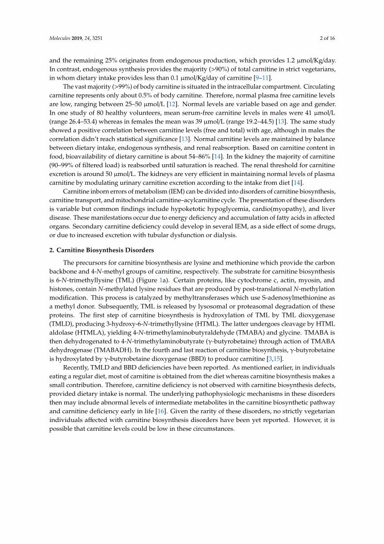
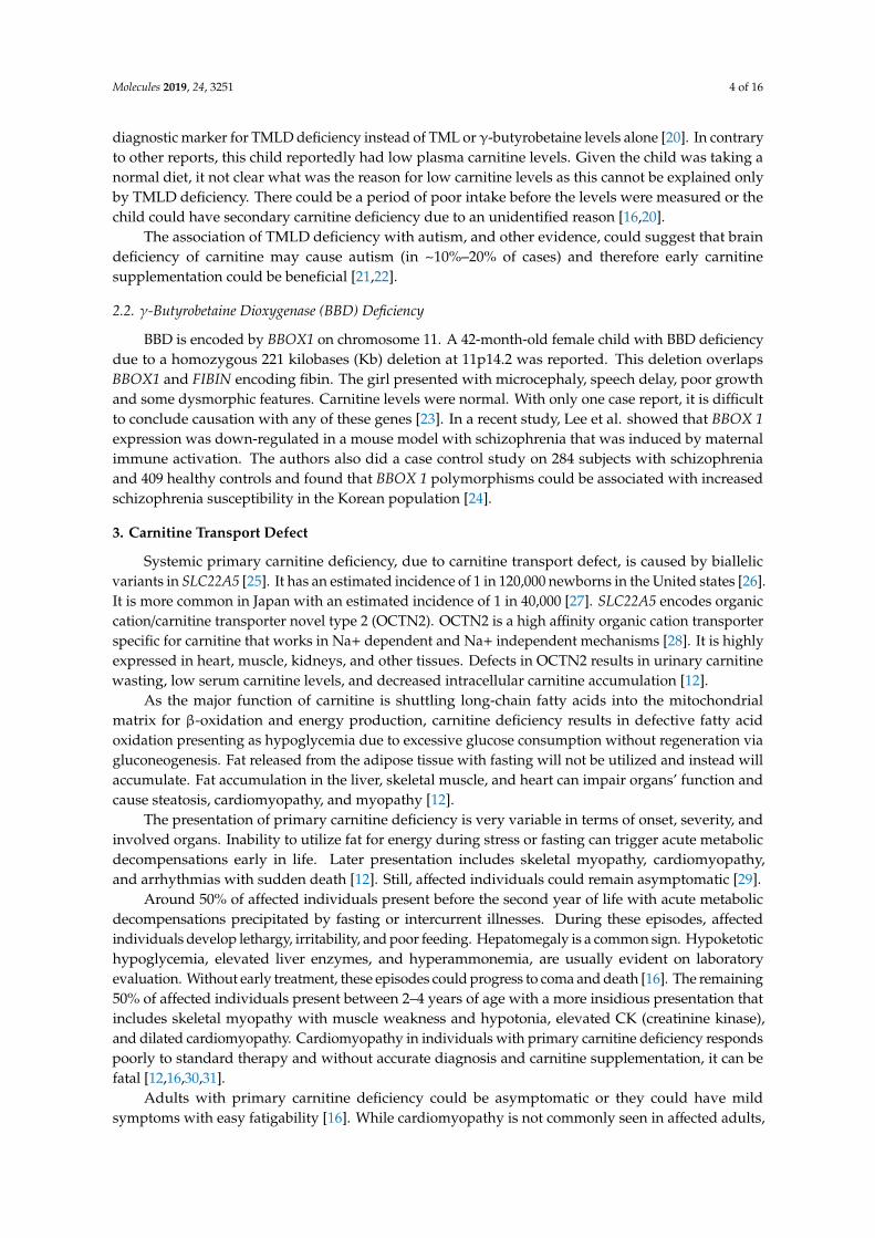
The precursors for carnitine biosynthesis are lysine and methionine which provide the carbonbackbone and 4-N-methyl groups of carnitine, respectively. The substrate for carnitine biosynthesisis 6-N-trimethyllysine (TML) (Figure 1a). Certain proteins, like cytochrome c, actin, myosin, andhistones, contain N-methylated lysine residues that are produced by post-translational N-methylationmodification. This process is catalyzed by methyltransferases which use S-adenosylmethionine asa methyl donor. Subsequently, TML is released by lysosomal or proteasomal degradation of theseproteins. The first step of carnitine biosynthesis is hydroxylation of TML by TML dioxygenase(TMLD), producing 3-hydroxy-6-N-trimethyllysine (HTML). The latter undergoes cleavage by HTMLaldolase (HTMLA), yielding 4-N-trimethylaminobutyraldehyde (TMABA) and glycine. TMABA isthen dehydrogenated to 4-N-trimethylaminobutyrate (γ-butyrobetaine) through action of TMABAdehydrogenase (TMABADH). In the fourth and last reaction of carnitine biosynthesis, γ-butyrobetaineis hydroxylated by γ-butyrobetaine dioxygenase (BBD) to produce carnitine [3,15].
Recently, TMLD and BBD deficiencies have been reported. As mentioned earlier, in individualseating a regular diet, most of carnitine is obtained from the diet whereas carnitine biosynthesis makes asmall contribution. Therefore, carnitine deficiency is not observed with carnitine biosynthesis defects,provided dietary intake is normal. The underlying pathophysiologic mechanisms in these disordersthen may include abnormal levels of intermediate metabolites in the carnitine biosynthetic pathwayand carnitine deficiency early in life [16]. Given the rarity of these disorders, no strictly vegetarianindividuals affected with carnitine biosynthesis disorders have been yet reported. However, it ispossible that carnitine levels could be low in these circumstances.
Molecules 2019, 24, 3251 3 of 16Molecules 2019, 24, x FOR PEER REVIEW 3 of 17
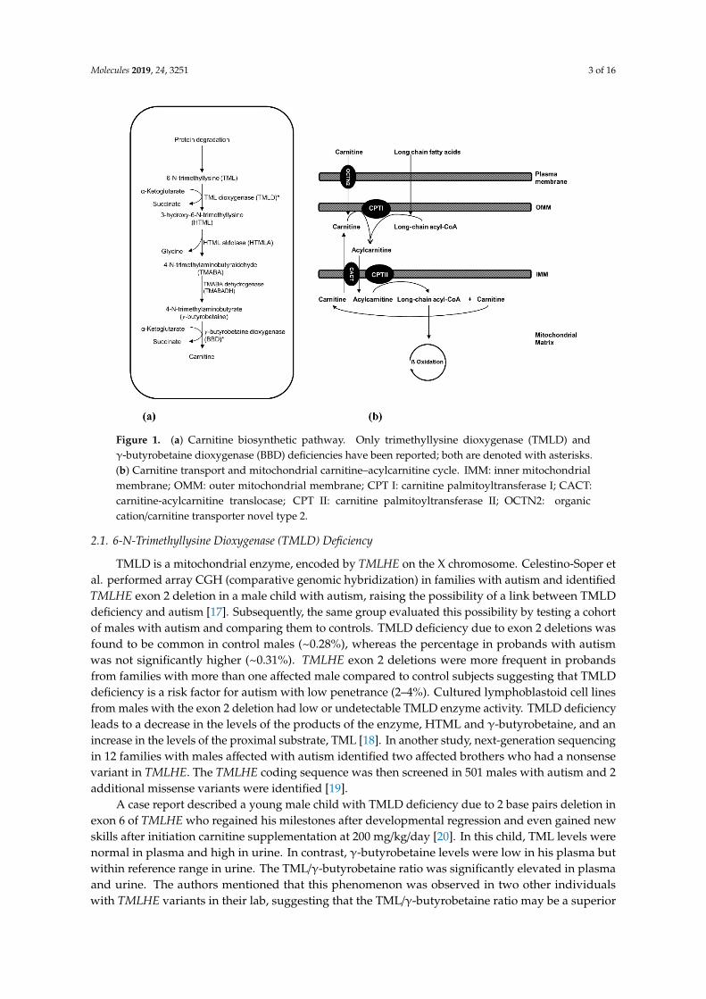
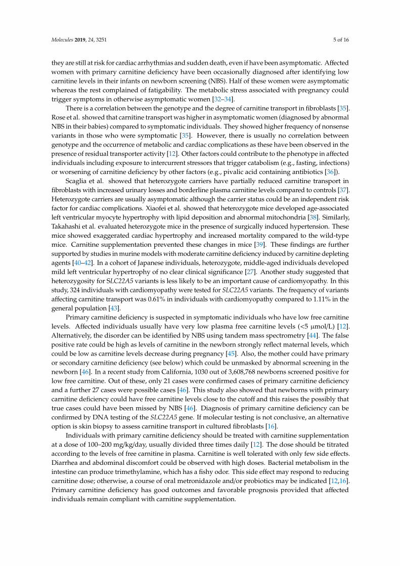
Figure 1. (a) Carnitine biosynthetic pathway. Only trimethyllysine dioxygenase (TMLD) and γ-butyrobetaine dioxygenase (BBD) deficiencies have been reported; both are denoted with asterisks. (b) Carnitine transport and mitochondrial carnitine–acylcarnitine cycle. IMM: inner mitochondrial membrane; OMM: outer mitochondrial membrane; CPT I: carnitine palmitoyltransferase I; CACT: carnitine-acylcarnitine translocase; CPT II: carnitine palmitoyltransferase II; OCTN2: organic cation/carnitine transporter novel type 2.
2.1. 6-N-Trimethyllysine Dioxygenase (TMLD) Deficiency
TMLD is a mitochondrial enzyme, encoded by TMLHE on the X chromosome. Celestino-Soper et al. performed array CGH (comparative genomic hybridization) in families with autism and identified TMLHE exon 2 deletion in a male child with autism, raising the possibility of a link between TMLD deficiency and autism [17]. Subsequently, the same group evaluated this possibility by testing a cohort of males with autism and comparing them to controls. TMLD deficiency due to exon 2 deletions was found to be common in control males (~0.28%), whereas the percentage in probands with autism was not significantly higher (~0.31%). TMLHE exon 2 deletions were more frequent in probands from families with more than one affected male compared to control subjects suggesting that TMLD deficiency is a risk factor for autism with low penetrance (2%–4%). Cultured lymphoblastoid cell lines from males with the exon 2 deletion had low or undetectable TMLD enzyme activity. TMLD deficiency leads to a decrease in the levels of the products of the enzyme, HTML and γ-butyrobetaine, and an increase in the levels of the proximal substrate, TML [18]. In another study, next-generation sequencing in 12 families with males affected with autism identified two affected brothers who had a nonsense variant in TMLHE. The TMLHE coding sequence was then screened in 501 males with autism and 2 additional missense variants were identified [19].
A case report described a young male child with TMLD deficiency due to 2 base pairs deletion in exon 6 of TMLHE who regained his milestones after developmental regression and even gained new skills after initiation carnitine supplementation at 200 mg/kg/day [20]. In this child, TML levels were normal in plasma and high in urine. In contrast, γ-butyrobetaine levels were low in his plasma but within reference range in urine. The TML/γ-butyrobetaine ratio was significantly elevated in plasma and urine. The authors mentioned that this phenomenon was observed in two other individuals with TMLHE variants in their lab, suggesting that the TML/γ-butyrobetaine ratio may be
Figure 1. (a) Carnitine biosynthetic pathway. Only trimethyllysine dioxygenase (TMLD) andγ-butyrobetaine dioxygenase (BBD) deficiencies have been reported; both are denoted with asterisks.(b) Carnitine transport and mitochondrial carnitine–acylcarnitine cycle. IMM: inner mitochondrialmembrane; OMM: outer mitochondrial membrane; CPT I: carnitine palmitoyltransferase I; CACT:carnitine-acylcarnitine translocase; CPT II: carnitine palmitoyltransferase II; OCTN2: organiccation/carnitine transporter novel type 2.
2.1. 6-N-Trimethyllysine Dioxygenase (TMLD) Deficiency
TMLD is a mitochondrial enzyme, encoded by TMLHE on the X chromosome. Celestino-Soper etal. performed array CGH (comparative genomic hybridization) in families with autism and identifiedTMLHE exon 2 deletion in a male child with autism, raising the possibility of a link between TMLDdeficiency and autism [17]. Subsequently, the same group evaluated this possibility by testing a cohortof males with autism and comparing them to controls. TMLD deficiency due to exon 2 deletions wasfound to be common in control males (~0.28%), whereas the percentage in probands with autismwas not significantly higher (~0.31%). TMLHE exon 2 deletions were more frequent in probandsfrom families with more than one affected male compared to control subjects suggesting that TMLDdeficiency is a risk factor for autism with low penetrance (2–4%). Cultured lymphoblastoid cell linesfrom males with the exon 2 deletion had low or undetectable TMLD enzyme activity. TMLD deficiencyleads to a decrease in the levels of the products of the enzyme, HTML and γ-butyrobetaine, and anincrease in the levels of the proximal substrate, TML [18]. In another study, next-generation sequencingin 12 families with males affected with autism identified two affected brothers who had a nonsensevariant in TMLHE. The TMLHE coding sequence was then screened in 501 males with autism and 2additional missense variants were identified [19].
A case report described a young male child with TMLD deficiency due to 2 base pairs deletion inexon 6 of TMLHE who regained his milestones after developmental regression and even gained newskills after initiation carnitine supplementation at 200 mg/kg/day [20]. In this child, TML levels werenormal in plasma and high in urine. In contrast, γ-butyrobetaine levels were low in his plasma butwithin reference range in urine. The TML/γ-butyrobetaine ratio was significantly elevated in plasmaand urine. The authors mentioned that this phenomenon was observed in two other individualswith TMLHE variants in their lab, suggesting that the TML/γ-butyrobetaine ratio may be a superior
Molecules 2019, 24, 3251 4 of 16
diagnostic marker for TMLD deficiency instead of TML or γ-butyrobetaine levels alone [20]. In contraryto other reports, this child reportedly had low plasma carnitine levels. Given the child was taking anormal diet, it not clear what was the reason for low carnitine levels as this cannot be explained onlyby TMLD deficiency. There could be a period of poor intake before the levels were measured or thechild could have secondary carnitine deficiency due to an unidentified reason [16,20].
The association of TMLD deficiency with autism, and other evidence, could suggest that braindeficiency of carnitine may cause autism (in ~10%–20% of cases) and therefore early carnitinesupplementation could be beneficial [21,22].
2.2. γ-Butyrobetaine Dioxygenase (BBD) Deficiency
BBD is encoded by BBOX1 on chromosome 11. A 42-month-old female child with BBD deficiencydue to a homozygous 221 kilobases (Kb) deletion at 11p14.2 was reported. This deletion overlapsBBOX1 and FIBIN encoding fibin. The girl presented with microcephaly, speech delay, poor growthand some dysmorphic features. Carnitine levels were normal. With only one case report, it is difficultto conclude causation with any of these genes [23]. In a recent study, Lee et al. showed that BBOX 1expression was down-regulated in a mouse model with schizophrenia that was induced by maternalimmune activation. The authors also did a case control study on 284 subjects with schizophreniaand 409 healthy controls and found that BBOX 1 polymorphisms could be associated with increasedschizophrenia susceptibility in the Korean population [24].
3. Carnitine Transport Defect
Systemic primary carnitine deficiency, due to carnitine transport defect, is caused by biallelicvariants in SLC22A5 [25]. It has an estimated incidence of 1 in 120,000 newborns in the United states [26].It is more common in Japan with an estimated incidence of 1 in 40,000 [27]. SLC22A5 encodes organiccation/carnitine transporter novel type 2 (OCTN2). OCTN2 is a high affinity organic cation transporterspecific for carnitine that works in Na+ dependent and Na+ independent mechanisms [28]. It is highlyexpressed in heart, muscle, kidneys, and other tissues. Defects in OCTN2 results in urinary carnitinewasting, low serum carnitine levels, and decreased intracellular carnitine accumulation [12].
As the major function of carnitine is shuttling long-chain fatty acids into the mitochondrialmatrix for β-oxidation and energy production, carnitine deficiency results in defective fatty acidoxidation presenting as hypoglycemia due to excessive glucose consumption without regeneration viagluconeogenesis. Fat released from the adipose tissue with fasting will not be utilized and instead willaccumulate. Fat accumulation in the liver, skeletal muscle, and heart can impair organs’ function andcause steatosis, cardiomyopathy, and myopathy [12].
The presentation of primary carnitine deficiency is very variable in terms of onset, severity, andinvolved organs. Inability to utilize fat for energy during stress or fasting can trigger acute metabolicdecompensations early in life. Later presentation includes skeletal myopathy, cardiomyopathy,and arrhythmias with sudden death [12]. Still, affected individuals could remain asymptomatic [29].
Around 50% of affected individuals present before the second year of life with acute metabolicdecompensations precipitated by fasting or intercurrent illnesses. During these episodes, affectedindividuals develop lethargy, irritability, and poor feeding. Hepatomegaly is a common sign. Hypoketotichypoglycemia, elevated liver enzymes, and hyperammonemia, are usually evident on laboratoryevaluation. Without early treatment, these episodes could progress to coma and death [16]. The remaining50% of affected individuals present between 2–4 years of age with a more insidious presentation thatincludes skeletal myopathy with muscle weakness and hypotonia, elevated CK (creatinine kinase),and dilated cardiomyopathy. Cardiomyopathy in individuals with primary carnitine deficiency respondspoorly to standard therapy and without accurate diagnosis and carnitine supplementation, it can befatal [12,16,30,31].
Adults with primary carnitine deficiency could be asymptomatic or they could have mildsymptoms with easy fatigability [16]. While cardiomyopathy is not commonly seen in affected adults,
Molecules 2019, 24, 3251 5 of 16
they are still at risk for cardiac arrhythmias and sudden death, even if have been asymptomatic. Affectedwomen with primary carnitine deficiency have been occasionally diagnosed after identifying lowcarnitine levels in their infants on newborn screening (NBS). Half of these women were asymptomaticwhereas the rest complained of fatigability. The metabolic stress associated with pregnancy couldtrigger symptoms in otherwise asymptomatic women [32–34].
There is a correlation between the genotype and the degree of carnitine transport in fibroblasts [35].Rose et al. showed that carnitine transport was higher in asymptomatic women (diagnosed by abnormalNBS in their babies) compared to symptomatic individuals. They showed higher frequency of nonsensevariants in those who were symptomatic [35]. However, there is usually no correlation betweengenotype and the occurrence of metabolic and cardiac complications as these have been observed in thepresence of residual transporter activity [12]. Other factors could contribute to the phenotype in affectedindividuals including exposure to intercurrent stressors that trigger catabolism (e.g., fasting, infections)or worsening of carnitine deficiency by other factors (e.g., pivalic acid containing antibiotics [36]).
Scaglia et al. showed that heterozygote carriers have partially reduced carnitine transport infibroblasts with increased urinary losses and borderline plasma carnitine levels compared to controls [37].Heterozygote carriers are usually asymptomatic although the carrier status could be an independent riskfactor for cardiac complications. Xiaofei et al. showed that heterozygote mice developed age-associatedleft ventricular myocyte hypertrophy with lipid deposition and abnormal mitochondria [38]. Similarly,Takahashi et al. evaluated heterozygote mice in the presence of surgically induced hypertension. Thesemice showed exaggerated cardiac hypertrophy and increased mortality compared to the wild-typemice. Carnitine supplementation prevented these changes in mice [39]. These findings are furthersupported by studies in murine models with moderate carnitine deficiency induced by carnitine depletingagents [40–42]. In a cohort of Japanese individuals, heterozygote, middle-aged individuals developedmild left ventricular hypertrophy of no clear clinical significance [27]. Another study suggested thatheterozygosity for SLC22A5 variants is less likely to be an important cause of cardiomyopathy. In thisstudy, 324 individuals with cardiomyopathy were tested for SLC22A5 variants. The frequency of variantsaffecting carnitine transport was 0.61% in individuals with cardiomyopathy compared to 1.11% in thegeneral population [43].
Primary carnitine deficiency is suspected in symptomatic individuals who have low free carnitinelevels. Affected individuals usually have very low plasma free carnitine levels (<5 µmol/L) [12].Alternatively, the disorder can be identified by NBS using tandem mass spectrometry [44]. The falsepositive rate could be high as levels of carnitine in the newborn strongly reflect maternal levels, whichcould be low as carnitine levels decrease during pregnancy [45]. Also, the mother could have primaryor secondary carnitine deficiency (see below) which could be unmasked by abnormal screening in thenewborn [46]. In a recent study from California, 1030 out of 3,608,768 newborns screened positive forlow free carnitine. Out of these, only 21 cases were confirmed cases of primary carnitine deficiencyand a further 27 cases were possible cases [46]. This study also showed that newborns with primarycarnitine deficiency could have free carnitine levels close to the cutoff and this raises the possibly thattrue cases could have been missed by NBS [46]. Diagnosis of primary carnitine deficiency can beconfirmed by DNA testing of the SLC22A5 gene. If molecular testing is not conclusive, an alternativeoption is skin biopsy to assess carnitine transport in cultured fibroblasts [16].
Individuals with primary carnitine deficiency should be treated with carnitine supplementationat a dose of 100–200 mg/kg/day, usually divided three times daily [12]. The dose should be titratedaccording to the levels of free carnitine in plasma. Carnitine is well tolerated with only few side effects.Diarrhea and abdominal discomfort could be observed with high doses. Bacterial metabolism in theintestine can produce trimethylamine, which has a fishy odor. This side effect may respond to reducingcarnitine dose; otherwise, a course of oral metronidazole and/or probiotics may be indicated [12,16].Primary carnitine deficiency has good outcomes and favorable prognosis provided that affectedindividuals remain compliant with carnitine supplementation.
Molecules 2019, 24, 3251 6 of 16
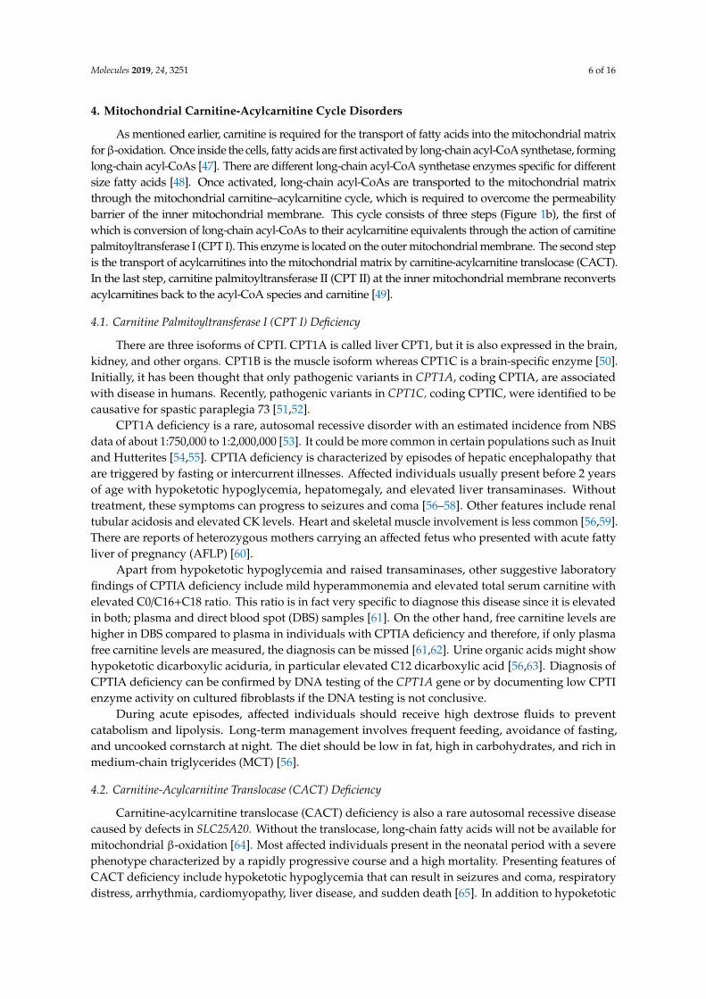
4. Mitochondrial Carnitine-Acylcarnitine Cycle Disorders
As mentioned earlier, carnitine is required for the transport of fatty acids into the mitochondrial matrixforβ-oxidation. Once inside the cells, fatty acids are first activated by long-chain acyl-CoA synthetase, forminglong-chain acyl-CoAs [47]. There are different long-chain acyl-CoA synthetase enzymes specific for differentsize fatty acids [48]. Once activated, long-chain acyl-CoAs are transported to the mitochondrial matrixthrough the mitochondrial carnitine–acylcarnitine cycle, which is required to overcome the permeabilitybarrier of the inner mitochondrial membrane. This cycle consists of three steps (Figure 1b), the first ofwhich is conversion of long-chain acyl-CoAs to their acylcarnitine equivalents through the action of carnitinepalmitoyltransferase I (CPT I). This enzyme is located on the outer mitochondrial membrane. The second stepis the transport of acylcarnitines into the mitochondrial matrix by carnitine-acylcarnitine translocase (CACT).In the last step, carnitine palmitoyltransferase II (CPT II) at the inner mitochondrial membrane reconvertsacylcarnitines back to the acyl-CoA species and carnitine [49].
4.1. Carnitine Palmitoyltransferase I (CPT I) Deficiency
There are three isoforms of CPTI. CPT1A is called liver CPT1, but it is also expressed in the brain,kidney, and other organs. CPT1B is the muscle isoform whereas CPT1C is a brain-specific enzyme [50].Initially, it has been thought that only pathogenic variants in CPT1A, coding CPTIA, are associatedwith disease in humans. Recently, pathogenic variants in CPT1C, coding CPTIC, were identified to becausative for spastic paraplegia 73 [51,52].
CPT1A deficiency is a rare, autosomal recessive disorder with an estimated incidence from NBSdata of about 1:750,000 to 1:2,000,000 [53]. It could be more common in certain populations such as Inuitand Hutterites [54,55]. CPTIA deficiency is characterized by episodes of hepatic encephalopathy thatare triggered by fasting or intercurrent illnesses. Affected individuals usually present before 2 yearsof age with hypoketotic hypoglycemia, hepatomegaly, and elevated liver transaminases. Withouttreatment, these symptoms can progress to seizures and coma [56–58]. Other features include renaltubular acidosis and elevated CK levels. Heart and skeletal muscle involvement is less common [56,59].There are reports of heterozygous mothers carrying an affected fetus who presented with acute fattyliver of pregnancy (AFLP) [60].
Apart from hypoketotic hypoglycemia and raised transaminases, other suggestive laboratoryfindings of CPTIA deficiency include mild hyperammonemia and elevated total serum carnitine withelevated C0/C16+C18 ratio. This ratio is in fact very specific to diagnose this disease since it is elevatedin both; plasma and direct blood spot (DBS) samples [61]. On the other hand, free carnitine levels arehigher in DBS compared to plasma in individuals with CPTIA deficiency and therefore, if only plasmafree carnitine levels are measured, the diagnosis can be missed [61,62]. Urine organic acids might showhypoketotic dicarboxylic aciduria, in particular elevated C12 dicarboxylic acid [56,63]. Diagnosis ofCPTIA deficiency can be confirmed by DNA testing of the CPT1A gene or by documenting low CPTIenzyme activity on cultured fibroblasts if the DNA testing is not conclusive.
During acute episodes, affected individuals should receive high dextrose fluids to preventcatabolism and lipolysis. Long-term management involves frequent feeding, avoidance of fasting,and uncooked cornstarch at night. The diet should be low in fat, high in carbohydrates, and rich inmedium-chain triglycerides (MCT) [56].
4.2. Carnitine-Acylcarnitine Translocase (CACT) Deficiency
Carnitine-acylcarnitine translocase (CACT) deficiency is also a rare autosomal recessive diseasecaused by defects in SLC25A20. Without the translocase, long-chain fatty acids will not be available formitochondrial β-oxidation [64]. Most affected individuals present in the neonatal period with a severephenotype characterized by a rapidly progressive course and a high mortality. Presenting features ofCACT deficiency include hypoketotic hypoglycemia that can result in seizures and coma, respiratorydistress, arrhythmia, cardiomyopathy, liver disease, and sudden death [65]. In addition to hypoketotic
Molecules 2019, 24, 3251 7 of 16
hypoglycemia, other laboratory abnormalities include hyperammonemia, elevated liver enzymes,and elevated CK levels. Late onset presentation with a milder phenotype has been reported but lesscommonly [66,67]. This phenotype presents in the form of episodes of hypoketotic hypoglycemia thatare triggered by fasting or intercurrent illnesses. Milder phenotype is associated with some degree ofresidual enzyme activity [66].
CACT deficiency is diagnosed based on elevated levels of long-chain fatty acid acylcarnitine esters(especially C16 and C18:1) along with low free carnitine [65]. This biochemical pattern is indistinguishablefrom CPT II deficiency (see below) and therefore, either DNA testing or measuring enzyme activity incultured fibroblasts is required to confirm the diagnosis. Urine organic acids can reveal non-specificdicarboxylic aciduria.
Treatment involves avoidance of fasting and frequent meals. The diet should be rich in carbohydratesand low in fat. Most fat intake should come from MCT. Carnitine is commonly used, but it is controversial.There have been concerns about the possible toxicity of acylcarnitine accumulation in long chain fattyacid oxidation disorders (see Section 6).
4.3. Carnitine Palmitoyltransferase II (CPT II) Deficiency
CPT II deficiency is an autosomal recessive disorder caused by biallelic pathogenic variants in CPT2.The presentation is variable in regard to age of onset, severity, and involved organs. Three phenotypesare described including, myopathic form, lethal neonatal form, and severe infantile form [68].
By far, the myopathic form is the most common form of CPT II deficiency and it is a common causefor hereditary rhabdomyolysis [69]. CPT II deficiency is considered as the most common disorder oflipid metabolism affecting skeletal muscles [68]. The myopathic form of CPT II deficiency can presentat any age with 70% of cases presented during childhood (0–12 years of age). In one quarter of cases,the disease presented during adolescence (13–22 years) [70,71]. The myopathic form is characterizedby recurrent attacks of rhabdomyolysis that are triggered by prolonged exercise in the majority ofcases. Other triggers include infections and fasting [71]. During these episodes, myalgia is the mostconsistent symptom, and it can be associated with muscles weakness and myoglobinuria [68,70,71],and CK levels are elevated. Episodes of rhabdomyolysis can lead to acute renal failure in 7–23% ofcases [71]. The myopathic form of CPT II deficiency is more commonly observed in males, although it isan autosomal recessive disorder with expected equal gender distribution [72]. This observation couldbe due to ascertainment bias as males are more likely to do strenuous exercise and therefore developmyoglobinuria. Hormonal factors were also suggested to be involved in this gender bias [72,73].
The other two forms of CPT II deficiency are very rare. Affected individuals with the neonatalform present soon after birth (hours to a few days) with lethargy, respiratory distress, hypoketotichypoglycemia, liver failure, cardiac arrhythmias, cardiomyopathy, seizures, and coma [74,75]. Affectedneonates could have dysmorphic features, renal dysgenesis, neuronal migration defects, and otherbrain malformations [68,76]. The prognosis is poor, with death occurring within days to months [68].
The severe infantile form usually presents between 6–24 months of age [57]. It is characterizedby hypoketotic hypoglycemia, hepatomegaly, liver failure, cardiomyopathy, and cardiac arrhythmias.Seizures and coma could develop secondary to hypoglycemia [57,68,77]. Episodes of decompensationcould be triggered by fasting or intercurrent illnesses. Cardiac arrhythmias can result in suddendeath [75]. In both neonatal and infantile forms, laboratory evaluation usually shows hyperammonemia,metabolic acidosis, hypoketotic hypoglycemia, and high CK levels.
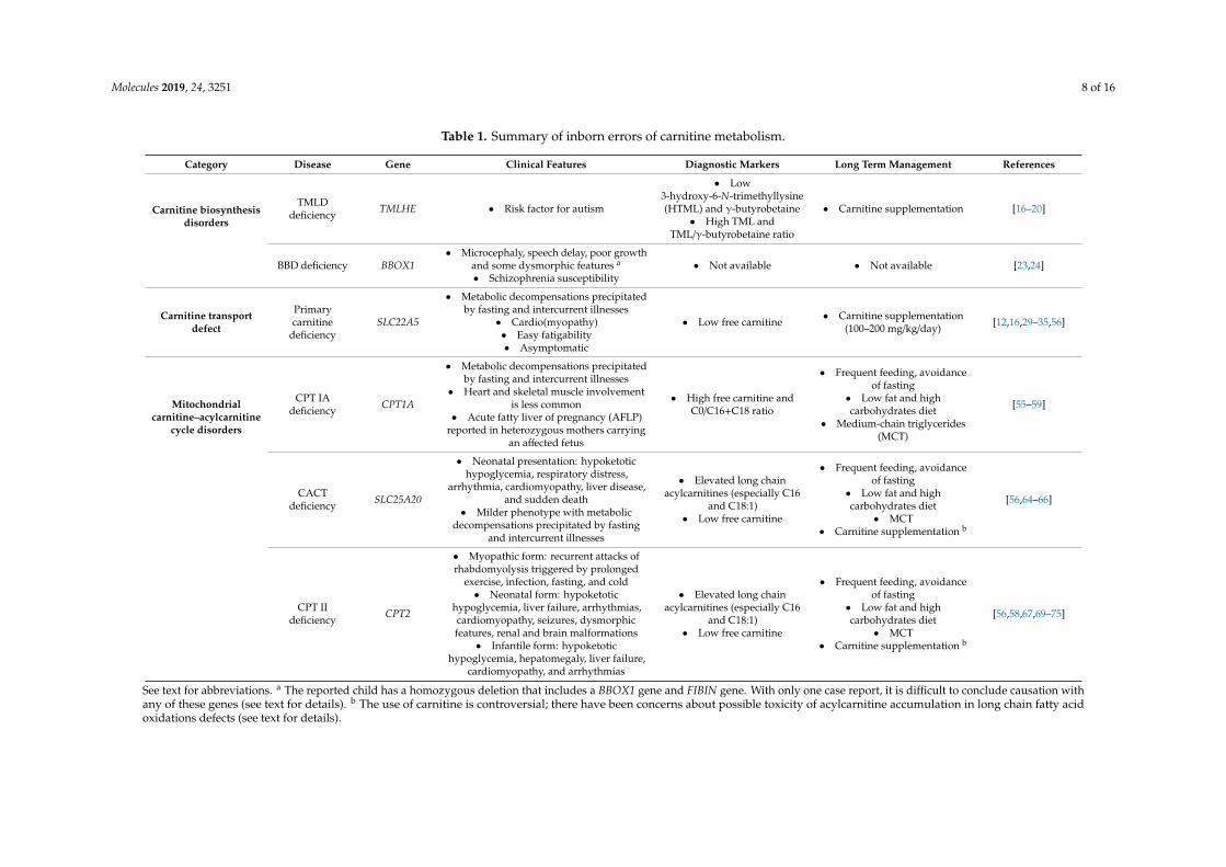
CPT II deficiency is diagnosed based on elevation of C12 to C18 acylcarnitines, in particular C16and C18:1 [68]. Free carnitine levels could be reduced. (C16+C18:1)/C2 ratio is a more sensitive markerand could improve the sensitivity of the NBS [78]. It should be noted that CPT II deficiency is betterdiagnosed from plasma samples compared to DBS [61]. Diagnosis can be confirmed by DNA testing orenzyme assays in fibroblast. Treatment principles are the same as discussed before for CACT deficiency.Prolonged exercise and other known triggers should be avoided [57,68]. Table 1 summarizes carnitineinborn errors of metabolism.
Molecules 2019, 24, 3251 8 of 16
Table 1. Summary of inborn errors of carnitine metabolism.
Category Disease Gene Clinical Features Diagnostic Markers Long Term Management References
Carnitine biosynthesisdisorders
TMLDdeficiency TMLHE • Risk factor for autism
• Low3-hydroxy-6-N-trimethyllysine(HTML) and γ-butyrobetaine
• High TML andTML/γ-butyrobetaine ratio
• Carnitine supplementation [16–20]
BBD deficiency BBOX1• Microcephaly, speech delay, poor growth
and some dysmorphic features a
• Schizophrenia susceptibility• Not available • Not available [23,24]
Carnitine transportdefect
Primarycarnitine
deficiencySLC22A5
• Metabolic decompensations precipitatedby fasting and intercurrent illnesses
• Cardio(myopathy)• Easy fatigability• Asymptomatic
• Low free carnitine • Carnitine supplementation(100–200 mg/kg/day) [12,16,29–35,56]
Mitochondrialcarnitine–acylcarnitine
cycle disorders
CPT IAdeficiency CPT1A
• Metabolic decompensations precipitatedby fasting and intercurrent illnesses
• Heart and skeletal muscle involvementis less common
• Acute fatty liver of pregnancy (AFLP)reported in heterozygous mothers carrying
an affected fetus
• High free carnitine andC0/C16+C18 ratio
• Frequent feeding, avoidanceof fasting
• Low fat and highcarbohydrates diet
• Medium-chain triglycerides(MCT)
[55–59]
CACTdeficiency SLC25A20
• Neonatal presentation: hypoketotichypoglycemia, respiratory distress,
arrhythmia, cardiomyopathy, liver disease,and sudden death
• Milder phenotype with metabolicdecompensations precipitated by fasting
and intercurrent illnesses
• Elevated long chainacylcarnitines (especially C16
and C18:1)• Low free carnitine
• Frequent feeding, avoidanceof fasting
• Low fat and highcarbohydrates diet
• MCT• Carnitine supplementation b
[56,64–66]
CPT IIdeficiency CPT2
• Myopathic form: recurrent attacks ofrhabdomyolysis triggered by prolonged
exercise, infection, fasting, and cold• Neonatal form: hypoketotic
hypoglycemia, liver failure, arrhythmias,cardiomyopathy, seizures, dysmorphicfeatures, renal and brain malformations
• Infantile form: hypoketotichypoglycemia, hepatomegaly, liver failure,
cardiomyopathy, and arrhythmias
• Elevated long chainacylcarnitines (especially C16
and C18:1)• Low free carnitine
• Frequent feeding, avoidanceof fasting
• Low fat and highcarbohydrates diet
• MCT• Carnitine supplementation b
[56,58,67,69–75]
See text for abbreviations. a The reported child has a homozygous deletion that includes a BBOX1 gene and FIBIN gene. With only one case report, it is difficult to conclude causation withany of these genes (see text for details). b The use of carnitine is controversial; there have been concerns about possible toxicity of acylcarnitine accumulation in long chain fatty acidoxidations defects (see text for details).
Molecules 2019, 24, 3251 9 of 16
4.4. Carnitine Palmitoyltransferase (CPT) Inhibitors
While the mitochondrial carnitine–acylcarnitine cycle is essential for fatty acid oxidation andenergy supply, modulation of these processes, through CPT inhibitors, could be of a potential therapeuticvalue in several disorders, including diabetes, cancer, and heart diseases [79]. For example, in type 2diabetes, the underlying reduced insulin sensitivity causes increased fatty acid oxidation which byitself aggravates the hyperglycemia by reducing glucose utilization and increasing production throughgluconeogenesis [80]. Selective inhibition of liver CPTI reduces gluconeogenesis and improves glucosehomeostasis [81]. In the diseased heart, there is an increase in fatty acid oxidation and a decrease inglucose consumption, which in turn impair energy utilization and decrease cardiac efficiency [82].Lionetti et al. showed that CPTI inhibition in dogs with heart failure delayed the time to end-stagefailure [83]. Etomoxir, an irreversible inhibitor of CPTI, was found to prevent the development ofheart failure in rats with pressure overload-induced cardiac hypertrophy [84]. Placebo-controlled trialin humans was stopped prematurely due to hepatotoxicity, although it showed trends for improvedcardiac function in treated subjects [85]. Finally, through inhibition of fatty acid oxidation which fuelsthe tumor cells, and other mechanisms like apoptosis and modulating gene expression, CPT inhibitioncould be a potential target in cancer [86].
5. Secondary Carnitine Deficiency
Apart from primary carnitine deficiency, which is caused by carnitine transport defect, secondarycarnitine deficiency could also develop due to a variety of reasons [87]. As compared to the primaryform, secondary carnitine deficiency is less severe and associated with higher carnitine levels that canbe treated with small doses of carnitine [16]. Carnitine deficiency is seen in several IEM, in particular,fatty acid oxidation disorders and organic acidemias. In these disorders, carnitine deficiencydevelops secondary to increased urinary excretion of carnitine in the form of acylcarnitines [88].It is uncommon to develop carnitine deficiency secondary to poor intake as carnitine biosynthesis alongwith renal reabsorption are effective in maintaining normal carnitine levels [89]. Still, malnutritionand malabsorption can cause carnitine deficiency. An adult individual with severe malnutritionand history of gastric bypass surgery developed hyperammonemia and was found to have very lowcarnitine levels. Hyperammonemia was refractory to standard therapy but normalized with carnitinesupplementation [90]. Children may be more prone to dietary carnitine deficiency [89]. In particular,preterm infants are born with limited carnitine reserves as placental transfer and biosynthesis ofcarnitine occurs mainly during the third trimester. They also have limited ability to synthesize carnitinedue to immaturity of biosynthetic enzymes [91]. Therefore, carnitine levels may need to be monitoredin those neonates, especially with prolonged use of total parental nutrition [92]. However, the evidencefor routine administration of carnitine in preterm infants is lacking and more studies are needed [93,94].
Valproic acid (VPA) is one of the well-known drugs causing secondary carnitine deficiency, whichcan contribute to VPA-induced toxicity [95]. VPA depletes carnitine through different mechanismsincluding increased urinary excretion in the form valproylcarnitine, reduction in tubular reabsorptionand endogenous biosynthesis, and inhibition of the carnitine transporter [95]. Pivalic acid containingantibiotics can also cause carnitine depletion as pivalate conjugates with free carnitine, forming pivaloylcarnitine, which is excreted in the urine [36,96]. Several drugs can cause secondary carnitine deficiencythough OCTN2 inhibition including omeprazole [97], zwitterionic drugs (e.g., levofloxacin) [98],and anticancer drugs (e.g., etoposide, vinblastine, actinomycin D) [99].
Secondary carnitine deficiency could also develop from increased urinary loss in Fanconi syndromeor with renal replacement therapy (hemodialysis and peritoneal dialysis). As mentioned earlier,the kidneys are very efficient in maintaining carnitine homeostasis. Several mechanisms contribute tocarnitine depletion with chronic dialysis including efficient free carnitine removal through dialysis,decreased intake, and decreased endogenous production [100]. In individuals with end stage renaldisease, free carnitine levels are higher than healthy individuals prior to initiation of dialysis. Oncedialysis starts, this level declines significantly, about 30% in 4 weeks and 40% in 12 months [101].
Molecules 2019, 24, 3251 10 of 16
This is associated with increased acylcarnitine and acylcarnitine/free carnitine ratio. Dialysis alsoresults in reduction in muscle carnitine content [101]. Dialysis-related carnitine disorder (DLD)is the term used to describe carnitine deficiency in dialysis patients and can be associated withseveral complications including fatigue, intradialytic hypotension, anemia that is poorly responsiveto erythropoietin, and cardiomyopathy [102]. Many clinical trials described the benefits of carnitineto treat these complications although definite evidence is still lacking [103]. To attain desired effectson multiple pathways, higher than physiologic levels of carnitine should be achieved in the plasma,and therefore, in the intracellular compartment. This is possible in individuals with end stage renaldisease on dialysis who receive parenteral carnitine supplementation, given their underlying renaldisease will impair rapid clearance of carnitine when transporter saturation reached. In this way,carnitine acts as a “conditionally essential nutrient” [104].
6. Carnitine Use in Inborn Errors of Metabolism
Carnitine supplementation is commonly used in several IEM and other disorders. It is themainstay of treatment in primary carnitine deficiency. Carnitine is also used in other IEM that areassociated with secondary carnitine deficiency [105,106], although a Cochrane review concludedthat evidence for carnitine supplementation in IEM is lacking [107]. Moreover, there have beenconcerns about possible toxicity of acylcarnitine accumulation in long chain fatty acid oxidationdisorders. In one report, two siblings with very long-chain acyl-CoA dehydrogenase (VLCAD)deficiency developed more frequent episodes of rhabdomyolysis after carnitine supplementation [108].In a mouse model, accumulating long-chain acylcarnitines in ischemic heart mitochondria inhibitedoxidative phosphorylation [109]. Long chain acylcarnitines were found to regulate the hERG (humanether-a-go-go-related gene) channel and contribute to the development of cardiac arrhythmias [110].Based on this potential risk, the use of carnitine in long chain fatty acid oxidation disorders should beavoided during acute metabolic crises [111]. Carnitine is often used in patients with mitochondrialdisorders [112]. In these disorders, plasma free carnitine tends to be lower than normal with increasedesterified carnitine due to a partial impairment of β-oxidation [113]. Metabolism of carnitine throughgut microbiota produces trimethylamine N-oxide (TMAO). Marked elevation in plasma levels ofTMAO were documented in patients with mitochondrial disorders treated with oral carnitine [114].Recent studies showed a positive correlation between elevated levels of TMAO and an increasedrisk for cardiovascular disease [115]. Given this potential risk and lack of evidence for the benefit ofcarnitine in patients with mitochondrial disorders, more studies are needed to evaluate the long-termsafety and efficacy of carnitine in these patients.
7. Summery and Conclusions
Carnitine is involved in several biochemical pathways, either directly or indirectly. Carnitine isobtained from the diet, synthesized endogenously, and excreted in the urine. In normal situations,these processes are very efficient in maintaining normal carnitine levels. It is important to recognizeinborn errors of carnitine metabolism as early treatment could prevent serious sequelae. Disordersof carnitine biosynthesis were recently identified and are still to be fully elucidated. The associationof TMLD deficiency with autism is interesting and could help open venues to better understand thepathophysiology of autism, in a subset of patients. Other inborn errors of carnitine metabolism, carnitinetransport defect, and mitochondrial carnitine–acylcarnitine cycle disorders, have been recognized forlong time and a common theme in these disorders is energy failure with hypoketotic hypoglycemia,cardio(myopathy), and liver disease. These disorders are part of NBS programs in several areas of theworld. The myopathic CPTII deficiency is a common cause for hereditary rhabdomyolysis and shouldbe always considered with such presentations. Carnitine supplementation and the use of compoundsto modulate carnitine metabolizing enzymes, like CPT inhibitors and potentiators, have been the focusof many research projects. These agents could be promising targets to treat disorders characterized byalterations in physiologic biochemical pathways, especially those concerning with energy production.
Molecules 2019, 24, 3251 11 of 16
Author Contributions: M.A. (Mohammed Almannai) drafted the initial manuscript and reviewed and revised themanuscript. A.W.E.-H. and M.A. (Majid Alfadhel) reviewed and revised the manuscript. All authors approvedthe final manuscript as submitted.
Funding: This research received no external funding.
Conflicts of Interest: The authors declare no conflicts of interest.
References
1. Pietrocola, F.; Galluzzi, L.; Bravo-San Pedro, J.M.; Madeo, F.; Kroemer, G. Acetyl Coenzyme A: A CentralMetabolite and Second Messenger. Cell Metab. 2015, 21, 805–821. [CrossRef]
2. Bene, J.; Hadzsiev, K.; Melegh, B. Role of carnitine and its derivatives in the development and managementof type 2 diabetes. Nutr. Diabetes 2018, 8, 8. [CrossRef]
3. Vaz, F.M.; Wanders, R.J.A. Carnitine biosynthesis in mammals. Biochem. J. 2002, 361, 417–429. [CrossRef]4. Carter, A.L.; Abney, T.O.; Lapp, D.F. Biosynthesis and metabolism of carnitine. J. Child Neurol. 1995, 10
(Suppl. 2), S3–S7. [CrossRef]5. Ribas, G.S.; Vargas, C.R.; Wajner, M. L-carnitine supplementation as a potential antioxidant therapy for
inherited neurometabolic disorders. Gene 2014, 533, 469–476. [CrossRef]6. Lee, B.-J.; Lin, J.-S.; Lin, Y.-C.; Lin, P.-T. Effects of L-carnitine supplementation on oxidative stress and
antioxidant enzymes activities in patients with coronary artery disease: A randomized, placebo-controlledtrial. Nutr. J. 2014, 13, 79. [CrossRef]
7. Komlósi, K.; Havasi, V.; Bene, J.; Süle, N.; Pajor, L.; Nicolai, R.; Benatti, P.; Calvani, M.; Melegh, B.Histopathologic abnormalities of the lymphoreticular tissues in organic cation transporter 2 deficiency:Evidence for impaired B cell maturation. J. Pediatr. 2007, 150, 109–111.e2. [CrossRef]
8. Xu, Y.; Jiang, W.; Chen, G.; Zhu, W.; Ding, W.; Ge, Z.; Tan, Y.; Ma, T.; Cui, G. L-carnitine treatment of insulinresistance: A systematic review and meta-analysis. Adv. Clin. Exp. Med. Off. Organ Wroclaw Med. Univ.2017, 26, 333–338.
9. Rebouche, C.J. Carnitine function and requirements during the life cycle. FASEB J. Off. Publ. Fed. Am. Soc.Exp. Biol. 1992, 6, 3379–3386.
10. Stanley, C.A. Carnitine deficiency disorders in children. Ann. N. Y. Acad. Sci. 2004, 1033, 42–51. [CrossRef]11. Tein, I.; Bukovac, S.W.; Xie, Z.W. Characterization of the human plasmalemmal carnitine transporter in
cultured skin fibroblasts. Arch. Biochem. Biophys. 1996, 329, 145–155. [CrossRef]12. Longo, N.; Frigeni, M.; Pasquali, M. Carnitine transport and fatty acid oxidation. Biochim. Biophys. Acta 2016,
1863, 2422–2435. [CrossRef]13. Opalka, J.R.; Gellerich, F.N.; Zierz, S. Age and sex dependency of carnitine concentration in human serum
and skeletal muscle. Clin. Chem. 2001, 47, 2150–2153.14. Rebouche, C.J. Kinetics, pharmacokinetics, and regulation of L-carnitine and acetyl-L-carnitine metabolism.
Ann. N. Y. Acad. Sci. 2004, 1033, 30–41. [CrossRef]15. Strijbis, K.; Vaz, F.M.; Distel, B. Enzymology of the carnitine biosynthesis pathway. IUBMB Life 2010, 62,
357–362. [CrossRef]16. El-Hattab, A.W.; Scaglia, F. Disorders of carnitine biosynthesis and transport. Mol. Genet. Metab. 2015, 116,
107–112. [CrossRef]17. Celestino-Soper, P.B.S.; Shaw, C.A.; Sanders, S.J.; Li, J.; Murtha, M.T.; Ercan-Sencicek, A.G.; Davis, L.;
Thomson, S.; Gambin, T.; Chinault, A.C.; et al. Use of array CGH to detect exonic copy number variantsthroughout the genome in autism families detects a novel deletion in TMLHE. Hum. Mol. Genet. 2011, 20,4360–4370. [CrossRef]
18. Celestino-Soper, P.B.S.; Violante, S.; Crawford, E.L.; Luo, R.; Lionel, A.C.; Delaby, E.; Cai, G.; Sadikovic, B.;Lee, K.; Lo, C.; et al. A common X-linked inborn error of carnitine biosynthesis may be a risk factor fornondysmorphic autism. Proc. Natl. Acad. Sci. USA 2012, 109, 7974–7981. [CrossRef]
19. Nava, C.; Lamari, F.; Héron, D.; Mignot, C.; Rastetter, A.; Keren, B.; Cohen, D.; Faudet, A.; Bouteiller, D.;Gilleron, M.; et al. Analysis of the chromosome X exome in patients with autism spectrum disordersidentified novel candidate genes, including TMLHE. Transl. Psychiatry 2012, 2, e179. [CrossRef]
Molecules 2019, 24, 3251 12 of 16
20. Ziats, M.N.; Comeaux, M.S.; Yang, Y.; Scaglia, F.; Elsea, S.H.; Sun, Q.; Beaudet, A.L.; Schaaf, C.P. Improvementof regressive autism symptoms in a child with TMLHE deficiency following carnitine supplementation.Am. J. Med. Genet. A. 2015, 167A, 2162–2167. [CrossRef]
21. Beaudet, A.L. Brain carnitine deficiency causes nonsyndromic autism with an extreme male bias: A hypothesis.BioEssays News Rev. Mol. Cell. Dev. Biol. 2017, 39. [CrossRef]
22. Demarquoy, C.; Demarquoy, J. Autism and carnitine: A possible link. World J. Biol. Chem. 2019, 10, 7–16.[CrossRef]
23. Rashidi-Nezhad, A.; Talebi, S.; Saebnouri, H.; Akrami, S.M.; Reymond, A. The effect of homozygous deletionof the BBOX1 and Fibin genes on carnitine level and acyl carnitine profile. BMC Med. Genet. 2014, 15, 75.[CrossRef]
24. Lee, H.; Kim, H.-K.; Kwon, J.-T.; Park, S.; Park, H.J.; Kim, S.K.; Park, J.K.; Kang, W.S.; Kim, Y.J.; Chung, J.-H.;et al. BBOX1 is down-regulated in maternal immune-activated mice and implicated in genetic susceptibilityto human schizophrenia. Psychiatry Res. 2018, 259, 197–202. [CrossRef]
25. Nezu, J.; Tamai, I.; Oku, A.; Ohashi, R.; Yabuuchi, H.; Hashimoto, N.; Nikaido, H.; Sai, Y.; Koizumi, A.;Shoji, Y.; et al. Primary systemic carnitine deficiency is caused by mutations in a gene encoding sodiumion-dependent carnitine transporter. Nat. Genet. 1999, 21, 91–94. [CrossRef]
26. Therrell, B.L.; Lloyd-Puryear, M.A.; Camp, K.M.; Mann, M.Y. Inborn errors of metabolism identified vianewborn screening: Ten-year incidence data and costs of nutritional interventions for research agendaplanning. Mol. Genet. Metab. 2014, 113, 14–26. [CrossRef]
27. Koizumi, A.; Nozaki, J.; Ohura, T.; Kayo, T.; Wada, Y.; Nezu, J.; Ohashi, R.; Tamai, I.; Shoji, Y.; Takada, G.; et al.Genetic epidemiology of the carnitine transporter OCTN2 gene in a Japanese population and phenotypiccharacterization in Japanese pedigrees with primary systemic carnitine deficiency. Hum. Mol. Genet. 1999, 8,2247–2254. [CrossRef]
28. Ohashi, R.; Tamai, I.; Nezu Ji, J.; Nikaido, H.; Hashimoto, N.; Oku, A.; Sai, Y.; Shimane, M.; Tsuji, A.Molecular and physiological evidence for multifunctionality of carnitine/organic cation transporter OCTN2.Mol. Pharmacol. 2001, 59, 358–366. [CrossRef]
29. Spiekerkoetter, U.; Huener, G.; Baykal, T.; Demirkol, M.; Duran, M.; Wanders, R.; Nezu, J.; Mayatepek, E.Silent and symptomatic primary carnitine deficiency within the same family due to identical mutations inthe organic cation/carnitine transporter OCTN2. J. Inherit. Metab. Dis. 2003, 26, 613–615. [CrossRef]
30. El-Hattab, A.W. Systemic Primary Carnitine Deficiency. In GeneReviews®; Adam, M.P., Ardinger, H.H.,Pagon, R.A., Wallace, S.E., Bean, L.J., Stephens, K., Amemiya, A., Eds.; University of Washington, Seattle:Seattle, WA, USA, 1993.
31. Magoulas, P.L.; El-Hattab, A.W. Systemic primary carnitine deficiency: An overview of clinical manifestations,diagnosis, and management. Orphanet J. Rare Dis. 2012, 7, 68. [CrossRef]
32. Vijay, S.; Patterson, A.; Olpin, S.; Henderson, M.J.; Clark, S.; Day, C.; Savill, G.; Walter, J.H. Carnitinetransporter defect: Diagnosis in asymptomatic adult women following analysis of acylcarnitines in theirnewborn infants. J. Inherit. Metab. Dis. 2006, 29, 627–630. [CrossRef]
33. Schimmenti, L.A.; Crombez, E.A.; Schwahn, B.C.; Heese, B.A.; Wood, T.C.; Schroer, R.J.; Bentler, K.;Cederbaum, S.; Sarafoglou, K.; McCann, M.; et al. Expanded newborn screening identifies maternal primarycarnitine deficiency. Mol. Genet. Metab. 2007, 90, 441–445. [CrossRef]
34. El-Hattab, A.W.; Li, F.-Y.; Shen, J.; Powell, B.R.; Bawle, E.V.; Adams, D.J.; Wahl, E.; Kobori, J.A.; Graham, B.;Scaglia, F.; et al. Maternal systemic primary carnitine deficiency uncovered by newborn screening: Clinical,biochemical, and molecular aspects. Genet. Med. Off. J. Am. Coll. Med. Genet. 2010, 12, 19–24. [CrossRef]
35. Rose, E.C.; di San Filippo, C.A.; Ndukwe Erlingsson, U.C.; Ardon, O.; Pasquali, M.; Longo, N.Genotype-phenotype correlation in primary carnitine deficiency. Hum. Mutat. 2012, 33, 118–123. [CrossRef]
36. Rasmussen, J.; Nielsen, O.W.; Lund, A.M.; Køber, L.; Djurhuus, H. Primary carnitine deficiency and pivalicacid exposure causing encephalopathy and fatal cardiac events. J. Inherit. Metab. Dis. 2013, 36, 35–41.[CrossRef]
37. Scaglia, F.; Wang, Y.; Singh, R.H.; Dembure, P.P.; Pasquali, M.; Fernhoff, P.M.; Longo, N. Defective urinarycarnitine transport in heterozygotes for primary carnitine deficiency. Genet. Med. Off. J. Am. Coll. Med. Genet.1998, 1, 34–39. [CrossRef]
Molecules 2019, 24, 3251 13 of 16
38. Xiaofei, E.; Wada, Y.; Dakeishi, M.; Hirasawa, F.; Murata, K.; Masuda, H.; Sugiyama, T.; Nikaido, H.;Koizumi, A. Age-associated cardiomyopathy in heterozygous carrier mice of a pathological mutation ofcarnitine transporter gene, OCTN2. J. Gerontol. A Biol. Sci. Med. Sci. 2002, 57, B270–B278. [CrossRef]
39. Takahashi, R.; Asai, T.; Murakami, H.; Murakami, R.; Tsuzuki, M.; Numaguchi, Y.; Matsui, H.; Murohara, T.;Okumura, K. Pressure overload-induced cardiomyopathy in heterozygous carrier mice of carnitine transportergene mutation. Hypertens. Dallas Tex. 1979 2007, 50, 497–502. [CrossRef]
40. Giudice, P.L.; Bonomini, M.; Arduini, A. A Moderate Carnitine Deficiency Exacerbates Isoproterenol-InducedMyocardial Injury in Rats. Cardiovasc. Drugs Ther. 2016, 30, 119–127. [CrossRef]
41. Roussel, J.; Labarthe, F.; Thireau, J.; Ferro, F.; Farah, C.; Roy, J.; Horiuchi, M.; Tardieu, M.; Lefort, B.; FrançoisBenoist, J.; et al. Carnitine deficiency induces a short QT syndrome. Heart Rhythm 2016, 13, 165–174.[CrossRef]
42. Wong, G.K.; Pehora, C.; Crawford, M.W. L-carnitine reduces susceptibility to bupivacaine-induced cardiotoxicity:An experimental study in rats. Can. J. Anaesth. J. Can. Anesth. 2017, 64, 270–279. [CrossRef]
43. Amat di San Filippo, C.; Taylor, M.R.G.; Mestroni, L.; Botto, L.D.; Longo, N. Cardiomyopathy and carnitinedeficiency. Mol. Genet. Metab. 2008, 94, 162–166. [CrossRef]
44. Wilcken, B.; Wiley, V.; Sim, K.G.; Carpenter, K. Carnitine transporter defect diagnosed by newborn screeningwith electrospray tandem mass spectrometry. J. Pediatr. 2001, 138, 581–584. [CrossRef]
45. Winter, S.C.; Linn, L.S.; Helton, E. Plasma carnitine concentrations in pregnancy, cord blood, and neonatesand children. Clin. Chim. Acta Int. J. Clin. Chem. 1995, 243, 87–93. [CrossRef]
46. Gallant, N.M.; Leydiker, K.; Wilnai, Y.; Lee, C.; Lorey, F.; Feuchtbaum, L.; Tang, H.; Carter, J.; Enns, G.M.;Packman, S.; et al. Biochemical characteristics of newborns with carnitine transporter defect identified bynewborn screening in California. Mol. Genet. Metab. 2017, 122, 76–84. [CrossRef]
47. Yan, S.; Yang, X.-F.; Liu, H.-L.; Fu, N.; Ouyang, Y.; Qing, K. Long-chain acyl-CoA synthetase in fatty acidmetabolism involved in liver and other diseases: An update. World J. Gastroenterol. 2015, 21, 3492–3498.[CrossRef]
48. Watkins, P.A.; Maiguel, D.; Jia, Z.; Pevsner, J. Evidence for 26 distinct acyl-coenzyme A synthetase genes inthe human genome. J. Lipid Res. 2007, 48, 2736–2750. [CrossRef]
49. McGarry, J.D.; Brown, N.F. The mitochondrial carnitine palmitoyltransferase system. From concept tomolecular analysis. Eur. J. Biochem. 1997, 244, 1–14. [CrossRef]
50. Houten, S.M.; Wanders, R.J.A. A general introduction to the biochemistry of mitochondrial fatty acidβ-oxidation. J. Inherit. Metab. Dis. 2010, 33, 469–477. [CrossRef]
51. Hong, D.; Cong, L.; Zhong, S.; Liu, L.; Xu, Y.; Zhang, J. A novel CPT1C variant causes pure hereditary spasticparaplegia with benign clinical course. Ann. Clin. Transl. Neurol. 2019, 6, 610–614. [CrossRef]
52. Rinaldi, C.; Schmidt, T.; Situ, A.J.; Johnson, J.O.; Lee, P.R.; Chen, K.-L.; Bott, L.C.; Fadó, R.; Harmison, G.H.;Parodi, S.; et al. Mutation in CPT1C Associated with Pure Autosomal Dominant Spastic Paraplegia.JAMA Neurol. 2015, 72, 561–570. [CrossRef]
53. Lindner, M.; Hoffmann, G.F.; Matern, D. Newborn screening for disorders of fatty-acid oxidation: Experienceand recommendations from an expert meeting. J. Inherit. Metab. Dis. 2010, 33, 521–526. [CrossRef]
54. Collins, S.A.; Sinclair, G.; McIntosh, S.; Bamforth, F.; Thompson, R.; Sobol, I.; Osborne, G.; Corriveau, A.;Santos, M.; Hanley, B.; et al. Carnitine palmitoyltransferase 1A (CPT1A) P479L prevalence in live newbornsin Yukon, Northwest Territories, and Nunavut. Mol. Genet. Metab. 2010, 101, 200–204. [CrossRef]
55. Prasad, C.; Johnson, J.P.; Bonnefont, J.P.; Dilling, L.A.; Innes, A.M.; Haworth, J.C.; Beischel, L.; Thuillier, L.;Prip-Buus, C.; Singal, R.; et al. Hepatic carnitine palmitoyl transferase 1 (CPT1 A) deficiency in NorthAmerican Hutterites (Canadian and American): Evidence for a founder effect and results of a pilot study ona DNA-based newborn screening program. Mol. Genet. Metab. 2001, 73, 55–63. [CrossRef]
56. Bennett, M.J.; Santani, A.B. Carnitine Palmitoyltransferase 1A Deficiency. In GeneReviews®; Adam, M.P.,Ardinger, H.H., Pagon, R.A., Wallace, S.E., Bean, L.J., Stephens, K., Amemiya, A., Eds.; University ofWashington, Seattle: Seattle, WA, USA, 1993.
57. Longo, N.; Amat di San Filippo, C.; Pasquali, M. Disorders of carnitine transport and the carnitine cycle. Am.J. Med. Genet. C Semin. Med. Genet. 2006, 142C, 77–85. [CrossRef]
58. Olpin, S.E.; Allen, J.; Bonham, J.R.; Clark, S.; Clayton, P.T.; Calvin, J.; Downing, M.; Ives, K.; Jones, S.;Manning, N.J.; et al. Features of carnitine palmitoyltransferase type I deficiency. J. Inherit. Metab. Dis. 2001,24, 35–42. [CrossRef]
Molecules 2019, 24, 3251 14 of 16
59. Bonnefont, J.-P.; Djouadi, F.; Prip-Buus, C.; Gobin, S.; Munnich, A.; Bastin, J. Carnitine palmitoyltransferases1 and 2: Biochemical, molecular and medical aspects. Mol. Aspects Med. 2004, 25, 495–520. [CrossRef]
60. Innes, A.M.; Seargeant, L.E.; Balachandra, K.; Roe, C.R.; Wanders, R.J.; Ruiter, J.P.; Casiro, O.; Grewar, D.A.;Greenberg, C.R. Hepatic carnitine palmitoyltransferase I deficiency presenting as maternal illness inpregnancy. Pediatr. Res. 2000, 47, 43–45. [CrossRef]
61. de Sain-van der Velden, M.G.M.; Diekman, E.F.; Jans, J.J.; van der Ham, M.; Prinsen, B.H.C.M.T.;Visser, G.; Verhoeven-Duif, N.M. Differences between acylcarnitine profiles in plasma and bloodspots.Mol. Genet. Metab. 2013, 110, 116–121. [CrossRef]
62. Primassin, S.; Spiekerkoetter, U. ESI-MS/MS measurement of free carnitine and its precursor γ-butyrobetainein plasma and dried blood spots from patients with organic acidurias and fatty acid oxidation disorders.Mol. Genet. Metab. 2010, 101, 141–145. [CrossRef]
63. Korman, S.H.; Waterham, H.R.; Gutman, A.; Jakobs, C.; Wanders, R.J.A. Novel metabolic and molecularfindings in hepatic carnitine palmitoyltransferase I deficiency. Mol. Genet. Metab. 2005, 86, 337–343.[CrossRef]
64. Indiveri, C.; Iacobazzi, V.; Tonazzi, A.; Giangregorio, N.; Infantino, V.; Convertini, P.; Console, L.; Palmieri, F.The mitochondrial carnitine/acylcarnitine carrier: Function, structure and physiopathology. Mol. Aspects Med.2011, 32, 223–233. [CrossRef]
65. Rubio-Gozalbo, M.E.; Bakker, J.A.; Waterham, H.R.; Wanders, R.J.A. Carnitine-acylcarnitine translocasedeficiency, clinical, biochemical and genetic aspects. Mol. Aspects Med. 2004, 25, 521–532. [CrossRef]
66. Lopriore, E.; Gemke, R.J.; Verhoeven, N.M.; Jakobs, C.; Wanders, R.J.; Roeleveld-Versteeg, A.B.;Poll-The, B.T. Carnitine-acylcarnitine translocase deficiency: Phenotype, residual enzyme activity andoutcome. Eur. J. Pediatr. 2001, 160, 101–104. [CrossRef]
67. Morris, A.A.; Olpin, S.E.; Brivet, M.; Turnbull, D.M.; Jones, R.A.; Leonard, J.V. A patient withcarnitine-acylcarnitine translocase deficiency with a mild phenotype. J. Pediatr. 1998, 132, 514–516. [CrossRef]
68. Wieser, T. Carnitine Palmitoyltransferase II Deficiency. In GeneReviews®; Adam, M.P., Ardinger, H.H.,Pagon, R.A., Wallace, S.E., Bean, L.J., Stephens, K., Amemiya, A., Eds.; University of Washington, Seattle:Seattle, WA, USA, 1993.
69. Nance, J.R.; Mammen, A.L. Diagnostic evaluation of rhabdomyolysis. Muscle Nerve 2015, 51, 793–810.[CrossRef]
70. Wieser, T.; Deschauer, M.; Olek, K.; Hermann, T.; Zierz, S. Carnitine palmitoyltransferase II deficiency:Molecular and biochemical analysis of 32 patients. Neurology 2003, 60, 1351–1353. [CrossRef]
71. Deschauer, M.; Wieser, T.; Zierz, S. Muscle carnitine palmitoyltransferase II deficiency: Clinical and moleculargenetic features and diagnostic aspects. Arch. Neurol. 2005, 62, 37–41. [CrossRef]
72. Anichini, A.; Fanin, M.; Vianey-Saban, C.; Cassandrini, D.; Fiorillo, C.; Bruno, C.; Angelini, C.Genotype-phenotype correlations in a large series of patients with muscle type CPT II deficiency. Neurol. Res.2011, 33, 24–32. [CrossRef]
73. Vladutiu, G.D.; Bennett, M.J.; Fisher, N.M.; Smail, D.; Boriack, R.; Leddy, J.; Pendergast, D.R. Phenotypicvariability among first-degree relatives with carnitine palmitoyltransferase II deficiency. Muscle Nerve 2002,26, 492–498. [CrossRef]
74. Malik, S.; Paldiwal, A.A.; Korday, C.S.; Jadhav, S.S. Neonatal Carnitine Palmitoyltransferase II Deficiency: ALethal Entity. J. Clin. Diagn. Res. JCDR 2015, 9, SD01–SD02. [CrossRef]
75. Vladutiu, G.D.; Quackenbush, E.J.; Hainline, B.E.; Albers, S.; Smail, D.S.; Bennett, M.J. Lethal neonataland severe late infantile forms of carnitine palmitoyltransferase II deficiency associated with compoundheterozygosity for different protein truncation mutations. J. Pediatr. 2002, 141, 734–736. [CrossRef]
76. Boemer, F.; Deberg, M.; Schoos, R.; Caberg, J.-H.; Gaillez, S.; Dugauquier, C.; Delbecque, K.; François, A.;Maton, P.; Demonceau, N.; et al. Diagnostic pitfall in antenatal manifestations of CPT II deficiency. Clin. Genet.2016, 89, 193–197. [CrossRef]
77. Isackson, P.J.; Bennett, M.J.; Lichter-Konecki, U.; Willis, M.; Nyhan, W.L.; Sutton, V.R.; Tein, I.; Vladutiu, G.D.CPT2 gene mutations resulting in lethal neonatal or severe infantile carnitine palmitoyltransferase IIdeficiency. Mol. Genet. Metab. 2008, 94, 422–427. [CrossRef]
Molecules 2019, 24, 3251 15 of 16
78. Tajima, G.; Hara, K.; Tsumura, M.; Kagawa, R.; Okada, S.; Sakura, N.; Maruyama, S.; Noguchi, A.; Awaya, T.;Ishige, M.; et al. Newborn screening for carnitine palmitoyltransferase II deficiency using (C16+C18:1)/C2:Evaluation of additional indices for adequate sensitivity and lower false-positivity. Mol. Genet. Metab. 2017,122, 67–75. [CrossRef]
79. Ceccarelli, S.M.; Chomienne, O.; Gubler, M.; Arduini, A. Carnitine palmitoyltransferase (CPT) modulators:A medicinal chemistry perspective on 35 years of research. J. Med. Chem. 2011, 54, 3109–3152. [CrossRef]
80. Foley, J.E. Rationale and application of fatty acid oxidation inhibitors in treatment of diabetes mellitus.Diabetes Care 1992, 15, 773–784. [CrossRef]
81. Conti, R.; Mannucci, E.; Pessotto, P.; Tassoni, E.; Carminati, P.; Giannessi, F.; Arduini, A. Selective reversibleinhibition of liver carnitine palmitoyl-transferase 1 by teglicar reduces gluconeogenesis and improves glucosehomeostasis. Diabetes 2011, 60, 644–651. [CrossRef]
82. Wang, W.; Lopaschuk, G.D. Metabolic therapy for the treatment of ischemic heart disease: Reality andexpectations. Expert Rev. Cardiovasc. Ther. 2007, 5, 1123–1134. [CrossRef]
83. Lionetti, V.; Linke, A.; Chandler, M.P.; Young, M.E.; Penn, M.S.; Gupte, S.; d’Agostino, C.; Hintze, T.H.;Stanley, W.C.; Recchia, F.A. Carnitine palmitoyl transferase-I inhibition prevents ventricular remodeling anddelays decompensation in pacing-induced heart failure. Cardiovasc. Res. 2005, 66, 454–461. [CrossRef]
84. Rupp, H.; Zarain-Herzberg, A.; Maisch, B. The use of partial fatty acid oxidation inhibitors for metabolictherapy of angina pectoris and heart failure. Herz 2002, 27, 621–636. [CrossRef]
85. Holubarsch, C.J.F.; Rohrbach, M.; Karrasch, M.; Boehm, E.; Polonski, L.; Ponikowski, P.; Rhein, S. Adouble-blind randomized multicentre clinical trial to evaluate the efficacy and safety of two doses of etomoxirin comparison with placebo in patients with moderate congestive heart failure: The ERGO (etomoxir for therecovery of glucose oxidation) study. Clin. Sci. Lond. Engl. 1979 2007, 113, 205–212. [CrossRef]
86. Qu, Q.; Zeng, F.; Liu, X.; Wang, Q.J.; Deng, F. Fatty acid oxidation and carnitine palmitoyltransferase I:Emerging therapeutic targets in cancer. Cell Death Dis. 2016, 7, e2226. [CrossRef]
87. Scaglia, F.; Longo, N. Primary and secondary alterations of neonatal carnitine metabolism. Semin. Perinatol.1999, 23, 152–161. [CrossRef]
88. Flanagan, J.L.; Simmons, P.A.; Vehige, J.; Willcox, M.D.; Garrett, Q. Role of carnitine in disease. Nutr. Metab.2010, 7, 30. [CrossRef]
89. Lombard, K.A.; Olson, A.L.; Nelson, S.E.; Rebouche, C.J. Carnitine status of lactoovovegetarians and strictvegetarian adults and children. Am. J. Clin. Nutr. 1989, 50, 301–306. [CrossRef]
90. Limketkai, B.N.; Zucker, S.D. Hyperammonemic Encephalopathy Caused by Carnitine Deficiency. J. Gen.Intern. Med. 2008, 23, 210–213. [CrossRef]
91. Clark, M.A.; Stein, R.E.K.; Silver, E.J.; Khalid, S.; Fuloria, M.; Esteban-Cruciani, N.V. Carnitine deficiency inpreterm infants: A national survey of knowledge and practices. J. Neonatal-Perinat. Med. 2017, 10, 381–386.[CrossRef]
92. Schmidt-Sommerfeld, E.; Penn, D. Carnitine and total parenteral nutrition of the neonate. Biol. Neonate 1990,58 (Suppl. 1), 81–88. [CrossRef]
93. Van Aerde, J.E. In preterm infants, does the supplementation of carnitine to parenteral nutrition improve thefollowing clinical outcomes: Growth, lipid metabolism and apneic spells? Paediatr. Child Health 2004, 9, 573.[CrossRef]
94. Salguero Olid, A.; Blanco Sánchez, G.; Alonso Ojembarrena, A. A systematic review about prophylacticL-carnitine administration in parenteral nutrition of extremely preterm infants. Farm. Hosp. Organo ExpresionCient. Soc. Espanola Farm. Hosp. 2018, 42, 168–173.
95. Lheureux, P.E.R.; Hantson, P. Carnitine in the treatment of valproic acid-induced toxicity. Clin. Toxicol.Phila. Pa 2009, 47, 101–111. [CrossRef]
96. Kobayashi, H.; Fukuda, S.; Yamada, K.; Hasegawa, Y.; Takahashi, T.; Purevsuren, J.; Yamaguchi, S. ClinicalFeatures of Carnitine Deficiency Secondary to Pivalate-Conjugated Antibiotic Therapy. J. Pediatr. 2016, 173,183–187. [CrossRef]
97. Pochini, L.; Scalise, M.; Indiveri, C. Inactivation by omeprazole of the carnitine transporter (OCTN2)reconstituted in liposomes. Chem. Biol. Interact. 2009, 179, 394–401. [CrossRef]
98. Hirano, T.; Yasuda, S.; Osaka, Y.; Kobayashi, M.; Itagaki, S.; Iseki, K. Mechanism of the inhibitory effectof zwitterionic drugs (levofloxacin and grepafloxacin) on carnitine transporter (OCTN2) in Caco-2 cells.Biochim. Biophys. Acta 2006, 1758, 1743–1750. [CrossRef]
Molecules 2019, 24, 3251 16 of 16
99. Hu, C.; Lancaster, C.S.; Zuo, Z.; Hu, S.; Chen, Z.; Rubnitz, J.E.; Baker, S.D.; Sparreboom, A. Inhibition ofOCTN2-mediated transport of carnitine by etoposide. Mol. Cancer Ther. 2012, 11, 921–929. [CrossRef]
100. Evans, A. Dialysis-related carnitine disorder and levocarnitine pharmacology. Am. J. Kidney Dis. Off. J. Natl.Kidney Found. 2003, 41, S13–S26. [CrossRef]
101. Evans, A.M.; Faull, R.J.; Nation, R.L.; Prasad, S.; Elias, T.; Reuter, S.E.; Fornasini, G. Impact of hemodialysison endogenous plasma and muscle carnitine levels in patients with end-stage renal disease. Kidney Int. 2004,66, 1527–1534. [CrossRef]
102. Hedayati, S.S. Dialysis-related carnitine disorder. Semin. Dial. 2006, 19, 323–328. [CrossRef]103. Wasserstein, A.G. L-carnitine supplementation in dialysis: Treatment in quest of disease. Semin. Dial. 2013,
26, 11–15. [CrossRef]104. Arduini, A.; Bonomini, M.; Savica, V.; Amato, A.; Zammit, V. Carnitine in metabolic disease: Potential for
pharmacological intervention. Pharmacol. Ther. 2008, 120, 149–156. [CrossRef]105. Merritt, J.L.; Norris, M.; Kanungo, S. Fatty acid oxidation disorders. Ann. Transl. Med. 2018, 6. [CrossRef]106. Baumgartner, M.R.; Hörster, F.; Dionisi-Vici, C.; Haliloglu, G.; Karall, D.; Chapman, K.A.; Huemer, M.;
Hochuli, M.; Assoun, M.; Ballhausen, D.; et al. Proposed guidelines for the diagnosis and management ofmethylmalonic and propionic acidemia. Orphanet J. Rare Dis. 2014, 9, 130. [CrossRef]
107. Nasser, M.; Javaheri, H.; Fedorowicz, Z.; Noorani, Z. Carnitine supplementation for inborn errors ofmetabolism. Cochrane Database Syst. Rev. 2012, CD006659. [CrossRef]
108. Watanabe, K.; Yamada, K.; Sameshima, K.; Yamaguchi, S. Two siblings with very long-chain acyl-CoAdehydrogenase (VLCAD) deficiency suffered from rhabdomyolysis after l-carnitine supplementation.Mol. Genet. Metab. Rep. 2018, 15, 121–123. [CrossRef]
109. Liepinsh, E.; Makrecka-Kuka, M.; Volska, K.; Kuka, J.; Makarova, E.; Antone, U.; Sevostjanovs, E.; Vilskersts, R.;Strods, A.; Tars, K.; et al. Long-chain acylcarnitines determine ischaemia/reperfusion-induced damage inheart mitochondria. Biochem. J. 2016, 473, 1191–1202. [CrossRef]
110. Ferro, F.; Ouillé, A.; Tran, T.-A.; Fontanaud, P.; Bois, P.; Babuty, D.; Labarthe, F.; Le Guennec, J.-Y. Long-ChainAcylcarnitines Regulate the hERG Channel. PLoS ONE 2012, 7, e41686. [CrossRef]
111. Spiekerkoetter, U.; Lindner, M.; Santer, R.; Grotzke, M.; Baumgartner, M.R.; Boehles, H.; Das, A.; Haase, C.;Hennermann, J.B.; Karall, D.; et al. Treatment recommendations in long-chain fatty acid oxidation defects:Consensus from a workshop. J. Inherit. Metab. Dis. 2009, 32, 498–505. [CrossRef]
112. Parikh, S.; Saneto, R.; Falk, M.J.; Anselm, I.; Cohen, B.H.; Haas, R. A Modern Approach to the Treatment ofMitochondrial Disease. Curr. Treat. Options Neurol. 2009, 11, 414–430. [CrossRef]
113. DiMauro, S.; Hirano, M.; Schon, E.A. Approaches to the treatment of mitochondrial diseases. Muscle Nerve2006, 34, 265–283. [CrossRef]
114. Vallance, H.D.; Koochin, A.; Branov, J.; Rosen-Heath, A.; Bosdet, T.; Wang, Z.; Hazen, S.L.; Horvath, G.Marked elevation in plasma trimethylamine-N-oxide (TMAO) in patients with mitochondrial disorderstreated with oral l-carnitine. Mol. Genet. Metab. Rep. 2018, 15, 130–133. [CrossRef]
115. Janeiro, M.H.; Ramírez, M.J.; Milagro, F.I.; Martínez, J.A.; Solas, M. Implication of Trimethylamine N-Oxide(TMAO) in Disease: Potential Biomarker or New Therapeutic Target. Nutrients 2018, 10, 1398. [CrossRef]
© 2019 by the authors. Licensee MDPI, Basel, Switzerland. This article is an open accessarticle distributed under the terms and conditions of the Creative Commons Attribution(CC BY) license (http://creativecommons.org/licenses/by/4.0/).