Use of Long-term Cultured Embryoid Bodies May Enhance Cardiomyocyte Differentiation by BMP2
Upload
yamagata-uCategory
view
3download
0
J O U R N A L O F T H E AM E R I C A N C O L L E G E O F C A R D I O L O G Y V O L - N O - 2 0 1 5
ordf 2 0 1 5 B Y T H E AM E R I C A N C O L L E G E O F C A R D I O L O G Y F O U N DA T I O N I S S N 0 7 3 5 - 1 0 9 7 $ 3 6 0 0
P U B L I S H E D B Y E L S E V I E R I N C h t t p d x d o i o r g 1 0 1 0 1 6 j j a c c 2 0 1 4 1 2 0 2 7
Human Ventricular Unloading InducesCardiomyocyte Proliferation
Diana C Canseco PHD Wataru Kimura PHD Sonia Garg MD Shibani Mukherjee PHDySouparno Bhattacharya MSz Salim Abdisalaam PHDy Sandeep Das MD Aroumougame Asaithamby PHDyPradeep PA Mammen MD Hesham A Sadek MD PHD
ABSTRACT
Fro
Psy
ve
by
All
Ma
BACKGROUND The adult mammalian heart is incapable of meaningful regeneration after substantial cardiomyocyte
loss primarily due to the inability of adult cardiomyocytes to divide Our group recently showed that mitochondria-
mediated oxidative DNA damage is an important regulator of postnatal cardiomyocyte cell cycle arrest However it is not
known whether mechanical load also plays a role in this process We reasoned that the postnatal physiological increase in
mechanical load contributes to the increase in mitochondrial content with subsequent activation of DNA damage
response (DDR) and permanent cell cycle arrest of cardiomyocytes
OBJECTIVES The purpose of this study was to test the effect of mechanical unloading on mitochondrial mass DDR and
cardiomyocyte proliferation
METHODS We examined the effect of human ventricular unloading after implantation of left ventricular assist devices
(LVADs) on mitochondrial content DDR and cardiomyocyte proliferation in 10 matched left ventricular samples collected
at the time of LVAD implantation (pre-LVAD) and at the time of explantation (post-LVAD)
RESULTS We found that post-LVAD hearts showed up to a 60 decrease in mitochondrial content and up to a 45
decrease in cardiomyocyte size compared with pre-LVAD hearts Moreover we quantified cardiomyocyte nuclear foci of
phosphorylated ataxia telangiectasia mutated protein an upstream regulator of the DDR pathway and we found a
significant decrease in the number of nuclear phosphorylated ataxia telangiectasia mutated foci in the post-LVAD hearts
Finally we examined cardiomyocyte mitosis and cytokinesis and found a statistically significant increase in both phos-
phorylated histone H3ndashpositive and Aurora Bndashpositive cardiomyocytes in the post-LVAD hearts Importantly these
results were driven by statistical significance in hearts exposed to longer durations of mechanical unloading
CONCLUSIONS Prolonged mechanical unloading induces adult human cardiomyocyte proliferation possibly through
prevention of mitochondria-mediated activation of DDR (J Am Coll Cardiol 2015--ndash-) copy 2015 by the American
College of Cardiology Foundation
A lthough modest but measurable cardiomyo-cyte turnover occurs in the adult heart (12)it is insufficient for the restoration of con-
tractile function after substantial cardiomyocyteloss In patients with heart failure persistent pres-sure or volume overload results in progression ofthe underlying cardiomyopathy (34) Although thiscardiac remodeling can be slowed or sometimes
m the Department of Internal Medicine University of Texas Southwes
chiatry University of Texas Southwestern Medical Center Dallas Texas
rsity of Texas Southwestern Medical Center Dallas Texas This work was s
R01-HL115275 to Dr Sadek Dr Sadek also received support from the
other authors have reported that they have no relationships relevant to
nuscript received October 24 2014 revised manuscript received Decemb
reversed by intense pharmacological therapy thisprocess is often progressive (5) In advanced heartfailure patients left ventricular assist devices(LVADs) result in improved cardiac output systemicperfusion and end-organ function (67) which haveled to an exponential increase in their implantationover the past decade (89) Intriguingly myocardialrecovery allowing for LVAD explantation has been
tern Medical Center Dallas Texas yDepartment of
and the zDepartment of Radiation Oncology Uni-
upported by NIH R01-HL102478 to Dr Mammen and
Foundation for Heart Failure Research New York
the contents of this paper to disclose
er 5 2014 accepted December 9 2014
ABBR EV I A T I ON S
AND ACRONYMS
DDR = DNA damage response
LVAD = left ventricular
assist device
mtDNA = mitochondrial DNA
pATM = phosphorylated ataxia
telangiectasia mutated protein
PBS = phosphate-buffered
saline
pH3 = phosphorylated
histone H3
ROS = reactive oxygen species
Canseco et al J A C C V O L - N O - 2 0 1 5
Cardiomyocyte Proliferation in LVAD Patients - 2 0 1 5 - ndash-
2
reported in small subsets of patients (10ndash12)and is thought to result from functionalrecovery of viable myocardium due to a com-bination of ventricular unloading and phar-macological therapy (13)
Our group recently showed that activationof the DNA damage response (DDR) is animportant mechanism of cell cycle arrest inpostnatal mammalian cardiomyocytes (14)We showed that the buildup of mitochondrialmass postnatally results in increased reactiveoxygen species (ROS) production oxidativeDNA damage and activation of DDR Althoughthe relative hyperoxemia of the postnatal
heart plays an important role in up-regulation ofoxidative metabolism increased mechanical load isalso known to activate cardiac mitochondrial biogen-esis (15) We therefore reasoned that mechanicalunloading might reverse the metabolic cascade thatresults in cell cycle arrest of cardiomyocytes In thisrespect human LVAD hearts provide the unique op-portunity to perform histological analysis in the samepatient in 2 drastically variable physiological statesWe conducted this study to test the effect of mechan-ical unloading on mitochondrial mass DDR andcardiomyocyte proliferation in patients who receivedLVADs
METHODS
PATIENT SAMPLES Human heart tissue sampleswereobtained from patients with advanced heart failureafter informed consent under 2 overlapping institu-tional review board protocols approved by the UTSouthwestern Medical Center Clinical InstitutionalReview Board Committee (Institutional Review BoardSTU 092010-193 and STU 092010-093) The patientshad been referred to the UT Southwestern MedicalCenter Heart Failure Ventricular Assist Device amp HeartTransplant Program for consideration of either im-plantation of an LVAD andor a heart transplantation
Paired heart tissue samples were obtained fromeach patient first at the time of LVAD implantationand again at the time of heart transplantationPre-LVAD samples were acquired from the left ven-tricular apex whereas the post-LVAD samples wereobtained from the lateral wall of the left ventricleOnce the left ventricular tissue was removed from thepatient the tissue was either fixed for 48 h in 10formalin or snap-frozen in liquid nitrogen The fixedtissue samples were submitted to the UT South-western Medical Center Cardiovascular HistologicalLaboratory for paraffin embedding and processing forvarious immunohistological studies
MITOCHONDRIAL DNA QUANTIFICATION BY REAL-TIME
POLYMERASE CHAIN REACTION For mitochondrialDNA (mtDNA) quantification DNA was extracted andpurified from tissue samples with proteinase Kdigestion and subsequent phenolchloroform extrac-tion mtDNA was quantified with real-time poly-merase chain reaction with the following primersmtDNA F CTAAATAGCCCACACGTTCCC R AGAGCTCCCGTGAGTGGTTA (targeting a relatively stable site inmitochondrial DNA minimal arc [16]) and nuclear DNAF GCTGGGTAGCTCTAAACAATGTATTCA R CCATGTACTAACAAATGTCTAAAATGGT (targeting single-copynuclear DNA within the beta-2M gene [16]) usingSYBR Green PCR Master Mix and the 7000 SequenceDetection System (Applied Biosystems Foster CityCalifornia) The relative mtDNA copy number wascalculated from the ratio of mtDNA copies to nuclearDNA copies per gram of tissue The relative fold changewas then calculated using the DDCT method
PROTEIN EXTRACTION FROM HEART TISSUE AND
WESTERN BLOTTING Whole-cell extracts from hu-man heart samples were prepared as described pre-viously (14) Briefly samples were homogenized inradioimmunoprecipitation assay buffer using a hand-held homogenizer (Thermo Fisher Scientific Wal-tham Massachusetts) on ice for 30 min Cell extractswere centrifuged at 14000 rpm for 30 min at 4C toremove insoluble material Radioimmunoprecipita-tion assay buffer contained phenylmethylsulfonylfluoride aprotinin (1 mgml) leupeptin (1 mgml)pepstatin A (1 mgml) sodium fluoride (150 mM)and sodium metavandate (1 mM) Aliquots con-taining 200 mg protein were resolved by 8 sodiumdodecylsulfatendashpolyacrylamide gel electrophoresisand then transferred onto nitrocellulose membrane at30 V at 4C overnight Membranes were blocked with5 milk in Tris-buffered salinendash01 Tween 20) atroom temperature for 20 min and incubated withdifferent antibodies in 5 milk in Tris-bufferedsalinendash01 Tween 20 at 4C overnight Membraneswere subsequently washed 3 times for 5 min eachwith Tris-buffered salinendash01 Tween 20 and thenincubated with horseradish peroxidasendashconjugatedsecondary antibodies (anti-mouserabbitgoat) in 5milk for 2 h at room temperature The primary anti-bodies used for Western blotting are as followsphosphorylated ataxia telangiectasia mutated (pATM)protein 10H11E12) (sc-47732 mouse 1500 SantaCruz Biotechnology Dallas Texas) and cardiactroponin T (13-11 mouse 110000 Thermo FisherScientific Richardson Texas) Quantification analysisof the Western blot signal was done using ImageJ(National Institutes of Health Bethesda Maryland)
J A C C V O L - N O - 2 0 1 5 Canseco et al- 2 0 1 5 - ndash- Cardiomyocyte Proliferation in LVAD Patients
3
IMMUNOSTAINING Samples for immunostaining wereprepared as described previously (14) Briefly afterantigen retrieval with 1 mM ethylenediamine tetraaceticacid containing 005 Tween in boiling water sectionswere blocked with 10 serum from the secondary anti-body host animal and 03 Triton-X100 and incubatedwith primary antibodies overnight at 4C Sections weresubsequently washed with phosphate-buffered saline(PBS) and incubated with corresponding secondary an-tibodies conjugated to Alexa Fluor 488 or 555 (LifeTechnologies Carlsbad California) Primary antibodiesused were as follows antindashphosphorylated histoneH3 (pH3) Ser10 (06-570 1100 Millipore BillericaMassachusetts) antindashAurora B (A5102 125 Sigma StLouis Missouri) antindashtroponin T Cardiac Isoform Ab-1Clone 13-11 (MS-295-P1 1100 Thermo Fisher Scien-tific) antindashsarcomeric alpha-actinin (ab68167 1100Abcam Cambridge Massachusetts) and anti-pATM(sc-47739 1100 Santa Cruz Biotechnology)
CELL SIZEmdashWHEAT GERM AGGLUTININ STAINING
After antigen retrieval slides were rinsed 3 times inPBS and then incubated overnight at 4C withtroponin T antibody The following day the slideswere rinsed 3 times with PBS and incubated for 1 h atroom temperature with a primary antibody againstwheat germ agglutinin conjugated to 50 mgml AlexaFluor 488 (Life Technologies) and secondary antibodyfor troponin T Slides were then rinsed in PBS andmounted in Vectashield (Vector Laboratories Bur-lingame California) To quantify cell size images at20were captured and ImageJ (National Institutes ofHealth) was used to determine the area of each cellQuantitative analyses involved the counting of mul-tiple fields from 3 independent samples per group(w50 cells per field assessed total w250 cells pergroup)
IMAGE ACQUISITION AND pATM FOCI QUANTIFICATION
Analysis and quantification of pATM were per-formed of images captured by a high-resolution LSM510 Meta laser scanning confocal microscope (CarlZeiss Inc Thornwood New York) equipped with a63 14 NA Plan-Apochromat oil immersion objective(Carl Zeiss Inc) Images were taken at z-sections(24 sections) at 035-mm intervals using the 488 nm(Alexa 488 Life Technologies) 543 nm (Alexa555 Life Technologies) and 405 nm (for 2-[4-amidinophenyl]-1H-indole-6-carboxamidine) lasersThe tube current of the 488-nm argon laser was set at61 Aring The laser power was typically set at 3 to 5transmission with the pinhole opened to 1 to 2 Airyunits The z-sections were subsequently assembledusing Imaris software (Bitplane South WindsorConnecticut) and then used for further analysis To
count pATM foci we used the spot detection functionof the Imaris software which determined the spatialposition along the x-axis y-axis and z-axis and theintensity of the pATM focus that the spot representsWe confirmed the accuracy of foci counting using thesame softwarersquos colocalization function and observedgt99 colocalization of the detected spots with pATMfoci For each sample the average number of foci permyocyte was quantified for images of 10 to 13 fields
IMAGE ACQUISITION FOR pH3 QUANTIFICATION
Analysis and quantification of pH3-positive cells wereperformed using fluorescent microscopy and high-resolution confocal microscopy as described in thepreceding section Only cells displaying a positivepH3 signal in the nucleus with z-stackingndashconfirmed2-(4-amidinophenyl)-1H-indole-6-carboxamidine andactinin colocalization in the same cell were countedMitotic cardiomyocytes were counted only if a pH3-positive nucleus was entirely within the boundaries ofan actinin-positive cell as determined by continuousactinin staining surrounding the nucleus False-positive cells with unclear colocalization of pH3 witheither 2-(4-amidinophenyl)-1H-indole-6-carboxamidineor actinin or with punctate pH3 staining wereexcluded
STATISTICAL ANALYSIS Pre- and post-LVAD sam-ples were compared using the paired Student t testTo assess the impact of dependence on the assump-tion of normality a sensitivity analysis was per-formed using the nonparametric Wilcoxon signedrank test for matched pairs Results of the nonpara-metric sensitivity analyses were similar to those ofthe primary analyses for all comparisons (data notshown) On the basis of previous research suggestinga lack of change in myocardial viability with short(2 to 3 months) LVAD duration (17) an a priori strat-ification was performed at an LVAD duration of6 months or less (group 1 short LVAD duration)versus longer than 6 months (group 2 long LVADduration) Results are expressed as mean SEMStatistical analyses were performed using SAS version92 (SAS Institute Cary North Carolina) All statisticaltests were 2-tailed with p lt 005 considered statisti-cally significant
RESULTS
This study included 10 patients (3 female and 7 male)from whom we were able to collect matched tissuesamples pre- and post-LVAD at the time of hearttransplantation The average age of these patientswas 51 years and 2 of the patients have sincedied The etiology of cardiomyopathy included
FIGURE 1 LVAD Su
Clinical Ch
Cell Size
A
C
Patient Study
Ag(yea
12 60
W white AA
4038
476306
6060
1316182023374785
109
(A) Samples from va
showed that in both
significantly decreas
analyzed by immuno
groups 1 and 2 LVA
Canseco et al J A C C V O L - N O - 2 0 1 5
Cardiomyocyte Proliferation in LVAD Patients - 2 0 1 5 - ndash-
4
nonischemic ischemic familial and chemotherapy-induced cardiomyopathies The duration of left ven-tricular mechanical unloading with the LVAD rangedfrom 1 to 25 months (Figure 1A) Quantification wasperformed on the entire population as well as ongroups 1 and 2
To test the effect of mechanical unloading with anLVAD on cardiomyocyte mitochondrial content weanalyzed mtDNA in ventricular chambers with orwithout LVAD support Quantitative real-time poly-merase chain reaction analysis of mtDNA copynumber standardized to nuclear DNA copy numberin post-LVAD tissue samples showed a decrease ofup to 60 compared with matched pre-LVAD sam-ples (n frac14 10) (Central Illustration A Figure 1B)Interestingly heart failure patients maintained onLVADs for longer than 6 months (group 2) (Figure 1B)
pport for Heart Failure Patients Leads to Decreased Mitochondrial DNA C
aracteristics
Pre-LVAD Post-LVAD
ers) Sex Race
F W Alive N 21
12
17
7617
NNN
N
N
NN
Y
AliveDead
DeadAlive
AliveAliveAliveAliveAlive
WWWWWWW
WAA
F
FM
MMMMMM
African American CABG coronary artery bypass grafting CM Cardiom
815
6
Etiology of CM
ChemotherapyNon-ischemic
NeuromuscularFamilialIschemic
Ischemic
Ischemic
Non-ischemic
Non-ischemicNon-ischemic
History ofCABG
Duration(mon
Current VitalStatus
rious heart failure patients with different durations of LVAD support were us
groups 1 (short LVAD duration 6 months or less) and 2 (long LVAD duratio
ed compared with the nuclear DNA copy number in post-LVAD supported he
staining using anti-WGA and antindashcardiac TnT antibodies showed a marked d
D frac14 left ventricular assist device TnT frac14 troponin T WGA frac14 wheat germ agg
showed a greater decrease in mtDNA contentcompared with patients with LVADs for less than 6months (group 1) (Figure 1B) indicating that mtDNAcontent progressively decreases with longer LVADduration
We then examined the effect of ventricularunloading on cardiomyocyte size To test this weperformed antindashwheat germ agglutinin staining tovisualize cardiomyocyte boundaries and to facilitatemeasurement of cardiomyocyte cell size (CentralIllustration B Figure 1C Online Figure S3A) Adecrease of as much as 45 in cardiomyocyte cell sizein post-LVAD ventricles compared with pre-LVADventricles was observed suggesting that ventricularunloading can reverse cardiomyocyte hypertrophy inthe human heart Consistent with the effect of longerLVAD duration on reduction of mitochondrial mass a
opy Number and Cardiomyocyte Cell Size
B
1200
60000
Pixe
lsA
rea 40000
20000
p=0043
p=0411 p=0042
p=0028Pre-LVAD
Post-LVAD
Pre-LVAD
Post-LVAD
Group 1 Group 2
Group 1 Group 2
0800
0400
0
0
3
45
4
yopathy
on LVADths)
Mitochondrial DNA Content
ed for the study (B) Quantitative polymerase chain reaction analysis
n longer than 6 months) the mitochondrial DNA copy number was
arts versus pre-LVAD supported hearts (C) Cardiomyocyte cell size
ecrease in cardiomyocyte size in the overall population as well as in
lutinin
CENTRAL ILLUSTRATION Cardiomyocyte Proliferation in LVAD Patients Prolonged Mechanical Unloading Results in aSwitch From Hypertrophic to Hyperplastic Cardiomyocyte Growth
Both mtDNA content (A) and cell size (B) markedly decreased post-LVAD in the combined samples The DNA damage response was not significantly
decreased in the combined samples post-LVAD as shown by measurements of pixelsarea (C) and by the number of pATM protein foci per myocyte in the
combined samples Post-LVAD cardiomyocyte mitosis shown by increased pH3-positive cardiomyocytes (D) and cardiomyocyte cytokinesis shown by
increased Aurora B localization to cytokinetic furrows (E) were both significantly increased in the combined samples Collectively these results
suggest that mechanical unloading results in cardiomyocyte cell cycle re-entry LVAD frac14 left ventricular assist device mtDNA frac14 mitochondrial DNA
pATM frac14 phosphorylated ataxia telangiectasia mutated protein PH3 frac14 phosphorylated histone H3
J A C C V O L - N O - 2 0 1 5 Canseco et al- 2 0 1 5 - ndash- Cardiomyocyte Proliferation in LVAD Patients
5
statistically significant decrease in cardiomyocytesize was only observed in patients with LVADs forlonger than 6 months (group 2)
As we previously showed (14) reduced mtDNAcontent is suggestive of lower mitochondrial ROS leveland as a consequence reduced oxidative DNA dam-age Importantly due to significant variability intiming of access to tissue samples (investigators didnot have immediate access to tissue at the time ofharvesting) measuring ROS was not feasible in these
human samples We hypothesized that ventricularunloading reduces activation of the DDR which wepreviously found to be an important mediator of car-diomyocyte cell cycle arrest (14) To examine the acti-vation of the DDR in cardiomyocytes of pre-LVAD orpost-LVAD hearts we used immunofluorescence andhigh-resolution confocal microscopy to quantify nu-clear pATM (Central Illustration C Figure 2 OnlineFigure S3B) which is recruited to DNA lesion foci andacts as an upstream regulator of the DDR pathway (18)
FIGURE 2 LVAD-Mediated Pressure Unloading Leads to Reduced DDR in Adult Human Cardiomyocytes
pATM α-Actinin DAPI Merged
Pre-LVAD
Post-LVAD
Pre-LVAD
Post-LVAD
pATM α-Actinin DAPI Merged
pATM
Foc
i Per
Myo
cyte
210 p=0232p=0400 p=0025
140
70
0
210
140
70
0CombinedSamples
Group 1 Group 2
pATM
Foc
i Per
Myo
cyte
Pre-LVADPost-LVAD
Representative Images for Group 1 (Patient 12) Representative Images for Group 2 (Patient 85)
10um
High-resolution confocal microscopy of immunostaining with pATM antibody shows a significant decrease in nuclear pATM foci and the DDR only in longer duration
group 2 post-LVAD cardiomyocytes DAPI frac14 2-(4-amidinophenyl)-1H-indole-6-carboxamidine DDR frac14 DNA damage response pATM frac14 phosphorylated ataxia telangi-
ectasia mutated protein
Canseco et al J A C C V O L - N O - 2 0 1 5
Cardiomyocyte Proliferation in LVAD Patients - 2 0 1 5 - ndash-
6
Although cardiomyocytes of patients with shorterdurations of LVAD (group 1) did not show a statisticallysignificant difference in the number of nuclear pATMfoci cardiomyocytes from patients with longer dura-tions of LVAD (group 2) showed a significant decreasein the number of nuclear pATM foci indicating thatthe DDR is deactivated after prolonged ventricularunloading (Figure 2) These findings were also sup-ported by quantitative Western blot analysis whichrevealed a trend toward down-regulation of pATMlevels in protein extracts from hearts with longerLVAD durations (Online Figures S1A and S3C)However this analysis did not reach statisticalsignificance likely due to inclusion of noncardio-myocytes in the protein extract (compared withthe more specific quantification of pATM foci incardiomyocyte nuclei)
When mammalian cardiomyocytes exit the cellcycle postnatally they display an increase in mito-chondrial mass cell size and DDR activation (14) allof which are decreased after long-term unloading asdescribed earlier Therefore we next tested whetherpressure unloading reverses cardiomyocyte cell cyclearrest We examined cardiomyocyte mitosis by im-munostaining using anti-pH3 Ser 10 a specific markerof G2-M progression Quantification of the numberof cardiomyocytes with nuclear pH3 signal (CentralIllustration D Figure 3A upper panels) revealed a sta-tistically significant increase in pH3-positive car-diomyocytes in all post-LVAD patients combined Thisincrease was not significant in shorter duration LVADpatients (group 1) consistent with the lack of anappreciable decrease in pATM foci in these car-diomyocytes However the number of pH3-positivecardiomyocytes was significantly increased in
patients with longer LVAD duration (group 2)(Figure 3A Online Figure S3D) It is important to notethat myocyte proliferation was confirmed by stringentcriteria using high-resolution confocal z-stacking mi-croscopy the gold standard method for identificationof cardiomyocyte nuclei (Figure 3A upper panels)These findings were further supported by quantifica-tion of the localization of the cytokinesis markerAurora B kinase to the cleavage furrow between 2 car-diomyocytes (19) (representative images of all samplesexamined in this study are shown in Online Figure S2)Quantification of confocal images indicated a signifi-cant increase of Aurora Bndashpositive cells in all post-LVAD samples combined with a similar statisticallysignificant increasewith longer LVADduration (CentralIllustration D Figure 3B Online Figures 2 and 3E)It is important to note here that we also used strictz-stack quantificationwithout the use of 2-dimensionalimaging This was necessary because Aurora B can befound in the cleavage furrow between 2 myocytes thatare not necessarily in the same horizontal plane(as demonstrated in the figure inset) This techniquemarkedly increased the sensitivity and specificity ofidentifying true cardiomyocyte cytokinesis Collec-tively these results indicate that ventricular unload-ing especially for longer durations induces cell cyclere-entry in adult human cardiomyocytes
DISCUSSION
Cell cycle re-entry of mammalian adult car-diomyocytes has been previously demonstrated inanimal models (20ndash25) however similar findingshave not been reported in humans because of thedifficulty of performing such studies The current
FIGURE 3 LVAD-Mediated Pressure Unloading Induces Cardiomyocyte Proliferation in Adult Human Heart
of
Pos
itive
PH3
Card
iom
yocy
tes p
er S
ectio
n
Combined Samples
0
of
Pos
itive
PH3
Card
iom
yocy
tes p
er S
ectio
n
4
8
12
Pre-LVADPost-LVAD
p=0012
p=0189
p=0047
16
0
4
8
12
16
Auro
raB
(+) C
ardi
omyo
cyte
s p
er S
ectio
n
0 0
Pre-LVADPost-LVAD
CombinedSamples
1
2
3 p=0009p=0054p=00001
1
2
3
Auro
raB
(+) C
ardi
omyo
cyte
s p
er S
ectio
n
cTnTAuroraBDAPI
Cardiomyocyte mitosis
Cardiomyocyte mitosis
Z
XZ
YFalse positiveNegativePre-LVAD
Z
XZ
Y
Z
X
YPositive
Z
Z
X
Y
Z
Z
X
Y
Z
Positive Positive
TnTpH3DAPITnTpH3DAPI
TnTpH3DAPI
TnTpH3DAPITnTpH3DAPI
Post-LVAD Post-LVAD Post-LVAD Post-LVAD
100 um Group 1 Group 2
Group 1 Group 2
B
A
(A)Confocal z-stack imaging after pH3 antibody staining showed a significant increase in cardiomyocytemitosis in the longer duration group 2LVADhearts Scale barfrac145mm
Note that stringent criteria were used for localization of pH3 staining to cardiomyocytes True- and false-positive examples are provided (B) Confocal z-stack imaging of
Aurora B kinase showed a marked increase in cardiomyocyte cytokinesis in the longer duration group 2 LVAD hearts Scale bar frac14 100 mm Note the localization of Aurora B
kinase to the cleavage following between 2 cardiomyocytes (right inset) cTnT frac14 cardiac troponin T PH3 frac14 phosphorylated histone H3 other abbreviations as in Figure 2
J A C C V O L - N O - 2 0 1 5 Canseco et al- 2 0 1 5 - ndash- Cardiomyocyte Proliferation in LVAD Patients
7
study builds on our recent work (14) which suggestedthat cardiomyocyte cell cycle exit is a regulated pro-cess and likely to play a protective role againstreplication in the setting of oxidative DNA damage
We previously demonstrated that by enhancingmitochondrial ROS production and oxidative DNAdamage the postnatal hyperoxic environment is animportant mediator of cardiomyocyte cell cycle exit
PERSPECTIVES
COMPETENCY IN MEDICAL KNOWLEDGE
Mechanical unloading of the human heart decreases
the size of cardiomyocytes and mitochondrial content
and when prolonged induces cardiomyocyte
proliferation through DNA damage response-
dependent effects on the cell cycle
TRANSLATIONAL OUTLOOK Additional studies
are needed to explore the regenerative effects on the
myocardium of prolonged mechanical unloading
Canseco et al J A C C V O L - N O - 2 0 1 5
Cardiomyocyte Proliferation in LVAD Patients - 2 0 1 5 - ndash-
8
We now show that mechanical loading is an impor-tant regulator of mitochondrial biogenesis DDR andcardiomyocyte cell cycle in the adult human heartImportantly although the effects of mechanicalunloading on cardiomyocyte size and nucleation werepreviously described (101226) the current study in-dicates that the reverse remodeling that occurs withunloading may represent a switch from hypertrophicto hyperplastic growth The current study highlightsthe association between the duration of LVAD me-chanical support and molecular changes in car-diomyocytes where decreased DDR and induction ofcardiomyocyte proliferation were restricted to longerLVAD durations Consistent with these observationsa recent study showed no post-LVAD change inmyocardial viability after a short duration on LVADhighlighting the need for a better understanding ofthe temporal effects of mechanical unloading oncardiomyocyte cell cycle and heart regeneration (17)
The current findings are consistent with those ofprevious reports suggesting that functional recoveryon an LVAD may be possible (42728) although var-iations in protocol duration on LVAD and outcomeshighlight the need for in-depth examination ofmyocardial viability and contractile function aftermechanical unloading
STUDY LIMITATIONS Despite the significance ofthese findings the current study falls short ofproviding a clear understanding of the mechanism ofcell cycle entry after mechanical unloading Otherlimitations also include the small sample size inparticular in group 1 and the difference in anatomiclocation of pre-LVAD and post-LVAD samples (apexand lateral wall respectively) More importantlyalthough there is clear evidence of cardiomyocyteproliferation in the unloaded human heart it is un-clear whether this leads to an appreciable regenera-tive effect and restores contractile function in thefailing heart Certainly any attempts at reloadingpost-LVAD ventricles must take into account the
significant cardiomyocyte atrophy that occurs afterprolonged mechanical unloading which will likelyhamper any appreciable restoration of cardiac outputwithout reactivation of cardiomyocyte hypertrophy
CONCLUSIONS
Prolonged mechanical unloading of the human heartresults in a switch from hypertrophic to hyperplasticcardiomyocyte growth
ACKNOWLEDGMENTS The authors thank Dr MiltonPacker for his insightful remarks as well as Drs JamesRichardson and John Shelton for their valuable input
REPRINT REQUESTS AND CORRESPONDENCE DrHesham A Sadek UT Southwestern InternalMedicineCardiology 6000 Harry Hines BoulevardNB10230A Dallas Texas 75039 E-mail heshamsadekutsouthwesternedu OR Dr Pradeep PAMammen UT Southwestern Medical Center Divisionof CardiologyDepartment of Internal MedicineHeart Failure VAD amp Heart Transplant Program 5323Harry Hines Boulevard Dallas Texas 75390 E-mailpradeepmammenutsouthwesternedu
RE F E RENCE S
1 Bergmann O Bhardwaj RD Bernard S et alEvidence for cardiomyocyte renewal in humansScience 200932498ndash102
2 Kajstura J Urbanek K Perl S et al Car-diomyogenesis in the adult human heart Circ Res2010107305ndash15
3 Zafeiridis A Jeevanandam V Houser SR et alRegression of cellular hypertrophy after left ven-tricular assist device support Circulation 199898656ndash62
4 Drakos SG Kfoury AG Hammond EH et alImpact of mechanical unloading on microvasculature
and associated central remodeling features of thefailing human heart J Am Coll Cardiol 201056382ndash91
5 Gheorghiade M Pang PS Acute heart failuresyndromes J Am Coll Cardiol 200953557ndash73
6 Gupte AA Hamilton DJ Cordero-Reyes AMet al Mechanical unloading promotes myocardialenergy recovery in human heart failure Circ Car-diovasc Genet 20147266ndash76
7 Birks EJ Molecular changes after left ventricu-lar assist device support for heart failure Circ Res2013113777ndash91
8 Miller LW Pagani FD Russell SD et alHeartMate II Clinical Investigators Use of acontinuous-flow device in patients awaiting hearttransplantation N Engl J Med 2007357885ndash96
9 Kirklin JK Naftel DC Pagani FD et al SixthINTERMACS annual report a 10000-patientdatabase J Heart Lung Transplant 201433555ndash64
10 Ambardekar AV Buttrick PM Reverse remod-eling with left ventricular assist devices a reviewof clinical cellular and molecular effects CircHeart Fail 20114224ndash33
J A C C V O L - N O - 2 0 1 5 Canseco et al- 2 0 1 5 - ndash- Cardiomyocyte Proliferation in LVAD Patients
9
11 Kirklin JK Naftel DC Kormos RL et al FifthINTERMACS annual report risk factor analysis frommore than 6000 mechanical circulatory supportpatients J Heart Lung Transplant 201332141ndash56
12 Nsair A Liem DA Cadeiras M et al Molecularbasis of recovering on mechanical circulatorysupport Heart Fail Clin 201410S57ndash62
13 Birks EJ George RS Hedger M et al Reversalof severe heart failure with a continuous-flow leftventricular assist device and pharmacologicaltherapy a prospective study Circulation 2011123381ndash90
14 Puente BN Kimura W Muralidhar SA et alThe oxygen-rich postnatal environment inducescardiomyocyte cell-cycle arrest through DNAdamage response Cell 2014157565ndash79
15 Piquereau J Caffin F Novotova M et al Mito-chondrial dynamics in the adult cardiomyocyteswhich roles for a highly specialized cell FrontPhysiol 20134102
16 Phillips NR Sprouse ML Roby RK Simulta-neous quantification of mitochondrial DNA copynumber and deletion ratio a multiplex real-timePCR assay Sci Rep 201443887
17 Gupta DK Skali H Rivero J et al Assessmentof myocardial viability and left ventricular function
in patients supported by a left ventricular assistdevice J Heart Lung Transplant 201433372ndash81
18 Jackson SP Bartek J The DNA-damageresponse in human biology and disease Nature20094611071ndash8
19 Goto H Yasui Y Kawajiri A et al Aurora-Bregulates the cleavage furrow-specific vimentinphosphorylation in the cytokinetic process J BiolChem 20032788526ndash30
20 Bersell K Arab S Haring B et al Neuregulin1ErbB4 signaling induces cardiomyocyte prolifera-tion and repair of heart injury Cell 2009138257ndash70
21 Chen J Huang ZP Seok HY et al mir-17-92cluster is required for and sufficient to inducecardiomyocyte proliferation in postnatal and adulthearts Circ Res 20131121557ndash66
22 Eulalio A Mano M Dal Ferro M et al Func-tional screening identifies miRNAs inducing cardiacregeneration Nature 2012492376ndash81
23 Mahmoud AI Kocabas F Muralidhar SA et alMeis1 regulates postnatal cardiomyocyte cell cyclearrest Nature 2013497249ndash53
24 Sdek P Zhao P Wang Y et al Rb and p130control cell cycle gene silencing to maintain the
postmitotic phenotype in cardiac myocytes J CellBiol 2011194407ndash23
25 Xin M Kim Y Sutherland LB et al Hippopathway effector Yap promotes cardiac regener-ation Proc Natl Acad Sci U S A 201311013839ndash44
26 Wohlschlaeger J Levkau B Brockhoff G et alHemodynamic support by left ventricular assistdevices reduces cardiomyocyte DNA content inthe failing human heart Circulation 2010121989ndash96
27 Klotz S Jan Danser AH Burkhoff D Impact ofleft ventricular assist device (LVAD) support onthe cardiac reverse remodeling process ProgBiophys Mol Biol 200897479ndash96
28 Yacoub MH A novel strategy to maximize theefficacy of left ventricular assist devices as abridge to recovery Eur Heart J 200122534ndash40
KEY WORDS DNA damage responseheart failure heart regenerationmechanical unloading ventricular assist device
APPENDIX For supplemental figures pleasesee the online version of this article

J A C C V O L - N O - 2 0 1 5 Canseco et al- 2 0 1 5 - ndash- Cardiomyocyte Proliferation in LVAD Patients
3
IMMUNOSTAINING Samples for immunostaining wereprepared as described previously (14) Briefly afterantigen retrieval with 1 mM ethylenediamine tetraaceticacid containing 005 Tween in boiling water sectionswere blocked with 10 serum from the secondary anti-body host animal and 03 Triton-X100 and incubatedwith primary antibodies overnight at 4C Sections weresubsequently washed with phosphate-buffered saline(PBS) and incubated with corresponding secondary an-tibodies conjugated to Alexa Fluor 488 or 555 (LifeTechnologies Carlsbad California) Primary antibodiesused were as follows antindashphosphorylated histoneH3 (pH3) Ser10 (06-570 1100 Millipore BillericaMassachusetts) antindashAurora B (A5102 125 Sigma StLouis Missouri) antindashtroponin T Cardiac Isoform Ab-1Clone 13-11 (MS-295-P1 1100 Thermo Fisher Scien-tific) antindashsarcomeric alpha-actinin (ab68167 1100Abcam Cambridge Massachusetts) and anti-pATM(sc-47739 1100 Santa Cruz Biotechnology)
CELL SIZEmdashWHEAT GERM AGGLUTININ STAINING
After antigen retrieval slides were rinsed 3 times inPBS and then incubated overnight at 4C withtroponin T antibody The following day the slideswere rinsed 3 times with PBS and incubated for 1 h atroom temperature with a primary antibody againstwheat germ agglutinin conjugated to 50 mgml AlexaFluor 488 (Life Technologies) and secondary antibodyfor troponin T Slides were then rinsed in PBS andmounted in Vectashield (Vector Laboratories Bur-lingame California) To quantify cell size images at20were captured and ImageJ (National Institutes ofHealth) was used to determine the area of each cellQuantitative analyses involved the counting of mul-tiple fields from 3 independent samples per group(w50 cells per field assessed total w250 cells pergroup)
IMAGE ACQUISITION AND pATM FOCI QUANTIFICATION
Analysis and quantification of pATM were per-formed of images captured by a high-resolution LSM510 Meta laser scanning confocal microscope (CarlZeiss Inc Thornwood New York) equipped with a63 14 NA Plan-Apochromat oil immersion objective(Carl Zeiss Inc) Images were taken at z-sections(24 sections) at 035-mm intervals using the 488 nm(Alexa 488 Life Technologies) 543 nm (Alexa555 Life Technologies) and 405 nm (for 2-[4-amidinophenyl]-1H-indole-6-carboxamidine) lasersThe tube current of the 488-nm argon laser was set at61 Aring The laser power was typically set at 3 to 5transmission with the pinhole opened to 1 to 2 Airyunits The z-sections were subsequently assembledusing Imaris software (Bitplane South WindsorConnecticut) and then used for further analysis To
count pATM foci we used the spot detection functionof the Imaris software which determined the spatialposition along the x-axis y-axis and z-axis and theintensity of the pATM focus that the spot representsWe confirmed the accuracy of foci counting using thesame softwarersquos colocalization function and observedgt99 colocalization of the detected spots with pATMfoci For each sample the average number of foci permyocyte was quantified for images of 10 to 13 fields
IMAGE ACQUISITION FOR pH3 QUANTIFICATION
Analysis and quantification of pH3-positive cells wereperformed using fluorescent microscopy and high-resolution confocal microscopy as described in thepreceding section Only cells displaying a positivepH3 signal in the nucleus with z-stackingndashconfirmed2-(4-amidinophenyl)-1H-indole-6-carboxamidine andactinin colocalization in the same cell were countedMitotic cardiomyocytes were counted only if a pH3-positive nucleus was entirely within the boundaries ofan actinin-positive cell as determined by continuousactinin staining surrounding the nucleus False-positive cells with unclear colocalization of pH3 witheither 2-(4-amidinophenyl)-1H-indole-6-carboxamidineor actinin or with punctate pH3 staining wereexcluded
STATISTICAL ANALYSIS Pre- and post-LVAD sam-ples were compared using the paired Student t testTo assess the impact of dependence on the assump-tion of normality a sensitivity analysis was per-formed using the nonparametric Wilcoxon signedrank test for matched pairs Results of the nonpara-metric sensitivity analyses were similar to those ofthe primary analyses for all comparisons (data notshown) On the basis of previous research suggestinga lack of change in myocardial viability with short(2 to 3 months) LVAD duration (17) an a priori strat-ification was performed at an LVAD duration of6 months or less (group 1 short LVAD duration)versus longer than 6 months (group 2 long LVADduration) Results are expressed as mean SEMStatistical analyses were performed using SAS version92 (SAS Institute Cary North Carolina) All statisticaltests were 2-tailed with p lt 005 considered statisti-cally significant
RESULTS
This study included 10 patients (3 female and 7 male)from whom we were able to collect matched tissuesamples pre- and post-LVAD at the time of hearttransplantation The average age of these patientswas 51 years and 2 of the patients have sincedied The etiology of cardiomyopathy included
FIGURE 1 LVAD Su
Clinical Ch
Cell Size
A
C
Patient Study
Ag(yea
12 60
W white AA
4038
476306
6060
1316182023374785
109
(A) Samples from va
showed that in both
significantly decreas
analyzed by immuno
groups 1 and 2 LVA
Canseco et al J A C C V O L - N O - 2 0 1 5
Cardiomyocyte Proliferation in LVAD Patients - 2 0 1 5 - ndash-
4
nonischemic ischemic familial and chemotherapy-induced cardiomyopathies The duration of left ven-tricular mechanical unloading with the LVAD rangedfrom 1 to 25 months (Figure 1A) Quantification wasperformed on the entire population as well as ongroups 1 and 2
To test the effect of mechanical unloading with anLVAD on cardiomyocyte mitochondrial content weanalyzed mtDNA in ventricular chambers with orwithout LVAD support Quantitative real-time poly-merase chain reaction analysis of mtDNA copynumber standardized to nuclear DNA copy numberin post-LVAD tissue samples showed a decrease ofup to 60 compared with matched pre-LVAD sam-ples (n frac14 10) (Central Illustration A Figure 1B)Interestingly heart failure patients maintained onLVADs for longer than 6 months (group 2) (Figure 1B)
pport for Heart Failure Patients Leads to Decreased Mitochondrial DNA C
aracteristics
Pre-LVAD Post-LVAD
ers) Sex Race
F W Alive N 21
12
17
7617
NNN
N
N
NN
Y
AliveDead
DeadAlive
AliveAliveAliveAliveAlive
WWWWWWW
WAA
F
FM
MMMMMM
African American CABG coronary artery bypass grafting CM Cardiom
815
6
Etiology of CM
ChemotherapyNon-ischemic
NeuromuscularFamilialIschemic
Ischemic
Ischemic
Non-ischemic
Non-ischemicNon-ischemic
History ofCABG
Duration(mon
Current VitalStatus
rious heart failure patients with different durations of LVAD support were us
groups 1 (short LVAD duration 6 months or less) and 2 (long LVAD duratio
ed compared with the nuclear DNA copy number in post-LVAD supported he
staining using anti-WGA and antindashcardiac TnT antibodies showed a marked d
D frac14 left ventricular assist device TnT frac14 troponin T WGA frac14 wheat germ agg
showed a greater decrease in mtDNA contentcompared with patients with LVADs for less than 6months (group 1) (Figure 1B) indicating that mtDNAcontent progressively decreases with longer LVADduration
We then examined the effect of ventricularunloading on cardiomyocyte size To test this weperformed antindashwheat germ agglutinin staining tovisualize cardiomyocyte boundaries and to facilitatemeasurement of cardiomyocyte cell size (CentralIllustration B Figure 1C Online Figure S3A) Adecrease of as much as 45 in cardiomyocyte cell sizein post-LVAD ventricles compared with pre-LVADventricles was observed suggesting that ventricularunloading can reverse cardiomyocyte hypertrophy inthe human heart Consistent with the effect of longerLVAD duration on reduction of mitochondrial mass a
opy Number and Cardiomyocyte Cell Size
B
1200
60000
Pixe
lsA
rea 40000
20000
p=0043
p=0411 p=0042
p=0028Pre-LVAD
Post-LVAD
Pre-LVAD
Post-LVAD
Group 1 Group 2
Group 1 Group 2
0800
0400
0
0
3
45
4
yopathy
on LVADths)
Mitochondrial DNA Content
ed for the study (B) Quantitative polymerase chain reaction analysis
n longer than 6 months) the mitochondrial DNA copy number was
arts versus pre-LVAD supported hearts (C) Cardiomyocyte cell size
ecrease in cardiomyocyte size in the overall population as well as in
lutinin
CENTRAL ILLUSTRATION Cardiomyocyte Proliferation in LVAD Patients Prolonged Mechanical Unloading Results in aSwitch From Hypertrophic to Hyperplastic Cardiomyocyte Growth
Both mtDNA content (A) and cell size (B) markedly decreased post-LVAD in the combined samples The DNA damage response was not significantly
decreased in the combined samples post-LVAD as shown by measurements of pixelsarea (C) and by the number of pATM protein foci per myocyte in the
combined samples Post-LVAD cardiomyocyte mitosis shown by increased pH3-positive cardiomyocytes (D) and cardiomyocyte cytokinesis shown by
increased Aurora B localization to cytokinetic furrows (E) were both significantly increased in the combined samples Collectively these results
suggest that mechanical unloading results in cardiomyocyte cell cycle re-entry LVAD frac14 left ventricular assist device mtDNA frac14 mitochondrial DNA
pATM frac14 phosphorylated ataxia telangiectasia mutated protein PH3 frac14 phosphorylated histone H3
J A C C V O L - N O - 2 0 1 5 Canseco et al- 2 0 1 5 - ndash- Cardiomyocyte Proliferation in LVAD Patients
5
statistically significant decrease in cardiomyocytesize was only observed in patients with LVADs forlonger than 6 months (group 2)
As we previously showed (14) reduced mtDNAcontent is suggestive of lower mitochondrial ROS leveland as a consequence reduced oxidative DNA dam-age Importantly due to significant variability intiming of access to tissue samples (investigators didnot have immediate access to tissue at the time ofharvesting) measuring ROS was not feasible in these
human samples We hypothesized that ventricularunloading reduces activation of the DDR which wepreviously found to be an important mediator of car-diomyocyte cell cycle arrest (14) To examine the acti-vation of the DDR in cardiomyocytes of pre-LVAD orpost-LVAD hearts we used immunofluorescence andhigh-resolution confocal microscopy to quantify nu-clear pATM (Central Illustration C Figure 2 OnlineFigure S3B) which is recruited to DNA lesion foci andacts as an upstream regulator of the DDR pathway (18)
FIGURE 2 LVAD-Mediated Pressure Unloading Leads to Reduced DDR in Adult Human Cardiomyocytes
pATM α-Actinin DAPI Merged
Pre-LVAD
Post-LVAD
Pre-LVAD
Post-LVAD
pATM α-Actinin DAPI Merged
pATM
Foc
i Per
Myo
cyte
210 p=0232p=0400 p=0025
140
70
0
210
140
70
0CombinedSamples
Group 1 Group 2
pATM
Foc
i Per
Myo
cyte
Pre-LVADPost-LVAD
Representative Images for Group 1 (Patient 12) Representative Images for Group 2 (Patient 85)
10um
High-resolution confocal microscopy of immunostaining with pATM antibody shows a significant decrease in nuclear pATM foci and the DDR only in longer duration
group 2 post-LVAD cardiomyocytes DAPI frac14 2-(4-amidinophenyl)-1H-indole-6-carboxamidine DDR frac14 DNA damage response pATM frac14 phosphorylated ataxia telangi-
ectasia mutated protein
Canseco et al J A C C V O L - N O - 2 0 1 5
Cardiomyocyte Proliferation in LVAD Patients - 2 0 1 5 - ndash-
6
Although cardiomyocytes of patients with shorterdurations of LVAD (group 1) did not show a statisticallysignificant difference in the number of nuclear pATMfoci cardiomyocytes from patients with longer dura-tions of LVAD (group 2) showed a significant decreasein the number of nuclear pATM foci indicating thatthe DDR is deactivated after prolonged ventricularunloading (Figure 2) These findings were also sup-ported by quantitative Western blot analysis whichrevealed a trend toward down-regulation of pATMlevels in protein extracts from hearts with longerLVAD durations (Online Figures S1A and S3C)However this analysis did not reach statisticalsignificance likely due to inclusion of noncardio-myocytes in the protein extract (compared withthe more specific quantification of pATM foci incardiomyocyte nuclei)
When mammalian cardiomyocytes exit the cellcycle postnatally they display an increase in mito-chondrial mass cell size and DDR activation (14) allof which are decreased after long-term unloading asdescribed earlier Therefore we next tested whetherpressure unloading reverses cardiomyocyte cell cyclearrest We examined cardiomyocyte mitosis by im-munostaining using anti-pH3 Ser 10 a specific markerof G2-M progression Quantification of the numberof cardiomyocytes with nuclear pH3 signal (CentralIllustration D Figure 3A upper panels) revealed a sta-tistically significant increase in pH3-positive car-diomyocytes in all post-LVAD patients combined Thisincrease was not significant in shorter duration LVADpatients (group 1) consistent with the lack of anappreciable decrease in pATM foci in these car-diomyocytes However the number of pH3-positivecardiomyocytes was significantly increased in
patients with longer LVAD duration (group 2)(Figure 3A Online Figure S3D) It is important to notethat myocyte proliferation was confirmed by stringentcriteria using high-resolution confocal z-stacking mi-croscopy the gold standard method for identificationof cardiomyocyte nuclei (Figure 3A upper panels)These findings were further supported by quantifica-tion of the localization of the cytokinesis markerAurora B kinase to the cleavage furrow between 2 car-diomyocytes (19) (representative images of all samplesexamined in this study are shown in Online Figure S2)Quantification of confocal images indicated a signifi-cant increase of Aurora Bndashpositive cells in all post-LVAD samples combined with a similar statisticallysignificant increasewith longer LVADduration (CentralIllustration D Figure 3B Online Figures 2 and 3E)It is important to note here that we also used strictz-stack quantificationwithout the use of 2-dimensionalimaging This was necessary because Aurora B can befound in the cleavage furrow between 2 myocytes thatare not necessarily in the same horizontal plane(as demonstrated in the figure inset) This techniquemarkedly increased the sensitivity and specificity ofidentifying true cardiomyocyte cytokinesis Collec-tively these results indicate that ventricular unload-ing especially for longer durations induces cell cyclere-entry in adult human cardiomyocytes
DISCUSSION
Cell cycle re-entry of mammalian adult car-diomyocytes has been previously demonstrated inanimal models (20ndash25) however similar findingshave not been reported in humans because of thedifficulty of performing such studies The current
FIGURE 3 LVAD-Mediated Pressure Unloading Induces Cardiomyocyte Proliferation in Adult Human Heart
of
Pos
itive
PH3
Card
iom
yocy
tes p
er S
ectio
n
Combined Samples
0
of
Pos
itive
PH3
Card
iom
yocy
tes p
er S
ectio
n
4
8
12
Pre-LVADPost-LVAD
p=0012
p=0189
p=0047
16
0
4
8
12
16
Auro
raB
(+) C
ardi
omyo
cyte
s p
er S
ectio
n
0 0
Pre-LVADPost-LVAD
CombinedSamples
1
2
3 p=0009p=0054p=00001
1
2
3
Auro
raB
(+) C
ardi
omyo
cyte
s p
er S
ectio
n
cTnTAuroraBDAPI
Cardiomyocyte mitosis
Cardiomyocyte mitosis
Z
XZ
YFalse positiveNegativePre-LVAD
Z
XZ
Y
Z
X
YPositive
Z
Z
X
Y
Z
Z
X
Y
Z
Positive Positive
TnTpH3DAPITnTpH3DAPI
TnTpH3DAPI
TnTpH3DAPITnTpH3DAPI
Post-LVAD Post-LVAD Post-LVAD Post-LVAD
100 um Group 1 Group 2
Group 1 Group 2
B
A
(A)Confocal z-stack imaging after pH3 antibody staining showed a significant increase in cardiomyocytemitosis in the longer duration group 2LVADhearts Scale barfrac145mm
Note that stringent criteria were used for localization of pH3 staining to cardiomyocytes True- and false-positive examples are provided (B) Confocal z-stack imaging of
Aurora B kinase showed a marked increase in cardiomyocyte cytokinesis in the longer duration group 2 LVAD hearts Scale bar frac14 100 mm Note the localization of Aurora B
kinase to the cleavage following between 2 cardiomyocytes (right inset) cTnT frac14 cardiac troponin T PH3 frac14 phosphorylated histone H3 other abbreviations as in Figure 2
J A C C V O L - N O - 2 0 1 5 Canseco et al- 2 0 1 5 - ndash- Cardiomyocyte Proliferation in LVAD Patients
7
study builds on our recent work (14) which suggestedthat cardiomyocyte cell cycle exit is a regulated pro-cess and likely to play a protective role againstreplication in the setting of oxidative DNA damage
We previously demonstrated that by enhancingmitochondrial ROS production and oxidative DNAdamage the postnatal hyperoxic environment is animportant mediator of cardiomyocyte cell cycle exit
PERSPECTIVES
COMPETENCY IN MEDICAL KNOWLEDGE
Mechanical unloading of the human heart decreases
the size of cardiomyocytes and mitochondrial content
and when prolonged induces cardiomyocyte
proliferation through DNA damage response-
dependent effects on the cell cycle
TRANSLATIONAL OUTLOOK Additional studies
are needed to explore the regenerative effects on the
myocardium of prolonged mechanical unloading
Canseco et al J A C C V O L - N O - 2 0 1 5
Cardiomyocyte Proliferation in LVAD Patients - 2 0 1 5 - ndash-
8
We now show that mechanical loading is an impor-tant regulator of mitochondrial biogenesis DDR andcardiomyocyte cell cycle in the adult human heartImportantly although the effects of mechanicalunloading on cardiomyocyte size and nucleation werepreviously described (101226) the current study in-dicates that the reverse remodeling that occurs withunloading may represent a switch from hypertrophicto hyperplastic growth The current study highlightsthe association between the duration of LVAD me-chanical support and molecular changes in car-diomyocytes where decreased DDR and induction ofcardiomyocyte proliferation were restricted to longerLVAD durations Consistent with these observationsa recent study showed no post-LVAD change inmyocardial viability after a short duration on LVADhighlighting the need for a better understanding ofthe temporal effects of mechanical unloading oncardiomyocyte cell cycle and heart regeneration (17)
The current findings are consistent with those ofprevious reports suggesting that functional recoveryon an LVAD may be possible (42728) although var-iations in protocol duration on LVAD and outcomeshighlight the need for in-depth examination ofmyocardial viability and contractile function aftermechanical unloading
STUDY LIMITATIONS Despite the significance ofthese findings the current study falls short ofproviding a clear understanding of the mechanism ofcell cycle entry after mechanical unloading Otherlimitations also include the small sample size inparticular in group 1 and the difference in anatomiclocation of pre-LVAD and post-LVAD samples (apexand lateral wall respectively) More importantlyalthough there is clear evidence of cardiomyocyteproliferation in the unloaded human heart it is un-clear whether this leads to an appreciable regenera-tive effect and restores contractile function in thefailing heart Certainly any attempts at reloadingpost-LVAD ventricles must take into account the
significant cardiomyocyte atrophy that occurs afterprolonged mechanical unloading which will likelyhamper any appreciable restoration of cardiac outputwithout reactivation of cardiomyocyte hypertrophy
CONCLUSIONS
Prolonged mechanical unloading of the human heartresults in a switch from hypertrophic to hyperplasticcardiomyocyte growth
ACKNOWLEDGMENTS The authors thank Dr MiltonPacker for his insightful remarks as well as Drs JamesRichardson and John Shelton for their valuable input
REPRINT REQUESTS AND CORRESPONDENCE DrHesham A Sadek UT Southwestern InternalMedicineCardiology 6000 Harry Hines BoulevardNB10230A Dallas Texas 75039 E-mail heshamsadekutsouthwesternedu OR Dr Pradeep PAMammen UT Southwestern Medical Center Divisionof CardiologyDepartment of Internal MedicineHeart Failure VAD amp Heart Transplant Program 5323Harry Hines Boulevard Dallas Texas 75390 E-mailpradeepmammenutsouthwesternedu
RE F E RENCE S
1 Bergmann O Bhardwaj RD Bernard S et alEvidence for cardiomyocyte renewal in humansScience 200932498ndash102
2 Kajstura J Urbanek K Perl S et al Car-diomyogenesis in the adult human heart Circ Res2010107305ndash15
3 Zafeiridis A Jeevanandam V Houser SR et alRegression of cellular hypertrophy after left ven-tricular assist device support Circulation 199898656ndash62
4 Drakos SG Kfoury AG Hammond EH et alImpact of mechanical unloading on microvasculature
and associated central remodeling features of thefailing human heart J Am Coll Cardiol 201056382ndash91
5 Gheorghiade M Pang PS Acute heart failuresyndromes J Am Coll Cardiol 200953557ndash73
6 Gupte AA Hamilton DJ Cordero-Reyes AMet al Mechanical unloading promotes myocardialenergy recovery in human heart failure Circ Car-diovasc Genet 20147266ndash76
7 Birks EJ Molecular changes after left ventricu-lar assist device support for heart failure Circ Res2013113777ndash91
8 Miller LW Pagani FD Russell SD et alHeartMate II Clinical Investigators Use of acontinuous-flow device in patients awaiting hearttransplantation N Engl J Med 2007357885ndash96
9 Kirklin JK Naftel DC Pagani FD et al SixthINTERMACS annual report a 10000-patientdatabase J Heart Lung Transplant 201433555ndash64
10 Ambardekar AV Buttrick PM Reverse remod-eling with left ventricular assist devices a reviewof clinical cellular and molecular effects CircHeart Fail 20114224ndash33
J A C C V O L - N O - 2 0 1 5 Canseco et al- 2 0 1 5 - ndash- Cardiomyocyte Proliferation in LVAD Patients
9
11 Kirklin JK Naftel DC Kormos RL et al FifthINTERMACS annual report risk factor analysis frommore than 6000 mechanical circulatory supportpatients J Heart Lung Transplant 201332141ndash56
12 Nsair A Liem DA Cadeiras M et al Molecularbasis of recovering on mechanical circulatorysupport Heart Fail Clin 201410S57ndash62
13 Birks EJ George RS Hedger M et al Reversalof severe heart failure with a continuous-flow leftventricular assist device and pharmacologicaltherapy a prospective study Circulation 2011123381ndash90
14 Puente BN Kimura W Muralidhar SA et alThe oxygen-rich postnatal environment inducescardiomyocyte cell-cycle arrest through DNAdamage response Cell 2014157565ndash79
15 Piquereau J Caffin F Novotova M et al Mito-chondrial dynamics in the adult cardiomyocyteswhich roles for a highly specialized cell FrontPhysiol 20134102
16 Phillips NR Sprouse ML Roby RK Simulta-neous quantification of mitochondrial DNA copynumber and deletion ratio a multiplex real-timePCR assay Sci Rep 201443887
17 Gupta DK Skali H Rivero J et al Assessmentof myocardial viability and left ventricular function
in patients supported by a left ventricular assistdevice J Heart Lung Transplant 201433372ndash81
18 Jackson SP Bartek J The DNA-damageresponse in human biology and disease Nature20094611071ndash8
19 Goto H Yasui Y Kawajiri A et al Aurora-Bregulates the cleavage furrow-specific vimentinphosphorylation in the cytokinetic process J BiolChem 20032788526ndash30
20 Bersell K Arab S Haring B et al Neuregulin1ErbB4 signaling induces cardiomyocyte prolifera-tion and repair of heart injury Cell 2009138257ndash70
21 Chen J Huang ZP Seok HY et al mir-17-92cluster is required for and sufficient to inducecardiomyocyte proliferation in postnatal and adulthearts Circ Res 20131121557ndash66
22 Eulalio A Mano M Dal Ferro M et al Func-tional screening identifies miRNAs inducing cardiacregeneration Nature 2012492376ndash81
23 Mahmoud AI Kocabas F Muralidhar SA et alMeis1 regulates postnatal cardiomyocyte cell cyclearrest Nature 2013497249ndash53
24 Sdek P Zhao P Wang Y et al Rb and p130control cell cycle gene silencing to maintain the
postmitotic phenotype in cardiac myocytes J CellBiol 2011194407ndash23
25 Xin M Kim Y Sutherland LB et al Hippopathway effector Yap promotes cardiac regener-ation Proc Natl Acad Sci U S A 201311013839ndash44
26 Wohlschlaeger J Levkau B Brockhoff G et alHemodynamic support by left ventricular assistdevices reduces cardiomyocyte DNA content inthe failing human heart Circulation 2010121989ndash96
27 Klotz S Jan Danser AH Burkhoff D Impact ofleft ventricular assist device (LVAD) support onthe cardiac reverse remodeling process ProgBiophys Mol Biol 200897479ndash96
28 Yacoub MH A novel strategy to maximize theefficacy of left ventricular assist devices as abridge to recovery Eur Heart J 200122534ndash40
KEY WORDS DNA damage responseheart failure heart regenerationmechanical unloading ventricular assist device
APPENDIX For supplemental figures pleasesee the online version of this article
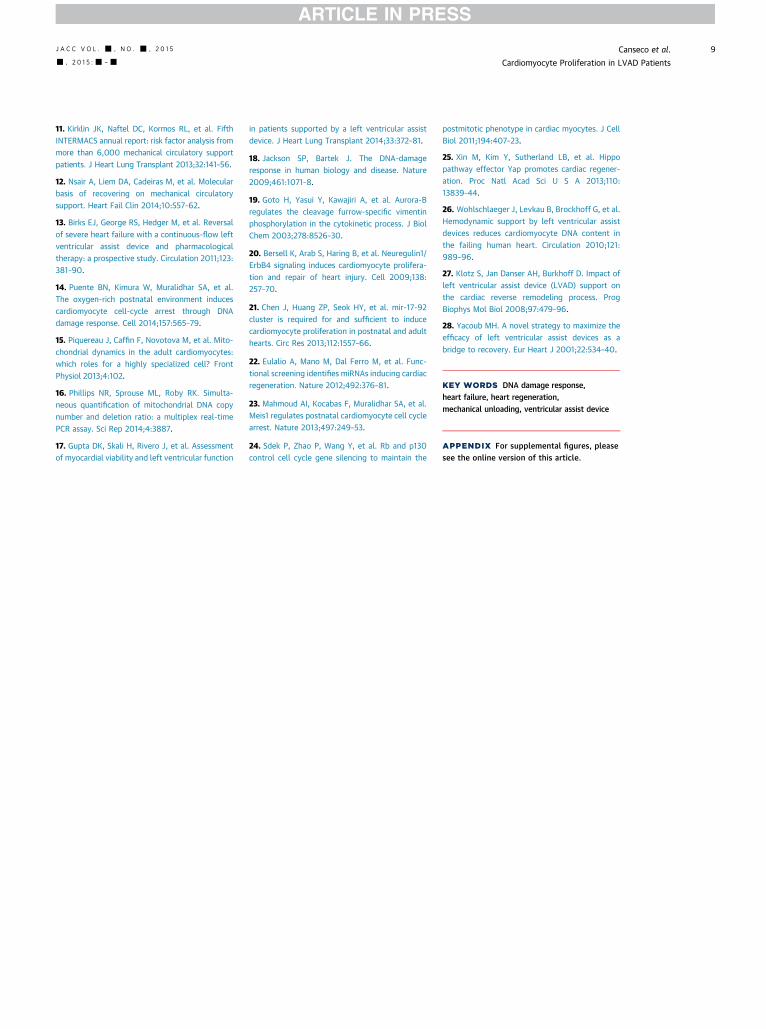
Copyright © 2022 FDOKUMEN