Percutaneous Ventricular Assist Devices: New Deus Ex Machina?
Upload
bartsandthelondonCategory
view
3download
0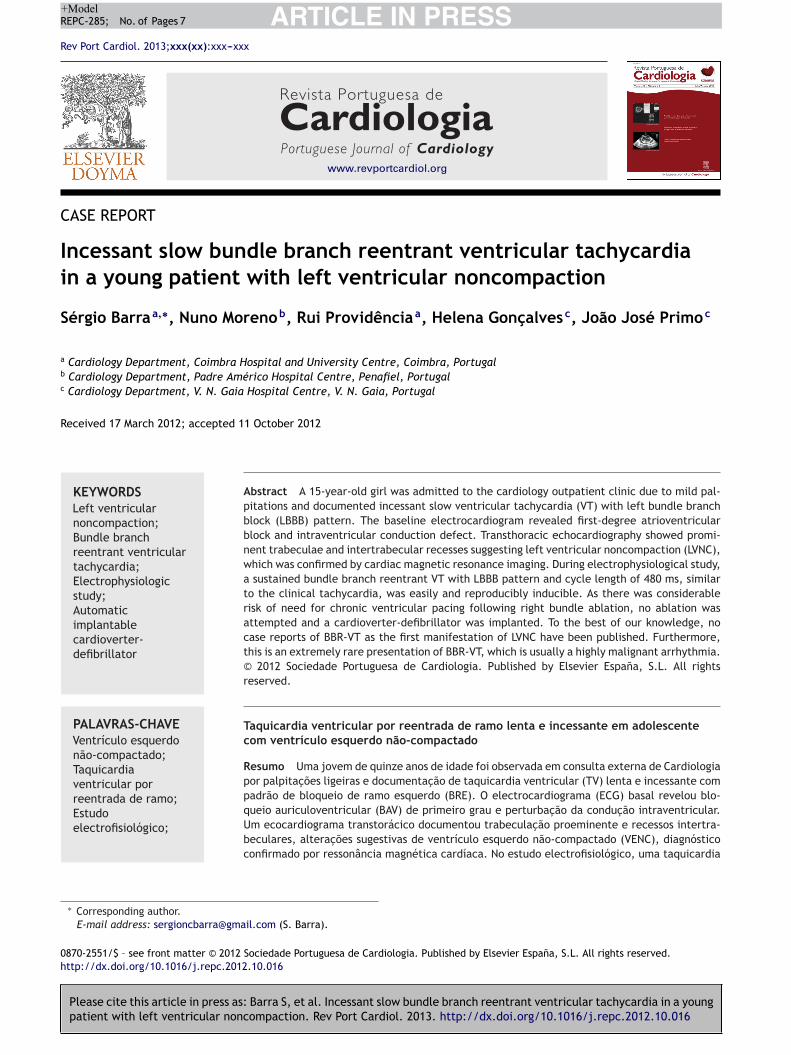
ARTICLE IN PRESS+ModelREPC-285; No. of Pages 7
Rev Port Cardiol. 2013;xxx(xx):xxx---xxx
Revista Portuguesa de
CardiologiaPortuguese Journal of Cardiology
www.revportcardiol.org
CASE REPORT
Incessant slow bundle branch reentrant ventricular tachycardiain a young patient with left ventricular noncompaction
Sérgio Barraa,∗, Nuno Morenob, Rui Providênciaa, Helena Goncalvesc, João José Primoc
a Cardiology Department, Coimbra Hospital and University Centre, Coimbra, Portugalb Cardiology Department, Padre Américo Hospital Centre, Penafiel, Portugalc Cardiology Department, V. N. Gaia Hospital Centre, V. N. Gaia, Portugal
Received 17 March 2012; accepted 11 October 2012
KEYWORDSLeft ventricularnoncompaction;Bundle branchreentrant ventriculartachycardia;Electrophysiologicstudy;Automaticimplantablecardioverter-defibrillator
Abstract A 15-year-old girl was admitted to the cardiology outpatient clinic due to mild pal-pitations and documented incessant slow ventricular tachycardia (VT) with left bundle branchblock (LBBB) pattern. The baseline electrocardiogram revealed first-degree atrioventricularblock and intraventricular conduction defect. Transthoracic echocardiography showed promi-nent trabeculae and intertrabecular recesses suggesting left ventricular noncompaction (LVNC),which was confirmed by cardiac magnetic resonance imaging. During electrophysiological study,a sustained bundle branch reentrant VT with LBBB pattern and cycle length of 480 ms, similarto the clinical tachycardia, was easily and reproducibly inducible. As there was considerablerisk of need for chronic ventricular pacing following right bundle ablation, no ablation wasattempted and a cardioverter-defibrillator was implanted. To the best of our knowledge, nocase reports of BBR-VT as the first manifestation of LVNC have been published. Furthermore,this is an extremely rare presentation of BBR-VT, which is usually a highly malignant arrhythmia.© 2012 Sociedade Portuguesa de Cardiologia. Published by Elsevier España, S.L. All rightsreserved.
PALAVRAS-CHAVEVentrículo esquerdonão-compactado;Taquicardia
Taquicardia ventricular por reentrada de ramo lenta e incessante em adolescentecom ventrículo esquerdo não-compactado
Resumo Uma jovem de quinze anos de idade foi observada em consulta externa de Cardiologia
cumentacão de taquicardia ventricular (TV) lenta e incessante com por palpitacões ligeiras e doPlease cite this article in press as: Barra S, et al. Incessant slow bundle branch reentrant ventricular tachycardia in a youngpatient with left ventricular noncompaction. Rev Port Cardiol. 2013. http://dx.doi.org/10.1016/j.repc.2012.10.016
ventricular porreentrada de ramo;Estudoelectrofisiológico;
padrão de bloqueio de ramo esquerdo (BRE). O electrocardiograma (ECG) basal revelou blo-queio auriculoventricular (BAV) de primeiro grau e perturbacão da conducão intraventricular.Um ecocardiograma transtorácico documentou trabeculacão proeminente e recessos intertra-beculares, alteracões sugestivas de ventrículo esquerdo não-compactado (VENC), diagnósticoconfirmado por ressonância magnética cardíaca. No estudo electrofisiológico, uma taquicardia
∗ Corresponding author.E-mail address: [email protected] (S. Barra).
0870-2551/$ – see front matter © 2012 Sociedade Portuguesa de Cardiologia. Published by Elsevier España, S.L. All rights reserved.http://dx.doi.org/10.1016/j.repc.2012.10.016
ARTICLE IN PRESS+ModelREPC-285; No. of Pages 7
2 S. Barra et al.
Cardioversor-desfibrilhadorimplantável
ventricular sustentada por reentrada de ramo, com padrão de BRE e ciclo de base de 480 ms,semelhante à taquicardia clínica, foi repetidamente induzida. Considerando o risco elevadode necessidade de pacing ventricular crónico em caso de ablacão do ramo direito (BAV deprimeiro grau e BRE no ECG basal e intervalo HV 100 ms no estudo electrofisiológico), não foiefetuado qualquer procedimento ablativo e um cardioversor-desfibrilhador foi implantado. Atéao momento atual, nenhum caso de TV por reentrada de ramo como primeira manifestacão deVENC foi publicado. O caso descrito revela uma apresentacão extremamente atípica deste tipode TV, que habitualmente é rápida e maligna.© 2012 Sociedade Portuguesa de Cardiologia. Publicado por Elsevier España, S.L. Todos os
I
BaepuawAifiaati
C
Am(whdwdAt
I
TdcwaahiawirfiL
t
wlra
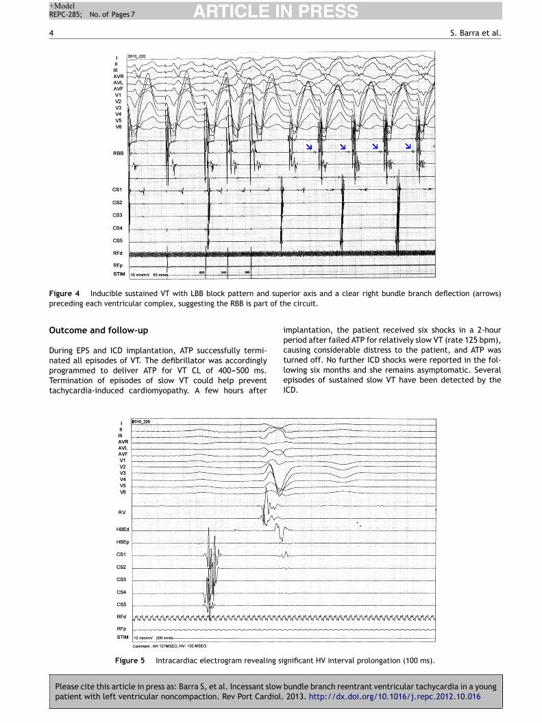
TcgdqHcsavV360 ms after an eight-beat drive-cycle length of 600 ms. Ithad a LBBB pattern, superior axis and a clear right bundledeflection preceding each ventricular complex, suggestingthe RBB was part of the circuit (Figure 4). A short postpacing
direitos reservados.
ntroduction
undle branch reentrant ventricular tachycardia (BBR-VT),n uncommon form of macroreentrant tachycardia, gen-rally occurs in the context of dilated cardiomyopathy,revious valve surgery or other cardiac conditions withnderlying His-Purkinje system (HPS) disease. This case is
very unusual presentation of BBR-VT in a young patientith isolated left ventricular noncompaction (LVNC).lthough our patient presented with HPS disease allowing
nitiation of this arrhythmia, it is rare for BBR-VT to be therst manifestation of isolated LVNC. Furthermore, BBR-VT is
highly malignant arrhythmia, yet our patient was almostsymptomatic due to the surprisingly long cycle length ofhe ventricular tachycardia (VT) and despite its unusualncessancy.
ase report
15-year-old girl was referred to our arrhythmology depart-ent for mild palpitations and documented incessant VT
hemodynamically stable VT lasting hours). She was other-ise healthy, with no relevant medical history and no familyistory of significant cardiomyopathy or sudden cardiaceath (SCD). Her palpitations were not related to effort andere persistent but otherwise extremely well tolerated. Sheenied precordial pain, dizziness, or presyncope/syncope.
previous electrocardiogram (ECG) had revealed wide-QRSachycardia (WCT) at 115 beats per minute (bpm).
nvestigations
he patient was examined shortly after being referred, andenied any symptoms, including palpitations. The physi-al exam was unremarkable. The first ECG showed a VTith left bundle branch block (LBBB) pattern and superiorxis at 118 bpm. A transthoracic echocardiogram revealed
normal-sized LV and preserved overall systolic function,ypertrabeculation of the LV posterior and lateral walls andntertrabecular recesses communicating with the LV cavitys demonstrated by color Doppler flow, suggestive of LVNC,hich was confirmed by cardiac magnetic resonance imag-
ng (Figure 1). Subsequent ECGs alternated between sinushythm with intraventricular conduction abnormalities and
Please cite this article in press as: Barra S, et al. Incessant slow
patient with left ventricular noncompaction. Rev Port Cardiol.
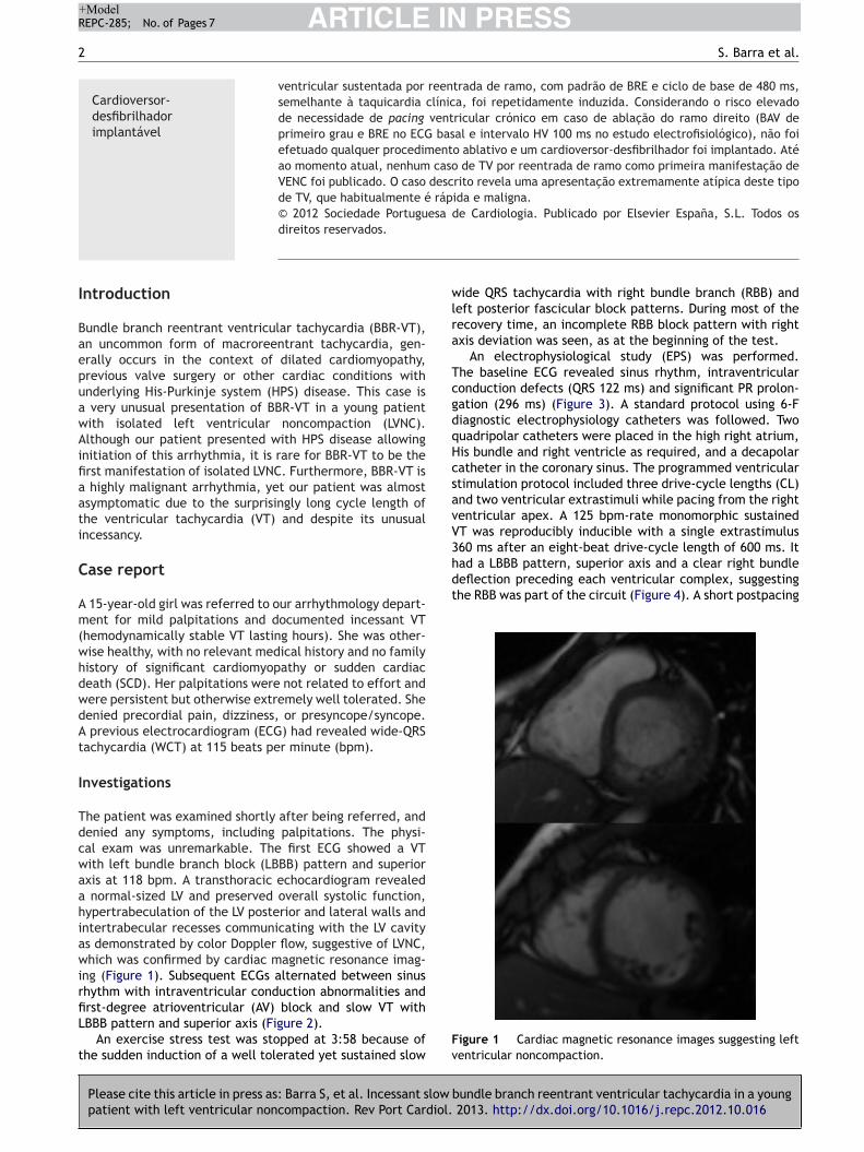
rst-degree atrioventricular (AV) block and slow VT withBBB pattern and superior axis (Figure 2).
An exercise stress test was stopped at 3:58 because ofhe sudden induction of a well tolerated yet sustained slow
Fv
ide QRS tachycardia with right bundle branch (RBB) andeft posterior fascicular block patterns. During most of theecovery time, an incomplete RBB block pattern with rightxis deviation was seen, as at the beginning of the test.
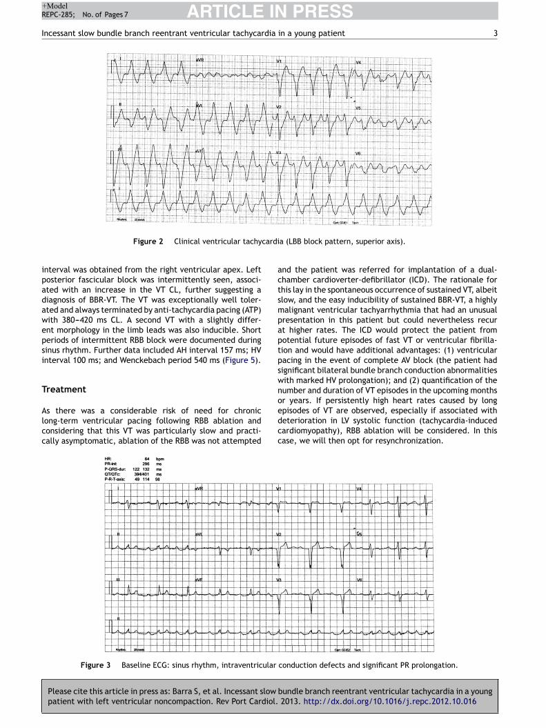
An electrophysiological study (EPS) was performed.he baseline ECG revealed sinus rhythm, intraventricularonduction defects (QRS 122 ms) and significant PR prolon-ation (296 ms) (Figure 3). A standard protocol using 6-Fiagnostic electrophysiology catheters was followed. Twouadripolar catheters were placed in the high right atrium,is bundle and right ventricle as required, and a decapolaratheter in the coronary sinus. The programmed ventriculartimulation protocol included three drive-cycle lengths (CL)nd two ventricular extrastimuli while pacing from the rightentricular apex. A 125 bpm-rate monomorphic sustainedT was reproducibly inducible with a single extrastimulus
bundle branch reentrant ventricular tachycardia in a young 2013. http://dx.doi.org/10.1016/j.repc.2012.10.016
igure 1 Cardiac magnetic resonance images suggesting leftentricular noncompaction.
ARTICLE IN PRESS+ModelREPC-285; No. of Pages 7
Incessant slow bundle branch reentrant ventricular tachycardia in a young patient 3
cardi
actsmpaptpswno
Figure 2 Clinical ventricular tachy
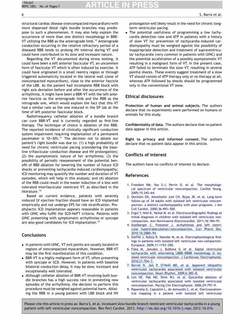
interval was obtained from the right ventricular apex. Leftposterior fascicular block was intermittently seen, associ-ated with an increase in the VT CL, further suggesting adiagnosis of BBR-VT. The VT was exceptionally well toler-ated and always terminated by anti-tachycardia pacing (ATP)with 380---420 ms CL. A second VT with a slightly differ-ent morphology in the limb leads was also inducible. Shortperiods of intermittent RBB block were documented duringsinus rhythm. Further data included AH interval 157 ms; HVinterval 100 ms; and Wenckebach period 540 ms (Figure 5).
Treatment
Please cite this article in press as: Barra S, et al. Incessant slow bpatient with left ventricular noncompaction. Rev Port Cardiol.
As there was a considerable risk of need for chroniclong-term ventricular pacing following RBB ablation andconsidering that this VT was particularly slow and practi-cally asymptomatic, ablation of the RBB was not attempted
edcc
Figure 3 Baseline ECG: sinus rhythm, intraventricular
a (LBB block pattern, superior axis).
nd the patient was referred for implantation of a dual-hamber cardioverter-defibrillator (ICD). The rationale forhis lay in the spontaneous occurrence of sustained VT, albeitlow, and the easy inducibility of sustained BBR-VT, a highlyalignant ventricular tachyarrhythmia that had an unusualresentation in this patient but could nevertheless recurt higher rates. The ICD would protect the patient fromotential future episodes of fast VT or ventricular fibrilla-ion and would have additional advantages: (1) ventricularacing in the event of complete AV block (the patient hadignificant bilateral bundle branch conduction abnormalitiesith marked HV prolongation); and (2) quantification of theumber and duration of VT episodes in the upcoming monthsr years. If persistently high heart rates caused by long
undle branch reentrant ventricular tachycardia in a young 2013. http://dx.doi.org/10.1016/j.repc.2012.10.016
pisodes of VT are observed, especially if associated witheterioration in LV systolic function (tachycardia-inducedardiomyopathy), RBB ablation will be considered. In thisase, we will then opt for resynchronization.
conduction defects and significant PR prolongation.
ARTICLE IN PRESS+ModelREPC-285; No. of Pages 7
4 S. Barra et al.
F supp of th
O
DnpTt
ipc
igure 4 Inducible sustained VT with LBB block pattern andreceding each ventricular complex, suggesting the RBB is part
utcome and follow-up
uring EPS and ICD implantation, ATP successfully termi-
Please cite this article in press as: Barra S, et al. Incessant slow
patient with left ventricular noncompaction. Rev Port Cardiol.
ated all episodes of VT. The defibrillator was accordinglyrogrammed to deliver ATP for VT CL of 400---500 ms.ermination of episodes of slow VT could help preventachycardia-induced cardiomyopathy. A few hours after
tleI
Figure 5 Intracardiac electrogram revealing si
erior axis and a clear right bundle branch deflection (arrows)e circuit.
mplantation, the patient received six shocks in a 2-houreriod after failed ATP for relatively slow VT (rate 125 bpm),ausing considerable distress to the patient, and ATP was
bundle branch reentrant ventricular tachycardia in a young 2013. http://dx.doi.org/10.1016/j.repc.2012.10.016
urned off. No further ICD shocks were reported in the fol-owing six months and she remains asymptomatic. Severalpisodes of sustained slow VT have been detected by theCD.
gnificant HV interval prolongation (100 ms).

IN+Model
dia
oaw
f
•
•••
•
•
•
mostpoLVwcvLitaotii
VBdpdhdfEdQdnsdb
ARTICLEREPC-285; No. of Pages 7
Incessant slow bundle branch reentrant ventricular tachycar
Discussion
LVNC is a rare form of a primary genetic cardiomyopa-thy considered to be the result of abnormal intrauterinearrest of the myocardial compaction process.1 Heart fail-ure, arrhythmias (including SCD) and embolic events are itsclassical triad of complications. Ventricular tachyarrhyth-mias are reported in 38---47% and SCD in 13---18% of adultpatients with LVNC.2
Although 80---90% of LVNC patients show ECG abnor-malities, no ECG features are specific to the disease.3
Conversely, intraventricular conduction defects are uncom-mon in children with LVNC, in whom the most frequentarrhythmias or conduction defects are Wolff-Parkinson-White syndrome, AV block (mainly second-degree), VT andbradycardia, while adults usually present with LV hypertro-phy, LBBB, VT, atrial fibrillation, QT prolongation and AVblock.4 Our patient had LBBB type intra/interventricularconduction defect and first-degree AV block. She also hadintermittent RBB block, rare in these patients. Alternat-ing bundle branch block is a class I recommendation, levelof evidence C, for cardiac pacing according to the Euro-pean Society of Cardiology and European Heart RhythmAssociation guidelines for cardiac pacing and cardiac resyn-chronization.
The first comprehensive analysis of electrophysiologi-cal (EP) findings in a relatively large cohort of patientswith LVNC (n=24) proposed that life-threatening ventricu-lar tachyarrhythmias were likely due to the noncompactedmyocardium serving as the arrhythmic substrate. Impairedflow reserve in structurally noncompacted myocardial seg-ments with resultant intermittent ischemia could playan important role. However, in that cohort, sustainedmonomorphic VT was rarely induced, even with iso-proterenol infusion. Non-sustained polymorphic VT wasobserved more commonly and, while it was believed tobe nonspecific, three patients with non-sustained polymor-phic VT demonstrated malignant ventricular arrhythmias onfollow-up. The authors concluded that no specific clinical,electrocardiographic or echocardiographic finding was pre-dictive of VT inducibility, except for the potential protectiveeffect of younger age and LV ejection fraction above 50%.Nevertheless, their findings suggested a negative EPS couldidentify a subset of patients at low risk of developing malig-nant tachyarrhythmias.5
The differential diagnosis of a WCT with a typical LBBBmorphology is limited to five entities: supraventriculartachycardia (SVT) with fixed LBBB, SVT with functionalaberrancy, pre-excited reentrant tachycardias using an atri-ofascicular accessory pathway as the anterograde limb, SVTwith a ‘‘bystander’’ atriofascicular pathway, and BBR-VT.In our patient, the lack of a 1:1 AV relationship excludeda pre-excited reentrant tachycardia using an atriofascicu-lar accessory pathway, while ventricular rate greater thanatrial rate strongly suggested a VT (rarely, an AV nodal reen-trant tachycardia may present with 2:1 retrograde block,but a longer postpacing interval is typically obtained fromthe right ventricular apex). Other potential diagnoses were
Please cite this article in press as: Barra S, et al. Incessant slow bpatient with left ventricular noncompaction. Rev Port Cardiol.
automatic fascicular VT and intramyocardial VT. The formeris catecholamine-dependent, not induced with programmedstimulation and shows a variable HV interval in tachycar-dia, while the latter rarely produces entirely typical RBBB
assi
PRESSin a young patient 5
r LBBB patterns on surface ECG and does not depend on critical delay in the HPS. Therefore, these two diagnosesere considered extremely unlikely.
The diagnosis of BBR-VT was suggested by a number ofactors:
The presence of clear intraventricular conduction abnor-malities and first-degree AV block on the baseline ECG;
A prolonged baseline HV interval; The occurrence of a VT with typical LBBB pattern; The induction and interruption of the arrhythmia with
pacing (suggestive of a reentrant mechanism); The need for a critical conduction delay in the HPS for
induction of the tachycardia; The presence of a clear right bundle deflection preceding
each ventricular complex, suggesting the RBB was part ofthe circuit;
A short postpacing interval from the right ventricularapex.
The current literature contains limited data regardingapping and ablation of premature ventricular complexes
r VT in the presence of LVNC. Fiala et al. describeduccessful ablation of a VT originating in the interven-ricular septum.6 Derval et al. reported two symptomaticatients who underwent successful radiofrequency ablationf a monomorphic VT in the basolateral aspect of the LV.7
im et al. described epicardial ablation of a monomorphicT located in the epicardial surface of the anterolateralall.8 The mechanism underlying these arrhythmias is notompletely understood. Paparella et al. reconstructed aentricular electroanatomical mapping in a patient withVNC and monomorphic VT. No areas of low voltage weredentified, probably excluding the presence of scar-basedissue reentry as a possible mechanism.9 There is consider-ble evidence that most VT episodes in LVNC arise from areasf noncompacted myocardium, and there have been reportshat, in some cases, the focus of arrhythmia is not necessar-ly related to LVNC, indicating the possibility of concomitantdiopathic arrhythmias in this population.10
To the best of our knowledge, no case report of BBR-T as the first manifestation of LVNC has been published.BR-VT, an uncommon form of VT incorporating both bun-le branches into the reentrant circuit, usually occurs inatients with structural heart disease, mainly dilated car-iomyopathy, although patients with structurally normalearts have been described. The critical prerequisite for itsevelopment is conduction delay in the HPS, which mani-ests as nonspecific conduction delay or LBBB on the surfaceCG and prolonged HV interval in the intracardiac recor-ings, although some patients may have relatively narrowRS complexes, suggesting a role of functional conductionelay in the genesis of BBR,11 or even an HV interval withinormal limits.12 Patients typically present with presyncope,yncope or SCD because of very rapid rates. QRS morphologyuring VT is a typical bundle branch block pattern and maye identical to that in sinus rhythm.11
Unlike typical presentations, our patient was practically
undle branch reentrant ventricular tachycardia in a young 2013. http://dx.doi.org/10.1016/j.repc.2012.10.016
symptomatic, reporting mild palpitations despite the inces-ant nature of her VT (continuing for hours). This lack ofymptoms was due to the low rate of the arrhythmia, whichs also very unusual for BBR-VT. Preexisting RBB disease or
IN+ModelR
6
smpoVcdc
cfctnorarrht
ctTsppnl(pesIeotl
repwLa
C
•
•
•
•
E
Pda
Cd
Rd
C
T
R
ARTICLEEPC-285; No. of Pages 7
tructural cardiac disease (noncompacted myocardium) withore dispersed distal right bundle branches may predis-ose to such a phenomenon. It may also help explain theccurrence of more than one distinct morphology in BBR-T utilizing the RBB as the anterograde limb.13 Anterogradeonduction occurring in the relative refractory period of aiseased RBB tends to prolong HV interval during VT andould have contributed to its slow and incessant nature.
Regarding the VT documented during stress testing, itould have been a left anterior fascicular VT, an uncommonorm of fascicular VT which is often induced by exercise. Itould have originated in a small reentry region or throughriggered automaticity located in the lateral wall (zone ofoncompacted myocardium), close to the anterior fasciclef the LBB. As the patient had incomplete RBB block withight axis deviation before and after the occurrence of therrhythmia, it might have been a BBR-VT with the left ante-ior fascicle as the anterograde limb and the RBB as theetrograde one, which would explain the fact that this VTad a similar rate as the one induced in the EP lab at theime of left posterior fascicular block.
Radiofrequency catheter ablation of a bundle branchan cure BBR-VT and is currently regarded as first-lineherapy. The technique of choice is ablation of the RBB.he reported incidence of clinically significant conductionystem impairment requiring implantation of a permanentacemaker is 10---30%.11 The decision not to ablate ouratient’s right bundle was due to: (1) a high probability ofeed for chronic ventricular pacing (considering the base-ine trifascicular conduction disease and HV prolongation);2) the asymptomatic nature of her arrhythmia; (3) theossibility of periodic reassessment of the potential ben-fit of RBB ablation for lowering the number of future ICDhocks or preventing tachycardia-induced cardiomyopathy;CD monitoring may quantify the number and duration of VTpisodes, which will help in this analysis; and (4) ablationf the RBB could result in the easier induction of a less wellolerated interfascicular reentrant VT, as described in theiterature.14
Based on current evidence, patients with severelyeduced LV ejection fraction should have an ICD implantedmpirically and not undergo EPS for risk stratification. Pro-hylactic ICD implantation appears reasonable in patientsith LVNC who fulfill the SCD-HeFT criteria. Patients with
VNC presenting with symptomatic arrhythmias or syncopere also good candidates for ICD implantation.15
onclusions
In patients with LVNC, VT exit points are usually located inregions of noncompacted myocardium. However, BBR-VTmay be the first manifestation of this condition.
BBR-VT is a highly malignant form of VT, often presentingwith syncope or SCD. However, in patients with baselinebilateral conduction delay, it may be slow, incessant andexceptionally well tolerated.
Although catheter ablation of BBR-VT involving both bun-
Please cite this article in press as: Barra S, et al. Incessant slow
patient with left ventricular noncompaction. Rev Port Cardiol.
dle branches has a high success rate in preventing newepisodes of the arrhythmia, the decision to perform thisprocedure must be weighed against potential harm. Ablat-ing the RBB in a young patient with LBB block and HV
PRESSS. Barra et al.
prolongation will likely result in the need for chronic long-term ventricular pacing.
The potential usefulness of programming a low tachy-cardia detection rate and ATP in patients with a historyof slow VT for prevention of tachycardia-induced car-diomyopathy must be weighed against the possibility ofinappropriate detection and treatment of supraventricu-lar tachycardia (very common in patients with LVNC) andthe potential acceleration of a possibly asymptomatic VTresulting in a malignant form of VT. In the present case,ATP failed to terminate the slow VT, resulting in severalpainful shocks. These events suggest treatment of a slowVT should consist of ATP therapy only or no therapy at all,whereas ATP followed by shocks should be programmedonly in the conventional VT zone.
thical disclosures
rotection of human and animal subjects. The authorseclare that no experiments were performed on humans ornimals for this study.
onfidentiality of data. The authors declare that no patientata appear in this article.
ight to privacy and informed consent. The authorseclare that no patient data appear in this article.
onflicts of interest
he authors have no conflicts of interest to declare.
eferences
1. Freedom RM, Yoo S-J, Perrin D, et al. The morphologi-cal spectrum of ventricular noncompaction. Cardiol Young.2005;15:345---64.
2. Oechslin EN, Attenhofer Jost CH, Rojas JR, et al. Long-termfollow-up of 34 adults with isolated left ventricular noncom-paction: a distinct cardiomyopathy with poor prognosis. J AmColl Cardiol. 2000;36:493---500.
3. Ergul Y, Nisli K, Varkal M, et al. Electrocardiographic findings atinitial diagnosis in children with isolated left ventricular non-compaction. Ann Noninvasive Electrocardiol. 2011;16:184---91.
4. Stollberger C, Finsterer J. Arrhythmias and left ventri-cular hypertrabeculation/noncompaction. Curr Pharm Des.2010;16:2880---94.
5. Steffel J, Kobza R, Namdar M, et al. Electrophysiological find-ings in patients with isolated left ventricular non-compaction.Europace. 2009;11:1193---200.
6. Fiala M, Januska J, Bulkova V, et al. Septal ventriculartachycardia with alternating LBBB---RBBB morphology in iso-lated ventricular noncompaction. J Cardiovasc Electrophysiol.2010;21:704---7.
7. Derval N, Jais P, O’Neill MD, et al. Apparent idiopathicventricular tachycardia associated with isolated ventricularnoncompaction. Heart Rhythm. 2009;6:385---8.
8. Lim HE, Pak HN, Shim WJ, et al. Epicardial ablation of
bundle branch reentrant ventricular tachycardia in a young 2013. http://dx.doi.org/10.1016/j.repc.2012.10.016
ventricular tachycardia associated with isolated ventricularnoncompaction. Pacing Clin Electrophysiol. 2006;29:797---9.
9. Paparella G, Capulzini L, de Asmundis C, et al. Electroanatom-ical mapping in a patient with isolated left ventricular
IN+Model
dia
1
1
1
ARTICLEREPC-285; No. of Pages 7
Incessant slow bundle branch reentrant ventricular tachycar
non-compaction and left ventricular tachycardia. Europace.2009;11:1227---9.
10. Tondato F, Daniel NG, Altemose G, et al. Non-sustained ven-tricular tachycardia in a patient with non-compaction of leftventricle. Natural history or associated condition? J Innov Car-diac Rhythm Manage. 2011;2:327---31.
11. Mazur A, Kusniec J, Strasberg B. Bundle branch reen-trant ventricular tachycardia. Indian Pacing ElectrophysiolJ. 2005;5:86---95.
Please cite this article in press as: Barra S, et al. Incessant slow bpatient with left ventricular noncompaction. Rev Port Cardiol.
12. Li Y-G, Gronefeld G, Israel C, et al. Bundle branch reen-trant tachycardia in patients with apparent normal His-Purkinjeconduction. The role of functional conduction impairment.J Cardiovasc Electrophysiol. 2002;13:1233---9.
PRESSin a young patient 7
3. Wang CW, Sterba R, Tchou P. Bundle branch reentry ven-tricular tachycardia with two distinct left bundle branchblock morphologies. J Cardiovasc Electrophysiol. 1997;8:688---93.
4. Blanck Z, Sra J, Akhtar M. Incessant interfascicular reentrantventricular tachycardia as a result of catheter ablation of theright bundle branch: case report and review of the literature.J Cardiovasc Electrophysiol. 2009;11:1279---83.
5. Caliskan K, Szili-Torok T, Theuns DA, et al. Indications
undle branch reentrant ventricular tachycardia in a young 2013. http://dx.doi.org/10.1016/j.repc.2012.10.016
and outcome of implantable cardioverter-defibrillators forprimary and secondary prophylaxis in patients with noncom-paction cardiomyopathy. J Cardiovasc Electrophysiol. 2011;22:898---904.
Copyright © 2022 FDOKUMEN