Reentrant waves in a ring of embryonic chick ventricular cells imaged with a Ca 2+ sensitive dye
10
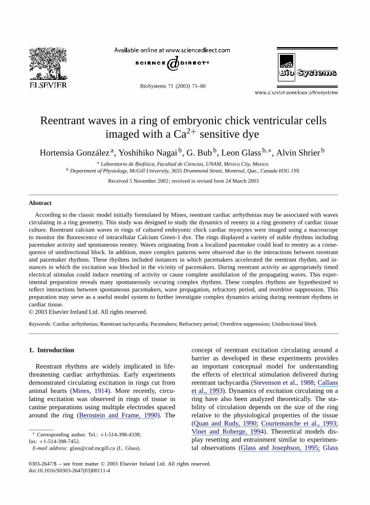
BioSystems 71 (2003) 71–80 Reentrant waves in a ring of embryonic chick ventricular cells imaged with a Ca 2+ sensitive dye Hortensia González a , Yoshihiko Nagai b , G. Bub b , Leon Glass b,∗ , Alvin Shrier b a Laboratorio de Biofísica, Facultad de Ciencias, UNAM, México City, Mexico b Department of Physiology, McGill University, 3655 Drummond Street, Montreal, Que., Canada H3G 1Y6 Received 5 November 2002; received in revised form 24 March 2003 Abstract According to the classic model initially formulated by Mines, reentrant cardiac arrhythmias may be associated with waves circulating in a ring geometry. This study was designed to study the dynamics of reentry in a ring geometry of cardiac tissue culture. Reentrant calcium waves in rings of cultured embryonic chick cardiac myocytes were imaged using a macroscope to monitor the fluorescence of intracellular Calcium Green-1 dye. The rings displayed a variety of stable rhythms including pacemaker activity and spontaneous reentry. Waves originating from a localized pacemaker could lead to reentry as a conse- quence of unidirectional block. In addition, more complex patterns were observed due to the interactions between reentrant and pacemaker rhythms. These rhythms included instances in which pacemakers accelerated the reentrant rhythm, and in- stances in which the excitation was blocked in the vicinity of pacemakers. During reentrant activity an appropriately timed electrical stimulus could induce resetting of activity or cause complete annihilation of the propagating waves. This exper- imental preparation reveals many spontaneously occuring complex rhythms. These complex rhythms are hypothesized to reflect interactions between spontaneous pacemakers, wave propagation, refractory period, and overdrive suppression. This preparation may serve as a useful model system to further investigate complex dynamics arising during reentrant rhythms in cardiac tissue. © 2003 Elsevier Ireland Ltd. All rights reserved. Keywords: Cardiac arrhythmias; Reentrant tachycardia; Pacemakers; Refractory period; Overdrive suppression; Unidirectional block 1. Introduction Reentrant rhythms are widely implicated in life- threatening cardiac arrhythmias. Early experiments demonstrated circulating excitation in rings cut from animal hearts (Mines, 1914). More recently, circu- lating excitation was observed in rings of tissue in canine preparations using multiple electrodes spaced around the ring (Bernstein and Frame, 1990). The ∗ Corresponding author. Tel.: +1-514-398-4338; fax: +1-514-398-7452. E-mail address: [email protected] (L. Glass). concept of reentrant excitation circulating around a barrier as developed in these experiments provides an important conceptual model for understanding the effects of electrical stimulation delivered during reentrant tachycardia (Stevenson et al., 1988; Callans et al., 1993). Dynamics of excitation circulating on a ring have also been analyzed theoretically. The sta- bility of circulation depends on the size of the ring relative to the physiological properties of the tissue (Quan and Rudy, 1990; Courtemanche et al., 1993; Vinet and Roberge, 1994). Theoretical models dis- play resetting and entrainment similar to experimen- tal observations (Glass and Josephson, 1995; Glass 0303-2647/$ – see front matter © 2003 Elsevier Ireland Ltd. All rights reserved. doi:10.1016/S0303-2647(03)00111-4
Transcript of Reentrant waves in a ring of embryonic chick ventricular cells imaged with a Ca 2+ sensitive dye
BioSystems 71 (2003) 71–80
Reentrant waves in a ring of embryonic chick ventricular cellsimaged with a Ca2+ sensitive dye
Hortensia Gonzáleza, Yoshihiko Nagaib, G. Bubb, Leon Glassb,∗, Alvin Shrierba Laboratorio de Biofísica, Facultad de Ciencias, UNAM, México City, Mexico
b Department of Physiology, McGill University, 3655 Drummond Street, Montreal, Que., Canada H3G 1Y6
Received 5 November 2002; received in revised form 24 March 2003
Abstract
According to the classic model initially formulated by Mines, reentrant cardiac arrhythmias may be associated with wavescirculating in a ring geometry. This study was designed to study the dynamics of reentry in a ring geometry of cardiac tissueculture. Reentrant calcium waves in rings of cultured embryonic chick cardiac myocytes were imaged using a macroscopeto monitor the fluorescence of intracellular Calcium Green-1 dye. The rings displayed a variety of stable rhythms includingpacemaker activity and spontaneous reentry. Waves originating from a localized pacemaker could lead to reentry as a conse-quence of unidirectional block. In addition, more complex patterns were observed due to the interactions between reentrantand pacemaker rhythms. These rhythms included instances in which pacemakers accelerated the reentrant rhythm, and in-stances in which the excitation was blocked in the vicinity of pacemakers. During reentrant activity an appropriately timedelectrical stimulus could induce resetting of activity or cause complete annihilation of the propagating waves. This exper-imental preparation reveals many spontaneously occuring complex rhythms. These complex rhythms are hypothesized toreflect interactions between spontaneous pacemakers, wave propagation, refractory period, and overdrive suppression. Thispreparation may serve as a useful model system to further investigate complex dynamics arising during reentrant rhythms incardiac tissue.© 2003 Elsevier Ireland Ltd. All rights reserved.
Keywords: Cardiac arrhythmias; Reentrant tachycardia; Pacemakers; Refractory period; Overdrive suppression; Unidirectional block
1. Introduction
Reentrant rhythms are widely implicated in life-threatening cardiac arrhythmias. Early experimentsdemonstrated circulating excitation in rings cut fromanimal hearts(Mines, 1914). More recently, circu-lating excitation was observed in rings of tissue incanine preparations using multiple electrodes spacedaround the ring(Bernstein and Frame, 1990). The
∗ Corresponding author. Tel.:+1-514-398-4338;fax: +1-514-398-7452.
E-mail address: [email protected] (L. Glass).
concept of reentrant excitation circulating around abarrier as developed in these experiments providesan important conceptual model for understandingthe effects of electrical stimulation delivered duringreentrant tachycardia(Stevenson et al., 1988; Callanset al., 1993). Dynamics of excitation circulating on aring have also been analyzed theoretically. The sta-bility of circulation depends on the size of the ringrelative to the physiological properties of the tissue(Quan and Rudy, 1990; Courtemanche et al., 1993;Vinet and Roberge, 1994). Theoretical models dis-play resetting and entrainment similar to experimen-tal observations(Glass and Josephson, 1995; Glass
0303-2647/$ – see front matter © 2003 Elsevier Ireland Ltd. All rights reserved.doi:10.1016/S0303-2647(03)00111-4

72 H. Gonzalez et al. / BioSystems 71 (2003) 71–80
et al., 2002; Sinha et al., 2002; Comtois and Vinet,2002).
The current work was motivated by our interestin developing an in vitro experimental system thatcontains both excitable medium and pacemakers, andis suitable for experimental manipulations. In earlywork, cardiac tissue that was grown in strands andrings was used to analyze biophysical properties andmodel arrhythmias(Lieberman et al., 1972, 1981;Shigeto et al., 1982). More recently, the develop-ment of optical recording techniques has facilitatedthe analysis of dynamics of propagation for cardiaccells in tissue culture grown in a variety of geometricpatterns(Koidl et al., 1980; Rohr et al., 1991; Rohr,1995; Bub et al., 1998, 2002; Fast et al., 1998; Nagaiet al., 2000; González et al., 2000; Arutunyan et al.,2001, 2002).
In this work, we describe a tissue culture model inwhich cells are grown as a ring and monitored by amacroscopic imaging system. In recent articles(Nagaiet al., 2000), we described reentrant rhythms in a ringgeometry and developed mathematical models of theobserved dynamics(Nagai et al., 2000; González et al.,2000). In this work we extend our experimental ob-servations to a wide variety of complex rhythms gen-erated by the interaction of spontaneous pacemakeractivity and propagating waves.
2. Materials and methods
2.1. Cell culture
Cell cultures were prepared according to previouslyestablished procedures(Bub et al., 1998). Briefly, ven-tricular cells were isolated from White Leghorn chickembryo hearts after 7 days of incubation in ovo. Theventricles were dissociated into single cells and platedat densities of 4.0× 10⁴ cells/ml in plastic dishes pat-terned with a ring design (see below). The cells weremaintained in the medium at 5% CO2 at 36◦C for 3–4days before experiments were undertaken. After sev-eral of the experiments, the rings were fixed for mi-croscopic examination using a Masson trichrome stain(Sigma Diagnostics).Fig. 1shows a microphotographof a portion of a ring. The optical recording experi-ments were carried out in preparations during the thirdand fourth days after culturing.
2.2. Construction of the ring
Dishes were first coated with a thin film of cell ad-hesive (Cell-Tak, Collaborative Biomedical Products).To create the ring geometry, we constructed a templatefrom a cylindrical piece of Plexiglas. On one end ofthe template, an annular depression with dimensionsof 8 mm outer diameter and 6 mm inner diameter wasmachined into the Plexiglas. The raised portions of thetemplate were first coated with silicon polymer (3140Dow-Corning) and then applied to the culture dishes.The regions of culture dishes that contacted the sili-conized regions of template did not favor cell adhe-sion. Cells that contacted siliconized areas tended toform beating aggregates that poorly adhered and wereeasily aspirated leaving an annular region.
2.3. Fluorescent imaging
Cellular activity was measured by monitoring thefluorescence signal generated by an intracellular cal-cium sensitive dye, Calcium Green-1 (MolecularProbes) using methods described previously(Bubet al., 1998).
Cell cultures were maintained at 36± 1◦C duringthe recordings. In some experiments, electrical stim-ulation was delivered by means of a bipolar tefloncoated platinum electrode (diameter 120�m, sepa-ration 1 mm). Cells were stimulated with rectangularpulses of 20 or 30 ms duration at twice diastolicthreshold current delivered from a Frederick-Haer(Pulsar 4i) stimulator.
The still figures in the manuscript are supplementedby animations on a public web sitehttp://www.cnd.mcgill.ca/bios/glass/hortensia/index.htm.
3. Results
In five preparations there was only evidence of lo-calized spontaneous pacemakers. The activity patternassociated with a single pacemaker is characterized bya pair of bidirectional waves originating from one po-sition on the ring and colliding at the antipodal point.Table 1gives a summary of the experiments in whichthere was a stable pacemaker.
Although the above pattern was frequently ob-served, in most cases it was not maintained stably over

H. Gonzalez et al. / BioSystems 71 (2003) 71–80 73
Fig. 1. Microscopic images of a section of the preparation stained with Masson trichrome stain. (A) The entire ring. The inner diameter ofthe ring is 6 mm and the outer diameter is 8 mm. (B) A section of the ring. The denser regions corresponds to the region of a pacemaker.
74 H. Gonzalez et al. / BioSystems 71 (2003) 71–80
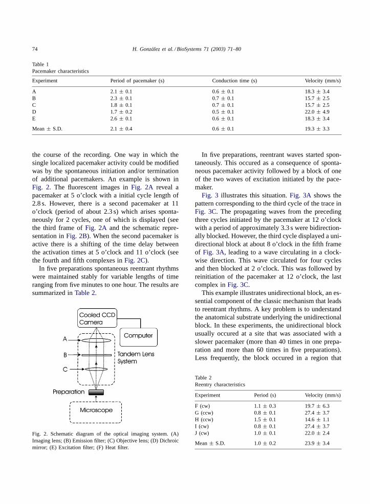
Table 1Pacemaker characteristics
Experiment Period of pacemaker (s) Conduction time (s) Velocity (mm/s)
A 2.1 ± 0.1 0.6± 0.1 18.3± 3.4B 2.3 ± 0.1 0.7± 0.1 15.7± 2.5C 1.8 ± 0.1 0.7± 0.1 15.7± 2.5D 1.7 ± 0.2 0.5± 0.1 22.0± 4.9E 2.6 ± 0.1 0.6± 0.1 18.3± 3.4
Mean± S.D. 2.1± 0.4 0.6± 0.1 19.3± 3.3
the course of the recording. One way in which thesingle localized pacemaker activity could be modifiedwas by the spontaneous initiation and/or terminationof additional pacemakers. An example is shown inFig. 2. The fluorescent images inFig. 2A reveal apacemaker at 5 o’clock with a initial cycle length of2.8 s. However, there is a second pacemaker at 11o’clock (period of about 2.3 s) which arises sponta-neously for 2 cycles, one of which is displayed (seethe third frame ofFig. 2A and the schematic repre-sentation inFig. 2B). When the second pacemaker isactive there is a shifting of the time delay betweenthe activation times at 5 o’clock and 11 o’clock (seethe fourth and fifth complexes inFig. 2C).
In five preparations spontaneous reentrant rhythmswere maintained stably for variable lengths of timeranging from five minutes to one hour. The results aresummarized inTable 2.
Fig. 2. Schematic diagram of the optical imaging system. (A)Imaging lens; (B) Emission filter; (C) Objective lens; (D) Dichroicmirror; (E) Excitation filter; (F) Heat filter.
In five preparations, reentrant waves started spon-taneously. This occured as a consequence of sponta-neous pacemaker activity followed by a block of oneof the two waves of excitation initiated by the pace-maker.
Fig. 3 illustrates this situation.Fig. 3A shows thepattern corresponding to the third cycle of the trace inFig. 3C. The propagating waves from the precedingthree cycles initiated by the pacemaker at 12 o’clockwith a period of approximately 3.3 s were bidirection-ally blocked. However, the third cycle displayed a uni-directional block at about 8 o’clock in the fifth frameof Fig. 3A, leading to a wave circulating in a clock-wise direction. This wave circulated for four cyclesand then blocked at 2 o’clock. This was followed byreinitiation of the pacemaker at 12 o’clock, the lastcomplex inFig. 3C.
This example illustrates unidirectional block, an es-sential component of the classic mechanism that leadsto reentrant rhythms. A key problem is to understandthe anatomical substrate underlying the unidirectionalblock. In these experiments, the unidirectional blockusually occured at a site that was associated with aslower pacemaker (more than 40 times in one prepa-ration and more than 60 times in five preparations).Less frequently, the block occured in a region that
Table 2Reentry characteristics
Experiment Period (s) Velocity (mm/s)
F (cw) 1.1± 0.3 19.7± 6.3G (ccw) 0.8± 0.1 27.4± 3.7H (ccw) 1.5± 0.1 14.6± 1.1I (cw) 0.8 ± 0.1 27.4± 3.7J (cw) 1.0± 0.1 22.0± 2.4
Mean± S.D. 1.0± 0.2 23.9± 3.4

H. Gonzalez et al. / BioSystems 71 (2003) 71–80 75
Fig. 3. Fluorescence from a ring showing the propagation from two pacemakers. There is a pacemaker at 5:00 o’clock with a periodof 2.8 s and a second pacemaker at 11:00 o’clock with a cycle of 2.3 s that is evident for two cycles. The excitation waves from bothpacemakers collide at 1 o’clock and 9 o’clock. The data are represented using three different methods. (A) Fluorescent images recordedby the CCD camera at 200 ms intervals. The lighter region corresponds to the region of higher Ca2+ concentration. (B) Schematic diagramof wave propagation corresponding to the images in panel A. (C) Normalized intensity of fluorescence. In this and the following figures,the normalized intensity of fluorescence F recorded from 11 o’clock is indicated by a dashed curve and 5 o’clock is indicated by a solidcurve. The bar over the trace in panel C indicates the time interval for which fluorescence is displayed in panel A. The same format andconventions are used in the following figures. In the fourth and fifth complexes the lag between the excitation maxima at 5 o’clock and11 o’clock is reduced due to the presence of the second pacemaker at 11 o’clock.
appeared to be of lower density (six times in two prepa-rations).
In three cases, following initiation of reentry, theperiod increased from the initial value which was from40 to 70% of the final value. The period increased toits final value over a time scale of 5–15 revolutionsto a value which was maintained for the duration ofthe observations. The combinations of multiple pace-makers and spontaneous initiation and termination ofreentrant waves led to complex rhythms.
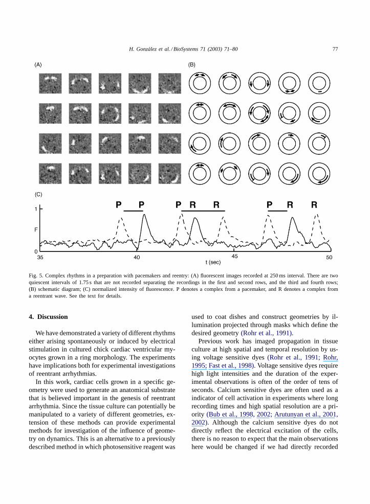
Fig. 4 shows an example of complex rhythms fromthe same preparation. Following a period of quies-cence there is a wave initiated by the pacemaker at 12o’clock (first row of fluorescent images). Followinga quiescent period of 1.75 s (not displayed), there is asecond wave initiated by the pacemaker at 12 o’clock(second and third rows of fluorescent images). Inaddition, a second pacemaker at 5 o’clock sponta-neously fires (third panel, second row of fluorescentimages). The counterclockwise circulating wavesfrom the pacemaker at 5 o’clock collides with theclockwise wave from the pacemaker at 12 o’clock and
both annihilate. However, the clockwise wave fromthe pacemaker at 5 o’clock continues circulating, pre-sumably because the original counterclockwise wavefrom the pacemaker at 12 o’clock was unidirection-ally blocked at 8 o’clock (second row, fourth panel)as previously shown inFig. 4. The newly initiatedcounterclockwise reentrant wave is itself blocked be-fore a complete circulation at 2 o’clock (third row,fifth panel). Following another quiescent period of1.75 s (not shown), there is again an initiation of areentrant wave by the pacemaker at 12 o’clock (fourthrow of fluorescent images). This time however, thereis initiation of a reentrant wave.
Although the main purpose of this work was to es-tablish whether the tissue cultured cells could supportcirculating waves, and to investigate the spontaneousrhythms set up by the interaction of reentry and pace-makers, it is important to verify whether the responseof the preparation to electrical stimulation is simi-lar to responses observed in experimental(Bernsteinand Frame, 1990), clinical (Callans et al., 1993)andtheoretical(Glass and Josephson, 1995; Glass et al.,

76 H. Gonzalez et al. / BioSystems 71 (2003) 71–80
Fig. 4. Initiation of reentrant excitation. There is a pacemaker at 12 o’clock and unidirectional block at 8 o’clock. Following unidirectionalblock, there is a single wave circulating in a counter clockwise direction with a velocity of 8.4 mm/s and a period of 2.8 s. (A) Fluorescentimages recorded at 250 ms intervals; (B) schematic diagram; (C) normalized intensity of fluorescence.
2002; Sinha et al., 2002; Comtois and Vinet, 2002)studies.
Electrical stimulation in this preparation has a va-riety of effects, depending on the precise timing andlocation of the stimulus and the number of pulses de-livered (González et al., 2000). If the stimulus is ap-plied a sufficiently long time after a wave travelingin a clockwise direction traverses a region, two newtraveling waves will be induced. The antidromic wavecollides with the original wave and both annihilate,while the newly initiated orthodromic wave continuescirculating. The net effect is that there is once again asingle clockwise wave which is reset.
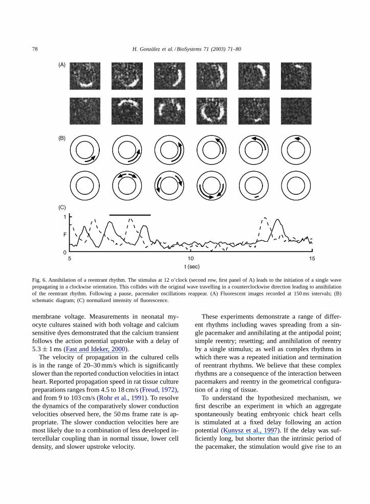
A different effect is observed from electrical stim-ulation delivered with a short latency (approximatelyin the range of 80 and 120 ms) following an activation(Fig. 5). Electrical stimulation applied with a shortlatency following the passage of a wave circulating
in a counterclockwise direction (Fig. 5A second row,second panel) initiated only a single wave, travelingclockwise. This wave collides with the original coun-terclockwise traveling wave to lead to annihilationof the reentrant rhythm. The initiation of a singlewave verifies theoretical predictions of a “vulnerablephase” of cardiac tissue(Starmer et al., 1993). Theannihilation of the reentrant wave following colli-sion of the induced wave with the original reentrantwave also follows theoretical predictions(Glass andJosephson, 1995; Sinha et al., 2002; Comtois andVinet, 2002).
In one ring the tissue remained indefinitely qui-escent following the annihilation. In the other twopreparations, one of which is illustrated inFig. 5, theoriginal pacemaker pattern reinitiated after a silentperiod of 1–5 s. Following the annihilation, waveswere generated by a pacemaker oscillationFig. 6.
H. Gonzalez et al. / BioSystems 71 (2003) 71–80 77
Fig. 5. Complex rhythms in a preparation with pacemakers and reentry: (A) fluorescent images recorded at 250 ms interval. There are twoquiescent intervals of 1.75 s that are not recorded separating the recordings in the first and second rows, and the third and fourth rows;(B) schematic diagram; (C) normalized intensity of fluorescence. P denotes a complex from a pacemaker, and R denotes a complex froma reentrant wave. See the text for details.
4. Discussion
We have demonstrated a variety of different rhythmseither arising spontaneously or induced by electricalstimulation in cultured chick cardiac ventricular my-ocytes grown in a ring morphology. The experimentshave implications both for experimental investigationsof reentrant arrhythmias.
In this work, cardiac cells grown in a specific ge-ometry were used to generate an anatomical substratethat is believed important in the genesis of reentrantarrhythmia. Since the tissue culture can potentially bemanipulated to a variety of different geometries, ex-tension of these methods can provide experimentalmethods for investigation of the influence of geome-try on dynamics. This is an alternative to a previouslydescribed method in which photosensitive reagent was
used to coat dishes and construct geometries by il-lumination projected through masks which define thedesired geometry(Rohr et al., 1991).
Previous work has imaged propagation in tissueculture at high spatial and temporal resolution by us-ing voltage sensitive dyes(Rohr et al., 1991; Rohr,1995; Fast et al., 1998). Voltage sensitive dyes requirehigh light intensities and the duration of the exper-imental observations is often of the order of tens ofseconds. Calcium sensitive dyes are often used as aindicator of cell activation in experiments where longrecording times and high spatial resolution are a pri-ority (Bub et al., 1998, 2002; Arutunyan et al., 2001,2002). Although the calcium sensitive dyes do notdirectly reflect the electrical excitation of the cells,there is no reason to expect that the main observationshere would be changed if we had directly recorded
78 H. Gonzalez et al. / BioSystems 71 (2003) 71–80
Fig. 6. Annihilation of a reentrant rhythm. The stimulus at 12 o’clock (second row, first panel of A) leads to the initiation of a single wavepropagating in a clockwise orientation. This collides with the original wave travelling in a counterclockwise direction leading to annihilationof the reentrant rhythm. Following a pause, pacemaker oscillations reappear. (A) Fluorescent images recorded at 150 ms intervals; (B)schematic diagram; (C) normalized intensity of fluorescence.
membrane voltage. Measurements in neonatal my-ocyte cultures stained with both voltage and calciumsensitive dyes demonstrated that the calcium transientfollows the action potential upstroke with a delay of5.3 ± 1 ms(Fast and Ideker, 2000).
The velocity of propagation in the cultured cellsis in the range of 20–30 mm/s which is significantlyslower than the reported conduction velocities in intactheart. Reported propagation speed in rat tissue culturepreparations ranges from 4.5 to 18 cm/s(Freud, 1972),and from 9 to 103 cm/s(Rohr et al., 1991). To resolvethe dynamics of the comparatively slower conductionvelocities observed here, the 50 ms frame rate is ap-propriate. The slower conduction velocities here aremost likely due to a combination of less developed in-tercellular coupling than in normal tissue, lower celldensity, and slower upstroke velocity.
These experiments demonstrate a range of differ-ent rhythms including waves spreading from a sin-gle pacemaker and annihilating at the antipodal point;simple reentry; resetting; and annihilation of reentryby a single stimulus; as well as complex rhythms inwhich there was a repeated initiation and terminationof reentrant rhythms. We believe that these complexrhythms are a consequence of the interaction betweenpacemakers and reentry in the geometrical configura-tion of a ring of tissue.
To understand the hypothesized mechanism, wefirst describe an experiment in which an aggregatespontaneously beating embryonic chick heart cellsis stimulated at a fixed delay following an actionpotential(Kunysz et al., 1997). If the delay was suf-ficiently long, but shorter than the intrinsic period ofthe pacemaker, the stimulation would give rise to an

H. Gonzalez et al. / BioSystems 71 (2003) 71–80 79
immediate firing of the pacemaker. This in turn wouldtrigger another stimulus and another firing of the pace-maker. However, as the process proceeded there wasa buildup of overdrive suppression of the pacemaker(Vassalle, 1977), and eventually the stimulus wouldfail to elicit an action potential. This would lead to along delay until the next spontaneous firing of the ac-tion potential. However, once there was a spontaneousaction potential, the stimulus delivered after a fixeddelay could once again elicit an action potential andthe process could repeat, leading to bursting rhythms(e.g. seeFig. 2 in (Kunysz et al., 1997)).
In the current experiments localized pacemakersserve to initiate bidirectional waves. However, dueto local heterogeneities, there is often unidirectionalblock. Once unidirectional block occurs there is areentrant rhythm. This effectively leads to excitationof the pacemaker at a fixed delay following the initialexcitation, similar to what was artificially generatedin (Kunysz et al., 1997). Thus, from a theoretical per-spective, even a single pacemaker in a ring with hetero-geneities could generate repetitive rhythms. However,in our experiments the situation is more complex sincein several preparations there is clear evidence for mul-tiple pacemaker regions simultaneously in the ring.We hypothesize that the interactions of these multiplepacemakers via the ring of tissue combined with phys-iological mechanisms that lead to decreased excitabil-ity following rapid activity lead to complex rhythmsobserved in these experiments(Nagai et al., 2000).Ectopic activity originating from a localized sourceoccurs in paroxysmal bursts in cell culture models ofischemic border zones(Arutunyan et al., 2001, 2002).
The above description gives insight into mech-anisms that lead to complex rhythms observed inphysiological settings. Although concepts such asrefractory period, reentry, pacemakers, and overdrivesuppression may seem simple when taken in isolation,when these are all combined in experimental systems,they lead to a wide diversity of rhythms that evolvein unexpected ways.
Acknowledgements
We thank Johanne Ouellette for technical assistanceand Diana Hernández Alonso for useful suggestionsconcerning image processing. This work was partially
carried out while HG was visiting McGill Universityduring a research leave. We thank Michael Guevarafor helpful comments. This research was supported bythe Canadian Institute for Health Research, the Heartand Stroke Foundation (Québec), the Research Re-source for Complex Physiologic Signals (NIH), MI-TACS (Canada), DGAPA (Mexico) and the Facultadde Ciencias of UNAM.
References
Arutunyan, A., Webster, D., Swift, L., Sarvasyan, N., 2001.Localized injury in a cardiomyocyte network: a newexperimental model of ischemia-reperfusion arrhythmias. Am.J. Physiol. Heart Circ. Physiol. 280, H1905–H1915.
Arutunyan, A., Swift, L., Sarvasyan, N., 2002. Initiation andpropagation of ectopic waves: insights from an in vitro modelof ischemia-reperfusion injury. Am. J. Physiol. Heart Circ.Physiol. 283, H741–H749.
Bernstein, R.C., Frame, L.H., 1990. Ventricular reentry around afixed barrier: resetting with advancement in an in vitro model.Circulation 81, 267–280.
Bub, G., Glass, L., Publicover, N., Shrier, A., 1998. Burstingcalcium rotors in cultured cardiac myocyte monolayers. Proc.Natl. Acad. Sci. (U.S.A.) 95, 10283–10287.
Bub, G., Shrier, A., Glass, L., 2002. Spiral wave generation inheterogeneous excitable media. Phys. Rev. Lett. 88, 058101.
Callans, D.J., Hook, B.G., Josephson, M.E., 1993. Comparisonof resetting and entrainment of uniform sustained ventriculartachycardia: further insights into the characteristics of theexcitable gap. Circulation 87, 1229–1338.
Comtois, P., Vinet, A., 2002. Resetting and annihilation of reentrantactivity in a model of a one-dimensional loop of ventriculartissue. Chaos 12, 903–922.
Courtemanche, M., Glass, L., Keener, J.P., 1993. Instabilities ofa propagating pulse in a ring of excitable media. Phys. Rev.Lett. 70, 2182–2185.
Fast, V.G., Rohr, S., Gillis, A.M., Kleber, A.G., 1998. Activationof cardiac tissue by extracellular electrical shocks: formationof ‘secondary sources’ at intercellular clefts in monolayers ofcultured myocytes. Circ. Res. 82, 375–385.
Fast, V.G., Ideker, R.E., 2000. Simultaneous optical mapping oftransmembrane potential and intracellular calcium in myocytecultures. J. Cardiovasc. Electrophysiol. 11, 547–556.
Freud, G.E., 1972. The Heart Cell Tissue Culture Swado OffsetN.V., Amsterdam.
Glass, L., Josephson, M.E., 1995. Resetting and annihilation ofreentrant abnormally rapid heartbeat. Phys. Rev. Lett. 75, 2059–2063.
Glass, L., Nagai, Y., Hall, K., Talajic, M., Nattel, S., 2002.Predicting the entrainment of reentrant cardiac waves usingphase resetting curves. Phys. Rev. E 65, 21908.
González, H., Nagai, Y., Bub, G., Glass, L., Shrier, A., 2000.Resetting and annihilating reentrant waves in a ring of cardiac

80 H. Gonzalez et al. / BioSystems 71 (2003) 71–80
tissue: theory and experiment. Prog. Theor. Phys. Suppl. 139,83–89.
Koidl, B., Tritthart, H.A., Erkinger, S., 1980. The effects ofouabain on the electrical and mechanical activities of embryonicchick heart cells in culture. J. Mol. Cell Cardiol. 12, 663–673.
Kunysz, A., Shrier, A., Glass, L., 1997. Bursting behavior duringfixed delay stimulation of spontaneously beating chick heartcell aggregates. Am. J. Physiol. 42, C331–C346.
Lieberman, M., Roggeveen, A., Purdy, J.E., Johnson, E.A., 1972.Synthetic strands of cardiac muscle: growth and physiologicalimplication. Science 175, 909–911.
Lieberman, M., Horres, C.R., Shigeto, N., Ebihara, L., Aiton, J.F.,Johnson, E.A., 1981. Cardiac muscle with controlled geometry:application to electrophysiological and ion transport studies. In:Nelson, P.G., Lieberman, M. (Eds.), Excitable Cells in TissueCulture. Plenum Press, New York.
Mines, G.R., 1914. On circulating excitations in heart muscles andtheir possible relation to tachycardia and fibrillation. Trans. R.Soc. Can. 4, 43–53.
Nagai, Y., González, H., Shrier, A., Glass, L., 2000. Paroxysmalstarting and stopping of circulating waves in excitable media.Phys. Rev. Lett. 84, 4248–4251.
Quan, W., Rudy, Y., 1990. Unidirectional block and reentry ofcardiac excitation: a model study. Circ. Res. 66, 367–382.
Rohr, S., Schölly, D.M., Kleber, A.G., 1991. Patterned growthof neonatal rat heart cells in culture: morphological andelctrophysiological characterization. Circ. Res. 68, 114–130.
Rohr, S., 1995. Determination of impulse conduction chara-cteristics at a microscopic scale in patterned growth heart cellcultures using multiple site optical recording of transmembranevoltage. J. Cardiovasc. Electrophysiol. 6, 551–568.
Shigeto, N., Shigeto, E., Lieberman, M., 1982. Unidirectionalconduction block and extrasystoles in synthetic strands ofcultured heart cells. Jpn. Heart J. (Suppl. 1) 66–68.
Sinha, S., Stein, K.M., Christini, D.J., 2002. Critical role ofinhomogeneities in pacing termination of cardiac reentry. Chaos12, 893–902.
Starmer, C.F., Biktashev, V.N., Romashko, D.N., et al., 1993.Vulnerability in an excitable medium: analytical and numericalstudies of initiating unidirectional propagation. Biophys. J. 65,1775–1787.
Stevenson, W.G., Weiss, J.N., Wiener, I., et al., 1988. Resettingof ventricular tachycardia: implications for localizing the areaof slow conduction. J. Am. Coll. Cardiol. 11, 522–529.
Vinet, A., Roberge, F.A., 1994. The dynamics of sustained reentryin a ring model of cardiac tissue. Ann. Biomed. Eng. 22, 568–591.
Vassalle, M., 1977. The relationship among cardiac pacemakers:overdrive suppression. Circ. Res. 41, 269–277.