Ontogenetical development of the chick and duck subcommissural organ
Upload
independentCategory
view
2download
0
Gene expression pattern
Expression pattern of the Tbr2 (Eomesodermin) gene during mouseand chick brain development
Alessandro Bulfonea,*, Salvador Martinezb, Valeria Marigoa, Marilena Campanellaa,Andrea Basilea, Nandita Quaderia, Claudio Gattusoa, John L.R. Rubensteinc, Andrea Ballabioa
aTelethon Institute of Genetics and Medicine (TIGEM), San Raffaele Biomedical Science Park, Via Olgettina, 58, 20132 Milan, ItalybDepartment of Morphological Sciences, University of Murcia, 30071 Murcia, Spain
cNina Ireland Laboratory of Developmental Neurobiology, Department of Psychiatry, University of California, San Francisco 94143-0984, CA, USA
Received 11 December 1998; received in revised form 17 February 1999; accepted 27 February 1999
Abstract
The members of the T-box gene family share a highly conserved DNA binding domain named the T-domain, and important develop-
mental functions. Here we report the cloning of chicken Tbr1 and of murine and chicken Tbr2 (orthologs of the Xenopus eomesodermin
gene), the mapping of the murine Tbr2 to chromosome 9, and their pattern of expression during mouse and chick embryogenesis. Both Tbr
1 and 2 have a restricted and conserved domain of expression in the telencephalic pallium of the two species. Chick Tbr2 has a speci®c and
dynamic expression in the gastrulating embryo. q 1999 Elsevier Science Ireland Ltd. All rights reserved.
Keywords: T-box genes; Eomesodermin; Tbr1; Tbr2; Mouse embryo; Chick embryo; Development; Expression pattern; Forebrain; Telencephalon; Cerebral
cortex; Cerebellum; Eye; Limb
1. Introduction
T-box genes are a family of transcription factors that have
a highly conserved DNA binding domain named the T-
domain (Papaioannou and Silver, 1998). T-box genes have
important developmental functions during gastrulation (e.g.
Brachyury, Wilkinson et al., 1990), mesodermal speci®ca-
tion (Tbx6, Chapman and Papaioannou, 1998), limb pattern-
ing, (Tbx2-Tbx5, Gibson-Brown et al., 1998; Logan et al.,
1998), and brain development (Tbr1, Bulfone et al., 1995;
Bulfone et al., 1998; OMB, P¯ugfelder and Heisenberg,
1995). Mutations in hu-TBX3 (Bamshad et al., 1997) and
hu-TBX5 (Basson et al., 1997; Li et al., 1997) are respon-
sible for the autosomal dominant developmental syndromes
known as Ulnar-Mammary and Holt-Oram, respectively,
and the human abnormalities are consistent with the
embryonic expression patterns of the mouse orthologs
(Chapman et al., 1996; Gibson-Brown et al., 1996). Here
we describe the isolation, sequence and expression of
chicken Tbr1 (Bulfone et al., 1995) and of murine and
chicken Tbr2, a homolog of Tbr1 and the ortholog (Wattler
et al., 1998) of the Xenopus eomesodermin gene (Ryan et al.,
1996, 1998), which has been implicated in gastrulation
(Ryan et al., 1996). Both Tbr1 and 2 have a highly restricted
pattern of central nervous system expression which is
largely limited to the telencephalic pallium.
2. Results
The mouse and chicken orthologs of Tbr1 and Tbr2 were
identi®ed using degenerate PCR. Comparison of the T-box
regions reveals a considerable degree of sequence conserva-
tion across species between the chicken and mouse genes
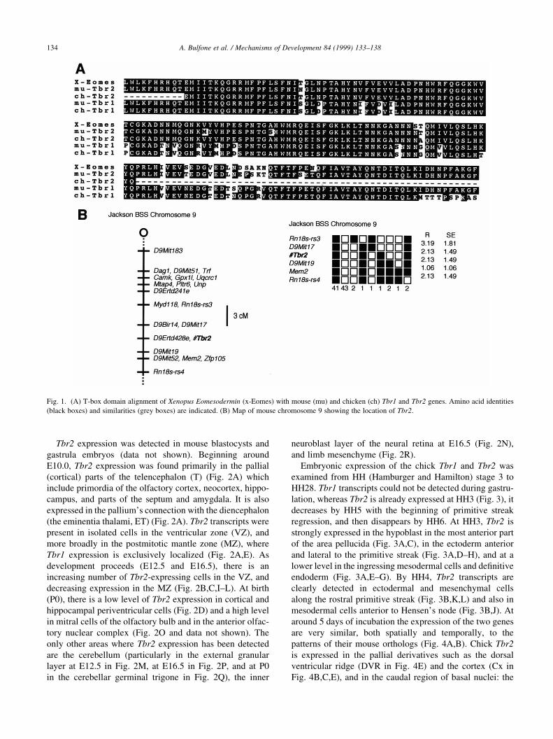
(Fig. 1A). At the amino acid level the T-domain of the
chicken and mouse Tbr1 and Tbr2 orthologs exhibit 98.5
and 99% identity, respectively, within the T-box domain.
The T-domains of Tbr1 and Tbr2 (mouse and chicken) are
87.5% identical; outside of the T-domain there is very little
similarity (data not shown).
The genomic location of mouse Tbr2 was determined
using the Jackson BSS backcross maps from two interspe-
ci®c backcross DNA panels (Rowe et al., 1994), exploiting a
Tbr2 PCR ampli®cation size polymorphism between the
C57/BL6 and mus spretus genomic DNAs. Tbr2 maps
between the markers D9Mit17 and D9Bir15 on distal chro-
mosome 9 (Fig. 1B), where three neurological mutants are
known to map: ducky, tippy and spinner.
Mechanisms of Development 84 (1999) 133±138
0925-4773/99/$ - see front matterq 1999 Elsevier Science Ireland Ltd. All rights reserved.
PII: S0925-4773(99)00053-2
* Corresponding author. Tel.: 1 39-02-21560234; fax: 1 39-02-
21560220.
E-mail address: [email protected] (A. Bulfone)
Tbr2 expression was detected in mouse blastocysts and
gastrula embryos (data not shown). Beginning around
E10.0, Tbr2 expression was found primarily in the pallial
(cortical) parts of the telencephalon (T) (Fig. 2A) which
include primordia of the olfactory cortex, neocortex, hippo-
campus, and parts of the septum and amygdala. It is also
expressed in the pallium's connection with the diencephalon
(the eminentia thalami, ET) (Fig. 2A). Tbr2 transcripts were
present in isolated cells in the ventricular zone (VZ), and
more broadly in the postmitotic mantle zone (MZ), where
Tbr1 expression is exclusively localized (Fig. 2A,E). As
development proceeds (E12.5 and E16.5), there is an
increasing number of Tbr2-expressing cells in the VZ, and
decreasing expression in the MZ (Fig. 2B,C,I±L). At birth
(P0), there is a low level of Tbr2 expression in cortical and
hippocampal periventricular cells (Fig. 2D) and a high level
in mitral cells of the olfactory bulb and in the anterior olfac-
tory nuclear complex (Fig. 2O and data not shown). The
only other areas where Tbr2 expression has been detected
are the cerebellum (particularly in the external granular
layer at E12.5 in Fig. 2M, at E16.5 in Fig. 2P, and at P0
in the cerebellar germinal trigone in Fig. 2Q), the inner
neuroblast layer of the neural retina at E16.5 (Fig. 2N),
and limb mesenchyme (Fig. 2R).
Embryonic expression of the chick Tbr1 and Tbr2 was
examined from HH (Hamburger and Hamilton) stage 3 to
HH28. Tbr1 transcripts could not be detected during gastru-
lation, whereas Tbr2 is already expressed at HH3 (Fig. 3), it
decreases by HH5 with the beginning of primitive streak
regression, and then disappears by HH6. At HH3, Tbr2 is
strongly expressed in the hypoblast in the most anterior part
of the area pellucida (Fig. 3A,C), in the ectoderm anterior
and lateral to the primitive streak (Fig. 3A,D±H), and at a
lower level in the ingressing mesodermal cells and de®nitive
endoderm (Fig. 3A,E±G). By HH4, Tbr2 transcripts are
clearly detected in ectodermal and mesenchymal cells
along the rostral primitive streak (Fig. 3B,K,L) and also in
mesodermal cells anterior to Hensen's node (Fig. 3B,J). At
around 5 days of incubation the expression of the two genes
are very similar, both spatially and temporally, to the
patterns of their mouse orthologs (Fig. 4A,B). Chick Tbr2
is expressed in the pallial derivatives such as the dorsal
ventricular ridge (DVR in Fig. 4E) and the cortex (Cx in
Fig. 4B,C,E), and in the caudal region of basal nuclei: the
A. Bulfone et al. / Mechanisms of Development 84 (1999) 133±138134
Fig. 1. (A) T-box domain alignment of Xenopus Eomesodermin (x-Eomes) with mouse (mu) and chicken (ch) Tbr1 and Tbr2 genes. Amino acid identities
(black boxes) and similarities (grey boxes) are indicated. (B) Map of mouse chromosome 9 showing the location of Tbr2.

caudal ganglionic eminence (CGE in Fig. 4C), anlage of the
amygdala. At HH25, cTbr2 is expressed in scattered cells
which appear to be in the proliferative zone and more diffu-
sely in the MZ, whereas cTbr1 is solely expressed in the MZ
(Fig. 4C±F). By HH stage 28, besides being expressed in the
proliferative zone, strong cTbr2 expression is concentrated
in a layer near the interface of the VZ and MZ (Fig. 4G,H).
3. Methods
Mouse and chicken Tbr2 and Tbr1 cDNA clones were
obtained by degenerate PCR and by screening embryonic
libraries. In situ hybridization was performed according to
published protocols (Riddle et al., 1993; Rugarli et al.,
1993).
A. Bulfone et al. / Mechanisms of Development 84 (1999) 133±138 135
Fig. 2. Comparison of mouse Tbr2 and Tbr1 brain expression during embryonic development by in situ hybridization on tissue sections. (A±H) Adjacent
sagittal sections showing the differential distribution of mTbr1 and mTbr2 transcripts during embryonic development. (A,E) At E10.5, Tbr2 and Tbr1 are
coexpressed in the mantle zone (MZ) but Tbr2 is also expressed in cells of the germinative epithelium of the cortical anlage. (B,F) At E12.5, the Tbr2 pattern of
expression follows the rostro-caudal (arrow in B) and latero-medial (arrow in I) gradients of differentiation and expression in the mantle is restricted to the deep
layers of the incipient cortical plate. (I±M) Set of coronal sections showing Tbr2 expression at E12.5. (N±R) Tbr2 expression in the neural retina at E16.5 (N),
in the olfactory bulb at P0 (O), in the developing cerebellum (at E16.5 in P, and P0 in Q), and in the fore limb bud (R, arrow indicates expression in a posterior
metacarpal pre-cartilage condensation). Other abbreviations: CGE, caudal ganglionic eminence; CGT, cerebellar germinal trigone; Cp, cerebellar plate; CP,
cortical plate; CPU, caudo-putamen; Cx, cortex; DG, dentate gyrus; EGL, external granular layer; ET, eminentia thalami; H, hippocampus; INL, inner
neuroblast layer; LGE, lateral ganglionic eminence; M, mesencephalon; MCL, mitral cell layer; MGE, medial ganglionic eminence; ONL, outer neuroblast
layer; Rh, rhombencephalon; T, telencephalon; VZ, ventricular zone; WM, white matter.

A. Bulfone et al. / Mechanisms of Development 84 (1999) 133±138136
Fig. 3. Early chick embryonic expression of Tbr2. Whole-mount in situ hybridization of embryos at stages HH3 (A) and HH4 (B). Transverse sections of
embryo 4A (C±H) and 4B (I±P) at levels indicated. The location of Hensen's node is indicated (arrowheads in A and B).

Acknowledgements
We thank Celia Pardini, Cristina Mocchetti and Micaela
Quarto for technical assistance, Andreas Russ for valuable
discussions, and Melissa Smith for preparation of this manu-
script. This work was supported by the Italian Telethon
Foundation (Grant no. B.37), the Merck Genome Research
Institute (Grant no. 37 to A.B.), the EC (Grant no. BMH4-
CT97-2341 to A.B.), and NINDS Grant no. NS34661-01A1
to J.L.R.R.
A. Bulfone et al. / Mechanisms of Development 84 (1999) 133±138 137
Fig. 4. (A,B) Tbr2 transcript distribution in E12.5 mouse and HH stage 25 chick embryos (sagital sections). (C±H) Comparison of chicken Tbr2 and Tbr1
pallial expression, in sagital sections, at HH stages 25 and 28. Other abbreviations: CGE, caudal ganglionic eminence; Cx, cortex; DVR, dorsal ventricular
ridge; EGL, external granular layer; ET, eminentia thalami; Hy, hippocampus; Is, isthmus; M, mesencephalon; MGE, medial ganglionic eminence; MZ, mantle
zone; OB, olfactory bulb; OS, optic stalk; P1-4, prosomeres 1-4; S, septum; VZ, ventricular zone; ZL, zona limitans.

References
Bamshad, M., Lin, R.C., Law, D.J., Watkins, W.C., Krakowiak, P.A.,
Moore, M.E., Franceschini, P., Lala, R., Holmes, L.B., Gebuhr, T.C.,
Bruneau, B.G., Schinzel, A., Seidman, J.G., Seidman, C.E., Jorde, L.B.,
1997. Mutations in human TBX3 alter limb, apocrine and genital devel-
opment in ulnar-mammary syndrome. Nat. Genet. 16, 311±315.
Basson, C.T., Bachinsky, D.R., Lin, R.C., Levi, T., Elkins, J.A., Soults, J.,
Grayzel, D., Kroumpouzou, E., Traill, T.A., Leblanc-Straceski, J.,
Renault, B., Kucherlapati, R., Seidman, J.G., Seidman, C.E., 1997.
Mutations in human TBX5 cause limb and cardiac malformation in
Holt-Oram syndrome. Nat. Genet. 15, 30±35.
Bulfone, A., Smiga, S.M., Shimamura, K., Peterson, A., Puelles, L., Ruben-
stein, J.L., 1995. T-brain-1: a homolog of Brachyury whose expression
de®nes molecularly distinct domains within the cerebral cortex. Neuron
15, 63±78.
Bulfone, A., Wang, F., Hevner, R., Anderson, S., Cutforth, T., Chen, S.,
Meneses, J., Pedersen, R., Axel, R., Rubenstein, J.L.R., 1998. An olfac-
tory sensory map develops in the absence of normal projection neurons
or gabaergic interneurons. Neuron 21, 1273±1282.
Chapman, D.L., Garvey, N., Hancock, S., Alexiou, M., Agulnik, S.I.,
Gibson-Brown, J.J., Cebra-Thomas, J., Bollag, R.J., Silver, L.M.,
Papaioannou, V.E., 1996. Expression of the T-box family genes,
Tbx1-Tbx5, during early mouse development. Dev. Dyn. 206, 379±390.
Chapman, D.L., Papaioannou, V.E., 1998. Three neural tubes in mouse
embryos with mutations in the T-box gene Tbx6. Nature 391, 695±697.
Gibson-Brown, J.J., Agulnik, S.I., Chapman, D.L., Alexiou, M., Garvey,
N., Silver, L.M., Papaioannou, V.E., 1996. Evidence of a role for T-box
genes in the evolution of limb morphogenesis and the speci®cation of
forelimb/hindlimb identity. Mech. Dev. 56, 93±101.
Gibson-Brown, J.J., Agulnik, S.I., Silver, L.M., Niswander, L., Papaioan-
nou, V.E., 1998. Involvement of T-box genes Tbx2-Tbx5 in vertebrate
limb speci®cation and development. Development 125, 2499±2509.
Li, Q.Y., Newbury-Ecob, R.A., Terrett, J.A., Wilson, D.I., Curtis, A.R., Yi,
C.H., Gebuhr, T., Bullen, P.J., Robson, S.C., Strachan, T., Bonnet, D.,
Lyonnet, S., Young, I.D., Raeburn, J.A., Buckler, A.J., Law, D.J.,
Brook, J.D., 1997. Holt-Oram syndrome is caused by mutations in
TBX5, a member of the Brachyury (T) gene family. Nat. Genet. 15,
21±29.
Logan, M., Simon, H.G., Tabin, C., 1998. Differential regulation of T-box
and homeobox transcription factors suggests roles in controlling chick
limb-type identity. Development 125, 2825±2835.
Papaioannou, V.E., Silver, L.M., 1998. The T-box gene family. Bioessays
20, 9±19.
P¯ugfelder, G.O., Heisenberg, M., 1995. Optomotor-blind of Drosophila
melanogaster: a neurogenetic approach to optic lobe development and
optomotor behaviour. Comp. Biochem. Physiol. A: Physiol. 110, 185±
202.
Riddle, R.D., Johnson, R.L., Laufer, E., Tabin, C., 1993. Sonic hedgehog
mediates the polarizing activity of the ZPA. Cell 75, 1401±1416.
Rowe, L.B., Nadeau, J.H., Turner, R., Frankel, W.N., Letts, V.A., Eppig,
J.T., Ko, M.S., Thurston, S.J., Birkenmeier, E.H., 1994. Maps from two
interspeci®c backcross DNA panels available as a community genetic
mapping resource. Mamm. Genome 5, 253±274.
Rugarli, E.I., Lutz, B., Kuratani, S.C., Wawersik, S., Borsani, G., Ballabio,
A., Eichele, G., 1993. Expression pattern of the Kallmann syndrome
gene in the olfactory system suggests a role in neuronal targeting. Nat.
Genet. 4, 19±26.
Ryan, K., Garrett, N., Mitchell, A., Gurdon, J.B., 1996. Eomesodermin, a
key early gene in Xenopus mesoderm differentiation. Cell 87, 989±
1000.
Ryan, K., Butler, K., Bellefroid, E., Gurdon, J.B., 1998. Xenopus eomeso-
dermin is expressed in neural differentiation. Mech. Dev. 75, 167±170.
Wattler, S., Russ, A., Evans, M., Nehls, M., 1998. A combined analysis of
genomic and primary protein structure de®nes the phylogenetic rela-
tionship of new members of the T-box family. Genomics 48, 24±33.
Wilkinson, D.G., Bhatt, S., Herrmann, B.G., 1990. Expression pattern of
the mouse T gene and its role in mesoderm formation. Nature 343, 657±
659.
A. Bulfone et al. / Mechanisms of Development 84 (1999) 133±138138
Copyright © 2022 FDOKUMEN